Article

Effects of Metal Oxide Nanoparticles on the Growth and Genotoxicity of Garden Cress (Lepidium sativum L.)

by
Aleksandra Mošenoka
1,*,
Inese Kokina
1,
Ilona Plaksenkova
1,
Marija Jermaļonoka
1,
Eriks Sledevskis
2 and
Marina Krasovska
2
1
Laboratory of Genomics and Biotechnology, Department of Technology, Institute of Life Sciences and Technology, Daugavpils University, Parādes Street 1A, LV-5401 Daugavpils, Latvia
2
G. Liberts’ Innovative Microscopy Centre, Department of Technology, Institute of Life Sciences and Technology, Daugavpils University, Parades Street 1a, LV-5401 Daugavpils, Latvia
*
Author to whom correspondence should be addressed.
Agronomy 2024, 14(10), 2324; https://doi.org/10.3390/agronomy14102324
Submission received: 30 July 2024 / Revised: 2 October 2024 / Accepted: 8 October 2024 / Published: 10 October 2024
(This article belongs to the Section Horticultural and Floricultural Crops)

Abstract

:
The interaction of nano-fertilizers with commercially important crops can be a promising solution to increase both crop yield and quality. This study investigated the effect of iron oxide nanoparticles (Fe3O4 NPs) on four-week-old garden cress (Lepidium sativum L.) seedlings. Iron is an essential micronutrient for plants but is not always available in sufficient quantities, which can lead to chlorosis and even plant death. The seedlings were grown hydroponically, with three concentrations (1 mg/L, 5 mg/L, and 10 mg/L) of the NPs, alongside a control group with no additions. During the experiment, the following methods were employed: measurement of stem and root length, spectrophotometry to determine chlorophyll absorbance and concentration, and the RAPD technique to assess the genotoxicity of Fe3O4 NPs. The study demonstrated a significant increase in the shoot length of cress at all concentrations compared to the control group (p < 0.05; p < 0.01). The light absorption and chlorophyll concentration levels in the experimental groups significantly increased compared to the control group (p < 0.001). Genotoxicity analysis revealed that the genotoxic impact of the NPs on the garden cress genome was only 10%, a statistically insignificant level. The findings suggest that Fe3O4 NPs exhibit low genotoxicity and have the potential to enhance the growth and chlorophyll content of cress seedlings in hydroponic conditions.

1. Introduction

Modern agriculture is increasingly confronted with challenges posed by climate change and a growing human population [1]. Moreover, current agricultural practices have contributed to many of these issues, such as soil depletion, loss of biodiversity, and worsening food security [2]. According to the Food and Agriculture Organization (FAO), the percentage of people suffering from chronic hunger increased from 7.2% to 9.2% between 2019 and 2022 [3]. One potential solution is the integration of nanotechnology into agriculture. It has been repeatedly suggested that such an approach could make agriculture economically sustainable by achieving higher yields in an environmentally responsible manner [4]. Although nanotechnology has already shown success in the agricultural sector, further improvements are needed [5]. Nanotechnologies offer a variety of applications that contribute to the development and maintenance of sustainable agriculture. Among the most promising areas in this field are nano-fertilizers and nano-pesticides [1].
Most nano-fertilizers contain various metal or metal oxide nanoparticles (NPs), with at least half of these NPs being less than 100 nm in size. Nano-fertilizers also contain organic compounds, such as humic acid [6,7]. The production of nano-fertilizers is based on the modification or synthesis of traditional fertilizers, their bulk components, or the extraction of certain plant parts [8]. The addition of suitable NPs to fertilizers can lead to the development of ecologically safe, qualitative, and valid methods to improve agricultural practices [4,9]. Traditionally used inorganic fertilizers can induce resistance in various harmful insects, weeds, and microorganisms [8,10]. Moreover, these inorganic chemicals enter the human body through the food chain, where they accumulate, leading to negative health effects [11]. Nano-fertilizers offer significant advantages over conventional fertilizers. NPs possess unique chemical and physical properties due to their small size [10]. Conventional chemicals often have adverse effects on the environment, causing contamination and negatively impacting non-target plant species [12]. Nano-fertilizers, in contrast, reach the target plant in much larger quantities than conventional fertilizers and do not disperse as widely through the environment [4,10]. Compared to conventional fertilizers, nano-fertilizers are absorbed by plants more quickly, meaning fewer fertilizer components are released into the environment, thus reducing the risk of pollution. A dosage tailored to the specific plant species, combined with the precise application of NPs, may offer an effective solution to nutrient deficiencies. Rapid absorption accelerates metabolism, enhancing the activity of physiological processes. Additional benefits of nano-fertilizers include their variable solubility and safe disposal methods [4,13,14]. Nano-fertilizers may also improve soil quality. A recent study has shown that nanoscale zerovalent iron-supported coconut-husk biochar (nZVI-CHB) at low doses can effectively improve the quality of soil contaminated with cadmium (Cd) and lead (Pb), while mitigating oxidative stress in Brassica rapa caused by these two toxic metals [15]. However, soil organisms such as nematodes, earthworms, bacteria, and plants can be affected both negatively and positively by iron oxide nanoparticles (IONPs). Furthermore, when introduced into the soil, iron oxide NP-related processes, such as aggregation, chemical transformation, or aging, may be altered by soil chemistry or the presence of microorganisms [16]. Additionally, if not applied properly, nano-fertilizers can reduce crop yield. Recently, it was reported that repeated sequential foliar treatment of plants with iron oxide nanoparticles (Fe3O4 NPs) may cause fluctuations in plant mineral distribution and contribute to plant growth retardation [17]. It is predicted that, due to their wide range of uses, iron oxide NPs will accumulate in soil, water, and air [18]. Unfortunately, there is a global lack of knowledge regarding the behavior, fate, and ecological hazards of nano-fertilizers [9]. Although numerous studies have demonstrated the effectiveness of nano-fertilizers [19,20,21,22,23,24], the lack of research on their long-term environmental impact raises concerns about their use and requires further investigation [25].
Metallic NPs are considered the most suitable for nano-fertilizers because many metals are essential for plant viability. Numerous studies have been conducted to explore the effects of metallic NPs on different plant species [26,27,28,29]. Metallic NPs possess unique physical properties, magnetic abilities, and biocompatibility. Some members of this group, such as iron oxides, exhibit low toxicity levels, making these NPs widely used in biological research [30]. Iron oxide, or magnetite (Fe3O4), is a compound from the iron oxide group, and along with other iron-containing complexes—especially maghemite (α-Fe2O3) and hematite (γ-Fe2O3)—it is one of the most common types of iron oxides in nature [31]. Iron compounds play a vital role in the physiological processes of living organisms. In plants, iron is a component of the photosynthetic reaction center (Photosystem I). It also participates in oxygen transport and uptake, protein stability regulation, and iron absorption as part of specific enzymes [32]. However, the availability of iron and its compounds for plants is unexpectedly low. Various soil characteristics, such as pH and redox potential, and significant soil processes such as chelation determine the availability of different elements and compounds, including iron and iron oxides [33]. Agriculture frequently faces iron deficiencies in soil, which negatively impacts crop health. A lack of iron often leads to chlorosis in plants, affecting their leaves by causing chloroplast degeneration, reducing photosynthesis, and halting growth [34]. Iron treatments can improve plant viability. Kim et al. [35] recently demonstrated that iron-based NPs can expand stomatal openings and increase their activity in mouse-ear cress (Arabidopsis thaliana (L.) Heynh). Exposure to these NPs also increases leaf area and significantly enhances the expression of AHA2, a gene responsible for stomatal opening. A similar experiment with Arabidopsis thaliana (L.) Heynh using NPs of other iron-based compounds showed an increase in biomass and reserve nutrients, such as starch and glucose [36]. Furthermore, depending on the growth conditions, iron-based NPs can enhance plant resilience under drought stress, support biosynthetic processes, boost phytohormone levels, and protect seedlings from the harmful effects of heavy metals [37]. Iron in conventional fertilizers is easily absorbed by soil particles, leading to iron leaching during irrigation. This reduces the effectiveness of such fertilizers, requiring repeated applications, and contributes to soil leaching. There is also a risk of plant damage if iron accumulates excessively in the soil. Controlled-release nano-fertilizers mitigate these risks by regulating the amount of nutrients available to plants. The ease with which plants absorb NPs is due to their size and physical and chemical properties [38,39].
Iron nanoparticles interact with living plant cells at the molecular level [40], allowing them to influence various physiological processes in plants. NPs can increase gene expression and enhance the production of photosynthetic pigments [41]. For example, iron oxide (Fe3O4) NPs, when applied at a suitable dose, can raise chlorophyll (Chl) levels in tomato plants (Solanum lycopersicum L.) [42]. Additionally, NPs improve nutrient uptake and transport through their mobility and enhance water absorption in plants [40]. In the right doses, NPs promote seed germination, increase root length, and positively affect photosynthesis. Moreover, they enhance the activity of antioxidant enzymes, helping plants combat oxidative stress [41]. Iron has been shown to reduce reactive oxygen species (ROS) and lipid peroxidation [43]. Despite numerous studies, the mechanisms underlying NP interactions with plant cells remain poorly understood [17]. Different NPs can alter gene expression in plants [44]. Genotoxicity refers to the damaging effects of a substance on the genetic material of a cell [45]. Assessing the genotoxicity of NPs is essential for understanding their environmental and human health risks [46]. Current research on the impact of NPs on plant genomes is relatively limited, and the level of toxicity may depend on the size, concentration, and origin of the NPs [47,48]. Prolonged exposure of genetic material to NPs can lead to excessive production of ROS, triggering strong oxidative reactions. This phenomenon is the most common cause of damage to the structure of DNA, lipids, and proteins [45,49]. However, it has been repeatedly demonstrated that the effectiveness of NPs at the same concentration can vary among different species, varieties, and even phenotypes of plants. For example, low concentrations of Fe3O4 NPs (1, 2, and 4 mg/L) significantly enhanced Chl a fluorescence (p < 0.05 and p < 0.01) in five-week-old yellow medick (Medicago falcata L.) seedlings [50]. Conversely, the same concentrations of Fe3O4 NPs caused a significant (p < 0.05) reduction in the absorption of Chl a and b in common wheat seedlings (Triticum aestivum L.) [51]. The negative effects of treatment with copper NPs (nCuO), as well as bulk (bCuO) and ionic copper (CuCl2) at high concentrations (75, 150, 300, and 600 mg/kg), depended on the cultivar of bok choy (Brassica rapa) treated. Additionally, the distribution of copper in the leaf midrib and parenchyma was influenced by the plant’s phenotype [52].
Iron is a vital macronutrient for plants, involved in physiological processes such as photosynthesis, respiration, nitrogen fixation, hormone and DNA synthesis, and the activation of various enzyme cofactors. However, excessive iron accumulation in plants can have negative effects. Treatment with higher concentrations of iron nanoparticles (NPs) can lead to phototoxic effects due to iron’s role in the Fenton reaction, which generates a significant number of hydroxyl radicals (OH) in plants [41]. Hydroxyl radicals, along with other reactive oxygen species (ROS) such as hydrogen peroxide (H2O2) and superoxide radicals ( O 2 ), contribute to abiotic stress, which can stunt plant growth and damage living cells, potentially leading to death [15]. Conversely, it has been demonstrated that treatment of plants with high concentrations of iron oxide (Fe2O3) NPs retards root growth, while lower concentrations can enhance root growth, plant biomass, and chlorophyll (Chl) content [41,53]. Treatment with iron oxide (Fe3O4) NPs at low concentrations (2 and 4 mg/L) significantly increased the shoot length of rocket (Eruca sativa Mill.) and the root length of yellow medick, whereas higher concentrations (17, 35, and 70 mg/L) of the same NPs showed no significant positive effect on the root length of barley (Hordeum vulgare L.) seedlings [47,50,54]. Fe3O4 NPs at a lower concentration of 25 mg/L increased the length of chamomile (Matricaria chamomilla L.) seedlings, while the highest concentration of 100 mg/L had a significant negative effect on both shoot and root lengths [55]. Moreover, Fe2O3 NPs at a very high concentration (320 mg/L) decreased Chl levels in three different plant species: broad bean (Vicia faba L.), common bean (Phaseolus vulgaris L.), and alfalfa (Medicago sativa L.) [56]. Additionally, iron oxide NPs (γ-Fe2O3 NPs) at a concentration of 100 mg/L also reduced Chl content and root activity in Citrus maxima (Burm.) Merr. plants, while the same NPs at half that concentration promoted increases in both Chl levels and root activity [57]. Based on this information, it was hypothesized that Fe3O4 NPs at low concentrations, such as 1, 5, and 10 mg/L, could improve growth and increase Chl levels in garden cress (Lepidium sativum L.) seedlings grown hydroponically without significant damage. To the best of our knowledge, this is the first time that Fe3O4 NPs at such low concentrations have been used for this plant.
Garden cress, also known as cress-salad or curly cress, is an edible herb from the cabbage family with high nutritional value. This cold-resistant plant, known for its spicy taste, is valued for its nutritional and medicinal properties [58,59]. Its cultivation has spread globally [59,60]. Garden cress is considered ideal for research due to its fast growth. However, despite its value, there have been few studies on its ability to accumulate iron-based NPs. In most experiments, a closely related plant, mouse-ear cress (Arabidopsis thaliana), is used instead [61]. This study aims to evaluate the effect of various concentrations of Fe3O4 NPs on Chl levels and morphological parameters of hydroponically grown garden cress seedlings, as well as to assess the genotoxicity of Fe3O4 NPs in relation to this plant.

2. Materials and Methods

2.1. Nanoparticles

Iron oxide (Fe3O4) nanoparticles (NPs) were provided by G. Libert’s Center of Innovative Microscopy at Daugavpils University. The NPs were synthesized using the Massart method by preparing aqueous magnetic fluid under inert atmosphere conditions, followed by washing and drying of the obtained NPs [62]. The morphology and size of the Fe3O4 NPs were analyzed using Field Emission Scanning Electron Microscopy (FESEM) (MAIA 3, Tescan, Brno, Czech Republic) and Atomic Force Microscopy (AFM) (NX 10, Park Systems Corp., Suwon, Republic of Korea). The chemical composition was examined using an Energy Dispersive Spectroscopy (EDS) system (Inca, Oxford Instruments, Oxford, UK). AFM images showed that the NPs had a spherical shape, with an average diameter of approximately d = 25 nm. In pH 5.8 medium, Fe3O4 NPs are expected to have a slightly positive charge or be near-neutral, depending on the exact conditions and any surface modifications. The positive charge arises from protonation of surface hydroxyl groups below the point of zero charge (PZC pH = 6.5). Under normal environmental conditions (pH 5–7), plant roots carry a negative surface charge due to deprotonation of carboxyl and phosphate groups in the root cell walls and membranes. This charge plays a key role in root interactions with soil particles, nutrients, and NPs, where electrostatic interactions facilitate the absorption [63].

2.2. Seedling Cultivation and Growth Conditions

For the experiment, garden cress seeds were purchased from a local shopping center. The seedlings were grown hydroponically using a standard hydroponic solution with ½ MS at a pH of 5.8. The hydroponic solution contained the following macronutrients: KNO3 (8001.1, ROTH, Karlsruhe, Germany), NH4NO3 (K299.1, ROTH, Karlsruhe, Germany), KH2PO4 (3804.1, ROTH, Karlsruhe, Germany), MgSO4 × 7H2O (P027.1, ROTH, Karlsruhe, Germany), CaCl2 × 2H2O (147.02, ROTH, Karlsruhe, Germany), and micronutrients: Na2EDTA × 2H2O (E6511-100G, Sigma-Aldrich, Burlington, MA, USA), FeSO4 × 7H2O (P015.1, ROTH, Karlsruhe, Germany), MnSO4 × 5H2O (4487.1, ROTH, Karlsruhe, Germany), H3BO3 (P010.1, ROTH, Karlsruhe, Germany), ZnSO4 × 7H2O (K301.1, ROTH, Karlsruhe, Germany), CoCl2 × 6H2O (7095.1, ROTH, Karlsruhe, Germany), CuSO4 × 5H2O (P024.1, ROTH, Karlsruhe, Germany), NaMoO4 × 2H2O (M1003, Sigma-Aldrich, Burlington, MA, USA), and KI (P0518.0100, Duchefa Biochemie, Haarlem, The Netherlands). The seeds were first soaked in water for 24 h. Then, 200 randomly selected seeds were used for germination. After one week, the seedlings were divided into four groups and transferred to tubes containing Fe3O4 NPs (~25 nm) at concentrations of 1 mg/L, 5 mg/L, and 10 mg/L. The fourth group served as the control and received no NPs. Each experimental group consisted of 50 plants (n = 50). The seedlings were grown for four weeks under laboratory conditions in a versatile environmental test chamber (MLR-351H, Sanyo, Moriguchi, Osaka, Japan) with constant humidity (80%), a temperature of +21 °C, light intensity of 2 Lx, and a photoperiod of 8/16 h (day/night).

2.3. Shoot and Root Length Measurements

After four weeks, 30 seedlings were randomly selected from each of the four experimental groups to measure the lengths of the stems (including the leaves) and roots separately.

2.4. Measurements of Chlorophyll a and b

Immediately after measuring the morphological parameters, chlorophyll (Chl) was extracted from the same samples. Prior to Chl extraction, the green part of each plant was separated from the roots and weighed in triplicate using a laboratory scale (440.35N, max 400 g, d = 0.01 g, KERN, Balingen, Germany). The average of these three replicates was used for each corresponding Chl sample. For the extraction, 3.5 mL of 96.6% ethanol was used. The green plant tissue was ground in ethanol using a mortar and pestle, and the solution was then filtered [64]. The Chl concentration was determined using the spectrophotometric method. Spectrophotometric analysis was performed using a NanoDrop 1000 spectrophotometer with standard parameters (full spectrum 220–750 nm, sample retention system, Thermo Scientific, Waltham, MA, USA). Measurements were taken at two different wavelengths: 645 nm for Chl a, and 663 nm for Chl b. The absorbance data were subsequently used to calculate the Chl concentration using Arnon’s equations (Equations (1)–(3)) [65]:
Chl   a   ( mg / g ) = [ 12.7 × A 663 2.69 × A 645 ] × V / 1000 × W
Chl   b   ( mg / g ) = [ 22.9 × A 645 4.86 × A 663 ] × V / 1000 × W
Chl   total   ( mg / g ) = [ 20.2 × A 645 + 8.02 × A 663 ] × V / 1000 × W
where A663 is the absorbance at 663 nm, A645 is the absorbance at 645 nm, V is the sample volume (mL), and W is the mass of the fresh sample (mg).

2.5. DNA Isolation, Polymerase Chain Reaction, and Randomly Amplified Polymorphic DNA Analysis

The remaining 20 plants from each group were used for DNA isolation and subsequent operations—polymerase chain reaction (PCR) assay and determination of the genotoxic effects caused by Fe3O4 NPs, using the randomly amplified polymorphic DNA (RAPD) method. The RAPD technique relies on PCR and requires a specially prepared DNA extract and specific primers. It is considered a sensitive, although not specific, genotoxicity assay. This quick and easy method detects polymorphisms and indicates a wide range of DNA damage types. The technique is widely used to demonstrate and evaluate the mutagenic effects of various genotoxicants [66]. For this study, three decamer primers (Bioneer, Daejeon, Republic of Korea) with the following sequences were selected: TIBM BE-18 (5′-CCAAGCCGTC-3′), OPC2 (5′-GTGAGGCGTC-3′), and OPJ-O4 (5′-CCGAACACGG-3′). DNA isolation was performed using a DNeasy Plant Mini Kit (Qiagen GmbH, Hilden, Germany) according to the manufacturer’s protocol. DNA was extracted from approximately 25 mg of the green parts (both stem and leaves) of the plants. The final elution volume was 100 μL. The concentration and absorbance of the extracted DNA were measured using a spectrophotometer with standard parameters (NanoDrop 1000, Thermo Scientific, Waltham, MA, USA). For PCR, the reaction solution was prepared according to the protocol of the Tag PCR Core Kit (Qiagen GmbH, Hilden, Germany). The amplification products were obtained using a reaction mixture comprising 2.5 μL PCR Buffer, 0.5 μL dNTP mix, 0.5 μL primer, 0.125 μL Taq DNA polymerase, 5 μL Q-Solution, and 11.375 μL RNase-free water. A 5 μL volume of each sample’s DNA was used as the template. The total reaction volume for each sample was 25 μL, with deionized water used as the negative control. The amplification process included an initial denaturation step at 94 °C for 3 min, followed by a three-stage cyclic reaction involving denaturation (1 min at 95 °C), annealing (1 min at 30–34 °C, depending on the melting temperature of each primer), and elongation (1 min at 72 °C). A total of 30 cycles were performed, followed by a final extension step lasting 10 min at 72 °C. The reactions for each DNA sample were conducted in triplicate for each primer, resulting in a total of 720 separate analyses.
The PCR products from three repetitions were subjected to analysis using agarose gel electrophoresis. This method is easy to use and allows for relatively rapid separation of DNA fragments. A 2% agarose gel was prepared manually from LE agarose (CSL-AG500, Cleaver Scientific Ltd., Rugby, UK) dissolved in 1× TAE buffer (A1691, AppliChem, Darmstadt, Germany) by heating the solution in a microwave oven. The liquid was then cooled to approximately 50 °C. After cooling, SimplySafe stain (E4600-01, EURx, Gdansk, Poland), an alternative to traditional ethidium bromide, was added to the solution, which was subsequently mixed. The liquid was poured into a gel casting tray equipped with two 20-well combs and allowed to solidify into a gel-like mass. After solidification, the tray with the gel was placed on a wide mini-sub cell GT horizontal electrophoresis system (MultiSUB Midi, Cleaver Scientific Ltd., Rugby, UK), and the entire system was filled with 1× TAE buffer (A1691, AppliChem, Darmstadt, Germany) to ensure the gel was adequately covered. To prepare the samples, a mixture of PCR product and loading dye (07-02-00001, Solis Biodyne, Tartu, Estonia) was prepared, with each sample consisting of 8 μL of PCR product and 2 μL of loading dye. The CSL-MDNA-50BP DNA Ladder RTU (range 50–1500 bp, 17 bands, concentration 112 µg/L, 2% agarose gel; Cleaver Scientific Ltd., Rugby, UK) was used as a DNA size marker, while clean water served as a negative control. Electrophoresis was conducted for 45 min at a voltage of 130 V (Power Supply EV3150, 300 V, 2000 mA, 300 W, Consort, Turnhout, Belgium) in a room with a stable temperature of 20 °C. Finally, the gel image was captured using the Fussion Solo 7S system (DarQ9 camera, Vilber, France).

2.6. Evaluation of Genomic Template Stability

To evaluate genomic template stability (GTS) (%), the following Equation (4) was applied:
G T S % = [ 1 a / n ] × 100
where a is the average number of bands from each DNA profile of every experimental group that differs from the control, and n is the total number of bands in the control [67].

2.7. Statistical Analysis

To determine significant differences between the experimental groups and the control, a Student’s t-test and ANOVA were performed. Values of p less than 0.05 or 0.01 were considered statistically significant. Each experimental value was compared to the corresponding control. The data obtained are presented as mean values with the standard deviation (±SD) of the means.

3. Results

3.1. Shoot and Root Lengths

A significant increase in seedling lengths was observed in the shoots treated with Fe3O4 NPs at concentrations of 1 mg/L and 5 mg/L (p < 0.05) and 10 mg/L (p < 0.01) compared to the control (Figure 1). However, there was no significant increase in root length for any of the NP treatments. The greatest effect on shoot and root length was observed at the 10 mg/L and 5 mg/L concentrations, respectively, with maximal lengths of 6.5 cm for shoots and 15 cm for roots. Overall, the treatment resulted in an average increase of 10.63% in shoot length and 7.7% in root length compared to the control. During the measurements, the accumulation of NPs was observed on the surface of the roots, indicating an intense process of accumulation in root tissues. A Polish study reported that over 90% of accumulated Fe3O4 NPs smaller than 50 nm were found in the roots of garden cress [68]. The Fe3O4 NPs used in this study were half the size of those in the Polish study; therefore, it can be assumed that at least some of the Fe3O4 NPs accumulated in the roots of hydroponically grown garden cress seedlings. Notably, during visual assessment, it was observed that plants in the experimental groups had thicker roots than those in the control group.

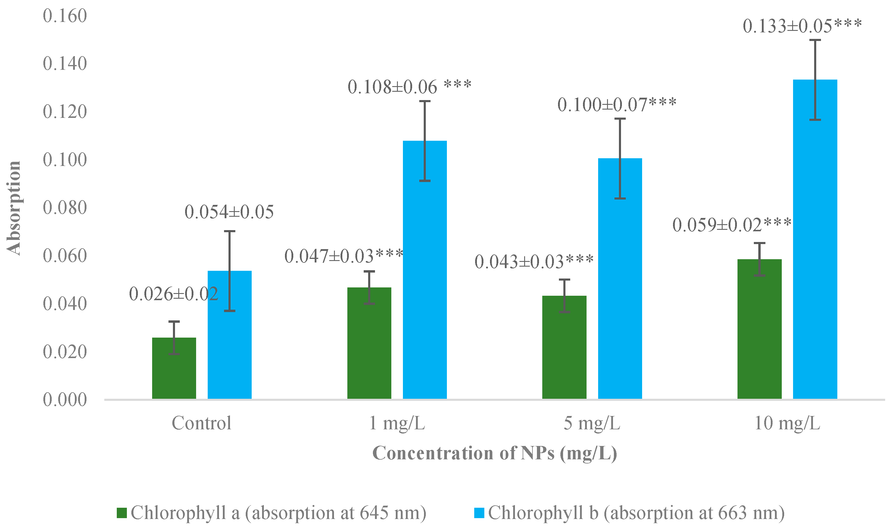
3.2. Measurement of Chlorophyll a and b Absorption

The absorption of chlorophyll (Chl) refers to the pigment’s ability to absorb light. The light absorption of Chl a and Chl b was measured at wavelengths of 645 nm and 663 nm, respectively. A significant increase in light absorption was observed in all experimental groups for both Chl a and Chl b compared to the control (p < 0.001) (Figure 2). The mean light absorption for control plants was 0.03 ± 0.02 at 645 nm and 0.05 ± 0.05 at 663 nm. Specifically, the light absorption of Chl a increased by an average of 52%, while the light absorption of Chl b increased by 48%. The maximum increase in light absorption at 645 nm was found in the group treated with 10 mg/L, with an increase of 0.113 compared to the control. In contrast, the maximum increase in absorption at 663 nm was 0.222 compared to the control and was observed in the group with a concentration of 5 mg/L.

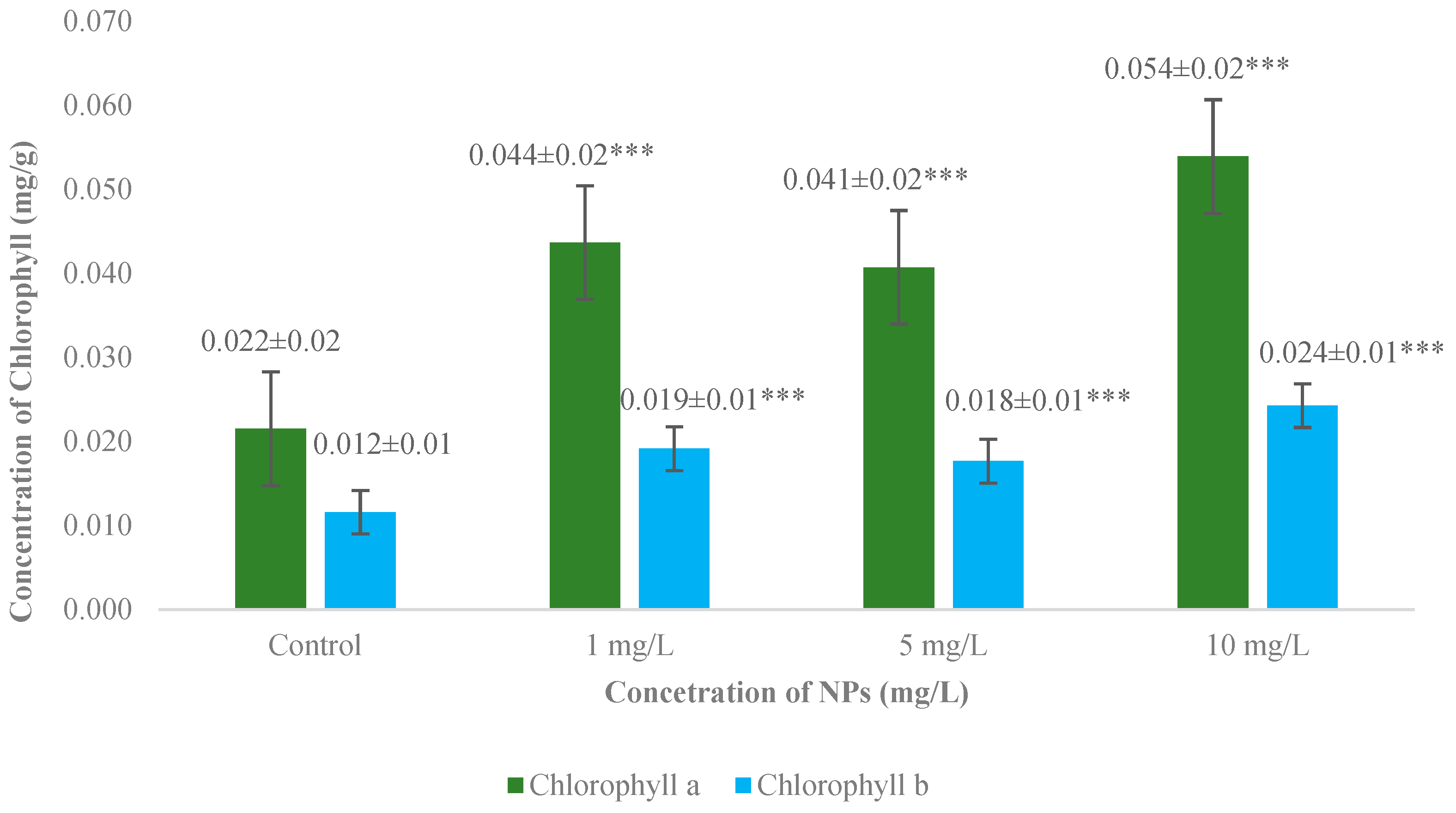
3.3. Measurement of Chlorophyll a and b Concentrations

The assessment of chlorophyll (Chl) concentration levels was carried out based on absorption data using the Arnon formula [65]. The data analysis showed a significant increase in the content of Chl a and Chl b in all experimental groups compared to the control (p < 0.001) (Figure 3). For Chl a, the average concentration in the control plants was 0.022 ± 0.015 mg/g, while for Chl b, it was 0.012 ± 0.007 mg/g. The samples from the group with a concentration of 10 mg/L showed the greatest impact, with an average increase of 40.7% for Chl a and 50% for Chl b. The most pronounced effect on Chl a concentration was observed in the group with a concentration of 5 mg/L, where the increase was 0.091 mg/g compared to the control.

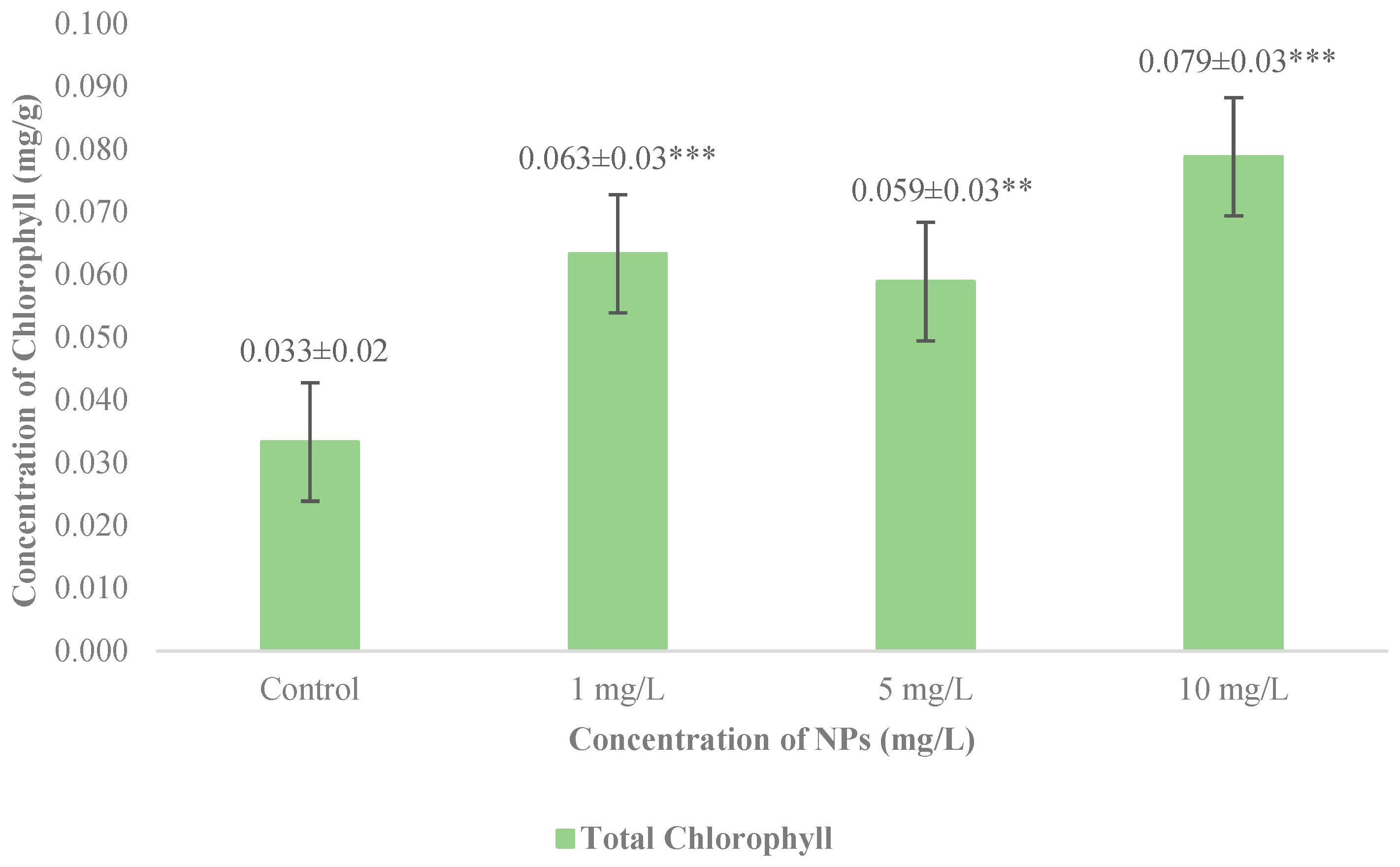
3.4. Measurements of Total Chlorophyll Concentration

The total chlorophyll (Chl) level was estimated based on the absorption data using the Arnon formula [65]. Data analysis showed that all three tested concentrations of Fe3O4 nanoparticles (NPs) significantly increased the total Chl concentration compared to the control. The significance level for the 5 mg/L group was p < 0.01, while for the 1 and 10 mg/L groups, it was p < 0.001 (Figure 4). The proposed hypothesis was confirmed: even low concentrations of Fe3O4 NPs can significantly increase Chl concentration in garden cress seedlings treated with these NPs and grown under hydroponic conditions for 4 weeks. The 10 mg/L concentration was the most effective, on average increasing the total Chl level by 139.39%. In contrast, the 5 mg/L concentration had the least effect, increasing the total Chl concentration by only 78.79%. Nevertheless, the highest value recorded in this group was 0.159 mg/g, while the minimum total Chl level was found in the 1 mg/L treatment group, which amounted to 0.007 mg/g.

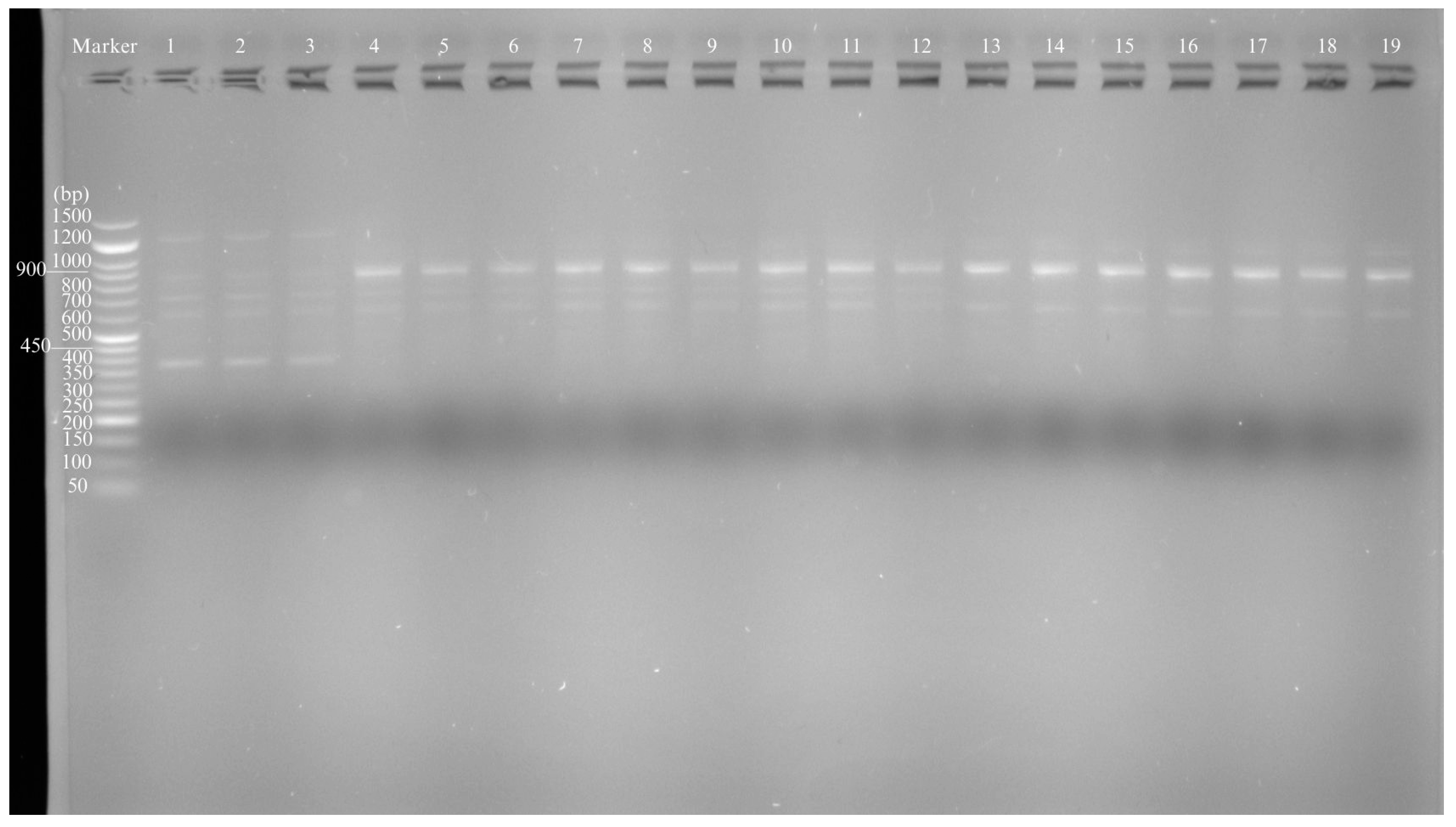
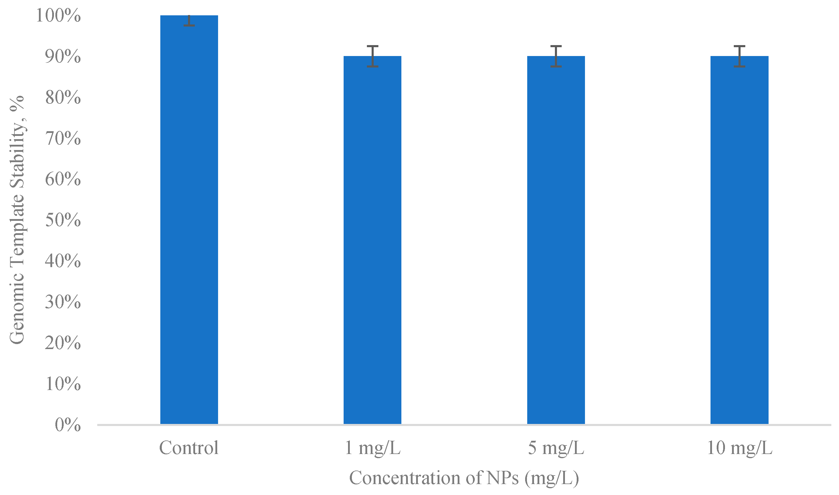
3.5. Evaluation of the Genotoxicity Level

Genomic template stability was assessed based on the appearance or disappearance of bands in the obtained RAPD profiles (Figure 5). All tested decamer primers (n = 3) were used to amplify representative bands in the control samples. The number of bands in the control RAPD profiles varied from one for TIBM BE-18 to six for OPJ 04. The comparison of profiles was based on the loss and appearance of new bands between the control and experimental RAPD profiles. Only one new band was detected in plants treated with 1 mg/L (OPC 02 and OPC 04). When plants were treated with 5 mg/L, bands disappeared in all RAPD profiles for all primers (n = 3) used. The same was observed with the 10 mg/L treatment: two normal bands were absent, and one new band appeared, in total. The total number of control and differing experimental bands was used to calculate genomic template stability (GTS, %). The level of genotoxicity in this study was determined based on the data obtained from assessing the GTS level. Genotoxicity indicators are inversely related to GTS. This means that the higher the genotoxicity, the lower the GTS level, and vice versa. For each experimental group, the total GTS value was calculated by combining all the data from the tested primers. The default GTS for the control group was recorded as 100%. The present study showed a very high GTS level and a very low genotoxicity level compared to the control. Statistical data analysis did not reveal any significant differences in genotoxicity levels or GTS values among the experimental groups compared to the control group (Figure 6). Interestingly, all experimental groups showed the same results: the genotoxicity level was 10%, and the GTS level was 90% for all NP concentrations compared to the control group. Such a high GTS indicator suggests that the tested DNA samples were weakly susceptible to the genotoxic effects of NP treatment. However, only three specific primers were used for the RAPD analysis. The consistency of the GTS percentage across all concentrations may be explained by the low levels of NP concentrations, which differed little from each other. In the future, additional analyses, such as specific gene expression studies, should be conducted.
In addition to the presented results, future study should include additional testing and analysis such as dissolution tests and elemental uptake data to help clarify the underlying mechanisms.

4. Discussion

Our study showed a significant increase (p < 0.05, p < 0.01) in the length of garden cress seedlings. However, no significant increase was observed in the roots. Currently, there is limited research on the effects of Fe3O4 NP treatment on the morphological parameters of garden cress. For example, an improvement in morphological parameters was observed in garden cress seedlings treated with powders of 80–110 nm sized Fe3O4 NPs at very high concentrations [69]. More commonly, studies have utilized different iron-based NPs rather than Fe3O4 NPs. Frequently, treatment of garden cress with iron-based NPs has resulted in improved morphological parameters. For example, an insignificant positive effect has been reported on the shoot and root lengths of garden cress seedlings treated with iron NPs coated with ethylenediaminetetraacetic acid (EDTA IONP). However, there was an eight-fold increase in overall biomass compared to the control. The authors suggest that these effects may be related to hydroponic growth conditions, which allow seedlings to access components from the growth medium continuously and without obstacles [61]. In another study using the hydroponic method, treatment with biogenic ferrihydrite or hydrous ferric oxide NPs at various concentrations resulted in a significant increase in the shoot length of eight-day-old garden cress seedlings in almost all experimental groups. However, similar to the present study, the root lengths decreased in most cases. At the same time, there was a significant increase in shoot length and biomass for both roots and shoots [58].
The effects of Fe3O4 NPs and iron-based NPs on the morphology of other plants have also been investigated. In a study utilizing three barley (Hordeum vulgare L.) cultivars and Fe3O4 NPs at three concentrations (1, 10, and 20 mg/L), almost all experimental groups observed a significant positive effect on both shoot and root length compared to the control [70]. An investigation on the impact of low-concentration Fe3O4 NPs (1, 2, and 4 mg/L) on the growth and morphology of common wheat (Triticum aestivum L.) demonstrated a significant (p < 0.05) elongation in shoot length at the two highest concentrations. However, there was no significant effect on root length in any of the experimental groups [51]. A powder of Fe3O4 NPs at two concentrations (50 and 5000 mg/kg) was introduced into the soil for maize (Zea mays L.) plants, and the results indicated a significant increase in root length (p < 0.05) in both cases, while no significant effect on leaf biomass was observed [71]. Nanoscale zerovalent iron supported by eggshell biochar and activated carbon (nZVI-ESB/AC) significantly increased both the dry and wet biomass, as well as the lengths of shoots and roots in Brassica chinensis L. [43]. Superparamagnetic iron oxide NPs synthesized by a biogenic route (BS-SPION) significantly elongated the roots of Raphanus sativa L. seedlings at high concentrations [72]. The ability of iron NPs to influence the morphological parameters of plants is well established. However, the results can be influenced by various factors, including plant species, cultivation method, NP size and concentration, as well as the origin and properties of the NPs themselves.
Chlorophyll is an essential pigment in plants, primarily responsible for light absorption and photosynthesis [73]. Measurements of Chl a, Chl b, total Chl, and their ratios are common techniques used to gain insight into photosynthetic rates, respiration, and metabolically active biomass [74]. The ratio of total to distributed Chls in a plant directly influences the photosynthetic rate. Additionally, Chl content (Chl a, Chl b, Chl a + b, and Chl a/b) is regulated by the plant to adapt to environmental conditions [75]. Photosynthesis rates depend on various factors, and numerous studies have shown that Chl responds differently to varying levels of stress. At low stress levels, Chl concentration increases, while at high stress levels, it decreases. Under low abiotic stress conditions, such as slight soil contamination, a protective reaction develops that supports vital processes in the plant [73,76]. For instance, high oxidative stress inhibits Chl synthesis [73]. Nanoparticles, which are inherently pollutants, also induce plant stress, altering Chl levels. The photosynthetic apparatus of plants is affected both positively and negatively by most NPs, depending on their dosage and chemical composition. This effect is thought to depend on NP characteristics and concentration, as well as the genetics of the plant itself. In a recent study, eight different metal and metal oxide NPs were examined, with iron oxide NPs (Fe2O3 NPs) showing the most significant effect on the Chl parameters of oakleaf lettuce (Lactuca sativa L. var. foliosa) [77]. It has also been reported that the method of plant cultivation can affect Chl concentration. Syafitri and Fevria [78] demonstrated, using kale (Ipomoea reptans Poir.) as an example, that hydroponic methods can decrease Chl concentration compared to traditional cultivation methods. Despite this, the present study showed that at all tested NP concentrations, the absorbance and content of Chl a, Chl b, and total Chl in hydroponically grown garden cress seedlings increased significantly (p < 0.001). However, other studies have reported varying results. To our knowledge, most research has focused on different types of iron-based NPs rather than Fe3O4 NPs, and very few studies have investigated the effects of iron-based NPs specifically on garden cress. One study examining the effect of iron oxide NPs (EDTA-iron oxide NPs) on garden cress showed a 1.4-fold increase in Chl levels compared to samples with added commercial chelated fertilizer [61]. Research conducted by Lithuanian scientists also indicated a positive result for Chl content in garden cress using cobalt ferrite NPs (CoFe2O4 NPs) of varying sizes, with results showing an increase in Chl b levels in the experimental groups. The resulting effect depended on the size and concentration of the NPs used [79]. A decrease in Chl fluorescence levels has also been reported in eight-day-old garden cress seedlings treated with biogenic ferrihydrite NPs at various concentrations [58].
The influence of Fe3O4 NPs and other types of iron-based NPs on Chl parameters in other plant species has been more frequently reported. The presence of nanoscale zerovalent iron supported with eggshell biochar and activated carbon (nZVI-ESB/AC) in soil increased the levels of Chl and carotenoids in Brassica chinensis L. plants [43]. In another study, all tested concentrations of Fe3O4 NPs had a significant (p < 0.05 and p < 0.01) positive effect on Chl a fluorescence in five-week-old yellow medick (Medicago falcata L.) seedlings [50]. Similarly, Chl a and b levels insignificantly increased in all experimental barley (Hordeum vulgare L.) samples treated with Fe3O4 NPs, although the Chl level in seedlings was inversely proportional to NP concentration [70]. Magnetite NPs (Fe3O4 NPs) coated with aspartic acid (A-MNPs) increased Chl levels in corn sprouts (Zea mays L.) by up to 41% compared to the control. However, a positive effect was only observed at a low NP concentration of 4.4 mg/L [80]. This suggests that higher NP concentrations likely induce excessive stress in maize. Additionally, the NP concentrations presented in this study are similar to those used in the current research. Therefore, the results of both studies confirm that low concentrations of iron-based NPs have a beneficial effect on the photosynthesis of certain plants. However, treatment with low concentrations of Fe3O4 NPs did not always lead to an improvement in Chl-related parameters. For instance, in garden rocket (Eruca sativa Mill.), significant increases in Chl a fluorescence were observed at NP concentrations of 1 and 2 mg/L, while a concentration of 4 mg/L decreased fluorescence [47]. Comparing the results of previous studies with those obtained in the present study, it can be concluded that the effect of iron and iron-based NPs largely depends on both the plant species and the concentration of the NPs themselves. Therefore, further research in this area is needed to determine the optimal dose and size of NPs to enhance crop photosynthesis.
The genotoxicity of Fe3O4 NPs in plants is a relatively novel topic with significant potential for research. Currently, it is believed that this issue remains poorly understood and requires further investigation. It is well established that toxicity levels are influenced by the size and concentration of NPs [47,81]. For example, larger NPs can penetrate actively dividing cells and may exert a direct physical effect on DNA, causing changes in gene expression and destabilizing the DNA helix. This mechanism of genotoxicity is referred to as direct genotoxicity. In contrast, indirect genotoxicity of NPs pertains to their effects on nuclear proteins, which can disrupt the cell cycle [81]. So far, there is no information available regarding the direct effect of these nanoparticles on the genetic material stability of garden cress. However, studies have investigated the impact of Fe3O4 NPs on other plant species. Even at lower concentrations, but of the same size as those used in this study, NPs caused greater genotoxicity and had a more pronounced effect on genomic template stability (GTS). For instance, the use of different primers revealed various changes in the genome of rocket (Eruca sativa Mill.), with GTS decreasing from 93.9% to 87.8% as the dosage increased. This study utilized unusually low concentrations of the same-sized Fe3O4 NPs (from 1 to 4 mg/L). Generally, genomic stability decreased in parallel with increasing treatment concentration [47]. However, another study using similar low concentrations of Fe3O4 NPs reported opposite results. In this case, GTS for yellow medick (Medicago falcata L.) increased with treatment concentrations, ranging from 86.7% to 87.5% [50]. Higher concentrations of Fe3O4 NPs of the same size often exhibited greater genotoxic effects than those observed in this study. For example, 25 nm Fe3O4 NPs at the lowest (17 mg/L) and highest (70 mg/L) concentrations reduced the stability of the barley (Hordeum vulgare L.) genome to 72%. Interestingly, Fe3O4 NPs were less toxic to this species than copper oxide NPs (CuO NPs), which were also included in the study [54]. In another investigation using the same plant (Hordeum vulgare L.) seedlings, similar-sized Fe3O4 NPs at lower concentrations (from 1 to 20 mg/L) caused significant DNA damage compared to the control group, with genotoxicity levels ranging from 6% to 19%. The negative effects increased with higher treatment concentrations [70]. High levels of genotoxicity have also been reported for other metal oxides. Iron oxide NPs (Fe2O3 NPs) with a size range of 20–40 nm caused DNA damage and significantly increased the number of mitotic abnormalities in wheat (Triticum aestivum L.) root meristem cells. The authors concluded that these NPs could be harmful to both the plants themselves and to consuming organisms, including humans, and suggested that bioaccumulation of these NPs can be mitigated through careful management of nano-fertilizers and their safe disposal in the future [18]. Treatment with high concentrations of iron oxide NPs (Fe2O3 NPs) significantly slowed cell division and increased the frequency of aberrations up to 62.03% and DNA damage in Allium cepa plants [82].
In all studies, including the present one, iron oxide NPs exhibited either significant or negligible genotoxic effects on the tested plants. This heterogeneity in results may be attributed to differences in plant species. In summary, the proposed hypothesis of this study was confirmed: low concentrations of Fe3O4 NPs, such as 1, 5, and 10 mg/L, could enhance growth and increase chlorophyll levels in garden cress seedlings without causing significant genomic damage. Although this study does not provide specific data on the mechanisms of NP–garden cress interactions, it remains valuable for its focus on other important endpoints, such as gene expression. Nonetheless, further research is needed to investigate the genotoxic effects of Fe3O4 NPs on cress and chlorophyll content, utilizing additional testing methods such as dissolution tests and elemental uptake data to gain new insights into the physiological and biochemical responses and mechanisms under Fe3O4 NP conditions.

5. Conclusions

The application of nanotechnology products in the agricultural sector may have significant importance in the near future, potentially becoming a primary solution to many agricultural and economic challenges. With human population growth, there will be an increasing demand for food sources, and one of the most vital of these sources is food crops. The development of nano-fertilizers and research into their interactions with plants and the environment may help address this problem. Iron oxide nanoparticles (Fe3O4 NPs) have repeatedly demonstrated their effectiveness in enhancing the growth and photosynthesis of various crops. However, studies indicate that the approach must be tailored to each plant species, as the same concentration and size of NPs can have different effects on different species. The aim of this study was to determine the effects of three low concentrations of 25 nm sized Fe3O4 NPs (1, 5, and 10 mg/L) on the morphological parameters and chlorophyll content of four-week-old hydroponically grown garden cress seedlings, as well as to assess whether these NPs are genotoxic to the seedlings. The results showed a significant elongation of the shoots across all tested concentrations (p < 0.05; p < 0.01). Spectrophotometric analysis revealed a significant increase in chlorophyll a and b concentrations in all experimental groups (p < 0.001). RAPD analysis indicated low (10%) and statistically insignificant genotoxicity of Fe3O4 NPs on the garden cress genome. These findings contribute to our understanding of the efficacy of Fe3O4 NPs at low concentrations for improving the productivity of garden cress, as well as the toxicity levels of these NPs in seedlings. To our knowledge, this is the first study to provide insights into the effects of 25 nm sized Fe3O4 NPs at low concentrations (1, 5, and 10 mg/L) on garden cress seedlings’ morphology, chlorophyll levels, and genomic stability. Further, more extensive research is needed on the genotoxic effects of Fe3O4 NPs on cress and chlorophyll content, utilizing additional testing methods. Such investigations will enhance our understanding of the physiological and biochemical responses and mechanisms under conditions involving Fe3O4 NPs.

Author Contributions

Conceptualization, I.K. and I.P.; methodology, A.M. and I.P.; formal analysis, A.M.; investigation, A.M. and M.J.; writing—original draft preparation, A.M.; writing—review and editing, I.K., I.P., E.S. and M.K.; visualization, A.M.; supervision, I.P.; project administration, A.M.; resources, E.S. and M.K.; funding acquisition, A.M. All authors have read and agreed to the published version of the manuscript.

Funding

This research and the APC was funded by Daugavpils University Student Research Project, grant number 14-89/2023/4.

Data Availability Statement

The data presented in this article are available upon request from the corresponding author.

Acknowledgments

The authors are thankful to G. Libert’s Center of Innovative Microscopy, Daugavpils University, for providing the nanoparticles.

Conflicts of Interest

The authors declare no conflicts of interest.

References

  1. Tang, Y.; Zhao, W.; Zhu, G.; Tan, Z.; Huang, L.; Zhang, P.; Rui, Y. Nano-pesticides and fertilizers: Solutions for global food security. Nanomaterials 2023, 14, 90. [Google Scholar] [CrossRef]
  2. Hasan, H.R.; Musamih, A.; Salah, K.; Jayaraman, R.; Omar, M.; Arshad, J.; Boscovic, D. Smart agriculture assurance: IoT and blockchain for trusted sustainable produce. Comput. Electron. Agric. 2024, 224, 109184. [Google Scholar] [CrossRef]
  3. World Health Organization. The State of Food Security and Nutrition in the World 2023: Urbanization, Agrifood Systems Transformation and Healthy Diets across the Rural–Urban Continuum; Food & Agriculture Org: Rome, Italy, 2023; Volume 2023.
  4. Kumar, N.; Samota, S.R.; Venkatesh, K.; Tripathi, S.C. Global trends in use of nano-fertilizers for crop production: Advantages and constraints—A review. Soil Tillage Res. 2023, 228, 105645. [Google Scholar] [CrossRef]
  5. Kaningini, A.G.; Nelwamondo, A.M.; Azizi, S.; Maaza, M.; Mohale, K.C. Metal nanoparticles in agriculture: A review of possible use. Coatings 2022, 12, 1586. [Google Scholar] [CrossRef]
  6. Yadav, A.; Yadav, K.; Abd-Elsalam, K.A. Exploring the potential of nanofertilizers for a sustainable agriculture. Plant Nano Biol. 2023, 5, 100044. [Google Scholar] [CrossRef]
  7. Bhaskar, M.; Kumar, A.; Rani, R. Application of nano formulations in agriculture. Biocatal. Agric. Biotechnol. 2023, 54, 102934. [Google Scholar] [CrossRef]
  8. Ahmed, F.; Gill, S.S.; Rao, T.N.; Arshi, N.; Kumar, S.; Prashanthi, Y. Nanofertilizers: As smart nanoformulations in the agriculture industry. In The Impact of Nanoparticles on Agriculture and Soil; Academic Press: Cambridge, MA, USA, 2023; pp. 285–299. [Google Scholar] [CrossRef]
  9. Kumar, R.; Kumar, M.; Luthra, G. Fundamental approaches and applications of nanotechnology: A mini review. Mater. Today Proc. 2023, in press. [CrossRef]
  10. Humbal, A.; Pathak, B. Application of Nanotechnology in Plant Growth and Diseases Management: Tool for Sustainable Agriculture. In Agricultural and Environmental Nanotechnology: Novel Technologies and Their Ecological Impact; Springer Nature Singapore: Singapore, 2023; pp. 145–168. [Google Scholar] [CrossRef]
  11. Dhankhar, N.; Kumar, J. Impact of increasing pesticides and fertilizers on human health: A review. Mater. Today Proc. 2023. [Google Scholar] [CrossRef]
  12. Ashitha, A.; Rakhimol, K.R.; Mathew, J. Fate of the conventional fertilizers in environment. In Controlled Release Fertilizers for Sustainable Agriculture; Academic Press: Cambridge, MA, USA, 2021; pp. 25–39. [Google Scholar] [CrossRef]
  13. Channab, B.E.; Idrissi, A.E.; Ammar, A.; Dardari, O.; Marrane, S.E.; El Gharrak, A.; Zahouily, M. Recent advances in nano-fertilizers: Synthesis, crop yield impact, and economic analysis. Nanoscale 2024, 16, 4484–4513. [Google Scholar] [CrossRef] [PubMed]
  14. Singh, N.; Singh, M.K.; Raghuvansi, J.; Yadav, R.K.; Azim, Z. Green synthesis of nano iron oxide using Emblica officinalis L. fruit extract and its impact on growth, chlorophyll content, and metabolic activity of Solanum lycopersicum L. J. Appl. Biol. Biotechnol. 2024, 12, 173–181. [Google Scholar] [CrossRef]
  15. Aborisade, M.A.; Geng, H.; Oba, B.T.; Kumar, A.; Ndudi, E.A.; Battamo, A.Y.; Zhao, L. Remediation of soil polluted with Pb and Cd and alleviation of oxidative stress in Brassica rapa plant using nanoscale zerovalent iron supported with coconut-husk biochar. J. Plant Physiol. 2023, 287, 154023. [Google Scholar] [CrossRef]
  16. Tao, Z.; Zhou, Q.; Zheng, T.; Mo, F.; Ouyang, S. Iron oxide nanoparticles in the soil environment: Adsorption, transformation, and environmental risk. J. Hazard. Mater. 2023, 459, 132107. [Google Scholar] [CrossRef] [PubMed]
  17. Tombuloglu, G.; Tombuloglu, H.; Slimani, Y.; Almessiere, M.A.; Baykal, A.; Bostancioglu, S.M.; Ercan, I. Effects of foliar iron oxide nanoparticles (Fe3O4) application on photosynthetic parameters, distribution of mineral elements, magnetic behaviour, and photosynthetic genes in tomato (Solanum lycopersicum var. cerasiforme) plants. Plant Physiol. Biochem. 2024, 210, 108616. [Google Scholar] [CrossRef] [PubMed]
  18. Tasar, N. Genotoxic effect of iron oxide (Fe2O3) nanoparticles on Triticum aestivum (wheat). Microsc. Res. Tech. 2023, 86, 1023–1036. [Google Scholar] [CrossRef] [PubMed]
  19. Alhasany, A.R.; Leiby, H.R.; Noaema, A.H. Effectiveness of Spraying Nano-Fertilizers of Iron And Potassium on the Growth And Yield of Faba Bean Crop (Vicia faba L.). Int. J. Agric. Stat. Sci. 2021, 1225, 012017. [Google Scholar]
  20. Wang, Y.; Deng, C.; Cota-Ruiz, K.; Peralta-Videa, J.R.; Sun, Y.; Rawat, S.; Gardea-Torresdey, J.L. Improvement of nutrient elements and allicin content in green onion (Allium fistulosum) plants exposed to CuO nanoparticles. Sci. Total Environ. 2020, 725, 138387. [Google Scholar] [CrossRef]
  21. Cieschi, M.T.; Polyakov, A.Y.; Lebedev, V.A.; Volkov, D.S.; Pankratov, D.A.; Veligzhanin, A.A.; Lucena, J.J. Eco-friendly iron-humic nanofertilizers synthesis for the prevention of iron chlorosis in soybean (Glycine max) grown in calcareous soil. Front. Plant Sci. 2019, 10, 413. [Google Scholar] [CrossRef]
  22. Faizan, M.; Faraz, A.; Yusuf, M.; Khan, S.T.; Hayat, S. Zinc oxide nanoparticle-mediated changes in photosynthetic efficiency and antioxidant system of tomato plants. Photosynthetica 2018, 56, 678–686. [Google Scholar] [CrossRef]
  23. Upadhyay, P.K.; Dey, A.; Singh, V.K.; Dwivedi, B.S.; Singh, R.K.; Rajanna, G.A.; Shukla, G. Changes in microbial community structure and yield responses with the use of nano-fertilizers of nitrogen and zinc in wheat–maize system. Sci. Rep. 2024, 14, 1100. [Google Scholar] [CrossRef]
  24. Sajyan, T.K.; Alturki, S.M.; Sassine, Y.N. Nano-fertilizers and their impact on vegetables: Contribution of Nano-chelate Super Plus ZFM and Lithovit®-standard to improve salt-tolerance of pepper. Ann. Agric. Sci. 2020, 65, 200–208. [Google Scholar] [CrossRef]
  25. Srivastava, P.; Das, A.; Gupta, K.; Muthukumaran, M.; Kurdekar, A.K.; Sharma, U.; Zaman, M.I. Impact of Nano and Non-nano Fertilizers on Rice Quality and Productivity: A Review. Int. J. Environ. Clim. Chang. 2023, 13, 973–987. [Google Scholar] [CrossRef]
  26. Jośko, I.; Kusiak, M.; Różyło, K.; Baranowska-Wójcik, E.; Sierocka, M.; Sheteiwy, M.; Świeca, M. The life cycle study revealed distinct impact of foliar-applied nano-Cu on antioxidant traits of barley grain comparing with conventional agents. Food Res. Int. 2023, 164, 112303. [Google Scholar] [CrossRef] [PubMed]
  27. López-Luna, J.; Nopal-Hormiga, Y.; López-Sánchez, L.; Mtz-Enriquez, A.I.; Pariona, N. Effect of methods application of copper nanoparticles in the growth of avocado plants. Sci. Total Environ. 2023, 880, 163341. [Google Scholar] [CrossRef] [PubMed]
  28. Ghouri, F.; Sarwar, S.; Sun, L.; Riaz, M.; Haider, F.U.; Ashraf, H.; Shahid, M.Q. Silicon and iron nanoparticles protect rice against lead (Pb) stress by improving oxidative tolerance and minimizing Pb uptake. Sci. Rep. 2024, 14, 5986. [Google Scholar] [CrossRef] [PubMed]
  29. Shukla, G.; Singh, A.; Chaudhary, N.; Singh, S.; Basnal, N.; Gaurav, S.S. Metal nanoparticles to improve the heat resilience in wheat (Triticum aestivum L.). Nanotechnology 2024, 35, 205101. [Google Scholar] [CrossRef] [PubMed]
  30. Liu, S.; Yu, B.; Wang, S.; Shen, Y.; Cong, H. Preparation, surface functionalization and application of Fe3O4 magnetic nanoparticles. Adv. Colloid Interface Sci. 2020, 281, 102165. [Google Scholar] [CrossRef]
  31. Ali, A.; Zafar, H.; Zia, M.; ul Haq, I.; Phull, A.R.; Ali, J.S.; Hussain, A. Synthesis, characterization, applications, and challenges of iron oxide nanoparticles. Nanotechnol. Sci. Appl. 2016, 9, 49–67. [Google Scholar] [CrossRef]
  32. Connorton, J.M.; Balk, J.; Rodríguez-Celma, J. Iron homeostasis in plants—A brief overview. Metallomics 2017, 9, 813–823. [Google Scholar] [CrossRef]
  33. Morrissey, J.; Guerinot, M.L. Iron uptake and transport in plants: The good, the bad, and the ionome. Chem. Rev. 2009, 109, 4553–4567. [Google Scholar] [CrossRef]
  34. Li, J.; Cao, X.; Jia, X.; Liu, L.; Cao, H.; Qin, W.; Li, M. Iron deficiency leads to chlorosis through impacting chlorophyll synthesis and nitrogen metabolism in Areca catechu L. Front. Plant Sci. 2021, 12, 710093. [Google Scholar] [CrossRef]
  35. Kim, J.H.; Oh, Y.; Yoon, H.; Hwang, I.; Chang, Y.S. Iron nanoparticle-induced activation of plasma membrane H+-ATPase promotes stomatal opening in Arabidopsis thaliana. Environ. Sci. Technol. 2015, 49, 1113–1119. [Google Scholar] [CrossRef]
  36. Yoon, H.; Kang, Y.G.; Chang, Y.S.; Kim, J.H. Effects of zerovalent iron nanoparticles on photosynthesis and biochemical adaptation of soil-grown Arabidopsis thaliana. Nanomaterials 2019, 9, 1543. [Google Scholar] [CrossRef] [PubMed]
  37. Landa, P. Positive effects of metallic nanoparticles on plants: Overview of involved mechanisms. Plant Physiol. Biochem. 2021, 161, 12–24. [Google Scholar] [CrossRef] [PubMed]
  38. Shirsat, S.; K, S. Iron oxide nanoparticles as iron micronutrient fertilizer—Opportunities and limitations. J. Plant Nutr. Soil Sci. 2023, 187, 565–588. [Google Scholar] [CrossRef]
  39. ul Ain, Q.; Hussain, H.A.; Zhang, Q.; Rasheed, A.; Imran, A.; Hussain, S.; Ali, K.S. Use of nano-fertilizers to improve the nutrient use efficiencies in plants. In Sustainable Plant Nutrition; Academic Press: Cambridge, MA, USA, 2023; pp. 299–321. [Google Scholar]
  40. Hasan, S.A.; Hayat, S.; Ahmad, A. Brassinosteroids protect photosynthetic machinery against the cadmium induced oxidative stress in two tomato cultivars. Chemosphere 2011, 84, 1446–1451. [Google Scholar] [CrossRef] [PubMed]
  41. Khan, S.; Akhtar, N.; Rehman, S.U.; Jamil, M. Iron Oxide Nanoparticles: Plant Response, Interaction, Phytotoxicity and Defense Mechanisms. In Nanomaterials and Nanocomposites Exposures to Plants: Response, Interaction, Phytotoxicity and Defense Mechanisms; Springer Nature Singapore: Singapore, 2023; pp. 227–245. [Google Scholar] [CrossRef]
  42. Rahmatizadeh, R.; Arvin, S.M.J.; Jamei, R.; Mozaffari, H.; Reza Nejhad, F. Response of tomato plants to interaction effects of magnetic (Fe3O4) nanoparticles and cadmium stress. J. Plant Interact. 2019, 14, 474–481. [Google Scholar] [CrossRef]
  43. Aborisade, M.A.; Oba, B.T.; Kumar, A.; Liu, J.; Chen, D.; Okimiji, O.; Zhao, L. Remediation of metal toxicity and alleviation of toxic metals-induced oxidative stress in Brassica chinensis L using biochar-iron nanocomposites. Plant Soil 2023, 493, 629–664. [Google Scholar] [CrossRef]
  44. Wang, H.; Wu, F.; Meng, W.; White, J.C.; Holden, P.A.; Xing, B. Engineered nanoparticles may induce genotoxicity. Environ. Sci. Technol. 2013, 47, 13212–13214. [Google Scholar] [CrossRef]
  45. Mohamed, S.A.K.S.; Sabita, U.; Rajendra, S.; Raman, D. Genotoxicity: Mechanisms, testing guidelines and methods. Glob. J. Pharm. Pharm. Sci. 2017, 1, 555575. [Google Scholar] [CrossRef]
  46. Wu, K.; Zhou, Q.; Ouyang, S. Direct and indirect genotoxicity of graphene family nanomaterials on DNA—A review. Nanomaterials 2021, 11, 2889. [Google Scholar] [CrossRef]
  47. Plaksenkova, I.; Jermaļonoka, M.; Bankovska, L.; Gavarāne, I.; Gerbreders, V.; Sledevskis, E.; Kokina, I. Effects of Fe3O4 nanoparticle stress on the growth and development of rocket Eruca sativa. J. Nanomater. 2019, 2019, 1–10. [Google Scholar] [CrossRef]
  48. Abdelsalam, N.R.; Abdel-Megeed, A.; Ghareeb, R.Y.; Ali, H.M.; Salem, M.Z.; Akrami, M.; Desoky, E.S.M. Genotoxicity assessment of amino zinc nanoparticles in wheat (Triticum aestivum L.) as cytogenetical perspective. Saudi J. Biol. Sci. 2022, 29, 2306–2313. [Google Scholar] [CrossRef] [PubMed]
  49. Shaw, A.K.; Ghosh, S.; Kalaji, H.M.; Bosa, K.; Brestic, M.; Zivcak, M.; Hossain, Z. Nano-CuO stress induced modulation of antioxidative defense and photosynthetic performance of Syrian barley (Hordeum vulgare L.). Environ. Exp. Bot. 2014, 102, 37–47. [Google Scholar] [CrossRef]
  50. Kokina, I.; Plaksenkova, I.; Jermaļonoka, M.; Petrova, A. Impact of iron oxide nanoparticles on yellow medick (Medicago falcata L.). Plants. J. Plant Interact. 2020, 15, 1–7. [Google Scholar] [CrossRef]
  51. Jankovskis, L.; Kokina, I.; Plaksenkova, I.; Jermaļonoka, M. Impact of Different Nanoparticles on Common Wheat (Triticum aestivum L.) Plants, Course, and Intensity of Photosynthesis. Sci. World J. 2022, 2022, 3693869. [Google Scholar] [CrossRef]
  52. Deng, C.; Wang, Y.; Cota-Ruiz, K.; Reyes, A.; Sun, Y.; Peralta-Videa, J.; Gardea-Torresdey, J. Bok choy (Brassica rapa) grown in copper oxide nanoparticles-amended soils exhibits toxicity in a phenotype-dependent manner: Translocation, biodistribution and nutritional disturbance. J. Hazard. Mater. 2020, 398, 122978. [Google Scholar] [CrossRef]
  53. Palchoudhury, S.; Jungjohann, K.L.; Weerasena, L.; Arabshahi, A.; Gharge, U.; Albattah, A.; Holler, R.A. Enhanced legume root growth with pre-soaking in α-Fe2O3 nanoparticle fertilizer. RSC Adv. 2018, 8, 24075–24083. [Google Scholar] [CrossRef]
  54. Petrova, A.; Plaksenkova, I.; Kokina, I.; Jermaļonoka, M. Effect of Fe3O4 and CuO Nanoparticles on morphology, genotoxicity, and miRNA expression on different barley (Hordeum vulgare L.) genotypes. Sci. World J. 2021, 2021, 6644689. [Google Scholar] [CrossRef]
  55. Rastegaran, M.M.; Hassanpour, H.; Ziyadi, H. Synthesized Fe3O4 nanoparticles induced antioxidant activity and total phenolic and flavonoid content in Matricaria chamomilla seedlings. Iran. J. Plant Physiol. 2022, 12, 4003–4011. [Google Scholar] [CrossRef]
  56. Hamuda, H.E.B. Influence of engineered metal oxide nanoparticles on seed germination, seedling development and chlorophyll content. Obuda Univ. e-Bull. 2015, 5, 79. [Google Scholar]
  57. Hu, J.; Guo, H.; Li, J.; Gan, Q.; Wang, Y.; Xing, B. Comparative impacts of iron oxide nanoparticles and ferric ions on the growth of Citrus maxima. Environ. Pollut. 2017, 221, 199–208. [Google Scholar] [CrossRef]
  58. Muchkina, E.Y.; Subbotin, M.A.; Garmashova, M.K. The effect of nanoparticles of biogenic ferrihydrite on the development of Lepidium sativum L. IOP Conf. Ser. Earth Environ. Sci. 2019, 315, 042035. [Google Scholar] [CrossRef]
  59. Painuli, S.; Quispe, C.; Herrera-Bravo, J.; Semwal, P.; Martorell, M.; Almarhoon, Z.M.; Cho, W.C. Nutraceutical profiling, bioactive composition, and biological applications of Lepidium sativum L. Oxidative Med. Cell. Longev. 2022, 2022, 1–20. [Google Scholar] [CrossRef]
  60. Doke, S.; Guha, M. Garden cress (Lepidium sativum L.) seed-an important medicinal source: A Review. Cellulose 2014, 4, 69–80. [Google Scholar]
  61. Ju, M.; Navarreto-Lugo, M.; Wickramasinghe, S.; Milbrandt, N.B.; McWhorter, A.; Samia, A.C.S. Exploring the chelation-based plant strategy for iron oxide nanoparticle uptake in garden cress (Lepidium sativum) using magnetic particle spectrometry. Nanoscale 2019, 11, 18582–18594. [Google Scholar] [CrossRef]
  62. Massart, R. Preparation of aqueous magnetic liquids in alkaline and acidic media. IEEE Trans. Magn. 1981, 17, 1247–1248. [Google Scholar] [CrossRef]
  63. Wang, X.; Xie, H.; Wang, P.; Yin, H. Nanoparticles in plants: Uptake, transport and physiological activity in leaf and root. Materials 2023, 16, 3097. [Google Scholar] [CrossRef] [PubMed]
  64. Su, S.; Zhou, Y.; Qin, J.G.; Yao, W.; Ma, Z. Optimization of the method for chlorophyll extraction in aquatic plants. J. Freshw. Ecol. 2010, 25, 531–538. [Google Scholar] [CrossRef]
  65. Arnon, D.I. Copper enzymes in isolated chloroplasts. Polyphenoloxidase in Beta vulgaris. Plant Physiol. 1949, 24, 1–15. [Google Scholar] [CrossRef]
  66. Pandey, H.; Kumar, S. Butylated hydroxytoluene and Butylated hydroxyanisole induced cyto-genotoxicity in root cells of Allium cepa L. Heliyon 2021, 7, e07055. [Google Scholar] [CrossRef]
  67. Salarizadeh, S.; Kavousi, H.R. Application of random amplified polymorphic DNA (RAPD) to detect the genotoxic effect of cadmium on tow iranian ecotypes of cumin (cuminum cyminum). J. Cell Mol. Res. 2015, 7, 38–46. [Google Scholar] [CrossRef]
  68. Bystrzejewska-Piotrowska, G.; Asztemborska, M.; Stęborowski, R.; Polkowska-Motrenko, H.; Danko, B.; Ryniewicz, J. Application of neutron activation for investigation of Fe3O4 nanoparticles accumulation by plants. Nukleonika 2012, 57, 427–430. [Google Scholar]
  69. Galaktionova, L.V.; Terehova, N.A.; Osipova, E.A.; Gusev, N.F.; Lebedev, S.V.; Gavrish, I.A. Biological effects of iron nanoparticles entering the soil. IOP Conf. Ser. Earth Environ. Sci. 2020, 579, 012087. [Google Scholar] [CrossRef]
  70. Kokina, I.; Plaksenkova, I.; Galek, R.; Jermaļonoka, M.; Kirilova, E.; Gerbreders, V.; Sledevskis, E. Genotoxic evaluation of Fe3O4 nanoparticles in different three barley (Hordeum vulgare L.) genotypes to explore the stress-resistant molecules. Molecules 2021, 26, 6710. [Google Scholar] [CrossRef] [PubMed]
  71. Yan, L.; Li, P.; Zhao, X.; Ji, R.; Zhao, L. Physiological and metabolic responses of maize (Zea mays) plants to Fe3O4 nanoparticles. Sci. Total Environ. 2020, 718, 137400. [Google Scholar] [CrossRef]
  72. Hoffmann, N.; Tortella, G.; Hermosilla, E.; Fincheira, P.; Diez, M.C.; Lourenço, I.M.; Rubilar, O. Comparative toxicity assessment of eco-friendly synthesized superparamagnetic iron oxide nanoparticles (SPIONs) in plants and aquatic model organisms. Minerals 2022, 12, 451. [Google Scholar] [CrossRef]
  73. Agathokleous, E.; Feng, Z.; Peñuelas, J. Chlorophyll hormesis: Are chlorophylls major components of stress biology in higher plants? Sci. Total Environ. 2020, 726, 138637. [Google Scholar] [CrossRef] [PubMed]
  74. Ritchie, R.J. Universal chlorophyll equations for estimating chlorophylls a, b, c, and d and total chlorophylls in natural assemblages of photosynthetic organisms using acetone, methanol, or ethanol solvents. Photosynthetica 2008, 46, 115–126. [Google Scholar] [CrossRef]
  75. Li, Y.; He, N.; Hou, J.; Xu, L.; Liu, C.; Zhang, J.; Wu, X. Factors influencing leaf chlorophyll content in natural forests at the biome scale. Front. Ecol. Evol. 2018, 6, 64. [Google Scholar] [CrossRef]
  76. Muszyńska, E.; Labudda, M. Dual role of metallic trace elements in stress biology—From negative to beneficial impact on plants. Int. J. Mol. Sci. 2019, 20, 3117. [Google Scholar] [CrossRef]
  77. Kalisz, A.; Kornaś, A.; Skoczowski, A.; Oliwa, J.; Jurkow, R.; Gil, J.; Caruso, G. Leaf chlorophyll fluorescence and reflectance of oakleaf lettuce exposed to metal and metal (oid) oxide nanoparticles. BMC Plant Biol. 2023, 23, 329. [Google Scholar] [CrossRef]
  78. Syafitri, S.D.; Fevria, R. Chlorophyll Ratio of Kale (Ipomea reptans Poir.) Which Are Cultivation with Hydroponick and Non Hydroponick. Serambi Biol. 2021, 6. [Google Scholar] [CrossRef]
  79. Montvydienė, D.; Jagminas, A.; Jurgelėnė, Ž.; Kazlauskas, M.; Butrimienė, R.; Žukauskaitė, Z.; Kazlauskienė, N. Toxicological effects of different-sized Co–Fe (CoFe2O4) nanoparticles on Lepidium sativum L.: Towards better understanding of nanophytotoxicity. Ecotoxicology 2021, 30, 277–291. [Google Scholar] [CrossRef] [PubMed]
  80. Răcuciu, M.; Tecucianu, A.; Oancea, S. Impact of magnetite nanoparticles coated with aspartic acid on the growth, antioxidant enzymes activity and chlorophyll content of maize. Antioxidants 2022, 11, 1193. [Google Scholar] [CrossRef] [PubMed]
  81. Sharma, P. Genotoxicity of the nanoparticles. In The Impact of Nanoparticles on Agriculture and Soil; Academic Press: Cambridge, MA, USA, 2023; pp. 115–128. [Google Scholar]
  82. Kizilkaya, D.; Unal, F.; Beyzi, E.; Kulahci, M.B.; Calis Ismetoglu, G.; Yuzbasioglu, D.; Suludere, Z. Comparative investigation of iron oxide nanoparticles and microparticles using the in vitro bacterial reverse mutation and in vivo Allium chromosome aberration and comet assays. J. Nanopart. Res. 2023, 25, 173. [Google Scholar] [CrossRef]
Figure 1. Lengths of garden cress seedlings’ shoots and roots exposed to iron oxide (III) nanoparticles (Fe3O4 NPs) at concentrations of 1, 5, and 10 mg/L over 4 weeks, compared to the control. Values are presented as mean ± standard deviation (±SD) of three replicates. ** Indicates a significant difference from the control (p < 0.05). * Indicates a significant difference from the control (p < 0.01).
Agronomy 14 02324 g001
Figure 2. Absorption of Chl a and Chl b obtained from garden cress seedlings exposed to iron oxide (III) nanoparticles (Fe3O4 NPs) at concentrations of 1, 5, and 10 mg/L for 4 weeks, compared to the control. Values are presented as the mean ± standard deviation (±SD) of three replicates. *** Indicates a significant difference from the control (p < 0.001).
Agronomy 14 02324 g002
Figure 3. Concentrations of Chl a and Chl b obtained from garden cress seedlings exposed to iron oxide (III) nanoparticles (Fe3O4 NPs) at concentrations of 1, 5, and 10 mg/L for 4 weeks compared to the control. Values are presented as the mean ± standard deviation (±SD) of three replicates. *** Indicates a significant difference from the control (p < 0.001).
Agronomy 14 02324 g003
Figure 4. The concentration of total chlorophyll obtained from garden cress seedlings exposed to iron oxide (III) nanoparticles (Fe3O4 NPs) at concentrations of 1, 5, and 10 mg/L for 4 weeks, compared to the control. The values are presented as the mean of three replicates with standard deviation (±SD). ** Indicates a significant difference from the control (p < 0.01). *** Indicates a significant difference from the control (p < 0.001).
Agronomy 14 02324 g004
Figure 5. An example of a RAPD profile of genomic DNA from three replicates extracted from garden cress seedlings treated with iron oxide (III) nanoparticles (Fe3O4 NPs) at a concentration of 1 mg/L for 4 weeks. Primer used: OPC2: 5′-GTGAGGCGTC-3′.
Agronomy 14 02324 g005
Figure 6. Genomic template stability (GTS, %) levels for garden cress seedlings exposed to iron oxide (III) nanoparticles (Fe3O4 NPs) at concentrations of 1, 5, and 10 mg/L over the course of 4 weeks. The values are presented as the mean of three replicates with standard deviation (±SD).
Agronomy 14 02324 g006
Disclaimer/Publisher’s Note: The statements, opinions and data contained in all publications are solely those of the individual author(s) and contributor(s) and not of MDPI and/or the editor(s). MDPI and/or the editor(s) disclaim responsibility for any injury to people or property resulting from any ideas, methods, instructions or products referred to in the content.

