Article

Identification of miRNAs Interacting with Abscisic Acid to Regulate Fatty Acid Metabolism

by
Zhijun Xu
1,
Zhenxie Yi
1 and
Jing Zhao
2,*
1
College of Agronomy, Hunan Agricultural University, Changsha 410128, China
2
College of Bioscience and Biotechnology, Hunan Agricultural University, Changsha 410128, China
*
Author to whom correspondence should be addressed.
Agronomy 2024, 14(7), 1358; https://doi.org/10.3390/agronomy14071358
Submission received: 16 May 2024 / Revised: 8 June 2024 / Accepted: 20 June 2024 / Published: 23 June 2024
(This article belongs to the Section Plant-Crop Biology and Biochemistry)

Abstract

:
Fatty acids are synthesized and stored in seeds during development in Brassica napus. Understanding the molecular mechanism behind fatty acid biosynthesis during seed development is a crucial research objective. In this study, we proved that exogenous application of abscisic acid (ABA) to the siliques can efficiently improve unsaturated fatty acid content in rapeseeds. Then we identified a total of 97 novel microRNAs (miRNAs) and 211 known miRNAs in the seeds of B. napus by high-throughput sequencing. Among them, a total of 23 differentially expressed miRNAs were observed between siliques treated with ABA and the control group. These 23 miRNAs regulated target genes that were involved in lipid metabolism through the integration of gene ontology (GO) and the Kyoto Encyclopedia of Genes and Genomes (KEGG) annotations. Moreover, we validated selected members from these miRNAs and their predicted targets through quantitative RT-PCR. Among these, miR172a, miR395a, and novel13 were significantly down-regulated after ABA treatment, while novel3 was significantly up-regulated. Notably, the predicted target genes of miR172a and miR395a, namely, KAS I and DGAT, play crucial roles in fatty acid synthesis and exhibit up-regulated expression in response to ABA. Our findings suggest that a variety of miRNAs interact with ABA to regulate fatty acid biosynthesis, highlighting the important roles played by miRNAs in the process of fatty acid biosynthesis during seed development.

1. Introduction

Brassica napus L., a major crop known for its seed oil, which is used in human nutrition, is highly valued due to its economic importance [1,2]. It also serves as an excellent model for studying fatty acid metabolism and has, therefore, become a prominent focus of plant research [3,4]. Although the pathways of fatty acid biosynthesis in oilseed species have been extensively investigated, limited information is available regarding regulatory factors involved in the process, particularly in B. napus.
MicroRNAs (miRNAs) are a class of endogenous single-stranded small RNAs, which lack an open reading frame and the ability to encode proteins. They were initially discovered in C. elegans [5]. The biogenesis process of plant miRNAs is intricate and complex. The genes encoding miRNAs are usually transcribed by RNA polymerase I or II to produce the primary transcript pri-miRNA. Subsequently, pri-miRNA is cleaved in the nucleus by the Dicer-like enzyme (DCL1) into a hairpin-structured pre-miRNA, which is further processed into a mature miRNA:miRNA* duplex structure [6]. This structure undergoes modification by the RNA methyltransferase HEN1 and is transported to the cytoplasm by the Hasty protein (HST). The mature miRNA strand, together with the complementary strand, binds to AGO family proteins to form the RNA-induced silencing complex [6,7]. Through direct cleavage of mRNA or inhibition of translation, miRNAs regulate gene expression [7,8]. miRNAs have been found to play crucial roles in various metabolic and physiological processes in plants, including growth, development, and responses to stresses [9,10,11].
While the regulatory function of miRNAs in lipid metabolism has been extensively studied in mammals [12,13], research in this area is relatively limited in B. napus. With the rapid development of high-throughput sequencing technologies, many highly conserved, tissue-specific, and inducible miRNAs have been identified in rapeseed, some of which are involved in seed development and lipid metabolism. Körbes et al. and Wang et al. measured the expression levels of miRNAs at different developmental stages of rapeseed seeds [14,15]. Chen et al. identified 17 miRNAs that were differentially expressed between long and short silique materials, potentially being involved in regulating silique length [16]. Wang et al. conducted small RNA sequencing and degradation component analysis, revealing the important roles of miRNAs and target genes in fatty acid and lipid metabolism [17]. Furthermore, the differential expression of miRNAs was analyzed between high- and low-oil-content rapeseeds [18,19]. By sequencing high- and low-oil-content rapeseed seeds, 302 differentially expressed miRNAs were identified, and further analysis through degradation sequencing and bioinformatics revealed that nine target genes may be involved in seed oil synthesis [19]. Analysis of the transcriptomes of different rapeseed varieties with varying harvest indices revealed that miRNAs are associated with the rapeseed harvest index, with bna-miR396 potentially controlling seed development and enhancing the harvest index [20]. In addition to rapeseed, miRNAs involved in oil and lipid biosynthesis have been identified in other oil crops as well. In perilla seed, 112 miRNAs were identified to potentially regulate 610 target genes involved in lipid metabolism, among which miR159b and miR167a were found to directly participate in oil biosynthesis [21]. By sequencing small RNAs in high-oil-content and low-oil-content lines of yellowhorn, 141 differentially expressed miRNAs were identified, possibly involved in regulating seed development and lipid biosynthesis [22]. Using high- and low-oil Camellia oleifera cultivars, 196 miRNAs were identified, including 156 known miRNAs and 40 novel miRNAs, with 55 significantly differentially expressed miRNAs [23].
Plant hormones also play pivotal roles in regulating plant growth and development [24]. Extensive studies have explored the interactions between auxins and specific miRNAs, along with their targets, to regulate root development and biotic stress responses [25,26,27,28,29]. These studies have served as a model for understanding signaling crosstalk in plants [30]. Abscisic acid (ABA) is recognized as one of the fundamental “big five” plant hormones, exerting a crucial influence on the growth and development of plants [31,32,33]. ABA is integral to regulating a wide array of physiological processes in plants, including seed germination, dormancy, stomatal closure, synthesis of seed storage proteins, and response to abiotic stress [31,32,33,34,35]. Notably, it plays a significant role in the accumulation of fatty acids during oilseed development [36]. It achieves this by regulating the expression of transcription factors and fatty acid elongation enzyme genes [37]. However, the molecular mechanisms through which plant hormones influence fatty acid metabolism remain unclear.
Given the lack of reported studies on the identification of miRNAs interacting with phytohormones to regulate fatty acid metabolism during seed development in B. napus, we aimed to systematically identify miRNAs that may be involved in regulating seed fatty acid biosynthesis. We constructed small RNA libraries from siliques treated with ABA and the control group, respectively. Through high-throughput sequencing and the subsequent analysis, we identified potential interactions between miRNAs and ABA in the regulation of fatty acid biosynthesis in B. napus.

2. Materials and Methods

2.1. Plant Material and Treatment Procedures

The B. napus cultivar Xiangyou15 with stable oil content, was initially grown in experimental fields during its early development stage. Consistently growing sprouts were then carefully transplanted into pots to continue growing under natural environmental conditions. The flowering shoots (siliques) were treated with a spray of 50 μM ABA or deionized water (ddH2O) four times at 14, 21, 28, and 35 days after flowering (DAF). Six hours post-treatment at 35-DAF, siliques from both treatment groups were harvested, respectively; then they were immediately frozen in liquid nitrogen and stored at −80 °C for later analysis. The entire experimental procedure was performed in triplicate.

2.2. RNA Extraction and Real-Time Quantitative PCR (RT-qPCR)

Total RNA including miRNA and mRNA was extracted from each sample harvested at 35-DAF using Planzol reagent (Magen, Foshan, China) following the protocols. RNA quality was evaluated through electrophoresis on a 1% agarose gel (Monad, Suzhou, China). RNA concentration was measured using the Multifunctional enzyme marker (Thermo, Waltham, MA, USA).
The generated cDNA of miRNA from the extracted RNA was prepared using the miRNA 1st Strand cDNA Synthesis Kit (by stem-loop) from Vazyme (Nanjing, China), with miRNA-specific primers synthesized by TSINGKE (Changsha, China). The stem-loop RT-PCR method as previously reported [38,39] and U6 snRNA as a control were used to quantify miRNAs. The cDNAs of mRNAs from the extracted RNA were synthesized using HiScript® II RT SuperMix for qPCR (+gDNA wiper) from Vazyme (Nanjing, China). Target gene validation employed RT-qPCR using SYBR Premix (Vazyme, Nanjing, China) with primer designs from Vector NTI software (v.11.5.0), and Actin8 served as the endogenous control. The expression levels of miRNAs and their target genes were quantified using RT-qPCR on a CFX96 Real-Time System (BIO-RAD, Hercules, CA, USA). All samples were subjected to three technical replicates. Primers used in all RT-qPCR experiments are listed in Table S1.

2.3. Small RNA Sequencing Data Analysis and miRNA Identification

RNA from the 35-DAF treated groups (weighing over 6 mg) were used for the small RNA sequencing, performed by OE Biotech Co., Ltd. (Shanghai, China). Total RNA was extracted using the CTAB method, followed by adapter ligation, reverse transcription, amplification, and separation. Subsequently, a small RNA library was constructed, and sequencing was conducted on the Illumina (San Diego, CA, USA) HiSeq 2000 platform following established protocols, with three biological replicates for each sample. Base calling converted the basic reads into sequence data (raw data/reads). Low-quality reads, primer contaminants, reads with poly (A), and reads outside the 15–41 nt range were filtered out to produce clean reads.
Non-coding RNAs were annotated as rRNAs, tRNAs, small nuclear RNAs (snRNAs), and small nucleolar RNAs (snoRNAs). These RNAs were aligned and then subjected to the BLAST search against Rfam v.10.1 (https://www.sanger.ac.uk/tool/pfam/) and GenBank databases (http://www.ncbi.nlm.nih.gov/genbank/). The known miRNAs were identified by aligning against the miRBase v.21 database (http://www.mirbase.org/), and the known miRNA expression patterns in different samples were analyzed. Unannotated small RNAs were analyzed by miRDeep2.0.0.8 with default parameters to predict novel miRNAs. Based on the hairpin structure of a pre-miRNA and the miRBase database, the corresponding miRNA star sequence was also identified.
The miRNA expression levels were determined using the TPM (transcripts per million) metric. The significance of differential expression was assessed using the DEG algorithm in the R package v.4.2.3 with default parameters to calculate the p-value, and miRNAs with a p-value < 0.05 were then selected.

2.4. Target Gene Prediction for miRNAs

The prediction of target genes for differentially expressed miRNAs was carried out using the TargetFinder software v.1.4 for plants, with the parameter as follows: S ≥ 150 ΔG ≤ −30 kcal/mol, and we demanded strict 5′ seed pairing. The alignment of miRNAs to possible target sequences was executed using Smith–Waterman algorithm-based EMBOSS water (https://www.ebi.ac.uk/jdispatcher/psa/emboss_water). Putative miRNA-binding sites were identified using a scoring system modified from a previous report [40] as follows: (1) a penalty score of 1 for each mismatch or gap between the pairing of miRNAs and transcripts; (2) a penalty score of 0.5 for each G::U pair; (3) doubled penalty scores for mismatches, gaps, or G::U pairs located within the 2 to 13 positions of miRNAs. A total penalty score of no more than 5 was used as the cutoff to select miRNA targets.

2.5. Gene Ontology (GO) and Kyoto Encyclopedia of Genes and Genomes (KEGG) Pathway Analysis

All predicted target genes of miRNAs were classified using GO terms and KEGG pathway analysis to understand the related functions, classifications, and metabolic regulatory networks tied to the B. napus miRNAs and their target genes. GO enrichment and KEGG pathway enrichment analysis of differently expressed miRNA target genes were, respectively, performed using R package clusterProfiler v.4.0 with default parameters based on the hypergeometric distribution, considering a p-value <0.05 as statistically significant.

2.6. Determination of Fatty Acid

Following the Chinese National Standard (GB/T17376–2008 [41]), a 20 mg sample and steel bead were processed in a 2 mL tube using a triturator, with subsequent removal of the steel beads. A total of 800 μL of a 5% v/v H2SO4-methanol solution was added and thoroughly mixed. The mixture was then subjected to ultrasonic treatment at 42 °C for 30 min, followed by incubation at 64 °C for 3 h or overnight. After the incubation, 800 μL of N-hexane and 500 μL of 0.9% NaCl were added to the mixture and mixed again. The mixture was then centrifuged at 12,000 rpm for 2 min. The upper organic phase, which contained the FAME, was carefully transferred to a vial and stored at −20 °C for subsequent GC-MS analysis.
The GC-MS analysis was performed using a Shimadzu Corporation GC MS-TQ8050 instrument (Kyoto, Japan). Nitrogen was used as the carrier gas, and an SH-Rt-2526 capillary column (100 m × 0.25 mm × 0.25 μm) (Kyoto, Japan) was used. The constant flow rate was set at 100 mL/min. The inlet temperature was set at 240 °C, and the detector temperature was set at 330 °C. The heating program began with an initial temperature of 125 °C, which was held for 1 min. It was then raised to 180 °C at a rate of 10 °C/min and held for 2 min. The temperature was then raised to 200 °C at a rate of 3 °C/min and held for 20 min. Finally, the temperature was raised to 230 °C at a rate of 5 °C/min and held for 10 min. The split ratio was set at 30:1, and the injection volume was 1.0 μL. The ion source used was electron ionization (EI), and the detector temperature was maintained at 240 °C. The solvent delay was set at 12 min. Standard curves were constructed using a mixed fatty acid standard (Yuanye, Shanghai, China), and the fatty acid content in the test samples was calculated based on this curve.

3. Results

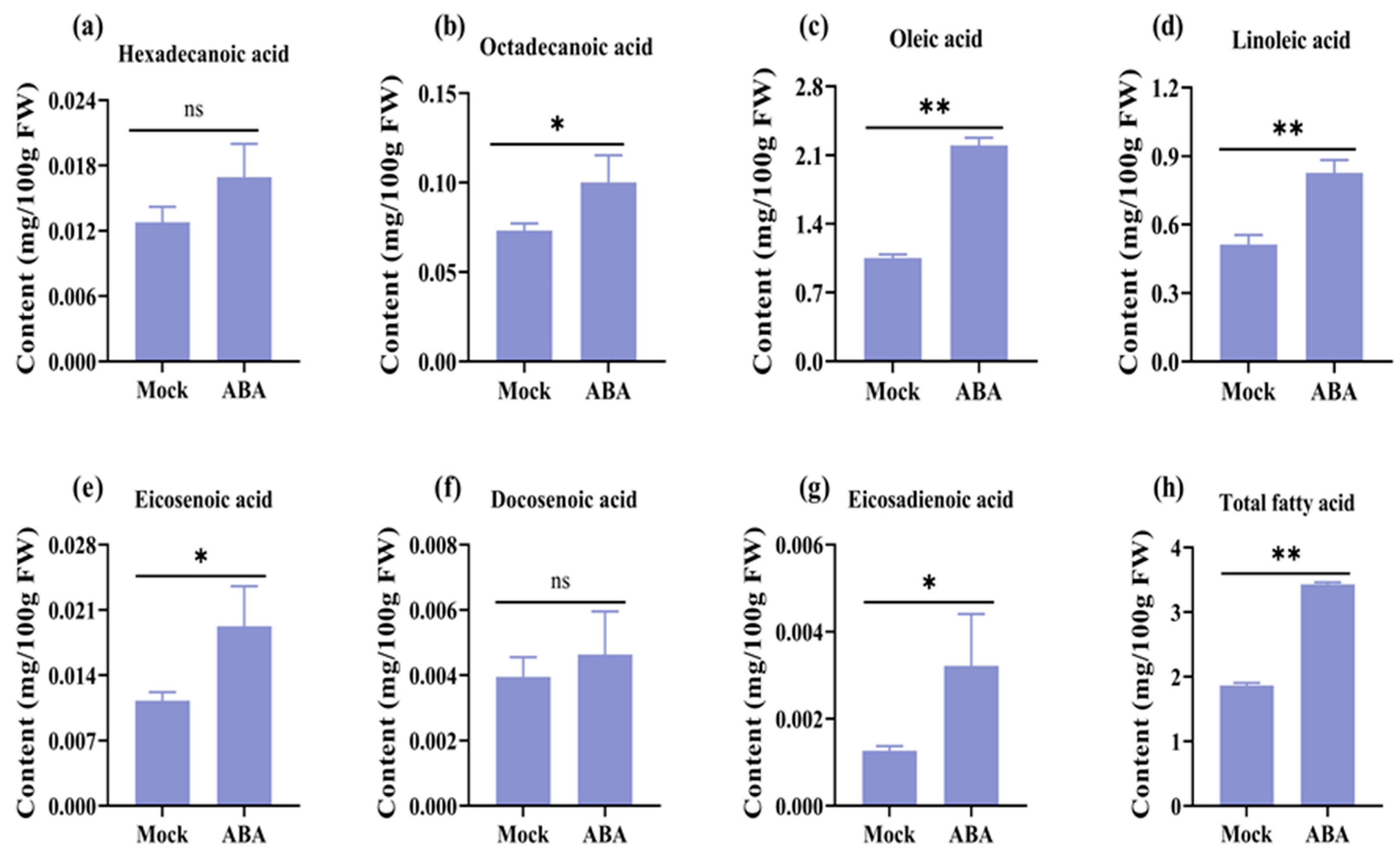
3.1. Exogenous Application of ABA Can Efficiently Improve Unsaturated Fatty Acids Content in Rapeseeds

We treated the rapeseed with exogenous ABA to detect the effect of ABA on fatty acid synthesis. Analysis using GC-MS/MS revealed that seeds treated with ABA showed a significant increase in both total fatty acid content and the content of multiple fatty acid components compared to the control group (Figure 1). Specifically, the proportion of oleic acid content was the highest and exhibited the largest increase in value (Figure 1). Notably, the total fatty acid content nearly doubled under ABA treatment compared to the control group (Figure 1h). These results suggest that exogenous ABA can increase the content of free fatty acids in rapeseed, especially the content of oleic acid, aligning to improve fatty acid content in rapeseed.

3.2. Overview of Small RNA Library Sequencing for Rapeseeds

To investigate the connection between fatty acid content and ABA treatment, we performed high-throughput small RNA sequencing using ABA-treated and control-treated seeds collected at 35-DAF. Each treatment group was represented by three biological replicates. Total RNA extraction and subsequent cDNA library construction were performed. We obtained clean read counts ranging from 13.18 to 15.08 million per sample, with genome alignment rates between 87.81% and 88.66% (Table S2).
The majority of original miRNA reads fell within the 19 to 22 nt range, with the highest abundance observed in 21 nt miRNA reads, showing a prominent preference for uracil at the miRNA mature sequence’s first base (Figure 2 and Figure S1a,b). Moreover, miRNA reads in the 20–21 nt range constitute more than half of all small RNAs of that size (Figure 2). The expression pattern data of miRNAs in each sample exhibits symmetry and a relatively concentrated distribution (Figure S1c). Correlation tests confirmed reasonable sample selection and reliable expression level correlations between samples (Figure S1d).

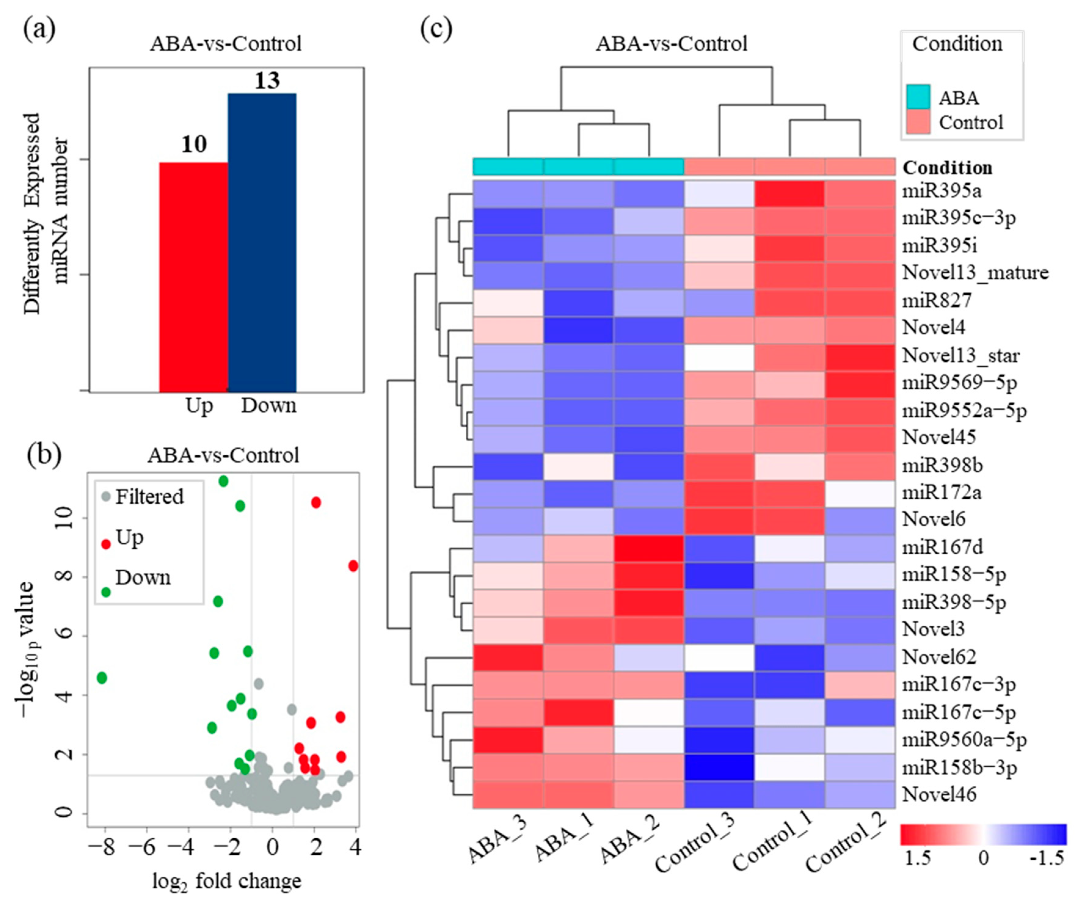
3.3. Identification of ABA-Interacting miRNAs Involved in Fatty Acid Metabolism in B. napus

After aligning unique small RNA (sRNA) sequences with miRBase 21.0, we identified 211 known miRNAs in the libraries (Table S3). For novel miRNA prediction, we employed the miRDeep2 tool using default parameters. In total, 97 novel miRNAs were predicted, and the lengths of the novel miRNAs ranged from 21 to 25 nt, most commonly measuring 24 nt in length (Table S4). Given that the occurrence of 24 nt miRNAs is exceptionally uncommon, these newly identified miRNAs at the 24 nt length should be considered as potential candidates and will require specific confirmation following the biogenesis guidelines outlined by Axtell and Meyers [42].
By using a threshold of |log2FC| ≥ 1.0 and a p-value ≤ 0.05, we determined that 23 miRNAs were differentially expressed between the control and ABA-treated samples—with 10 up-regulated and 13 down-regulated (Figure 3 and Figure S2 and Table S5). Among these, eight novel miRNAs were included, and their secondary structures can be found in Figure S3. These differentially expressed miRNAs may be involved in interacting with ABA to modulate fatty acid metabolism in B. napus.

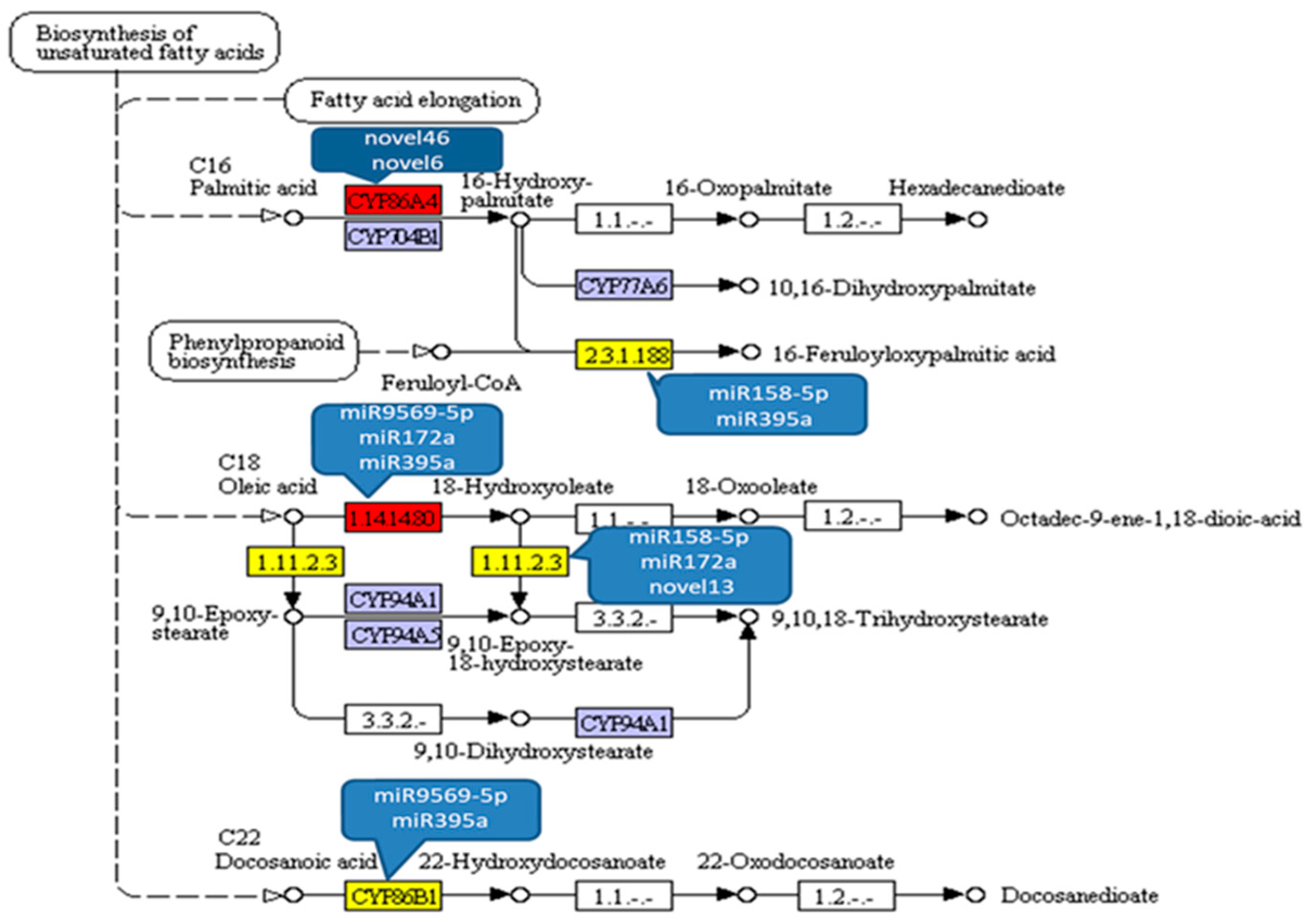
3.4. Prediction of Target Genes by miRNAs Interacting with ABA and Functional Analysis

We then predicted the potential target genes of these miRNAs using the miRNA target gene prediction software TargetFinder (Table S6). KEGG enrichment analysis was performed to predict the function of genes targeted by differentially expressed miRNAs, with a focus on metabolic regulation. Lipid metabolism and signal transduction pathways were involved (Figure 4), indicating the potential importance of the identified miRNAs in these regulatory processes.
Furthermore, we used GO analysis to classify the functions of target genes by the differentially expressed miRNAs interacting with ABA. These target genes were found to be involved in 21 different molecular functions, 23 biological processes, and 20 cellular components (Figure S4). Several important biological processes were identified, including biological regulation and metabolic processes (Figure S4).

3.5. Expression Profiles of miRNAs Interacting with ABA Involved in Fatty Acids Biosynthesis

To further investigate the molecular mechanism of fatty acid biosynthesis during seed development, we examined the target genes associated with the pathway of fatty acid biosynthesis for differentially expressed miRNAs that interact with ABA. A total of 199 target genes were involved in pathways of fatty acid biosynthesis, degradation, and metabolism (Table S7). Among them, 74 genes were predicted to participate in the biosynthesis of unsaturated fatty acid and to be regulated by more than 10 miRNAs (Table S8). As shown in Figure 5, known miRNAs including miR9569-5p, miR158-5p, miR172a, miR395a and novel miRNAs including Novel13, Novel46, and Novel6 are involved in the main regulatory network of unsaturated fatty acid synthesis pathway.

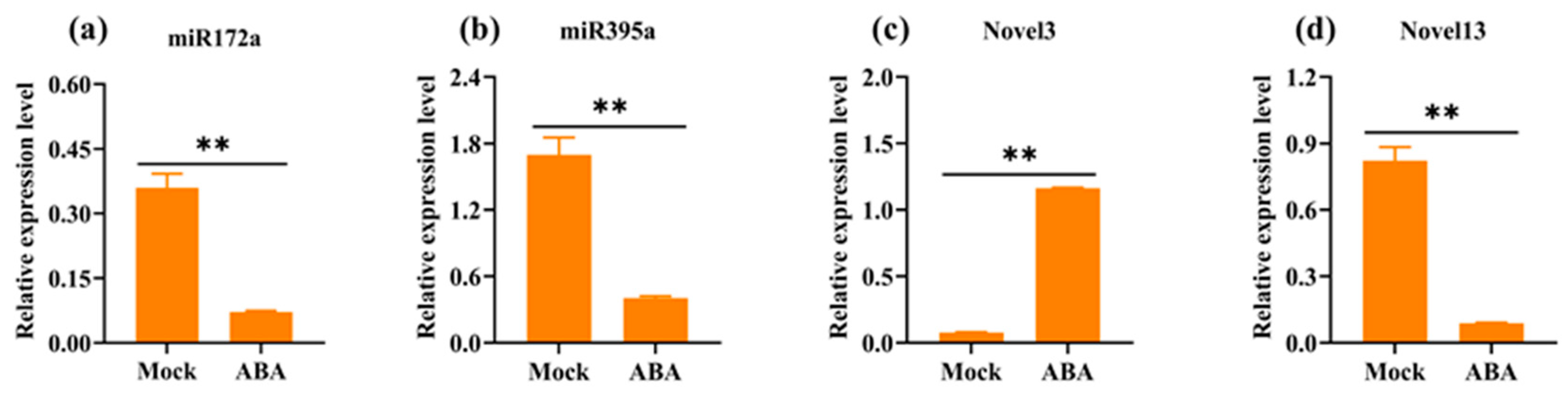
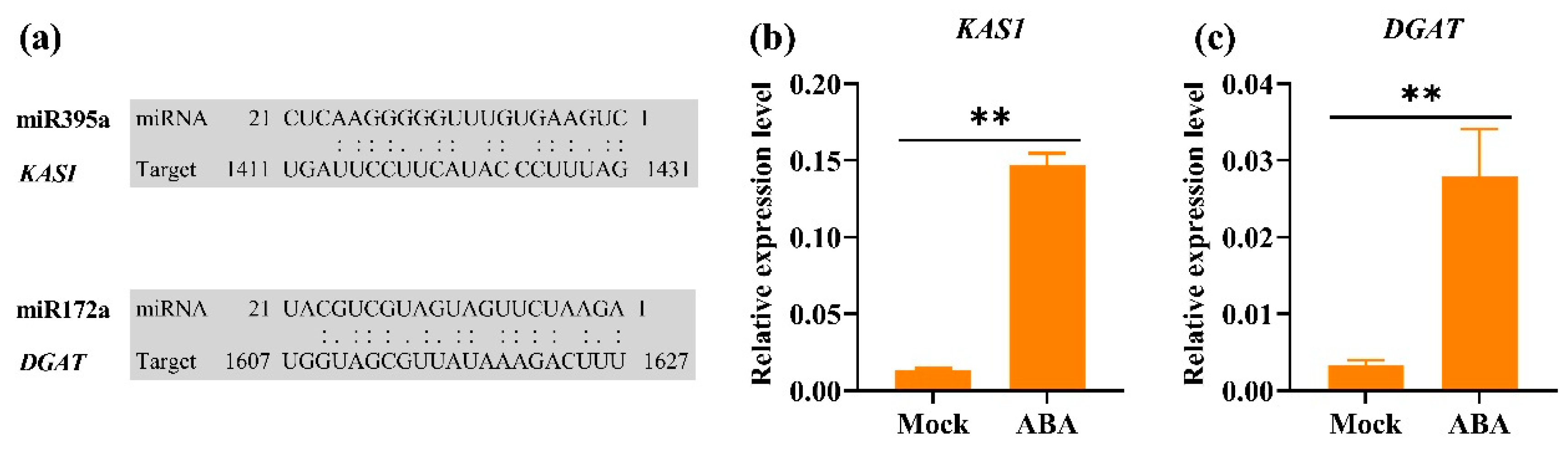
3.6. RT-qPCR Validation of miRNAs and Corresponding Target Genes

We analyzed the expression of differentially expressed miRNAs using Stem-Loop qPCR. Specifically, the expression levels of miR172a, miR395a, Novel13, and Novel3 were quantified. The target genes predicted by miR172a, miR395a, and Novel13 are implicated in various stages of fatty acid synthesis (Figure 5). Additionally, Novel3 is significantly up-regulated upon ABA treatment and exhibits the highest expression level among all differentially expressed miRNAs (Table S5). Our results revealed that under ABA treatment, miR172a, miR395a, and Novel13 exhibited significant down-regulation compared to the control, with fold changes of 5.0, 4.2, and 9.3, respectively, while Novel3 exhibited significant up-regulation with fold changes of 14.8 (Figure 6). These findings were generally consistent with the small RNA sequencing.
We then analyzed the possibility of the regulatory pathway using the classic website for plant miRNA and target gene interaction analysis, psRNATarget v.2 (https://www.zhaolab.org/psRNATarget/). Results revealed that miRNA395a can bind to the mRNA of the ketoacyl-ACP synthase (KAS I) and miR172a could bind to the mRNA of the triglyceride acyltransferase (DGAT). Both KAS I and DGAT are involved in the fatty acid synthesis pathway (Figure 7a). Further expression level of KAS I and DGAT was detected by RT-qPCR, revealing that both were up-regulated by ABA induction (Figure 7b,c). The results suggest the targeting relationship of miRNA395a—KAS I and miRNA172a—DGAT, respectively.

4. Discussion

The process of seed development plays a crucial role in determining both the quantity and the quality of fatty acids in B. napus [43,44]. To enhance fatty acid content and quality, researchers need to gain a comprehensive understanding of the molecular mechanisms that regulate the various stages of fatty acid biosynthesis within seed development. miRNAs are integral post-transcriptional regulators that control the expression of a multitude of genes involved in fatty acid biosynthesis [17]. High-throughput sequencing techniques are uncovering an expanding array of miRNAs connected to fatty acid biosynthesis, with these miRNAs and their target genes forming an extensive network that governs fatty metabolism [40,45]. ABA is known to suppress the synthesis of long-chain fatty acids such as erucic acid by down-regulating the expression of the FAE1 gene [46].
ABA and miRNA play crucial roles in regulating plant growth and development. While their individual impacts are well-documented, the interplay between the two in regulating rapeseed fatty acid synthesis and metabolism remains unexplored. Treatment with ABA during the rapeseed seed development stage has been shown to markedly enhance the seed fatty acid content (Figure 1). Identifying the miRNAs involved in this process is pivotal in uncovering the mechanisms of interactive regulation between ABA and miRNA, as well as establishing a framework for leveraging miRNA to modulate rapeseed fatty acid metabolism in the future. Through the application of high-throughput technologies, we have identified the presence of 211 known miRNAs and 97 novel miRNAs during seed development in B. napus (Tables S3 and S4). Furthermore, some miRNAs demonstrated a significant increase in expression following ABA treatment and other miRNAs showed an inverse correlation with fatty acid content and composition post-ABA treatment (Table S5). These outcomes indicate that miRNAs working with ABA operate at different stages of fatty acid biosynthesis through various regulatory paths.
A previous study conducted a systematic analysis of small RNA expression profiles and identified 50 conserved and 9 novel miRNAs in B. napus seeds at early embryonic stages, across both high- and low-oil-content cultivars, and the miR156 and miR167 were found in higher abundance in the high-oil-content cultivar [18]. In our study, the expression of miR167c was also notably up-regulated in response to ABA treatment (Table S5), aligning somewhat with prior studies, indicating the potential involvement of miR167 in the regulation of ABA-induced fatty acid biosynthesis in developing B. napus seeds. However, the expression of miR156, which targets the SPL gene to regulate early embryo development and affects seed oil content in B. napus [47], was hardly changed by ABA treatment.
The enzymatic cascade responsible for generating C18 unsaturated fatty acids has been extensively studied and documented [48]. In plastids, fatty acids are synthesized de novo from acetyl-coenzyme A (CoA) [48,49]. Once produced, 18:0 is bound to an acyl carrier protein (ACP) and enters the unsaturation program catalyzed by a series of fatty acid desaturases (FADs) [50]. These steps involve stearoyl-ACP desaturase (EC: 2.3.1.188), delta-12 oleate desaturase (EC: 1.11.2.3), and fatty acid omega-3 desaturase (EC: 1.14.1480), resulting in the production of unsaturated fatty acids [51]. Specifically, several miRNAs, such as miR9569-5p, miR158-5p, miR172a, miR395, Novel13, Novel46, and Novel6, are believed to interact with ABA and subsequently regulate the aforementioned metabolic processes (Figure 5).
Furthermore, miRNA and target gene interaction analysis using psRNATarget indicates that miRNA395a can bind to the mRNA of the ketoacyl-ACP synthase gene KAS I, which is a component of the fatty acid synthesis pathway (Figure 7). The responsive behavior of miRNA395a and its potential target gene KAS I to ABA at the expression level (Figure 6 and Figure 7), exhibiting opposite changes, further suggests the targeting relationship between miRNA395a and KAS I. Additionally, miR172a, which was predicted to regulate DGAT, was markedly reduced following ABA treatment (Figure 6 and Figure 7). Notably, DGAT is a rate-limiting enzyme for triglyceride synthesis and is crucial for the accumulation of oil in seeds [52]. And expression of DGAT was up-regulated 8-fold after treatment with ABA (Figure 7). These findings highlight the significance of miR172’s interactions with ABA in fatty acid biosynthesis during seed development.
Results from this research broaden our comprehension of the molecular mechanisms underpinning seed development and fatty acid biosynthesis in B. napus. However, this study also has some limitations. Firstly, ABA is a hormone with broad effects, and the variations expressed miRNA observed in ABA treatment may not necessarily pertain to fatty acid synthesis. While we identified potential miRNAs implicated in fatty acid synthesis through functional analysis of predicted target genes (Table S7) and conducted preliminary research on the expression and regulatory relationships of miR172a and miRNA395a along with their target genes (Figure 7), additional evidence is required to validate their interactions. Previous independent studies have confirmed that miR172 and miR395 have highly conserved targets such as AP2 or AP2-like transcript TOE3, APS, and SULTR2 [53,54,55,56,57,58,59]. Although miRNAs often have alternative targets, more direct or indirect evidence is necessary to establish a causal relationship between these miRNAs and their targets, thus clearly delineating their involvement in ABA’s impact on fatty acid biosynthesis. Additionally, integrating information from relevant studies on degradome data [19,60] for comprehensive comparisons can facilitate the identification of new target sites and regulatory processes, enhancing our understanding of the ABA-mediated miRNA regulation of fatty acid synthesis.

5. Conclusions

Our current research uncovered that exogenous application of ABA can efficiently improve unsaturated fatty acid content in rapeseeds, and further identified a total of 308 miRNAs—97 that are novel and 211 that are known—in B. napus seeds. Within this group, 23 differentially expressed miRNAs have been determined to play roles in fatty acid biosynthesis in response to ABA treatment. Of these, miR172a and miR395a were notably down-regulated, whereas corresponding potential target genes experienced significant up-regulation. Through comprehensive GO and KEGG functional annotation, these 23 miRNAs were found to regulate numerous genes, including genes within lipid metabolism or specifically associated with fatty acid biosynthesis. The involvement of partial miRNAs and their target genes in fatty acid synthesis was further confirmed via RT-qPCR validation. To our knowledge, this study is the first to provide a comparative analysis of miRNA profiles that respond to ABA with significant changes in fatty acid composition.

Supplementary Materials

The following supporting information can be downloaded at: https://www.mdpi.com/article/10.3390/agronomy14071358/s1, Figure S1: General features of identified miRNAs in B. napus; Figure S2: MA-plot of expressed miRNAs; Figure S3: Secondary structures of differentially expressed novel miRNAs; Figure S4: Gene ontology classification of genes targeted by differentially expressed miRNAs; Table S1: Primers used for quantitative RT-PCR experiments; Table S2: Genome alignment statistics; Table S3: Identified known miRNAs; Table S4: Identified novel miRNAs; Table S5: ABA-vs-Control-differentially expressed miRNAs; Table S6: Predicted target genes of miRNA; Table S7: Target genes involved in fatty acid biosynthesis, degradation, and metabolism; Table S8: Target genes involved in biosynthesis of unsaturated fatty acid.

Author Contributions

Conceptualization, J.Z.; methodology, J.Z.; formal analysis, J.Z.; investigation, J.Z. and Z.X.; resources, J.Z. and Z.Y.; writing—original draft preparation, J.Z.; writing—review and editing, J.Z., Z.Y. and Z.X.; visualization, J.Z. and Z.X.; supervision, J.Z. and Z.Y.; funding acquisition, J.Z. All authors have read and agreed to the published version of the manuscript.

Funding

This research was funded by grants from the Hunan Provincial Natural Science Foundation of China 2022JJ30304 and the Scientific Research Fund of the Hunan Provincial Education Department 23B0217.

Data Availability Statement

Data are contained within the article and Supplementary Materials.

Conflicts of Interest

The authors declare no conflicts of interest.

References

  1. Petrie, J.R.; Zhou, X.R.; Leonforte, A.; McAllister, J.; Shrestha, P.; Kennedy, Y.; Belide, S.; Buzza, G.; Gororo, N.; Gao, W.; et al. Development of a Brassica napus (Canola) Crop Containing Fish Oil-like Levels of DHA in the Seed Oil. Front. Plant Sci. 2020, 11, 727. [Google Scholar] [CrossRef]
  2. Wang, Z.; Wang, F.; Yu, Z.; Shi, X.; Zhou, X.; Wang, P.; Song, Y.; Hong, D.; Yang, G. Pyramiding of multiple genes generates rapeseed introgression lines with clubroot and herbicide resistance, high oleic acid content, and early maturity. Crop J. 2023, 11, 895–903. [Google Scholar] [CrossRef]
  3. Wang, H.; Wang, Q.; Pak, H.; Yan, T.; Chen, M.; Chen, X.; Wu, D.; Jiang, L. Genome-wide association study reveals a patatin-like lipase relating to the reduction of seed oil content in Brassica napus. BMC Plant Biol. 2021, 21, 6. [Google Scholar] [CrossRef]
  4. He, M.; Qin, C.X.; Wang, X.; Ding, N.-Z. Plant Unsaturated Fatty Acids: Biosynthesis and Regulation. Front. Plant Sci. 2020, 11, 390. [Google Scholar] [CrossRef]
  5. Lee, R.C.; Feinbaum, R.L.; Ambros, V. The C. elegans heterochronic gene lin-4 encodes small RNAs with antisense complementarity to lin-14. Cell 1993, 75, 843–854. [Google Scholar] [CrossRef] [PubMed]
  6. Zhan, J.; Meyers, B.C. Plant Small RNAs: Their Biogenesis, Regulatory Roles, and Functions. Annu. Rev. Plant Biol. 2023, 74, 21–51. [Google Scholar] [CrossRef]
  7. Voinnet, O. Origin, biogenesis, and activity of plant microRNAs. Cell 2009, 136, 669–687. [Google Scholar] [CrossRef]
  8. Llave, C.; Xie, Z.; Kasschau, K.D.; Carrington, J.C. Cleavage of Scarecrow-like mRNA targets directed by a class of Arabidopsis miRNA. Science 2002, 297, 2053–2056. [Google Scholar] [CrossRef] [PubMed]
  9. Zhang, B.; Pan, X.; Cobb, G.P.; Anderson, T.A. Plant microRNA: A small regulatory molecule with big impact. Dev. Biol. 2006, 289, 3–16. [Google Scholar] [CrossRef]
  10. Li, J.; Reichel, M.; Li, Y.; Millar, A.A. The functional scope of plant microRNA-mediated silencing. Trends Plant Sci. 2014, 19, 750–756. [Google Scholar] [CrossRef]
  11. Yuan, S.; Zhao, J.; Li, Z.; Hu, Q.; Yuan, N.; Zhou, M.; Xia, X.; Noorai, R.; Saski, C.; Li, S.; et al. MicroRNA396-mediated alteration in plant development and salinity stress response in creeping bentgrass. Hortic. Res. 2019, 6, 48. [Google Scholar] [CrossRef]
  12. Iliopoulos, D.; Drosatos, K.; Hiyama, Y.; Goldberg, I.J.; Zannis, V.I. MicroRNA-370 controls the expression of MicroRNA-122 and Cpt1α and affects lipid metabolism. J. Lipid Res. 2010, 51, 1513–1523. [Google Scholar] [CrossRef] [PubMed]
  13. Nakanishi, N.; Nakagawa, Y.; Tokushige, N.; Aoki, N.; Matsuzaka, T.; Ishii, K.; Yahagi, N.; Kobayashi, K.; Yatoh, S.; Takahashi, A.; et al. The up-regulation of microRNA-335 is associated with lipid metabolism in liver and white adipose tissue of genetically obese mice. Biochem. Biophys. Res. Commun. 2009, 385, 492–496. [Google Scholar] [CrossRef]
  14. Körbes, A.P.; Machado, R.D.; Guzman, F.; Almerão, M.P.; de Oliveira, L.F.; Loss-Morais, G.; Turchetto-Zolet, A.C.; Cagliari, A.; dos Santos Maraschin, F.; Margis-Pinheiro, M.; et al. Identifying conserved and novel microRNAs in developing seeds of Brassica napus using deep sequencing. PLoS ONE 2012, 7, e50663. [Google Scholar] [CrossRef]
  15. Wang, J.; Jian, H.; Wang, T.; Wei, L.; Li, J.; Li, C.; Liu, L. Identification of microRNAs Actively Involved in Fatty Acid Biosynthesis in Developing Brassica napus Seeds Using High-Throughput Sequencing. Front. Plant Sci. 2016, 7, 1570. [Google Scholar] [CrossRef]
  16. Chen, L.; Chen, L.; Zhang, X.; Liu, T.; Niu, S.; Wen, J.; Yi, B.; Ma, C.; Tu, J.; Fu, T.; et al. Identification of miRNAs that regulate silique development in Brassica napus. Plant Sci. 2018, 269, 106–117. [Google Scholar] [CrossRef] [PubMed]
  17. Wang, Z.; Qiao, Y.; Zhang, J.; Shi, W.; Zhang, J. Genome wide identification of microRNAs involved in fatty acid and lipid metabolism of Brassica napus by small RNA and degradome sequencing. Gene 2017, 619, 61–70. [Google Scholar] [CrossRef]
  18. Zhao, Y.-T.; Wang, M.; Fu, S.X.; Yang, W.C.; Qi, C.K.; Wang, X.-J. Small RNA Profiling in Two Brassica napus Cultivars Identifies MicroRNAs with Oil Production- and Development-Correlated Expression and New Small RNA Classes. Plant Physiol. 2012, 158, 813–823. [Google Scholar] [CrossRef] [PubMed]
  19. Wei, W.; Li, G.; Jiang, X.; Wang, Y.; Ma, Z.; Niu, Z.; Wang, Z.; Geng, X. Small RNA and degradome profiling involved in seed development and oil synthesis of Brassica napus. PLoS ONE 2018, 13, e0204998. [Google Scholar] [CrossRef]
  20. Zhang, C.; Chang, W.; Li, X.; Yang, B.; Zhang, L.; Xiao, Z.; Li, J.; Lu, K. Transcriptome and Small RNA Sequencing Reveal the Mechanisms Regulating Harvest Index in Brassica napus. Front. Plant Sci. 2022, 13, 855486. [Google Scholar] [CrossRef]
  21. Zou, X.; Zhang, K.; Wu, D.; Lu, M.; Wang, H.; Shen, Q. Integrated analysis of miRNA, transcriptome, and degradome sequencing provides new insights into lipid metabolism in perilla seed. Gene 2024, 895, 147953. [Google Scholar] [CrossRef] [PubMed]
  22. Wang, L.; Ruan, C.; Bao, A.; Li, H. Small RNA profiling for identification of microRNAs involved in regulation of seed development and lipid biosynthesis in yellowhorn. BMC Plant Biol. 2021, 21, 464. [Google Scholar] [CrossRef] [PubMed]
  23. Wu, B.; Ruan, C.; Shah, A.H.; Li, D.; Li, H.; Ding, J.; Li, J.; Du, W. Identification of miRNA-mRNA Regulatory Modules Involved in Lipid Metabolism and Seed Development in a Woody Oil Tree (Camellia oleifera). Cells 2021, 11, 71. [Google Scholar] [CrossRef] [PubMed]
  24. Li, M.; Zhu, Y.; Li, S.; Zhang, W.; Yin, C.; Lin, Y. Regulation of Phytohormones on the Growth and Development of Plant Root Hair. Front. Plant Sci. 2022, 13, 865302. [Google Scholar] [CrossRef] [PubMed]
  25. Gutierrez, L.; Bussell, J.D.; Păcurar, D.I.; Schwambach, J.l.; Păcurar, M.; Bellini, C. Phenotypic Plasticity of Adventitious Rooting in Arabidopsis Is Controlled by Complex Regulation of AUXIN RESPONSE FACTOR Transcripts and MicroRNA Abundance. Plant Cell 2009, 21, 3119–3132. [Google Scholar] [CrossRef] [PubMed]
  26. Guo, H.-S.; Xie, Q.; Fei, J.F.; Chua, N.-H. MicroRNA Directs mRNA Cleavage of the Transcription Factor NAC1 to Downregulate Auxin Signals for Arabidopsis Lateral Root Development. Plant Cell 2005, 17, 1376–1386. [Google Scholar] [CrossRef]
  27. Kinoshita, N.; Wang, H.; Kasahara, H.; Liu, J.; MacPherson, C.; Machida, Y.; Kamiya, Y.; Hannah, M.A.; Chua, N.-H. IAA-Ala Resistant3, an Evolutionarily Conserved Target of miR167, MediatesArabidopsisRoot Architecture Changes during High Osmotic Stress. Plant Cell 2012, 24, 3590–3602. [Google Scholar] [CrossRef] [PubMed]
  28. Yoon, E.K.; Yang, J.H.; Lim, J.; Kim, S.H.; Kim, S.K.; Lee, W.S. Auxin regulation of the microRNA390-dependent transacting small interfering RNA pathway in Arabidopsis lateral root development. Nucleic Acids Res. 2010, 38, 1382–1391. [Google Scholar] [CrossRef] [PubMed]
  29. Yu, H.; Zhang, Y.; Moss, B.L.; Bargmann, B.O.R.; Wang, R.; Prigge, M.; Nemhauser, J.L.; Estelle, M. Untethering the TIR1 auxin receptor from the SCF complex increases its stability and inhibits auxin response. Nat. Plants 2015, 1, 14030. [Google Scholar] [CrossRef]
  30. Jodder, J. miRNA-mediated regulation of auxin signaling pathway during plant development and stress responses. J Biosci. 2020, 45, 91. [Google Scholar] [CrossRef]
  31. Yan, A.; Chen, Z. The pivotal role of abscisic acid signaling during transition from seed maturation to germination. Plant Cell Rep. 2017, 36, 689–703. [Google Scholar] [CrossRef] [PubMed]
  32. Cutler, S.R.; Rodriguez, P.L.; Finkelstein, R.R.; Abrams, S.R. Abscisic acid: Emergence of a core signaling network. Annu. Rev. Plant Biol. 2010, 61, 651–679. [Google Scholar] [CrossRef] [PubMed]
  33. Brookbank, B.P.; Patel, J.; Gazzarrini, S.; Nambara, E. Role of Basal ABA in Plant Growth and Development. Genes 2021, 12, 1936. [Google Scholar] [CrossRef] [PubMed]
  34. Lim, C.W.; Baek, W.; Jung, J.; Kim, J.H.; Lee, S.C. Function of ABA in Stomatal Defense against Biotic and Drought Stresses. Int. J. Mol. Sci. 2015, 16, 15251–15270. [Google Scholar] [CrossRef] [PubMed]
  35. Sano, N.; Marion-Poll, A. ABA Metabolism and Homeostasis in Seed Dormancy and Germination. Int. J. Mol. Sci. 2021, 22, 5069. [Google Scholar] [CrossRef] [PubMed]
  36. Wilmer, J.A.; Helsper, J.P.F.G.; van der Plas, L.H.W. Effects of abscisic acid and temperature on erucic acid accumulation in oilseed rape (Brassica napus L.). J. Plant Physiol. 1997, 150, 414–419. [Google Scholar] [CrossRef]
  37. Ma, H.; Wang, S. Histidine Regulates Seed Oil Deposition through Abscisic Acid Biosynthesis and β-oxidation. Plant Physiol. 2016, 172, 848–857. [Google Scholar] [CrossRef] [PubMed]
  38. Chen, C.; Ridzon, D.A.; Broomer, A.J.; Zhou, Z.; Lee, D.H.; Nguyen, J.T.; Barbisin, M.; Xu, N.L.; Mahuvakar, V.R.; Andersen, M.R.; et al. Real-time quantification of microRNAs by stem-loop RT-PCR. Nucleic Acids Res. 2005, 33, e179. [Google Scholar] [CrossRef] [PubMed]
  39. Abel, Y.; Rederstorff, M. Stem-Loop qRT-PCR-Based Quantification of miRNAs. Methods Mol. Biol. 2021, 2300, 59–64. [Google Scholar]
  40. Huang, D.; Koh, C.; Feurtado, J.A.; Tsang, E.W.; Cutler, A.J. MicroRNAs and their putative targets in Brassica napusseed maturation. BMC Genom. 2013, 14, 1–25. [Google Scholar] [CrossRef]
  41. GB/T17376–2008; Animal and vegetable fats and oils-Preparation of methyl esters of fatty acids. Standards Press of China: Beijing, China, 2008.
  42. Axtell, M.J.; Meyers, B.C. Revisiting Criteria for Plant MicroRNA Annotation in the Era of Big Data. Plant Cell 2018, 30, 272–284. [Google Scholar] [CrossRef] [PubMed]
  43. Zhao, C.; Xie, M.; Liang, L.; Yang, L.; Han, H.; Qin, X.; Zhao, J.; Hou, Y.; Dai, W.; Du, C.; et al. Genome-Wide Association Analysis Combined With Quantitative Trait Loci Mapping and Dynamic Transcriptome Unveil the Genetic Control of Seed Oil Content in Brassica napus L. Front. Plant Sci. 2022, 13, 929197. [Google Scholar] [CrossRef]
  44. Boulard, C.; Bardet, M.; Chardot, T.; Dubreucq, B.; Gromova, M.; Guillermo, A.; Miquel, M.; Nesi, N.; Yen-Nicolaÿ, S.; Jolivet, P. The structural organization of seed oil bodies could explain the contrasted oil extractability observed in two rapeseed genotypes. Planta 2015, 242, 53–68. [Google Scholar] [CrossRef] [PubMed]
  45. Liu, D.; Yu, L.; Wei, L.; Yu, P.; Wang, J.; Zhao, H.; Zhang, Y.; Zhang, S.; Yang, Z.; Chen, G.; et al. BnTIR: An online transcriptome platform for exploring RNA-seq libraries for oil crop Brassica napus. Plant Biotechnol. J. 2021, 19, 1895–1897. [Google Scholar] [CrossRef] [PubMed]
  46. Finkelstein, R.; Somerville, C. Abscisic acid or high osmoticum promote accumulation of long-chain fatty acids in developing embryos of Brassica napus. Plant Sci. 1989, 61, 213–217. [Google Scholar] [CrossRef]
  47. Yu, S.; Galvão, V.C.; Zhang, Y.C.; Horrer, D.; Zhang, T.Q.; Hao, Y.H.; Feng, Y.Q.; Wang, S.; Schmid, M.; Wang, J.-W. Gibberellin regulates the Arabidopsis floral transition through miR156-targeted SQUAMOSA promoter binding-like transcription factors. Plant Cell 2012, 24, 3320–3332. [Google Scholar] [CrossRef]
  48. Zhang, S.; Wu, S.; Hou, Q.; Zhao, J.; Fang, C.; An, X.; Wan, X. Fatty acid de novo biosynthesis in plastids: Key enzymes and their critical roles for male reproduction and other processes in plants. Plant Physiol. Biochem. 2024, 210, 108654. [Google Scholar] [CrossRef] [PubMed]
  49. Ke, J.; Behal, R.H.; Back, S.L.; Nikolau, B.J.; Wurtele, E.S.; Oliver, D.J. The Role of Pyruvate Dehydrogenase and Acetyl-Coenzyme A Synthetase in Fatty Acid Synthesis in Developing Arabidopsis Seeds. Plant Physiol. 2000, 123, 497–508. [Google Scholar] [CrossRef]
  50. Meï, C.; Michaud, M.; Cussac, M.; Albrieux, C.; Gros, V.; Maréchal, E.; Block, M.A.; Jouhet, J.; Rébeillé, F. Levels of polyunsaturated fatty acids correlate with growth rate in plant cell cultures. Sci. Rep. 2015, 5, 15207. [Google Scholar] [CrossRef]
  51. Wang, S.H.; Pan, Y.; Li, J.; Chen, H.-q.; Zhang, H.; Chen, W.; Gu, Z.N.; Chen, Y.Q. Endogenous omega-3 long-chain fatty acid biosynthesis from alpha-linolenic acid is affected by substrate levels, gene expression, and product inhibition. RSC Adv. 2017, 7, 40946–40951. [Google Scholar] [CrossRef]
  52. Perez-Arcoiza, A.; Luisa Hernández, M.; Dolores Sicardo, M.; Hernandez Santana, V.; Diaz Espejo, A.; Martinez Rivas, J.M. Carbon supply and water status regulate fatty acid and triacylglycerol biosynthesis at transcriptional level in the olive mesocarp. Plant Cell Environ. 2022, 45, 2366–2380. [Google Scholar] [CrossRef]
  53. Liang, G.; Yu, D. Reciprocal regulation among miR395, APS and SULTR2;1 in Arabidopsis thaliana. Plant Signal Behav. 2010, 5, 1257–1259. [Google Scholar] [CrossRef] [PubMed]
  54. Liang, G.; Yang, F.; Yu, D. MicroRNA395 mediates regulation of sulfate accumulation and allocation in Arabidopsis thaliana. Plant J. 2010, 62, 1046–1057. [Google Scholar] [CrossRef]
  55. Chung, M.Y.; Nath, U.K.; Vrebalov, J.; Gapper, N.; Lee, J.M.; Lee, D.J.; Kim, C.K.; Giovannoni, J. Ectopic expression of miRNA172 in tomato (Solanum lycopersicum) reveals novel function in fruit development through regulation of an AP2 transcription factor. BMC Plant Biol. 2020, 20, 283. [Google Scholar] [CrossRef]
  56. Nowak, K.; Morończyk, J.; Grzyb, M.; Szczygieł-Sommer, A.; Gaj, M.D. miR172 Regulates WUS during Somatic Embryogenesis in Arabidopsis via AP2. Cells 2022, 11, 718. [Google Scholar] [CrossRef] [PubMed]
  57. Zeng, V.; Uauy, C.; Chen, Y. Identification of a novel SNP in the miR172 binding site of Q homoeolog AP2L-D5 is associated with spike compactness and agronomic traits in wheat (Triticum aestivum L.). Theor. Appl. Genet. 2023, 137, 13. [Google Scholar] [CrossRef]
  58. Jung, J.H.; Lee, S.; Yun, J.; Lee, M.; Park, C.M. The miR172 target TOE3 represses AGAMOUS expression during Arabidopsis floral patterning. Plant Sci. 2014, 215–216, 29–38. [Google Scholar] [CrossRef] [PubMed]
  59. Wang, X.; Li, Q.; Zhu, H.; Song, M.; Zhang, K.; Ge, W. Molecular mechanisms of miR172a and its target gene LbrTOE3 regulating maturation in Lilium. Planta 2023, 258, 53. [Google Scholar] [CrossRef]
  60. Liu, J.; Liu, X.; Zhang, S.; Liang, S.; Luan, W.; Ma, X. TarDB: An online database for plant miRNA targets and miRNA-triggered phased siRNAs. BMC Genom. 2021, 22, 348. [Google Scholar] [CrossRef]
Figure 1. Fatty acid content in B. napus seeds in response to ABA. (a) Hexadecanoic acid content. (b) Octadecanoic acid content. (c) Oleic acid content. (d) Linoleic acid. (e) Eicosenoic acid content. (f) Docosenoic acid content. (g) Eicosadienoic acid content. (h) Total fatty acid content. Data are means ± SD, n = 3, with * and ** indicating a significant difference at the p < 0.05 and p < 0.01 level, respectively. ns indicating no significant difference at the p < 0.05.
Agronomy 14 01358 g001
Figure 2. Size distribution of total genome-mapped read counts for various types of small RNAs.
Agronomy 14 01358 g002
Figure 3. Expression of miRNAs in seeds in response to ABA treatment. (a) Number of significantly differentially expressed miRNAs in response to ABA treatment (p-value < 0.05 and |log2FC| > 1). (b) Volcanic plot displaying expressed miRNAs. Gray dots represent non-differentially expressed miRNAs (p-value > 0.05 or |log2FC| < 1), red dots signify significantly up-regulated miRNAs, and green dots denote significantly down-regulated miRNAs. (c) Clustered heatmap illustrating differentially expressed miRNAs. Red rectangles denote miRNAs up-regulated by ABA treatment, while blue rectangles indicate miRNAs down-regulated by ABA treatment.
Agronomy 14 01358 g003
Figure 4. KEGG pathway classification of genes targeted by differentially expressed miRNAs. The numbers on the right side of the columns represent a number of genes targeted by differentially expressed miRNAs and annotated to the corresponding pathway. Gene ontology classification of genes targeted by differentially expressed miRNAs.
Agronomy 14 01358 g004
Figure 5. Differential miRNAs involved in unsaturated fatty acid synthesis pathway. The map displays selected steps from KEGG pathways of fatty acid biosynthesis and exhibits differentially expressed miRNAs interacting with ABA-regulating unsaturated fatty acid biosynthesis pathways.
Agronomy 14 01358 g005
Figure 6. Stem-Loop qPCR analysis of miRNA expression in response to ABA. (ad) Expression level of miR172a (a), miR395a (b), Novel3 (c), and Novel13 (d) in siliques in response to ABA. Data are means ± SD, n = 3, with ** indicating a significant difference at the p < 0.01 level. Similar fold change results were obtained in another experiment.
Agronomy 14 01358 g006
Figure 7. Gene expression in response to ABA. (a) Interaction of miRNA and target gene. (b,c) Expression level of KAS I (b) and DGAT (c) in siliques in response to ABA. Data are means ± SD, n = 3, with ** indicating a significant difference at the p < 0.01 level. For (b,c), similar fold change results were obtained in other experiments.
Agronomy 14 01358 g007
Disclaimer/Publisher’s Note: The statements, opinions and data contained in all publications are solely those of the individual author(s) and contributor(s) and not of MDPI and/or the editor(s). MDPI and/or the editor(s) disclaim responsibility for any injury to people or property resulting from any ideas, methods, instructions or products referred to in the content.

