Article

Stress Response of Citrus Leaves under Mechanical Damage and Huanglongbing Disease Infection Using Plasmonic TiO2 Nanotube Substrate-Based Imprinting Mass Spectrometry Imaging

National Key Laboratory of Green Pesticide and Key Laboratory of Natural Pesticide and Chemical Biology of the Ministry of Education, South China Agricultural University, Guangzhou 510642, China
*
Author to whom correspondence should be addressed.
The authors contributed equally to this work.
Agronomy 2024, 14(8), 1797; https://doi.org/10.3390/agronomy14081797
Submission received: 11 July 2024 / Revised: 5 August 2024 / Accepted: 12 August 2024 / Published: 15 August 2024
(This article belongs to the Special Issue Pests, Pesticides, Pollinators and Sustainable Farming)

Abstract

:
Mapping the molecular signatures and metabolic regulation of plant tissues under biotic/abiotic stresses and defensive responses has become a subject of increasing interest in plant biology and systems biology, but determining when and where specialized metabolites are produced and accumulated currently remains a somewhat elusive goal. Herein, we demonstrated the use of a TiO2 nanotube-based composite substrate modified with plasmonic gold nanoparticles and hydrophobic polydopamine (AuNP-hPDA-TDNT) for surface-assisted laser desorption/ionization mass spectrometry (SALDI-MS) analysis of a wide range of pesticides and for visualizing the stress-responsive metabolites of citrus leaves during various plant defense processes. This method enabled the visualization of non-uniform and tissue-specific distribution patterns of functional metabolites of citrus leaves that were mechanically damaged, fed to larvae, and infected by Huanglongbing disease. Interestingly, some specialized metabolites exhibited different accumulation and regulation patterns for mechanical damage and larval feeding, suggesting that plant-derived secondary metabolites exercise specific defensive functions with respect to various damage processes. Moreover, the early diagnosis and detection of HLB disease-associated biomarkers can facilitate the prevention of citrus HLB diseases. Overall, this imprinting MS imaging strategy will expand the scope of MS techniques in plant biology, providing more biologically relevant insights into the biosynthesis, accumulation, and defensive role of bioactive metabolites in economically important plants.

1. Introduction

Citrus (Citrus japonica Thunb.) Huanglongbing (HLB) is a disease of worldwide incidence that causes considerable economic losses during the development, growth, and production stages of citrus plants [1,2]. Citrus HLB is caused by infection with Liberbacter asianticum jagoueix, which is propagated mainly by the citrus psyllid and grafting [3]. HLB has an incubation period during which newly infected trees do not show symptoms, posing a challenge for the early detection of HLB [4,5]. As the main pest of young citrus shoots, the citrus psyllid is a propagation medium for citrus HLB. The adults mostly lay eggs on the susceptible young treetops and begin to suck the sap of young shoots after the emergence of larvae. The adult citrus psyllid can then fly to new plants to transmit the citrus HLB. Currently, there is no completely effective treatment for citrus HLB, but early detection and treatment are the preferred strategies [6,7]. Thus, the early detection of HLB and the control of citrus psyllids are the keys to the prevention and control of HLB. Additionally, insights into the stress-induced secondary metabolites of plant tissues that are mechanically damaged and fed larvae will help researchers evaluate their defensive roles and decipher how plants deploy defenses for their maximum benefit [8]. However, the interactions between defensive responses and secondary metabolites produced by plants remain largely unclear.
Given that primary metabolites are closely associated with the growth and development of plant systems, plant secondary metabolites have received a significant amount of attention because of their specific biological functions under biotic/abiotic stresses [9,10,11]. Thus, gas chromatography or liquid chromatography coupled with mass spectrometry (GC−/LC−MS) has been well established for direct component analysis [12,13]. Unfortunately, spatial distribution information on minute amounts of metabolites where stress responses occur is normally missing during tissue homogenization and metabolite extraction processes. To circumvent these problems, mass spectrometry imaging (MSI) has evolved as a promising and molecule-specific tool for spatially and temporally characterizing a wide range of metabolites in complex plant systems [14,15,16,17,18,19]. Among the available techniques, three mainstream types of MSI techniques, namely, matrix-assisted laser desorption/ionization (MALDI), desorption electrospray ionization (DESI), and laser ablation electrospray ionization (LA-ESI), have become the most routinely used ionization sources of choice for plant metabolomics studies [20,21,22]. Despite being extensively used, several key challenges are currently facing conventional MALDI-MSI, such as background interference at m/z < 500 from organic matrices, poor reproducibility resulting from “sweet spots”, and potential imaging artifacts due to inhomogeneous co-crystallization [23,24,25]. Even worse, unlike plant stem or root tissues, which can be frozen-sectioned for MALDI-MSI, leaf and flower tissues cannot be sliced, which makes it exceedingly difficult to perform MSI analysis [26,27]. Although direct leaf and petal MSI can be obtained via DESI techniques, the MSI results might be compromised to a large extent because of wax layer protection and fluctuating surfaces. To this end, several porous materials have been proposed for use in imprinting strategies in DESI-MSI applications [28,29,30]. However, the limited spatial resolution of ambient MSI methods limits their ability to acquire high-quality images. Thus, there is a high demand for developing matrix-free LDI approaches that are compatible with plant leaves and flowers for capturing molecular signatures under biotic/abiotic stresses.
Inspired by the pioneering efforts of Gary Siuzdak’s group [31], a plethora of ultraviolet (UV)-absorbing nanostructured substrates, such as semiconductor-based [32,33,34], metal-based [35,36], carbon-based [37,38], and silicon-based [33,39] substrates, have enabled matrix-free LDI for MS analysis, termed surface-assisted laser desorption/ionization (SALDI). More valuably, these SALDI substrates are very suitable for MSI analysis of plant leaves and flowers and can be simply imprinted onto nanostructured material surfaces, regardless of matrix deposition and tissue sectioning. Benefiting from the advantages of high light absorption, a high surface area-to-volume ratio, and low thermal conductivity [40,41], several recent examples of these methods include visualizing endogenous glycoalkaloids, flavonoids, and carbohydrates within Catharanthus roseus flowers and spearmint leaves by imprinting SALDI-MSI [42,43]. Recently, we constructed a TiO2 nanotube (TDNT)-based composite material with plasmonic gold nanoparticles (AuNPs) and hydrophobic polydopamine (PDA) modification (AuNP-hPDA-TDNT) for primary and secondary metabolite mapping in a wide range of plant tissues and fruits [34,44]. The synergistic effects between n-type semiconductor TDNTs and plasmonic AuNPs make this SALDI-MSI strategy effective and sensitive. Despite considerable efforts, the great potential of imprinting MSI in real-case applications with respect to the stress response of plant tissues to disease infection and mechanical damage remains to be explored.
In this study, we demonstrated the potential of AuNP-hPDA-TDNT-based nanostructured substrates for SALDI-MS analysis of a wide range of pesticides and exploited the imprinting MSI capability in mapping secondary metabolites during various plant defense processes. Moreover, various nonuniform and tissue-specific distribution patterns of functional metabolites can be clearly visualized in plant leaves that are mechanically damaged, fed to larvae, and infected by Huanglongbing disease via AuNP-hPDA-TDNT-based SALDI-MSI. Taken together, these results demonstrate that this imprinting MSI method has great potential for visualizing plant defense-derived metabolites, providing insights into the biosynthesis, accumulation, and defensive role of a diverse variety of bioactive metabolites in economically important plants.

2. Materials and Methods

2.1. Chemicals and Materials

Titanium sheets (99.95% purity) were purchased from Qingyuan Metal Materials Ltd. (Shijiazhuang, China). Chloroauric acid (HAuCl4, 98% purity and 47.8% Au content), sodium citrate (98% purity), sodium fluoride (99% purity), and anhydrous sodium sulfate (99% purity) were purchased from Energy Chemistry Ltd. (Shanghai, China). Dopamine hydrochloride (98% purity) and 1H,1H,2H,2H-perfluorooctyltrichlorosilane (FOTS, 97% purity) were purchased from Shanghai Macklin Biochemical Co., Ltd. (Shanghai, China). α-Cyano-4-hydroxycinnamic acid (CHCA, 99.0% purity) and Tris buffer were purchased from Sigma-Aldrich (St. Louis, MO, USA). Acephate, dinotefuran, thiamethoxam, spirotetramat, rotenone, azoxystrobin, cyantraniliprole, chlorantraniliprole, and abamectin were purchased from J&K Scientific Ltd. (Beijing, China). HPLC-grade acetonitrile was purchased from Tedia Company, Inc. (Fairfield, OH, USA). All the chemicals were used without further purification.

2.2. Preparation of the Pesticide Standards and AuNPs

Thiamethoxam, rotenone, chlorobenzamide, and abamectin were dissolved in acetone. Spirotetramat, dinotefuran, acephate, cyantraniliprole, and azoxystrobin were dissolved in an acetonitrile/water (1:1, v/v) mixture. All the analytes were dissolved in the stock solution at a concentration of 10 mM. Then, the standard individual solution and mixed solution were prepared by progressively diluting the stock solution to the desired concentration. Then, 0.1 μL of standard solution was manually deposited onto the AuNP-hPDA-TDNT substrate, which was naturally air-dried prior to MS analysis. AuNPs were synthesized via thermal treatment according to previous reports [17,27].

2.3. Preparation of the AuNP-hPDA-TDNT Substrate

The composite nanostructured substrate was prepared via an electrochemical anodization method according to previous reports [34,44]. Briefly, anhydrous Na2SO4 and NaF were dissolved in deionized water as the electrolyte solution, a Ti sheet was adopted as the anode, and a Pt electrode was adopted as the cathode. The oxidation reaction proceeded at a voltage of 20 V at room temperature for 1 h. To avoid adverse side effects, the voltage was increased slowly. Then, TiO2 nanotube (TDNT) materials were produced and washed ultrasonically with deionized water for 1 min. After air drying at room temperature, the TDNT substrates were stored in clean containers until future use.
The TDNT substrates were placed on a mixed solution containing dopamine hydrochloride and Tris-HCl buffer (pH 8.5) that was heated with stirring at 600 rpm in a 90 °C water bath for 1 h to prepare the PDA-TDNT substrate. The sample was subjected to ultrasonic cleaning twice with ethanol and deionized water to remove surface residues. Then, the hPDA-TDNT substrate was obtained by spraying 10 times with 0.25% (v/v) FOTS at a flow rate of 1 mL/min onto the PDA-TDNT surface in a high-performance ultrasonic sprayer (UAM4000, Hangzhou, China). Finally, a 0.1 mg/mL AuNP suspension was evenly sprayed on the hPDA-TDNT substrate surface 200 times at a flow rate of 0.03 mL/min to prepare the AuNP-hPDA-TDNT substrate. The detailed workflow of the AuNP-hPDA-TDNT substrate fabrication is shown in Figure S1.

2.4. Preparation of Imprinted Citrus Leaves That Were Infected by Huanglongbing Disease, Mechanically Damaged, and Fed Larvae for MSI Analysis

The experimental citrus (Citrus japonica Thunb.) variety was sour orange (a native wild species in Hainan), and susceptible citrus plants were provided by South China Agricultural University. Four- to five-month-old leaves were chosen for infection with HLB (Liberbacter asianticum jagoueix), which is an early pathogen. Susceptible leaves with obvious diseased spots were selected. Additionally, one- to two-month-old leaves that were mechanically damaged and fed larvae were chosen for imprinting MSI analysis. Specifically, the mechanically damaged leaves were subsequently crushed with needle-nose pliers and then harvested after they had grown for 2 h, 12 h, and 24 h. The leaves that were fed with larvae were placed on young leaves to eat and then collected after they had grown for 12 h. After the leaves were collected, the leaf surface was simply wiped with a water-soaked paper towel and then placed on the AuNP-hPDA-TDNT substrate for imprinting, and pressure was slowly applied at 4 MPa, where the duration of the imprinting process was 20 min. Then, the leaves were removed, and the leaf-imprinted substrates were transferred to room temperature for drying and subjected to MSI analysis.

2.5. Metabolite Extraction of Citrus Leaves for Ultra-Performance Liquid Chromatography Cou-Pled with Mass Spectrometry (UPLC–MS) Experiments

For metabolite extraction from citrus plant leaves, 50 mg of each sample was weighed and placed into centrifuge tubes, where 800 µL of precooled extraction solvent containing methanol and water (7:3, v/v) at −20 °C was added for metabolite extraction. After the tissue was ground with two small steel balls at 50 Hz for 5 min, it was placed in an ultrasonic water bath at 4 °C for 30 min and then transferred to −20 °C for 1 h. Following centrifugation at 25,000 rpm for 15 min, 600 μL of the supernatant was collected through a 0.22 μm filter and analyzed via UPLC-MS.

2.6. UPLC-MS Conditions for Metabolite Identification in Citrus Leaves

For metabolite identification in citrus leaves, a two-dimensional UPLC (Waters, Milford, MA, USA) coupled to a high-resolution Orbitrap mass spectrometer (Q Exactive HF, Thermo Fisher Scientific, Waltham, MA, USA) was used. In brief, 0.1% formic acid in water (Solvent A) and 0.1% formic acid in methanol (Solvent B) composed the mobile phases. The gradient elution conditions were as follows: 0–1 min, 2% Solvent B; 1–9 min, 2–98% Solvent B; 9–12 min, 98% Solvent B; 12–12.1 min, 98–2% Solvent B; and 12.1–15 min, 2% Solvent B. The flow rate and injection volume were set at 0.35 mL/min and 5 μL, respectively. The column oven was maintained at 45 °C. LC-eluted fractions were subjected to MS through electrospray ionization. In this study, the positive ion mode for electrospray ionization (ESI) was used with the spray voltage set at 3.80 kV, where 40 L/min of sheath gas and 10 L/min of auxiliary gas at 350 °C were used. The ion transfer capillary temperature was set at 320 °C.
Mass spectra were acquired in the m/z range from 70 to 1050 Da with a mass resolution of 70,000 at m/z 200. The automatic gain control (AGC) target value was set to 3 × 10⁶ for full MS mode. For MS/MS analysis, data-dependent acquisition (DDA) mode was employed with a mass resolution of 17,500 and an AGC setting of 1 × 10⁵. Step-increase collision voltages of 20−40−60 eV were adopted to activate and fragment the precursor ions of interest. Metabolite identification was performed by matching the measured m/z values and fragmental patterns with the Human Metabolome Database (HMDB) (http://www.hmdb.ca, accessed on 5 October 2022).

2.7. SALDI-MS and Imprinting MSI Experiments

For AuNP-hPDA-TDNT-based SALDI-MS analysis, 0.1 μL of sample solution was spotted onto the AuNP-hPDA-TDNT substrate. After it was naturally air-dried, the AuNP-hPDA-TDNT substrate was mounted on a stainless steel target (MTP Slide Adapter II, Bruker Daltonics, Billerica, MA, USA). All SALDI-MS analyses were performed on an UltrafleXtreme mass spectrometer (Bruker Daltonics, Billerica, MA, USA) equipped with a Nd:YAG laser at a wavelength of 355 nm, a laser frequency of 1000 Hz, and a laser energy of 45% in position ion mode. The mass spectral data were acquired with a mass range from 100 to 1000 Da. The spot size was selected as “Ultra”, and mass axis calibration was performed using the signals of the Aun+ ions (n = 1 to 5) generated by the AuNPs. Each SALDI mass spectrum was obtained by accumulating the MS signals from 400 laser shots. For MSI analysis, the laser power was set at 80–90%, and the mass spectrum at each pixel was obtained by accumulating 200 laser shots unless otherwise indicated. The optimized voltage parameters, such as a reflector voltage of 20.84 kV, a lens voltage of 11.00 kV, an ion source voltage of 20.00 kV, and an extraction delay time of 100 ns, were adopted throughout the MSI experiments.

2.8. Data Analysis

FlexControl 5.0 and flexAnalysis 5.0 software were used to acquire and analyze the data. For MSI data analysis, the raw data were visualized via flexImaging 5.0 software and then imported into SCiLS Lab 2016b software for further processing. The acquired raw mass spectra were normalized to the total ion count for each image. The statistical data were processed using SPSS software (version 19.0; SPSS, Inc., Chicago, IL, USA). One-way analysis of variance (ANOVA) and Tukey’s test were performed to exhibit statistical significance.

3. Results and Discussion

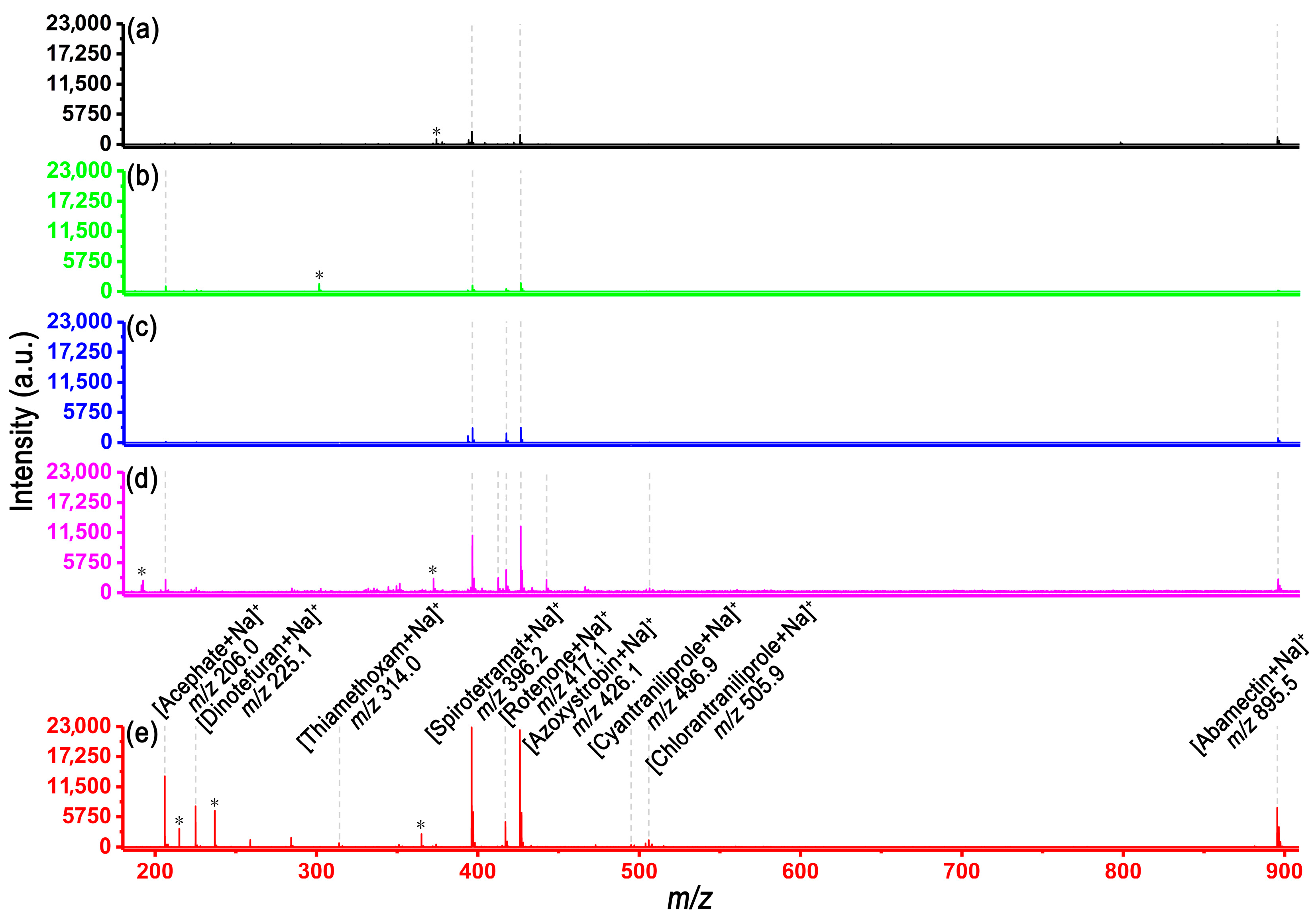
3.1. SALDI-MS Analysis of Various Pesticide Molecules

Previous results have demonstrated that the AuNP-hPDA-TDNT material shows great promise as a SALDI substrate for primary and secondary metabolites in plant tissues, whereas the great potential of this SALDI-MS method in pesticide mixture detection remains to be explored. To this end, a total of nine pesticides were selected for SALDI-MS analysis via a conventional MALDI matrix and four SALDI substrates at different construction stages. Information on the detected pesticides and their ion forms is listed in Tables S1 and S2. As shown in Figure 1, only three pesticides could be detected when CHCA was used as the matrix (Figure 1a), whereas all of the pesticides could be clearly detected using the AuNP-hPDA-TDNT substrate (Figure 1e). With the same laser energy, the other substrates, including TDNT, PDA-TDNT, and hPDA-TDNT, afforded three, four, and eight pesticides, respectively. Notably, the excellent photothermal conversion efficiency and enhanced ion production contributed to the improved desorption/ionization performance in the analysis of agrichemicals from the AuNP-hPDA-TDNT substrate compared with those from other substrates or conventional MALDI matrices, which was in accordance with previous studies [34,44]. More specifically, the deposited PDA layer and AuNPs facilitate enhanced photothermal conversion efficiency and surface plasmon excitation for high-density charges [45,46]. These results suggest that the enhanced desorption/ionization performance of these molecules of interest can be greatly enhanced by step-by-step modifications of the TDNT substrate.
Additionally, we further assessed the repeatability, linearity, and sensitivity of this method for detecting pesticides. Thus, azoxystrobin, thiamethoxam, and rotenone were chosen for the assessment of the AuNP-hPDA-TDNT material, as the relative standard deviation (RSD) values were lower than 6% for both the spot-to-spot and batch-to-batch homogeneity investigations (Figures S2–S4). This good reproducibility can be attributed to the fact that this SALDI substrate afforded uniform nanoscale microregions on the surface, facilitating subsequent MSI analysis regardless of the presence of imaging artifacts. Given that the stability of the SALDI material is crucial for practical MSI applications, the day-to-day stability of these composite materials was also investigated, and the results indicated that all of the RSD values were lower than 6.2% for the three selected species (Figures S5–S7). Furthermore, good linearities of the calibration curves with correlation coefficient (R2) values better than 0.98 for all the model pesticides can be acquired (Figures S8–S10). We further performed SALDI-MS analysis of azoxystrobin, rotenone, and thiamethoxam at absolute concentrations of ~10 fmol, ~100 fmol, and ~100 fmol, respectively, at each spot (Figure S11). These results indicate that methodological limits of detection below 100 fmol can be reached for some pesticides.

3.2. Imprinting SALDI-MSI of Citrus Leaves

Histologic sectioning has been widely applied to plant roots and stems, but this strategy is incompatible with plant leaves and petals because of their inability to be sliced. Thus, prior to performing imprinting MSI of citrus leaves during various plant defense processes, we visualized the spatial distribution of primary and secondary metabolites within a citrus leaf. Previous studies have revealed that citrus leaves are the main source of botanical drugs, so visualizing their intrinsic distribution patterns is highly important [47]. As shown in Figure 2, some tentatively assigned metabolites, such as leucine ([M+Na]+, m/z 154.1), 7-hydroxycoumarin ([M+K]+, m/z 201.0), 8-methoxypsoralen ([M+H]+, m/z 217.1), scopoletin ([M+K]+, m/z 231.0), scoparone ([M+K]+, m/z 633.1), apigenin ([M+Na]+, m/z 293.0), oxypeucedanin ([M+Na]+, m/z 309.1), astragalin ([M+H]+, m/z 449.1), and kaempferitrin ([M+K]+, m/z 617.1), were found to be homogeneously distributed across the whole leaf. In contrast, cathinone ([M+H]+, m/z 150.1), caffeic acid ([M+K]+, m/z 219.0), and hesperidin ([M+K]+, m/z 649.2) were co-localized with a tissue-specific distribution pattern and accumulated near the leaf stalk. A representative mass spectrum acquired via SALDI-MSI is shown in Figure S12. Some typical tandem mass spectra of secondary metabolites are displayed in Figure S13. Taken together, these results support the idea that leaf-imprinted MSI can be used for the investigation of bioactive compounds in some functional plants.

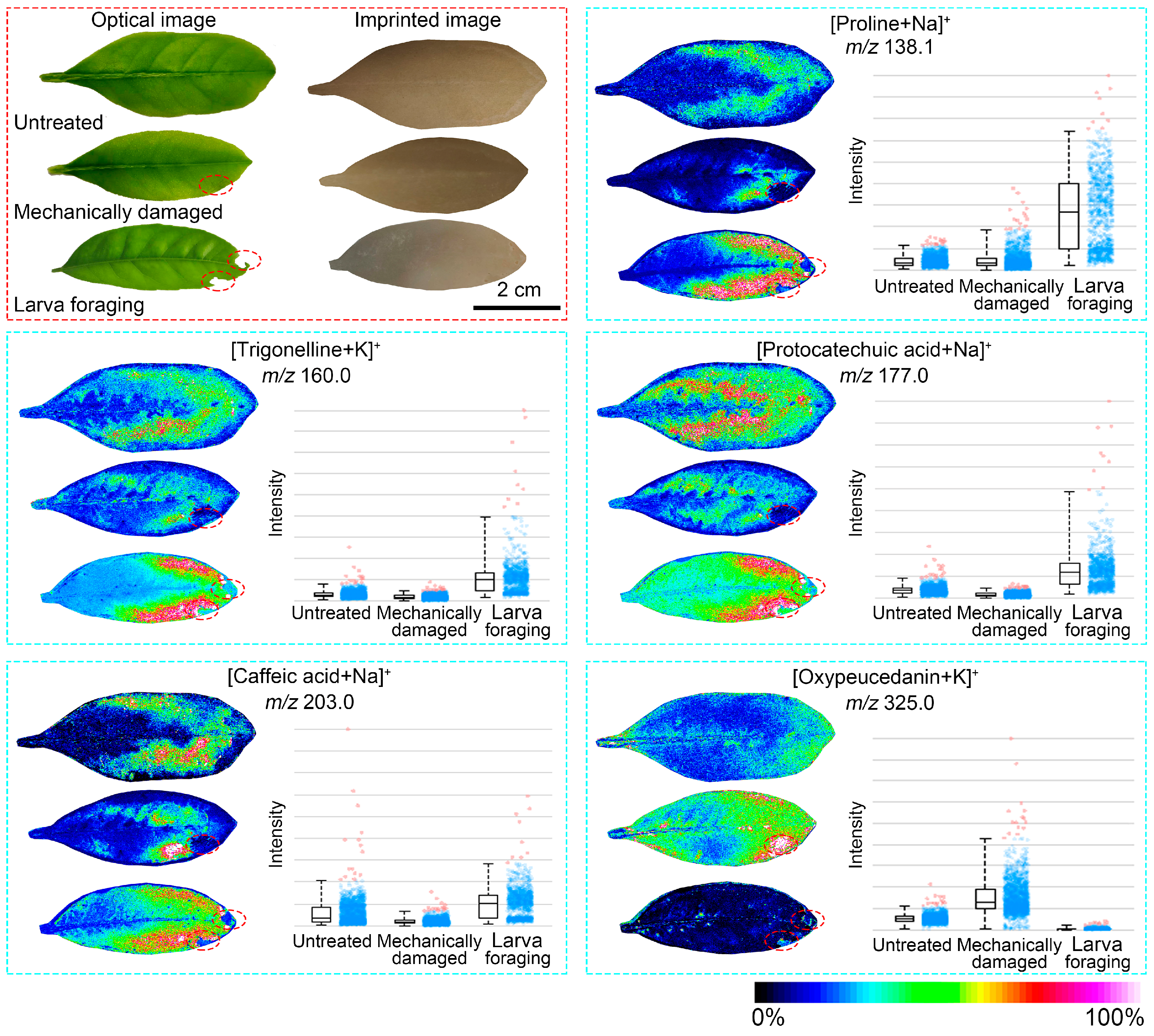
3.3. Stress Response of Citrus Leaves to Insect Feeding and Mechanical Damage

Having optimized the AuNP-hPDA-TDNT-based SALDI-MSI, we investigated its potential for mapping stress-responsive metabolites of citrus leaves in real cases. As a proof-of-concept study, we visualized the spatial distribution of some secondary metabolites in citrus leaves subjected to insect feeding and mechanical damage by imprinting MSI based on the AuNP-hPDA-TDNT substrate. Figure 3 shows that when citrus leaves were damaged by insect feeding and mechanical damage for 12 h, some metabolic marker substances exhibited specific distributions. Specifically, the levels of protocatechuic acid ([M+Na]+, m/z 177.0), trigonelline ([M+Na]+, m/z 160.0), and caffeic acid ([M+Na]+, m/z 203.0) decreased after citrus leaves were subjected to mechanical damage, but increased to a great extent after insect feeding. Figure 3 shows that these compounds significantly accumulated at insect feeding sites, but did not significantly change at mechanically damaged sites or in the CK group. Previous studies have reported that an increase in the levels of protocatechuic acid and caffeic acid may be the main stress defense substance produced in response to insect mouth injury [4]. Interestingly, trigonelline is a plant alkaloid that is closely related to coffee and fenugreek, the main components of which have antioxidant, anti-inflammatory, and neuroprotective effects. This study suggested that trigonelline has important antioxidant effects in response to damage caused by insect feeding. On the other hand, oxypeucedanin ([M+K]+, m/z 325.0) obviously accumulated at sites of mechanical damage, which may be closely related to tissue damage and repair. Additionally, given that the levels of most amino acids also tend to increase in leaves damaged by larval bites [48], proline ([M+Na]+, m/z 138.1) significantly accumulated at sites of larval damage, but there was no significant change at sites of mechanical damage.

3.4. Stress Response of Citrus after Mechanical Damage for Different Repair Durations

In addition to the static spatial distribution variation in mechanically damaged leaves, we further investigated the dynamic stress response of citrus leaves in the face of mechanical damage after different repair durations (i.e., 2 h, 12 h, and 24 h). Using the AuNP-hPDA-TDNT-based imprinting MSI method, we visualized several metabolic markers with specific spatial distributions in citrus leaves at different repair times (Figure 4). The results revealed that the levels of various amino acids, such as leucine ([M+Na]+, m/z 132.1) and methionine ([M+Na]+, m/z 172.0), increased, and that the methionine content in the CK group was greater than that at different repair times, indicating that the level decreased sharply after mechanical damage and then gradually increased. Although the defensive role of the amino acid methionine in mechanical damage is not yet fully understood, the dynamic variations in methionine levels for different durations of repair are interesting. Several studies have revealed that Met, a fundamental metabolite in plant cells, controls the levels of several essential metabolites, such as polyamines, biotin, ethylene, and phytosiderophores, after its conversion to S-adenosylmethionine (SAM) [49,50,51]. SAM, which serves as a primary methyl group donor, can regulate key processes, including the formation of chlorophyll and the cell wall, as well as the synthesis of many secondary metabolites [52]. Additionally, Met can also be catabolized to produce a group of plant secondary metabolites (e.g., glucosinolates), which exhibit repellent activity against herbivorous insects and pathogens [8,53]. When a leaf is mechanically damaged, the cell wall can be severely destroyed so that Met levels can decrease within damaged regions. As the repair mechanism is triggered, the level of the amino acid methionine gradually recovers to a normal level. More specifically, the leucine content increased significantly at the injury site after mechanical damage, while its contents at 2 h, 12 h, and 24 h were not significantly different, which was in agreement with previous studies showing that tryptophan and serine are common biomarkers of wound stress in citrus plants [54]. In sharp contrast, N-formylglycine ([M+H]+, m/z 104.1) increased slowly after mechanical damage and peaked at 12 h but began to decrease slowly from 12 h to 24 h. Notably, both the oxobutanedioic acid ([M+K]+, m/z 171.0) and citric acid ([M+H]+, m/z 193.0) levels increased after mechanical damage, which is part of the TCA cycle. Taken together, these results suggest that the TCA cycle produces increased amounts of energy in response to the threat posed by mechanical damage.

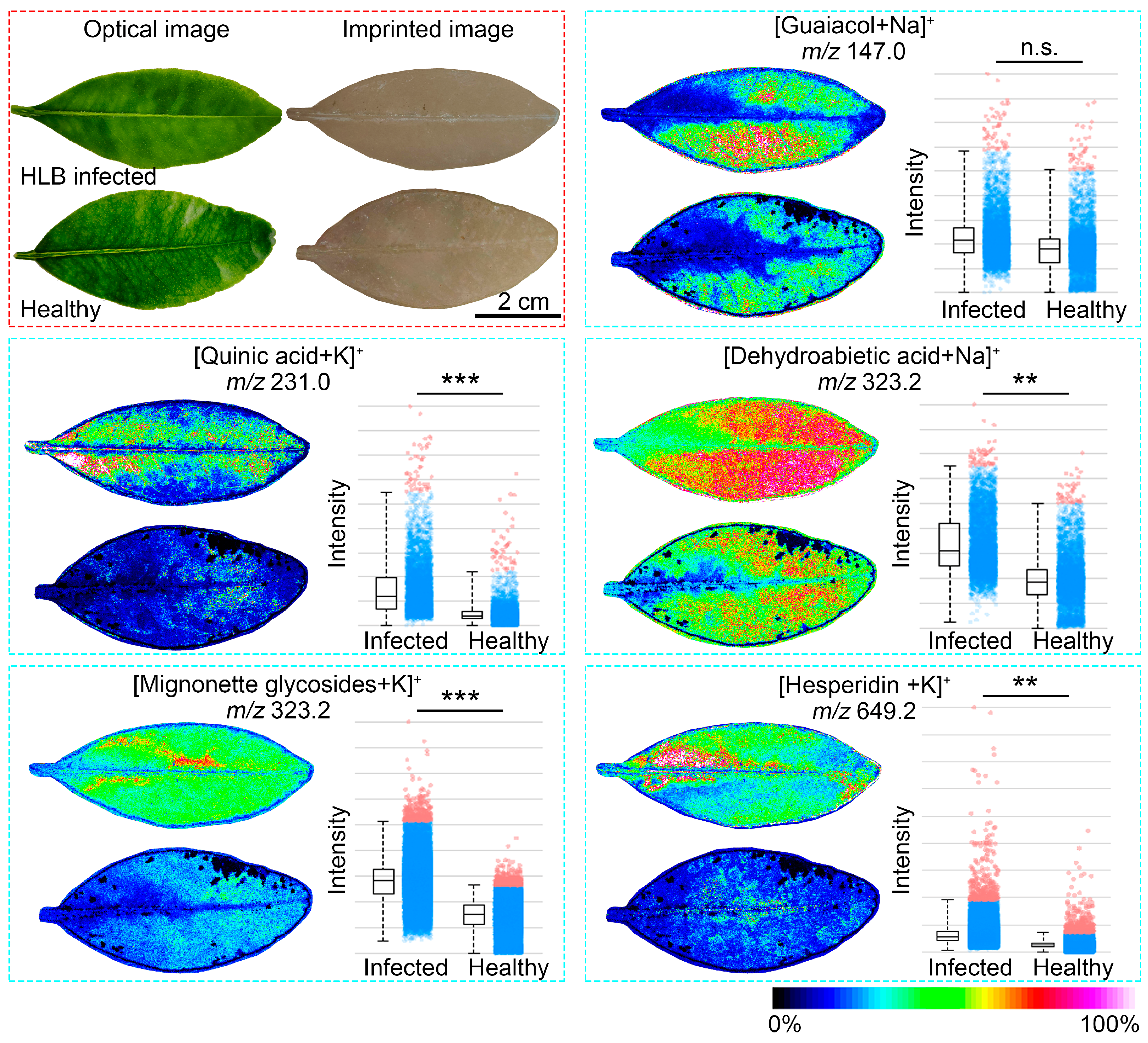
3.5. Stress Response of Citrus Leaves during Infection with HLB

In this study, in addition to insect feeding and mechanical damage, the spatial distribution of some secondary metabolites in citrus leaves before and after HLB infection was also visualized by imprinting MSI based on the AuNP-hPDA-TDNT substrate. We found that some metabolic markers related to defense response processes were generated when citrus leaves were infected by HLB and exhibited a specific distribution on the leaves (Figure 5). The MSI results revealed that the contents of several metabolites, such as guaiacol ([M+Na]+, m/z 147.0), quinic acid ([M+K]+, m/z 231.0), dehydroabietic acid ([M+Na]+, m/z 323.2), mignonette glycosides ([M+K]+, m/z 487.1), and hesperidin ([M+K]+, m/z 649.2), were greater in the HLB-infected citrus leaves than in the healthy leaves. Notably, the concentration of quinic acid markedly increased throughout the infected leaves, whereas guaiacol was present throughout the whole leaf except in the vein. Previous studies have demonstrated that guaiacol and quinic acid can be candidate biomarkers of HLB disease infection [55], but their specific spatial distributions are poorly understood. Specifically, Hijaz et al. and Jones et al. demonstrated that the organic acid quinic acid can be detected at a relatively high level in symptomatic or CLas-infected leaves, corroborating our imprinting MSI results [56,57]. As a key metabolite associated with plant stress and defense, quinic acid has been reported to exhibit a significantly different profile in asymptomatic leaves of Citrus sinensis [54,55], suggesting that quinic acid can be a vital biomarker candidate with potential for HLB infection recognition, even in the asymptomatic stage. Additionally, dehydroabietic acid, a natural product isolated from the nonvolatile residue of citric acid essential oil, was markedly increased in symptomatic citrus leaves, suggesting that it might be another candidate biomarker that is directly associated with HLB disease [55]. Given that HLB is a disease of worldwide incidence that could result in great losses to the citrus industry, the early diagnosis and detection of HLB disease-associated biomarkers can facilitate the prevention of citrus HLB diseases.

4. Conclusions

In summary, this work demonstrates a AuNP-hPDA-TDNT-based SALDI-MS method for the detection of a wide range of pesticides and the spatial visualization of primary and secondary metabolites during various plant defense processes by imprinting MSI. This nanostructured substrate, with the synergistic advantages of imprinting capability toward plant tissues and enhanced ionization efficiency, enables simplified sample preparation in real-case MSI applications, particularly for plant leaves that are incompatible with conventional histologic sectioning procedures. Moreover, the results presented here revealed that various nonuniform and tissue-specific distribution patterns of stress-induced metabolites can be clearly visualized in citrus leaves facing mechanical damage, insect feeding, and HLB disease infection via AuNP-hPDA-TDNT-based SALDI-MSI. Intriguingly, several potential biomarkers associated with the stress response of citrus leaves, such as quinic acid, could be visualized during mechanical damage and HLB disease infection, opening new avenues for a deeper understanding of crucial metabolites that participate in plant defense and infection processes. Efforts will be made in the future to decipher the biosynthesis, accumulation, and defensive role of various bioactive metabolites in economically important plants.

Supplementary Materials

The following supporting information can be downloaded at: https://www.mdpi.com/article/10.3390/agronomy14081797/s1. Figure S1: Workflow of the AuNP-hPDA-TDNT substrate fabrication. Figure S2: Repeatability test of the AuNP-hPDA-TDNT substrates from 3 batches for SALDI-MS analysis of azoxystrobin. Figure S3: Repeatability test of the AuNP-hPDA-TDNT substrates from 3 batches for SALDI-MS analysis of thiamethoxam. Figure S4: Repeatability test of the AuNP-hPDA-TDNT substrates from 3 batches for SALDI-MS analysis of rotenone. Figure S5: Stability test of the AuNP-hPDA-TDNT substrates for SALDI-MS analysis of azoxystrobin on different days. Figure S6: Stability test of the AuNP-hPDA-TDNT substrates for SALDI-MS analysis of thiamethoxam on different days. Figure S7: Stability test of the AuNP-hPDA-TDNT substrates for SALDI-MS analysis of rotenone on different days. Figure S8: Calibration curve of azoxystrobin using the AuNP-hPDA-TDNT substrate. Figure S9: Calibration curve of thiamethoxam using the AuNP-hPDA-TDNT substrate. Figure S10: Calibration curve of rotenone using the AuNP-hPDA-TDNT substrate. Figure S11: AuNP-hPDA-TDNT-based SALDI mass spectra of three pesticides with 10 amol amount. Figure S12: Representative mass spectrum acquired from an imprinted Citrus madurensis leaf using the AuNP-hPDA-TDNT substrate. Figure S13: MS/MS spectra of some typical metabolites from Citrus madurensis leaves in the positive ion mode. Table S1: Information on the pesticides used in the experiments. Table S2: Ion forms of standards of pesticides observed in SALDI-MS.

Author Contributions

Conceptualization, D.C. and X.W.; methodology, D.C.; validation, Y.S. and X.C.; formal analysis, Y.S.; investigation, Y.S. and X.C.; visualization, Y.S. and X.C.; writing—original draft preparation, Y.S., D.C. and X.C.; writing—review and editing, X.W.; supervision, X.W.; project administration, X.W.; funding acquisition, X.W. All authors have read and agreed to the published version of the manuscript.

Funding

This research was funded by the Spring Sunshine Programme by the Ministry of Education of China (No. HZKY20220113) and the Natural Science Foundation of Guangdong Province (No. 2023A1515030241). This work was also supported by the Original Innovation Cultivation Project of South China Agricultural University (No. 8400-223377).

Data Availability Statement

Data are contained within the article and Supplementary Materials.

Acknowledgments

We gratefully acknowledge the contributions of Yuhui Zhao and Wenlin Wen to data curation for this work.

Conflicts of Interest

The authors declare no conflict of interest.

References

  1. Gottwald, T.R. Current epidemiological understanding of citrus Huanglongbing. Annu. Rev. Phytopathol. 2010, 48, 119–139. [Google Scholar] [CrossRef] [PubMed]
  2. Ma, W.; Pang, Z.; Huang, X.; Xu, J.; Pandey, S.S.; Li, J.; Achor, D.S.; Vasconcelos, F.N.C.; Hendrich, C.; Huang, Y.; et al. Citrus Huanglongbing is a pathogen-triggered immune disease that can be mitigated with antioxidants and gibberellin. Nat. Commun. 2022, 13, 529. [Google Scholar] [CrossRef]
  3. Li, X.; Ruan, H.; Zhou, C.; Meng, X.; Chen, W. Controlling citrus Huanglongbing: Green sustainable development route is the future. Front. Plant Sci. 2021, 12, 760481. [Google Scholar] [CrossRef] [PubMed]
  4. Liu, Y.; Xue, A.; Wang, S.; Hao, Y.; Cui, M.; Liu, L.; Luo, L. Metabolic response of Citrus limon to Asian citrus psyllid infestation revealed by EESI-MS and HPLC. Anal. Biochem. 2020, 609, 113973. [Google Scholar] [CrossRef]
  5. Lee, J.A.; Halbert, S.E.; Dawson, W.O.; Robertson, C.J.; Keesling, J.E.; Singer, B.H. Asymptomatic spread of Huanglongbing and implications for disease control. Proc. Natl. Acad. Sci. USA 2015, 112, 7605–7610. [Google Scholar] [CrossRef]
  6. Wang, K.; Liao, Y.; Meng, Y.; Jiao, X.; Huang, W.; Liu, T.C.-y. The early, rapid, and non-destructive detection of citrus Huanglongbing (HLB) based on microscopic confocal Raman. Food Anal. Methods 2019, 12, 2500–2508. [Google Scholar] [CrossRef]
  7. Xue, A.; Liu, Y.; Li, H.; Cui, M.; Huang, X.; Wang, W.; Wu, D.; Guo, X.; Hao, Y.; Luo, L. Early detection of Huanglongbing with EESI-MS indicates a role of phenylpropanoid pathway in citrus. Anal. Biochem. 2022, 639, 114511. [Google Scholar] [CrossRef] [PubMed]
  8. Shroff, R.; Vergara, F.; Muck, A.; Svatoš, A.; Gershenzon, J. Nonuniform distribution of glucosinolates in Arabidopsis thaliana leaves has important consequences for plant defense. Proc. Natl. Acad. Sci. USA 2008, 105, 6196–6201. [Google Scholar] [CrossRef]
  9. Yan, S.; Bhawal, R.; Yin, Z.; Thannhauser, T.W.; Zhang, S. Recent advances in proteomics and metabolomics in plants. Mol. Hortic. 2022, 2, 17. [Google Scholar] [CrossRef]
  10. Aharoni, A.; Goodacre, R.; Fernie, A.R. Plant and microbial sciences as key drivers in the development of metabolomics research. Proc. Natl. Acad. Sci. USA 2023, 120, e2217383120. [Google Scholar] [CrossRef] [PubMed]
  11. Maeda, H.A.; Fernie, A.R. Evolutionary history of plant metabolism. Annu. Rev. Plant Biol. 2021, 72, 185–216. [Google Scholar] [CrossRef]
  12. Perez de Souza, L.; Alseekh, S.; Scossa, F.; Fernie, A.R. Ultra-high-performance liquid chromatography high-resolution mass spectrometry variants for metabolomics research. Nat. Methods 2021, 18, 733–746. [Google Scholar] [CrossRef]
  13. Alseekh, S.; Aharoni, A.; Brotman, Y.; Contrepois, K.; D’Auria, J.; Ewald, J.; Ewald, J.C.; Fraser, P.D.; Giavalisco, P.; Hall, R.D.; et al. Mass spectrometry-based metabolomics: A guide for annotation, quantification and best reporting practices. Nat. Methods 2021, 18, 747–756. [Google Scholar] [CrossRef] [PubMed]
  14. Horn, P.J.; Chapman, K.D. Imaging plant metabolism in situ. J. Exp. Bot. 2023, 75, 1654–1670. [Google Scholar] [CrossRef]
  15. Yin, Z.; Huang, W.; Fernie, A.R.; Yan, S. Mass spectrometry imaging techniques: A versatile toolbox for plant metabolomics. Trends Plant Sci. 2023, 28, 250–251. [Google Scholar] [CrossRef]
  16. Yin, Z.; Dong, T.; Huang, W.; Du, M.; Chen, D.; Fernie, A.R.; Yi, G.; Yan, S. Spatially resolved metabolomics reveals variety-specific metabolic changes in banana pulp during postharvest senescence. Food Chem. X 2022, 15, 100371. [Google Scholar] [CrossRef] [PubMed]
  17. Qin, R.; Li, P.; Du, M.; Ma, L.; Huang, Y.; Yin, Z.; Zhang, Y.; Chen, D.; Xu, H.; Wu, X. Spatiotemporal visualization of insecticides and fungicides within fruits and vegetables using gold nanoparticle-immersed paper imprinting mass spectrometry imaging. Nanomaterials 2021, 11, 1327. [Google Scholar] [CrossRef]
  18. Yin, Z.; Huang, W.; Li, K.; Fernie, A.R.; Yan, S. Advances in mass spectrometry imaging for plant metabolomics—Expanding the analytical toolbox. Plant J. 2024; early view. [Google Scholar] [CrossRef]
  19. Wang, T.; Cheng, X.; Xu, H.; Meng, Y.; Yin, Z.; Li, X.; Hang, W. Perspective on advances in laser-based high-resolution mass spectrometry imaging. Anal. Chem. 2020, 92, 543–553. [Google Scholar] [CrossRef]
  20. Ma, S.; Leng, Y.; Li, X.; Meng, Y.; Yin, Z.; Hang, W. High spatial resolution mass spectrometry imaging for spatial metabolomics: Advances, challenges, and future perspectives. TrAC Trends Anal. Chem. 2023, 159, 116902. [Google Scholar] [CrossRef]
  21. Chen, Y.-J.; Zeng, H.-S.; Jin, H.L.; Wang, H.B. Applications of mass spectrometry imaging in botanical research. Adv. Biotechnol. 2024, 2, 6. [Google Scholar] [CrossRef]
  22. Dong, Y.; Aharoni, A. Image to insight: Exploring natural products through mass spectrometry imaging. Nat. Prod. Rep. 2022, 39, 1510–1530. [Google Scholar] [CrossRef] [PubMed]
  23. Müller, W.H.; Verdin, A.; De Pauw, E.; Malherbe, C.; Eppe, G. Surface-assisted laser desorption/ionization mass spectrometry imaging: A review. Mass Spectrom. Rev. 2022, 41, 373–420. [Google Scholar] [CrossRef]
  24. Lu, M.; Yang, X.; Yang, Y.; Qin, P.; Wu, X.; Cai, Z. Nanomaterials as assisted matrix of laser desorption/ionization time-of-flight mass spectrometry for the analysis of small molecules. Nanomaterials 2017, 7, 78. [Google Scholar] [CrossRef] [PubMed]
  25. Pomastowski, P.; Buszewski, B. Complementarity of matrix- and nanostructure-assisted laser desorption/ionization approaches. Nanomaterials 2019, 9, 260. [Google Scholar] [CrossRef] [PubMed]
  26. Dong, Y.; Li, B.; Malitsky, S.; Rogachev, I.; Aharoni, A.; Kaftan, F.; Svatoš, A.; Franceschi, P. Sample preparation for mass spectrometry imaging of plant tissues: A review. Front. Plant Sci. 2016, 7, 60. [Google Scholar] [CrossRef]
  27. Wu, X.; Qin, R.; Wu, H.; Yao, G.; Zhang, Y.; Li, P.; Xu, Y.; Zhang, Z.; Yin, Z.; Xu, H. Nanoparticle-immersed paper imprinting mass spectrometry imaging reveals uptake and translocation mechanism of pesticides in plants. Nano Res. 2020, 13, 611–620. [Google Scholar] [CrossRef]
  28. Xia, J.; Lou, G.; Zhang, L.; Huang, Y.; Yang, J.; Guo, J.; Qi, Z.; Li, Z.; Zhang, G.; Xu, S.; et al. Unveiling the spatial distribution and molecular mechanisms of terpenoid biosynthesis in Salvia miltiorrhiza and S. grandifolia using multi-omics and DESI–MSI. Hortic. Res. 2023, 10, uhad109. [Google Scholar] [CrossRef] [PubMed]
  29. Hemalatha, R.G.; Pradeep, T. Understanding the molecular signatures in leaves and flowers by desorption electrospray ionization mass spectrometry (DESI MS) imaging. J. Agric. Food Chem. 2013, 61, 7477–7487. [Google Scholar] [CrossRef]
  30. Hemalatha, R.G.; Ganayee, M.A.; Pradeep, T. Electrospun nanofiber mats as “smart surfaces” for desorption electrospray ionization mass spectrometry (DESI MS)-based analysis and imprint imaging. Anal. Chem. 2016, 88, 5710–5717. [Google Scholar] [CrossRef] [PubMed]
  31. Wei, J.; Buriak, J.M.; Siuzdak, G. Desorption-ionization mass spectrometry on porous silicon. Nature 1999, 399, 243–246. [Google Scholar] [CrossRef]
  32. Samarah, L.Z.; Tran, T.H.; Stacey, G.; Vertes, A. Mass spectrometry imaging of bio-oligomer polydispersity in plant tissues by laser desorption ionization from silicon nanopost arrays. Angew. Chem. Int. Ed. 2021, 60, 9071–9077. [Google Scholar] [CrossRef] [PubMed]
  33. Liu, X.; Chen, Z.; Wang, T.; Jiang, X.; Qu, X.; Duan, W.; Xi, F.; He, Z.; Wu, J. Tissue imprinting on 2D nanoflakes-capped silicon nanowires for lipidomic mass spectrometry imaging and cancer diagnosis. ACS Nano 2022, 16, 6916–6928. [Google Scholar] [CrossRef] [PubMed]
  34. Chen, D.; Du, M.; Huang, Y.; Xu, Y.; Chen, Y.; Ma, L.; Xie, Q.; Zhu, X.; Chen, Z.; Xu, H.; et al. Plasmonic polydopamine-modified TiO2 nanotube substrates for surface-assisted laser desorption/ionization mass spectrometry imaging. Nano Res. 2023, 16, 3028–3039. [Google Scholar] [CrossRef]
  35. Yang, J.; Zhang, W.; Zhang, H.; Zhong, M.; Cao, W.; Li, Z.; Huang, X.; Nie, Z.; Liu, J.; Li, P.; et al. Polydopamine-modified substrates for high-sensitivity laser desorption ionization mass spectrometry imaging. ACS Appl. Mater. Interfaces 2019, 11, 46140–46148. [Google Scholar] [CrossRef] [PubMed]
  36. Yang, J.; Zhang, H.; Jia, J.; Zhang, X.; Ma, X.; Zhong, M.; Ouyang, Z. Antireflection surfaces for biological analysis using laser desorption ionization mass spectrometry. Research 2018, 2018, 5439729. [Google Scholar] [CrossRef] [PubMed]
  37. Sakai, R.; Kondo, H.; Ishikawa, K.; Ohta, T.; Hiramatsu, M.; Tanaka, H.; Hori, M. Effects of high-quality carbon nanowalls ionization-assisting substrates on surface-assisted laser desorption/ionization mass spectrometry performance. Nanomaterials 2023, 13, 63. [Google Scholar] [CrossRef]
  38. Kim, Y.-K.; Na, H.-K.; Kwack, S.-J.; Ryoo, S.-R.; Lee, Y.; Hong, S.; Hong, S.; Jeong, Y.; Min, D.-H. Synergistic effect of graphene oxide/MWCNT films in laser desorption/ionization mass spectrometry of small molecules and tissue imaging. ACS Nano 2011, 5, 4550–4561. [Google Scholar] [CrossRef]
  39. Iakab, S.A.; Ràfols, P.; Tajes, M.; Correig-Blanchar, X.; García-Altares, M. Gold nanoparticle-assisted black silicon substrates for mass spectrometry imaging applications. ACS Nano 2020, 14, 6785–6794. [Google Scholar] [CrossRef]
  40. Sun, S.; Liu, W.; Yang, J.; Wang, H.; Qian, K. Nanoparticle-assisted cation adduction and fragmentation of small metabolites. Angew. Chem. Int. Ed. 2021, 60, 11310–11317. [Google Scholar] [CrossRef]
  41. Manjavacas, A.; Liu, J.G.; Kulkarni, V.; Nordlander, P. Plasmon-induced hot carriers in metallic nanoparticles. ACS Nano 2014, 8, 7630–7638. [Google Scholar] [CrossRef] [PubMed]
  42. Dutkiewicz, E.P.; Su, C.-H.; Lee, H.-J.; Hsu, C.-C.; Yang, Y.-L. Visualizing vinca alkaloids in the petal of Catharanthus roseus using functionalized titanium oxide nanowire substrate for surface-assisted laser desorption/ionization imaging mass spectrometry. Plant J. 2021, 105, 1123–1133. [Google Scholar] [CrossRef] [PubMed]
  43. Wang, X.-N.; Li, B. Monolithic gold nanoparticles/thiol-β-cyclodextrin-functionalized TiO2 nanowires for enhanced SALDI MS detection and imaging of natural products. Anal. Chem. 2022, 94, 952–959. [Google Scholar] [CrossRef] [PubMed]
  44. Chen, D.; Xu, Y.; Huang, Y.; Chen, Y.; Zhao, Y.; Yan, B.; Zhu, X.; Chen, Z.; Xu, H.; Yin, Z.; et al. Mapping molecular signatures in plant leaves, flowers, and fruits by a TiO2 nanotube-based plasmonic chip for imprint mass spectrometry imaging. ACS Agric. Sci. Technol. 2023, 3, 119–130. [Google Scholar] [CrossRef]
  45. Wu, Q.; Chu, J.L.; Rubakhin, S.S.; Gillette, M.U.; Sweedler, J.V. Dopamine-modified TiO2 monolith-assisted LDI MS imaging for simultaneous localization of small metabolites and lipids in mouse brain tissue with enhanced detection selectivity and sensitivity. Chem. Sci. 2017, 8, 3926–3938. [Google Scholar] [CrossRef] [PubMed]
  46. Sun, X.; Huang, L.; Zhang, R.; Xu, W.; Huang, J.; Gurav, D.D.; Vedarethinam, V.; Chen, R.; Lou, J.; Wang, Q.; et al. Metabolic fingerprinting on a plasmonic gold chip for mass spectrometry based in vitro diagnostics. ACS Cent. Sci. 2018, 4, 223–229. [Google Scholar] [CrossRef] [PubMed]
  47. Alam, F.; Mohammadin, K.; Shafique, Z.; Amjad, S.T.; Asad, M.H.H.b. Citrus flavonoids as potential therapeutic agents: A review. Phytother. Res. 2022, 36, 1417–1441. [Google Scholar] [CrossRef] [PubMed]
  48. Dong, Z.; Liu, X.; Srivastava, A.K.; Tan, Q.; Low, W.; Yan, X.; Wu, S.; Sun, X.; Hu, C. Foliar nutrition alleviate citrus plants from Asian citrus psyllid feeding by affecting leaf structure and secondary metabolism. Sci. Hortic. 2023, 309, 111667. [Google Scholar] [CrossRef]
  49. Galili, G.; Amir, R.; Fernie, A.R. The regulation of essential amino acid synthesis and accumulation in plants. Annu. Rev. Plant Biol. 2016, 67, 153–178. [Google Scholar] [CrossRef] [PubMed]
  50. Galili, G.; Amir, R. Fortifying plants with the essential amino acids lysine and methionine to improve nutritional quality. Plant Biotechnol. J. 2013, 11, 211–222. [Google Scholar] [CrossRef]
  51. Gonda, I.; Lev, S.; Bar, E.; Sikron, N.; Portnoy, V.; Davidovich-Rikanati, R.; Burger, J.; Schaffer, A.A.; Tadmor, Y.; Giovannonni, J.J.; et al. Catabolism of L–methionine in the formation of sulfur and other volatiles in melon (Ucumis melo L.) fruit. Plant J. 2013, 74, 458–472. [Google Scholar] [CrossRef]
  52. Roje, S. S-Adenosyl-L-methionine: Beyond the universal methyl group donor. Phytochemistry 2006, 67, 1686–1698. [Google Scholar] [CrossRef] [PubMed]
  53. Hirai, M.Y.; Sugiyama, K.; Sawada, Y.; Tohge, T.; Obayashi, T.; Suzuki, A.; Araki, R.; Sakurai, N.; Suzuki, H.; Aoki, K.; et al. Omics-based identification of Arabidopsis Myb transcription factors regulating aliphatic glucosinolate biosynthesis. Proc. Natl. Acad. Sci. USA 2007, 104, 6478–6483. [Google Scholar] [CrossRef] [PubMed]
  54. Asai, T.; Matsukawa, T.; Kajiyama, S. Metabolomic analysis of primary metabolites in citrus leaf during defense responses. J. Biosci. Bioeng. 2017, 123, 376–381. [Google Scholar] [CrossRef] [PubMed]
  55. de Moraes Pontes, J.G.; Vendramini, P.H.; Fernandes, L.S.; de Souza, F.H.; Pilau, E.J.; Eberlin, M.N.; Magnani, R.F.; Wulff, N.A.; Fill, T.P. Mass spectrometry imaging as a potential technique for diagnostic of Huanglongbing disease using fast and simple sample preparation. Sci. Rep. 2020, 10, 13457. [Google Scholar] [CrossRef]
  56. Hijaz, F.M.; Manthey, J.A.; Folimonova, S.Y.; Davis, C.L.; Jones, S.E.; Reyes-De-Corcuera, J.I. An HPLC-MS characterization of the changes in sweet orange leaf metabolite profile following infection by the bacterial pathogen Candidatus Liberibacter asiaticus. PLoS ONE 2013, 8, e79485. [Google Scholar] [CrossRef] [PubMed]
  57. Jones, S.; Hijaz, F.; Davis, C.; Folimonova, S.; Manthey, J.; Reyes-De-Corcuera, J. GC-MS analysis of secondary metabolites in leaves from orange trees infected with HLB: A 9-month study. Proc. Fla. State Hortic. Soc. 2012, 125, 75–83. [Google Scholar]
Figure 1. SALDI mass spectra of nine pesticide mixtures in positive ion mode. (a) Comparison of mass spectra of a mixture of nine pesticides, including acephate, thiamethoxam, spirotetramat, rotenone, azoxystrobin, cyantraniliprole, chlorantraniliprole, and abamectin, using (a) CHCA and four SALDI substrates, including (b) TDNT, (c) PDA-TDNT, (d) hPDA-TDNT, and (e) AuNP-hPDA-TDNT. The concentration of each analyte was 1 mM, and * represents the background interference peaks.
Agronomy 14 01797 g001
Figure 2. Spatial distribution of various small-molecule metabolites resulting from an imprinted citrus leaf. Optical image of a citrus leaf and the corresponding image imprinted onto the surface of the AuNP-hPDA-TDNT substrate and representative ion images of a citrus leaf obtained via SALDI-MSI. The colored bars indicate the relative signal intensity. Scale bar: 10 mm.
Agronomy 14 01797 g002
Figure 3. Mass spectrometry images of mechanically damaged and larva-fed citrus leaves. Optical images of citrus leaves, corresponding imprinted images of the surface of the AuNP-hPDA-TDNT substrate, and representative ion images of the citrus leaves obtained via SALDI-MSI with a pixel size of 160 μm. The red dashed ellipses indicate the specific locations that are mechanically damaged and larva foraged. The blue and red data points indicate the reasonable values and outliers, respectively. Scale bar: 20 mm.
Agronomy 14 01797 g003
Figure 4. Imprinting MS images of mechanically damaged citrus leaves for different durations after repair. Optical images of citrus leaves, corresponding imprinted images of the surface of the AuNP-hPDA-TDNT substrate, and representative ion images of the citrus leaves obtained via SALDI-MSI with a pixel size of 160 μm. The red dashed ellipses indicate the specific locations that are mechanically damaged. The blue and red data points indicate the reasonable values and outliers, respectively. Scale bar: 20 mm.
Agronomy 14 01797 g004
Figure 5. Imprinting MS images of leaves infected with citrus HLB and healthy leaves. Optical images of citrus leaves, corresponding imprinted images of the surface of the AuNP-hPDA-TDNT substrate, and representative ion images of the citrus leaves obtained via SALDI-MSI with a pixel size of 160 μm. The blue and red data points indicate the reasonable values and outliers, respectively. The colored bars indicate the relative signal intensity. The asterisks indicate the level of significance: ** p < 0.01, *** p < 0.001, and p > 0.05 (not significant, n.s.). Scale bar, 20 mm.
Agronomy 14 01797 g005
Disclaimer/Publisher’s Note: The statements, opinions and data contained in all publications are solely those of the individual author(s) and contributor(s) and not of MDPI and/or the editor(s). MDPI and/or the editor(s) disclaim responsibility for any injury to people or property resulting from any ideas, methods, instructions or products referred to in the content.

