Article

Genome-Wide Identification, Characterization, and Expression Profile of PDCB Gene Family in Zea mays L.

Laboratory of Plant Epigenetics and Evolution, School of Life Sciences, Liaoning University, Shenyang 110036, China
*
Author to whom correspondence should be addressed.
These authors contributed equally to this work.
Agronomy 2024, 14(8), 1858; https://doi.org/10.3390/agronomy14081858
Submission received: 15 July 2024 / Revised: 14 August 2024 / Accepted: 20 August 2024 / Published: 21 August 2024

Abstract

:
The plant kingdom harbors the Plasmodesmata Callose Binding Protein (PDCB) gene family, which plays essential roles in plant growth, development, environmental adaptation, and yield. PDCB genes are closely involved in regulating cell-to-cell communication and controlling callose deposition at plasmodesmata (PD) throughout the whole plant. Remarkably, their functions remain largely unknown in many crops, including maize. This study sought to identify the members of the PDCB gene family within the maize genome and analyze their physicochemical properties and expression patterns. Utilizing bioinformatics methodologies, a comprehensive genome-wide analysis of the PDCB gene family was performed. The findings revealed that PDCB genes were highly abundant in maize, with a total of 56 PDCB genes identified and categorized into six distinct groups. Members of the PDCB family were dispersed across all chromosomes. The PDCBs within each group exhibited significant similarity in their conserved motifs and gene structures; all members contained the X8 domain, comprising one to five exons, while displaying a straightforward genomic structure. Numerous cis-acting elements associated with plant growth and development, light response, stress-associated responses, and plant hormones were identified in the promoter regions of PDCB genes. Moreover, the PDCBs exhibited diverse expression patterns across various tissues. This study improves the comprehension of the PDCB gene family and provides a robust foundation for further research on maize.

1. Introduction

Plants are profoundly reliant on plasmodesmata (PD) for intercellular communication [1]. The growth and development of plants are significantly influenced by PD [2]. These tiny apertures in the plant cell wall enable adjacent cells to connect through extensions of their cell membranes into these pores. The smooth endoplasmic reticulums of the two cells are also interconnected, forming the PD. It is articulated that PD plays a pivotal role in facilitating symplasmic cell-to-cell transfer in plants [3,4]. Homeodomain transcription factors, small RNA molecules, and viral genomic material are specifically transported to neighboring cells via PD microchannels [5]. Additionally, sucrose is conveyed from cell to cell through PD into the sieve elements (SEs), which are sequentially arranged to form sieve tubes. These tubes act as a conduit for the long-distance transport of sucrose from source tissues to the sinks [6,7]. In maize xylem exudate, sucrose was found to be the sole sugar present [8]. It stands as the primary product of photosynthesis and is instrumental in the distribution of assimilated carbon within plants [9]. The process known as whole-plant carbohydrate partitioning is employed by plants to transfer carbon, produced through photosynthesis, via the phloem. Carbohydrate allocation profoundly impacts all aspects of plant growth and crop yield; thus, PD are potential factors influencing crop development, stress tolerance and yield [10].
PD are associated with numerous proteins, including plasmodesmata callose binding proteins (PDCBs), callose synthases (CalS), remorin (REM), plasmodesmata-located proteins (PDLPs), and β-1,3-glucanase (BGs), among others [11]. The pores of PD permit the selective deposition or degradation of callose [12]. Callose, a type of β-(1,3)-D-glucan found in many higher plants, is critical for proper growth and development and plays a crucial role in plant defense mechanisms. Notably, callose accumulates between the plasma membrane and cell wall at the sites of pathogen attack, including PD, thereby impeding pathogen entry and dissemination [13]. Although in the model plant Arabidopsis thaliana, callose constitutes merely about 0.3% of the cell wall, it regulates various biological processes such as phloem and pollen development, cell division, organ formation, response to pathogenic invasion, and adaptation to changing soil nutrients and toxic metal levels [14]. The size exclusion limit (SEL) of PD and consequently the mobility of large macromolecules and signaling proteins and RNA molecules that mediate plant developmental and environmental responses are influenced by callose deposition [14,15].
Key contributors to the accumulation of callose at PD include AtBG_PP, PDCB, glycan synthase-like (GSL), and PDLP [16]. Despite lacking apparent enzymatic activity, the PDCB family has been shown to regulate callose deposition at the neck of PD. Thus, PDCBs have emerged as significant regulators of cell-to-cell communication [17,18]. Studies have demonstrated that overexpression of PDCB1 results in increased callose accumulation [18], and PDCB1, PDCB2, and PDCB3 showed overlapping tissue-specific expression suggesting the functional redundancy in this gene family. Due to possessing callose-binding domains, PDCBs have been linked to the closure of PD during plant development [19].
The potential correlation between PDCB-mediated callose deposition and intercellular communication in Arabidopsis thaliana has been implicated in the regulation of callose homeostasis in plasmodesmata [16,18]. The callose synthases GSL1, GSL2, GSL5, GSL8, and GSL10 are essential for callose production during pollen development, playing a vital role in ensuring the fertility and/or survivability of the pollen [19]. In maize mutants, abnormal callose and lignin accumulation has been observed in the phloem; and genetic modifications in poplar trees that induce callose deposition can alter the permeability, structure, water-holding capacity, and potentially the lignin content of the secondary cell wall, leading to an increased distance between lignin and cellulose molecules, which ultimately enhances biomass resistance to degradation [20,21].
Although callose holds great significance and the research on the PDCB gene family is being conducted extensively, pertinent reports are rare. It has been documented that there were 37 AtPDCBs in Arabidopsis thaliana, 25 PbPDCBs in pear fruit, and 45 GhPDCBs in cotton [18,20,21]. The Arabidopsis PDCB proteins belong to a larger protein superfamily, which encompasses proteins characterized by conserved X8 domains adjacent to β-1,3-glucanase catalytic domains. PDCBs interact with callose through their X8 domain, and the presence of PDCBs affects the transference of substances through PD between cells by influencing callose deposition [18]. In pear fruit, the formation of stone cells is associated with the lignification transmitted by PD. PbPDCB16 is considered to enhance callose deposition at PD, thereby affecting intercellular connectivity mediated by PD and resulting in a reduction in lignin concentration [20]. Furthermore, GhPDCB9 was believed to be related to fiber elongation in cotton, contributing positively to increasing cotton production [21]. Although the PDCB gene family in Arabidopsis, pear, and cotton has been studied, they remain largely unknown in most crops, such as maize. Maize is a widely cultivated cereal crop, grown globally for fuel, fiber, food, and feed. Moreover, maize serves as an important model crop for studying various biological phenomena [22], and has its genome information sufficiently available [23].
Maize is also a representative of the C4 photosynthetic pathway [24]. In contrast to C3 plants, C4 plants possess two structurally and biochemically specialized photosynthetic cell types: mesophyll (M) and bundle sheath (BS) [25]. These BS and M cells surround the leaf veins, forming a wreath-like structure [26]. A significant number of PD exist between BS and M cells, facilitating material exchange between these cellular types and enabling their coordinated effort in achieving carbon assimilation during plant photosynthesis [25,27]. Currently, there is little research on the PDCB gene family. Even with regard to rice, one of the most important food crops and relevant model plants, the PDCBs have not been reported. Thus, investigating PDCBs in important crops could elucidate their role in plant growth and development, and provide new insights into the study of carbohydrate transport and potential for agronomy trait improvement. In this study, we identified members of the maize PDCB gene family and subsequently analyzed their gene structures, conserved domains, evolutionary relationships, cis-acting elements, and expression patterns across different tissues. This study could furnish valuable information for comprehending the functions of the PDCB gene family in maize and provide new insights for crop breeding potential including yield, quality, and so on. Additionally, it might promote further exploration of PDCB genes in other significant crops, such as rice, and also facilitate a more comprehensive grasp of plant growth and metabolism.

2. Materials and Methods

2.1. Detection of PDCB Gene Family Members in Maize and Rice

We downloaded the Arabidopsis thaliana PDCB gene family proteins from TAIR (https://www.arabidopsis.org, accessed on 29 February 2024) [28]. The maize genome (Zm-B73-REFERENCE-NAM-5.0) and annotated files were obtained from Ensembl plants (http://plants.ensembl.org/index.html, accessed on 29 February 2024) [29].
Then, the Sequence Files program of TBtools was used to search PDCBs within the maize database, with the PDCB proteins of Arabidopsis as the query for identification of the PDCB genes. Furthermore, the HMM profile of the X8 domain (Pfam: PF07983) was downloaded from the Pfam database (http://pfam.xfam.org, accessed on 1 March 2024) [30] and was effectively utilized to accurately identify the PDCB genes with an E-value lower than 1 × 10⁻⁵. The proteins lacking X8 domain were eliminated. The candidate sequences were calculated using the TBtools Protein Parameter Calc (ProtParam-based) software (v2.119) for determining the molecular weights (MW) and theoretical isoelectric points (pI) and the WOLF PSORT (https://wolfpsort.hgc.jp, accessed on 2 March 2024) for subcellular localization prediction [31].
The rice genome (T2T-NIP) was downloaded from the Rice Super Pan-genome Information Resource Database (http://www.ricesuperpir.com/web/nip, accessed on 6 August 2024) [32]. The genome-wide identification of PDCB proteins in rice was conducted by using the same process mentioned above.

2.2. Developing Phylogenetic Trees, Analyzing Motifs, and Determining Gene Structures

A phylogenetic tree was constructed by following the neighbor-joining (NJ) method and employing MEGA11 with 1000 replicates [33]. TBtools was utilized to extract PDCB location data and view the gene structure. The conservative pattern was found using TBtools’ Simple MEME Wrapper program, which was configured to yield a maximum of 20 motifs. Following this, the motif was shown using TBtools’ Visualize MEME Motif Pattern application.

2.3. Chromosome Location and Synteny Analysis for PDCBs

PDCB positions on chromosomes were displayed using TBtools using genomic annotation files from the Zea mays species [34]. PDCB collinearity was examined using MCScanX, which analyzes intra- and extra-genomic collinearity based on genome annotation files. Collinear gene pairs were shown using TBtools’ Advanced Circossoftware (v2.119).2.4. Examination of the Cis-Acting Element inside Promoters
The 2000 bp region chosen as the promoter was located upstream of the PDCB gene’s translation start codon ATG. The cis-acting elements present in the promoter region of the PDCBs were estimated using the PlantCare website [35]. Then, the number of cis-acting elements was counted, and the cis-acting elements were visualized using the TBtools HeatMap program (v2.119).

2.4. ZmPDCBs Expression Patterns under Different Tissues

Transcriptome data for expression patterns of ZmPDCBs in 18 different tissues were downloaded from MaizeGDB (https://www.maizegdb.org/genome/assembly/Zm-B73-REFERENCE-NAM-5.0,accessed on 5 March 2024) [36,37,38]. The tissues include internode, vegetative meristem, ear primordium, embryo, endosperm, endosperm crown, germination kernels, pericarp aleurone, female spikelet, silk, root cortex, root meristem zone, secondary root, root elongation zone, primary root, mature pollen, mature leaf, and leaf zone. Genes with FPKM values greater than 1 were classified as expressed genes. Using the value of log2 (FPKM+1), the HeatMap software (v2.119) of TBtools was used to visualize the expression patterns of the ZmPDCBs.

3. Results

3.1. Zea mays PDCB Identification and Characterization

Fifty-six members of the PDCB gene family were identified in Zea mays. The X8 domain (PF07983) was present in every protein sequence. Further analysis was conducted on the characteristics of ZmPDCBs (Table S1). The protein length ranged from 81 aa (amino acids) in ZmPDCB9 to 665 aa in ZmPDCB43 and ZmPDCB54. The molecular weight (MW) of PDCBs varied from 9.07 kDa (ZmPDCB9) to 73.12 kDa (ZmPDCB54), with an average MW of 41.19 kDa. The isoelectric point (pI) values of ZmPDCB proteins were either above or below 7, demonstrating both alkaline and acidic characteristics. The GAH ranged from −0.532 (ZmPDCB24 and ZmPDCB55) to 0.382 (ZmPDCB51), revealing their differences of hydrophilicity. The PSL (predicted subcellular location) indicated that the subcellular localization of the ZmPDCBs was also highly diverse. It may be located on membrane structures such as the plasma membrane and vacuolar membrane, or in organelles such as chloroplasts, nucleus, and Golgi apparatuses. These results indicate that ZmPDCB proteins present multiplicity in their physicochemical properties.

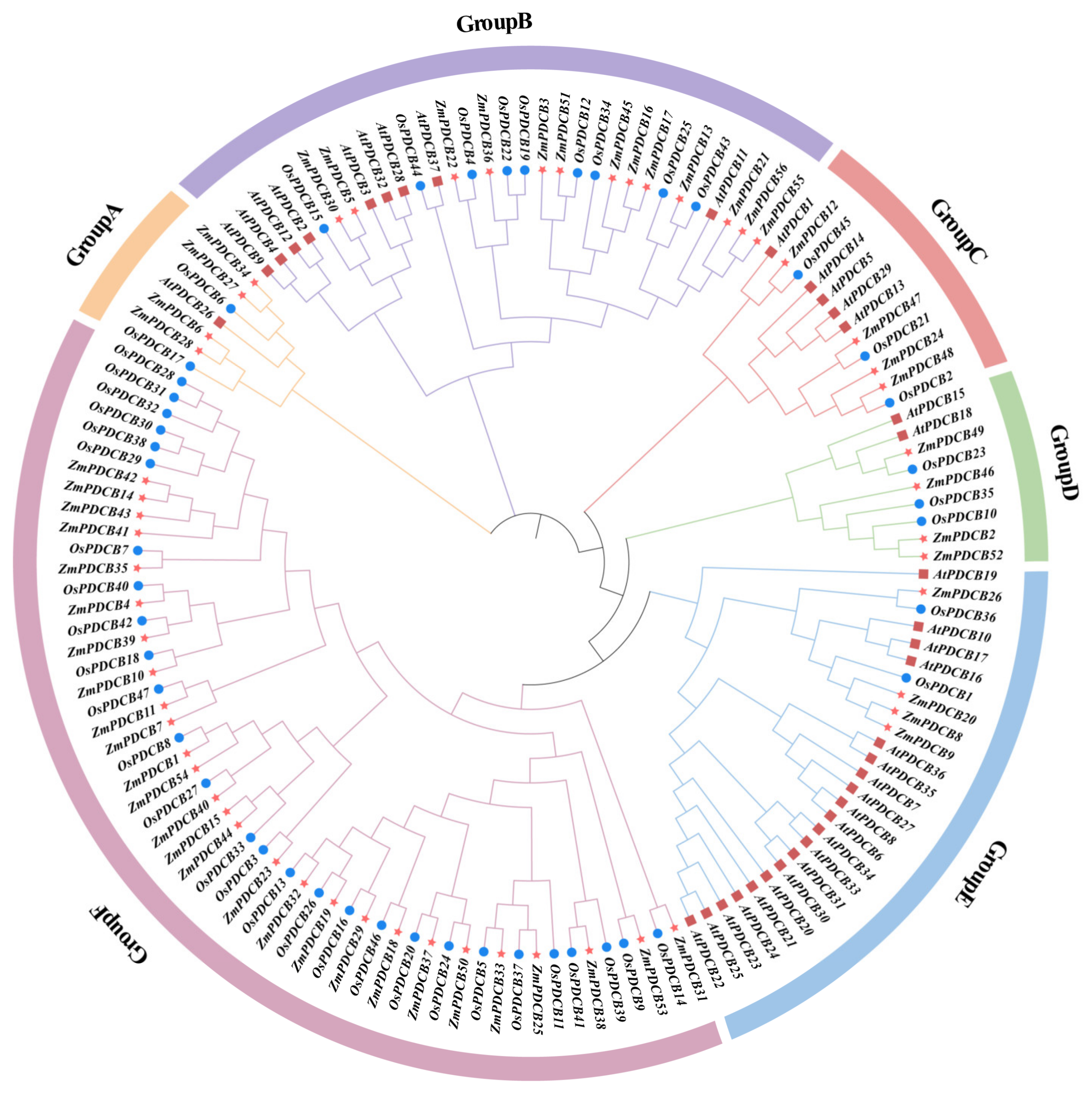
3.2. Phylogenetic Study and Classification of Maize PDCBs

To investigate the evolutionary relationships among the PDCB protein family in maize and other plants, we constructed a phylogenetic tree using the PDCB protein sequences from maize (Zea mays) and Arabidopsis thaliana. Furthermore, we identified 47 PDCB proteins from rice (Oryza sativa) to better characterize the evolutionary traits of the PDCB proteins in the grass family (Table S2). The phylogenetic relationships among PDCB proteins from these three species are illustrated in Figure 1. The phylogenetic tree divided the PDCB proteins into six branches. ZmPDCBs were distributed in all branches with an uneven pattern. Branches B and F contained more PDCB members, with 13 members in branch B and 27 members in branch F. In contrast, branches A, C, D, and E contained only four members, respectively. Notably, each branch contained PDCB proteins from both maize and rice, but not all branches included PDCB proteins from Arabidopsis (e.g., branch F), indicating an evolutionary divergence between monocots and dicots. Meanwhile, in branch F, there was a subbranch containing only ZmPDCB14, 41, 42, 43, which was significantly different from the subbranch nearby constituted by OsPDCBs (28, 29, 30, 31, 32, 38). These results indicate that the functions of the PDCB homolog genes in maize might be similar, and the phylogenetic relationship between maize and rice, both members of the grass family, is closer than that between maize and Arabidopsis, which belong to the Brassicaceae family.

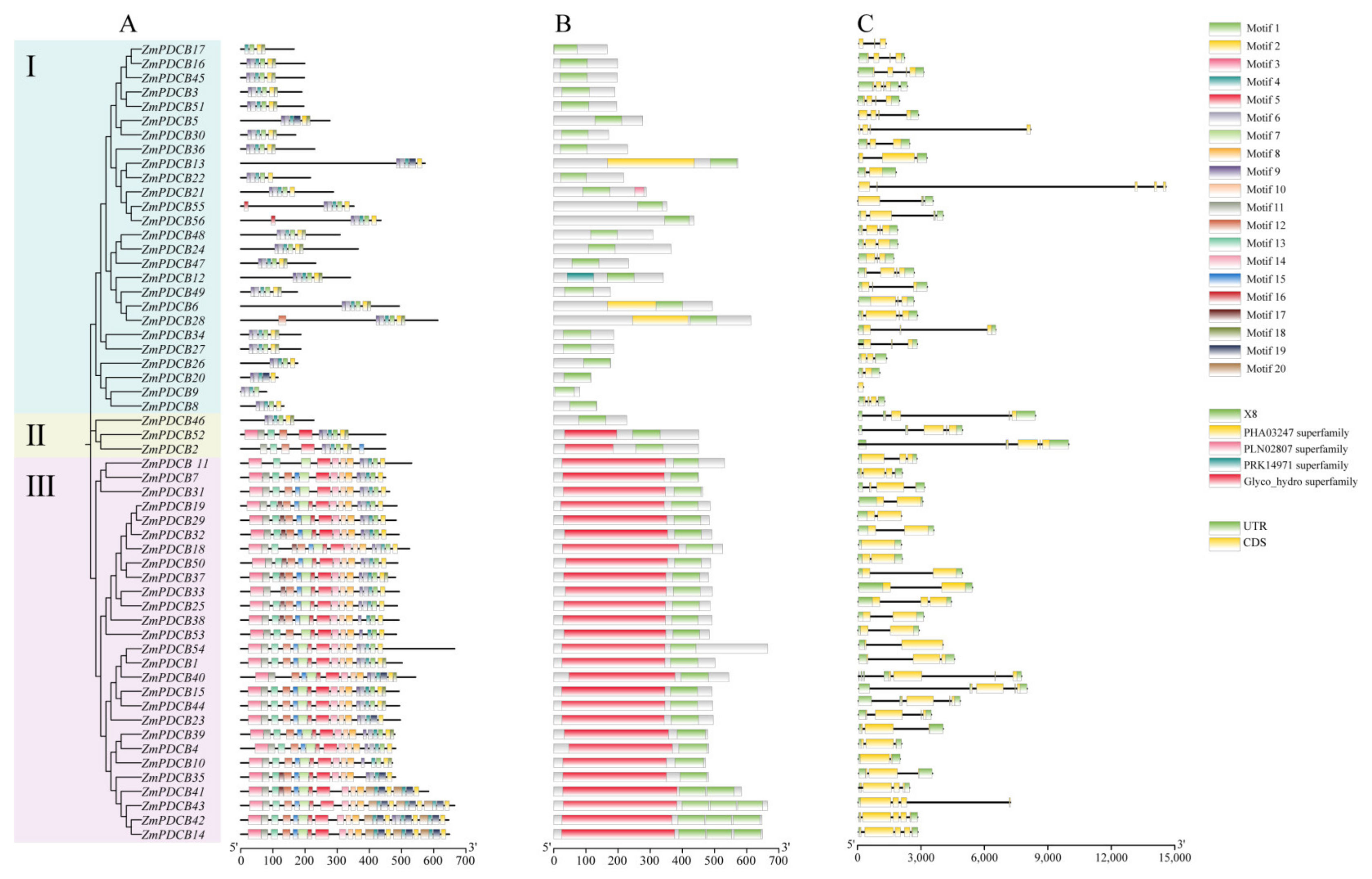
3.3. Gene Structure and Conserved Motif Analysis of ZmPDCB Genes

Variations in conserved motifs and the diversity of gene architecture serve as signs of the evolution of multigene families [39]. To comprehend the variation in gene structure, the spatial arrangement of PDCB exon/intron sections was investigated (Figure 2). The majority of PDCBs (36%) had four exons, out of a total of five exons. The exons of PDCB genes showed a higher degree of similarity within the same group (Figure 2C).
The fundamental framework of any feature sequence is defined by the motif, a conserved segment within the sequence. There were 20 possible motifs identified (Figure 2A). Remarkably, only motif 4 was present in every PDCB. The majority of PDCBs were ordered in the motif 4-1-2 pattern. The conserved motifs of PDCBs exhibited variations among groups, whereas the amount and configuration of motifs within a particular group of PDCBs were more similar compared to those of other groups. The remaining PDCBs, except for PDCB9, were placed in the sequence of motif 4-19-2.
The PDCB proteins were shown to have five conserved domains: PHA03247, X8, PRK14971, PLN02807, and Glyco_hydro superfamily domains (Figure 2B). The X8 domain was present in all PDCBs, and approximately half of them contained Glyco_hydro superfamily domains. The PHA03247 superfamily was present in ZmPDCB6, ZmPDCB13, and ZmPDCB28; the PRK14971 superfamily was present in ZmPDCB12; and the PLN02807 superfamily was present in ZmPDCB21. Based on these findings, we deduced that while the majority of PDCB members were conserved in maize, there was a minor degree of divergence in terms of gene structure and protein sequences.

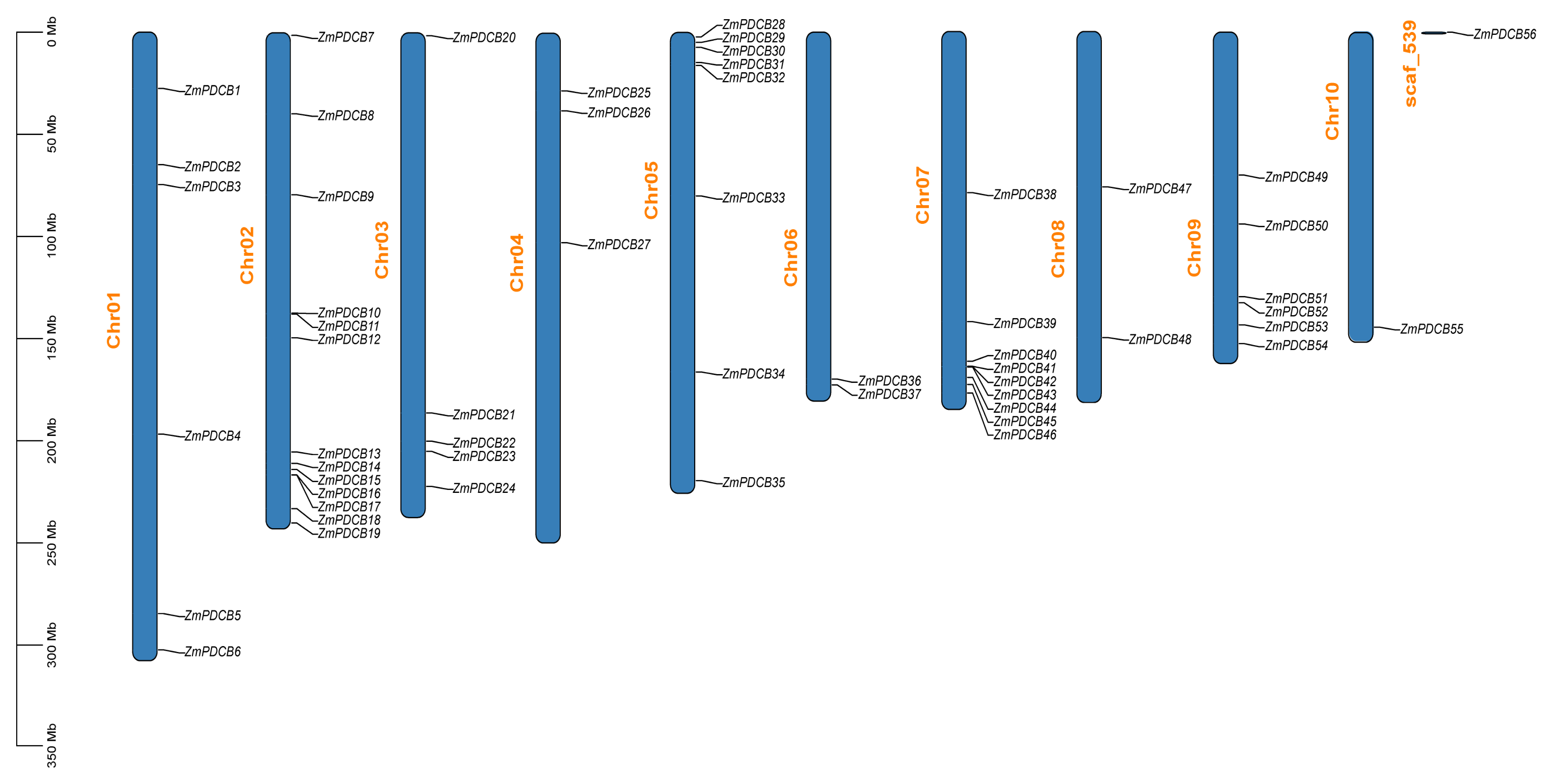
3.4. Chromosomal Location and Synteny Analysis of the PDCBs in Zea mays

Chromosome mapping showed that there were 55 ZmPDCBs distributed across 10 chromosomes, with 6 ZmPDCBs on Chr01, 13 ZmPDCBs on Chr02, 5 ZmPDCBs on Chr03, 3 ZmPDCBs on Chr04, 8 ZmPDCBs on Chr05, 2 ZmPDCBs on Chr06, 9 ZmPDCBs on Chr07, 2 ZmPDCBs on Chr08, 6 ZmPDCBs on Chr09, and 1 ZmPDCB on Chr10. Additionally, 1 ZmPDCB was located on scaffold_539. Most PDCBs were found near the telomeres or centromeres of chromosomes (Figure 3).
The distribution or arrangement of homologous genes within or between species is referred to as collinearity [40]. We examined the intra-genomic collinearities to demonstrate the collinearity of PDCB genes (Figure 4A). A total of 16 pairs of duplicated genes were distributed across 7 chromosomes, and most of them spanned chromosomes, suggesting that segmental duplication was a crucial aspect in the evolution of the PDCB gene family in maize. Then, we conducted an analysis of the collinearity among maize, rice, and Arabidopsis to reveal their evolutionary relationships. The synteny relationships in ZmPDCBs and OsPDCBs, and in ZmPDCBs and AtPDCBs were denoted by red and blue lines, respectively. A total of 48 pairs of collinearity were identified, of which 47 pairs of collinearity occurred in ZmPDCBs and OsPDCBs, while ZmPDCBs and AtPDCBs had only 1 pair of collinearity. The number of homologous gene pairs between maize and rice was much higher than the number between maize and Arabidopsis, and which was consistent with the evolutionary relationship shown in Figure 1, further indicating that maize and rice are both grass family members with a closer relationship.

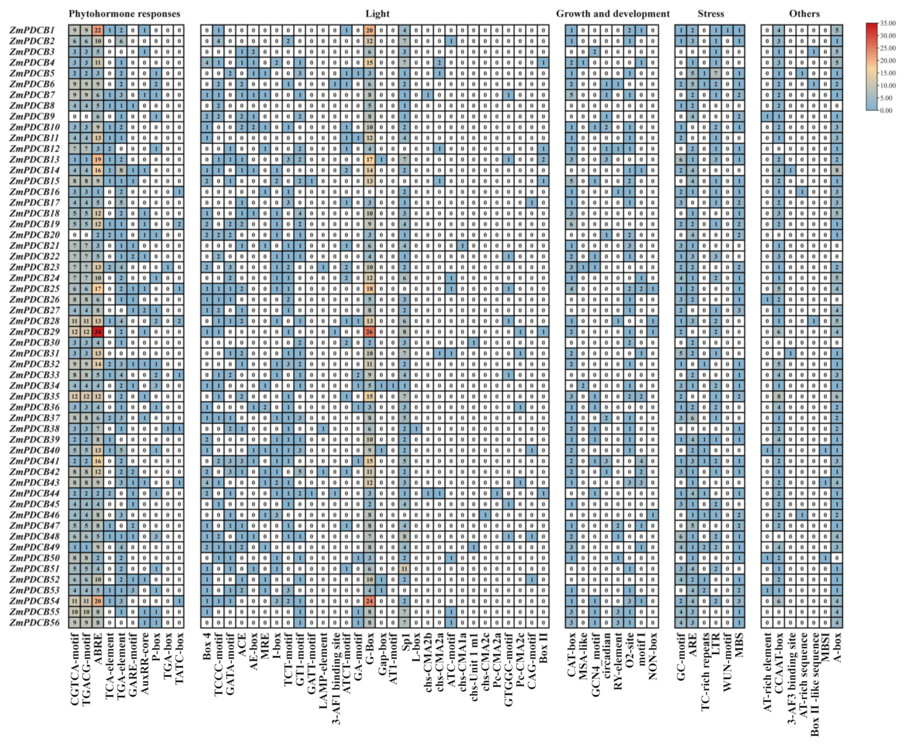
3.5. Cis-Acting Elements in the Promoter of PDCB Genes

The cis-acting elements in the promoter region may facilitate the regulation of gene transcription. An analysis of the cis-acting elements upstream of PDCB genes can illuminate their potential functions and enhance our comprehension of their regulatory mechanisms (Figure 5). A total of 10 cis-acting elements linked to phytohormone responses were identified. For instance, there was 1 salicylic acid-responsive element (TCA-element), 3 auxin-responsive elements (TGA-element, TGA-box, and AuxRR-core), 3 gibberellin-responsive elements (GARE-motif, P-box, and TATC-box), 2 methyl jasmonate-responsive elements (TGACG-motif and CGTCA-motif), and 1 abscisic acid-responsive element (ABRE). All ZmPDCB promoters contained the ABRE, with ZmPDCB29 having the largest number of this element. Given that maize is a C4 plant, these components, especially auxRR-core and ABRE, may play a role in regulating ZmPDCBs, thereby enhancing maize agronomy traits like deep rooting and stress tolerance.
Additionally, 30 light-responsive elements were identified, with G-Box and Sp1 existing in almost all ZmPDCB promoters. Six environmental stress-responsive cis-acting elements were identified, including defense and stress responsiveness (TC-rich repeats), wound-responsive elements (WUN-motif), anaerobic induction (ARE), enhancer-like elements involved in anoxia-specific inducibility (GC-motif), the MYB binding site involved in drought inducibility (MBS), and low-temperature responsiveness (LTR). Additional cis-acting elements associated with plant development were also found, including those regulating the cell cycle (MSA-like), zein metabolism (O2-site), and MYB binding sites, which are involved in the regulation of flavonoid biosynthesis genes (MBSI), light responsiveness (MRE), and drought inducibility (MBS). One MYBHv1 binding site, termed CCAAT-box, was also identified, with HvMYB1 known as a positive regulator of drought tolerance [41]. In conclusion, the quantity and diversity of cis-acting elements in the ZmPDCB promoter region were not only indicators of functional complexity, but also shown to be key regulators that contribute significantly to the unique growth characteristics and stress tolerance of maize as a C4 plant.

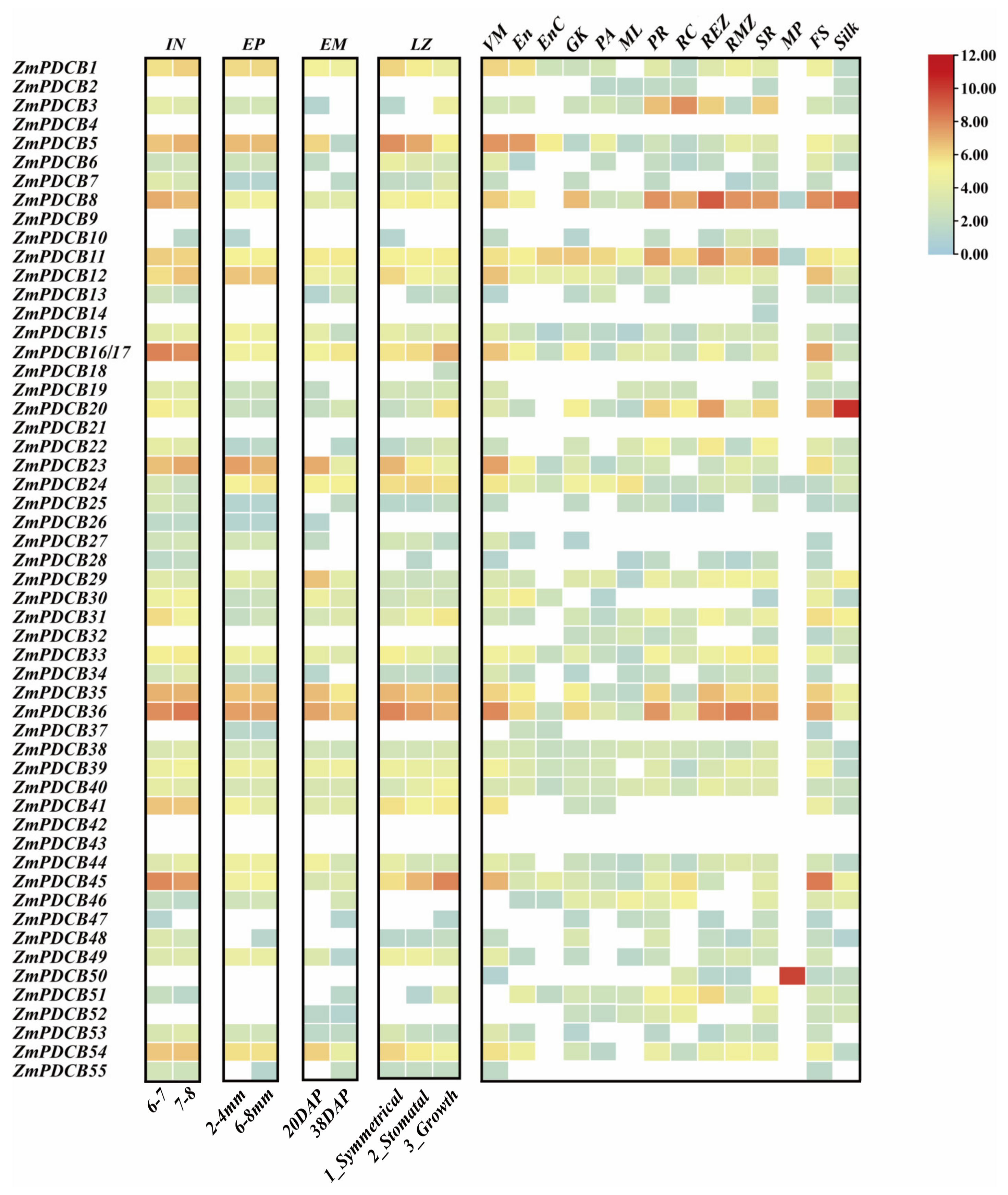
3.6. ZmPDCBs Expression Patterns in Various Tissues

Transcriptomic data from specific tissues can be utilized to predict the functionality of a gene during particular plant developmental stages. Various tissues displayed expression of ZmPDCBs, with some exhibiting high expression levels. Notably, 2 out of the 56 ZmPDCB genes, namely ZmPDCB11 and ZmPDCB24, were expressed across all 18 tissues, albeit at varying levels (Figure 6). Conversely, five genes, ZmPDCB4, 9, 21, 42, and 43, showed no expression in any tissue. Eighteen PDCBs, namely ZmPDCB2, 4, 9, 10, 13, 14, 18, 21, 25, 26, 27, 28, 32, 37, 42, 43, 47, and 55, exhibited extremely low expression (FPKM value < 10) across all tissues. The remaining genes were generally expressed in at least six tissues. Furthermore, ZmPDCB5, 8, 23, 35, 36, and 45 displayed elevated expression levels in most tissues relative to other genes. ZmPDCB5 demonstrated significant expression (FPKM value > 90) in internode, vegetative meristem, ear primordium, endosperm, and leaf. ZmPDCB8 and ZmPDCB36 demonstrated significant expression (FPKM value > 80) in 10 and 15 samples in different tissues, respectively, with higher expression (FPKM value > 190) in root, indicating that these genes may play important roles in root development. ZmPDCB23 demonstrated significant expression (FPKM value > 90) in internode, vegetative meristem, ear primordium, leaf, and embryo. ZmPDCB35 demonstrated significant expression (FPKM value > 80) in internode, ear primordium, embryo, leaf, and root. ZmPDCB45 demonstrated significant expression (FPKM value > 110) in internode, vegetative meristem, leaf, and female spikelet, with the highest expression patterns in female spikelet compared to other genes. Notably, one gene (ZmPDCB14) was expressed only in secondary root. Five genes showed significant tissue-specific expression in reproductive tissues. For instance, ZmPDCB8, 11, 24, and 50 were specifically expressed in mature pollen, with ZmPDCB50 exhibiting an extraordinarily high expression level, suggesting a potential role for the genes in pollen maturation and function. Furthermore, the expression levels of ZmPDCB20 increased extremely in silk, indicating its significance in the development of pistils in maize. In summary, the expression profiles of ZmPDCBs revealed the diverse and specific expression patterns of ZmPDCB genes in different tissues and developmental stages of maize. The prominent expression of certain genes, such as ZmPDCB8, 20, 36, 45, and B50, in vegetative tissues like roots and reproductive tissues like mature pollen, female spikelets, and silk indicates their essential regulatory roles in these processes.

4. Discussion

Maize holds a pivotal position in human nutrition as a staple food, provides a valuable source of raw materials for various industries, and is highly esteemed as the primary feed for livestock [42], but its PDCB coding gene family remains unknown. PDCBs are involved in regulating the function and localization of callose in the vicinity of PD [18]. Callose is crucial for numerous biological processes related to plant development and growth, as well as plant defense against adverse environmental factors [43]. In plants, the best studied PDCB gene families were those of Arabidopsis thaliana and pear fruit [20,44]. This study employs bioinformatics approaches to identify and evaluate PDCBs in Zea mays, aiming to gain insights into the involvement of the PDCB family in maize development.
A total of 56 PDCBs were identified in maize genome, categorized into six groups based on their evolutionary relationships (Figure 1). Not all groups contained homologous genes from Arabidopsis. Each group comprised between 4 to 27 ZmPDCB members; ZmPDCBs and OsPDCBs were predominantly found in group F, in which there were 27 ZmPDCBs, implying they might be specific orthologs of cereal. On the other hand, the majority of group E were 20 AtPDCBs, with just four ZmPDCBs and two OsPDCBs included. Group F, a subbranch containing only ZmPDCB14, 41, 42, and 43, was significantly different from the nearby subbranch constituted by OsPDCBs (28, 29, 30, 31, 32, and 38). Based on FPKM values obtained from RNA-sequencing data, the expression patterns of ZmPDCBs in 18 tissues were scrutinized to identify which PDCB genes might be implicated in regulating the growth of specific tissues or organs in maize (Figure 5). Interestingly, the transcription level in various tissues indicated the expression of ZmPDCB41 was general in nine tissues investigated, but the expression of ZmPDCB14 was only detected in secondary root, and the expressions of ZmPDCB42, 43 were absent. According to the relationship demonstrated by the phylogenetic tree, the ZmPDCB41 showed a big genetic distance from ZmPDCB14, 42, 43, which might explain partially the difference in their expression profile. However, as showed in Figure 3, it is very likely that ZmPDCB41, 42, 43 were in one gene cluster, thus ZmPDCB14 possibly originated from them by chromosome duplication and merging. The expression profile of ZmPDCB42, 43 remained unknown. It is clear that PDCB genes have been linked to callose accumulation in maize [11]. The study on tissue-specific expression of this gene family provides insights into its potential roles in various developmental processes [22].
Considering the vital roles of structural diversity in the evolution of gene families, we conducted the gene structure and conserved motif analysis of ZmPDCB genes (Figure 2). The ZmPDCBs exhibited notable congruence in their conserved motifs and gene architectures within the same group. However, for group II, there was an exception. This may be because some motifs in these three PDCBs were not conserved. The PDCB proteins typically featured five conserved domains: X8, PLN02807, PHA03247, PRK14971, and Glyco_hydro superfamily domains. However, not all ZmPDCB proteins contained the Glyco_hydro superfamily, PLN02807, PHA03247, or PRK14971 domains. All ZmPDCB proteins contained the X8 domain, which belongs to a broader family and is essential for callose-binding activity [18,20]. Additionally, 16 pairs of homologous genes were identified within the maize genome.
Moreover, cis-acting elements relevant to plant growth and development, light response, stress-associated responses, and plant hormones were analyzed within the promoter sequences of the ZmPDCB genes (as showed in Figure 5). These components may play a role in regulating ZmPDCBs. C4 plants are well-known for their superior agronomy traits, like deep rooting and stress tolerance, of which maize is typical and outstanding [24,45]. In plants, auxin (IAA) is a master regulator of morphogenesis, pattern formation, tropic responses, and development, including root architecture. Present research checked expression profiles of the ZmPDCBs in root meristem zone (RMZ), root cortex (RC), primary root (PR), root elongation zone (REZ), and secondary root (SR). More than half of the ZmPDCBs showed expression in these root tissues (Figure 6). Among these genes, ZmPDCB3, 8, 11, 20, 35, and 36 had relatively higher expression levels than the others. The presentation of cis-regulatory element auxRR-core was identified in the promoter regions of ZmPDCB3, 20, and 35, which hinted at the involvement of an auxin signal regulating the expression of ZmPDCBs in the process of root development. There is also research reporting that the quantity of callose at the plasmodesmal region is controlled by the auxin-GSL8 feedback circuit [46]. The results also showed that all the promoter regions of these ZmPDCBs have cis-regulatory elements concerned with stress responding and tolerance, and ABRE were detected and relatively abundant. These are key factors determining abscisic-acid-mediated transcriptional regulation of stress-related genes. ABA is vital in responding to various adverse conditions and is significantly involved in callose production [47]. Furthermore, environmental stressors, such as low temperatures, reactive oxygen species, and early viral defense systems, also influence callose deposition [15]. All these results implied that ZmPDCBs are highly involved in the maize root development and stress response.

5. Conclusions

Within this scholarly investigation, we delineated 56 ZmPDCBs within the maize genome, categorizing them into six distinct groups according to their evolutionary profiles. The ZmPDCBs exhibited notable congruence in their conserved motifs and gene architectures within the same group. Numerous cis-acting elements associated with plant hormones, light, plant growth dynamics, and stress conditions were identified within the promotor regions of the ZmPDCB genes. Significantly, every ZmPDCB promoter encompassed the abscisic-acid-responsive element. Through transcriptomics analysis of the ZmPDCBs across 18 distinct tissues, diverse and specific expression patterns emerged across various developmental stages of maize. Certain genes displayed extraordinarily high expression levels in vegetative tissues, including roots and reproductive tissues such as mature pollen, female spikelets, and silk, underscoring their pivotal regulatory functions in tissue-specific gene expression. These outcomes lay the groundwork for forthcoming endeavors in elucidating the crucial roles of the PDCBs in cereal crops, especially in the study of carbohydrate transport mechanisms.

Supplementary Materials

The following supporting information can be downloaded at https://www.mdpi.com/article/10.3390/agronomy14081858/s1, Table S1: Detailed information and analysis of the physicochemical analysis of the Plasmodesmata Callose Binding Proteins (PDCBs) in Zea mays L.; Table S2: Detailed information of the PDCB gene family in Oryza sativa L.

Author Contributions

Conceptualization, H.W., J.G. and S.W.; methodology, J.G.; validation, S.W., M.Z. and X.S.; formal analysis, J.G., S.W., M.Z. and X.S.; writing—original draft preparation, H.W., J.G. and S.W.; writing—review and editing, H.W., J.G. and S.W.; visualization, J.G., S.W. and H.W.; project administration, H.W.; funding acquisition, H.W. All authors have read and agreed to the published version of the manuscript.

Funding

This research was funded by the National Natural Science Foundation of China (Nos. 31100172, 31970238), Natural Science Foundation of Liaoning Province (No. 2022-MS-174), Key Research Projects of Liaoning Provincial Education Department (No. JYTZD2023067), and Shenyang Science and Technology Bureau project (No. RC230254).

Data Availability Statement

The original contributions presented in the study are included in the article/Supplementary Material, further inquiries can be directed to the corresponding author.

Conflicts of Interest

The authors declare no conflicts of interest.

References

  1. Ehlers, K.; Kollmann, R. Primary and secondary plasmodesmata: Structure, origin, and functioning. Protoplasma 2001, 216, 1–30. [Google Scholar] [CrossRef] [PubMed]
  2. Burch-Smith, T.M.; Stonebloom, S.; Xu, M.; Zambryski, P.C. Plasmodesmata during development: Re-examination of the importance of primary, secondary, and branched plasmodesmata structure versus function. Protoplasma 2011, 248, 61–74. [Google Scholar] [CrossRef]
  3. Sager, R.E.; Lee, J.-Y. Plasmodesmata at a glance. J. Cell Sci. 2018, 131, jcs209346. [Google Scholar] [CrossRef] [PubMed]
  4. Lucas, W.J.; Ham, B.-K.; Kim, J.-Y. Plasmodesmata—Bridging the gap between neighboring plant cells. Trends Cell Biol. 2009, 19, 495–503. [Google Scholar] [CrossRef] [PubMed]
  5. Ruiz-Medrano, R.; Xoconostle-Cazares, B.; Kragler, F. The plasmodesmatal transport pathway for homeotic proteins, silencing signals and viruses. Curr. Opin. Plant Biol. 2004, 7, 641–650. [Google Scholar] [CrossRef]
  6. Evert, R.F. Sieve-Tube Structure in Relation to Function. BioScience 1982, 32, 789–795. [Google Scholar] [CrossRef]
  7. Dhungana, S.R.; Braun, D.M. Sugar transporters in grasses: Function and modulation in source and storage tissues. J. Plant Physiol. 2021, 266, 153541. [Google Scholar] [CrossRef]
  8. Heyser, W.; Evert, R.F.; Fritz, E.; Eschrich, W. Sucrose in the Free Space of Translocating Maize Leaf Bundles. Plant Physiol. 1978, 62, 491–494. [Google Scholar] [CrossRef]
  9. Ayre, B.G. Membrane-Transport Systems for Sucrose in Relation to Whole-Plant Carbon Partitioning. Mol. Plant 2011, 4, 377–394. [Google Scholar] [CrossRef]
  10. Slewinski, T.L.; Braun, D.M. Current perspectives on the regulation of whole-plant carbohydrate partitioning. Plant Sci. 2010, 178, 341–349. [Google Scholar] [CrossRef]
  11. Grison, M.S.; Brocard, L.; Fouillen, L.; Nicolas, W.; Wewer, V.; Dörmann, P.; Nacir, H.; Benitez-Alfonso, Y.; Claverol, S.; Germain, V.; et al. Specific Membrane Lipid Composition Is Important for Plasmodesmata Function in Arabidopsis. Plant Cell 2015, 27, 1228–1250. [Google Scholar] [CrossRef] [PubMed]
  12. Zavaliev, R.; Ueki, S.; Epel, B.L.; Citovsky, V. Biology of callose (β-1,3-glucan) turnover at plasmodesmata. Protoplasma 2011, 248, 117–130. [Google Scholar] [CrossRef] [PubMed]
  13. Wang, Y.; Li, X.; Fan, B.; Zhu, C.; Chen, Z. Regulation and Function of Defense-Related Callose Deposition in Plants. Int. J. Mol. Sci. 2021, 22, 2393. [Google Scholar] [CrossRef] [PubMed]
  14. German, L.; Yeshvekar, R.; Benitez-Alfonso, Y. Callose metabolism and the regulation of cell walls and plasmodesmata during plant mutualistic and pathogenic interactions. Plant Cell Environ. 2022, 46, 391–404. [Google Scholar] [CrossRef] [PubMed]
  15. Wu, S.-W.; Kumar, R.; Iswanto, A.B.B.; Kim, J.-Y. Callose balancing at plasmodesmata. J. Exp. Bot. 2018, 69, 5325–5339. [Google Scholar] [CrossRef] [PubMed]
  16. Ohba, Y.; Yoshihara, S.; Sato, R.; Matsuoka, K.; Asahina, M.; Satoh, S.; Iwai, H. Plasmodesmata callose binding protein 2 contributes to the regulation of cambium/phloem formation and auxin response during the tissue reunion process in incised Arabidopsis stem. J. Plant Res. 2023, 136, 865–877. [Google Scholar] [CrossRef] [PubMed]
  17. Maule, A.J.; Gaudioso-Pedraza, R.; Benitez-Alfonso, Y. Callose deposition and symplastic connectivity are regulated prior to lateral root emergence. Commun. Integr. Biol. 2013, 6, e26531. [Google Scholar] [CrossRef]
  18. Simpson, C.; Thomas, C.; Findlay, K.; Bayer, E.; Maule, A.J. An Arabidopsis GPI-anchor plasmodesmal neck protein with callose binding activity and potential to regulate cell-to-cell trafficking. Plant Cell 2009, 21, 581–594. [Google Scholar] [CrossRef] [PubMed]
  19. De Storme, N.; Geelen, D. Callose homeostasis at plasmodesmata: Molecular regulators and developmental relevance. Front. Plant Sci. 2014, 5, 138. [Google Scholar] [CrossRef]
  20. Li, W.; Yuan, K.; Ren, M.; Xie, Z.; Qi, K.; Gong, X.; Wang, Q.; Zhang, S.; Tao, S. PbPDCB16-mediated callose deposition affects the plasmodesmata blockage and reduces lignification in pear fruit. Plant Sci. 2023, 337, 111876. [Google Scholar] [CrossRef] [PubMed]
  21. Zhang, H.; Xiao, X.; Li, Z.; Chen, Y.; Li, P.; Peng, R.; Lu, Q.; Wang, Y. Exploring the plasmodesmata callose-binding protein gene family in upland cotton: Unraveling insights for enhancing fiber length. PeerJ 2024, 12, e17625. [Google Scholar] [CrossRef] [PubMed]
  22. Das Jyoti, S.; Bin Azim, J.; Robin, A.H.K. Genome-wide characterization and expression profiling of EIN3/EIL family genes in Zea mays. Plant Gene 2021, 25, 100270. [Google Scholar] [CrossRef]
  23. Schnable, P.S.; Ware, D.; Fulton, R.S.; Stein, J.C.; Wei, F.; Pasternak, S.; Liang, C.; Zhang, J.; Fulton, L.; Graves, T.A.; et al. The B73 Maize Genome: Complexity, Diversity, and Dynamics. Science 2009, 326, 1112–1115. [Google Scholar] [CrossRef]
  24. Xie, J.; Fei, X.; Yan, Q.; Jiang, T.; Li, Z.; Chen, H.; Wang, B.; Chao, Q.; He, Y.; Fan, Z.; et al. The C4 photosynthesis bifunctional enzymes, PDRPs, of maize are co-opted to cytoplasmic viral replication complexes to promote infection of a prevalent potyvirus sugarcane mosaic virus. Plant Biotechnol. J. 2024, 22, 1812–1832. [Google Scholar] [CrossRef]
  25. Edwards, G.E.; Franceschi, V.R.; Voznesenskaya, E.V. Single-cell C(4) photosynthesis versus the dual-cell (Kranz) paradigm. Annu. Rev. Plant Biol. 2004, 55, 173–196. [Google Scholar] [CrossRef] [PubMed]
  26. Langdale, J.A. C4 cycles: Past, present, and future research on C4 photosynthesis. Plant Cell 2011, 23, 3879–3892. [Google Scholar] [CrossRef]
  27. Danila, F.R.; Quick, W.P.; White, R.G.; Furbank, R.T.; von Caemmerer, S. The Metabolite Pathway between Bundle Sheath and Mesophyll: Quantification of Plasmodesmata in Leaves of C3 and C4 Monocots. Plant Cell 2016, 28, 1461–1471. [Google Scholar] [CrossRef]
  28. Garcia-Hernandez, M.; Berardini, T.; Chen, G.; Crist, D.; Doyle, A.; Huala, E.; Knee, E.; Lambrecht, M.; Miller, N.; Mueller, L.A.; et al. TAIR: A resource for integrated Arabidopsis data. Funct. Integr. Genom. 2002, 2, 239–253. [Google Scholar] [CrossRef]
  29. Bolser, D.; Staines, D.M.; Pritchard, E.; Kersey, P. Ensembl Plants: Integrating Tools for Visualizing, Mining, and Analyzing Plant Genomics Data; Springer: New York, NY, USA, 2016. [Google Scholar] [CrossRef]
  30. Mistry, J.; Chuguransky, S.; Williams, L.; Qureshi, M.; Salazar, G.A.; Sonnhammer, E.L.L.; Tosatto, S.C.; Paladin, L.; Raj, S.; Richardson, L.J.; et al. Pfam: The protein families database in 2021. Nucleic Acids Res. 2020, 49, D412–D419. [Google Scholar] [CrossRef]
  31. Horton, P.; Park, K.-J.; Obayashi, T.; Fujita, N.; Harada, H.; Adams-Collier, C.J.; Nakai, K. WoLF PSORT: Protein localization predictor. Nucleic Acids Res. 2007, 35, 585–587. [Google Scholar] [CrossRef]
  32. Shang, L.; He, W.; Wang, T.; Yang, Y.; Xu, Q.; Zhao, X.; Yang, L.; Zhang, H.; Li, X.; Lv, Y.; et al. A complete assembly of the rice Nipponbare reference genome. Mol. Plant 2023, 16, 1232–1236. [Google Scholar] [CrossRef]
  33. Tamura, K.; Stecher, G.; Kumar, S. MEGA11: Molecular Evolutionary Genetics Analysis Version 11. Mol. Biol. Evol. 2021, 38, 3022–3027. [Google Scholar] [CrossRef]
  34. Chen, C.J.; Chen, H.; Zhang, Y.; Thomas, H.R.; Frank, M.H.; He, Y.H.; Xia, R. TBtools: An Integrative Toolkit Developed for Interactive Analyses of Big Biological Data. Mol. Plant 2020, 13, 1194–1202. [Google Scholar] [CrossRef]
  35. Lescot, M.; Déhais, P.; Thijs, G.; Marchal, K.; Moreau, Y.; Van de Peer, Y.; Rouzé, P.; Rombauts, S. PlantCARE, a database of plant cis-acting regulatory elements and a portal to tools for in silico analysis of promoter sequences. Nucleic Acids Res. 2002, 30, 325–327. [Google Scholar] [CrossRef]
  36. Walley, J.W.; Sartor, R.C.; Shen, Z.; Schmitz, R.J.; Wu, K.J.; Urich, M.A.; Nery, J.R.; Smith, L.G.; Schnable, J.C.; Ecker, J.R.; et al. Integration of omic networks in a developmental atlas of maize. Science 2016, 353, 814–818. [Google Scholar] [CrossRef]
  37. Hufford, M.B.; Seetharam, A.S.; Woodhouse, M.R.; Chougule, K.M.; Ou, S.; Liu, J.; Ricci, W.A.; Guo, T.; Olson, A.; Qiu, Y.; et al. De novo assembly, annotation, and comparative analysis of 26 diverse maize genomes. Science 2021, 373, 655–662. [Google Scholar] [CrossRef] [PubMed]
  38. Stelpflug, S.C.; Sekhon, R.S.; Vaillancourt, B.; Hirsch, C.N.; Buell, C.R.; De Leon, N.; Kaeppler, S.M. An Expanded Maize Gene Expression Atlas based on RNA Sequencing and its Use to Explore Root Development. Plant Genome 2016, 9. [Google Scholar] [CrossRef]
  39. Feng, J.; Chen, Y.; Xiao, X.; Qu, Y.; Li, P.; Lu, Q.; Huang, J. Genome-wide analysis of the CalS gene family in cotton reveals their potential roles in fiber development and responses to stress. PeerJ 2021, 9, e12557. [Google Scholar] [CrossRef] [PubMed]
  40. Wang, Y.; Tang, H.; Wang, X.; Sun, Y.; Joseph, P.V.; Paterson, A.H. Detection of colinear blocks and synteny and evolutionary analyses based on utilization of MCScanX. Nat. Protoc. 2024, 19, 2206–2229. [Google Scholar] [CrossRef]
  41. Alexander, R.D.; Wendelboe-Nelson, C.; Morris, P.C. The barley transcription factor HvMYB1 is a positive regulator of drought tolerance. Plant Physiol. Biochem. 2019, 142, 246–253. [Google Scholar] [CrossRef]
  42. Cao, L.; Lu, X.; Zhang, P.; Wang, G.; Wei, L.; Wang, T. Systematic Analysis of Differentially Expressed Maize ZmbZIP Genes between Drought and Rewatering Transcriptome Reveals bZIP Family Members Involved in Abiotic Stress Responses. Int. J. Mol. Sci. 2019, 20, 4103. [Google Scholar] [CrossRef]
  43. Piršelová, B.; Matušíková, I. Callose: The plant cell wall polysaccharide with multiple biological functions. Acta Physiol. Plant. 2013, 35, 635–644. [Google Scholar] [CrossRef]
  44. Simpson, C. GPI-Anchored Proteins as Potential Plasmodesmal Components; University of East Anglia: Norwich, UK, 2008. [Google Scholar]
  45. Singh, J.; Garai, S.; Das, S.; Thakur, J.K.; Tripathy, B.C. Role of C4 photosynthetic enzyme isoforms in C3 plants and their potential applications in improving agronomic traits in crops. Photosynth. Res. 2022, 154, 233–258. [Google Scholar] [CrossRef] [PubMed]
  46. Han, X.; Hyun, T.K.; Zhang, M.; Kumar, R.; Koh, E.-J.; Kang, B.-H.; Lucas, W.J.; Kim, J.-Y. Auxin-callose-mediated plasmodesmal gating is essential for tropic auxin gradient formation and signaling. Dev. Cell 2014, 28, 132–146. [Google Scholar] [CrossRef] [PubMed]
  47. Liu, J.; Du, H.; Ding, X.; Zhou, Y.; Xie, P.; Wu, J. Mechanisms of callose deposition in rice regulated by exogenous abscisic acid and its involvement in rice resistance to Nilaparvata lugens Stål (Hemiptera: Delphacidae). Pest. Manag. Sci. 2017, 73, 2559–2568. [Google Scholar] [CrossRef] [PubMed]
Figure 1. Plasmodesmata Callose Binding Proteins (PDCBs) from Zea mays, Arabidopsis, and rice underwent phylogenetic analysis. Zea mays, Arabidopsis and rice PDCBs are denoted by pink stars, red squares, and blue circles, respectively.
Agronomy 14 01858 g001
Figure 2. Conserved motif, conserved domain, and exon–intron distribution of PDCB genes in maize. (A) The conservative motif of PDCBs and MEGA11 were used to build the evolutionary tree of PDCBs. The 20 motifs are shown in various colored boxes. (B) Conserved domains of PDCB proteins. (C) Gene structures of PDCB genes. I, II, III represent the groups of the phylogenetic tree.
Agronomy 14 01858 g002
Figure 3. The distribution of 56 PDCBs on the chromosomes of maize. The chromosome name is located to the left of each chromosome, whereas the gene ID is situated to the right.
Agronomy 14 01858 g003
Figure 4. Collinearity analysis of PDCB genes. (A) Synteny analysis of PDCB genes in Zea mays. (B) Synteny analysis of PDCB genes among Zea mays, Arabidopsis, and rice.
Agronomy 14 01858 g004
Figure 5. Cis-regulatory elements identified in the promoter region of ZmPDCBs. The different colors and numbers of the squares represent the numbers of different promoter elements in ZmPDCB genes.
Agronomy 14 01858 g005
Figure 6. Transcriptome analyses of ZmPDCB genes in 18 tissues. The expression levels of ZmPDCB genes in distinct tissues can be observed by plotting the normalized Log2 (FPKM+1) values against the relevant tissues. Internode (IN), vegetative meristem (Vm), ear primordium (EP), embryo (Em), endosperm (En), endosperm crown (EnC), germination kernels (GK), pericarp aleurone (PA), leaf zone (LZ), root meristem zone (RMZ), root cortex (RC), primary root (PR), mature leaf (ML), root elongation zone (REZ), secondary root (SR), mature pollen (MP), and female spikelets collected on the same day as silk (FS) are presented in abbreviated forms.
Agronomy 14 01858 g006
Disclaimer/Publisher’s Note: The statements, opinions and data contained in all publications are solely those of the individual author(s) and contributor(s) and not of MDPI and/or the editor(s). MDPI and/or the editor(s) disclaim responsibility for any injury to people or property resulting from any ideas, methods, instructions or products referred to in the content.

