Article

Chemical and Biological Investigation on the Potential Ornamental Plant Ophiorrhiza chinensis

College of Chemistry and Chemical Engineering, Central South University of Forestry and Technology, Changsha 410004, China
*
Author to whom correspondence should be addressed.
Agronomy 2024, 14(8), 1872; https://doi.org/10.3390/agronomy14081872
Submission received: 4 July 2024 / Revised: 11 August 2024 / Accepted: 21 August 2024 / Published: 22 August 2024
(This article belongs to the Section Horticultural and Floricultural Crops)

Abstract

:
An investigation of the potential ornamental plant Ophiorrhiza chinensis H.S. Lo (Rubiaceae) was conducted, which resulted in the discovery of eight structurally diverse compounds, including two triterpenes, two steroids, two anthraquinones, one alkaloid, and one coumarin. These chemical constituents were isolated by repeated column chromatography and identified by analysis of their NMR spectral data. All of these substances were found in this species for the first time, and four of them were first isolated from the genus Ophiorrhiza. The chemotaxonomic importance of these isolates was discussed, indicating four chemotaxonomic markers for O. chinensis. The tyrosinase inhibitory activity of these isolates was evaluated by a colorimetric method. As a result, six phytochemicals demonstrated moderate tyrosinase inhibitory effects with IC50 values ranging from 25.7 μM to 68.1 μM. Moreover, the binding modes between the active compounds and the mushroom tyrosinase were analyzed preliminarily assisted by molecular docking calculations. This study filled up the knowledge gap of the unreported phytochemical and pharmacological profiles of secondary metabolites from the species O. chinensis.

1. Introduction

The plant Ophiorrhiza japonica Blume (Rubiaceae) is under cultivation, which could be used as a new kind of ornamental plant because of its beautiful flowers [1]. Indeed, almost all species of the genus Ophiorrhiza have graceful blossoms. From this perspective, Ophiorrhiza chinensis H.S. Lo (Rubiaceae) is a candidate for ornamental plants. However, how to effectively utilize the rest of the materials of the ornamental plants after flowering is an important challenge. One way is to explore their bioactive chemical compounds, which might be developed as lead compounds for drugs or used as cosmetic ingredients.
Indeed, some species of the genus Ophiorrhiza have been used as traditional medicines worldwide for a long time. For instance, four species O. japonica, Ophiorrhiza cantonensis Hance (Rubiaceae), Ophiorrhiza pumila Champ. & Benth. (Rubiaceae), and Ophiorrhiza succirubra King ex Hook. f. (Rubiaceae) were recorded in the monumental book Chinese Materia Medica, which exhibit a variety of pharmacological effects such as detoxifying, treating snakebites, traumatic injuries, and neurasthenia [2]. To find the chemical constituents with promising bioactivities, the plants of genus Ophiorrhiza were extensively studied. As reported, the Ophiorrhiza plants harbor a rich set of chemical compositions, including terpenes, alkaloids, anthraquinones, steroids, flavonoids, and coumarins [3,4,5]. Until now, ca. 30 species of this genus have been studied on the aspects of their chemical components and/or biological activities. Notably, Ophiorrhiza is a species-rich genus with more than 300 species. Therefore, the phytochemical components of most species in this genus, including O. chinensis, are poorly known. The wild O. chinensis distributes in southern China, including Hunan, and generally grows at altitudes of 300–1500 m [6]. There was only one report regarding the different contents of chlorogenic acid in various organs of O. chinensis [7]. However, no detailed investigation on the chemical and biological aspects of compounds from the title plant has been conducted till now.
Tyrosinase, also known as polyphenol oxidase, is widely distributed in various organisms and serves as a crucial and rate-limiting enzyme in melanin synthesis within living organisms [8]. Tyrosinase inhibitors are widely used in multiple fields, including pharmaceuticals and cosmetics. In the past decades, many natural tyrosinase inhibitors have been reported [9,10,11,12,13]. From the viewpoint of structural types, these tyrosinase inhibitors could be classified as terpenoids, flavonoids, lignans, etc. They were obtained from plants of different families, such as Rosaceae, Theaceae, Moraceae, etc. [14], some of which were found in the traditional Chinese material medica [15].
In our previous study on the plants of the genus Ophiorrhiza, a new anthraquinone and 10 known compounds with antioxidant activity were found in O. japonica [16]. Inspired by this work, the chemical components from the title plant O. chinensis and their bioactivities were investigated. The aim of this study is to disclose the chemical and biological profiles of O. chinensis, which could provide the basis for the future utilization of this plant in the field of health care or cosmetical products. Herein, the isolation, structure elucidation, chemotaxonomic significance, tyrosinase inhibitory, and molecular docking study of the major chemical components of O. chinensis were described.

2. Materials and Methods

2.1. General Experimental Procedures

NMR spectra were measured on a Bruker DRX-600 spectrometer (Bruker Biospin AG, Fällanden, Germany). Commercial silica gel (200–300 and 300–400 mesh, Qingdao Haiyang Chemical Group Co., Ltd., Qingdao, China) and Sephadex LH-20 gel (Amersham Biosciences, Amersham, UK) were used for column chromatography, and precoated silica gel plates (GF-254, Yan Tai Zi Fu Chemical Group Co., Yantai, China) were used for analytical TLC. All solvents including ethanol, petroleum ether, EtOAc, CHCl3, dichromethane, and methanol used for column chromatography were of analytical grade (Shanghai Chemical Reagents Co., Ltd., Shanghai, China).

2.2. Plant Material

The aerial parts of the plants O. chinensis were collected from Mang Mountain (27.3° N, 112.9° E), Chenzhou, Hunan, China, in May 2018, and identified by Prof. Lei Wu at Central South University of Forestry and Technology (CSUFT, Changsha, China). A specimen (voucher No. P-2018-HN-3) was stored in the Laboratory of Natural Products Chemistry, CSUFT.

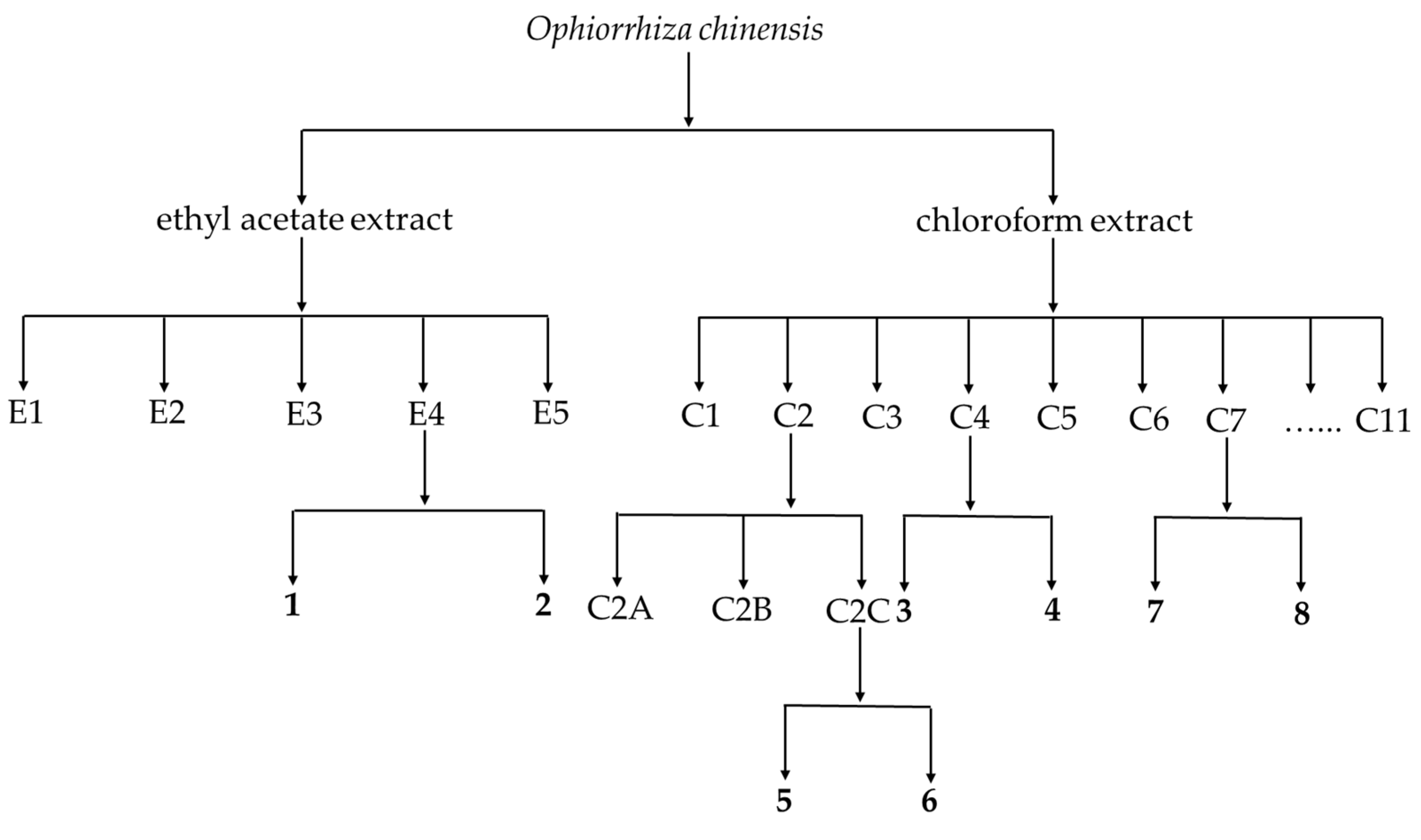
2.3. Extraction and Isolation

The air-dried aerial parts of the plants O. chinensis (dried weight 0.2 kg) were pulverized and soaked in 99.7% ethanol at room temperature once a week for a total of three times. The resulting ethanol extracts were combined and concentrated by rotavapor to remove the solvent, yielding a dark residue. Then, the residue was suspended in H2O and successively partitioned with EtOAc (E) and CHCl3 (C) to afford two corresponding E and C extracts.
The E extract (182.0 mg) was separated using silica gel (200–300 mesh) column chromatography (CC) (petroleum ether (P)/E 50:1 → 0:1) to afford five fractions (Fr. E1–E5). Fr. E4 (52.5 mg) was divided into six subfractions (Fr. E4A–E4F) by silica gel CC (300–400 mesh, P/E 5:1 → 0:1). Compounds 1 (2.4 mg) and 2 (2.0 mg) were got from Fr. E4E (23.1 mg) after a two-stage separation starting with Sephadex LH-20 gel CC (P/dichromethane (D)/methanol (M) 2:1:1) and continued by silica gel CC (300–400 mesh, D/M 80:1).
The C extract (60.0 mg) was purified by silica gel CC (200–300 mesh, D/M 1:1), yielding eleven fractions (Fr. C1–C11). Fr. C2 (8.7 mg) was purified by silica gel CC (300–400 mesh, P/E 7:1 → 3:1) to yield three subfractions (Fr. C2A–C2C). Compounds 5 (1.4 mg) and 6 (1.5 mg) were obtained from Fr. C2C (3.8 mg) by Sephadex LH-20 gel CC (300–400 mesh, P/D/M 2:1:1). Compounds 3 (1.6 mg) and 4 (14.8 mg) were isolated from Fr. C4 (23.7 mg) by chromatographing over silica gel (300–400 mesh, P/E 9:1) and then Sephadex LH-20 CC (P/D/M 2:1:1). Similarly, compounds 7 (2.1 mg) and 8 (1.2 mg) were got from Fr. C7 (6.9 mg) by a two-stage separation beginning with silica gel CC (300–400 mesh, D/M 16:1) and continued by Sephadex LH-20 CC (P/D/M 2:1:1).
The above-mentioned key experimental procedures of compounds 18 were summarized in Figure 1.

2.4. Characteristic 1H and 13C NMR Spectral Data of Isolates

2.5. In Vitro Tyrosinase Inhibitory Activity Assay

The tyrosinase inhibition of sample solutions was performed as previously reported methods with minor modification as described [17,18]: 50 μL of phosphate buffer (0.1 M, pH 6.8), 25 μL of tyrosinase (100 U/mL), 25 μL of sample solutions and 100 μL L-tyrosine were added into the wells of a 96-well plate, then incubated at 37 °C for 30 min. The absorbance of each well was measured at 475 nm by a microplate reader. Vitamin C was used as the positive control. The inhibition rate of the test compound against tyrosinase was calculated as follows: inhibition rate (%) = [1 − (A1 − A2)/(A3 − A0)] × 100, where A1 is the absorbance of the sample solution, A2 is the absorbance of the sample solution control, A3 is the absorbance of blank; A0 is the absorbance of blank control. Each concentration was tested in parallel three times. Half maximal inhibitory concentration (IC50) was derived from the fitting curve of inhibition rates vs. different concentrations of the test compound.

2.6. Molecular Docking Calculations

Molecular docking calculations were performed using the software AutoDock Vina (v4.2). The X-ray crystal structure of mushroom tyrosinase with the ligand (PDB: 2Y9X with 2.78 Å resolution) was downloaded from the RCSB Protein Data Bank. The structures of compounds 16 were depicted by the software ChemDraw (v20.0), which were further converted to 3D structures using the software Chem3D (v20.0). The optimizations of the structures were done using energy minimization with the MM2 method, and the optimized molecules were subsequently subjected to docking calculations with mushroom tyrosinase. The 2Y9X receptor was imported into the software Pymol (v2.4) and removed the ligand ORT as well as water molecules from the 2Y9X file. The resulting file was imported into AutoDock Vina (v4.2), hydrated, merged with non-polar hydrogen atoms, and then saved as 2Y9X.pdbqt. The active site of 2Y9X was detected by the software AutoDock Vina (v4.2), and the coordinates were set to (−7.397 −23.551 −32.508), while the grid box size was set at 70.35 Å × 70.35 Å × 70.35 Å. When the calculations were completed, the results were analysed using the software Discovery Studio Visualizer (v19.1).

3. Results and Discussion

3.1. Structure Elucidation of the Isolates

A phytochemical examination of the aerial parts of the O. chinensis plants led to the isolation of eight structurally diverse compounds 18 (Figure 2). Their chemical structures were established based on the analysis of NMR data together with a comparison of the data recorded in the published work (Table 1 and Table S1–S8).
Chemical constituent 1 was obtained as a white amorphous powder. The 1H and 13C NMR spectra showed the signals attributable to one double bond (δH 5.27 (1H, t, J = 3.5 Hz, H-12); δC 126.0 (C-12), 138.1 (C-13)), one oxygen-bearing methine (δH 3.22 (1H, dd, J = 11.3, 4.7 Hz, H-3); δC 79.2 (C-3)), and seven methyls (δH 1.25 (3H, s, H-27), 1.08 (3H, s, H-25), 0.99 (3H, s, H-24), 0.95 (3H, d, J = 6.3 Hz, H-29), 0.93 (3H, s, H-23), 0.86 (3H, d, J = 6.5 Hz, H-30), and 0.78 (3H, s, H-26); δC28.2 (C-23), 23.7 (C-29), 23.5 (C-27), 21.3 (C-30), 17.2 (C-26), 15.8 (C-25), 15.6 (C-24)). Considering the features revealed by the 1H and 13C NMR spectra, this compound likely belonged to the family of ursane triterpenes [29]. It was found that the overall 1H and 13C NMR spectral data of 1 (Figure S17, Table S1) displayed a high degree of resemblance to those of the ursane-type triterpene ursolic acid [19]. Consequently, the structure of compound 1 was depicted as shown in Figure 2.
In the 1H and 13C NMR spectrum of compound 2, one double bond (δH 5.29 (1H, m, H-12); δC 143.7 (C-13), 122.7 (C-12)), one oxygen-bearing methine (δH 3.74 (1H, br s, H-3); δC 71.0 (C-3)), one oxygen-bearing methylene (δH 4.17 (1H, d, J = 11.3 Hz, H-24a), 3.96 (1H, d, J = 11.3 Hz, H-24b); δC 67.8 (C-24)), an acetyl (δH 2.03 (3H, s, H-32); δC 171.4 (C-31), 21.1 (C-32)), and six methyls (δH 1.15 (3H, s, H-27), 1.06 (3H, s, H-25), 0.93 (3H, s, H-23), 0.91 (3H, s, H-30), 0.88 (3H, s, H-29), 0.75 (3H, s, H-26); δC 26.2 (C-27), 15.7 (C-25), 22.5 (C-23), 23.7 (C-30), 32.9 (C-29), 17.0 (C-26)) were observed. Inspection of the characteristic 1H and 13C NMR data (Figure S18, Table S2) indicated this compound was a oleane-type triterpene [29]. Finally, compound 2 was identified as 24-acetoxy-3α-hydroxyolean-12-en-28-oic, on the basis of comparable data in the published work [20,21].
Compound 3 was isolated as colorless needles. As shown in the 1H and 13C NMR spectra of 3, one double bond (δH 5.54 (1H, s, H-4); δC 147.8 (C-5), 128.8 (C-4)), two oxygenated methines (δH 4.23 (1H, t, J = 2.8 Hz, H-6), 4.18 (1H, m. H-3); δC 68.2 (C-6), 74.4 (C-3)), and six methyls (δH 1.26 (3H, s, H-19), 0.91 (3H, d, J = 6.5 Hz, H-21), 0.84 (3H, t, J = 7.4 Hz, H-29), 0.83 (3H, d, J = 6.8 Hz, H-26), 0.81 (3H, d, J = 6.8 Hz, H-27), 0.71 (3H, s, H-18); δC 21.7 (C-19), 18.9 (C-21), 12.2 (C-29), 20.0 (C-26), 19.2 (C-27), 12.1 (C-18)) were recognized. It was found that the 1H and 13C NMR spectral data of 3 (Figure S19, Table S3) was identical to those of the sterol stigmast-4-ene-3β,6β-diol [22]. Consequently, the structure of chemical composition 3 was assigned, as displayed in Figure 2.
Compound 5 was obtained as a white solid. In the 1H NMR spectrum, the group of four protons resonated at δH 8.26 (1H, dd, J = 7.6, 1.3 Hz, H-8), 7.77 (1H, td, J = 7.4, 1.6 Hz, H-7), 7.74 (1H, td, J = 7.4, 1.6 Hz, H-6), and 8.24 (1H, dd, J = 7.6, 1.4 Hz, H-5) revealed the presence of ortho-disubstituted benzene. Moreover, one singlet signal with the chemical shift 7.72 (1H, s, H-4) revealed there was penta-substituted benzene ring as a partial structure. The up-field 1H NMR peaks at δH 4.10 (3H, s, H-1′) and 4.04 (3H, s, H-2′) corresponded to two methoxyls. According to their chemical shifts, both two methoxyls were attached to a benzene ring. In the 13C NMR spectrum, 12 carbon signals were found, including two carbonyls (δC 182.6 (C-10), 181.9 (C-9)), twelve aromatic carbons (δC 51.7 (C-3), 147.5 (C-1), 145.0 (C-2), 134.8 (C-8a), 134.1 (C-7), 133.5 (C-6), 132.9 (C-10a), 127.8 (C-4a), 127.2 (C-8), 126.8 (C-5), 121.0 (C-9a), 106.4 (C-4)), and two methoxyls (δC 62.0 (C-1′), 56.8 (C-2′)). These data (Figure S21, Table S5) were indicative of the spectral features of anthraquinones [3]. Based on the comparison with those reported in the literature [24,25], 5 was identified as anthragallol 1,3-dimethyl ether, as shown in Figure 2.
Inspection of 1H NMR spectrum of compound 6, it exhibited close structural similarity with 5, including one ortho-disubstituted benzene (δH 8.26 (1H, dd, J = 7.6, 1.3 Hz, H-5), 8.22 (1H, dd, J = 7.6, 1.3 Hz, H-8), 7.77 (1H, td, J = 7.5, 1.4 Hz, H-7), 7.73 (1H, td, J = 7.5, 1.4 Hz, H-6)), one penta-substituted benzene ring (δH 7.72 (1H, s, H-4)), and two methoxyls (δH 4.12 (3H, s, H-1′), 4.00 (3H, s, H-2′)). However, the remarkable different 13C NMR data of one methoxyl (δC 61.8 (C-2′) in 6 (Figure S22, Table S6) vs. δC 56.8 (C-2′) in 5) was observed, which indicated the different positions for this methoxyl in these two compounds. Through an extensive literature survey, compound 6 was identified as anthragallol 1,2-dimethyl ether, based on the NMR data in the literature, that was identical [26].
Chemical composition 7 was obtained as white solid. In the 1H NMR spectrum, the signals at δH 8.35 (1H, d, J = 5.4 Hz, H-3), 7.82 (1H, d, J = 5.4 Hz, H-4), 8.11 (1H, d, J = 7.9 Hz, H-5), 7.29 (ddd, J = 7.9, 5.6, 2.4 Hz, H-6), 7.53 (2H, m, H-7, H-8) represented six aromatic protons. In addition, a methyl attached to an aromatic ring was recognized by its NMR data (δH 2.83 (3H, s, H-1′); δC 20.4 (C-1′)). The 13C NMR spectrum of 7 displayed 12 carbons, consisting of 11 aromatic carbons and one methyl. These characteristic NMR data (Figure S23, Table S7) suggested this compound was likely an aromatic alkaloid [3]. This compound was identified as harmane through a literature survey and comparison with reported data [27].
In the 1H and 13C NMR spectrum of compound 8, one α,β-conjugated ketone (δH 7.60 (1H, d, J = 9.5 Hz, H-4), 6.27 (1H, d, J = 9.5 Hz, H-3); δC 143.4 (C-4), 113.6 (C-3), 161.6 (C-2)), one tetra-substituted benzene (δH 6.92 (1H, s, H-5), 6.85 (1H, s, H-8); δC 107.6 (C-5), 150.4 (C-6), 149.8 (C-7), 103.3 (C-8), 144.1 (C-9), 111.6 (C-10)), and one methoxyl (δH 3.96 (3H, s, H-1′); δC 56.6 (C-1′)) were observed. It was revealed that the 1H and 13C NMR spectral data of 8 (Figure S24, Table S8) resembled those of the coumarin scopoletin [28]. As a result, the structure of chemical composition 8 was determined, as displayed in Figure 2.

3.2. Chemotaxonomic Significance

In the present work, two triterpenes (1 and 2), two steroids (3 and 4), two anthraquinones (5 and 6), one alkaloid (7), and one coumarin (8) were found from the species O. chinensis for the first time. Structurally, two triterpenes were further classified as one ursane (1) and one oleane (2) based on their different types of skeletons. The presence of these isolates disclosed the diverse carbon frameworks of secondary metabolites produced by the title species. It might be worth pointing out that no detailed chemical investigation of the species O. chinensis has been performed before. Thus, the chemotaxonomic significance of chemical compositions 18 was discussed.
It was reported that triterpene ursolic acid (1) had been previously obtained from the other four species Ophiorrhiza liukiuensis Hayata (Rubiaceae) [30], Ophiorrhiza nicobarica N.P.Balakr. (Rubiaceae) [31], Ophiorrhiza rosea Hook. f. (Rubiaceae) [32], and Ophiorrhiza austroyunnanensis H.S. Lo (Rubiaceae) [33], besides the species O. chinensis. This compound co-existed in these five different species, indicating they likely shared the same metabolic pathway for this triterpene. It might be worth to point out that β-stiosterol (4) were presented in additional eight species including Ophiorrhiza mungos L. (Rubiaceae) [34], O. japonica [16,35], O. liukiuensis [30], O. nicobarica [31], O. rosea [32], O. austroyunnanensis [33], O. cantoniensis [36], and O. pumila [37], apart from the species O. chinensis. Consequently, the widespread presence of this steroid in various species limited its chemotaxonomic significance. Interestingly, five species including O. japonica [16,35,38], Ophiorrhiza communis Ridl. (Rubiaceae) [39], Ophiorrhiza acuminata DC. (Rubiaceae) [40], O. rosea [32], and O. liukiuensis [30], in addition to O. chinensi, yielded the alkaloid harmane (7). This finding disclosed the common biogenetical pathway of this alkaloid in these six species. Additionally, scopoletin (8) was found in the chemical investigations of three species O. japonica [16], O. liukiuensis [30], and O. austroyunnanensis [33]. The co-existence of this metabolite could indicate the close phylogenetic relationships for these four species O. chinensis, O. liukiuensis, O. austroyunnanensis, and O. japonica to some extent.
Worth mentioning was that components 2, 3, 5, and 6 had never been found in other species of the genus Ophiorrhiza except O. chinensis. Therefore, these four isolates could serve as unique markers for this species chemotaxonomically. In addition, compounds 1, 4, 7, and 8 might be evidential secondary metabolites to show the phylogenetic relationship between this species and the rest of the genus Ophiorrhiza from the chemical perspective.

3.3. Tyrosinase Inhibitory Bioassay

Compounds 16 were evaluated for their tyrosinase inhibitory effects. The results (Table 2) showed that these compounds exhibited different levels of tyrosinase inhibition, with IC50 values differing from 25.7 μM to 68.1 μM.

3.4. Preliminary Analysis of Structure–Activity Relationships for Tyrosinase Inhibition

Since triterpenes 1 and 2 possessed close structural similarity, their different levels of tyrosinase inhibition indicated that acetylation at C-24 likely played a key role in enhancing the activity. A comparison of the structures of steroids 3 and 4 suggested that the hydroxyl group substituted at C-6 could potentially be a benefit for tyrosinase inhibitory activity. Interestingly, anthraquinones 5 and 6 displayed almost identical tyrosinase inhibitory effects, revealing that the patterns of substitutions of the methoxy and hydroxyl groups at the benzene ring hardly had an impact on the bioactivity. In the future, more in-depth research will be conducted to explore the structure–activity relationships and potential mechanisms of these compounds along with the chemical modifications.

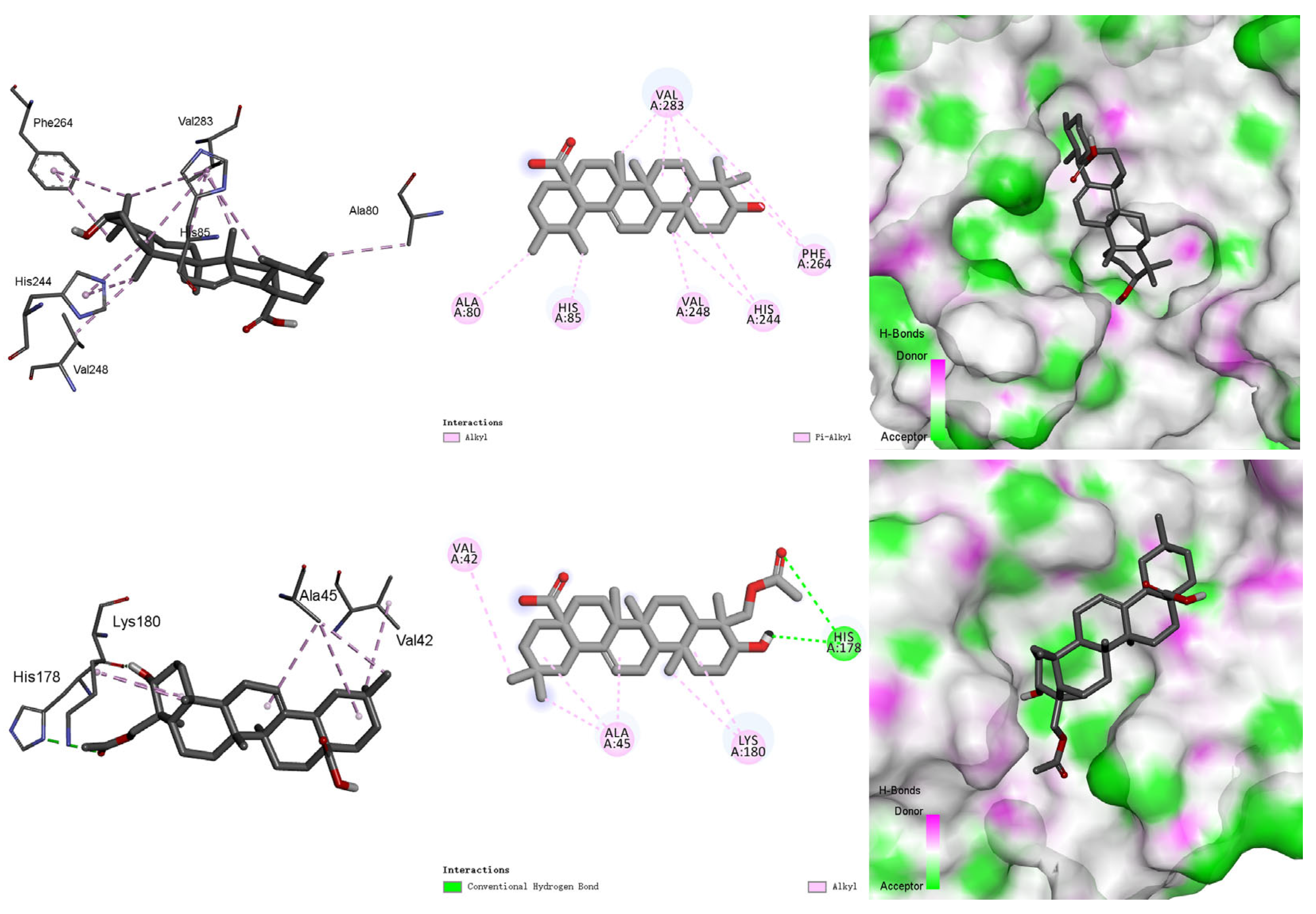
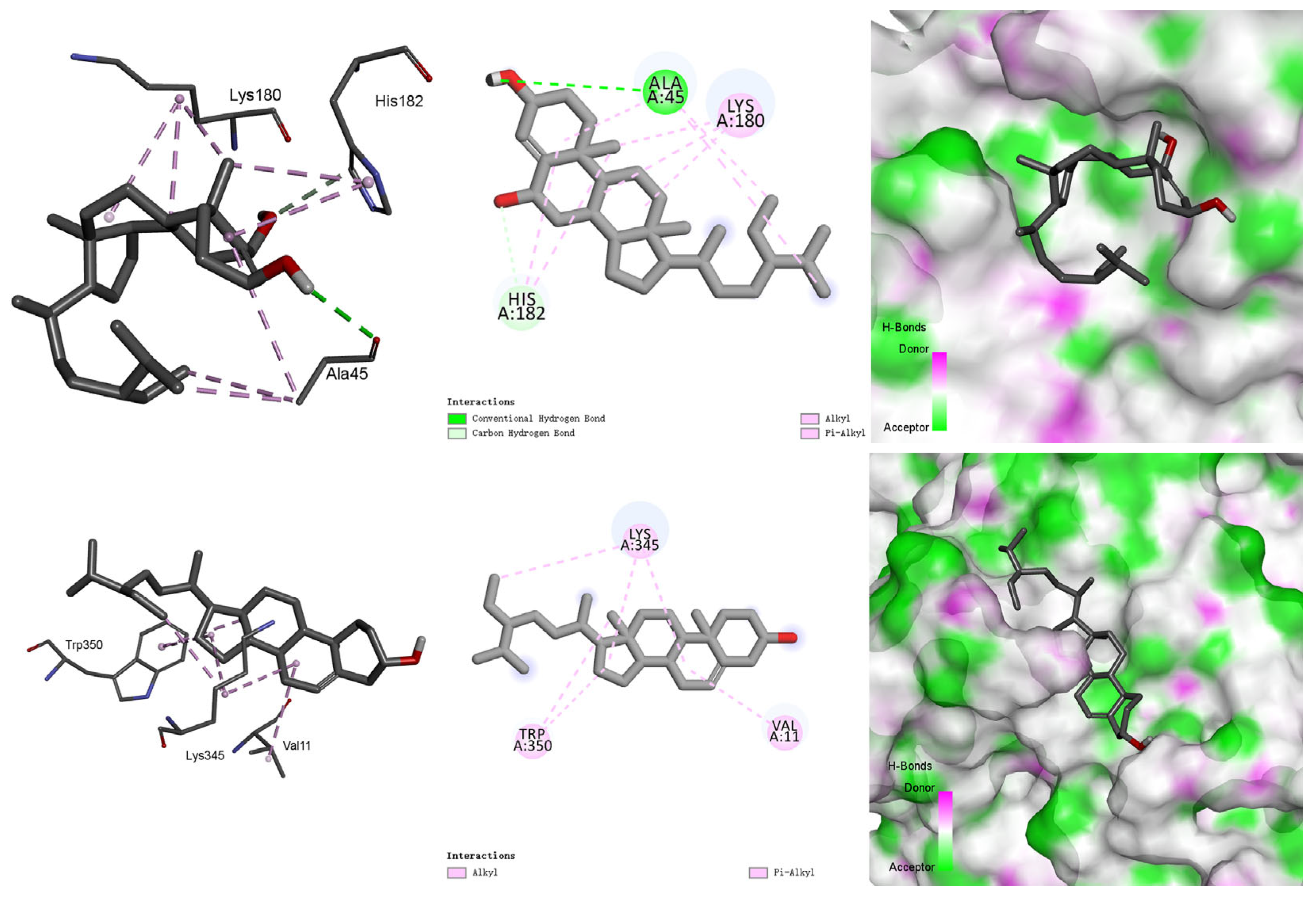
3.5. Molecular Docking Study

Molecular docking calculations were conducted to decipher the binding mechanism between compounds 16 and the mushroom tyrosinase (PDB ID: 2Y9X) [41]. The images of docked complexes and molecular surfaces, namely 2D and 3D interactive plots for these compounds with tyrosinase, were shown in Figure 3, Figure 4 and Figure 5.
As illustrated in Figure 3, compound 1 engaged in alkyl interactions with amino acid residues Val283, Ala80, and Val248, and also formed π–alkyl stacking interactions with His85, His244, and Phe264 in the mushroom tyrosinase. The carbonyl of the acetyl group at C-24 of 2 formed a hydrogen bond with amino acid residue His178, while its hydroxyl group also established a hydrogen bond with His178. Furthermore, 2 exhibited π-alkyl stacking interactions with Val42, Lys180, and Ala45 in the protein cavity [42]. The aforementioned illustration revealed that the acetyl group at C-24 of 2 had an important impact on the enhancement of tyrosinase inhibitory activity.
As shown in Figure 4, the interactions between compound 3 and the mushroom tyrosinase involved hydrogen bonds and alkyl and π–alkyl interactions. The π–alkyl interaction with the amino acid residue His182 and alkyl stacking interactions with Lys180 and Ala45 were observed. Furthermore, the hydroxyl groups at C-3 and C-6 of 3 formed hydrogen bonds with Ala45 and His182, respectively. Compound 4 exhibited π–alkyl interaction with the Trp350 and alkyl stacking interactions with Val11 and Lys345. The above analysis indicated the benefits brought by substituting the hydroxyl group at C-6 of 3.
As for compound 5, its methoxy and hydroxyl groups were involved in hydrogen bonds with Asn253, Asn230, and Glu226. Furthermore, 5 also engaged in an π–π stacked interaction with Trp227, as well as an π–alkyl stacking interaction with Ala252, which were both located in the active site (Figure 5). Compound 6 also formed hydrogen bonds between its methoxy and hydroxyl groups and Asn253, Asn230, and Glu226. Additionally, it established an π–alkyl interaction between its benzene rings and Trp227. The above study revealed that hydrogen bonds always formed regardless of the positions of the methoxy and hydroxyl groups at the benzene ring.

4. Conclusions

The present research on the chemical constituents of the potential ornamental plant O. chinensis led to the isolation and identification of a series of compounds with diverse structural features, including two triterpenes (1 and 2), two steroids (3 and 4), two anthraquinones (5 and 6), one alkaloid (7), and one coumarin (8). All of them were first found in this species. Components 2, 3, 5, and 6 were obtained from the genus Ophiorrhiza for the first time, which could serve as markers for this Chinese endemic species chemotaxonomically. In the tyrosinase inhibitory bioassay, compounds 16 demonstrated moderate tyrosinase inhibitory properties with IC50 values ranging from 25.7 μM to 68.1 μM. This study not only disclosed the chemical diversity and pharmacological properties of the species O. chinensis, but also provided the basis for the future utilization of this species as a source of health care or cosmetical products.

Supplementary Materials

The following supporting information can be downloaded at: https://www.mdpi.com/article/10.3390/agronomy14081872/s1, Figures S1–S16: The 1H and 13C NMR spectra of compounds 18; Figures S17–S24: The key 1H and 13C NMR spectral data (pink and blue texts, respectively) of compounds 18; Tables S1–S8: The comparison of both 1H and 13C NMR data of compounds 18 with those in the literature.

Author Contributions

Conceptualization, L.-F.L.; methodology, Q.B. and Z.-Y.G.; software, Z.-Y.G.; validation, Q.B. and Z.-Y.G.; formal analysis, Q.B. and Z.-Y.G.; investigation, Q.B. and Z.-Y.G.; data curation, Q.B. and Z.-Y.G.; writing—original draft preparation, Q.B.; writing—review and editing, L.-F.L.; visualization, Q.B. and Z.-Y.G.; supervision, L.-F.L.; project administration, L.-F.L.; funding acquisition, L.-F.L. All authors have read and agreed to the published version of the manuscript.

Funding

This research was funded by the Natural Science Foundation of Changsha (No. kq2402254).

Data Availability Statement

Data is contained within the article.

Acknowledgments

We thank Lei Wu at CSUFT for the sample collection and taxonomic identification of the plant.

Conflicts of Interest

The authors declare no conflicts of interest.

References

  1. Ding, H. The valuable utilizationand reproductive cultivation techniques of Ophiorrhiza japonica. S. China Agric. 2021, 15, 24–25. [Google Scholar] [CrossRef]
  2. Chinese Materia Medica Committee of the State Administration of Traditional Chinese Medicine. Zhushacao, Shengencao, Duanxiaoshegencao, Diguicao. In Chinese Materia Medica; Shanghai Science and Technology Press: Shanghai, China, 1999; Volume 18, pp. 457–460. [Google Scholar]
  3. Zhong, L.-J.; Liang, L.-F.; Chen, J.-K.; Bu, Q.; Xu, M.-J. Research progress on chemical constituents and pharmacological activities from the genus Ophiorrhiza. Chin. Pharm. J. 2022, 57, 1221–1231. [Google Scholar] [CrossRef]
  4. Anil, J.J.; Renjith, R.; Sabulal, B. Secondary metabolites from Ophiorrhiza. Nat. Prod. J. 2018, 8, 248–267. [Google Scholar] [CrossRef]
  5. Taher, M.; Shaari, S.S.; Susanti, D.; Arbain, D.; Zakaria, Z.A. Genus Ophiorrhiza: A review of its distribution, traditional uses, phytochemistry, biological activities and propagation. Molecules 2020, 25, 2611. [Google Scholar] [CrossRef]
  6. Flora of China Editorial Committee. Ophiorrhiza Chinensis H. S. Lo. In Flora of China; Science Press: Beijing, China, 1999; Volume 71, p. 168. [Google Scholar]
  7. Shi, J.; Xu, M.; Zhou, C.; Zou, Q.; Zhang, X. Comparative study on the content of chlorogenic acid in Ophiorrhiza chinensis from different organs. Hunan Zhongyiyao Daxue Xuebao 2016, 36, 28–30. [Google Scholar] [CrossRef]
  8. Yang, S.; Jiang, J.; Zhou, A.; Zhou, Y.; Ye, W.; Cao, D.-S.; Yang, R. Substrate-photocaged enzymatic fluorogenic probe enabling sequential activation for light-controllable monitoring of intracellular tyrosinase activity. Anal. Chem. 2020, 92, 7194–7199. [Google Scholar] [CrossRef]
  9. Parvez, S.; Kang, M.; Chung, H.-S.; Bae, H. Naturally occurring tyrosinase inhibitors: Mechanism and applications in skin health, cosmetics and agriculture industries. Phytother. Res. 2007, 21, 805–816. [Google Scholar] [CrossRef]
  10. Chang, T.-S. An updated review of tyrosinase inhibitors. Int. J. Mol. Sci. 2009, 10, 2440–2475. [Google Scholar] [CrossRef]
  11. Baber, M.A.; Crist, C.M.; Devolve, N.L.; Patrone, J.D. Tyrosinase inhibitors: A perspective. Molecules 2023, 28, 5762. [Google Scholar] [CrossRef]
  12. Kim, H.-D.; Choi, H.; Abekura, F.; Park, J.-Y.; Yang, W.-S.; Yang, S.-H.; Kim, C.-H. Naturally-occurring tyrosinase inhibitors classified by enzyme kinetics and copper chelation. Int. J. Mol. Sci. 2023, 24, 8226. [Google Scholar] [CrossRef]
  13. Ullah, S.; Son, S.; Yun, H.Y.; Kim, D.H.; Chun, P.; Moon, H.R. Tyrosinase inhibitors: A patent review (2011-2015). Expert. Opin. Ther. Pat. 2016, 26, 347–362. [Google Scholar] [CrossRef] [PubMed]
  14. Bonesi, M.; Xiao, J.; Tundis, R.; Aiello, F.; Sicari, V.; Loizzo, R.M. Advances in the tyrosinase inhibitors from plant source. Curr. Med. Chem. 2019, 26, 3279–3299. [Google Scholar] [CrossRef]
  15. Li, J.; Li, C.; Peng, X.; Li, S.; Liu, B.; Chu, C. Recent discovery of tyrosinase inhibitors in traditional Chinese medicines and screening methods. J. Ethnopharmacol. 2023, 303, 115951. [Google Scholar] [CrossRef]
  16. Bu, Q.; Jin, Y.; Xu, M.-J.; Wu, L.; Liang, L.-F. Structurally diverse metabolites from the Ophiorrhiza japonica Bl. and their antioxidant activities in vitro and PPARα agonistic activities in silico. Molecules 2022, 27, 5301. [Google Scholar] [CrossRef] [PubMed]
  17. Si, X.-y.; Wang, Y.-f.; He, R.-j.; Yang, B.-y.; Huang, Y.-l. Chemical constituents from leaves of Castanopsis lamontii and its tyrosinase inhibitory activities. Chin. Tradit. Herb. Drugs 2023, 54, 6214–6219. [Google Scholar]
  18. Souza, P.M.; Elias, S.T.; Simeoni, L.A.; de Paula, J.E.; Gomes, S.M.; Guerra, E.N.S.; Fonseca, Y.M.; Silva, E.C.; Silveira, D.; Magalhães, P.O. Plants from Brazilian Cerrado with potent tyrosinase inhibitory activity. PLoS ONE 2012, 7, e48589. [Google Scholar] [CrossRef]
  19. Luo, P.; Deng, Z.; Wang, J.; Shen, Z.; Pan, W. Chemical constituents from Primulina linearifolia (W. T. Wang) Yin Z. Wang. Guangxi Sci. 2023, 30, 883–890. [Google Scholar] [CrossRef]
  20. Pereda-Miranda, R.; Delgado, G.; de Vivar, A.R. New triterpenoids from Salvia nicolsoniana. J. Nat. Prod. 1986, 49, 225–230. [Google Scholar] [CrossRef]
  21. Kalinovskii, A.I. 13C NMR spectroscopy of oleanane derivatives. Chem. Nat. Compd. 1992, 28, 1–22. [Google Scholar] [CrossRef]
  22. Wang, J.; Fei, J.-d.; Ding, G.; Li, R.-t.; Yu, C.-y.; Zou, Z.-m. Chemical constituents in the petroleum ether extract of the whole herb of Flickingeria fimbriata. Chin. Pharm. J. 2013, 48, 1343–1346. [Google Scholar]
  23. Kim, J.S.; Kim, J.C.; Shim, S.H.; Lee, E.J.; Jin, W.Y.; Bae, K.; Son, K.H.; Kim, H.P.; Kang, S.S.; Chang, H.W. Chemical constituents of the root of Dystaenia takeshimana and their anti-inflammatory activity. Arch. Pharm. Res. 2006, 29, 617–623. [Google Scholar] [CrossRef] [PubMed]
  24. Wu, K.-s.; Zhang, K.; Tan, G.-s.; Zeng, G.-y.; Zhou, Y.-j. Studies on constituents of Oldenladia diffusa. Chin. Pharm. J. 2005, 40, 817–819. [Google Scholar]
  25. Fraga, B.M.; Quintana, N.; Díaz, C.E. Anthraquinones from natural and transformed roots of Plocama pendula. Chem. Biodivers. 2009, 6, 182–192. [Google Scholar] [CrossRef] [PubMed]
  26. Mahibalan, S.; Rao, P.C.; Khan, R.; Basha, A.; Siddareddy, R.; Masubuti, H.; Fujimoto, Y.; Begum, A.S. Cytotoxic constituents of Oldenlandia umbellata and isolation of a new symmetrical coumarin dimer. Med. Chem. Res. 2016, 25, 466–472. [Google Scholar] [CrossRef]
  27. Ying, Y.-M.; Shan, W.-G.; Liu, W.-H.; Zhan, Z.-J. Alkaloids and nucleoside derivatives from a fungal endophyte of Huperzia serrata. Chem. Nat. Compd. 2013, 49, 184–186. [Google Scholar] [CrossRef]
  28. Li, Y.-H.; Luo, F.; Peng, S.-L.; Liang, J.; Ding, L.-S. A new dihydroisocoumarin from the rhizomes of Notopterygium forbesii. Nat. Prod. Res. 2006, 20, 860–865. [Google Scholar] [CrossRef]
  29. Mahato, S.B.; Kundu, A.P. 13C NMR spectra of pentacyclic triterpenoids—A compilation and some salient features. Phytochemistry 1994, 37, 1517–1575. [Google Scholar] [CrossRef]
  30. Kitajima, M.; Fujii, N.; Yoshino, F.; Sudo, H.; Saito, K.; Aimi, N.; Takayama, H. Camptothecins and two new monoterpene glucosides from Ophiorrhiza liukiuensis. Chem. Pharm. Bull. 2005, 53, 1355–1358. [Google Scholar] [CrossRef]
  31. Chattopadhyay, D.; Das, S.; Mandal, A.B.; Arunachalam, G.; Bhattacharya, S.K. Evaluation of analgesic and anti-inflammatory activity of Ophiorrhiza nicobarica, an ethnomedicine from Nicobar Islands, India. Orient. Pharm. Exp. Med. 2007, 7, 395–408. [Google Scholar] [CrossRef]
  32. Huang, Y.-L.; Xiang, W.; Song, Q.-S. Chemical constituents of Ophiorrhiza rosea. Chin. Tradit. Herb. Drugs 2009, 40, 519–521. [Google Scholar] [CrossRef]
  33. Huang, Y.-L.; Li, J.; Xiang, W.; Song, Q.-S. Chemical constituents of Ophiorrhiza austroyunnanensis. Nat. Prod. Res. Dev. 2011, 23, 652–654. [Google Scholar] [CrossRef]
  34. Agarwal, K.P.; Dhar, M.M. Chemical examination of Ophiorrhiza mungos. J. Sci. Ind. Res. 1959, 18B, 114–115. [Google Scholar]
  35. Fujita, E.; Sumi, A. Studies on the constituents of Ophiorrhiza japonica Bl. Yakugaku Zasshi 1967, 87, 1153–1155. [Google Scholar] [CrossRef] [PubMed]
  36. Li, W.-F.; Song, Q.-S.; Xiang, W.; Wang, Y.-L. Study on chemical constituents and antibacterial activity of Ophiorrhiza cantoniensis Hance. Nat. Prod. Res. Dev. 2014, 26, 683–686. [Google Scholar] [CrossRef] [PubMed]
  37. Kitajima, M.; Nakamura, M.; Takayama, H.; Saito, K.; Stöckigt, J.; Aimi, N. Constituents of regenerated plants of Ophiorrhiza pumila; formation of a new glycocamptothecin and predominant formation of (3R)-deoxypumiloside over (3S)-congener. Tetrahedron Lett. 1997, 38, 8997–9000. [Google Scholar] [CrossRef]
  38. Aimi, N.; Murakami, H.; Tsuyuki, T.; Nishiyama, T.; Sakai, S.-i.; Haginiwa, J. Hydrolytic degradation of β-carboline-type monoterpenoid glucoindole alkaloids: A possible mechanism for harman formation in Ophiorrhiza and related rubiaceous plants. Chem. Pharm. Bull. 1986, 34, 3064–3066. [Google Scholar] [CrossRef]
  39. Hamzah, A.S.; Arbain, D.; Mahyudin; Sargent, M.V.; Lajis, N.H. The alkaloids of Ophiorrhiza communis and O. tomentosa. Pertanika J. Sci. Technol. 1994, 2, 33–38. [Google Scholar]
  40. Nonato, M.G.; Truscott, R.J.W.; Carver, J.A.; Hemling, M.E.; Garson, M.J. Glucoindole alkaloids from Ophiorrhiza acuminata. Planta Med. 1995, 61, 278–280. [Google Scholar] [CrossRef]
  41. Ashraf, Z.; Rafiq, M.; Nadeem, H.; Hassan, M.; Afzal, S.; Waseem, M.; Afzal, K.; Latip, J. Carvacrol derivatives as mushroom tyrosinase inhibitors; synthesis, kinetics mechanism and molecular docking studies. PLoS ONE 2017, 12, e0178069. [Google Scholar] [CrossRef]
  42. Ismaya, W.T.; Rozeboom, H.J.; Weijn, A.; Mes, J.J.; Fusetti, F.; Wichers, H.J.; Dijkstra, B.W. Crystal structure of Agaricus bisporus mushroom tyrosinase: Identity of the tetramer subunits and interaction with tropolone. Biochemistry 2011, 50, 5477–5486. [Google Scholar] [CrossRef]
Figure 1. The summary of key experimental procedures of compounds 18.
Agronomy 14 01872 g001
Figure 2. The chemical structures and names of the isolates 18 from O. chinensis.
Agronomy 14 01872 g002
Figure 3. The binding modes between compounds 1 and 2 and the mushroom tyrosinase (PDB: 2Y9X).
Agronomy 14 01872 g003
Figure 4. The binding modes between compounds 3 and 4 and the mushroom tyrosinase (PDB: 2Y9X).
Agronomy 14 01872 g004
Figure 5. The binding modes between compounds 5 and 6 and the mushroom tyrosinase (PDB: 2Y9X).
Agronomy 14 01872 g005
Table 1. The identification information of compounds 18 from O. chinensis.
CompoundsStructural TypesNamesReferences
1ursane-type triterpeneursolic acid[19]
2oleane-type triterpene24-acetoxy-3α-hydroxyolean-12-en-28-oic[20,21]
3stigmastane-type steroidstigmast-4-ene-3β,6β-diol[22]
4stigmastane-type steroidβ-stiosterol[23]
5anthraquinoneanthragallol 1,3-dimethyl ether[24,25]
6anthraquinoneanthragallol 1,2-dimethyl ether[26]
7aromatic alkaloidharmane[27]
8coumarinscopoletin[28]
Table 2. The tyrosinase inhibitory results of compounds 16 and positive control vitamin C.
CompoundsIC50 (μM)
143.8 ± 0.1
225.7 ± 1.3
335.5 ± 2.0
465.8 ± 7.8
568.1 ± 1.4
667.4 ± 4.4
1 Vitamin C3.6 ± 0.3
1 Positive control.
Disclaimer/Publisher’s Note: The statements, opinions and data contained in all publications are solely those of the individual author(s) and contributor(s) and not of MDPI and/or the editor(s). MDPI and/or the editor(s) disclaim responsibility for any injury to people or property resulting from any ideas, methods, instructions or products referred to in the content.

