Article

A Phytochrome-Interacting Factor Gene CaPIF7a Positively Regulates the Defense Response against Phytophthora capsici Infection in Pepper (Capsicum annuum L.)

1
Jiangxi Provincial Key Laboratory for Postharvest Storage and Preservation of Fruits & Vegetables, College of Agronomy, Jiangxi Agricultural University, Nanchang 330045, China
2
Nanchang Academy of Agricultural Sciences, Nanchang 330038, China
3
Key Laboratory of Crop Physiology, Ecology and Genetic Breeding, Ministry of Education, College of Bioscience and Bioengineering, Jiangxi Agricultural University, Nanchang 330045, China
*
Authors to whom correspondence should be addressed.
Agronomy 2024, 14(9), 2035; https://doi.org/10.3390/agronomy14092035
Submission received: 11 August 2024 / Revised: 31 August 2024 / Accepted: 4 September 2024 / Published: 6 September 2024

Abstract

:
Phytochrome-interacting factor (PIF) is a subfamily of the basic helix–loop–helix (bHLH) transcription factors (TFs) and plays key roles in plant responses to diverse biotic and abiotic stresses. In this work, a PIF gene named CaPIF7a was cloned and its role in the regulation of pepper’s resistance to Phytophthora capsici infection (PCI) was studied. The cloned CaPIF7a gene has a CDS length of 1383 bp, encodes a hydrophilic protein containing bHLH and APB characteristic domains, and subcellular localization results showed that CaPIF7a was located in the nucleus. Expression analysis showed that CaPIF7a gene has the highest expression level in leaf, and its expression was regulated under PCI and salicylic acid (SA) treatment. Silencing of CaPIF7a in pepper plants by virus-induced gene silencing (VIGS) reduces the resistance of pepper to PCI, with decreased expression of SA-responsive and SA-biosynthesis genes and obviously decreased SA content. DNA affinity purification sequencing (DAP-seq) was employed to identify the potential targets of CaPIF7a, and yeast one-hybrid (Y1H) verified that CaPIF7a could regulate the expression of CaHY5 by binding its promoter. These findings indicated that CaPIF7a might be a key modulator in plant immune response and presented a possible regulatory network of CaPIF7a in PCI.

1. Introduction

Pepper (Capsicum annuum L.) is an annual herbaceous plant that is rich in various vitamins and minerals and is an essential vegetable crop in people’s daily lives. However, the Phytophthora blight caused by Phytophthora capsici is one of the most harmful soil-borne diseases in the pepper production process across the globe. P. capsici is a destructive oomycete pathogen that can infect the leaves, stems, and fruits of the pepper plants, and in severe cases, it can cause fruit decay, whole plant wilting, damping-off, and even death, making an obvious decline in yield and quality of the host plants. Therefore, identification of resistance genes in pepper will provide valuable information for further breeding to strength of the defense response and enhance the yield and quality under P. capsici infection (PCI). To cope with pathogen infection, plants have evolved a series of defense mechanisms along with inducible innate immunity to defend themselves against pathogen infection. Amongst them, transcription factors (TFs) play significant roles in monitoring inducible innate immunity and combating the impact of pathogens through controlling transcriptional regulation of defense genes.
Among the vast TFs, phytochrome-interacting factor (PIF) is a subfamily of the basic helix–loop–helix (bHLH) family, which is well-known to work in interacting with the red (R) and far-red (FR) light photoreceptors phytochrome (phy) to mediate multiple developmental and physiological processes in plants. Besides the conserved bHLH domain, PIFs harbor an N-terminal active phytochrome B (phyB)-binding (APB) domain and/or an active phytochrome A (phyA)-binding (APA) domain, which are important for phyB- and phyA-specific binding. AtPIF3 was the first identified PIF member by using the C-terminus of phyB as bait, and AtPIF3 can physically interact with phyB only upon its activated form (Pfr) [1]. Subsequently, seven other PIFs were identified in Arabidopsis, and all AtPIFs were found to contain the APB motif, but only AtPIF1 and AtPIF3 contain the APA motif.
Recently, the roles of PIFs were comprehensively characterized in Arabidopsis and other plants, and these reports indicated that PIFs are conservatively worked in the downstream targets of phytochromes and involved in plant growth and development in the light signal transduction pathway [2,3]. For example, activated phyB regulates the phosphorylation and degradation of PIFs and results in inactivation of PIF functions; the pif1 pif3 pif4 pif5 quadruple mutant (pifq, disruption of AtPIF1, AtPIF3, AtPIF4, and AtPIF5) resulted in a phenotype of constitutive photomorphogenesis under dark conditions, suggesting that these PIFs play redundant roles in photomorphogenesis and skotomorphogenesis [4,5,6]. phyB-mediated PIF activity also plays vital roles in thermomorphogenesis and shade avoidance response. For example, phyB converts to its inactive form (Pr) under high temperatures, thus relieving the repression of the AtPIF4 and target gene activation to mediate AtPIF4-induced cell elongation. In the shade, the low R/FR ratio also leads to the inactivation of phyB, thus relieving and allowing unbound AtPIF7 to activate the expression of shade-related genes [7]. PIFs are also found to be widely involved in other physiological and biochemical processes of plants, such as leaf senescence [8,9], flowering time regulation [10,11], fruit development [12], seed size elongation [13], carotenoid and sterol biosynthesis [14,15].
Multiple studies have shown that plant PIFs have important roles in responses to various biotic stresses. For example, sweet potato (Ipomoea batatas) IbPIF3.1 was demonstrated to be involved in Fusarium wilt resistance in transgenic tobacco plants [16]. In Arabidopsis, AtPIFs are found to participate in defense responses towards fungi and bacteria [17,18]. AtPIF1, AtPIF3, AtPIF4 and AtPIF5 have redundant functions over plant defense response to Botrytis cinerea [18]. AtPIF3 was reported to be involved in plant immunity to Pseudomonas syringae pv. tomato (Pto) DC3000 by negatively regulating defense gene expression [19]. AtPIF4-mediated thermosensory signaling plays an important role in the suppression of defense to Pto DC3000 by elevated temperature, suggesting that AtPIF4 can coordinate thermosensory growth and immunity in Arabidopsis [20]. NbPIF4 activates the expression of RDR6 and AGO1 in the RNAi pathway and positively regulates near-infrared-mediated plant defense against viruses [21]. CmPIF8 could directly and indirectly interact with the promoter of CmACS10 to repress its expression, and thus regulates red light-induced ethylene biosynthesis to compromise the resistance of melon seedlings to powdery mildew [22]. Therefore, PIFs play key roles in the regulation of plant responses to pathogen invasion by being associated with a set of downstream target genes.
Although the biological roles of PIFs have been studied in different plants, there have been few reports on the functional study of pepper PIF genes so far. Our previous research has shown that the pepper genome contained six CaPIF genes, and CaPIF8 plays positively roles in cold and salt tolerance [23], but little is known about whether the CaPIF genes participate in the process of pepper resistance to PCI. In this study, we isolated the CaPIF7a gene and its expression levels in different tissues and response to PCI were determined with qRT-PCR. In addition, we explored the function of CaPIF7a in plant defense response to P. capsici by virus-induced gene silencing (VIGS) and DNA affinity purification followed by sequencing (DAP-seq). This study provides a foundation for revealing the function of PIF genes in plant defense response to P. capsici.

2. Materials and Methods

2.1. Plant Material and Growth Conditions

The seeds of pepper (Capsicum annuum L.) cultivar “Hangjiao 12” were sown in plastic pots and grown in a growth chamber under the conditions of a 12 h/12 h light cycle, a day/night temperature of 22/20 °C, and a light intensity of 200 μmol·m−2·s−1. During the fruiting period, different tissues including root, stem, leaf, flower, pulp, and placenta were taken from 5 individual plants, and each tissue sample was divided into 3 parts for RNA isolation. Harvested samples were frozen in liquid nitrogen and immediately stored at −80 °C for future use.

2.2. Cloning and Sequence Analyses of CaPIF7a of Pepper

Total RNA was isolated from “Hangjiao 12” leaves using a MolPure®Plant RNA Kit, and about 1 μg total RNA was reverse-transcribed using Hifair®III 1st Strand cDNA Synthesis SuperMix for qPCR (Yisheng, Shanghai, China) according to the manufacturer’s instructions. Using the cDNA of the leaves as a template, the coding sequence (CDS) of CaPIF7a was amplified by 2× Phanta Max Master Mix (Vazyme, Nanjing, China). The RT-PCR program was as follows: pre-denaturation at 95 °C for 3 min; 35 cycles of 95 °C denaturation for 15 s, 60 °C annealing for 15 s, and 72 °C extension for 1 min. The PCR products were electrophoretic on 1% agarose gel and subsequently sequenced. The protein structure and functional domains of CaPIF7a were analyzed by a SOPMA secondary structure prediction tool (https://npsa-prabi.ibcp.fr/cgi-bin/npsa_automat.pl?page=npsa_sopma.html/, accessed on 20 May 2024) and SMART tool (http://smart.embl-heidelberg.de/, accessed on 20 May 2024). The cis-acting elements on the CaPIF7a promoter were analyzed using PlantCARE (http://bioinformatics.psb.ugent.be/webtools/plantcare/html/, accessed on 20 May 2024). To predict the subcellular localization of CaPIF7a, its amino acid sequence was analyzed using Plant-mPLoc server (http://www.csbio.sjtu.edu.cn/bioinf/plant-multi/, accessed on 20 May 2024).

2.3. Subcellular Localization Assay

The CDS without stop codon of CaPIF7a were cloned into the PC1300S-GFP vector to yield CaPIF7a-GFP. And then, the plasmids of CaPIF7a-GFP and empty vector PC1300S-GFP were transformed into Arabidopsis protoplasts with nuclear markers and cultured under low light for 8–10 h. The subcellular localization of CaPIF7a was observed under a confocal laser-scanning microscope (Nikon C2-ER, Tokyo, Japan).

2.4. Quantitative Real-Time PCR (qRT-PCR)

For qRT-PCR analysis of CaPIF7a in response to P. capsici infection (PCI), total RNA extraction was conducted at 0, 24, 48, 72, 90 and 120 h after inoculation. For qRT-PCR analysis of CaPIF7a in response to SA treatment, the pepper leaves sprayed with SA solution were sampled at four time points (0, 1, 3, 12, and 24 h). qRT-PCR was performed in triplicate using Hieff qPCR SYBR Green Master Mix (Yisheng, Shanghai, China) with special primers listed in Table S1. The relative expression levels of genes were analyzed using the 2−∆∆Ct method by normalizing the internal reference actin gene (CA12g08730/Capana12g001934).

2.5. Virus-Induced Gene Silencing (VIGS) of CaPIF7a in Pepper

Specific primers were used to amplify the partial cDNA fragment of CaPIF7a (360 bp) and CaPDS (690 bp) genes for VIGS, which were designed and detected the specificity in the VIGS tool of the SGN database (https://vigs.solgenomics.net/, accessed on 20 May 2024). The target gene PCR products were obtained and inserted into the TRV2 vector with restriction endonucleases Xba I and Apa I using homologous recombination ligase (Yisheng, Shanghai, China). Subsequently, TRV2:CaPIF7a, TRV2:00 (control), and TRV2:CaPDS (phytoene desaturase gene, positive control as a photobleaching phenotype) were transformed into Agrobacterium tumefaciens strain GV3101 and injected into 2-week-old pepper plants’ cotyledons by a needless syringe according to the protocol in our previous study [24]. After 48 h of dark cultivation at 18 °C, the Agrobacterium-injected pepper plants were placed in a growth chamber under the conditions of a 12 h/12 h light cycle, a day/night temperature of 22/20 °C, and a light intensity of 200 μmol·m−2·s−1. The silencing efficiency of TRV2:CaPIF7a was analyzed by qRT-PCR when the TRV2:CaPDS pepper plants exhibited the photobleaching phenotype.

2.6. Pathogens Growth and Inoculation Procedures

The P. capsici isolate JX202105 was grown on potato dextrose agar (PDA) medium (containing 200 g of potato, 20 g of glucose, and 17 g of agar per 1 L of medium) in a dark incubator at 28 °C for 3–5 days for activation. Then, the activated mycelia were cultured in a 90 mm culture dish containing 10% V8 medium (containing 100 mL of V8 vegetable juice, 1 g of CaCO3, and 17 g of agar per 1 L of medium) at 28 °C in the dark for 10 days to induce the production of zoospores. And then, the mycelia of P. capsici were washed 3 times at an interval of 30 min using sterilized water and then incubated at 25 °C until zoospores were released; the concentration of zoospores was adjusted to 1 × 10⁵ spores per mL. For the P. capsici infection in pepper plants, 3 mL of zoospore suspension was injected into the basal stem (1 cm away of root) of pepper seedlings at the 5-leaf stage, and the injected seedlings were grown in a growth chamber at 25 °C with a 12 h light/12 h dark photoperiod. Then, 3 mL distilled water was used as a control, and a resistance assessment was conducted using the injury index of disease. P. capsici infection in detached pepper leaves was performed according to our previous study [24].

2.7. Determination of Resistance Indexes

To determine the accumulation of hydrogen peroxide (H2O2) and superoxide (O2−) of detached pepper leaves after PCI, the 3,3ʹ-diaminobenzidine (DAB) staining and nitro-blue tetrazolium (NBT) staining were carried out using the methods on the basis of our previous study [24]. Free SA levels were determined using High-Performance Liquid Chromatography (HPLC, Waters 2695, Milford, MA, USA).

2.8. DNA Affinity Purification Followed by Sequencing (DAP-Seq)

DAP-seq was conducted using the methods of the previous studies [25,26]. Genomic DNA was extracted from leaves of pepper plants, and the genomic DNA library was prepared. Simultaneously, the CDS of CaPIF7a was cloned into the pFN19K HaloTag T7 SP6 Flexi expression vector, and Halo-CaPIF7a fusion protein was expressed with the Promega TNT SP6 wheat germ master mix kit protein according to the manufacturer’s instructions. With a mixed protein and genomic DNA library, sequencing of the protein-bound DNA was performed by IGENEBOOK Biotechnology Co., Ltd. (Wuhan, China). Data analysis with a comparison of the DAP-seq reads against reference pepper “zunla 1” genome sequences was carried out with the BOWTIE2 software (version 2.3.5). The MACS2 algorithm at a threshold of q-value < 0.05 was used to detect the DAP-seq peaks [27], and the MEME-ChIP tool was employed to identify motif sequences with default parameters [28].

2.9. Yeast One-Hybrid (Y1H) Assay

For Y1H assay, the promoter of CaHY5 was cloned into pAbAi vector as the bait, then linearized by BstB I/Bbs I digestion, and transformed into Y1HGold to generate reporter strains. The CDS of CaPIF7a was cloned into the pGADT7 vector as prey, and then transferred into the bait strain and grown on SD/-Ura plates for 3 days at 30 °C; the positive clones were then transferred onto the SD/-Ura/-Leu/AbA plates at 30 °C for 3–5 days to check the interactions using the yeast cell growth status. The primers used in the Y1H assay are listed in Supplementary Table S1.

2.10. Statistical Analysis

All data were analyzed by Duncan’s test in SPSS software (version 19.0), and different letters were employed to indicate the statistical significance of the data (p < 0.05).

3. Results

3.1. Cloning and Sequence Analyses of Pepper CaPIF7a Gene and Its Encoded Protein

In our previous study, a total of six CaPIF genes (CaPIF1, CaPIF3, CaPIF4, CaPIF7a, CaPIF7b, and CaPIF8) were identified in the pepper genome database. PCR was used to amplify the CDS of CaPIF7a by using cDNA from pepper leaf tissues as a template. Sequencing of a cloned CaPIF7a fragment showed that it was 1383 bp in length (Figure 1A), encoding a 460-amino acid protein with a calculated molecular weight of 50.68 kDa and isoelectric point of 7.31. In addition, the grand average of hydropathicity of CaPIF7a was −0.711, indicating that it was a hydrophilic protein. Sequence analysis via SMART tool revealed that CaPIF7a harbored the typical bHLH domain for DNA binding and APB domain for phyB-specific binding, but not the APA domain (Figure 1A). The predicted secondary structure results showed that CaPIF7a contains 61.96% random coils, 31.96% alpha helices, 4.35% extend strands and 1.74% beta turns (Figure 1B). These findings provide key clues for further exploration of the structure and function of CaPIF7a.

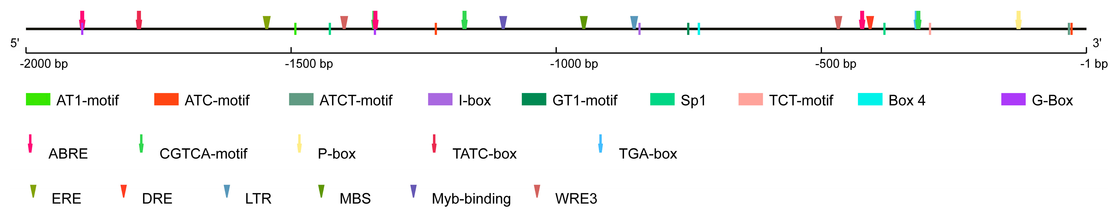
3.2. Promoter Analysis of the Pepper CaPIF7a Gene

To study how the CaPIF7a gene is regulated, the 2,000 bp upstream region sequence of its CDS was obtained and analyzed by PlantCARE tool. As shown in Figure 2, three types of cis-acting regulatory elements were identified, including light-, hormone-, and stress-related elements. Amongst these cis-elements, light-related elements were the most abundant in the promoter region of CaPIF7a, implying the possible role of CaPIF7a in light response. Six types of hormone-related cis-elements, including ABRE (abscisic acid-responsive element), CGTCA-motif (MeJA-responsive element), ERE (ethylene-responsive element), TGA-box (auxin-responsive element), P-box and TATC-box (gibberellin-responsive element) were mapped onto the promoter region. In addition, the promoter region of CaPIF7a harbored four types of stress-related cis-elements, including DRE (dehydration-responsive element), LTR (low-temperature responsive), WRE3 (wounding responsive), Myb-binding site and MBS (drought-inducibility responsive) (Figure 2). The cis-elements identified in this analysis indicated that these may be involved in light, hormone and stress responses.

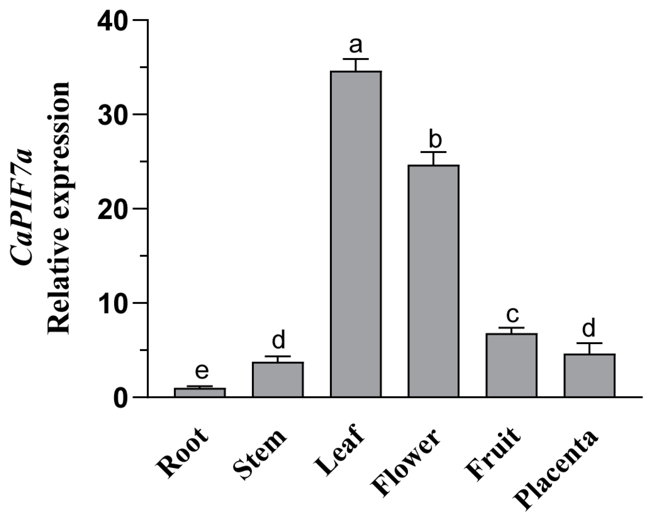
3.3. Expression Profiles of CaPIF7a in Different Tissues

Our previous study showed that CaPIF7a displayed a higher expression level in the leaves than in other tested tissues on the basis of publicly available RNA-seq data [23]. To further understand CaPIF7a expression characteristics, qRT-PCR was used to examine the expression profiles of CaPIF7a in different tissues. As a result, CaPIF7a was expressed in all the tested tissues, with the highest expression in leaf, followed by flower and fruit, with the lowest expression in root, stem and tendril (Figure 3).

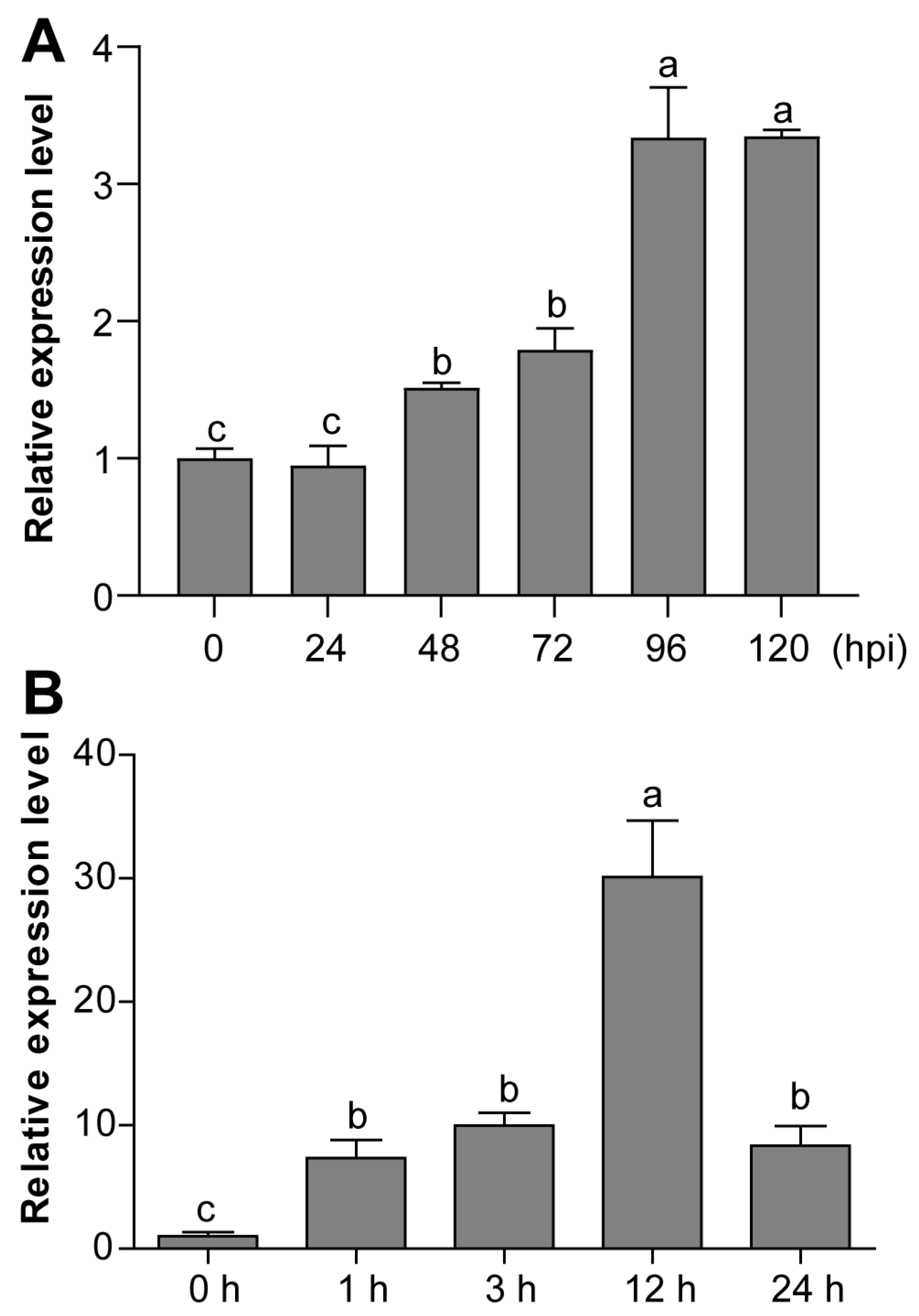
3.4. Expression Profiles of CaPIF7a under PCI and SA Treatment

To dissect the potential functions of CaPIF7a, its expression was investigated under PCI and SA treatment by qRT-PCR. The results showed that the expression of CaPIF7a was remarkably induced under PCI, with the maximum levels at 96 and 120 hpi (Figure 4A). Under SA treatment, the expression of CaPIF7a was significantly increased throughout the treatment, and its expression reached the highest at 12 h (Figure 4B). These findings indicated that CaPIF7a may contribute to pepper’s response against PCI in the SA signaling pathway.

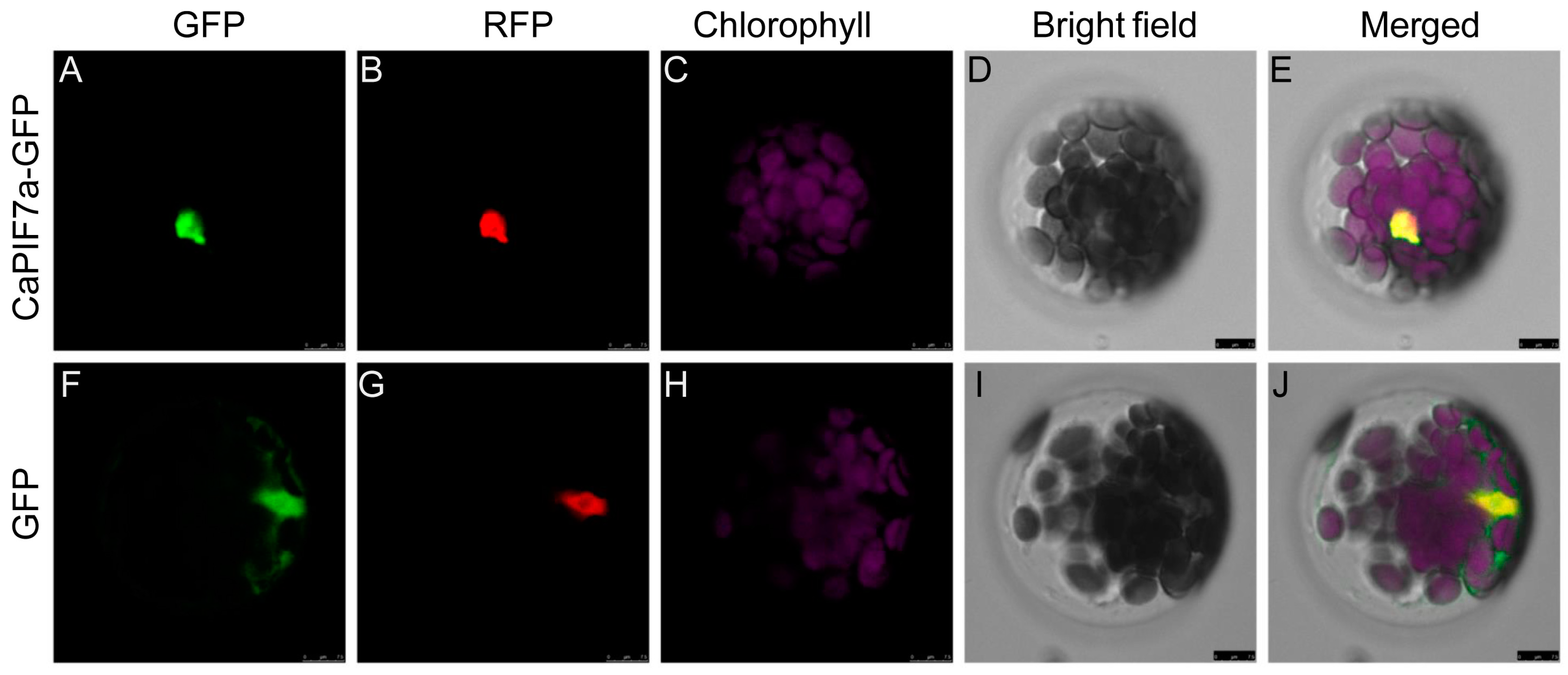
3.5. Subcellular Localization of CaPIF7a

The predicted results by the Plant-mPLoc server showed that CaPIF7a may be located in the nucleus. To determine whether CaPIF7a is active in the nucleus, a CaPIF7a-GFP fusion protein driven by a CaMV 35S promoter was constructed and transformed into Arabidopsis mesophyll protoplasts. The empty vector was used as a control. The confocal microscopic examination indicated that the control 35S::GFP exhibited GFP signals in the whole cell, while the protoplasts carrying the CaPIF7a-GFP fusion protein and RFP nuclear marker only exhibited fluorescence in the nucleus (Figure 5). These results indicated that CaPIF7a is a nucleus-localized protein.

3.6. VIGS-Silenced of CaPIF7a Increased Pepper Sensitivity to PCI

To determine the possible role of CaPIF7a in pepper’s response to PCI, virus-induced gene silencing (VIGS) was used. After about one month of injection, the positive control plants with TRV2:CaPDS (phytoene desaturase gene) were photo-bleached (Figure S1A). At the same time, qRT-PCR analysis showed that the expression of CaPIF7a in the silenced plants (TRV2:CaPIF7a) was about 40% compared to that of the control plants (TRV2:00) (Figure S1B), confirming that CaPIF7a was successfully knocked down.
To investigate whether the silencing of CaPIF7a regulates the resistance of pepper plants to PCI, the TRV2:00 and TRV2:CaPIF7a plants were infected by P. capsici. After 3 days of infection (3 dpi), the TRV2:CaPIF7a plants exhibited wilting compared to the TRV2:00 plants. After 7 days of infection (7 dpi), the TRV2:CaPIF7a plants exhibited more noticeable necrotic symptoms compared with the TRV2:00 plants, including severe wilting leaves with blackening and constriction of the stems (Figure 6A). The disease index determination results showed that the TRV2:CaPIF7a plants exhibited a higher disease index than the TRV2:00 plants after PCI (Figure 6B). We also examined the SA content and expression of defense and SA-related genes in the TRV2:CaPIF7a and TRV2:00 plants after PCI. As a result, the expression levels of CaPR1 and CaPAL3 were observably reduced in TRV2:CaPIF7a plants compared with the TRV2:00 plants at 2 dpi, while the expression levels had no significant differences between the mock-treated TRV2:00 and TRV2:CaPIF7a plants (Figure 6C,D). Additionally, the SA contents were remarkably decreased in TRV2:CaPIF7a plants compared with the TRV2:00 plants (Figure 6E).
The sensitivity of TRV2:CaPIF7a plants to PCI was also examined through detached leaf assays by inoculation with zoospore suspension. As shown in Figure 6F, both TRV2:00 and TRV2:CaPIF7a pepper plants exhibited water-stained lesions on their detached leaves, but larger brown spots and blue dead cell areas were observed in the leaves of TRV2:CaPIF7a plants. The lesion areas on the leaves at TRV2:CaPIF7a and TRV2:00 were 4.22 cm2 and 1.93 cm2, respectively. The lesion area on the leaves of TRV2:CaPIF7a plants was significantly larger than that of the control (Figure 6G). We also examined the biomass of P. capsici on the leaves of TRV2:CaPIF7a and TRV2:00 pepper plants. The biomass of P. capsici on TRV2:CaPIF7a leaves was observably increased compared with that of TRV2:00 plants (Figure 6H). These results indicated that TRV2:CaPIF7a pepper plants were more severely damaged by the disease.

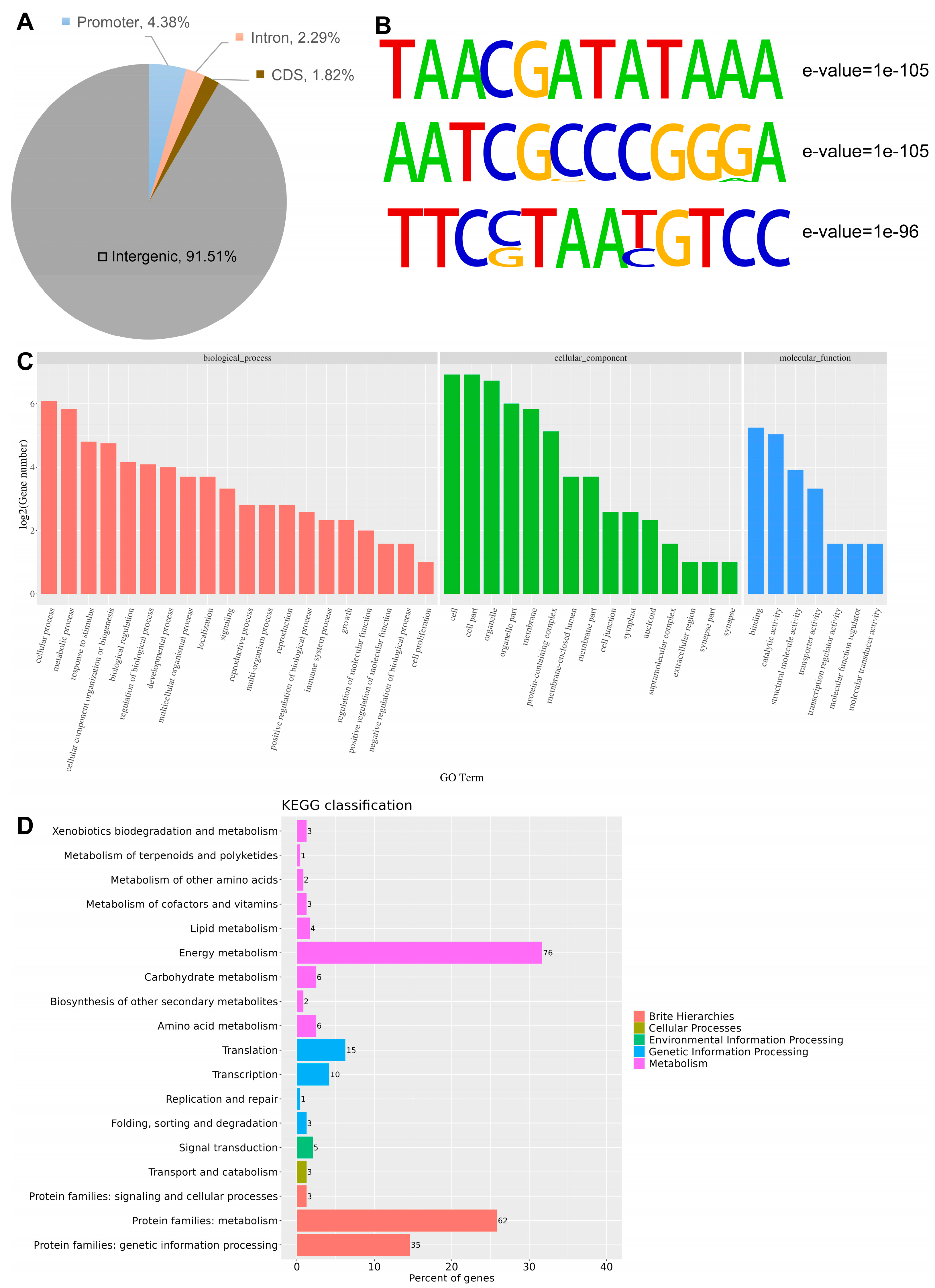
3.7. Genome-Wide Identification of CaPIF7a Binding Regions and Motifs by DAP-Seq

To investigate the regulatory mechanism mediated by CaPIF7a and to inspect its binding landscape, DNA affinity purification followed by sequencing (DAP-seq) was performed. A total of 7658 sequences with CaPIF7a binding peaks were identified, and they were distributed on all pepper chromosomes. Among these peaks, 91.51% were located in the intergenic region, 4.38% were located in the promoter region, and 2.29% were located in the intron, while the distribution of CDS was 1.82% (Figure 7A). MEME suite was employed to perform motif enrichment analysis involving the binding sites of CaPIF7a. As a result, 40 obviously enriched motifs were identified and they ranged from 8 to 12 bp in length (Table S2). In addition, the results showed that the main conservative motifs bound by CaPIF7a were “TAACGATATAAA” (E-value = 1 × 10−105), “AATCGCCCGGGA” (E-value = 1 × 10−105), and “TTCCTAATGTCC” (E-value = 1 × 10−96) (Figure 7B).
To gain an in-depth understanding of the function of CaPIF7a targeted genes, the functions of genes binding in promoter were analyzed and a total of 286 genes were identified (Table S3). Further gene ontology (GO) analysis indicated that these genes were enriched in 20, 15 and 7 different groups on the basis of their biological processes, cellular components, and molecular functions, respectively (Figure 7C; Table S4). Additionally, the results of KEGG enrichment analysis showed that the genes binding in the promoter could be assigned into 18 pathways, of which the most enriched pathways included energy metabolism, protein families in metabolism and genetic information processing (Figure 7D; Table S5).

3.8. CaPIF7a Regulates the Expression of CaHY5

Amongst the bound genes of CaPIF7a, we found the light signal transcription factor CaHY5 (Capana08g000273), which was found to have a significant role in SA-mediated resistance of pepper plants against PCI [24]. Therefore, a yeast one-hybrid experiment was employed for checking whether CaPIF7a can bind to the promoter of CaHY5. As expected, CaPIF7a can bind to the promoter sequence of CaHY5 in yeast cells (Figure 8A). In addition, the expression of CaHY5 in the TRV2:CaPIF7a and TRV2:00 plants after PCI was examined using RT-qPCR. As shown in Figure 8B, the expression of CaHY5 was observably decreased in TRV2:CaPIF7a plants compared with the TRV2:00 plants at 2 dpi, while its expression had no significant differences between the mock-treated TRV2:00 and TRV2:CaPIF7a plants (Figure 8B). These results illustrated that CaPIF7a could activate the expression of CaHY5 by specifically binding to its promoter.

4. Discussion

Plants continuously receive environmental signals during their growth process and regulate their physiological development by transducing the received environmental signals to adapt to environmental changes. As a subfamily of bHLH transcription factors, PIFs exist in a series of plant lineages from bryophytes to higher plants, act as downstream targets of photosensitive pigments and have been well known to work in regulating various light-dependent growth and development processes in plants [29,30]. In our previous study, six CaPIF genes were identified from the pepper genome, and their expression was found to respond to various abiotic stresses [23]. However, the roles of CaPIF genes in biotic stress were largely unknown. For the current study, one of the CaPIF genes (CaPIF7a) was isolated and functionally characterized and it was found to have a positive function in plant defense response against P. capsici.
CaPIF7a has a PIF gene of 1383-bp open reading frame, encoding a nucleus-localized protein of 460 amino acids (Figure 3). By further analysis of the deduced amino acid sequence of CaPIF7a, it possessed the bHLH and APB domains, but missed the APA domain. Additionally, CaPIF7a was expressed in all the tested tissues and with the highest expression in leaf (Figure 3), which was consistent with the findings of VvPIF7 in grape [31], and MdPIF7 in apple [32]. Moreover, expression analysis showed that CaPIF7a had altered expression profiles in response to PCI (Figure 4 and Figure 5), which is similar to the previous reported pepper genes in plant immune response to PCI. For example, CaSBP11 and CaSBP12 have increased expression levels in their VIGS-silenced and negative control pepper plants, and both of them negatively regulated the defense response against PCI [33,34]. The expression of CaHY5 is also induced by PCI, and CaHY5 positively modulates pepper resistance to PCI [24]. Therefore, it can be speculated that CaPIF7a might be involved in regulating the interaction of pepper to PCI.
To further confirm the contribution of CaPIF7a to plant immune response to PCI, VIGS was employed to successfully silence CaPIF7a. As a result, the silencing of CaPIF7a in pepper plants caused phenotypes to be highly vulnerable to infection with P. capsici, including higher disease index, larger lesion area, higher biomass of P. capsici, and higher in H2O2 content, compared to those of the TRV2:00 control plants (Figure 6 and Figure 7), indicating the positive role of CaPIF7a in defense response to the infection of P. capsici. PIFs are thought to be involved in a variety of hormone response pathways, including gibberellin (GA) [35], brassinosteroid [36], and abscisic acid (ABA) [37]. Especially, Arabidopsis PIFs negatively regulate disease resistance to Botrytis cinerea by repressing the expression of a number of defensive genes involved in jasmonic acid (JA) and ethylene (ET) signaling pathways [18,19,38]. AtPIF3 negatively regulates the expression of defense genes, including iron-deficiency overly sensitive 1 (IOS1) and Jasmonate ZIM-domain (JAZ), and resistance to Pto DC3000, and the phosphorylation of AtPIF3 is required for the negative control of plant immunity [19]. AtPIF4 directly promotes the expression of the sulfotransferase (ST2a) gene by binding with its promoter, and therefore negatively regulates the JA-mediated defense response [38]. However, there is little research on PIFs in the physiological and biochemical, and immune response pathways mediated by SA. It is well known that SA plays a key role in quickly initiating the plant responses to various biotic stresses, including P. capsici infection [39]. And several TFs were reported to take part in pepper plants against PCI by mediating the transcription levels of downstream genes in the SA-dependent signaling pathway. For example, CmPIF8 could bind to the promoter of CmICS to inhibit its transcription, and thus cause a decreased SA content and compromise the resistance of oriental melon to powdery mildew [40]. CaWRKY01-10 and CaWRKY08-4 act as positive regulators in pepper resistance to P. capsici by activating the expression of four pathogenesis-related protein (PR) genes by directly binding to their promoters [41]. When plants are challenged by biotic and abiotic stimuli, plants activate a variety of genes, including PR genes, which are disease-resistance-responsive genes involved in systemic acquired resistance (SAR) and pathogenesis in the SA signaling pathway [42]. In this study, the silencing of CaPIF7a negatively affected the transcription levels of SA-responsive gene CaPR1 and SA-biosynthesis gene CaPAL3 against P. capsici infection, and obviously decreased SA content was also observed in CaPIF7a-silenced pepper plants (Figure 6A–E), which showed the role of CaPIF7a in the defense mechanism of pepper plants via the SA-dependent signaling pathway. Previous studies also showed that PAL metabolism plays significant roles in pepper resistance to P. capsici [43,44]. In addition, DAP-seq results showed that CaPIF7a-targeted genes were enriched in pathways involved in energy metabolism, protein families in metabolism and genetic information processing (Figure 7), and one of the target genes, CaHY5, also displayed a significantly decreased expression in TRV2:CaPIF7a compared to the control plants (Figure 8B). Y1H results indicated that CaPIF7a could directly bind to the promoter of CaHY5 (Figure 8A), and our previous study showed that CaHY5 can activate the transcripts of SA-biosynthesis genes CaPAL3 and CaPAL7 by directly binding to their promoters and positively modulates pepper resistance to PCI [24]. Therefore, it is possible that CaPIF7a might be a positive regulator in plant immune response to PCI in the SA signaling pathway by activating the expression of CaHY5, and thus mediating downstream SA-responsive and SA-biosynthesis genes. Further studies should be focused on the interaction of CaPIF7a and its target genes.

5. Conclusions

In this study, CaPIF7a is a primary regulatory factor in pepper defense response to the infection of P. capsici, and loss of function of CaPIF7a in pepper plants by VIGS resulted in increased susceptibility of pepper plants to PCI by reducing SA accumulation and decreased expression of genes in SA biosynthesis and signal transduction. Furthermore, CaPIF7a could activate the expression of CaHY5 by specifically binding to its promoter, and it enhances pepper resistance to PCI probably through CaHY5-mediated gene expression. Our study provides a better understanding of the role of CaPIF7a in pepper immunity against PCI, and the generation of CaPIF7a stable transgenic pepper plants may further reveal the roles of CaPIF7a in PCI as well as in other biological pathways in the future.

Supplementary Materials

The following supporting information can be downloaded at: https://www.mdpi.com/article/10.3390/agronomy14092035/s1, Figure S1: Virus-induced gene silencing (VIGS) of CaPIF7a. (A) photo-bleached phenotypes of TRV2:CaPDS pepper plants. (B) Silencing efficiency of CaPIF7a in TRV2:00 and TRV2:CaPIF7a pepper plants; Table S1: The primers used in this study; Table S2: CaPIF7a genome-wide binding motif in pepper; Table S3: CaPIF7a.promoter_peak.peak_annotation; Table S4: GO enrichment analysis; Table S5: KEGG enrichment analysis.

Author Contributions

Data curation, Y.L. and D.W.; Formal Analysis, D.W. and T.Y.; Funding Acquisition, Y.Z. and Y.Y.; Investigation, Y.L., D.W. and B.L.; Methodology, Y.L. and Y.Y.; Project administration, Y.Y.; Resources, X.G., H.H. and J.C.; Software, T.Y.; Supervision, Y.Y.; Validation, Y.Z.; Writing—Original Draft, Y.Z. and Y.Y.; Writing—Review and Editing, Y.Z. and Y.Y. All authors have read and agreed to the published version of the manuscript.

Funding

This work was supported by the National Natural Science Foundation of China (32372797, 32460797 and 32460798), the National Key Research and Development Program of Jiangxi Province, China (20223BBF61017), the Natural Science Foundation of Jiangxi Province, China (20212ACB215006 and 20232BAB205039), and the Earmarked Fund for Jiangxi Agriculture Research System (JXARS-06).

Data Availability Statement

Relevant data are incorporated in this manuscript.

Conflicts of Interest

The authors declare that this research was conducted without any commercial or financial relationships that could be construed as potential conflicts of interest.

References

  1. Ni, M.; Tepperman, J.M.; Quail, P.H. Binding of phytochrome B to its nuclear signalling partner PIF3 is reversibly induced by light. Nature 1999, 400, 781–784. [Google Scholar] [CrossRef] [PubMed]
  2. Bian, Y.; Chu, L.; Lin, H.; Qi, Y.; Fang, Z.; Xu, D. PIFs- and COP1-HY5-mediated temperature signaling in higher plants. Stress Biol. 2022, 2, 35. [Google Scholar] [CrossRef] [PubMed]
  3. Wang, P.; Abid, M.A.; Qanmber, G.; Askari, M.; Zhou, L.; Song, Y.; Liang, C.; Meng, Z.; Malik, W.; Wei, Y.; et al. Photomorphogenesis in plants: The central role of phytochrome interacting factors (PIFs). Environ. Exp. Bot. 2022, 194, 104704. [Google Scholar] [CrossRef]
  4. Leivar, P.; Monte, E.; Oka, Y.; Liu, T.; Carle, C.; Castillon, A.; Huq, E.; Quail, P.H. Multiple phytochrome-interacting bHLH transcription factors repress premature seedling photomorphogenesis in darkness. Curr. Biol. CB 2008, 18, 1815–1823. [Google Scholar] [CrossRef]
  5. Leivar, P.; Monte, E. PIFs: Systems integrators in plant development. Plant Cell 2014, 26, 56–78. [Google Scholar] [CrossRef]
  6. Park, E.; Kim, Y.; Choi, G. Phytochrome B requires PIF degradation and sequestration to induce light responses across a wide range of light conditions. Plant Cell 2018, 30, 1277–1292. [Google Scholar] [CrossRef]
  7. Xie, Y.; Liu, W.; Liang, W.; Ling, X.; Ma, J.; Yang, C.; Li, L. Phytochrome B inhibits the activity of phytochrome-interacting factor 7 involving phase separation. Cell Rep. 2023, 42, 113562. [Google Scholar] [CrossRef]
  8. Song, Y.; Yang, C.; Gao, S.; Zhang, W.; Li, L.; Kuai, B. Age-triggered and dark-induced leaf senescence require the bHLH transcription factors PIF3, 4, and 5. Mol. Plant 2014, 7, 1776–1787. [Google Scholar] [CrossRef]
  9. Sakuraba, Y.; Jeong, J.; Kang, M.Y.; Kim, J.; Paek, N.C.; Choi, G. Phytochrome-interacting transcription factors PIF4 and PIF5 induce leaf senescence in Arabidopsis. Nat. Commun. 2014, 5, 4636. [Google Scholar] [CrossRef]
  10. Sun, J.; Lu, J.; Bai, M.; Chen, Y.; Wang, W.; Fan, C.; Liu, J.; Ning, G.; Wang, C. Phytochrome-interacting factors interact with transcription factor CONSTANS to suppress flowering in rose. Plant Physiol. 2021, 186, 1186–1201. [Google Scholar] [CrossRef]
  11. Liu, L.Y.; Jia, M.Z.; Wang, S.N.; Han, S.; Jiang, J. Identification and characterization of cotton PHYTOCHROME-INTERACTING FACTORS in temperature-dependent flowering. J. Exp. Bot. 2023, 74, 3765–3780. [Google Scholar] [CrossRef] [PubMed]
  12. Rosado, D.; Trench, B.; Bianchetti, R.; Zuccarelli, R.; Rodrigues Alves, F.R.; Purgatto, E.; Segal Floh, E.I.; Silveira Nogueira, F.T.; Freschi, L.; Rossi, M. Downregulation of PHYTOCHROME-INTERACTING FACTOR 4 influences plant development and fruit production. Plant Physiol. 2019, 181, 1360–1370. [Google Scholar] [CrossRef] [PubMed]
  13. Li, Q.; Hou, Y.; Wang, Q.; Pan, X.; Sun, Y.; Zhu, X.; Li, H.; Guo, M.; Gao, Y. Phytochrome interacting factor ZmPIF6 simultaneously enhances chilling tolerance and grain size in rice. Plant Physiol. Biochem. PPB 2024, 214, 108954. [Google Scholar] [CrossRef] [PubMed]
  14. Zhuge, Y.; Sheng, H.; Zhang, M.; Fang, J.; Lu, S. Grape phytochrome-interacting factor VvPIF1 negatively regulates carotenoid biosynthesis by repressing VvPSY expression. Plant Sci. Int. J. Exp. Plant Biol. 2023, 331, 111693. [Google Scholar] [CrossRef]
  15. Michael, R.; Ranjan, A.; Gautam, S.; Trivedi, P.K. HY5 and PIF antagonistically regulate HMGR expression and sterol biosynthesis in Arabidopsis thaliana. Plant Sci. Int. J. Exp. Plant Biol. 2024, 346, 112168. [Google Scholar] [CrossRef]
  16. Nie, N.; Huo, J.; Sun, S.; Zuo, Z.; Chen, Y.; Liu, Q.; He, S.; Gao, S.; Zhang, H.; Zhao, N.; et al. Genome-wide characterization of the PIFs family in sweet potato and functional identification of IbPIF3.1 under drought and Fusarium wilt stresses. Int. J. Mol. Sci. 2023, 24, 4092. [Google Scholar] [CrossRef]
  17. Sng, B.J.R.; Vu, K.V.; Choi, I.K.Y.; Chin, H.J.; Jang, I.C. LONG HYPOCOTYL IN FAR-RED 1 mediates a trade-off between growth and defence under shade in Arabidopsis. J. Exp. Bot. 2023, 74, 3560–3578. [Google Scholar] [CrossRef]
  18. Xiang, S.; Wu, S.; Zhang, H.; Mou, M.; Chen, Y.; Li, D.; Wang, H.; Chen, L.; Yu, D. The PIFs redundantly control plant defense response against Botrytis cinerea in Arabidopsis. Plants 2020, 9, 1246. [Google Scholar] [CrossRef]
  19. Zhao, Y.; Zheng, X.; Zhang, X.; Wang, W.; Cai, G.; Bi, G.; Chen, S.; Sun, C.; Zhou, J.M. PIF3 is phosphorylated by MAPK to modulate plant immunity. New Phytol. 2023, 240, 372–381. [Google Scholar] [CrossRef]
  20. Gangappa, S.N.; Berriri, S.; Kumar, S.V. PIF4 coordinates thermosensory growth and immunity in Arabidopsis. Curr. Biol. CB 2017, 27, 243–249. [Google Scholar] [CrossRef]
  21. Zhang, X.; Wang, D.; Zhao, P.; Sun, Y.; Fang, R.X.; Ye, J. Near-infrared light and PIF4 promote plant antiviral defense by enhancing RNA interference. Plant Commun. 2024, 5, 100644. [Google Scholar] [CrossRef] [PubMed]
  22. Wu, X.; Wang, L.; Xing, Q.; Zhao, Y.; Qi, H. CmPIF8-CmERF27-CmACS10-mediated ethylene biosynthesis modulates red light-induced powdery mildew resistance in oriental melon. Plant Cell Environ. [CrossRef]
  23. Yang, Y.; Guang, Y.; Wang, F.; Chen, Y.; Yang, W.; Xiao, X.; Luo, S.; Zhou, Y. Characterization of phytochrome-interacting factor genes in pepper and functional analysis of CaPIF8 in cold and salt stress. Front. Plant Sci. 2021, 12, 746517. [Google Scholar] [CrossRef]
  24. Yang, Y.; Li, Y.; Guang, Y.; Lin, J.; Zhou, Y.; Yu, T.; Ding, F.; Wang, Y.; Chen, J.; Zhou, Y.; et al. Red light induces salicylic acid accumulation by activating CaHY5 to enhance pepper resistance against Phytophthora capsici. Hortic. Res. 2023, 10, uhad213. [Google Scholar] [CrossRef] [PubMed]
  25. O’Malley, R.C.; Huang, S.C.; Song, L.; Lewsey, M.G.; Bartlett, A.; Nery, J.R.; Galli, M.; Gallavotti, A.; Ecker, J.R. Cistrome and epicistrome features shape the regulatory DNA landscape. Cell 2016, 165, 1280–1292. [Google Scholar] [CrossRef]
  26. Mai, Y.; Sun, P.; Suo, Y.; Li, H.; Han, W.; Diao, S.; Wang, L.; Yuan, J.; Wang, Y.; Ye, L.; et al. Regulatory mechanism of MeGI on sexuality in Diospyros oleifera. Front. Plant Sci. 2023, 14, 1046235. [Google Scholar] [CrossRef] [PubMed]
  27. Zhang, Y.; Liu, T.; Meyer, C.A.; Eeckhoute, J.; Johnson, D.S.; Bernstein, B.E.; Nusbaum, C.; Myers, R.M.; Brown, M.; Li, W.; et al. Model-based analysis of ChIP-Seq (MACS). Genome Biol. 2008, 9, R137. [Google Scholar] [CrossRef]
  28. Ma, W.; Noble, W.S.; Bailey, T.L. Motif-based analysis of large nucleotide data sets using MEME-ChIP. Nat. Protoc. 2014, 9, 1428–1450. [Google Scholar] [CrossRef]
  29. Roeber, V.M.; Bajaj, I.; Rohde, M.; Schmülling, T.; Cortleven, A. Light acts as a stressor and influences abiotic and biotic stress responses in plants. Plant Cell Environ. 2021, 44, 645–664. [Google Scholar] [CrossRef]
  30. Pham, V.N.; Kathare, P.K.; Huq, E. Phytochromes and phytochrome interacting factors. Plant Physiol. 2018, 176, 1025–1038. [Google Scholar] [CrossRef]
  31. Zhang, K.; Zheng, T.; Zhu, X.; Jiu, S.; Liu, Z.; Guan, L.; Jia, H.; Fang, J. Genome-wide identification of PIFs in grapes (Vitis vinifera L.) and their transcriptional analysis under lighting/shading conditions. Genes 2018, 9, 451. [Google Scholar] [CrossRef]
  32. Zheng, P.F.; Wang, X.; Yang, Y.Y.; You, C.X.; Zhang, Z.L.; Hao, Y.J. Identification of phytochrome-interacting factor family members and functional analysis of MdPIF4 in Malus domestica. Int. J. Mol. Sci. 2020, 21, 7350. [Google Scholar] [CrossRef] [PubMed]
  33. Zhang, H.X.; Feng, X.H.; Jin, J.H.; Khan, A.; Guo, W.L.; Du, X.H.; Gong, Z.H. CaSBP11 participates in the defense response of pepper to Phytophthora capsici through regulating the expression of defense-related genes. Int. J. Mol. Sci. 2020, 21, 9065. [Google Scholar] [CrossRef]
  34. Zhang, H.X.; Ali, M.; Feng, X.H.; Jin, J.H.; Huang, L.J.; Khan, A.; Lv, J.G.; Gao, S.Y.; Luo, D.X.; Gong, Z.H. A novel transcription factor CaSBP12 gene negatively regulates the defense response against Phytophthora capsici in pepper (Capsicum annuum L.). Int. J. Mol. Sci. 2019, 20, 48. [Google Scholar] [CrossRef] [PubMed]
  35. Wang, F.; Chen, X.; Dong, S.; Jiang, X.; Wang, L.; Yu, J.; Zhou, Y. Crosstalk of PIF4 and DELLA modulates CBF transcript and hormone homeostasis in cold response in tomato. Plant Biotechnol. J. 2020, 18, 1041–1055. [Google Scholar] [CrossRef]
  36. Oh, E.; Zhu, J.Y.; Wang, Z.Y. Interaction between BZR1 and PIF4 integrates brassinosteroid and environmental responses. Nat. Cell Biol. 2012, 14, 802–809. [Google Scholar] [CrossRef] [PubMed]
  37. Li, Q.; Zhou, L.; Chen, Y.; Xiao, N.; Zhang, D.; Zhang, M.; Wang, W.; Zhang, C.; Zhang, A.; Li, H.; et al. Phytochrome interacting factor regulates stomatal aperture by coordinating red light and abscisic acid. Plant Cell 2022, 34, 4293–4312. [Google Scholar] [CrossRef] [PubMed]
  38. Fernández-Milmanda, G.L.; Crocco, C.D.; Reichelt, M.; Mazza, C.A.; Köllner, T.G.; Zhang, T.; Cargnel, M.D.; Lichy, M.Z.; Fiorucci, A.S.; Fankhauser, C.; et al. A light-dependent molecular link between competition cues and defence responses in plants. Nat. Plants 2020, 6, 223–230. [Google Scholar] [CrossRef]
  39. Lei, G.; Zhou, K.H.; Chen, X.J.; Huang, Y.Q.; Yuan, X.J.; Li, G.G.; Xie, Y.Y.; Fang, R. Transcriptome and metabolome analyses revealed the response mechanism of pepper roots to Phytophthora capsici infection. BMC Genom. 2023, 24, 626. [Google Scholar] [CrossRef]
  40. Wang, L.; Wu, X.; Xing, Q.; Zhao, Y.; Yu, B.; Ma, Y.; Wang, F.; Qi, H. PIF8-WRKY42-mediated salicylic acid synthesis modulates red light induced powdery mildew resistance in oriental melon. Plant Cell Environ. 2023, 46, 1726–1742. [Google Scholar] [CrossRef]
  41. Cheng, W.; Wang, N.; Li, Y.; Zhou, X.; Bai, X.; Liu, L.; Ma, X.; Wang, S.; Li, X.; Gong, B.; et al. CaWRKY01-10 and CaWRKY08-4 confer pepper’s resistance to Phytophthora capsici infection by directly activating a cluster of defense-related genes. J. Agric. Food Chem. 2024, 72, 11682–11693. [Google Scholar] [CrossRef]
  42. Zribi, I.; Ghorbel, M.; Brini, F. Pathogenesis related proteins (PRs): From cellular mechanisms to plant defense. Curr. Protein Pept. Sci. 2021, 22, 396–412. [Google Scholar] [CrossRef] [PubMed]
  43. Li, Y.; Yu, T.; Wu, T.; Wang, R.; Wang, H.; Du, H.; Xu, X.; Xie, D.; Xu, X. The dynamic transcriptome of pepper (Capsicum annuum) whole roots reveals an important role for the phenylpropanoid biosynthesis pathway in root resistance to Phytophthora capsici. Gene 2020, 728, 144288. [Google Scholar] [CrossRef] [PubMed]
  44. Hao, C.; Xia, Z.; Fan, R.; Tan, L.; Hu, L.; Wu, B.; Wu, H. De novo transcriptome sequencing of black pepper (Piper nigrum L.) and an analysis of genes involved in phenylpropanoid metabolism in response to Phytophthora capsici. BMC Genom. 2016, 17, 822. [Google Scholar] [CrossRef] [PubMed]
Figure 1. Characterization of pepper CaPIF7a gene and its encoded protein. (A) Nucleic acid and translated amino acid sequences of CaPIF7a. The conserved APB and bHLH domains were underlined with different colors. (B) Secondary structure of the CaPIF7a protein.
Agronomy 14 02035 g001
Figure 2. Predicted cis-acting regulatory elements in the promoter region of CaPIF7a gene by PlantCARE tool. Different shapes and colors represent diverse cis-elements.
Agronomy 14 02035 g002
Figure 3. qRT-PCR analysis of the relative expression of CaPIF7a in different tissues. Data represent mean ± standard error of three biological replicates, and different letters above the columns indicate significant difference (p < 0.05).
Agronomy 14 02035 g003
Figure 4. Expression analysis of CaPIF7a under PCI and SA treatment. (A) qRT-PCR analysis of the relative expression of CaPIF7a under PCI at different time points. (B) qRT-PCR analysis of the relative expression of CaPIF7a in response to SA treatment. Data represent mean ± standard error of three biological replicates, and different letters above the columns indicate significant difference (p < 0.05).
Agronomy 14 02035 g004
Figure 5. Subcellular localization of the CaPIF7a protein in Arabidopsis mesophyll protoplasts. (A,F) GFP fluorescence, (B,G) RFP fluorescence, (C,H) chlorophyll fluorescence, (D,I) bright field images, (E,J) merged images. Bar = 75 μm.
Agronomy 14 02035 g005
Figure 6. Virus-induced gene silencing (VIGS) of CaPIF7a increased pepper susceptibility to P. capsici infection (PCI). (A) Disease symptoms of TRV2:00 and TRV2:CaPIF7a pepper plants at 0, 3, and 7 dpi (days post infection). (B) Disease index determination in TRV2:CaPIF7a and TRV2:00 pepper plants. (C,D) Relative expression levels of CaPR1 (C) and CaPAL3 (D) in mock-treated and P. capsici infected TRV2:00 and TRV2:CaPIF7a plants. (E) The determination of SA content of TRV2:00 and TRV2:CaPIF7a pepper plants. (F) Phenotypes of detached leaves of TRV2:CaPIF7a and TRV2:00 pepper plants after inoculation with P. capsici. Bar = 1 cm. (G) Lesion area determination in TRV2:CaPIF7a and TRV2:00 pepper plants. (H) Relative P. capsici biomass in infected pepper leaves of TRV2:CaPIF7a and TRV2:00 pepper plants determined by qRT-PCR. Data represent mean ± standard error of three biological replicates, and the asterisks above the columns indicate significant differences (p < 0.05).
Agronomy 14 02035 g006
Figure 7. Genome-wide binding regions and enriched motif analysis of CaPIF7a by DAP-seq. (A) Distribution of CaPIF7a peaks in the pepper genome based on the localization of peak summits. (B) Significantly enriched motif sequence bound by CaPIF7a. (C) The result of GO functional enrichment. (D) The result of KEGG functional enrichment.
Agronomy 14 02035 g007
Figure 8. CaPIF7a regulates the expression of CaHY5. (A) Interaction of CaPIF7a and the promoter fragment of CaHY5 in yeast. Interaction was confirmed by the growth status of yeast cells on SD/-Ura/-Leu/AbA plates. AD was used as the negative control. (B) Expression analysis of CaHY5 in mock-treated and P. capsici-infected TRV2:00 and TRV2:CaPIF7a plants by qRT-PCR. Data represent mean ± standard error of three biological replicates, and the asterisks above the columns indicate significant differences (p < 0.05).
Agronomy 14 02035 g008
Disclaimer/Publisher’s Note: The statements, opinions and data contained in all publications are solely those of the individual author(s) and contributor(s) and not of MDPI and/or the editor(s). MDPI and/or the editor(s) disclaim responsibility for any injury to people or property resulting from any ideas, methods, instructions or products referred to in the content.

