Article

Nodules of Medicago spp. Host a Diverse Community of Rhizobial Species in Natural Ecosystems

by
Andrei Stefan
1,
Jannick Van Cauwenberghe
2,3,
Craita Maria Rosu
4,
Catalina Stedel
4,
Crystal Chan
3,
Ellen L. Simms
3,
Catalina Iticescu
5,
Daniela Tsikou
6,
Emmanouil Flemetakis
7 and
Rodica Catalina Efrose
4,7,*
1
“Grigore Antipa” National Museum of Natural History, Șos. Kiseleff 1, 011341 Bucharest, Romania
2
Institute of Biodiversity, Faculty of Biological Sciences, Cluster of Excellence Balance of the Microverse, Friedrich-Schiller-University Jena, 07743 Jena, Germany
3
Department of Integrative Biology, University of California, Berkeley, CA 94720, USA
4
Department of Experimental and Applied Biology, NIRDBS-Institute of Biological Research Iași, Lascar Catargi 47, 700107 Iași, Romania
5
Department of Chemistry, Physics and Environment, Faculty of Sciences and Environment, ‘Dunarea de Jos’ University of Galati, 800008 Galati, Romania
6
Department of Biochemistry and Biotechnology, University of Thessaly, Biopolis, 41500 Larissa, Greece
7
Department of Biotechnology, Agricultural University of Athens, 75 Iera Odos Str., 11855 Athens, Greece
*
Author to whom correspondence should be addressed.
Agronomy 2024, 14(9), 2156; https://doi.org/10.3390/agronomy14092156
Submission received: 22 August 2024 / Revised: 18 September 2024 / Accepted: 19 September 2024 / Published: 21 September 2024

Abstract

:
Biological nitrogen fixation by rhizobia-nodulated legumes reduces the dependence on synthetic nitrogen fertilizers. Identification of locally adapted rhizobia may uncover economically valuable strains for sustainable agriculture. This study investigated the diversity and symbiotic potential of rhizobia associated with three Medicago species from Eastern Romania’s ecosystems. Phenotypic screening ensured that only rhizobial species were retained for molecular characterization. 16S rDNA sequencing clustered the isolates into four distinct groups: Sinorhizobium meliloti, Sinorhizobium medicae, Rhizobium leguminosarum, and Mesorhizobium spp. The chromosomal genes (atpD, glnII, recA) and nifH phylogenies were congruent, while the nodA phylogeny grouped the Mesorhizobium spp. isolates with R. leguminosarum. Medicago sativa was the most sampled plant species, and only S. meliloti and R. leguminosarum were found in its nodules, while Medicago falcata nodules hosted S. meliloti and Mesorhizobium spp. Medicago lupulina was the only species that hosted all four identified rhizobial groups, including S. medicae. This study provides the first report on the Mesorhizobium spp. associated with M. falcata nodules. Additionally, R. leguminosarum and two Mesorhizobium genospecies were identified as novel symbionts for Medicago spp. Comparative analysis of Medicago-associated rhizobia from other studies revealed that differences in 16S rDNA sequence type composition were influenced by Medicago species identity rather than geographic region.

1. Introduction

Rhizobia are motile, naturally occurring rod-shaped soil bacteria. Following a complex exchange of signaling molecules between the bacteria and plants of the legume family and activation of subsequent cellular pathways, the bacteria can enter plant root cells and initiate the formation of specialized organs on the roots called nodules [1,2,3]. In these nodules, the bacteria can differentiate and are capable of fixing atmospheric nitrogen into ammonia, which they transfer to the host plant in exchange for carbon-containing molecules and shelter [4,5]. Biological nitrogen fixed by this bacteria-plant symbiosis [6] contributes large amounts of nitrogen to agricultural systems [7]. Soils with poor nitrogen-fixing rhizobial communities can be supplemented by inoculation with superior nitrogen-fixing rhizobia. The application of rhizobial inoculants to crop fields can reduce the amount of synthetic nitrogen fertilizers, can reduce emissions of the potent greenhouse gas N2O by converting it to nitrogen [8], can improve crop productivity [9], nutrient mobilization and solubilization [10].
Crop rotation every few years with legume species and intercropping practices locks the nitrogen molecules into plant structures, thus reducing nitrogen depletion from the soil [11,12,13]. Some legume species (like alfalfa and clovers) are specifically planted to be incorporated into the soil while still green (‘green manuring’) to benefit subsequent crops [14,15,16]. The isolation and characterization of indigenous rhizobia can pave the way for their use as inoculants [17] and can lead to the discovery of strains of potential economic value. Native rhizobial strains, tolerant to local soil conditions, can be more efficient in nitrogen fixation and outcompete commercial inoculants [18,19,20,21,22].
One way of characterizing the rhizobial community is by sequencing multiple chromosomal and plasmid loci, a method that has been previously used in Medicago rhizobia studies [23,24,25]. This multilocus approach has been used extensively in rhizobial diversity studies in various legume species [26,27] across different regions and soil conditions, from subpolar soils [28] to arid desert soils [29] and in the characterization of indigenous rhizobia [30,31,32].
Among the most economically important legumes in temperate regions are those of the genus Medicago, colloquially known as medics. Medics comprise more than 80 species, both perennial and annual, with the main diversity region being the Mediterranean basin [33]. Used primarily for livestock fodder, medics can be cultivated or can grow wildly in a variety of environments, such as pastures, rock quarries, and urbanized environments, and are resilient to a broad range of stress factors [34,35,36]. The rhizobia most commonly found in medic nodules are Sinorhizobium meliloti and Sinorhizobium medicae [24,25,37,38], but some Medicago species can host a more diverse rhizobial community [39,40].
There are ten species of naturally occurring Medicago plants in Romania [41], with M. sativa being the most widely cultivated legume in terms of cultivated area [42]. In this study, rhizobia were isolated from nodules of three species of Medicago sampled from diverse landscapes in Eastern Romania: the perennial Medicago sativa (alfalfa, lucerne) and M. falcata (yellow lucerne), and the annual Medicago lupulina (black medic). The goals were to identify and characterize indigenous, locally adapted rhizobia associated with these Medicago species capable of establishing efficient nitrogen-fixing symbiosis and to gain insights into how Medicago spp. and the collection areas could shape the rhizobial community.

2. Materials and Methods

2.1. Nodule Collection and Isolation of Bacteria

During the spring of 2013, naturally occurring and cultivated Medicago plants were sampled in the Eastern part of Romania. To our knowledge, the cultivated fields have no history of inoculation. Sample sites spanned a variety of environments and elevations in the Eastern Carpathian Mountains and the Moldavian Plateau (Figure 1). The plant populations were grouped into four regions, named from North to South: Rarău, Lespezi, Iași, and Oituz. Each plant population was marked with a capital letter, from A to M, for readability (Supplementary Table S1).
Intact nodules were collected from three Medicago species (M. sativa, M. falcata, M. lupulina), surface sterilized, and individually stored in 20% glycerol following a brief visual inspection of the nodules until subsequent processing. Only active nodules were collected, as determined by the pinkish color indicating the presence of leghaemoglobin. A total of sixty-one rhizobial strains were isolated from plant root nodules using standard protocols [43,44] and purified by repeated plating of single colonies on yeast-extract-mannitol agar (YMA) supplemented with 25 µg/mL Congo red [44]. Bacterial isolates were incubated at 28 °C for 3 to 5 days and evaluated for Gram reaction, colony morphology, and acid/alkaline reaction on YMA plates containing 25 µg/mL bromothymol blue [45]. Pure cultures were maintained on YMA slants at 4 °C or preserved in yeast mannitol broth (YMB) containing 20% (v/v) glycerol at −80 °C. The pure cultures are part of the Romanian Microbial Culture Collection (RMCC) established at the Department of Experimental and Applied Biology, NIRDBS-Institute of Biological Research, Iași, Romania.

2.2. Phenotypic Characterization of Rhizobia

The rhizobial isolates and selected reference strains (Sinorhizobium meliloti LMG 6133, S. meliloti 1021, Rhizobium leguminosarum bv. trifolii LMG 8820 and Mesorhizobium loti MAFF 303099) were examined for several phenotypic features, according to Eaglesham’s technique [46] and further analyzed by numerical taxonomy [47]. Salt tolerance and pH growth range were evaluated on agar plates containing 0.1, 0.2, 0.3, 0.4, 0.75, 1, 2, and 3.5% (w/v) NaCl or YMA medium buffered with 1 M HCl or 1 M NaOH to adjust the pH to 4.8, 5.8, 6.8, 7.8 and 8.8, respectively. Rhizobial growth sensitivity to varying temperatures was assessed on YMA plates incubated at 15, 28, 33, 37, and 42 °C. Intrinsic antibiotic resistance (IAR) assays were performed on YMA solid medium supplemented with the following antibiotics (µg/mL): chloramphenicol (30; 100), streptomycin (10; 25), gentamicin (5; 25), rifampicin (2; 20), tetracycline (1; 10), neomycin (5; 15), kanamycin (10; 20), ampicillin (20; 50), carbenicillin (50; 200), and nalidixic acid (50; 300). The choice of antibiotics and concentrations were adapted from Josey et al. [48] and Mpepereki et al. [49]. For each strain, bacterial growth was recorded from colony counts of plates inoculated with serial dilutions of each culture after incubating for 5 days at 28 °C. The physiological traits of the bacterial strains were coded 1 and 0 for positive and negative reactions, respectively, and the simple matching similarity coefficient (SSM) was calculated to generate a similarity matrix. The Unweighted Pair Group Method using Arithmetic Means (UPGMA) was performed for cluster analysis of phenotypic features [50] using MVSP–Multivariate Statistical Package v3.2 (Kovach Computing Services, Anglesey, Wales).

2.3. Nodulation Test

The nodulation ability of the rhizobial isolates on their original host was assessed as previously described [51]. Each rhizobial isolate was used to inoculate seeds from the Medicago species from which they were isolated. Briefly, batches of five surface-sterilized germinated seeds were transferred to individual pots containing sterile perlite and inoculated with 1 mL (1 × 10⁸ cells/mL) of bacterial culture. Plants inoculated with strains Sinorhizobium meliloti 1021 and S. meliloti LMG6133 were used as a reference, and uninoculated plants were used as controls. The inoculated plants and controls were grown in strict conditions (18 h day/6 h night, 24 °C day/18 °C night cycles, 70% humidity) and watered every two days alternatively with N-free Hoagland solution and deionized water. Plants were harvested at 45 days post-inoculation, and the modulation and N-fixing abilities of the isolates were estimated by the number of pink nodules on the roots and the total dry weight of the plants.

2.4. DNA Extraction, Amplification and Sequencing

Pure cultures of rhizobial isolates were grown in liquid YMB and incubated at 28 °C for three days on a rotary shaker. Total genomic DNA was isolated from equal-sized aliquots of bacterial cultures. DNA isolation, PCR reactions, and sequencing were performed as described in detail elsewhere [51,52]. Six genetic markers were amplified, purified, and directly sequenced (CeMIA, Larissa, Greece) from each bacterial isolate: 16S rDNA, atpD (ATP synthase beta-chain), glnII (glutamine synthetase), recA (recombinase A), nodA (nodulation protein A), and nifH (nitrogen reductase).

2.5. Genetic Analyses

The raw chromatogram files were visually inspected and manually edited in Chromas v2.6.6 (Technelysium Pty Ltd., Brisbane, Australia). Alignments were assembled into contigs, and ambiguous regions were checked in CodonCode Aligner v3.7.1 (CodonCode Corporation, Centerville, MA, USA). Files were exported to fasta format, and basic sequence statistics were computed in DnaSP v6.12 [53]. To ensure that all isolates were indeed rhizobia, the 16S rDNA sequences were initially compared to GenBank [54] sequences using BLASTn [55]. If the top BLAST hits did not belong to putative rhizobial species, then the entire sequence set for that particular isolate was discarded. The most similar GenBank sequences were downloaded and included in the analyses to position isolates among known rhizobia. Identical sequences were collapsed into a single sequence type (ST), and the sequence types that contained a single sequence were denoted as singleton ST (sST). Phylogenetic and molecular evolutionary analyses were performed in MEGA X [56]. The phylogenies inferred separately from each gene sequence as well as from concatenated sequences of the atpD, glnII, and recA chromosomal genes were based on the Maximum Likelihood method using the best DNA substitution model. Bootstrap confidence levels were calculated for 1000 replicates.
The rhizobia obtained in the present study was compared with the previously described rhizobia associated with Medicago spp. across the world. Additional 16S rDNA sequences were obtained from studies found using the search term “Medicago rhizobi* diversity” on the Web of Science. A Medicago species × ST per sampling site matrix was constructed, as well as a Medicago species × rhizobia species per sampling site matrix. For each matrix, a Permutational Multivariate Analysis of Variance (PERMANOVA) based on Jaccard distances was performed using 999 permutations, with Medicago species and region as factors, in the ecodist [57] and vegan [58] packages in R v3.6.3 [59]. Differences among rhizobium communities were visualized by a Principal Coordinates Analysis (PCoA) with ade4 [60] and adegraphics [61] R packages. The map was made using QGIS v3.28 [62]. Further visual edits to the map, graphs, and phylogenetic trees were made in Inkscape v1.2 [63].

2.6. Nucleotide Sequence Data

The GenBank accession numbers for the sequences reported in the present study are MT706727-MT706790 (for 16S rDNA); OM677466-OM677522 (for atpD); OM735848-OM735904 (for glnII); OM735905-OM735961 (for recA); OM970805-OM970861 (for nodA) and ON016017-ON016073 (for nifH). Accession numbers of the related reference strains are individually specified in the corresponding phylograms.

3. Results

3.1. Phenotypic Characterization and Nodulation of the Rhizobial Isolates

The phenotypic screening produced sixty-one rhizobial isolates that were Gram-negative, fast-growing, and acid-producing bacteria. After 3–5 days of incubation at 28 °C on YMA medium, most isolates formed white–opaque and mucoid-type colonies, slightly domed with smooth margins (Sinorhizobium sp.). Other colonies were small (<1 mm in diameter), translucent, with entire margins (Mesorhizobium spp.), and only a few isolates formed single colonies with diameters of 1–3 mm, white or creamy, gummy, smooth, raised, and circular with entire margins (Rhizobium leguminosarum).
Based on their physiological features (47 variables), the native rhizobial isolates and reference strains were distributed into two main clusters at a similarity level of 77% (Supplementary Figure S1 and Supplementary Table S2). The majority of the native isolates composed the largest cluster (I) and were separated into two subclusters (I.1 and I.2) at a similarity level of 87% due to differences in temperature tolerance. The majority of subcluster I.1 strains grew best at temperatures between 15 and 37 °C, while strains belonging to subcluster I.2 exhibited thermotolerance between 28 and 42 °C. All isolates could grow at pH values between 5.8 and 8.8 and tolerate 2.0% (w/v) NaCl, but only 10% of them grew at 3.5% (w/v) NaCl. Moreover, all isolates manifested multiple intrinsic antibiotic resistances (IAR) against low concentrations of antibiotic substances, except streptomycin (34%). Two isolates were placed separately due to low salt tolerance (maximum 0.75% NaCl; RMCC MS1312) or higher resistance to streptomycin (25 µg/mL; RMCC MS1011). Cluster II included twelve rhizobial isolates with the most diverse and divergent physiological features and were distributed into two subclusters at a similarity level of 81%. They all grew at temperatures between 15 and 33 °C and in a broad range of pH values (4.8 to 8.8). Cluster II.1 was the smallest, with only four isolates, including the reference strain R. leguminosarum bv. trifolii LMG 8820. These isolates and the reference strain tolerated a maximum concentration of 0.1% (w/v) NaCl, and the majority of the isolates (75%) showed resistance to low concentrations of all antibiotics tested. The rhizobial isolates that grouped together with Mesorhizobium loti MAFF 303099 reference strain (subcluster II.2.) tolerated NaCl concentrations up to 0.75% (w/v), with only M. loti MAFF 303099 reference strain growing at 2.0% (w/v) NaCl. Almost all isolates and strains (90%) exhibited high resistance to low concentrations of all antibiotics tested.
In the nodulation tests, all the isolated rhizobia induced the formation of pink nodules on the inoculated plant roots, and the plants were well developed (Supplementary Figure S2).

3.2. Molecular Characterization of Rhizobia

Following the phenotypic characterization of the bacteria, fifty-seven rhizobial isolates obtained from thirteen plant populations, either cultivated or wildly grown Medicago sativa (n = 34), M. falcata (n = 8), and M. lupulina (n = 15), were subsequently used for the genetic analyses.
The low-quality ends of the chromatograms were trimmed, and the sequences were corrected for frame-shift errors. The resulting working dataset consisted of six partially sequenced genetic markers that varied in length: 1345 bp (16S rDNA), 435 bp (atpD), 588 bp (glnII), 330 bp (recA), 168 bp (nifH), 580 bp (nodA). The closely affiliated GenBank sequences were included in the analysis, and the whole sequence set was used to generate phylogenetic trees for each of the six loci.
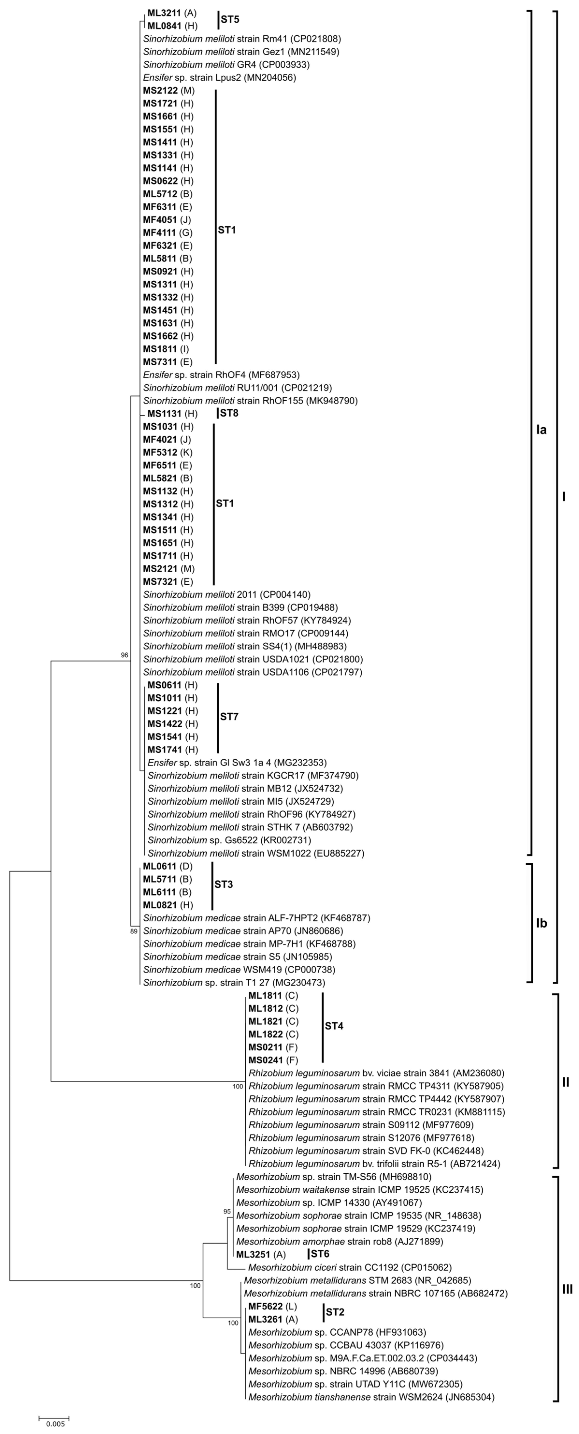
The 16S rDNA sequences from our isolates clustered into three major groups: forty-eight isolates clustered in the Sinorhizobium group (I), six isolates clustered in the Rhizobium leguminosarum group (II), and three isolates clustered in the Mesorhizobium spp. group (III) (Figure 2). Pairwise comparisons of 16S rDNA sequences revealed that the native isolates within the Sinorhizobium group shared a similarity of 99.6% to 100% and were similarly related to their reference strains. However, two well-defined subgroups (Ia and Ib) were distinguished within the main cluster. The S. meliloti group (Ia) contained isolates from all three sampled Medicago species, grouped into four different STs, one of which is a singleton ST. The S. medicae group (Ib) contained four isolates grouped into a single ST, all of which were from M. lupulina plants collected from three different locations. The R. leguminosarum group (II) contained only one ST, with isolates from M. lupulina and M. sativa plants from two different plant populations. The six R. leguminosarum native isolates were placed in a robust lineage with their reference strains, to which 100% similarity was shared. The Mesorhizobium sp. group (III) contained two STs, one of which was a singleton ST. These two STs contained isolates from M. lupulina and M. falcata plants from two different plant populations. The pairwise comparison revealed that isolates within the two STs shared 98.9% similarity.
Phylogenetic analysis of the individual and concatenated atpD, glnII, and recA chromosomal genes was used to classify the native isolates. All the individual chromosomal markers (atpD, glnII, recA) followed the same clustering pattern and maintained the same four distinct groups (Ia, Ib, II, III) with varying levels of intra-group diversity (Supplementary Figures S3–S5). The native bacterial isolates grouped into several well-defined phyletic lineages, together with their most closely affiliated strains, to which they shared sequence identity values ranging from 97.2 to 100% for atpD, 98 to 100% for glnII, and 96.4 to 100% for recA. The glnII phylogeny showed greater variability, and the isolates were placed in sixteen STs (Supplementary Figure S4). Ten STs were found in the recA phylogeny (Supplementary Figure S5), whereas the atpD gene was less discriminative at strain level, with nine STs being distributed across the phylogenetic tree (Supplementary Figure S3). Although they carried highly similar 16S rDNA sequences, the isolates comprising the Sinorhizobium cluster revealed a more diverse genetic background: in all chromosomal phylogenies, they could be confidently grouped into two groups, defined as S. meliloti (Ia) and S. medicae (Ib). Pairwise comparisons showed that the two Sinorhizobium clades were distantly related to one another, with sequence identity varying from 92% to 93.9% in the described chromosomal phylogenies.
The phylogeny inferred from the concatenated sequences of the three chromosomal markers and their corresponding most similar sequences of reference strains obtained from GenBank was consistent with the individual phylogenies but resolved the native rhizobial isolates into more robust lineages (Figure 3). Eighteen STs were found in the concatenated chromosomal set, of which ten were singleton STs. The S. meliloti group contained ten different STs, of which four were singleton STs; the S. medicae group contained two STs, of which one was a singleton ST; the R. leguminosarum group contained three STs, of which two were singleton STs, and the Mesorhizobium spp. group contained three STs, all of which were singleton STs. Pairwise comparisons revealed that of the STs in the MLST (Multilocus sequence typing) phylogeny, those within the S. meliloti, S. medicae, and R. leguminosarum groups shared high similarity ranging from 99.3 to 99.9%. In contrast, the three isolates positioned within the Mesorhizobium spp. clade were more distantly related to each other, with similarities ranging from 92 to 97.1%. Isolate ML3251 was the most divergent, and it only shared 92.1 to 93.6% relatedness to the Mesorhizobium spp. reference strains included in the tree.
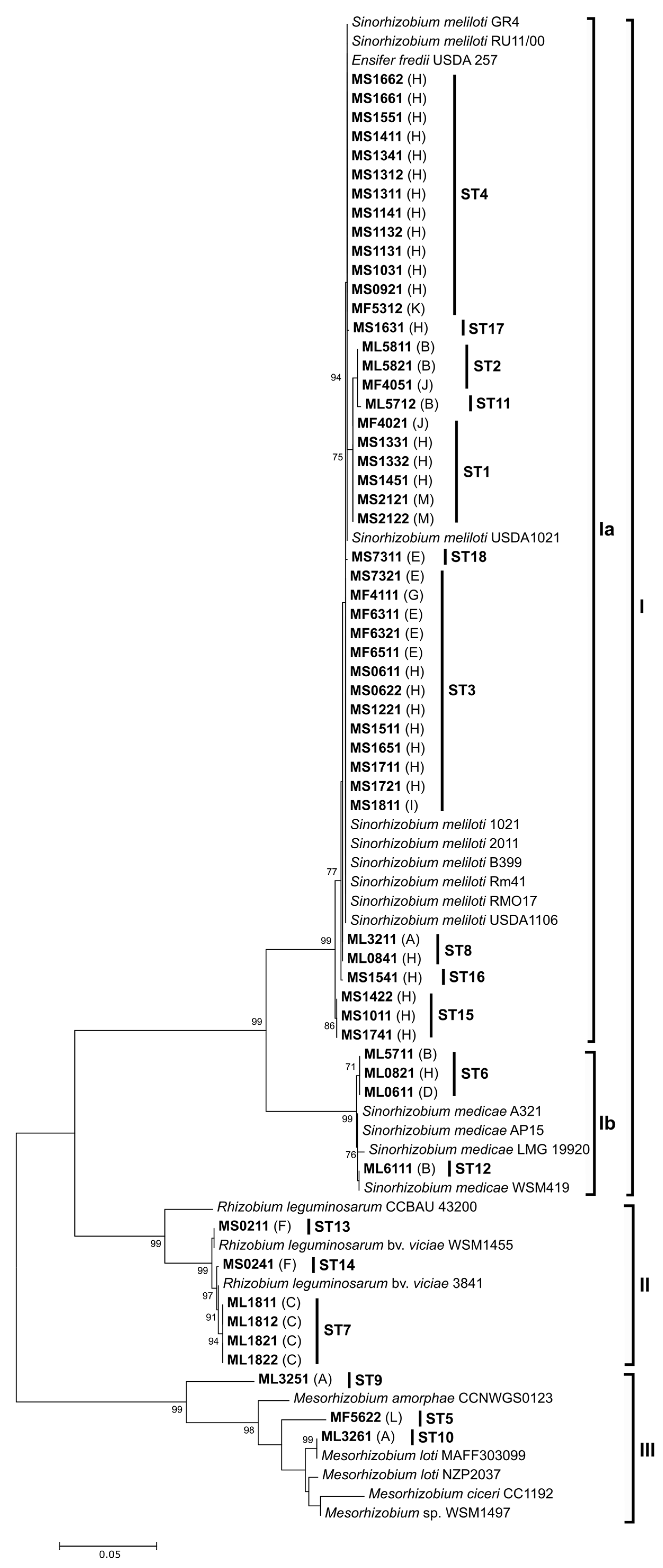
The phylogenies of symbiotic genes were partially consistent with those derived from the chromosomal genes. The nifH phylogeny maintained the same pattern of four distinct groups and the same assignment of the isolates to the groups (Supplementary Figure S6). For nodA, a slightly different pattern emerged, with the MF5622, ML3251, and ML3261 isolates being clustered with the Rhizobium leguminosarum group and not with the Mesorhizobium spp. group, as was the case for the chromosomal loci (Figure 4). A pairwise comparison of the nodA gene alignment revealed that within the defined Rhizobium leguminosarum group, the native isolates were placed in two distinct, distantly related lineages, sharing only 95.2 to 96% similarity. Notably, these strains exhibited only 53.9 to 55.8% similarity to the Mesorhizobium spp. reference strains included in the nodA phylogram. The number of sequence types varied for each marker, from 16 for glnII to six for nifH, and the percentage of singleton sequence types was the lowest for nifH (16.6%) and the highest for recA (70%), while the rest had a percentage between 25% and 50%. The number of variable sites varied, but the ratio of variable sites (VS)/parsimony informative (PI) sites had similar values, ranging from 1 (nodA) to 1.1 (atpD). Considering the length of the sequence, 16S had the lowest percentage of parsimony informative sites (7%), while the rest of the markers had values ranging from 20% for atpD and recA, to 29% for nodA (Table 1).
The sampled plant populations are located in four distinct regions: Rarău, Lespezi, Iași, and Oituz (Figure 1 and Supplementary Table S1). The Rarău region is located in the Northern part of the Eastern Carpathian Mountains and contains plant populations A, B, C, and D. The elevation for the sampled plants varies from ca. 500 m to just over 1000 m. Only Medicago lupulina plants were sampled here, and this region is the only one in this study where the rhizobial isolates belong to all of the four identified rhizobial groups. From the Lespezi region, only one plant population (E) was sampled, consisting of M. falcata and M. sativa plants. S. meliloti was the only rhizobial species identified and found in both plant species. The Iași region contains the F, G, H, I, and J plant populations and is the region with the lowest elevation (mean elevation less than 100 m). This is the only region where all three of the Medicago species were sampled; S. meliloti was the dominant isolate, having been found on all three Medicago species. The Oituz region contains the K, L, and M plant populations, with only M. falcata and M. sativa plants being sampled. The Mesorhizobium spp. isolate from the M. falcata population L (MF5622) has the same 16S sequence type as isolate ML3261 from the M. lupulina plant population A in the Rarău region, about 200 km away.
In the present study, S. meliloti was found in all sampled regions, regardless of the host plant, and was identified in nine out of the fourteen plant populations and on all three of the Medicago species. Sinorhizobium medicae was identified in three plant populations (B, D, and H) from two different regions; it was identified only in Medicago lupulina plants. All four of the S. medicae isolates belonged to the same 16S rDNA sequence type. Rhizobium leguminosarum was found in two plant populations (C and F) from two different regions: in population C, it was identified in M. lupulina plants, and in population F, it was found in M. sativa plants. All six R. leguminosarum isolates had the same 16S rDNA sequence type that was previously found in R. leguminosarum isolates from white clover and red clover plants from the same region [51,52]. Mesorhizobium spp. was identified in Medicago lupulina in plant population A and in M. falcata in plant population L, and the three rhizobial isolates belonged to two distinct 16S rDNA sequence types.

3.3. Diversity of Rhizobia Associated with Medicago: Comparative Analyses

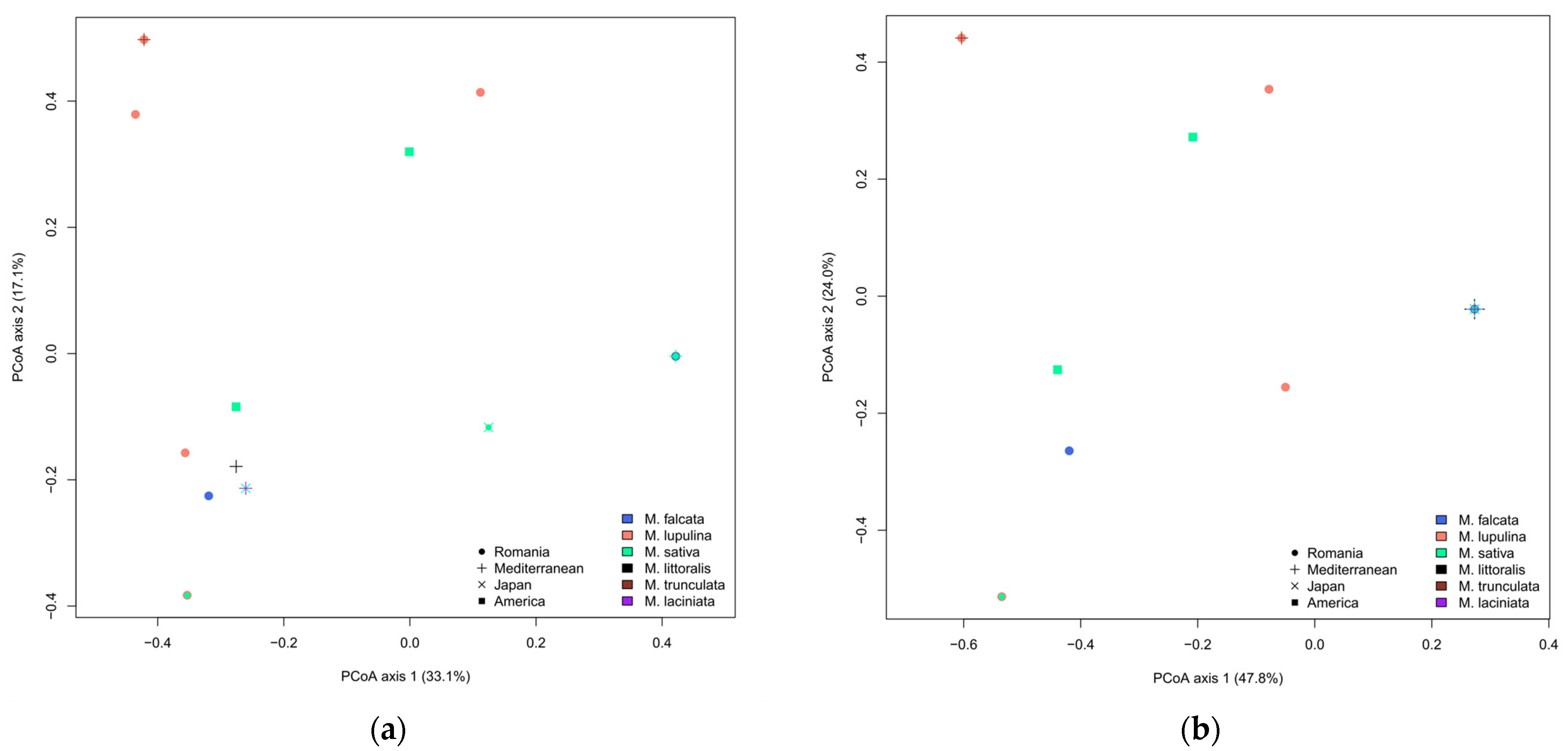
Rhizobial communities associated with Medicago hosts in Romania were compared with those previously observed in other geographic regions (Supplementary Tables S3 and S4) to examine whether these communities are shaped by Medicago species identity or by location. We found that differences in ST composition were significantly shaped by Medicago species identity (F5,14 = 1.629, p = 0.011) but not by geographic region (F3,14 = 1.229, p = 0.179; Figure 5a). This pattern arises because most commonly sampled Medicago species (M. falcata, M. lupulina, and M. sativa) share only 11–50% of STs and because ST10 is prevalent across most (61.5%) Medicago communities but less common in M. lupulina communities (20%; Table S3). Differences in species composition were not significantly affected by either Medicago species identity (F, = 1.389, p = 0.077) or geographic location (F, = 1.452, p = 0.071; Figure 5b).

4. Discussion

Building on our previous results from white clover and red clover populations [51,52], the present study adds another piece to the rhizobial diversity puzzle in Eastern Romania. Rhizobia were sampled from active root nodules of Medicago lupulina, M. falcata, and M. sativa plants. The initial screening and genus-level identification of rhizobia using partial 16S rDNA sequencing revealed that the rhizobial isolates belonged to four groups: Sinorhizobium meliloti, S. medicae, Rhizobium leguminosarum, and Mesorhizobium sp., and all were able to nodulate their respective host-plants in lab conditions. The 16S rDNA sequencing was sufficient to identify most isolates at the species level. Further sequencing of housekeeping and plasmid genes added sequence types and increased phylogenetic resolution among them, but the Mesorhizobium isolates could not be discerned at the species level.
Globally, Medicago species were often found to associate with Sinorhizobium meliloti, while other rhizobia (e.g., Sinorhizobium medicae) are less prevalent (Supplementary Table S3). In the present study, Sinorhizobium meliloti was the predominant rhizobial species, having been identified in all three Medicago species, regardless of elevation or soil characteristics. We identified Sinorhizobium medicae only in M. lupulina nodules. The two sinorhizobia were found nodulating both perennial and annual Medicago species in Mexico, with S. meliloti being the main symbiont of cultivated alfalfa plants [64]. In a field inoculation experiment, Roberts et al. [65] found no obvious preference among S. meliloti and S. medicae in nodulating either M. sativa or M. lupulina. In a legume survey in Tibet, Hou et al. [66] found that S. meliloti nodulates M. falcata and M. sativa plants, but Rhizobium species nodulate M. lupulina plants. In this study, the only sinorhizobia naturally found to form active nodules with M. falcata was S. meliloti, which has also been reported by Muntyan et al. [67]. Studies on rhizobial diversity in association with M. lupulina and M. falcata are scarce, and further sampling of natural populations of these plants could provide more information regarding their associated rhizobial community.
We found no underlying structure in the sinorhizobial isolates. The 16S rDNA ST1 of S. meliloti is shared among isolates from all four regions. Sinorhizobium meliloti isolate ML3211 from plant population A in the Rarău region shares the same 16S rDNA ST with S. meliloti isolate ML0841 from plant population H in the Iași region. All four S. medicae isolates found in plant populations B, D, and H in the two aforementioned regions also share between them the same 16S rDNA ST, showing no geographic structuring. The concatenated chromosomal set showed that the isolates from the Iași region shared identical STs with isolates from all the other three regions, but the other three regions did not share a common ST between them. The inability to discern structure could also occur because few plant populations were sampled. In a genotyping-by-sequencing study, Harrison et al. [68] similarly found no evidence of population structure within either S. meliloti or S. medicae in nodules of M. lupulina plants along a latitude gradient.
In Romanian Medicago communities, we also detected R. leguminosarum and two Mesorhizobium species. Both R. leguminosarum and Mesorhizobium spp. had previously been detected in M. lupulina by De Meyer et al. [40]. Consistent with the previous study in Belgium [40], which found that Medicago lupulina hosted the most diverse rhizobial community among all sampled Medicago species, we found that M. lupulina was the only plant species that harbored all four groups of rhizobial isolates that we identified.
Isolates found in M. lupulina also occurred in other host species. For example, R. leguminosarum bacteria harboring the same 16S rDNA sequence type that was identified in M. lupulina nodules in plant population C in the Rarău region are widespread in Eastern Romania, having been identified in three regions on at least four plant species, including M. sativa (population F from the Iaşi region), white clover (Trifolium repens), and red clover (Trifolium pratense) in multiple populations from both the Rarău and Oituz regions [51,52]. Three species of Rhizobium and S. meliloti were also identified as nodulating alfalfa varieties in China [23].
The mesorhizobia we identified in Romania belongs to two different 16S rDNA STs on well-supported clades. Both sequence types have been observed elsewhere in the world and are able to modulate a diversity of legume species. Isolate ML3251 shares a sequence type with mesorhizobia identified on Sophora microphylla from New Zealand [69], Oxytropis sp. from Canada [70], and Robinia pseudoacacia from South Korea [71] and Germany [72]. Isolates ML3261 and MF5622 share the same 16S rDNA ST with mesorhizobia from Cicer arietinum in Ethiopia [73], Glycine max in Latvia (unpublished, GenBank accession number OQ236321.1, https://www.ncbi.nlm.nih.gov/nuccore/OQ236321, accessed on 13 July 2023), Cicer canariense in the Canary Islands [74], Lotononis sp. in South Africa [75], and Lotus corniculatus in the USA [76]. Mesorhizobium species have previously been sampled from M. lupulina. For example, Zeng et al. [77] used 16S rDNA sequencing to identify two species of Mesorhizobium nodulating M. lupulina plants in a karstic region of China. To our knowledge, we also provide the first report of Mesorhizobium species in M. falcata nodules.
The two 16S rDNA mesorhizobia clades from this study are congruent with clades delineated by the other sequenced genetic markers, except nodA. The nodA sequences from the isolated Mesorhizobium species cluster together with the nodA sequences from Rhizobium leguminosarum isolates and are closely related to the nodA STs previously identified in red clover and white clover plants [51,52]. This phylogenetic incongruence could result from the horizontal transfer of symbiotic genes between rhizobia, a geographically widespread and common occurrence that is unrestricted between and within rhizobial genera [78]. Previous studies have detected the transfer of nodA genes from sinorhizobial species to mesorhizobia. Gerding et al. [79] provided evidence for lateral gene transfer in a Mesorhizobium strain nodulating Lessertia in South Africa. That strain harbored a nodA gene closely related to the nodA that we found in S. meliloti. Fterich et al. [80] identified a Mesorhizobium isolate from Prosopis farcta in Tunisia containing a nodA sequence identical to the one we found in S. medicae. This previous evidence, coupled with our own findings, could indicate that mesorhizobia are especially susceptible to the acquisition of foreign nodulation genes capable of establishing symbioses in specific soil conditions or under the constraint of a more selective plant species. Epstein and Tiffin [81] showed that the genes involved in nodule formation display rates of transfer 50% or higher than the nitrogen fixation genes. Our results could also explain why the nifH sequences of mesorhizobia from his study cluster with nifH sequences of other Mesorhizobium species, but the nodA sequences cluster outside of Mesorhizobium.
The 16S rDNA ST comparative analysis results, indicated that various Medicago species generally share the same rhizobial species across the globe. However, Mesorhizobium and R. leguminosarum, which were identified in this study, had not been reported before. Moreover, it is possible that different Medicago species prefer different STs. This may provide an explanation for contrasting results seen in different studies, where apparent species preferences are evident in some cases (e.g., here, [64,66]) while absent in others (e.g., [65]). It is unclear whether Medicago spp. have preferences for certain strains regardless of species identity or whether this is the result of a lack of studies on the diversity of rhizobia associated with Medicago. Clearly, more extensive sampling is needed to test this preference hypothesis.

Supplementary Materials

The following supporting information can be downloaded at: https://www.mdpi.com/article/10.3390/agronomy14092156/s1, Figure S1: Cluster analysis of phenotypic features by UPGMA; Figure S2: Basic descriptive statistics of inoculation experiments; Figure S3: Maximum Likelihood tree of the atpD sequences; Figure S4: Maximum Likelihood tree of the glnII sequences; Figure S5: Maximum Likelihood tree of the recA sequences; Figure S6: Maximum Likelihood tree of the nifH sequences; Table S1: Sampled Medicago populations, physical characteristics of sampling sites and rhizobial isolates used in the present study; Table S2: Phenotypic characterization of rhizobial isolates associated with Medicago plants and clusters defined by numerical taxonomy; Table S3: Global distribution of STs in Medicago spp.; Table S4: List of sequences used in the phylogenetic analysis.

Author Contributions

A.S.: Conceptualization, Investigation, Formal analysis, Visualization, Writing—original draft; J.V.C.: Conceptualization, Formal analysis, Writing—original draft; C.M.R.: Investigation, Formal analysis, Writing—original draft; C.S.: Investigation, Formal analysis; C.C.: Formal analysis, Writing—original draft; E.L.S.: Resources, Writing—review and editing; C.I.: Resources; D.T.: Resources, Writing—review and editing; E.F.: Conceptualization, Writing—review and editing; R.C.E.: Conceptualization, Investigation, Formal analysis, Writing—original draft, Resources, Project administration, Funding acquisition. All authors have read and agreed to the published version of the manuscript.

Funding

This research was funded by the UEFISCDI PN-II-ID-PCE-2011-3-1011 funding grant (contract no: 292/5.10.2011), the Core-Program, within the National Plan for Research, Development and Innovation 2022-2027 (project no. 7N/23020402/2023) and the project ResPonSE, (contract no. 760010/30.12.2022), developed with the support of the Romanian Ministry of Research, Innovation and Digitalization. This work was supported by NSF grants DEB-1457508 and IOS-1759048 awarded to Ellen Simms, which supported J.V.C. and C.C. J.V.C. was also supported by the Deutsche Forschungsgemeinschaft (DFG, German Research Foundation) under Germany’s Excellence Strategy–EXC 2051–Project-ID 390713860 and by the European Research Council (ERC) Consolidator grant 865694: DiversiPHI, the Deutsche Forschungsgemeinschaft (DFG, German Research Foundation) under Germany’s Excellence Strategy–EXC 2051–Project-ID 390713860, and the Alexander von Humboldt Foundation in the context of an Alexander von Humboldt-Professorship founded by German Federal Ministry of Education and Research.

Data Availability Statement

DNA sequences are deposited in GenBank and are publicly available. All data supporting the findings of this study are available in the paper and its Supplementary Information section.

Conflicts of Interest

The authors declare no conflicts of interest.

References

  1. Masson-Boivin, C.; Giraud, E.; Perret, X.; Batut, J. Establishing nitrogen-fixing symbiosis with legumes: How many rhizobium recipes? Trends Microbiol. 2009, 17, 458–466. [Google Scholar] [CrossRef]
  2. Wang, D.; Yang, S.; Tang, F.; Zhu, H. Symbiosis Specificity in the Legume: Rhizobial Mutualism. Cell. Microbiol. 2012, 14, 334–342. [Google Scholar] [CrossRef] [PubMed]
  3. Wang, Q.; Liu, J.; Zhu, H. Genetic and Molecular Mechanisms Underlying Symbiotic Specificity in Legume-Rhizobium Interactions. Front. Plant Sci. 2018, 9, 313. [Google Scholar] [CrossRef] [PubMed]
  4. Lindström, K.; Mousavi, S.A. Effectiveness of nitrogen fixation in rhizobia. Microb. Biotechnol. 2019, 13, 1314–1335. [Google Scholar] [CrossRef]
  5. Schulte, C.C.M.; Borah, K.; Wheatley, R.M.; Terpolilli, J.J.; Saalbach, G.; Crang, N.; de Groot, D.H.; Ratcliffe, R.G.; Kruger, N.J.; Papachristodoulou, A.; et al. Metabolic control of nitrogen fixation in rhizobium-legume symbioses. Sci. Adv. 2021, 7, eabh2433. [Google Scholar] [CrossRef] [PubMed]
  6. Masson-Boivin, C.; Sachs, J.L. Symbiotic nitrogen fixation by rhizobia—The roots of a success story. Curr. Opin. Plant Biol. 2018, 44, 7–15. [Google Scholar] [CrossRef]
  7. Herridge, D.F.; Peoples, M.B.; Boddey, R.M. Global inputs of biological nitrogen fixation in agricultural systems. Plant Soil 2008, 311, 1–18. [Google Scholar] [CrossRef]
  8. Hénault, C.; Barbier, E.; Hartmann, A.; Revellin, C. New Insights into the Use of Rhizobia to Mitigate Soil N2O Emissions. Agriculture 2022, 12, 271. [Google Scholar] [CrossRef]
  9. Li, J.; Wang, J.; Liu, H.; Macdonald, C.A.; Singh, B.K. Application of microbial inoculants significantly enhances crop productivity: A meta-analysis of studies from 2010 to 2020. J. Sustain. Agric. Environ. 2022, 1, 216–225. [Google Scholar] [CrossRef]
  10. Maitra, S.; Brestic, M.; Bhadra, P.; Shankar, T.; Praharaj, S.; Palai, J.B.; Shah, M.M.R.; Barek, V.; Ondrisik, P.; Skalický, M.; et al. Bioinoculants—Natural Biological Resources for Sustainable Plant Production. Microorganisms 2021, 10, 51. [Google Scholar] [CrossRef]
  11. Chamkhi, I.; Cheto, S.; Geistlinger, J.; Zeroual, Y.; Kouisni, L.; Bargaz, A.; Ghoulam, C. Legume-based intercropping systems promote beneficial rhizobacterial community and crop yield under stressing conditions. Ind. Crop. Prod. 2022, 183, 114958. [Google Scholar] [CrossRef]
  12. Kebede, E. Contribution, Utilization, and Improvement of Legumes-Driven Biological Nitrogen Fixation in Agricultural Systems. Front. Sustain. Food Syst. 2021, 5, 767998. [Google Scholar] [CrossRef]
  13. Layek, J.; Das, A.; Mitran, T.; Nath, C.; Meena, R.S.; Yadav, G.S.; Shivakumar, B.G.; Kumar, S.; Lal, R. Cereal+Legume Intercropping: An Option for Improving Productivity and Sustaining Soil Health. In Legumes for Soil Health and Sustainable Management; Meena, R.S., Das, A., Yadav, G.S., Lal, R., Eds.; Springer: Singapore, 2018; pp. 347–386. ISBN 9789811302534. [Google Scholar] [CrossRef]
  14. Fageria, N.K. Green Manuring in Crop Production. J. Plant Nutr. 2007, 30, 691–719. [Google Scholar] [CrossRef]
  15. Gatsios, A.; Ntatsi, G.; Celi, L.; Said-Pullicino, D.; Tampakaki, A.; Savvas, D. Legume-Based Mobile Green Manure Can Increase Soil Nitrogen Availability and Yield of Organic Greenhouse Tomatoes. Plants 2021, 10, 2419. [Google Scholar] [CrossRef] [PubMed]
  16. Meena, B.L.; Fagodiya, R.K.; Prajapat, K.; Dotaniya, M.L.; Kaledhonkar, M.J.; Sharma, P.C.; Meena, R.S.; Mitran, T.; Kumar, S. Legume Green Manuring: An Option for Soil Sustainability. In Legumes for Soil Health and Sustainable Management; Meena, R.S., Das, A., Yadav, G.S., Lal, R., Eds.; Springer: Singapore, 2018; pp. 387–408. ISBN 9789811302534. [Google Scholar] [CrossRef]
  17. Lindström, K.; Murwira, M.; Willems, A.; Altier, N. The biodiversity of beneficial microbe-host mutualism: The case of rhizobia. Res. Microbiol. 2010, 161, 453–463. [Google Scholar] [CrossRef]
  18. Argaw, A.; Tsigie, A. Indigenous rhizobia population influences the effectiveness of Rhizobium inoculation and need of inorganic N for common bean (Phaseolus vulgaris L.) production in eastern Ethiopia. Chem. Biol. Technol. Agric. 2015, 2, 19. [Google Scholar] [CrossRef]
  19. Athul, P.P.; Patra, R.K.; Sethi, D.; Panda, N.; Mukhi, S.K.; Padhan, K.; Sahoo, S.K.; Sahoo, T.R.; Mangaraj, S.; Pradhan, S.R.; et al. Efficient native strains of rhizobia improved nodulation and productivity of French bean (Phaseolus vulgaris L.) under rainfed condition. Front. Plant Sci. 2022, 13, 1048696. [Google Scholar] [CrossRef]
  20. Castellano-Hinojosa, A.; Mora, C.; Strauss, S.L. Native Rhizobia Improve Plant Growth, Fix N2, and Reduce Greenhouse Emissions of Sunnhemp More than Commercial Rhizobia Inoculants in Florida Citrus Orchards. Plants 2022, 11, 3011. [Google Scholar] [CrossRef] [PubMed]
  21. Koskey, G.; Mburu, S.W.; Njeru, E.M.; Kimiti, J.M.; Ombori, O.; Maingi, J.M. Potential of Native Rhizobia in Enhancing Nitrogen Fixation and Yields of Climbing Beans (Phaseolus vulgaris L.) in Contrasting Environments of Eastern Kenya. Front. Plant Sci. 2017, 8, 443. [Google Scholar] [CrossRef]
  22. Nguyen, T.T.; Atieno, M.; Herrmann, L.; Nakasathien, S.; Sarobol, E.; Wongkaew, A.; Nguyen, K.T.; Lesueur, D. Does inoculation with native rhizobia enhance nitrogen fixation and yield of cowpea through legume-based intercropping in the northern mountainous areas of Vietnam? Exp. Agric. 2020, 56, 825–836. [Google Scholar] [CrossRef]
  23. Kang, W.; Shi, S.; Xu, L. Diversity and symbiotic divergence of endophytic and non-endophytic rhizobia of Medicago sativa. Ann. Microbiol. 2018, 68, 247–260. [Google Scholar] [CrossRef]
  24. van Berkum, P.; Elia, P.; Eardly, B.D. Multilocus Sequence Typing as an Approach for Population Analysis of Medicago-Nodulating Rhizobia. J. Bacteriol. 2006, 188, 5570–5577. [Google Scholar] [CrossRef]
  25. van Berkum, P.; Badri, Y.; Elia, P.; Aouani, M.E.; Eardly, B.D. Chromosomal and Symbiotic Relationships of Rhizobia Nodulating Medicago truncatula and M. laciniata. Appl. Environ. Microbiol. 2007, 73, 7597–7604. [Google Scholar] [CrossRef]
  26. Lemaire, B.; Dlodlo, O.; Chimphango, S.; Stirton, C.; Schrire, B.; Boatwright, J.S.; Honnay, O.; Smets, E.; Sprent, J.; James, E.K.; et al. Symbiotic diversity, specificity and distribution of rhizobia in native legumes of the Core Cape Subregion (South Africa). FEMS Microbiol. Ecol. 2015, 91, 1–17. [Google Scholar] [CrossRef]
  27. Van Cauwenberghe, J.; Verstraete, B.; Lemaire, B.; Lievens, B.; Michiels, J.; Honnay, O. Population structure of root nodulating Rhizobium leguminosarum in Vicia cracca populations at local to regional geographic scales. Syst. Appl. Microbiol. 2014, 37, 613–621. [Google Scholar] [CrossRef] [PubMed]
  28. Kozieł, M.; Kalita, M.; Janczarek, M. Genetic diversity of microsymbionts nodulating Trifolium pratense in subpolar and temperate climate regions. Sci. Rep. 2022, 12, 12144. [Google Scholar] [CrossRef]
  29. Bessadok, K.; Navarro-Torre, S.; Fterich, A.; Caviedes, M.A.; Pajuelo, E.; Rodríguez-Llorente, I.D.; Mars, M. Diversity of rhizobia isolated from Tunisian arid soils capable of forming nitrogen-fixing symbiosis with Anthyllis henoniana. J. Arid. Environ. 2021, 188, 104467. [Google Scholar] [CrossRef]
  30. Efstathiadou, E.; Ntatsi, G.; Savvas, D.; Tampakaki, A.P. Genetic characterization at the species and symbiovar level of indigenous rhizobial isolates nodulating Phaseolus vulgaris in Greece. Sci. Rep. 2021, 11, 8674. [Google Scholar] [CrossRef]
  31. Tampakaki, A.P.; Fotiadis, C.T.; Ntatsi, G.; Savvas, D. Phylogenetic multilocus sequence analysis of indigenous slow-growing rhizobia nodulating cowpea (Vigna unguiculata L.) in Greece. Syst. Appl. Microbiol. 2017, 40, 179–189. [Google Scholar] [CrossRef]
  32. Simbine, M.G.; Mohammed, M.; Jaiswal, S.K.; Dakora, F.D. Functional and genetic diversity of native rhizobial isolates nodulating cowpea (Vigna unguiculata L. Walp.) in Mozambican soils. Sci. Rep. 2021, 11, 12747. [Google Scholar] [CrossRef]
  33. Steele, K.P.; Ickert-Bond, S.M.; Zarre, S.; Wojciechowski, M.F. Phylogeny and character evolution in Medicago (Leguminosae): Evidence from analyses of plastid trnK/matK and nuclear GA3ox1 sequences. Am. J. Bot. 2010, 97, 1142–1155. [Google Scholar] [CrossRef]
  34. Kang, Y.; Han, Y.; Torres-Jerez, I.; Wang, M.; Tang, Y.; Monteros, M.; Udvardi, M. System responses to long-term drought and re-watering of two contrasting alfalfa varieties. Plant J. 2011, 68, 871–889. [Google Scholar] [CrossRef]
  35. Zhang, L.-L.; Zhao, M.-G.; Tian, Q.-Y.; Zhang, W.-H. Comparative studies on tolerance of Medicago truncatula and Medicago falcata to freezing. Planta 2011, 234, 445–457. [Google Scholar] [CrossRef]
  36. Wang, H.; Coulman, B.; Bai, Y.; Tar’an, B.; Biligetu, B. Genetic diversity and local adaption of alfalfa populations (Medicago sativa L.) under long-term grazing. Sci. Rep. 2023, 13, 1632. [Google Scholar] [CrossRef]
  37. Epstein, B.; Branca, A.; Mudge, J.; Bharti, A.K.; Briskine, R.; Farmer, A.D.; Sugawara, M.; Young, N.D.; Sadowsky, M.J.; Tiffin, P. Population Genomics of the Facultatively Mutualistic Bacteria Sinorhizobium meliloti and S. medicae. PLoS Genet. 2012, 8, e1002868. [Google Scholar] [CrossRef]
  38. Rome, S.; Brunel, B.; Normand, P.; Fernandez, M.; Cleyet-Marel, J.C. Evidence that two genomic species of Rhizobium are associated with Medicago truncatula. Arch. Microbiol. 1996, 165, 285–288. [Google Scholar] [CrossRef]
  39. Bromfield, E.S.P.; Tambong, J.T.; Cloutier, S.; Prévost, D.; Laguerre, G.; van Berkum, P.; Thi, T.V.T.; Assabgui, R.; Barran, L.R. Ensifer, Phyllobacterium and Rhizobium species occupy nodules of Medicago sativa (alfalfa) and Melilotus alba (sweet clover) grown at a Canadian site without a history of cultivation. Microbiology 2010, 156, 505–520. [Google Scholar] [CrossRef]
  40. De Meyer, S.E.; Van Hoorde, K.; Vekeman, B.; Braeckman, T.; Willems, A. Genetic diversity of rhizobia associated with indigenous legumes in different regions of Flanders (Belgium). Soil Biol. Biochem. 2011, 43, 2384–2396. [Google Scholar] [CrossRef]
  41. Ciocârlan, V. Flora Ilustrată a României: Pteridophyta et Spermatophyta (The Illustrated Flora of Romania), 2nd ed.; Ceres: Bucharest, Romania, 2000; ISBN 973-40-0495-6. (In Romanian) [Google Scholar]
  42. Vîrdol, D.C. (Ed.) The Romanian Statistical Yearbook 2022; National Institute of Statistics (NIS): Bucharest, Romania, 2023; pp. 476–477. ISBN 1220-3246. Available online: https://insse.ro/cms/en/content/romanian-statistical-yearbook-book-format-5 (accessed on 11 October 2023).
  43. Beattie, G.A.; Handelsman, J. A rapid method for the isolation and identification of Rhizobium from root nodules. J. Microbiol. Methods 1989, 9, 29–33. [Google Scholar] [CrossRef]
  44. Vincent, J.M. A Manual for the Practical Study of the Root-Nodule Bacteria (IBP Handbuch No. 15 des International Biology Program, London); Blackwell Scientific Publishing: Oxford, UK; Edinburgh, UK, 1970. [Google Scholar] [CrossRef]
  45. Somasegaran, P.; Hoben, H.J. Quantifying the Growth of Rhizobia. In Handbook for Rhizobia: Methods in Legume-Rhizobium Technology; Somasegaran, P., Hoben, H.J., Eds.; Springer: New York, NY, USA, 1994; pp. 47–57. ISBN 978-1-4613-8375-8. [Google Scholar] [CrossRef]
  46. Frioni, L.; Rodríguez, A.; Meerhoff, M. Differentiation of rhizobia isolated from native legume trees in Uruguay. Appl. Soil Ecol. 2001, 16, 275–282. [Google Scholar] [CrossRef]
  47. Gao, J.-L.; Turner, S.L.; Kan, F.L.; Wang, E.T.; Tan, Z.Y.; Qiu, Y.H.; Gu, J.; Terefework, Z.; Young, J.P.W.; Lindström, K.; et al. Mesorhizobium septentrionale sp. nov. and Mesorhizobium temperatum sp. nov., isolated from Astragalus adsurgens growing in the northern regions of China. Int. J. Syst. Evol. Microbiol. 2004, 54, 2003–2012. [Google Scholar] [CrossRef] [PubMed]
  48. Josey, D.P.; Beynon, J.L.; Johnston, A.W.B.; Beringer, J.E. Strain Identification in Rhizobium Using Intrinsic Antibiotic Resistance. J. Appl. Bacteriol. 1979, 46, 343–350. [Google Scholar] [CrossRef]
  49. Mpepereki, S.; Makonese, F.; Wollum, A.G. Physiological characterization of indigenous rhizobia nodulating Vigna unguiculata in Zimbabwean soils. Symbiosis 1997, 22, 275–292. [Google Scholar]
  50. Sneath, P.H.A.; Sokal, R.R. Numerical Taxonomy: The Principles and Practice of Numerical Classification; W. H. Freeman: New York, NY, USA, 1973; ISBN 978-0-7167-0697-7. [Google Scholar]
  51. Efrose, R.C.; Rosu, C.M.; Stedel, C.; Stefan, A.; Sirbu, C.; Gorgan, L.D.; Labrou, N.E.; Flemetakis, E. Molecular diversity and phylogeny of indigenous Rhizobium leguminosarum strains associated with Trifolium repens plants in Romania. Antonie Van Leeuwenhoek 2017, 111, 135–153. [Google Scholar] [CrossRef]
  52. Stefan, A.; Van Cauwenberghe, J.; Rosu, C.M.; Stedel, C.; Labrou, N.E.; Flemetakis, E.; Efrose, R.C. Genetic diversity and structure of Rhizobium leguminosarum populations associated with clover plants are influenced by local environmental variables. Syst. Appl. Microbiol. 2018, 41, 251–259. [Google Scholar] [CrossRef]
  53. Rozas, J.; Ferrer-Mata, A.; Sánchez-DelBarrio, J.C.; Guirao-Rico, S.; Librado, P.; Ramos-Onsins, S.E.; Sánchez-Gracia, A. DnaSP 6: DNA Sequence Polymorphism Analysis of Large Data Sets. Mol. Biol. Evol. 2017, 34, 3299–3302. [Google Scholar] [CrossRef] [PubMed]
  54. Sayers, E.W.; Cavanaugh, M.; Clark, K.; Pruitt, K.D.; Schoch, C.L.; Sherry, S.T.; Karsch-Mizrachi, I. GenBank. Nucleic Acids Res. 2020, 49, D92–D96. [Google Scholar] [CrossRef] [PubMed]
  55. Altschul, S.F.; Gish, W.; Miller, W.; Myers, E.W.; Lipman, D.J. Basic Local Alignment Search Tool. J. Mol. Biol. 1990, 215, 403–410. [Google Scholar] [CrossRef]
  56. Kumar, S.; Stecher, G.; Li, M.; Knyaz, C.; Tamura, K. MEGA X: Molecular Evolutionary Genetics Analysis across Computing Platforms. Mol. Biol. Evol. 2018, 35, 1547–1549. [Google Scholar] [CrossRef]
  57. Goslee, S.C.; Urban, D.L. The ecodist Package for Dissimilarity-based Analysis of Ecological Data. J. Stat. Softw. 2007, 22, 1–19. [Google Scholar] [CrossRef]
  58. Oksanen, J.; Simpson, G.L.; Blanchet, F.G.; Kindt, R.; Legendre, P.; Minchin, P.R.; O’Hara, R.B.; Solymos, P.; Stevens, M.H.H.; Szoecs, E.; et al. Vegan: Community Ecology Package 2022. Available online: https://cran.r-project.org/web/packages/vegan/index.html (accessed on 11 October 2023).
  59. R Core Team. R: A Language and Environment for Statistical Computing; R Foundation for Statistical Computing: Vienna, Austria, 2020; Available online: https://www.r-project.org/ (accessed on 15 May 2023).
  60. Dray, S.; Dufour, A.-B. The ade4 Package: Implementing the Duality Diagram for Ecologists. J. Stat. Softw. 2007, 22, 1–20. [Google Scholar] [CrossRef]
  61. Siberchicot, A.; Julien-Laferrière, A.; Dufour, A.-B.; Thioulouse, J.; Dray, S. adegraphics: An S4 Lattice-Based Package for the Representation of Multivariate Data. R J. 2017, 9, 198–212. [Google Scholar] [CrossRef]
  62. QGIS Development Team. 2023. Available online: https://www.qgis.org/ (accessed on 15 May 2023).
  63. Inkscape Project, 2020. Available online: https://inkscape.org/ (accessed on 15 May 2023).
  64. Silva, C.; Kan, F.L.; Martínez-Romero, E. Population genetic structure of Sinorhizobium meliloti and S. medicae isolated from nodules of Medicago spp. in Mexico. FEMS Microbiol. Ecol. 2007, 60, 477–489. [Google Scholar] [CrossRef]
  65. Roberts, R.; Jackson, R.W.; Mauchline, T.H.; Hirsch, P.R.; Shaw, L.J.; Döring, T.F.; Jones, H.E. Is there sufficient Ensifer and Rhizobium species diversity in UK farmland soils to support red clover (Trifolium pratense), white clover (T. repens), lucerne (Medicago sativa) and black medic (M. lupulina)? Appl. Soil Ecol. 2017, 120, 35–43. [Google Scholar] [CrossRef]
  66. Hou, B.C.; Wang, E.T.; Li, Y.; Jia, R.Z.; Chen, W.F.; Man, C.X.; Sui, X.H.; Chen, W.X. Rhizobial Resource Associated with Epidemic Legumes in Tibet. Microb. Ecol. 2009, 57, 69–81. [Google Scholar] [CrossRef]
  67. Muntyan, V.S.; Baturina, O.A.; Afonin, A.M.; Cherkasova, M.E.; Laktionov, Y.V.; Saksaganskaya, A.S.; Kabilov, M.R.; Roumiantseva, M.L. Draft Genome Sequence of Sinorhizobium meliloti AK555. Microbiol. Resour. Announc. 2019, 8, e01567-18. [Google Scholar] [CrossRef]
  68. Harrison, T.L.; Wood, C.W.; Heath, K.D.; Stinchcombe, J.R. Geographically Structured Genetic Variation in the Medicago lupulina-Ensifer Mutualism. Evolution 2017, 71, 1787–1801. [Google Scholar] [CrossRef]
  69. De Meyer, S.E.; Tan, H.W.; Andrews, M.; Heenan, P.B.; Willems, A. Mesorhizobium calcicola sp. nov., Mesorhizobium waitakense sp. nov., Mesorhizobium sophorae sp. nov., Mesorhizobium newzealandense sp. nov. and Mesorhizobium kowhaii sp. nov. isolated from Sophora root nodules. Int. J. Syst. Evol. Microbiol. 2016, 66, 786–795. [Google Scholar] [CrossRef] [PubMed]
  70. Duan, Y.F.; Grogan, P.; Walker, V.K.; diCenzo, G.C. Whole genome sequencing of mesorhizobia isolated from northern Canada. Can. J. Microbiol. 2022, 68, 661–673. [Google Scholar] [CrossRef]
  71. Kang, J.W.; Song, J.; Doty, S.L.; Lee, D.K. Diversity of Rhizobia Associated with Leguminous Trees Growing in South Korea. J. Basic Microbiol. 2013, 53, 291–298. [Google Scholar] [CrossRef]
  72. Ulrich, A.; Zaspel, I. Phylogenetic diversity of rhizobial strains nodulating Robinia pseudoacacia L. Microbiology 2000, 146 Pt 11, 2997–3005. [Google Scholar] [CrossRef] [PubMed]
  73. Greenlon, A.; Chang, P.L.; Damtew, Z.M.; Muleta, A.; Carrasquilla-Garcia, N.; Kim, D.; Nguyen, H.P.; Suryawanshi, V.; Krieg, C.P.; Yadav, S.K.; et al. Global-level population genomics reveals differential effects of geography and phylogeny on horizontal gene transfer in soil bacteria. Proc. Natl. Acad. Sci. USA 2019, 116, 15200–15209. [Google Scholar] [CrossRef] [PubMed]
  74. Armas-Capote, N.; Pérez-Yépez, J.; Martínez-Hidalgo, P.; Garzón-Machado, V.; Del Arco-Aguilar, M.; Velázquez, E.; León-Barrios, M. Core and symbiotic genes reveal nine Mesorhizobium genospecies and three symbiotic lineages among the rhizobia nodulating Cicer canariense in its natural habitat (La Palma, Canary Islands). Syst. Appl. Microbiol. 2014, 37, 140–148. [Google Scholar] [CrossRef]
  75. Ardley, J.K.; Reeve, W.G.; O’Hara, G.W.; Yates, R.J.; Dilworth, M.J.; Howieson, J.G. Nodule morphology, symbiotic specificity and association with unusual rhizobia are distinguishing features of the genus Listia within the Southern African crotalarioid clade Lotononis s.l. Ann. Bot. 2013, 112, 1–15. [Google Scholar] [CrossRef]
  76. Qian, J.; Parker, M.A. Contrasting nifD and Ribosomal Gene Relationships Among Mesorhizobium from Lotus oroboides in Northern Mexico. Syst. Appl. Microbiol. 2002, 25, 68–73. [Google Scholar] [CrossRef] [PubMed]
  77. Zeng, Q.; Wei, X.; Wei, X.; Ou, E.; Ji, Y.; Shu, J.; Long, Z. Research on Resource Exploration, Nitrogen Fixation Characteristics and Diversity of Rhizobia of Medicago lupulina in Karst Mountainous Area of Guizhou. Acta Agrestia Sin. 2022, 30, 1891–1899. [Google Scholar] [CrossRef]
  78. Andrews, M.; De Meyer, S.; James, E.K.; Stępkowski, T.; Hodge, S.; Simon, M.F.; Young, J.P.W. Horizontal Transfer of Symbiosis Genes within and between Rhizobial Genera: Occurrence and Importance. Genes 2018, 9, 321. [Google Scholar] [CrossRef]
  79. Gerding, M.; O’Hara, G.W.; Bräu, L.; Nandasena, K.; Howieson, J.G. Diverse Mesorhizobium spp. with unique nodA nodulating the South African legume species of the genus Lessertia. Plant Soil 2012, 358, 385–401. [Google Scholar] [CrossRef]
  80. Fterich, A.; Mahdhi, M.; Caviedes, M.A.; Pajuelo, E.; Rivas, R.; Rodriguez-Llorente, I.D.; Mars, M. Characterization of root-nodulating bacteria associated to Prosopis farcta growing in the arid regions of Tunisia. Arch. Microbiol. 2011, 193, 385–397. [Google Scholar] [CrossRef]
  81. Epstein, B.; Tiffin, P. Comparative genomics reveals high rates of horizontal transfer and strong purifying selection on rhizobial symbiosis genes. Proc. Biol. Sci. 2021, 288, 20201804. [Google Scholar] [CrossRef]
Figure 1. Map of the sampled plant populations. The shapes represent the three medic species (triangle = Medicago lupulina, square = M. falcata, circle = M. sativa), and the colors represent the four identified rhizobial groups (blue = Sinorhizobium meliloti, yellow = S. medicae, green = Rhizobium leguminosarum, magenta = Mesorhizobium spp.). Each symbol represents one rhizobial isolate, with the exception of plant population H, where the enlarged circle stands for 27 isolates. The square part on the inset map represents the sampling area. The ellipses represent the four geographic regions. The position of the plant population letter (A–M) represents the sampling point.
Agronomy 14 02156 g001
Figure 2. Maximum Likelihood tree of the 16S rDNA sequences (1345 nt); the best-fit model was found to be Jukes-Cantor and a discrete Gamma distribution was used to model evolutionary rate differences among sites (5 categories). The isolates from this study are in bold, and the plant population of origin is in parentheses. The sequence types (ST) are also shown. The three main rhizobial groups are shown in Roman numerals (Ia, Ib, II, III). The ‘MF’, ‘ML’, and ‘MS’ in the rhizobial isolates names stand for the three Medicago species.
Agronomy 14 02156 g002
Figure 3. Maximum Likelihood tree based on the concatenated chromosomal gene sequences (atpD + glnII + recA, 1353 nt); the best-fit model was found to be Tamura 3-parameter and a discrete Gamma distribution was used to model evolutionary rate differences among sites (5 categories). The isolates from this study are in bold, and the plant population of origin is in parentheses. The sequence types (ST) are also shown. The three main rhizobial groups are shown in Roman numerals (Ia, Ib, II, III).
Agronomy 14 02156 g003
Figure 4. Maximum Likelihood tree based on the nodA sequences (580 nt); the best-fit model was found to be Tamura 3-parameter, and a discrete Gamma distribution was used to model evolutionary rate differences among sites (5 categories). The isolates from this study are in bold, and the plant population of origin is in parentheses. The sequence types (ST) are also shown. The three main rhizobial groups are shown in Roman numerals (Ia, Ib, II).
Agronomy 14 02156 g004
Figure 5. (a) PCoA plot comparing Sequence Type (ST) composition of Medicago spp. associated rhizobial communities using Jaccard distances. The first principal component (PCoA 1) and the second principal component (PCoA 2) explain 33.1% and 17.1% of the variance of the dataset, respectively. Distances between the points in the plot indicate the relative dissimilarity values of different regions and different Medicago spp. hosts. (b) PCoA plot comparing rhizobial species composition of Medicago spp., associated rhizobial communities using Jaccard distances. The first principal component (PCoA 1) and the second principal component (PCoA 2) explain 47.8% and 24.0% of the variance of the dataset, respectively. Distances between the points in the plot indicate the relative dissimilarity values of different regions and different Medicago spp. hosts.
Agronomy 14 02156 g005
Table 1. Sequence statistics of nucleotide polymorphisms and neutrality tests of 57 rhizobial isolates from Medicago species.
GMLST (sST)VarS/ParIπGCFu & Li’s
D*
Fu & Li’s
F*
Tajima’s
D
16S rDNA13458 (2)99/950.01255.11.465 *0.738−0.752
atpD4359 (3)97/880.04463.80.5470.261−0.311
glnII58816 (8)162/1510.05362.40.7980.426−0.344
recA33010 (7)75/690.04762.30.1980.077−0.153
nifH1686 (1)47/460.05560.71.806 **1.231−0.278
nodA58013 (5)171/1700.07856.92.066 **1.888 *0.803
GM = genetic marker; L = length of sequences (in nucleotides); ST = number of sequence types; sST = number of singleton sequence types; VarS = number of variable sites; ParI = number of parsimony-informative sites; π = nucleotide diversity; GC = percent of G + C content; Fu & Li’s D*, Fu & Li’s F*, Tajima’s D calculated using the total number of segregating sites. * Significant at p < 0.05, ** Significant at p < 0.02.
Disclaimer/Publisher’s Note: The statements, opinions and data contained in all publications are solely those of the individual author(s) and contributor(s) and not of MDPI and/or the editor(s). MDPI and/or the editor(s) disclaim responsibility for any injury to people or property resulting from any ideas, methods, instructions or products referred to in the content.