Communication

Strigolactone and Karrikin Signaling Influence the Recruitment of Wild Tobacco’s Root Microbiome in the Desert

1
Shenzhen Branch, Guangdong Laboratory of Lingnan Modern Agriculture, Key Laboratory of Synthetic Biology, Ministry of Agriculture and Rural Affairs, Agricultural Genomics Institute at Shenzhen, Chinese Academy of Agricultural Sciences, Shenzhen 518120, China
2
College of Life Sciences, South China Agricultural University, Guangzhou 510642, China
3
Department of Molecular Ecology, Max Planck Institute for Chemical Ecology, 07745 Jena, Germany
*
Authors to whom correspondence should be addressed.
These authors contributed equally to this work.
Agronomy 2025, 15(1), 44; https://doi.org/10.3390/agronomy15010044
Submission received: 31 October 2024 / Revised: 18 December 2024 / Accepted: 25 December 2024 / Published: 27 December 2024

Abstract

:
Survival in desert ecosystems poses significant challenges for plants due to harsh conditions. Plant microbiomes are thought to promote resilience; however, whether plant hormones, specifically strigolactones (SLs) and karrikins (KARs), shape plant microbiomes remains unknown. The recruitment of root-associated microbiomes in Nicotiana attenuata, a model desert plant, silenced in specific genes associated with SL biosynthesis (CCD7) and perception (D14), karrikin perception (KAI2), and in the shared receptor (MAX2), required for both pathways, was studied. SL and KAR signaling, with MAX2 as a co-regulator, fine-tuned the assembly of root-associated microbiomes, with unique and shared regulatory functions on bacterial microbiome recruitment, particularly in taproot. Significant variation among the different plant genotypes in bacterial diversity and composition in taproot and lateral roots provides a foundation for future research to explore how microbiomes function in plant resilience in these harsh environments.

1. Introduction

Plants that grow in deserts are profoundly challenged by harsh environmental conditions, such as intense solar radiation, extreme temperature fluctuations, limited water availability, and nutrient deficiencies. Several studies have demonstrated the essential role of root microbiomes in promoting plant growth and enhancing tolerance to different abiotic and biotic stresses [1,2,3]. The primary factors affecting the composition of plant microbial communities in desert environments are extreme conditions, such as drought, heat, and salinity [1]. Phytohormones are important regulators of growth and mediate adaptations to environmental stresses. Strigolactone (SL) and karrikin (KAR) signaling play essential roles in plant development and ecological interactions. However, how hormone signaling influences plant-microbe interactions, particularly in desert environments, remains unknown.
SLs are terpenoid lactones derived from carotenoids, and their biosynthesis is dependent on the key enzyme carotenoid cleavage dioxygenase 7 (CCD7) [4]. The receptor for SLs is DWARF14 (D14), an α/β hydrolase [4,5,6], whereas KARs are a group of butenolide compounds that bind to karrikin-insensitive 2 (KAI2), a paralogue of D14 [5]. The co-receptor involved in both SL and KAR pathways is more axillary growth 2 (MAX2) [7]. SLs play a crucial role in regulating plant architecture by inhibiting shoot branching, influencing yield and responding to biotic stress [8,9,10]. In addition, SLs also serve as signaling molecules in the rhizosphere, facilitating communication between host plants, parasitic plants, and symbiotic fungi [11,12]. The wild tobacco, Nicotiana attenuata, was used to study the effects of SL and KAR signaling on desert plant–microbiome interactions. N. attenuata is an ideal model, as it occurs naturally in the arid landscapes of the Great Basin Desert of Utah in the United States, and an Agrobacterium-mediated transformation system for this plant has been established [13].

2. Results

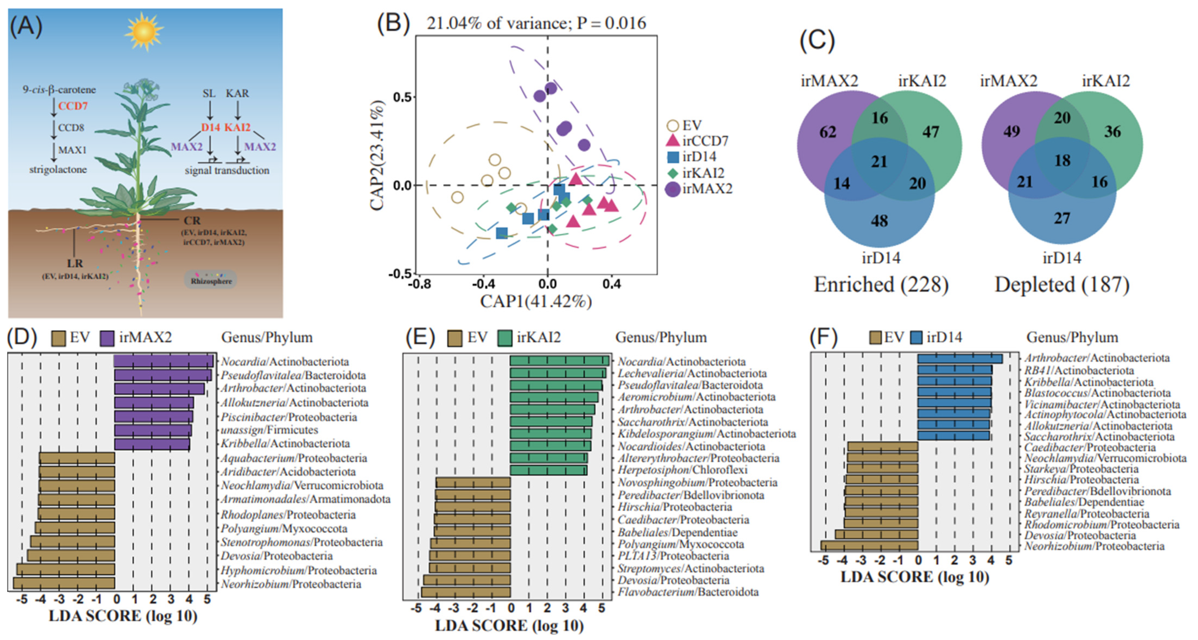
2.1. Distinct Roles of SL and KAR Signaling in Main Root Microbiome Recruitment

To clarify the function of SL and KAR signaling on the composition of the root-associated microbiome, we planted lines of N. attenuata, transformed with RNAi constructs targeting the expression of SL biosynthesis (irCCD7), SL perception (irD14), KAR perception (irKAI2), and the shared co-receptor (irMAX2), along with isogenic empty vector (EV)-transformed plants in a field plot in this species’ native habitat, the Great Basin Desert of Utah. Several independently transformed lines harboring single insertions of each construct were previously characterized [10,14]. The lines were planted during two field seasons with two field-planting procedures: 2018 (Jiffy pots) and 2020 (direct germination in the field). When plants are grown in Jiffy pots prior to field planting, they are unable to attain a natural root architecture consisting of a central tap root that enlarges into a carrot root (CR) with large lateral roots (LR) (Figure S1A). To allow for natural root development, a new planting procedure was implemented in 2020. Rather than using Jiffy pots for germination and transplanting early rosette-stage plants into the field, seeds were directly germinated in the field, allowing for the natural growth of a taproot system (Figure S1A and Figure 1B). After three months of growth, we carefully excavated roots and sampled the base of the taproot (CR) of EV, irCCD7, irD14, irKAI2, and irMAX2 lines. Each line was sampled with five replicates (Figure 1A). The root-associated bacterial community was then evaluated by the 16S ribosomal RNA (rRNA) gene sequencing with Illumina NovaSeq (Beijing, China), which generated 2,979,558 clean reads in total (averaging 119,182 and ranging from 90,170 to 127,997 reads per sample) (Table S1). After filtering and curating by QIIME 2 (v2023.2) (Quantitative Insights into Microbial Ecology 2), a total of 2,119,706 high-quality reads were used for further analysis (Table S1). Therefore, the amplicon sequence variants (ASVs) table was rarefied to 37,850 reads with 1,275 ASVs (Table S2). Rarefaction curves of observed richness and Shannon (H index) diversity were calculated. The curves of these values attained saturation in all examined samples (Figure S2), suggesting that the sequencing depth was sufficient to reflect species richness and to compare diversities.
The datasets from CR samples were then used for analyzing microbiome diversity and structure across different genotypes. To examine the impact of SL and KAR on the N. attenuata root microbiome communities, we analyzed the microbiome compositions (Figure S3) and conducted Chao1 richness and Shannon diversity analysis of CR samples from different genotypes (Figure S4). As a result, although Chao1 and Shannon index values were relatively higher in the irD14 CR samples, there was no significant difference between root bacterial community of these transgenic plants with suppressed SL and KAR signaling and EV plants (Figure S4). Constrained principal coordinate analysis (CPCoA) analysis of Bray–Curtis distances was conducted to present β-diversity of ASVs in the CR samples of different transgenic plants. Distinctly separated clusters in the CPCoA plot between EV samples versus irCCD7 and irD14 samples and EV samples versus irKAI2 samples (Figure 1B) reflect the critical roles of both SL and KAR signaling in microbiome recruitment. Moreover, the irMAX2 cluster was clearly separated from the EV and other transgenic lines clusters (Figure 1B), revealing that the absence of both SL and KAR signaling leads to significantly different microbiome diversity compared to impairing only one of the signaling systems. These findings highlight the diverse functions of SL and KAR signaling in recruiting diverse microbiomes.
To explore the role of receptors in SL and KAR signaling on root microbiome recruitment, Venn analyses were conducted on altered ASVs of irMAX2, irKAI2, and irD14 transgenic plants compared to the EV control. The analysis revealed a significant overlap of enriched and depleted ASVs in all three genotypes (Figure 1C). As D14 mediates SL reception, KAI2 acts as the KAR receptor, and MAX2 serves as a co-receptor for both pathways. The overlap of enriched and depleted ASVs in irMAX2 with either irD14 or irKAI2 samples was expected. Moreover, there were 41 enriched and 34 depleted ASVs common to both irD14 and irKAI2, although there are also uniquely altered ASVs in irD14 and irKAI2. This implies that SL and KAR signaling pathways distinctly regulate microbiome recruitment but also shared some regulatory mechanisms in recruiting specific microbiome species. LDA analysis revealed the unique genera recruited by each genotype, which mostly belonged to the Actinobacteria phylum. Actinobacteria have a significant impact on soil health, nutrient cycling, and plant productivity. They help to maintain the balance and resilience of the ecosystem [15]. irMAX2 shared specific ASVs with irKAI2 (e.g., Nocardia and Pseudoflavitalea) and irD14 (e.g., Kribella and Allokutzneria), suggesting the mutual function of irMAX2 in both SL and KAR pathways in regulating root microbiome recruitment (Figure 1D–F).

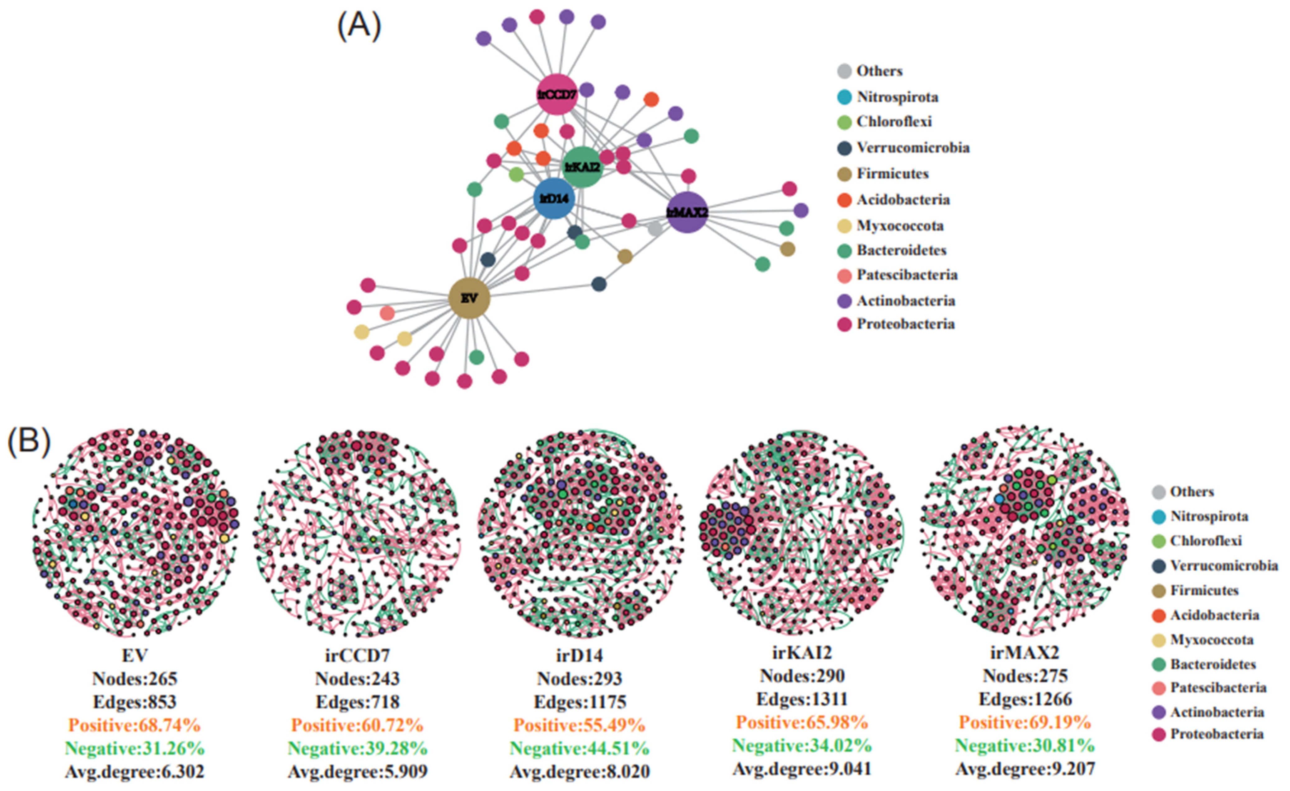
2.2. Distinct Roles of SL and KAR Signaling on Microbiome Network Connections

We next identified indicator species for each genotype and employed bipartite network analyses. The results showed a strong interconnection between indicator ASVs in different genotype’s CR samples, but also some unique indicators for each genotype. For example, irMAX2 plants shared indicator species with irCCD7, irD14, or irKAI2, such as ASV4334 (Devosia) and ASV3906 (Ellin6055), but also some unique indicators, including ASV1379 (Nocardia) and ASV2972 (Flavobacterium) (Figure 2A, Table S3). These results suggest cross talk between SL and KAR signaling and highlight the specific function of each gene in recruiting species. In EV’s CR, distinct bacteria species, such as ASV1236 (Polyangium), ASV3302 (Haliangium), ASV3583 (SM2D12), ASV3602 (Mesorhizobium), and ASV4291 (TM7a) (Figure 2A, Table S3), were found, suggesting that SL and KAR signaling are required for recruiting these bacterial species.
To investigate potential interactions between microbial taxa across different genotypes, co-occurrence network analysis was performed. The results showed that the EV bacterial community comprised 265 nodes (ASVs) and 853 edges with an average degree of 6.302. Notably, irCCD7 exhibited a reduced number of nodes and edges, whereas irD14, irKAI2, and irMAX2 exhibited an increased number. The network in irMAX2 consisted of 275 nodes, 1,266 edges, and an average degree of 9.207, with indices that were higher than those in EV (Figure 2B). These results suggest that the receptor genes play an essential role in regulating the microbiome network. Moreover, irMAX2 exhibited a more clustered topology due to the highly connected ASVs arranged in densely connected groups of nodes (Figure 2B, Table S4). For example, the structural analysis revealed that in irKAI2, ASVs from Actinobacteria and Proteobacteria exhibited a higher tendency to co-occur, possibly because of their important roles in plant–microbe interactions and ecological balance [15,16], while the co-occurrence of ASVs in irMAX2 was more complex (Figure 2B). These results indicate a central role of MAX2 in regulating microbial composition via connections through both SL and KAR signaling pathways.

2.3. SL and KAR Signaling Play Critical Roles in LR Microbiome Recruitment

The root system of N. attenuata is comprised of CR and LR. The CR is responsible for penetrating deep into the soil to acquire water, while the LR spread laterally from the plant to meet nutrient needs. As the CR and LR are produced at different times during plant growth and in different positions in the soil, it is possible that the hormone receptor genes regulate bacterial communities in each of these root types differently. To evaluate this, LR samples from EV, irD14, and irKAI2 plants were collected for analysis. As irMAX2 plants developed a leaf-bleaching phenotype in the field in response to exposure to high-light fluence [14] (Figure S5) and the CCD7 enzyme is involved in the biosynthesis of SLs and other carotenoid-derived molecules, such as 10′-apo-zeaxanthinal and blumenin [17], which may introduce other influences on root microbiome recruitment, irMAX2 and irCCD7 plants were excluded from this analysis. After assessing the data quality of the sequenced LR samples (Figures S6 and S7, Table S5), bacterial community differences between LR and CR samples from the same plants were compared. In a CPCoA analysis, the LR datasets separated from the CR datasets along the PCoA1 axis, which accounted for 35.28% of the correlation in bacterial community composition (Figure S8), indicating significant differences in bacterial diversity and community composition between the CR and LR of the same plant. The α-diversity values between all CR and LR samples (EV, irD14, and irKAI2 lines) was further analyzed. Compared with the CR samples, the LR samples exhibited significantly higher Chao1 richness and Shannon diversity indexes, indicating a significant increase in the complexity of bacterial composition of LR samples (Figure S9). This is perhaps not surprising, given that the LR develop first in the late stages of rosette growth and elongate through dry soil close to the soil surface, a process likely facilitated by hydraulic lift from the CR complex.
α-diversity metrics, including Chao1 richness and Shannon diversity, were then calculated for LR samples of EV, irD14, and irKAI2 lines. No significant differences were observed between genotypes in terms of both indices (Figure S10). CPCoA analysis was conducted to examine the β-diversity of ASVs in different transgenic plants’ LR samples. The CPCoA plot showed clear cluster separations between EV versus irD14 and irKAI2 samples (Figure S11). The results suggest critical roles for both SL and KAR signaling in LR microbiome recruitment. Venn analysis showed that LR samples of irD14 and irKAI2 plants had fewer enriched ASVs and similar enriched ASVs of LR samples compared to the CR samples; however, for the depleted ASVs, LR and CR samples had similar numbers (Figure 1C and Figure S12). LDA analysis revealed different unique ASVs in irD14 and irKAI2 LR samples compared to CR samples: in irD14 LR samples, only Ellin6067 from Proteobacteria was identified (Figure S13), which was not found in the CR samples (Figure 1D). Network analysis indicated that the LR of EV, irD14, and irKAI2 samples had similar node and edge numbers, and all exhibited similar levels of clustered topology with highly connected ASVs (Figure S14 and Table S6), which was different from the CR samples of these genotypes (Figure 2B). Therefore, the SL receptor, D14, and KAR receptor, KAI2, appear to play a more significant role in recruiting CR microbiomes than they do in the LR microbiomes.

3. Discussion

The root structure of plants, especially the CR and LR, play different roles in the absorption of water, minerals, and nutrients [18,19]. However, current research on the impact of plant root niche on microbial composition and structure mainly focuses on rhizosphere, rhizoplane, and endosphere rather than the CR and LR [20]. In this study, the results highlight the distinct contributions of CR and LR in shaping root-associated microbiomes in N. attenuata, with LR displaying greater diversity than CR. This could be attributed to the ability of LR to expand radially in the soil and determine the overall size of the root system, potentially interacting with a broader range of microorganisms [21]. However, the deep-root structure of the CR is crucial for uptaking deep soil water and nutrients [22], possibly adapting to specific microbial communities in deeper soil layers.
The involvement of SL and KAR signaling in plant–microbiome–soil interactions is critical for the recruitment of Arbuscular mycorrhizal fungi and for nodulation in soybeans [23,24]. Even though several studies have revealed the impact of SLs in shaping the rhizosphere bacterial community in rice and soybean [25,26,27], the role of KAI2 signaling remains unclear. Meanwhile, the impact of SL and KAR signaling on desert microbiomes has yet to be explored. This study provides evidence for SL and KAR signaling’s fine-tuning selection on the rhizosphere and endosphere bacterial microbiomes of N. attenuata, especially for the recruitment of microbiome in the taproot. It has been reported that silencing or knockout MAX2 and D14 produces higher SLs due to positive feedback on SL biosynthesis, while impairing CCD7 reduces SL concentrations [10,28]. Therefore, it is possible that part of the differences in microbiome recruitment of irMAX2, irD14 plants compared with irCCD7 plants result from the high concentration of SLs, which secrete to rhizosphere soil and attract particular microbes. The patterns observed in this study with manipulations of SL and KAR signaling will need to be tested more thoroughly in future research in more detailed mechanistic analyses with isolated bacterial species in synthetic root microbiomes to examine their effects on plant growth and stress resistance.

4. Methods

4.1. Plant Growth in Desert

All transgenic lines harbored a single, completely integrated inverted-repeat (ir) transformation construct containing 150–300 bp fragments of the genes targeted for silencing by RNAi. EV, irCCD7, irD14, irKAI2, and irMAX2 transgenic lines were characterized and reported in previous studies [10,14]. Transgenic seeds were imported, germinated, and planted at the Lytle Ranch Preserve under the U.S. Department of Agricultural Animal and Plant Health Inspection Service (APHIS) permits 07-341-101m, 18-046-102m, 18-282-102m.
In the 2018 field season (April–June, Snow Plot), seeds were first germinated in Jiffy pots and watered with borax solution supplemented with soil microbes collected from a natural N. attenuata population in the Utah desert. After approximately 3–4 weeks of growth in shade tents, seedlings were planted into the field plot. In the 2020 field season (April–June, Lytle Plot), after a 1 h soaking in a smoke and GA3 solution, seeds were sowed directly in the field to allow plants to develop a natural root, as described previously [14]. Briefly, a single seed was pipetted into the 3–4 mm depression, and 3-seed sowings were covered by a transparent plastic dome lid, which increased soil temperatures and protected young seedlings from wind storms. Germinated seedlings were subsequently thinned to a single seedling per dome, and domes were removed several days later.

4.2. Sample Collection

On the 5 June 2020 field season, entire plants of the EV, irCCD7, irD14, irKAI2, and irMAX2 genotypes were excavated with as much root tissue as possible, including taproot (CR) and lateral roots (LR). Soil adhering to the roots was carefully removed with a brush, and CR and LR were separated with a clean razor and harvested. For each genotype, root samples from five individual plants were collected. Samples were wrapped in labeled aluminum foil, immediately frozen on dry ice, and shipped to the laboratory in Germany in a dry-ice shipper. In the laboratory, samples were stored at −80°C until analysis. For 16S rRNA analysis, root samples were ground thoroughly in a liquid nitrogen pre-cooled mortar and sent to Novogene (Beijing, China) for analysis.

4.3. DNA Extraction, Library Preparation, and Sequencing

DNA was extracted from root and soil samples; DNA purity and concentration were monitored by nanodrop and gel electrophoresis. DNA was stored at −80°C until further analysis. Per sample, approximately 400 ng DNA was subjected to 16S rDNA sequencing at Novogene (Beijing, China). The specific primers (799F: AACMGGATTAGATACCCKG; 1193R: ACGTCATCCCCACCTTCC) were used to amplify the V5–V7 target region of the 16S rRNA gene. Sequencing libraries were generated using TruSeq® DNA PCR-Free Sample Preparation Kit (Illumina, San Diego, CA, USA) following the manufacturer’s recommendations, and index codes were added. The amplicon libraries were sequenced on an Illumina NovaSeq 6000 platform, generating 250 bp paired-end reads. The paired-end reads were assigned to samples based on their unique barcode and truncated by excising the barcode and primer sequences. The sequenced data were then merged, trimmed, filtered, aligned, and clustered to ASVs using scripts from the QIIME 2 (v2023.2) software [29]. Representative sequences were selected using DADA2 [30] and classified with naïve Bayesian classifiers against the SILVA 138.1 database [31].

4.4. Sequence Analysis

All analyses were conducted in the R environment (v4.1.2). The bacterial dataset was processed as follows: first, ASVs were assigned to mitochondrial and chloroplast genomes. Sequences assigned to bacteria were removed, as well as the ASVs that did not have 10 reads in at least 3 samples. Alpha diversity was calculated to determine the complexity of species diversity for individual samples using several indices, including observed species, Chao1 and Shannon. The microbial community variations between samples were ordinated by principal coordinates analysis (PCoA) using Bray–Cutis distances, and the effect of different factors, including compartment niches and genotypes on microbial community dissimilarity, were tested by ADONIS using the vegan package [32]. LDA and a significance test were used to explore the most discriminating biomarkers using LEfSe [33] and edgeR [34], respectively. The based indicator species analysis with the R package indicspecies (v1.7.12) [35] was used to calculate the point-biserial correlation coefficients (r) of an ASV’s positive association with plant genotype. The analysis was conducted with 104 permutations and considered significant at p < 0.05. Moreover, the functional abundances of the indicator species were predicted by PICRUSt2 [36] based on marker gene sequences, and the enrichment analysis was performed by the R package “ReporterScore” [37]. The co-occurrence network was calculated by ggClusterNet [38], with a significant correlation threshold set to |ρ| > 0.6 and p < 0.001; the network was visualized by Gpehi v0.9.7 [39].

Supplementary Materials

The following supporting information can be downloaded at: www.mdpi.com/article/10.3390/agronomy15010044/s1, Figure S1: Representative images of root tissues of EV and different genotypes in 2018 or 2020 field seasons. Figure S2: Rarefaction curves showing Observed Richness and Shannon Diversity in CR samples. Figure S3: The microbiome compositions of bacterial taxa at the phylum level of the CR samples. Figure S4: Chao1 Richness and Shannon Diversity analyses of bacterial communities in transgenic plants. Figure S5: Representative images of plants grown in the Utah desert of each genotype during the 2020 field season. Figure S6: Rarefaction curves showing Observed Richness and Shannon Diversity in LR samples. Figure S7: The microbiome compositions of bacterial taxa at the phylum level of the LR samples. Figure S8: A Principal Coordinates Analysis (PCoA) of root-associated bacteria communities. Figure S9: Alpha-diversity indices (Chao1 richness and Shannon diversity) of microbiome community between the CR and LR samples. Figure S10: Alpha-diversity indices (Chao1 richness and Shannon diversity) of microbiome communities among LR samples. Figure S11: CPCoA using Bray–Cutis distances for microbial communities of LR samples. Figure S12: Venn diagram analysis of enriched and depleted ASVs between LR samples. Figure S13: Differential abundance analysis was conducted using LefSe software across all taxonomic levels. Figure S14: Bacterial co-occurrence networks at ASV level based on the similarity of bacterial communities in LR samples. Table S1: Seq-reads statistic of CR. Table S2: ASV Bacteria taxonomy list. Table S3: Indicator species in CR of different genotypes. Table S4: Topological properties of the co-occurrence network in CR. Table S5: Seq-reads statistic of LR. Table S6: Topological properties of the co-occurrence network in LR.

Author Contributions

J.C.: Conceptualization (equal); methodology (equal); software (equal); writing—original draft (equal). S.L. (Shuai Luo): methodology (equal); software (equal); visualization (equal). G.B.: methodology (equal); resources-sample collection (equal). X.C.: Methodology (equal); visualization (equal); writing—review and editing (equal). I.T.B.: conceptualization (equal); resources-sample collection (equal); writing—review and editing (equal). S.L.: (Suhua Li): conceptualization (equal); methodology (equal); writing—review and editing (equal). All authors have read and agreed to the published version of the manuscript.

Funding

This research was funded by National Key R&D Program of China: 2020YFA0907900; Max Planck Society: SFB 1127; Major Projects for Talent Development in Guangdong Province of China: 2021QN02N756; Guangdong Basic and Applied Basic Research Foundation: 2022A1515111125.

Institutional Review Board Statement

This study did not involve any human participants or animal subjects.

Data Availability Statement

The 16S rRNA sequence data of N. attenuata roots are available at the National Center for Biotechnology Information (NCBI) database with the accession number PRJNA1203413.

Acknowledgments

We thank Ming Wang (Nanjing Agricultural University) for the suggestions to the manuscript. We thank to Novogene (Beijing, China) for the sequencing.

Conflicts of Interest

The authors declare no conflict of interests.

References

  1. Alsharif, W.; Saad, M.M.; Hirt, H. Desert microbes for boosting sustainable agriculture in extreme environments. Front. Microbiol. 2020, 11, 1666. [Google Scholar] [CrossRef]
  2. De Zelicourt, A.; Al-Yousif, M.; Hirt, H. Rhizosphere microbes as essential partners for plant stress tolerance. Mol. Plant. 2013, 6, 242–245. [Google Scholar] [CrossRef]
  3. Na, X.; Xu, T.; Li, M.; Zhou, Z.; Ma, S.; Wang, J.; He, J.; Jiao, B.; Ma, F. Variations of bacterial community diversity within the rhizosphere of three phylogenetically related perennial shrub plant species across environmental gradients. Front. Microbiol. 2018, 9, 709. [Google Scholar] [CrossRef]
  4. Booker, J.; Auldridge, M.; Wills, S.; McCarty, D.; Klee, H.; Leyser, O. MAX3/CCD7 is a carotenoid cleavage dioxygenase required for the synthesis of a novel plant signaling molecule. Curr. Biol. 2004, 14, 1232–1238. [Google Scholar] [CrossRef]
  5. Waters, M.T.; Nelson, D.C.; Scaffidi, A.; Flematti, G.R.; Sun, Y.K.; Dixon, K.W.; Smith, S.M. Specialisation within the DWARF14 protein family confers distinct responses to karrikins and strigolactones in Arabidopsis. Development 2012, 139, 1285–1295. [Google Scholar] [CrossRef]
  6. Yao, R.; Ming, Z.; Yan, L.; Li, S.; Wang, F.; Ma, S.; Yu, C.; Yang, M.; Chen, L.; Chen, L.; et al. DWARF14 is a non-canonical hormone receptor for strigolactone. Nature 2016, 536, 469–473. [Google Scholar] [CrossRef]
  7. Nelson, D.C.; Scaffidi, A.; Dun, E.A.; Waters, M.T.; Flematti, G.R.; Dixon, K.W.; Beveridge, C.A.; Ghisalberti, E.L.; Smith, S.M. F-box protein MAX2 has dual roles in karrikin and strigolactone signaling in Arabidopsis thaliana. Proc. Natl. Acad. Sci. USA 2011, 108, 8897–8902. [Google Scholar] [CrossRef]
  8. Gomez-Roldan, V.; Fermas, S.; Brewer, P.B.; Puech-Pagès, V.; Dun, E.A.; Pillot, J.-P.; Letisse, F.; Matusova, R.; Danoun, S.; Portais, J.-C.; et al. Strigolactone inhibition of shoot branching. Nature 2008, 455, 189–194. [Google Scholar] [CrossRef] [PubMed]
  9. Guan, J.-C.; Li, C.; Flint-Garcia, S.; Suzuki, M.; Wu, S.; Saunders, J.W.; Dong, L.; Bouwmeester, H.J.; McCarty, D.R.; Koch, K.E. Maize domestication phenotypes reveal strigolactone networks coordinating grain size evolution with kernel-bearing cupule architecture. Plant Cell 2022, 35, 1013–1037. [Google Scholar] [CrossRef]
  10. Li, S.; Joo, Y.; Cao, D.; Li, R.; Lee, G.; Halitschke, R.; Baldwin, G.; Baldwin, I.T.; Wang, M. Strigolactone signaling regulates specialized metabolism in tobacco stems and interactions with stem-feeding herbivores. PLoS Biol. 2020, 18, e3000830. [Google Scholar] [CrossRef]
  11. Akiyama, K.; Matsuzaki, K.-I.; Hayashi, H. Plant sesquiterpenes induce hyphal branching in arbuscular mycorrhizal fungi. Nature 2005, 435, 824–827. [Google Scholar] [CrossRef] [PubMed]
  12. Cook, C.W.; Whichard, L.P.; Turner, B.; Wall, M.E.; Egley, G.H. Germination of witchweed (Striga lutea Lour.): Isolation and properties of a potent stimulant. Science 1966, 154, 1189–1190. [Google Scholar] [CrossRef]
  13. Krügel, T.; Lim, M.; Gase, K.; Halitschke, R.; Baldwin, I.T. Agrobacterium-mediated transformation of Nicotiana attenuata, a model ecological expression system. Chemoecology 2002, 12, 177–183. [Google Scholar] [CrossRef]
  14. Li, S.; Baldwin, G.; Yang, C.; Lu, R.; Meng, S.; Huang, J.; Wang, M.; Baldwin, I.T. Field-work reveals a novel function for MAX2 in a native tobacco’s high-light adaptions. Plant Cell Environ. 2024, 47, 230–245. [Google Scholar] [CrossRef]
  15. Mitra, D.; Mondal, R.; Khoshru, B.; Senapati, A.; Radha, T.K.; Mahakur, B.; Uniyal, N.; Myo, E.M.; Boutaj, H.; Sierra, B.E.G.; et al. Actinobacteria-enhanced plant growth, nutrient acquisition, and crop protection: Advances in soil, plant, and microbial multifactorial interactions. Pedosphere 2022, 32, 149–170. [Google Scholar] [CrossRef]
  16. Glick, B.R. Plant growth-promoting bacteria: Mechanisms and applications. Scientifica 2012, 2012, 79–86. [Google Scholar] [CrossRef]
  17. Simkin, A.J. Carotenoids and apocarotenoids in planta: Their role in plant development, contribution to the flavour and aroma of fruits and flowers, and their nutraceutical benefits. Plants 2021, 10, 2321. [Google Scholar] [CrossRef]
  18. Yalamanchili, K.; Vermeer, J.E.M.; Scheres, B.; Willemsen, V. Shaping root architecture: Towards understanding the mechanisms involved in lateral root development. Biol. Direct 2024, 19, 87. [Google Scholar] [CrossRef]
  19. Gao, Y.-Q.; Su, Y.; Chao, D.-Y. Exploring the function of plant root diffusion barriers in sealing and shielding for environmental adaptation. Nat. Plants 2024, 10, 1865–1874. [Google Scholar] [CrossRef]
  20. Huang, Y.-H.; Liu, Y.; Geng, J.; Lü, H.; Zhao, H.-M.; Xiang, L.; Li, H.; Mo, C.-H.; Li, Y.-W.; Cai, Q.-Y. Maize root-associated niches determine the response variation in bacterial community assembly and function to phthalate pollution. J. Hazard. Mater. 2022, 429, 128280. [Google Scholar] [CrossRef]
  21. Waidmann, S.; Sarkel, E.; Kleine-Vehn, J. Same same, but different: Growth responses of primary and lateral roots. J. Exp. Bot. 2020, 71, 2397–2411. [Google Scholar] [CrossRef] [PubMed]
  22. Thorup-Kristensen, K.; Halberg, N.; Nicolaisen, M.; Olesen, J.E.; Crews, T.E.; Hinsinger, P.; Kirkegaard, J.; Pierret, A.; Dresbøll, D.B. Digging deeper for agricultural resources, the value of deep rooting. Trends Plant Sci. 2020, 25, 406–417. [Google Scholar] [CrossRef] [PubMed]
  23. Gutjahr, C.; Gobbato, E.; Choi, J.; Riemann, M.; Johnston, M.G.; Summers, W.; Carbonnel, S.; Mansfield, C.; Yang, S.Y.; Nadal, M.; et al. Rice perception of symbiotic arbuscular mycorrhizal fungi requires the karrikin receptor complex. Science 2015, 350, 1521–1524. [Google Scholar] [CrossRef] [PubMed]
  24. Ahmad, M.Z.; Rehman, N.U.; Yu, S.; Zhou, Y.; Haq, B.U.; Wang, J.; Li, P.; Zeng, Z.; Zhao, J. GmMAX2-D14 and -KAI interaction-mediated SL and KAR signaling play essential roles in soybean root nodulation. Plant J. 2020, 101, 334–351. [Google Scholar] [CrossRef]
  25. Kim, B.; Westerhuis, J.A.; Smilde, A.K.; Floková, K.; Suleiman, A.K.A.; Kuramae, E.E.; Bouwmeester, H.J.; Zancarini, A. Effect of strigolactones on recruitment of the rice root-associated microbiome. FEMS Microbiol. Ecol. 2022, 98, fiac010. [Google Scholar] [CrossRef] [PubMed]
  26. Nasir, F.; Shi, S.; Tian, L.; Chang, C.; Ma, L.; Li, X.; Gao, Y.; Tian, C. Strigolactones shape the rhizomicrobiome in rice (Oryza sativa). Plant Sci. 2019, 286, 118–133. [Google Scholar] [CrossRef] [PubMed]
  27. Liu, F.; Rice, J.H.; Lopes, V.; Grewal, P.; Lebeis, S.L.; Hewezi, T.; Staton, M.E. Overexpression of strigolactone-associated genes exerts fine-tuning selection on soybean rhizosphere bacterial and fungal microbiome. Phytobiomes J. 2020, 4, 239–251. [Google Scholar] [CrossRef]
  28. Arite, T.; Iwata, H.; Ohshima, K.; Maekawa, M.; Nakajima, M.; Kojima, M.; Sakakibara, H.; Kyozuka, J. DWARF10, an RMS1/MAX4/DAD1 ortholog, controls lateral bud outgrowth in rice. Plant J. 2007, 51, 1019–1029. [Google Scholar] [CrossRef]
  29. Bolyen, E.; Rideout, J.R.; Dillon, M.R.; Bokulich, N.A.; Abnet, C.C.; Al-Ghalith, G.A.; Alexander, H.; Alm, E.J.; Arumugam, M.; Asnicar, F.; et al. Reproducible, interactive, scalable and extensible microbiome data science using QIIME2. Nat. Biotechnol. 2019, 37, 852–857. [Google Scholar] [CrossRef]
  30. Callahan, B.J.; McMurdie, P.J.; Rosen, M.J.; Han, A.W.; Johnson, A.J.; Holmes, S.P. DADA2: High-resolution sample inference from Illumina amplicon data. Nat. Methods 2016, 13, 581–583. [Google Scholar] [CrossRef]
  31. Quast, C.; Pruesse, E.; Yilmaz, P.; Gerken, J.; Schweer, T.; Yarza, P.; Peplies, J.; Glöckner, F.O. The SILVA ribosomal RNA gene database project: Improved data processing and web-based tools. Nucleic Acids Res. 2013, 41, D590–D596. [Google Scholar] [CrossRef]
  32. Oksanen, J.; Blanchet, F.G.; Friendly, M.; Kindt, R.; Wagner, H.H. Vegan Community Ecology Package Version 2.5–7 November 2020. Available online: https://github.com/vegandevs/vegan (accessed on 9 September 2024).
  33. Segata, N.; Izard, J.; Waldron, L.; Gevers, D.; Miropolsky, L.; Garrett, W.S.; Huttenhower, C. Metagenomic biomarker discovery and explanation. Genome Biol. 2011, 12, R60. [Google Scholar] [CrossRef]
  34. Nearing, J.T.; Douglas, G.M.; Hayes, M.G.; MacDonald, J.; Desai, D.K.; Allward, N.; Jones, C.M.A.; Wright, R.J.; Dhanani, A.S.; Comeau, A.M.; et al. Microbiome differential abundance methods produce different results across 38 datasets. Nat. Commun. 2022, 13, 342–357. [Google Scholar] [CrossRef]
  35. De Cáceres, M.; Legendre, P.; Moretti, M. Improving indicator species analysis by combining groups of sites. Oikos 2010, 119, 1674–1684. [Google Scholar] [CrossRef]
  36. Douglas, G.M.; Maffei, V.J.; Zaneveld, J.R.; Yurgel, S.N.; Brown, J.R.; Taylor, C.M.; Huttenhower, C.; Langille, M.G.I. PICRUSt2 for prediction of metagenome functions. Nat. Biotechnol. 2020, 38, 685–688. [Google Scholar] [CrossRef]
  37. Peng, C.; Chen, Q.; Tan, S.; Shen, X.; Jiang, C. Generalized reporter score-based enrichment analysis for omics data. Brief. Bioinform. 2024, 25, bbae116. [Google Scholar] [CrossRef]
  38. Wen, T.; Xie, P.; Yang, S.; Niu, G.; Liu, X.; Ding, Z.; Xue, C.; Liu, Y.-X.; Shen, Q.; Yuan, J. ggclusternet: An R package for microbiome network analysis and modularity-based multiple network layouts. iMeta 2022, 1, e32. [Google Scholar] [CrossRef] [PubMed]
  39. Bastian, M.; Heymann, S.; Jacomy, M. Gephi: An open source software for exploring and manipulating networks. Proc. Int. AAAI Conf. Web Soc. Media 2009, 3, 361–362. [Google Scholar] [CrossRef]
Figure 1. SL and KAR signaling display distinct roles in main root microbiome recruitment. (A) A schematic of Nicotiana attenuata, with the sampled CR and LR labeled. (B) CPCoA using Bray–Cutis distance for bacterial communities of the CR samples of EV, irCCD7, irD14, irKAI2, and irMAX2 plants, p = 0.016. (C) Venn diagram analysis of enriched and depleted ASVs between CR samples of EV vs. irD14, EV vs. irKAI2, and EV vs. irMAX2 using a false discovery rate (FDR) < 0.05. (DF) Differential abundance analysis was conducted using LefSe (v1.1.2) software across all taxonomic levels. All of the listed taxa were either significantly enriched or depleted in the CR of EV versus irMAX2 (D), irKAI2 (E), or irD14 (F) plants. The effect size was estimated by linear discriminant analysis (LDA).
Agronomy 15 00044 g001
Figure 2. SL and KAR signaling have different functions on microbiome network connections. (A) Bipartite network of indicator species in the CR samples of EV, irCCD7, irD14, irKAI2, and irMAX2 plants. (B) Bacterial co-occurrence networks at the ASV level based on the similarity of bacterial communities in CR of EV, irCCD7, irD14, irKAI2, and irMAX2 plants. Each point represents an independent ASV; the size of each node is proportional to the number of connections (i.e., degree) ASVs colored by taxonomy. A connection represents a strong (Spearman’s |ρ| > 0.6) and statistically significant (p-value < 0.001) correlation. Edge colors represent the relationships among the ASVs in the network, with red indicating positive relationships and green indicating negative relationships.
Agronomy 15 00044 g002
Disclaimer/Publisher’s Note: The statements, opinions and data contained in all publications are solely those of the individual author(s) and contributor(s) and not of MDPI and/or the editor(s). MDPI and/or the editor(s) disclaim responsibility for any injury to people or property resulting from any ideas, methods, instructions or products referred to in the content.

