Article

Removal of Persistent Bacterial Contaminants from In Vitro Shoot Cultures of Raspberry (Rubus idaeus L.) Using Vacuum Infiltration and Its Effect on Multiplication Efficiency

by
Aleksandra Trzewik
*,
Tadeusz Malinowski
,
Angelika Niewiadomska-Wnuk
,
Katarzyna Mynett
and
Teresa Orlikowska
The National Institute of Horticultural Research, 96-100 Skierniewice, Poland
*
Author to whom correspondence should be addressed.
Agronomy 2025, 15(11), 2452; https://doi.org/10.3390/agronomy15112452
Submission received: 19 September 2025 / Revised: 16 October 2025 / Accepted: 18 October 2025 / Published: 22 October 2025

Abstract

The aim of this study was to find a way to remove persistent bacteria inhabiting in vitro shoot cultures of raspberry. Often, decontamination treatments fail to reach bacteria residing in internal tissues, leading to contaminated cultures later. Three raspberry cultivars, each harboring a unique bacterial contaminant, were used in this study. Experiments were conducted to assess the potential for eliminating these bacteria using biocide infiltration at 30 mbar. The following biocides were used: mercuric chloride (HgCl2 at 0.05 and 0.1%), Plant Preservative Mixture (PPMTM 0.2–4%), rifampicin (50–200 mg L−1), and sodium hypochlorite (NaOCl 0.1–60%). Only 0.05 or 0.1% HgCl2 applied via infiltration successfully eliminated all of the bacteria from the shoots, which remained bacteria-free for several years, as confirmed by indexing explants on bacterial media at each subculture. While most treated shoots became necrotic and died due to infiltration, the surviving shoots remained vital and provided bacteria-free material for long-term propagation. Results from experiments comparing micropropagation potential in bacteria-contaminated and bacteria-free cultures showed that bacteria-free shoots produced longer shoots, and the total number of shoots did not differ, except for ‘Norna’/Curtobacbacteria-free cultures, which were more productive. Bacteria-contaminated shoots rooted at higher percentages, but roots were much shorter, and plantlets initiated growth during acclimatization later. Cultures that were contaminated did not survive storage at 4 °C in the dark for 4–6 months.

1. Introduction

Contamination of in vitro plant cultures is often a crucial problem in micropropagation, especially in cultures maintained for long periods or stored at low temperatures, which are common in production cycles or active in vitro gene bank collections [1]. Standard methods of decontaminating primary explants involve removing bacteria from the surface, while endogenous bacteria remain within the tissues—in the intercellular spaces and inside the cells [2], including meristematic cells [3]. The presence of bacteria both inside and on the surface of plants reflects a natural way of organismal coexistence [4]. Only a few bacteria are pathogenic to plants, and the majority seem to be neutral or are recognized as partners in a mutualistic relationship with the plant, behaving as beneficial endosymbionts [4,5]. In the in vitro artificial environment, bacteria may continue living undetected, do not visibly cover explants, but do not migrate into the plant medium, remaining visually undetected. Some bacteria influence the morphogenesis of in vitro explants beneficially or detrimentally, but in most cases, their exact role in the plant organism remains unknown [6]. Sometimes bacteria begin to reproduce actively and migrate into the medium in search of nutritional resources, thereby negatively affecting explants’ condition and, therefore, micropropagation efficiency [7,8,9,10]. The sudden appearance of bacteria in the medium or on explants can result from changes in culture conditions, including temperature fluctuation or long intervals between subcultures, especially in long-maintained cultures [11,12,13]. During the initiation step, explants showing the presence of bacteria on plant medium or on bacterial medium should be eliminated [11]. When bacteria appear in most or all explants during mass production, additional measures are required to obtain bacteria-free cultures. Eliminating endogenous bacteria is difficult because antibacterial preparations are also toxic to plant tissues to varying degrees. The concentrations of the preparations must be precisely matched not only to the bacterial genotype but also to the tissue type and plant genotype. Broad-spectrum antibiotics, such as rifampicin, are most commonly used. Rifampicin is minimally toxic to in vitro explants due to its low phytotoxicity and broad spectrum of toxicity against Gram+ and Gram− bacteria [14]. It was used alone or in combination with other antibiotics [15,16]. Rifampicin did not negatively affect micropropagated cultures of raspberry and increased propagation efficiency, probably by limiting the bacterial populations, both cultivable and non-cultivable [6]. A recent popular option is Plant Propagation Mixture (PPMTM), a mixture of isothiazolones and mineral salts, recommended by the manufacturer, Plant Cell Technology (Washington, DC, USA), for maintaining microbiological purity in in vitro plant cultures [17]. PPMTM is recommended for sterilizing initial explants and for continuous addition to plant media. Many publications and our own experience show that it helps with mild contamination but not with severe cases. In our laboratory, various bacteria in raspberry shoot cultures gradually appeared during subsequent passages. To eliminate them, PPMTM, NaOCl, and antibiotics were used for decontamination based on antibiogram results. The presence of rifampicin and PPMTM prevented bacteria migration into the medium, but after subculturing the shoots into biocide-free media, the bacteria re-emerged [6]. It is generally accepted that antibiotics should not be used for prolonged periods due to the risk of developing resistance. By contrast, PPMTM, which has a lower ability to provoke bacterial resistance, increases propagation costs. To improve the efficiency of biocidal treatments, vacuum infiltration was tested to eliminate bacterial contamination of microshoots. The idea of introducing desired compounds into plants using a vacuum was incorporated in research some time ago and is known in plant microtechnique. More recently, vacuum infiltration was introduced for genetic transformation [18]. The use of a vacuum, which causes internal fluids and air to flow out of the tissues, should, after normalizing the pressure, result in more active penetration of the biocide solution into the tissues and into the bacteria’s neighborhood.
The study aimed to find a way to remove persistent bacteria inhabiting in vitro shoot cultures of raspberry, and, after building a population of bacteria-free cultures, the effect of bacterial elimination on shoot multiplication and rooting was studied.

2. Materials and Methods

2.1. Plant Material and Growth Conditions

The experiments were carried out from 2017 to 2023. Raspberry (Rubus idaeus L.) cultures of the cvs Norna, Polka, and Polana were initiated from virus-free 1-year-old plants grown in the greenhouse after completion of winter dormancy. In the laboratory, shoot tips were rinsed in running tap water for 1–2 h. Standard disinfection included shaking in water with detergent for 3 min, a 5 s dip in 70% ethanol, shaking in 0.1% aqueous mercuric chloride (HgCl2) solution for 1.5–2 min, and shaking in sterile water 3 times for 5 min. Shoot tips (5 mm long) were placed individually in test tubes containing medium with ½ Murashige and Skoog mineral salts [19] (MS), with the addition of vitamins according to Lloyd and McCown [20], 0.5 mg L−1 benzylaminopurine (BAP), 30 g L−1 sucrose, and 250 mg L−1 milk albumin. The pH was adjusted to 5.7 before autoclaving. The medium was solidified with 6 g L−1 Plant agar.
The initial cultures were repeatedly monitored, and those contaminated with bacteria or fungi were discarded. After 1 month, green explants showing no signs of microbial contamination were transferred individually to 50 mL Erlenmeyer flasks containing MS mineral salts plus an additional 85.45 mg L−1 magnesium sulfate (MgSO4), 50 mg L−1 ethylenediamine di-2-hydroxyphenyl acetate ferric (FeEDDHA), vitamins as above, 0.6 mg L−1 BAP, 0.1 mg L−1 3-indolebutyricacid (IBA), 30 g L−1 sucrose, and 6 g L−1 Plant agar, and adjusted to pH 5.7. Multi-shoot explants free of visible bacteria were divided and subcultured every 6 weeks. The conditions in the growth room were as follows: photoperiod of 16/8 h under fluorescent lamps emitting daylight-white at an intensity of 50–60 μmol m−2 s−1 (LF 36V, Philips, Pila, Poland), and room temperature of 22 °C ± 2 °C. All media compounds were purchased from Duchefa Biochemie (Haarlem, The Netherlands), except for mercuric chloride (Merck Life Science, Darmstadt, Germany).
During subculture (=passage), shoot bases were occasionally checked for the presence of bacteria on bacteriological media, Nutrient Agar (NA, BTL, Lodz, Poland), and 523 medium (BTL, Lodz, Poland) [21], which are used for the detection of bacteria from woody plant tissues, and contaminated explants were discarded (Figure 1).

2.2. Identification and Characteristics of Bacteria

Experiments were conducted using 3-year-old cultures when they were contaminated with one cultivable bacterial type persisting in each cultivar. Identifications of bacterial genera were made on the basis of DNA sequencing. DNA was isolated from single colonies grown on NA medium using the Genomic Mini Kit (A&A Biotechnology, Gdańsk, Poland) according to the manufacturer’s instructions. Sequenced DNA fragments were obtained using primer pair fd1/rp2 proposed by Weisburg [22]. The obtained sequences were assembled using SeqMan software 7.0 (Lasergene package, DNASTAR Inc., Madison, WI, USA) and compared with sequences available in the National Center for Biotechnology Information (NCBI) database. Bacteria were identified as Pseudomonas sp. (Gram−) in ‘Polka’, Curtobacterium sp.(Gram+) in ‘Norna’, and Luteibacter sp. (Gram−) in ’Polana’. All three bacteria were able to fix atmospheric nitrogen, which was proved as shown by Ribeiro and Cardoso [23]. None of the bacteria produced indole-3-acetic acid (IAA) according to modified Bric et al. [24] and Ribeiro and Cardoso [23]. None were able to digest chitin according to Figueredo de Vasconcellos [25] nor make calcium phosphate available according to Nautiyal [26]. Only Luteibacter sp. was able to synthesize siderophores (according to Alexander and Zuberer) [27]. The characterization of bacteria was performed on two Petri plates and repeated three times.

2.3. Antibiograms

The sensitivity of bacteria to biocides was determined by antibiograms performed in Mueller–Hinton medium (Merck Life Science, Darmstadt, Germany) with Oxoid test discs for rifampicin (Thermo Fisher Scientific, Waltham, MA, USA). For HgCl2, PPMTM, and NaOCl (Merck Life Science, Darmstadt, Germany), filter paper discs were saturated with aqueous solutions of the above biocides at different concentrations. Briefly, 50 µL of 24 h bacterial suspension was smeared on Petri dishes (90 mm × 14 mm). The inoculated dishes were incubated at 37 °C in the dark, and after 2, 4, and 10 days, the diameters of the growth inhibition zones were measured. The test was repeated twice with two plates.

2.4. Elimination of Bacteria Using Biocides

The contamination status of raspberry shoots subjected to infiltration was confirmed each time by placing shoot bases on NA and/or 523 medium. Seven experiments were performed to determine a procedure for removing bacteria from in vitro explants by vacuum-infiltration with biocides. Preliminary experiments (I–III) did not study the effect of mercuric chloride. Because PPMTM, rifampicin, and NaOCl did not eliminate bacteria from the shoots, HgCl2 was included in subsequent experiments. Biocides were used in the following concentrations: NaOCl (0.1 to 60%), PPMTM (0.2 to 4%), rifampicin (50 to 200 mg L−1), and HgCl2 (0.05 and 0.1%). For each treatment, 10 defoliated shoots about 1.5 cm long, collected from contaminated proliferating cultures, were placed in Falcon 50 mL sterile tubes containing 10 mL of the biocide solution. The control was sterile water. The tubes were incubated in a desiccator and subjected to a pressure of 30 mbar, reduced gradually by an electronically controlled vacuum pump for two periods of 15 min, one of 20 min, one of 30 min, or one of 90 min. Pressure reduction caused the shoots to float to the surface of the solution gradually, and after rapid equalization of pressure, they sank to the bottom as a result of taking up solution during the infiltration process. Shoots were removed after 5 min or subjected to a repeat vacuum treatment. Finally, the shoots were washed twice in sterile water, drained on sterile paper towel, and placed individually into test tubes containing proliferation medium without biocides. Only in Experiment VI, after completion of vacuum treatment, 10 µL sterilization solutions were poured onto the surface of 523 medium to determine if bacteria from the tissues survived the treatments (two plates per treatment).
After 4 weeks, the explants were visually evaluated for the leakage of bacteria into the propagation medium. The positively verified explants were discarded, and the bases of the shoots that were visually free of cultivable bacteria were indexed on NA or 523 medium. Bacteria-free shoots were subcultured on the multiplication medium and repeatedly checked at each subculture for up to one year. Thereafter, explants were indexed sporadically for up to 24 months.
Moreover, young shoot tips of the raspberry breeding line grown in the field were subjected to infiltration with HgCl2, to check if this method could be helpful for the decontamination of initial explants (Experiment VIII).
The following experiments were performed, using sterile water as a control:
I: rifampicin 50 and 100 mg L−1 and PPMTM 0.2 and 0.4% with and without vacuum for 20 min (cv. Norna);
II: rifampicin 150 and 200 mg L−1 and PPMTM 0.2 and 0.4% with and without vacuum for 20 min (cv. Norna);
III: rifampicin 50 and 100 mg L−1 and PPMTM 0.2 and 0.4% for 20 and 90 min under vacuum (cv. Norna);
IV: HgCl2 0.05% and NaOCl at 4, 7, 10, 20, 40, 50, and 60% for 30 min under vacuum (cv. Polana);
V: HgCl2 0.05 and 0.1%, rifampicin 100 and 200 mg L−1, PPM 0.2 and 0.4%, and NaOCl 1, 10, and 20% for 2 × 15 min vacuum (3 reps cv. Norna, 2 reps cv. Polka, 1 rep cv. Polana);
VI: HgCl2 0.05 and 0.1% and NaOCl at 0.1 and 0.5% for 2 × 15 min or 1 × 30 min vacuum (cvs. Norna, Polka, Polana);
VII: HgCl2 0.05, 0.1%; rifampicin 100 and 200 mg L−1; PPMTM 0.2 and 0.4%; and NaOCl 1, 10, and 20% for 2 × 15 min vacuum (cvs. Norna, Polka, Polana);
VIII: HgCl2 0.05 and 0.1%, 2 × 15 min vacuum, cv. Polana, initial shoot tips taken from plants grown in a field.

2.5. The Effect of Freeing Shoots from Bacteria on Shoot Multiplication, Rooting, and Surviving Storage Conditions

The effect of freeing shoots from bacteria on shoot multiplication and rooting was studied in experiments repeated four times (multiplication) and three times (rooting) with 10 jars of 300 mL, containing 5 shoots each, comparing bacteria-free (−) and bacteria-contaminated (+) shoot populations. These experiments were made using media for raspberry shoot multiplication (detailed in Section 2.1). Shoots measuring 1.5–2 cm in length were used for rooting experiments conducted on MS medium supplemented with 1 mg L−1 IBA. Shoot multiplication was evaluated after 6 weeks, and rooting after 4 weeks. To evaluate multiplication, shoots were divided into two classes: from 0.5 to 1 cm and higher than 1 cm, and presented as total and higher than 1 cm. As rooted, they were considered shoots with roots at least 0.5 cm long.
Moreover, bacteria-free and bacteria-contaminated cultures of all three cultivars were maintained in the dark at 4 °C for 4–6 months to observe how bacteria-contaminated and bacteria-free cultures would cope with cold conditions.

2.6. Statistical Analysis

The data from the experiments were subjected to a one-way analysis of variance (ANOVA) with STATISTICA software, version 13.1 (StatSoft Inc., Tulsa, OK, USA). Tukey’s test was performed to evaluate the significance of differences at p ≤ 0.05.

3. Results

3.1. Effect of Biocides on Bacterial Culture (Antibiograms)

Only rifampicin and HgCl2 produced inhibition zones. Rifampicin produced the largest inhibition zones for all the bacterial cultures (Table 1). The zones in cultures of Pseudomonas sp. and Curtobacterium sp. were 16 to 19 mm in diameter and remained constant over 10 days. Only Luteibacter sp. was more resistant to rifampicin, and the diameters of zones were from 6 to 9 mm on the second day, but after day 4, they decreased to 2–3 mm. Zones made by HgCl2 were 12 and 16 mm in Pseudomonas sp. and 6 and 8 mm in Luteibacter sp. for 0.05% and 0.1%, respectively, over 10 days. In Curtobacterium sp. cultures, zones were 8 and 10 mm on day 2, but after that time, the zones were overgrown with bacteria. PPMTM and NaOCl did not produce inhibition zones.

3.2. Survival of Bacteria in Biocide Solutions After Completion of Vacuum Treatment

Bacteria from HgCl2 treatment did not grow on the 523 agar medium within 10 days (Table 2). Curtobacterium sp. and Pseudomonas sp. did not grow after rifampicin treatment. Still, Luteibacter sp. survived with 10–20 colonies in the 100 mg L−1 treatment and 5–10 colonies in the 200 mg L−1 rifampicin treatment on the fifth day. Colonies of Curtobacterium sp. were present in large numbers on the fourth day, when shoots were treated with PPMTM and NaOCl solutions. Luteibacter sp. colonies were present on the third day in PPMTM solutions and from the first day in NaOCl solutions. A total of 5–10 colonies of Pseudomonas sp. were present in solutions containing PPM on the eighth day. Pseudomonas sp. did not grow within 10 days after NaOCl treatment at concentrations of 1%, 10%, and 20%.

3.3. Effect of Biocide Infiltration on Shoot Necrosis and Bacterial Survival in Raspberry Shoots

Preliminary studies were conducted on cv. Norna to determine basic parameters for effective treatment. Later experiments included all three cultivars. The first three experiments with cv. Norna contaminated with Curtobacterium sp., testing rifampicin and PPMTM at 20 and 90 min vacuum treatment, did not result in contamination-free shoots (Table 3). Rifampicin and PPM were not detrimental to the shoots, but all shoots contained bacteria, as confirmed on MS and NA media. Subsequent experiments (V and VI) on the shoots of cv. Norna confirmed that rifampicin and PPMTM, as well as NaOCl, did not cause necrosis on the explants, but also did not result in their release from bacteria (Table 4). HgCl2 treatment caused necrosis of 3.3–90% shoots, but the remaining shoots were free of bacteria after 4 weeks, as confirmed on MS and NA media. The number of infiltrations and concentration of HgCl2 did not appear to affect bacterial removal.
Experiments on cv. Polka (V, VI, and VII) contaminated with Pseudomonas sp. confirmed the results obtained for cv. Norna. The only treatment that enabled obtaining bacteria-free shoots was HgCl2 infiltration. Depending on the experiment, from 0 to 15 explants were necrotic, but from 2 to 6 shoots were decontaminated because they did not show bacteria on MS and NA medium (Table 5, Figure 2). Similarly effective was the HgCl2 treatment in cv. Polana contaminated with Luteibacter sp., where two to nine shoots per 10 were bacteria-free after 4 weeks (Table 6).
All initial shoot tips taken from the field in Experiment VIII, which were treated by infiltration of HgCl2, became fully necrotic and did not regenerate on the initial medium.

3.4. Effect of Freeing from Bacteria on Shoot Multiplication, Rooting, and Survival Storage Conditions

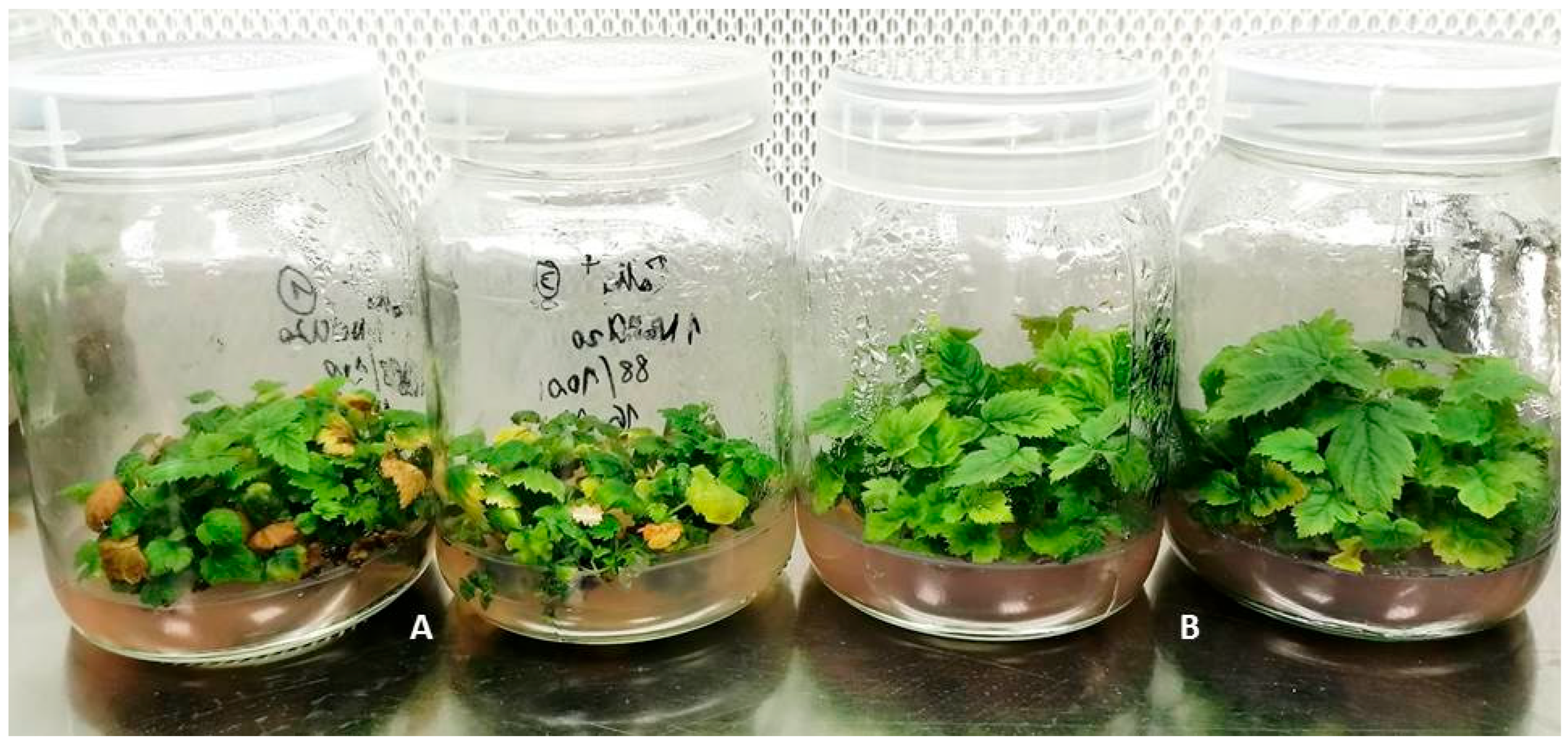
After 6 weeks, bacteria-free cultures were higher than contaminated ones and showed yellow leaves earlier (Figure 3). A detailed evaluation showed that only bacteria-free cv. Norna produced more shoots of 0.5 cm and higher, but all cultivars had only more shoots higher than 1 cm in bacteria-free cultures (Table 7, Table 8 and Table 9). More intensive yellowing was especially visible with a prolonged cycle exceeding 6 weeks. Maintaining bacteria-free cultures at 4 °C in the dark for 4–6 months caused shoot elongation with some leaves turning yellow. In contrast, almost all shoots contaminated with bacteria were necrotic (Figure S1).
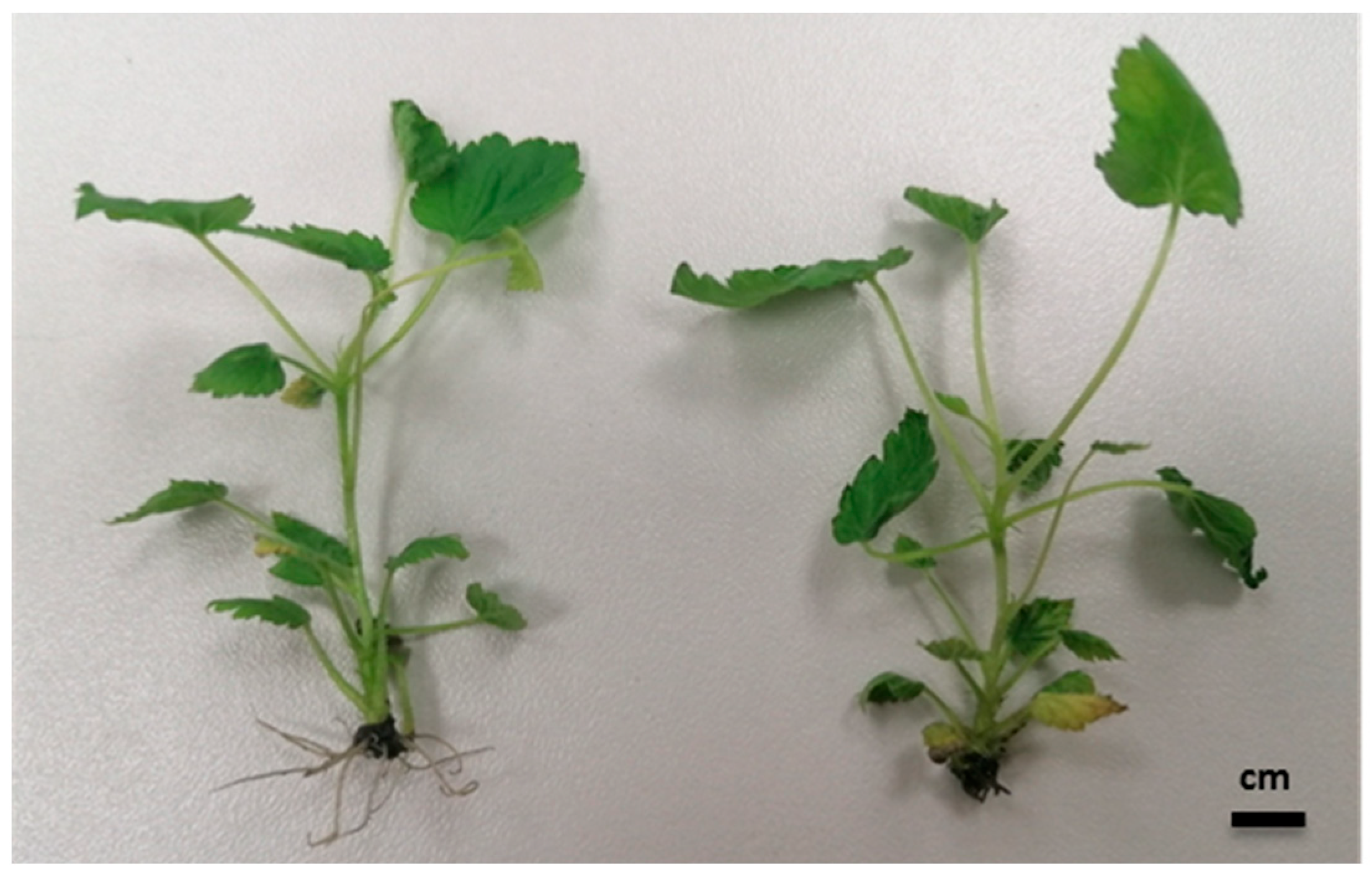
Evaluation of rooting showed more rooted shoots in the “contaminated” group (Table 10), but roots were very short (2–5 mm) and black in color (Figure 4).
Almost all rooted plantlets survived transferring to the greenhouse, although bacteria-contaminated plantlets started growing later.

4. Discussion

Persistent bacterial contamination has always been a problem. In scientific laboratories, it can contribute to false results as a potential producer of growth regulators or by stimulating plants to alter metabolism. Bacteria can also be a significant obstacle to the use of biotechnological tools in plant breeding. In commercial laboratories, they can affect yields at every stage of propagation and acclimation [28]. Most external bacteria can be eliminated by pretreatment of donor plants and careful surface decontamination [29]. Bacteria in the initial explants can be stimulated to visible growth by the addition of organic nitrogen to the medium. In our laboratory, we add albumin hydrolysate to the initiation medium. The more serious problem could arise when bacteria become visible en masse in commercially propagated plant cultures already well—adapted to in vitro conditions. The literature offers several solutions to minimize the problem. Various chemicals are used, most commonly antibiotics, chlorine compounds, and organic products, combined in a mixture. These must be selected according to the type of culture, the plant genotype, and their effectiveness against contaminating bacteria [30]. Some are unstable and gradually lose their effectiveness. Bacteria can become resistant to antibiotics.
PPM, which can be added to the medium before autoclaving and has relatively low toxicity to plants, has gained incredible popularity in recent years. However, some plants are sensitive, such as chrysanthemum and raspberry [31]. Furthermore, its constant demand at relatively high concentrations increases the cost of propagation [32,33,34].
Mercuric chloride, although harmful to the environment, is often used as an effective sterilant of the initial explants. Its use is minimally detrimental to the environment because it is used only in the initial stage and at low concentrations. It is not used as an additive to a propagation medium because it is harmful to the plants.
The goal of our study was to obtain raspberry cultures free of bacteria that colonized shoots over multiple passages. These bacteria did not leak into the medium immediately after transplantation. Still, they gradually became visible in the medium around the shoot bases, being particularly evident in the ’Polana’ cultures, which were contaminated with yellow Luteibacter sp. In the current study, HgCl2, rifampicin, PPM, and NaOCl were used to decontaminate in vitro raspberry shoot explants via infiltration under negative pressure.
Although rifampicin was very effective in the antibiogram tests, and it did not harm raspberry cultures, it did not eliminate bacteria in infiltration under vacuum. We suspect that rifampicin has a low ability to penetrate the shoot tissues. A similar idea was reported by Reed et al. [32].
In the current experiments, PPMTM infiltration prevented bacterial leakage into the proliferation medium, but at indexing on the NA medium, all shoots showed the presence of bacteria. Using vacuum infiltration of PPMTM at a concentration of 5 mg L−1, Miyazaki et al. [35] freed petunia explants from Sphingomonas paucimobilis. In this case, the vacuum lasted 2–3 min, after which the explants were held in PPMTM solution for 15 min. The presence of bacteria in the tissues was evaluated on a plant medium lacking PPMTM, not on the bacterial medium, containing organic nitrogen. The PPMTM was detected in the shoots with HPLC [35]. PPMTM is often used as an additive to the culture medium due to its low phytotoxicity and high thermostability, and its lower risk of bacterial resistance compared with antibiotics. PPMTM was recommended as a product that protects in vitro plant cultures against new contaminations when a small number of infective propagules is to be combated. Still, it is ineffective against a large population of bacteria, as shown in antibiogram tests. It should be understood as a bacteriostatic factor, but not as bactericidal [36].
NaOCl only weakly reduced bacterial growth, and this was observed in the proliferation medium, especially in Luteibacter sp-contaminated shoots. Rifampicin, PPMTM, and NaOCl were ineffective at killing internal bacteria under vacuum pressure, which means that they were inactivated in the shoots and/or did not penetrate [32].
Many authors have reported the effective use of bacteriocides for surface disinfection, but observations typically ended after the first passages. Subsequent passages may show varying results. Endogenous bacteria may appear, posing an additional problem and requiring further experiments. In this article, we highlight another option for bacterial control: mercuric chloride infiltration under negative pressure, which can eliminate the problem for many subsequent passages. This method may be particularly valuable for maintaining genetic collections and for micropropagation, where cyclical storage of cultures without transplantation at low temperatures is necessary.
To conclude, we recommend, for the first time, decontaminating persistent bacteria with 0.05 or 0.1% mercuric chloride applied by infiltration for two 15-minute periods under reduced pressure. Our conclusions regarding the effectiveness of HgCl2 in eliminating bacteria appear valid, as the same results were obtained for all plant/bacteria combinations. Differences in the number of bacteria-free and necrotic shoots were likely due to differences in shoot lignification. The concentration and exposure time may require adaptation to the culture time and plant species. This approach can be used to release from bacteria important, difficult-to-initiate cultures with high potential for in vitro multiplication. The bacteria we studied are not significantly harmful to raspberry micropropagation, but they can eliminate the cultures during low-temperature storage. While our attempts to eliminate bacteria from raspberry shoot cultures were successful, further optimization of the method is necessary, as its effectiveness may strongly depend on the specific plant/bacteria combination.

Supplementary Materials

The following supporting information can be downloaded at: https://www.mdpi.com/article/10.3390/agronomy15112452/s1. Figure S1: Cultures of cv. Polka after 6 months of maintenance at 4 °C in the dark. The contaminated culture is shown on the right.

Author Contributions

Conceptualization, A.T. and T.O.; data curation, T.O.; funding acquisition, A.T.; investigation, A.T., T.M., A.N.-W. and K.M.; methodology, A.T., T.M. and T.O.; project administration, A.T.; supervision, A.T. and T.O.; writing—original draft, A.T. and T.O.; writing—review and editing, A.T. and T.O. All authors have read and agreed to the published version of the manuscript.

Funding

The study was funded by the Polish Ministry of Science and Higher Education through statutory funds (ZBS/4/2019 and 11.1.1) at the National Institute of Horticultural Research, Skierniewice, Poland.

Data Availability Statement

The original contributions presented in this study are included in the article/Supplementary Materials. Further inquiries can be directed to the corresponding author.

Acknowledgments

The authors would like to thank Lucyna Ogórek for her excellent technical assistance.

Conflicts of Interest

The authors declare no conflicts of interest.

References

  1. Volk, G.M.; Bonnart, R.; Araújo de Oliveira, A.C.; Henk, A.D. Minimizing the deleterious effects of endophytes in plant shoot tip cryopreservation. Appl. Plant Sci. 2022, 10, e11489. [Google Scholar] [CrossRef]
  2. Thomas, P.; Sekhar, A.C. Live cell imaging reveals extensive intracellular cytoplasmic colonization of banana by normally non-cultivable endophytic bacteria. AoB Plants 2014, 6, plu002. [Google Scholar] [CrossRef] [PubMed]
  3. Pirttilä, A.M.; Laukkanen, H.; Pospiech, H.; Myllylä, R.; Hohtola, A. Detection of intracellular bacteria in the buds of Scotch pine (Pinus sylvestris L.) by in situ hybridization. Appl. Environ. Microbiol. 2000, 66, 3073–3077. [Google Scholar] [CrossRef]
  4. Partida-Martinez, L.P.; Heil, M. The microbe-free plant: Fact or artifact? Front. Plant Sci. 2011, 2, 100. [Google Scholar] [CrossRef]
  5. De Almeida, D.F.; Andreote, F.D.; Yara, R.; Tanaka, F.A.O.; Azevedo, J.L.; de Almeida, M. Bacteriosomes in axenic plants: Endophytes as stable endosymbionts. World J. Microbiol. Biotechnol. 2009, 25, 1757–1764. [Google Scholar] [CrossRef]
  6. Orlikowska, T.; Ogórek, L.; Trzewik, A.; Nowak, K.; Maciorowski, R. Micropropagation of raspberry (Rubus idaeus L.) using biocides for retarding bacterial contamination. Zesz. Nauk. Inst. Ogrod. 2018, 26, 77–83, (In Polish with English Abstract). [Google Scholar]
  7. Leifert, C.; Waites, W.M.; Camotta, H.; Nicholas, J.R. Lactobacillus plantarum: A deleterious contaminant of plant tissue culture. J. Appl. Bacteriol. 1989, 67, 363–370. [Google Scholar] [CrossRef]
  8. Marino, G.; Altan, A.D.; Biavati, B. The effect of bacterial contamination on the growth and gas evolution of in vitro cultured apricot shoots. In Vitro Cell. Dev. Biol. Plant 1996, 32, 51–56. [Google Scholar] [CrossRef]
  9. Thomas, P. In vitro decline in plant cultures: Detection of a legion of covert bacteria as the cause for degeneration of long-term micropropagated triploid watermelon cultures. Plant Cell Tissue Organ Cult. 2004, 77, 173–179. [Google Scholar] [CrossRef]
  10. Wojtania, A.; Puławska, J.; Gabryszewska, E. Identification and elimination of bacterial contaminants from Pelargonium tissue cultures. J. Fruit. Ornam. Plant Res. 2005, 13, 101–108. [Google Scholar]
  11. Reed, B.M.; Buckley, P.M.; DeWilde, T.N. Detection and eradication of endophytic bacteria from micropropagated mint plants. In Vitro Cell. Dev. Biol. Plant 1995, 31P, 53–57. [Google Scholar] [CrossRef]
  12. Thomas, P.; Prabhakara, B.S.; Pitchaimuthu, M. Cleansing the long term micropropagated triploid watermelon cultures from covert bacteria and field testing the plants for clonal fidelity and fertility during the 7–10 year period in vitro. Plant Cell Tissue Organ Cult. 2006, 85, 317–329. [Google Scholar] [CrossRef]
  13. Sarasan, V.A.; Cripps, R.; Ramsay, M.M.; Atherton, C.; McMichen, M.; Prendergast, G.; Rowntree, J.K. Conservation in vitro of threatened plants—Progress in the past decade. In Vitro Cell. Dev. Biol. Plant 2006, 42, 206–214. [Google Scholar] [CrossRef]
  14. Philips, R.; Arnott, S.M.; Kaplan, S.E. Antibiotics in plant tissue culture: Rifampicin effectively controls bacterial contaminants without affecting the growth of short-term explant cultures of Helianthus tuberosus. Plant Sci. Lett. 1981, 21, 235–240. [Google Scholar] [CrossRef]
  15. Leifert, C.; Camotta, H.; Waites, W.M. Effect of combinations of antibiotics on micropropagated Clematis, Delphinium, Hosta, Iris and Photinia. Plant Cell Tissue Organ Cult. 1992, 29, 153–160. [Google Scholar] [CrossRef]
  16. Mbah, E.I.; Wakil, S.M. Elimination of bacteria from in vitro yam tissue cultures using antibiotics. J. Plant Pathol. 2012, 94, 53–58. [Google Scholar]
  17. Plant Preservative Mixture. Available online: https://www.plantcelltechnology.com/plant-preservative-mixture-ppm-1/ (accessed on 7 June 2022).
  18. Bent, A.F. Arabidopsis in planta transformation. Uses, mechanisms, and prospects for transformation of other species. Plant Physiol. 2000, 124, 1540–1547. [Google Scholar] [CrossRef]
  19. Murashige, T.; Skoog, F. A revised medium for rapid growth and bioassays with tobacco tissue cultures. Physiol. Plant 1962, 15, 156–163. [Google Scholar] [CrossRef]
  20. Lloyd, G.; McCown, B. Commercially-feasible micropropagation of mountain laural, Kalmia latifolia, by use of shoot tip culture. Comb. Proc. Int. Plant Prop. Soc. 1980, 30, 421–427. [Google Scholar]
  21. Viss, P.R.; Brooks, E.M.; Driver, J.A. A simplified method for the control of bacterial contamination in woody plant tissue culture. In Vitro Cell. Dev. Biol. Plant 1991, 27, 42. [Google Scholar] [CrossRef]
  22. Weisburg, W.G.; Barns, S.M.; Pelletier, D.A.; Lane, D.J. 16S ribosomal DNA amplification for phylogenetic study. J. Bacteriol. 1991, 173, 697–703. [Google Scholar] [CrossRef]
  23. Ribeiro, C.M.; Cardoso, E.J.B.N. Isolation, selection and characterization of root-associated growth promoting bacteria In Brazil Pine (Araucaria angustifolia). Microbiol. Res. 2012, 167, 69–78. [Google Scholar] [CrossRef]
  24. Bric, J.M.; Bostock, R.M.; Silverstone, S.E. Rapid In Situ Assay for Indoleacetic Acid Production by Bacteria Immobilized on a Nitrocellulose Membrane. Appl. Environ. Microbiol. 1991, 57, 535–538. [Google Scholar] [CrossRef]
  25. Figueiredo de Vasconcellos, R.F.; Pinheiro da Silva, M.C.; Ribeiro, C.M.; Bran Nogueira Cardoso, E.J. Isolation and screening for plant growth-promoting (PGP) actinobacteria from Araucaria angustifolia rhizosphere soil. Sci. Agric. 2010, 67, 743–746. [Google Scholar] [CrossRef]
  26. Nautiyal, C.S. An effcient microbiological growth medium for screening phosphate solubilizing microorganisms. FEMS Microbiol. Lett. 1999, 170, 265–270. [Google Scholar] [CrossRef] [PubMed]
  27. Alexander, D.B.; Zuberer, D.A. Use of chrome azurol S reagents to evaluate siderophore production by rhizosphere bacteria. Biol. Fert. Soils 1991, 12, 39–45. [Google Scholar] [CrossRef]
  28. Thomas, P.; Kumari, S.; Swarna, G.K.; Prakash, D.P.; Dinesh, M.R. Ubiquitous presence of fastidious endophytic bacteria in field shoots and index-negative apparently clean shoot-tip cultures of papaya. Plant Cell. Rep. 2007, 26, 1491–1499. [Google Scholar] [CrossRef]
  29. Cassells, A.C. Pathogen and microbial contamination management in micropropagation—An overview. In Pathogen and Microbial Contamination Management in Micropropagation; Cassells, A.C., Ed.; Kluwer Academic Publishers: Alphen aan den Rhein, The Netherlands, 1997; pp. 1–13. [Google Scholar]
  30. Abdalla, N.; El-Ramady, H.; Seliem, M.K.; El-Mahrouk, M.E.; Taha, N.; Bayoumi, Y.; Shalaby, T.A.; Dobranszki, J. An Academic and Technical Overview on Plant Micropropagation Challenges. Hortuculturae 2022, 8, 677. [Google Scholar] [CrossRef]
  31. Orlikowska, T.; Zawadzka, M.; Zenkteler, E.; Sobiczewski, P. Influence of the biocides PPM tm and vitrofural on bacteria isolated from contaminated plant tissue cultures and on plant microshoots grown on various media. J. Hortic. Sci. Biotech. 2015, 87, 223–230. [Google Scholar] [CrossRef]
  32. Reed, B.M.; Tanprasert, P. Detection and control of bacterial contaminants of plant tissue cultures. A review of recent literature. Plant Tissue Cult. Biotechnol. 1995, 3, 137–142. [Google Scholar]
  33. Liang, C.; Wu, R.; Han, Y.; Wan, T.; Cai, Y. Optimizing Suitable Antibiotics for Bacterium Control in Micropropagation of Cherry Rootstock Using a Modified Leaf Disk Diffusion Method and E Test. Plants 2019, 8, 66. [Google Scholar] [CrossRef] [PubMed]
  34. Romadanova, N.V.; Tolegen, A.B.; Kushnarenko, S.V.; Zholdybayeva, E.V.; Bettoni, J.C. Effect of Plant Preservative MixtureTM on Endophytic Bacteria Eradication from In Vitro-Grown Apple Shoots. Plants 2022, 11, 2624. [Google Scholar] [CrossRef] [PubMed]
  35. Miyazaki, J.; Tan, B.H.; Errington, S.G. Eradication of endophytic bacteria via treatment for axillary buds of Petunia hybrida using Plant Preservative Mixture (PPMTM). Plant Cell Tissue Organ Cult. 2010, 102, 365–372. [Google Scholar] [CrossRef]
  36. Bunn, E.; Tan, B. Microbial contaminants in plant tissue culture propagation. In Microorganisms in Plant Conservation and Biodiversity; Sivasithamparam, K., Dixon, K.W., Barrett, R.L., Eds.; Kluwer Academic Publishers: Alphen aan den Rhein, The Netherlands, 2002; pp. 307–335. [Google Scholar]
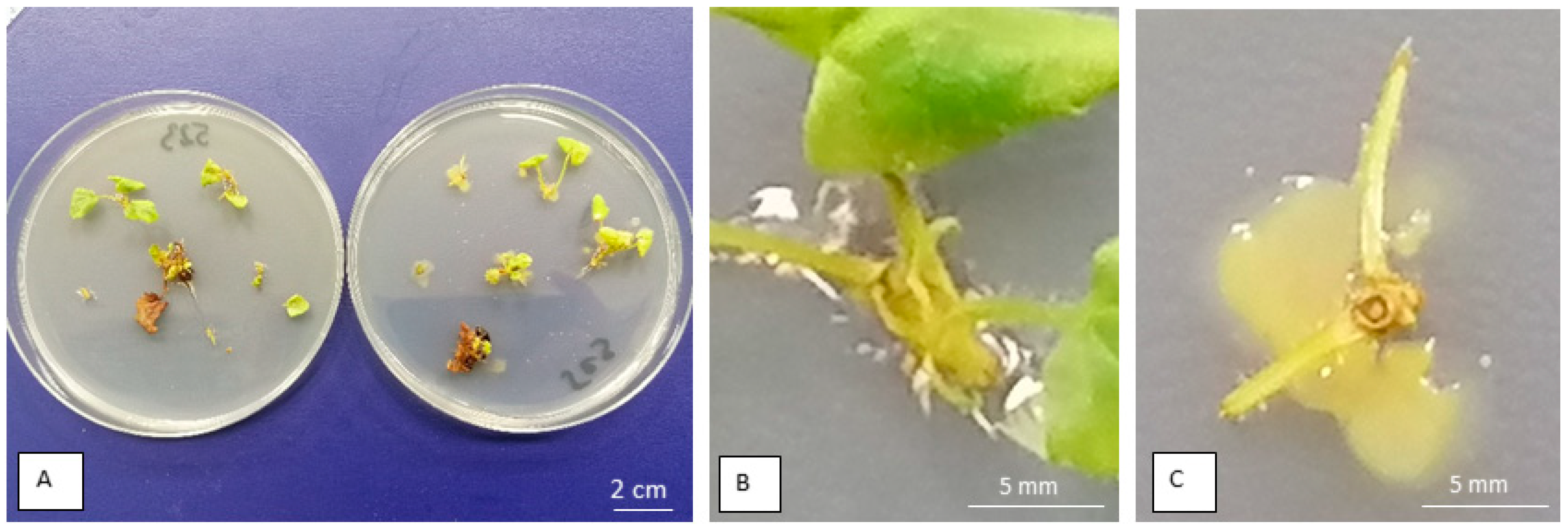
Figure 1. Indexing of ‘Polana’ raspberry shoot bases on 523 medium, to select bacteria-containing shoots for experiments on infiltration. (A) The plate on the left contains bacteria-free bases, while the plate on the right contains infected bases. (B) Enlargement of the left plate. (C) Enlargement from the right plate showing Luteibacter sp. growth.
Agronomy 15 02452 g001
Figure 2. ‘Polana’ raspberry shoot tips indexed on 523 medium one month after vacuum decontamination with HgCl2 0.05%, showed two shoots survived as free of bacteria (arrows).
Agronomy 15 02452 g002
Figure 3. Cultures of cv. Polka after 6 weeks on micropropagation medium: (A) contaminated cultures; (B) bacteria-free cultures.
Agronomy 15 02452 g003
Figure 4. Rooted shoots of cv. Polka after 4 weeks on the rooting medium. Shoot from contaminated culture is on the right. Natural size of plantlets.
Agronomy 15 02452 g004
Table 1. Diameters of bacterial growth inhibition (in mm) in the antibiogram test for three bacteria isolated from three raspberry cultivars (n = 4).
Cultivar/
Bacteria
Days
After
Inoculation
Biocides
HgCL2 0.05%HgCL2 0.1%PPMTM 0.2%PPMTM 0.4%Rif *
100 mg L−1
Rif. 200 mg L−1NaOCl 1%NaOCl 10%NaOCl 20%
‘Norna’/
Curtobacterium sp.
2810001719001
400001719000
1000001719000
‘Polka’/
Pseudomonas sp.
21216001617000
41216001617000
101216001617000
‘Polana’/
Luteibacter sp.
2680069000
4680023000
10680023000
* Rifampicin.
Table 2. Presence of bacterial colonies in 10 µL of biocide solutions on 523 medium after vacuum treatments (2 × 15 min). Observations were made over 10 days. Shown are the number of days after the experiment and the number of bacterial colonies.
Treatment Biocide ConcentrationBacteria/Cultivar
Curtobacterium sp./‘Norna’Pseudomonas sp./‘Polka’Luteibacter sp./‘Polana’
Water2nd *—>20 **4th—10–201st—>20
HgCl2 0.05%No bacteriaNo bacteriaNo bacteria
HgCl2 0.1%No bacteriaNo bacteriaNo bacteria
Rifampicin 100 mg/LNo bacteriaNo bacteria5th—10–20
Rifampicin 200 mg/LNo bacteriaNo bacteria5th—15–10
PPM 0.2%4th—>208th—5–103rd—10–20
PPM 0.4%4th—>208th—<53rd—>20
NaOCl 1%2nd—>20No bacteria1st—>20
NaOCl 10%2nd—>20No bacteria1st—>20
NaOCl 20%2nd—>20No bacteria3rd—10–20
* Days after plating. ** Number of bacterial colonies.
Table 3. Initial experiments on the influence of biocide infiltration on Curtobacterium in cv. Norna.
Exp. No
No. of Shoots
BiocideVacuumNo. of
Necrotic Shoots After 4 Weeks
No. of
Contaminated Shoots on MS Medium After 4 Weeks
No. of
Contaminated Shoots on NA Medium After 4 Weeks
No. of
Bacteria-Free Shoots
After 4 Weeks
I
n = 10
WaterNo vacuum010--
Water20 min010--
Rifamp.50 mg/LNo vacuum055-
Rifamp.50 mg/L20 min028-
Rifamp.100 mg/LNo vacuum028-
Rifamp.100 mg/L20 min028-
PPM 0.2%No vacuum0010-
PPM 0.2%20 min.0010-
PPM 0.4%No vacuum0010-
PPM 0.4%20 min0010-
II
n = 10
WaterNo vacuum010--
Water20 min010--
Rifamp.150 mg/LNo vacuum028-
Rifamp.150 mg/L20 min028-
Rifamp.200 mg/LNo vacuum028-
Rifamp.200 mg/L20 min028-
PPM 2%No vacuum0010-
PPM 2%20 min0010-
PPM 4%No vacuum0010-
PPM 4%20 min0010-
III
n = 10
Water20 min010--
Water90 min010--
Rifamp.50 mg/L20 min055-
Rifamp.50 mg/L90 min037-
Rifamp.100 mg/L20 min019-
Rifamp.100 mg/L90 min028-
PPM 0.2%20 min0010-
PPM 0.2%90 min0010-
PPM 0.4%20 min0010-
PPM 0.4%90 min0010-
Table 4. Influence of biocide vacuum infiltration on Curtobacterium in cv. Norna.
Exp. No
No. of Shoots
BiocideVacuumNo. of Necrotic Shoots After 4 WeeksNo. of Contaminated Shoots on MS Medium After 4 WeeksNo. of Contaminated Shoots on NA Medium After 4 WeeksNo. of Bacteria-Free Shoots After 4 Weeks
V
n = 10
Water2 × 15 min06--
HgCl2 0.05%2 × 15 min8002
HgCl2 0.1%2 × 15 min9001
Rifamp.100 mg/L2 × 15 min055-
Rifamp.200 mg/L2 × 15 min055-
PPM 0.2%2 × 15 min0010-
PPM 0.4%2 × 15 min0010-
NaOCl 1%2 × 15 min082-
NaOCl 10%2 × 15 min055-
NaOCl 20%2 × 15 min73--
VI
n = 30
Water2 × 15 min030--
Water1 × 30 min030--
HgCl2 0.05%2 × 15 min170013
HgCl2 0.05%1 × 30 min100020
HgCl2 0.1%2 × 15 min120018
HgCl2 0.1%1 × 30 min120018
NaOCl 0.1%2 × 15 min0822-
NaOCl 0.1%1 × 30 min0822-
NaOCl 0.5%2 × 15 min01515-
NaOCl 0.5%1 × 30 min01614-
VI
n = 10
Water2 × 15 min064-
HgCl2 0.05%2 × 15 min8002
HgCl2 0.1%2 × 15 min9001
Rifamp.100 mg/L2 × 15 min055-
Rifamp.200 mg/L2 × 15 min055-
PPM 0.2%2 × 15 min0010-
PPM 0.4%2 × 15 min0010-
NaOCl 1%2 × 15 min082-
NaOCl 10%2 × 15 min055-
NaOCl 20%2 × 15 min73--
Table 5. Influence of biocide vacuum infiltration on Pseudomonas in cv. Polka.
Exp. No
No. of Shoots
BiocideVacuumNo. of Necrotic Shoots After 4 WeeksNo. of Contaminated Shoots on MS Medium After 4 WeeksNo. of Contaminated Shoots on NA Medium After 4 WeeksNo. of Bacteria-Free Shoots After 4 Weeks
V
n = 10
Water2 × 15 min073-
HgCl2 0.05%2 × 15 min104
HgCl2 0.1%2 × 15 min6025
Rifamp.100 mg/L2 × 15 min00102
Rifamp.200 mg/L2 × 15 min0010-
PPM 0.2%2 × 15 min0010-
PPM 0.4%2 × 15 min0010-
NaOCl 1%2 × 15 min173-
NaOCl 10%2 × 15 min073-
NaOCl 20%2 × 15 min55--
VI
n = 20
Water2 × 15 min020--
Water1 × 30 min020--
HgCl2 0.05%2 × 15 min15005
HgCl2 0.05%1 × 30 min14006
HgCl2 0.1%2 × 15 min14006
HgCl2 0.1%1 × 30 min14006
NaOCl 0.1%2 × 15 min0164-
NaOCl 0.1%1 × 30 min0182-
NaOCl 0.5%2 × 15 min0164-
NaOCl 0.5%1 × 30 min0191-
VII
n = 10
Water2 × 15 min073-
HgCl2 0.05%2 × 15 min1063
HgCl2 0.1%2 × 15 min802-
Rifamp.100 mg/L2 × 15 min0010-
Rifamp.200 mg/L2 × 15 min0010-
PPM 0.2%2 × 15 min0010-
PPM 0.4%2 × 15 min0010-
NaOCl 1%2 × 15 min172-
NaOCl 10%2 × 15 min073-
NaOCl 20%2 × 15 min55--
Table 6. Influence of biocide vacuum infiltration on Luteibacter in cv. Polana.
Exp. No
No. of Shoots
BiocideVacuumNo. of Necrotic Shoots After 4 WeeksNo. of Contaminated Shoots on MS Medium After 4 WeeksNo. of Contaminated Shoots on NA Medium After 4 WeeksNo. of Bacteria-Free Shoots After 4 Weeks
IV
n = 10
Water30 min010--
HgCl2 0.05%30 min1009
NaOCl 4%30 min010--
NaOCl 7%30 min010--
NaOCl 10%30 min082-
NaOCl 20%30 min282-
NaOCl 30%30 min55--
NaOCl 40%30 min55--
NaOCl 50%30 min73--
NaOCl 60%30 min109--
V
n = 10
Water2 × 15 min073-
HgCl2 0.05%2 × 15 min2062
HgCl2 0.1%2 × 15 min4006
Rifamp.100 mg/L2 × 15 min0010-
Rifamp.200 mg/L2 × 15 min0010-
PPM 0.2%2 × 15 min0010-
PPM 0.4%2 × 15 min0010-
NaOCl 1%2 × 15 min055-
NaOCl 10%2 × 15 min055-
NaOCl 20%2 × 15 min46--
VI
n = 10
Water2 × 15 min010--
Water1 × 30 min010--
HgCl2 0.05%2 × 15 min7003
HgCl2 0.05%1 × 30 min6004
HgCl2 0.1%2 × 15 min6004
HgCl2 0.1%1 × 30 min5005
NaOCl 0.1%2 × 15 min082-
NaOCl 0.1%1 × 30 min082-
NaOCl 0.5%2 × 15 min082-
NaOCl 0.5%1 × 30 min091-
VII
n = 10
Water2 × 15 min073-
HgCl2 0.05%2 × 15 min406-
HgCl2 0.1%2 × 15 min6004
Rifamp.100 mg/L2 × 15 min0010-
Rifamp.200 mg/L2 × 15 min0010-
PPM 0.2%2 × 15 min0010-
PPM 0.4%2 × 15 min0010-
NaOCl 1%2 × 15 min055-
NaOCl 10%2 × 15 min055-
NaOCl 20%2 × 15 min46--
Table 7. Effect of contamination with Curtobacterium sp. on shoot propagation of raspberry cv. Norna (n = 10).
Repetition in Subsequent PassagesTotal ShootsShoots > 1 cm
Bacteria −Bacteria +Bacteria −Bacteria +
15.2 a *3.2 b3.9 a2.4 b
24.9 a2.9 b4.4 a2.5 b
33.5 a3.4 a3.1 a2.6 a
43.0 a2.7 a2.8 a2.3 a
Mean4.2 a3.1 b3.6 a2.5 b
* Means in the row followed by the same letter were not significantly different (p ≤ 0.05, one-way ANOVA, Tukey’s test).
Table 8. Effect of contamination with Pseudomonas sp. on shoot propagation of raspberry cv. Polka (n = 10).
Repetition in Subsequent PassagesTotal ShootsShoot > 1 cm
Bacteria −Bacteria +Bacteria −Bacteria +
13.1 b *4.0 a2.4 a 2.1 a
22.8 b3.3 a2.6 a1.7 b
34.3 a3.5 b3.8 a1.4 b
43.3 b4.0 a2.5 a1.7 b
Mean3.4 a3.7 a2.8 a1.7 b
* Means in the row followed by the same letter were not significantly different (p ≤ 0.05, one-way ANOVA, Tukey’s test).
Table 9. Effect of contamination with Luteibacter sp. on shoot propagation of raspberry cv. Polana (n = 10).
Repetition in Subsequent PassagesTotal ShootsShoots >1 cm
Bacteria −Bacteria +Bacteria −Bacteria +
13.1 b *4.0 a2.4 a2.1 a
22.8 b3.3 a2.6 a1.7 b
34.3 a3.5 b3.8 a1.4 b
43.3 b4.0 a2.5 a1.7 b
Mean3.4 a3.7 a2.8 a1.7 b
* Means in the row followed by the same letter were not significantly different (p ≤ 0.05, one-way ANOVA, Tukey’s test).
Table 10. Effect of bacterial contamination on the rooting percentage of three raspberry cultivars.
Repetition‘Norna’/
Curtobacterium sp
‘Polka’/Pseudomonas sp.‘Polana’/Luteibacter sp.
+++
137 b *48 a74 a86 a87 a84 a
238 b47 a22 b57 a22 b34 a
3----42 b81 a
Mean38 b48 a48 b72 a50 b66 a
* Means in the row followed by the same letter were not significantly different (p ≤ 0.05, one-way ANOVA, Tukey’s test).
Disclaimer/Publisher’s Note: The statements, opinions and data contained in all publications are solely those of the individual author(s) and contributor(s) and not of MDPI and/or the editor(s). MDPI and/or the editor(s) disclaim responsibility for any injury to people or property resulting from any ideas, methods, instructions or products referred to in the content.