Article

Appraising Endophyte–Plant Symbiosis for Improved Growth, Nodulation, Nitrogen Fixation and Abiotic Stress Tolerance: An Experimental Investigation with Chickpea (Cicer arietinum L.)

1
Department of Soil Science, University College of Agriculture and Environmental Sciences, the Islamia University of Bahawalpur, Bahawalpur-63100, Pakistan
2
Institute of Molecular Biology and Biotechnology, the University of Lahore, Defense Road, Lahore 54000, Pakistan
3
National Engineering Laboratory for Improving Quality of Arable Land, Institute of Agricultural Resources and Regional Planning, Chinese Academy of Agricultural Sciences, Beijing 100081, China
4
Institute of Agricultural Sciences in the Tropics (Hans-Ruthenberg Institute), University of Hohenheim, 70593 Stuttgart, Germany
5
Institute of Soil and Environmental Sciences, University of Agriculture Faisalabad, Faisalabad 38000, Pakistan
*
Authors to whom correspondence should be addressed.
Agronomy 2019, 9(10), 621; https://doi.org/10.3390/agronomy9100621
Submission received: 2 September 2019 / Revised: 3 October 2019 / Accepted: 3 October 2019 / Published: 9 October 2019
(This article belongs to the Special Issue Rhizobium–Legume Symbiosis)

Abstract

:
Chickpea is an important leguminous crop that improves soil fertility through atmospheric nitrogen fixation with the help of rhizobia present in nodules. Non-rhizobia endophytes are also capable of inducing nodulation and nitrogen fixation in leguminous crops. The aim of the current study was to isolate, characterize and identify the non-rhizobia endophytic bacterial strains from root nodules of chickpea. For this purpose, more than one hundred isolates were isolated from chickpea root nodules under aseptic conditions and were confirmed as endophytes through re-isolating them from root nodules of chickpea after their inoculation. Nineteen confirmed endophytic bacterial strains revealed significant production of indole acetic acid (IAA) both in presence and absence of L-tryptophan and showed their ability to grow under salt, pH and heavy metal stresses. These strains were evaluated for in vitro plant growth promoting (PGP) traits and results revealed that seven strains showed solubilization of P and colloidal chitin along with possessing catalase, oxidase, urease and chitinase activities. Seven P-solubilizing strains were further evaluated in a jar trial to explore their potential for promoting plant growth and induction of nodulation in chickpea roots. Two endophytic strains identified as Paenibacillus polymyxa ANM59 and Paenibacillus sp. ANM76 through partial sequencing of the 16S rRNA gene showed the maximum potential during in vitro PGP activities and improved plant growth and nodulation in chickpea under the jar trial. Use of these endophytic strains as a potential biofertilizer can help to reduce the dependence on chemical fertilizers while improving crop growth and soil health simultaneously.

1. Introduction

Chickpea (Cicer arietinum L.) is an important legume crop and is consumed all over the world as a source of protein, carbohydrate, fiber, oil, ash, minerals, vitamins, amino acids and unsaturated fatty acids [1]. Being a leguminous crop, chickpea forms a symbiotic relationship with rhizobia that support biological nitrogen fixation (BNF) in root nodules. Symbiotic N2 fixation facilitates larger proportions of nitrogen (N) for food production and agricultural sustainability [2]. Leguminous plants in association with a diverse variety of bacteria can reduce atmospheric N2 into a plant available form and as a result improve soil fertility and plant growth [3]. For decades, rhizobia were thought to be the only nitrogen-fixing inhabitants of legume nodules. However, non-rhizobial bacteria were detected within legume nodules revealing the existence of a phytomicrobiota where the interaction among the individuals is complex and affects the behaviour and fitness of the host plant [4]. These non-rhizobial bacteria are involved in BNF through inducing nitrogen fixing nodules on roots of host legume [5]. They benefit legume host by improving plant growth through producing phytohormones, fixing atmospheric N2, and solubilizing mineral nutrients [6]. In the past, non-rhizobial bacteria remained largely ignored due to lack of knowledge on diversity of non-rhizobial bacteria which co-exist with rhizobia and involved in N2 fixation [5]. Bacterial endophytes may took advantage over rhizobacteria as endophytes have more opportunity to be in contact with plant cells and exert direct beneficial effect on host plant [7,8] probably due to aggressive colonization patterns.
Plants are able to shape their rhizospheric and endophytic microbiome and can maintain environmental stress resistant bacterial communities having specific beneficial characteristics [9,10]. Bacterial endophytes have been found almost in every analysed plant species while, endophytic free plants are less able to cope with environmental stress conditions [11]. Previous reports described the diversity of bacterial endophytes in various plant species with agricultural interests. Diversity of endophytes found are comprised of various genera viz. Acinetobacter, Agromyces, Azoarus, Bacillus, Brevibacillus, Brevundimonas, Burkholderia, Comamonas, Corynebacterium, Delftia, Dietzia, Enterobacter, Frigoribacterium, Kocuria, Lysinibacillus, Methylobacterium, Microbacterium, Micrococcus, Paenibacillus, Pantoea, Pseudomonas, Rhizobium, Rhodococcus, Serria, Sphingobacterium, Sphingomonas, Sporosarcina, Staphylococcus, Stenotrophomonas, Xanthomonas, etc. [7,12,13,14,15,16,17,18,19,20,21,22]. All of these described bacterial endophytic genera are also common inhabitants of the rhizosphere [7,8]. Once symbiosis takes place, endophytes promote plant growth through the expression of the enzyme 1-aminocyclopropane-1-carboxylate (ACC) deaminase which can sequester and cleave the ethylene precursor ACC to α-ketobutyrate and ammonia (NH3) and decreases ethylene production in host plants [23,24]. They can alter the plant physiology including regulation in osmotic pressure, changes in stomatal responses, increase in root morphology and improved N accumulation and metabolism and uptake of minerals [7,23,24,25]. Bacterial endophytes can produce phytohormones as metabolites such indole-3-acetic acid (IAA), gibberellins and cytokinins [26]. These phytohormones are natural substances manufactured by plants to promote its growth and development however their supplementation through bacterial sources can be safeguarded from environmental stimuli [27]. Among phytohormones, IAA plays a vital role in several plant activities such as formation, initiation and development of plant leaf, root and fruit, and is involved in phototropism and geotropism, etc. It plays an important role in enhancement of root length, branches, hair and root lateral growth that increase uptake of nutrients from surrounding environments [28]. Its production is widespread among various class of endophytic bacterial genus such as Acinetobacter, Azospirillum, Azotobacter, Bacillus and Pseudomonas. These endophytic bacterial genera were reported to produce IAA through indole-3-pyruvic acid, indole-3-acetamide and indole-3-acetonitrile biosynthesis pathways, which are part of tryptophan-dependent and -independent pathways [29]. The application of IAA producing endophytic strains viz. Bacillus subtilis, Enterobacter ludwigii, Klebsiella sp. Sal 1, and Pseudomonas fluorescens in onion, rice, tomato and radish crops has shown significant increase in plant growth and yield [30,31]. Bacterial endophytes are involved in biocontrol through producing enzymes (e.g., hydrolases, chitinases, laminarinases, glucanases etc.) as reported by Chernin and Chet [32]. In addition, endophytes have been reported to induce systemic resistance (ISR)-based plant growth promotion in host plants once exposed to environmental stresses [33,34]. Much of the research hitherto has concentrated on rhizobia–legume interactions, whereas there is very limited work on exploring the role of non-rhizobial endophyte–legume symbiosis. Therefore, there is a need to investigate the role of non-rhizobial endophytes in improving crop growth, nodulation and nitrogen fixing ability of legumes. In addition, no information is available regarding abiotic stress tolerance capacity of non-rhizobial endophytes which in the present study constitutes the novelty.
Therefore, keeping in view the above facts, the current study was conducted for isolation of endophytic bacteria from chickpea nodules, the selection and identification of strains with better PGP abilities and analysis of the effect of these strains on chickpea plants compared with uninoculated control. The current study accomplished the potential utilization of PGP non-rhizobial endophytic bacteria (Paenibacillus polymyxa ANM59 and Paenibacillus sp. ANM76) to improve chickpea growth, nodulation and nitrogen fixation. Endophytic bacterial strains in this study were first isolated from chickpea nodules, screened for plant growth promotion and various types of stresses and identified as Paenibacillus spp. through 16S rRNA gene sequencing afterwards.

2. Materials and Methods

2.1. Isolation of Endophytic Strains

Plant samples along with roots were collected from chickpea grown in experimental fields of University College of Agriculture and Environmental Sciences (UCA and ES), the Islamia University of Bahawalpur (IUB), Pakistan. For isolation of endophytes, rhizosphere soil from chickpea roots was removed by washing with tap water. The healthy, unbroken and pink nodules were separated from roots by using sterile razor blade and placed in petri plates. These nodules were surface-disinfected with 95% ethanol for twenty seconds followed by immersing in 0.2% mercuric chloride (HgCl2) solution for three minutes and five times washing with sterile-distilled water [35]. After that, nodules were aseptically crushed with a glass rod in a test tube containing distilled water, and a final suspension of 1 mL was prepared. The obtained suspension was spread on yeast extract mannitol agar (YEMA) and incubated at 28 ± 1 °C for bacterial growth. Morphologically distinct single colonies were purified and preserved in glycerol stock (50%) at −20 °C until further use.

2.2. Authentication of Endophytic Nature of Strains

To confirm isolates as true nodule endophytes, surface-disinfected chickpea seeds were inoculated with respective strains and aseptically sown in plastic jars filled with sand. Jars were kept in a growth room and watered daily with sterilized nitrogen free Hoagland solution. After 40 days of incubation, plants were harvested and analysed for induction of nodules in chickpea and presence of bacterial strains. Nineteen isolates were termed as true endophytic bacterial strains after detecting them in nodules of chickpea and promoters of nodulation. These strains were screened in vitro by Gram staining. The standard method of Vincent [36] was adopted for Gram staining. Various morphological features of endophytic strains such as colour, transparency, colony and cell shape, colony border, mucus production and colony diameter were examined on Congo red amended YEMA after an incubation period of 48 h at 28 ± 1 °C [36].

2.3. In Vitro Screening of Endophytes for their PGP Characteristics

Nineteen confirmed endophytic strains were selected for evaluation of their PGP characteristics. P-solubilization ability of strains was accessed qualitatively on Pikovskaya agar medium [37]. Appearance of a halo zone around the colonies was considered P-solubilization positive and the halo zone was measured to calculate P-solubilization index (PSI) through following Formula (1) as reported by Vazquez et al. [38] Premono et al. [39] and Singh et al. [40]. P-solubilization efficiency (PSE) was calculated using following Formula (2).
S o l u b i l i z a t i o n   I n d e x   ( S I ) = C o l o n y   d i a m e t e r + H a l o   z o n e   d i a m e t e r C o l o n y   d i a m e t e r ,
S o l u b i l i z a t i o n   E f f i c i e n c y   ( S E )   =   H a l o   z o n e   d i a m e t e r C o l o n y   d i a m e t e r × 100 .
Qualitative zinc (Zn) solubilization test was performed on zinc oxide amended Tris-minimal salt agar (Tris-MSA) and halo zones around the colonies were observed [41,42]. For determination of ammonia production, strains were inoculated in 10 mL peptone broth in test tubes and incubated for 30 ± 1 °C at 72 h. After incubation, Nessler’s reagent (1 mL) was added and a change in colour was observed [43]. For determination of acid production by strains, YEMA medium was amended with bromothymol blue and bacterial cells were inoculated. After 48 h of incubation at 28 ± 1 °C, change in colour from blue to yellow around bacterial cells was reported positive for acid producing endophytes [36].
The method of Ali and Hasnain [44] was modified to access the auxins production by strains. Dworkin and Foster (DF) minimal salt broth was amended both with and without 1.0 g L−1 L-tryptophan. Strains having 0.5 optical density measured at 600 nm (OD600) by spectrophotometer (Agilent Technologies, mulgrave victoria, Melb, Australia) were inoculated and incubated at 28 ± 1 °C for 48 h. After incubation, cultures were centrifuged (10,000 rev/min for 10 min) and 1 mL Salkowski reagent was added in supernatant [45]. The absorbance of both with and without L-tryptophan amended media was observed through spectrophotometer at 600 nm and auxins production was calculated through drawing a standard curve for comparison by using indole acetic acid (Sigma-Aldrich, Steinheim, Germany) as a standard [42].
Selected strains were further screened for enzymatic activities, production of ammonia, triple sugar iron (TSI) test (related to glucose, sucrose, lactose) and hydrolysis of starch. Standard methods of Cappuccino and Welsh [43] were used for enzymatic activities viz. catalase, oxidase and urease and the TSI test. For chitinase activity, strains were grown on Dworkin and Foster (DF) minimal salt media modified with colloidal chitin (2% w/v). Formation of halo zones around the colonies was considered positive for chitinase activity [46]. Halo zone diameter of colloidal chitin solubilization was estimated and chitin solubilization efficiency (CSE) and chitin solubilization index (CSI) were calculated using Formulae 1,2. Starch agar was spot inoculated with overnight grown bacterial cells and incubated at 28 ± 1 °C. After 48 h, 0.1% iodine solution was spread in Petri plates and the result of starch hydrolysis was observed by adopting the method of Oliveira et al. [47]. P-solubilizing strains were selected for root colonization assay and the method of Mendis et al. [48] was followed to count bacterial colony forming units (CFUs) through serial dilution and pour plate techniques.

2.4. In Vitro Screening for Stress Tolerance

Nineteen selected endophytic strains were screened under heavy metals, temperature, pH and salt stress. Among heavy metals, mercury (Hg), Zn, copper (Cu) and chromium (Cr) tolerance was evaluated. The YEMA media was amended by adding HgCl2 (20 μg mL−1), ZnSO4 (10 μg mL−1), CuSO4 (10 μg mL−1), and K2Cr2O7 (25 μg mL−1) separately and autoclaved. Freshly grown strains were inoculated in all heavy metals amended media and incubated at 28 ± 1 °C for 72 h. After incubation, bacterial growth was observed as described by Carrasco et al. [49]. For temperature tolerance, strains were grown on YEMA media and incubated at 4, 28, 36 and 55 °C for 72 h to observe growth. Strains were also checked for their ability to grow under various pH levels. pH of yeast extract mannitol broth (YEMB) was maintained at 4.0, 5.0, 6.8, 8.0 and 9.0 by using 1 M HCl. Bacterial culture having 0.5 OD was inoculated and incubated at 28 ± 1 °C for 24 h. After incubation, OD was measured at 600 nm by spectrophotometer. Ability of strains to grow under salt stress was determined by growing strains in YEMB having salinity levels viz. 1.6 (normal), 4, 8 and 12 dS m−1 maintained by using NaCl. After 72 h of incubation at 28 ± 1 °C, optical density at 540 nm (OD540) was measured through spectrophotometer.

2.5. Screening of Endophytes for Growth Promotion of Chickpea (Jar Trial)

Seven P-solubilizing endophytic strains were evaluated for their ability to promote growth of chickpea under natural climatic conditions. The YEMB media was cultured with overnight grown bacterial strains and incubated at 28 ± 1 °C for 48 h. Seeds of chickpea were surface-disinfected by dipping in 95% ethanol for twenty seconds then 0.2% HgCl2 solution for three minutes and rinsed five times with sterilized distilled water [35]. Three surface-disinfected seeds were inoculated with each of selected bacterial culture by dipping in the respective broth for 30 min. In the case of control, surface-disinfected seeds were treated with sterilized broth without bacteria. Inoculated seeds were sown in glass jars filled with sterilized sand. Modified, N-free sterilized Hoagland solution was watered to fulfil nutrients needs of seedlings. Jars were arranged in completely randomized design (CRD) in triplicate and placed in a wire house of the Department of Soil Science, IUB under natural climatic conditions. After two months of sowing, data regarding nodule formation and growth promotion were recorded.

2.6. Identification of Selected Endophytic Strains

Endophytic strains ANM59 and ANM76 were grown in YEMB media at 28 ± 1 °C overnight and slants and glycerol stocks were prepared. Commercial service of Macrogen, Seoul, Korea [50] was used for sequencing of bacterial strains. The 16S rRNA partial gene sequence through using universal primer 785F 5’ (GGA TTA GAT ACC CTG GTA) 3’ 907R 5’ (CCG TCA ATT CMT TTR AGT TT) 3’ was carried out. The resulting partial sequence of 16S rRNA were blasted using MEGABLAST on National Center for Biotechnology Information (NCBI) servers. Sequences of closely related fifteen published strains were retrieved from the database. A neighbour-joining phylogenetic tree was constructed using MEGA 7.0.14 software as described by Kumar et al. [51]. The tree topology of the phylogenetic tree was assessed by bootstrap resampling method of Tamura et al. [52] with 500 replications. The sequences were submitted to the NCBI gene bank to get accession numbers of the strains.

2.7. Statistical Assessment

Data were statistically analysed in software Statistix 8.1 (Analytical Software, Tallahassee, FL, USA) and treatment means were compared using least significant difference (LSD) test at p ≤ 0.05 [53].

3. Results

3.1. Morphological Characteristics of Endophytes

More than one hundred non-rhizobial endophytic isolates were isolated from nodules of chickpea and nineteen of these were authenticated as true endophytes by observing their presence in root nodules and induction of nodulation in chickpea. These endophytic isolates were selected and analysed for their colony morphological characteristics. All the colonies appeared as milky white with an entire margin. The majority of isolates showed mucoid growth and their colonies were translucent, round and small (Table S1).

3.2. Endophytic Strains Possess PGP Characteristics

All the tested strains showed IAA production both in the presence as well as absence of L-tryptophan (Table 1). Maximum IAA production with and without L-tryptophan was reported by the strain ANM79 followed by ANM78 and ANM76, while, ANM58 and ANM75 produced minimum IAA in presence as well as absence of L-tryptophan. Seven out of 19 strains performed solubilization of tri-calcium phosphate by showing halo zones around colonies. The diameter of P halo zones and their solubilization efficiency and solubilization index are given in Table 1, Figure 1. The strain ANM76 followed by ANM63 showed the maximum solubilization efficiency and solubilization index whereas, ANM73, ANM59 and ANM16 gave minimum values of P-solubilization efficiency and solubilization index. Eleven out of 19 strains were chitinase positive (Table 2) and showed halo zone formation around the colonies. Their colloidal chitin solubilization zone with solubilization efficiency and solubilization index are given in Table 1.
The strain ANM76 illustrated the highest P-solubilization zone, solubilization efficiency and solubilization index. The P-solubilizing strains were evaluated for root colonization and results indicated that all of these strains possessed strong root colonization ability. Maximum root colonization of 5.45 × 10⁵ and 5.17 × 10⁵ were shown by strains ANM76 and ANM73 (Table 1). Further, in vitro plant growth promoting characteristics of selected endophytic strains are described in Table 2. None of the tested endophytic strains were able to solubilize zinc oxide. All tested strains were catalase positive, production of CO2 and H2S negative and were Gram negative (–ve) bacteria. The majority of strains were able to hydrolyse starch, produced NH3 and acid, and were able to utilize glucose and sucrose. Oxidase activity was performed by six strains out of 19 while five strains were urease positive.

3.3. Endophytic Strains were Salinity, pH and Heavy Metals Stress Tolerant

Nineteen selected strains were screened through various levels of salinity, pH, heavy metals and their results are given in Table 3. To assess salt tolerance by endophytic strains, these were grown in YEMB medium with 1% glucose as the sole carbon source at four salinity levels viz. 1.6, 4.0, 8.0 and 12.0 dS m−1 for determination of OD540. Strains growth was decreased with increasing salinity and their tolerance ability was variable at highest salinity (12.0 dS m−1). At this salinity level, the maximum OD was found in the case of ANM76 followed by ANM100 (Table 3). Yeast extract mannitol broth adjusted to starting pH values of 4.0, 5.0, 6.8, 9.0 and 10.0 was used to evaluate the effect of pH on growth of endophytic strains (their growth profiles are summarized in Table 3). All the strains showed optimum growth profiles at pH 6.8 but showed a drastic drop in growth with decrease in pH up to 4.0 as well as increase in pH up to 10.0. However, strains were still able to show their growth to some extent at acidic pH 4.0 as well as alkaline pH 10.0. To assess heavy metals tolerance, endophytic strains were grown on heavy metal (viz. Hg, Zn, Cu and Cr) amended YEMA plates and incubated for 48 h to observe their growth (growth is summarized in Table 3). Variable thirteen out of 19 strains were capable of growing on Hg and Zn amended media. Nine strains showed growth in Cu amended media while in Cr amended media only six strains showed their ability to grow.

3.4. Endophytic Strains Promoted the Growth and Nodulation in Chickpea (Jar Trial)

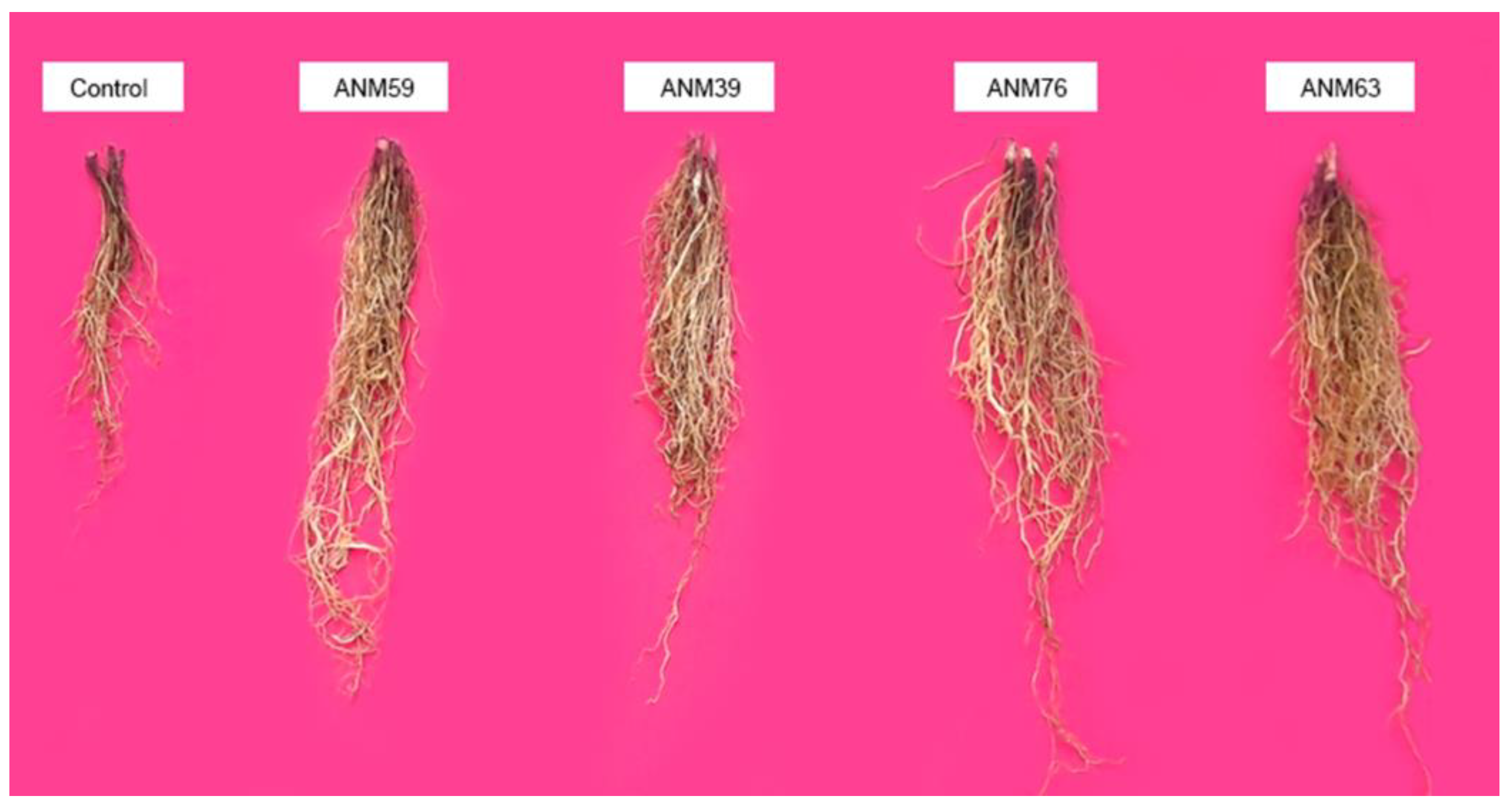
Seven P-solubilizing endophytic strains were selected to evaluate their ability to promote the growth and nodulation in chickpea by conducting a jar trial under natural environmental conditions and results are depicted in Table 4. Inoculation with endophytic strains significantly promoted the shoot and root growth (Figure 2 and Figure 3), biomass and nodulation in chickpea. Increase in these attributes was non-significant between tested strains however, significantly different from un-inoculated control. Maximum shoot and root length with 87% and 100% increase was observed due to inoculation with ANM76 as compared to un-inoculated control. This strain also showed the highest shoot and root fresh biomass with 77% and 81% increase which was significantly higher than un-inoculated control. Highest shoot and root dry biomass were also reported by strain ANM76 by showing 84% and 85% increase over un-inoculated control. Inoculation with endophytic strains significantly promoted the number of nodules as well as their fresh and dry weight over un-inoculated control (Table 4). No nodule formation was observed in the case of un-inoculated control while inoculation with endophytic strains promoted nodulation in chickpea. Maximum number of nodules (7.3), nodules fresh weight (0.15 g) and nodule dry weight (0.10 g) of three chickpea plants in a jar were reported by strain ANM76 which was non-significant to other strains however significantly better compared to un-inoculated control.

3.5. Identification of Selected Strains through 16S rRNA

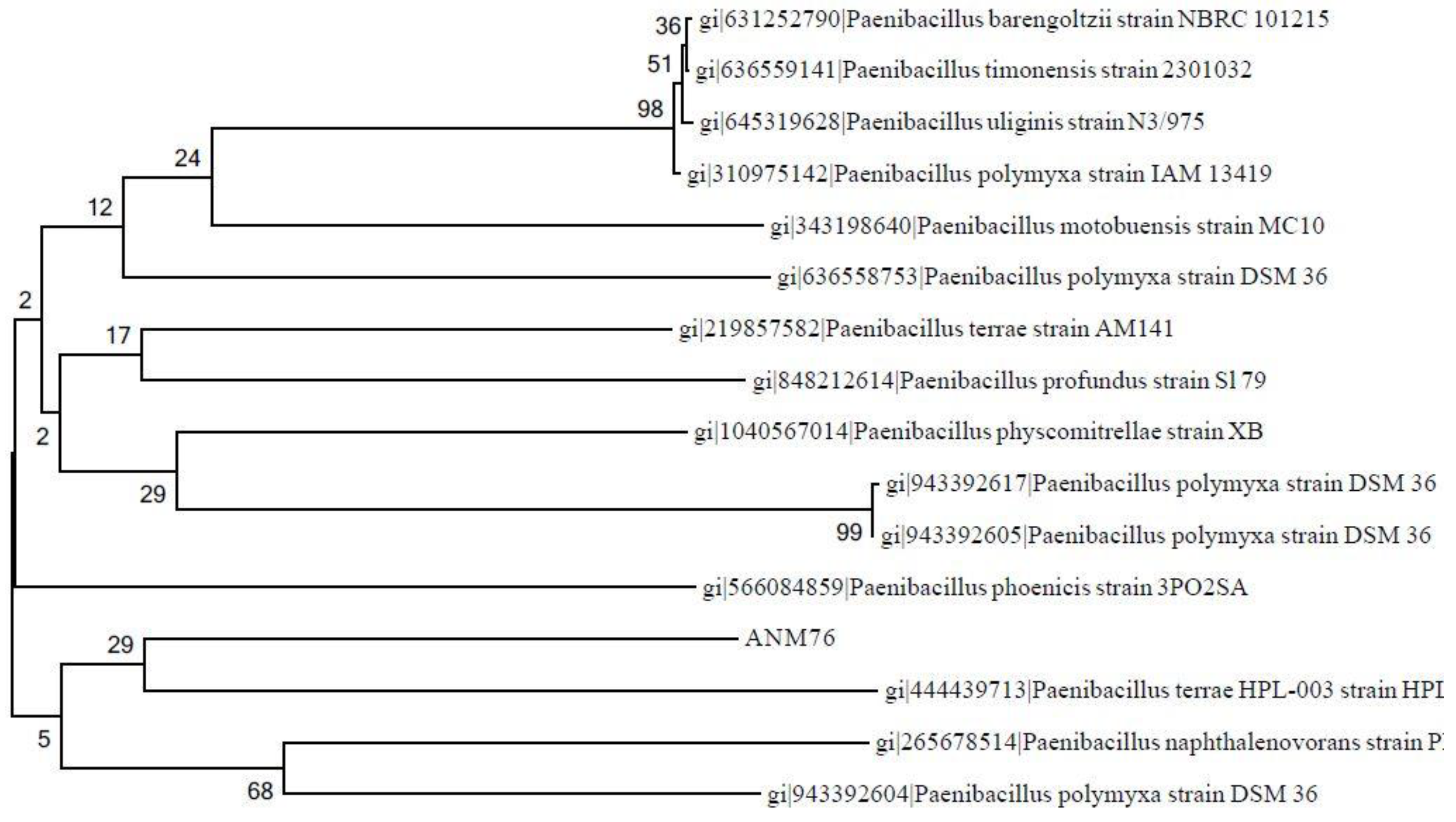
The two endophytic strains viz. ANM76 and ANM59 were selected on the basis of their potential for nodulation, to increase plant growth and difference in their colony morphology. Partial sequences of 16S rRNA coding genes were determined and submitted to NCBI with accession numbers KX788870 and KX788871 for strains ANM59 and ANM76, respectively. Nucleotide BLAST search of NCBI was used to find homologous sequences to identify the strain ANM59 as P. polymyxa and ANM76 as Paenibacillus sp. (Table 5). The phylogenetic tree of Paenibacillus polymyxa ANM59 and Paenibacillus sp. ANM76 are given in Figure 4 and Figure 5, respectively.

4. Discussion

Legumes have a specific trait of nodule formation which is a supreme habitat for nitrogen fixing rhizobia as well as non-rhizobial endophytes. Many non-rhizobial endophytes from roots and nodules of legumes are involved in plant growth and nodulation by making a symbiotic relationship with the host [17]. It is identified that thirteen bacterial genera are able to nodulate different legumes which include ten genera from α-proteobacteria while three genera belong to β-proteobacteria [54,55]. Most of these bacteria in nodules originated from the rhizosphere and phyllosphere or were transmitted through seed. Mostly, endophytic bacteria reside in nodules as compared to roots which is due to fact that nodules are much richer in nutrients. Isolation of both rhizobia as well as non-rhizobial endophytes from chickpea nodules need proper sterilization of nodules and every care taken to avoid surface contaminants [54]. In the current study, we isolated more than hundred non-rhizobial endophytic bacterial strains from the chickpea root nodules via the surface sterilization technique and authenticated them as endophytic in nature by re-isolation from chickpea root nodules after their inoculation under aseptic conditions. Similarly, Saini et al. [56] isolated endophytic Bacillus subtilis CNE215 and Bacillus licheniformis CRE1 from root or nodules of chickpea and authenticated them as true endophytic nature by re-isolating from chickpea nodules under sterilized conditions. In the current study, true endophytic Paenibacillus strains were characterized for PGP activities and selected strains able to promote chickpea growth and inducing nodulation were identified as Paenibacillus polymyxa ANM59 and Paenibacillus sp. ANM76 through 16S rRNA partial gene sequencing. Strain ANM76 was identified as Paenibacillus sp. with the possibility of having discovered a new species within the genus Paenibacillus in spite 86.9% similarity of the 16S rDNA, as this is low enough to speculate with this possibility. Moreover, in arid environments and soils submitted to several stresses such as drought, salinity, high temperatures, heavy metals, etc., new species of endophytes are being described as the phytomicrobiota of plants colonizing these areas.
Endophytic bacteria can promote plant growth through various direct and indirect mechanisms [7]. In the current study, endophytic bacterial strains were evaluated for plant growth promoting traits including IAA production, solubilization of P and colloidal chitin, enzymatic activity and production of ammonia. Various endophytic strains contributed in production of phytohormones and help to regulate developmental process in plants. It is now a matter-of-fact that a variety of endophytic bacteria can produce IAA [29,57,58]. Similarly, in the present study, all the tested endophytic strains showed production of IAA even without of precursor L-tryptophan however in presence of L-tryptophan there was significantly higher production of IAA (Table 1) which is according to the findings of Idris et al. [57] who revealed that IAA produced can be promoted by supplementing L-tryptophan. Khana et al. [58] also reported the production of IAA by endophytic Bacillus subtilis LK14, Pseudomonas aeruginosa LK17 and Sphingomonas LK18, while, application of Bacillus subtilis LK14 in Solanum lycopersicum showed significant increase in shoot and root biomass and chlorophyll contents. In vitro screening of endophytic bacteria to produce IAA could be a reliable trait for selection of effective plant growth promoting bacteria. The IAA production by bacteria is a prime trait for plant growth promotion as it is involved as a plant defence mechanism against stresses [59], may improve fitness of the plant–bacterium interaction [60], increases root exudation through loosening plant cell walls [61,62] and increases root surface area and length which provide plant access to soil nutrient and water uptake [63].
In the present study, seven strains were positive for P-solubilization and showed variation in halo zone formation (Table 1). These endophytic bacteria are able to convert insoluble P to soluble form possibly through producing low molecular weight organic acids which chelate the cation bound to phosphate through their OH and COOH groups or reduce the pH of medium by H+ to release P [64,65,66]. Paenibacillus is reportedly one of the most significant P-solubilizing bacteria. In our study, P. polymyxa ANM59 and Paenibacillus sp. ANM76 showed strong solubilization of P which are handy in increasing plant nutrition through increased P-uptake. P-solubilizing endophytic bacteria were evaluated for their ability to colonize chickpea roots and it was found that all the tested strains were well-capable of colonizing the chickpea roots. Plants respond to bacterial as well as fungal infection by activating the plant-encoded synthesis of cell wall degrading enzymes including chitinase, protease and lipase which degrade plant cell-associated chitin, proteins and lipids, respectively [32,46,67,68,69,70]. In the present study, the majority of strains were positive for chitinase which could strengthen plant defence mechanisms as well as improve plant growth and yield. Endophytic bacterial strains genetically transformed with genes encoding these cell wall degrading enzymes to become more effective biocontrol agents [70]. Kowsari et al. [71] reported that Trichoderma harzianum can transform plant encoded chitinase genes tested with the inserted acetamidase gene amds used as a selectable marker on plasmid could help the enzyme to bind better to insoluble chitin and showed higher antifungal activity towards Fusarium graminearum. The chitinase, peroxidases, lipase, protease are part of the pathogenesis-related proteins and their activation bring ISR in plants [7,72,73].
Colonies of endophytic strains in the current study appeared as mucoid milky white with an entire margin however there was a large variation in colony transparency, size and shape (Table 1). Other research also reported large variation in colony morphology of endophytic strains from nodules of Glycine max and Lespedeza sp. [74,75]. In our study, endophytic Paenibacillus strains were found Gram-negative which confirmed the findings of Dsouza et al. [75], Kittiwongwattana and Thawai [76], Valverde et al. [77] and Wang et al. [78] who reported several Paenibacillus strains including P. prosopidis PW21T, P. uliginis DSM 21861T, P. purispatti ES-MS17T, P. lactis PSM15596T and P. lamnae L7-75T as Gram-negative or Gram-variable. Generally, Paenibacillus species showed a Gram-positive reaction however due to a progressive increase in the numbers, cells became sensitive to the decolorizing step of the Gram-stain and electron microscopy showed these cells to be Gram-negative. With time, the peptidoglycan layer of Gram-positive bacteria became thinner, fragile, un-visible and cells relied more and more on the outermost surface layer to maintain cell shape and showed Gram-negative reaction during staining [79].
Evaluation of bacterial endophytic efficacy is important as these infect roots nodules, improve nutrients availability and may facilitate rhizobia for nitrogen fixation [56,80]. Therefore, efficacy of seven non-rhizobial endophytic strains to promote chickpea plant growth were evaluated to develop better inoculants of endophytic bacteria having multiple PGP traits. The shoot and root fresh and dry chickpea biomass were significantly boosted by the application of these endophytic bacteria (Table 4). Increase in chickpea seedlings biomass could be due to production of IAA, cell wall degrading enzymes, ammonia, acids and increased nutrient uptake which were found positive in the current in vitro study. Egamberdieva et al. [81] reported positive correlation between the production of IAA, hydrogen cyanide, siderophore and cell wall degrading enzymes by B. subtilis NUU4 which indicated that these strains are able to promote chickpea growth. Park et al. [80] and Parray et al. [82] also reported the several mechanisms in arrears of plant beneficial effects including synthesis of plant growth regulators, antibiotics, antifungal compounds, cell wall degrading enzymes or modulation of physio-biochemical processes in plants. In the nodulation test, we re-inoculated these strains to their host chickpea to reconfirm their nodulating capacity and all the strains were found positive in forming nodules. Among these endophytic strains, P. polymyxa ANM59 and Paenibacillus sp. ANM76 showed the maximum nodule formation along with improvement in its fresh and dry nodule weights while no nodulation was observed in control chickpea plants. Previous studies reported the nitrogen-fixing ability in few Paenibacillus strains including P. brasilensis, P. durus, P. sabinae, P. massiliensis, P. wynnii, P. peoriae and P. polymyxa [83,84,85]. Silva et al. [85] studied the effects of different Paenibacillus macerans, Paenibacillus durus, Paenibacillus polymyxa and Bacillus pumilus strains on the symbiosis between Bradyrhizobium and cowpea, and showed that these strains stimulated nodulation and improved nitrogen fixation efficiency. Latif et al. [86] assessed Paenibacillus sp. (T.P2-4) along with Rhizobia and Sinorhizobia for nodule formation and reported for the first time a unique symbiotic association of Paenibacillus sp. (T.P2-4) nodulating Trifolium pratense. They also screened nod D genes in strains for genetics involved in nitrogen fixation by using nod primers deduced from Sinorhizobium sp. and reported maximum amplified band size of nod D gene in the case of Paenibacillus sp. (T.P2-4). However, in future further work on the mode of co-existence is needed to be investigated about the respective role of various rhizobia and non-rhizobial strains along with their associated nodulation genes.

5. Conclusions

In this study, endophytic Paenibacillus strains isolated from root nodules successfully formed a symbiotic association with chickpea, which was a novel finding. Our results suggest that endophytic Paenibacillus polymyxa ANM59 and Paenibacillus sp. ANM76 are well capable of colonizing chickpea roots and induced nodulation in chickpea. These strains possess multiple plant growth promoting traits as well as the ability to tolerate abiotic stresses. Plant growth promotion and biological nitrogen fixation by Paenibacillus strains in addition to other PGP traits offer a potential candidature to these Paenibacillus strains as efficient plant growth promoting endophytes. In future, genetic characteristics of Paenibacillus strains involved in root nodulation and nitrogen fixation in combination with or without rhizobial strains should be evaluated.

Supplementary Materials

The following are available online at https://www.mdpi.com/2073-4395/9/10/621/s1. Table S1: Colony morphological characteristics of endophytic strains from chickpea grown on yeast extract mannitol agar.

Author Contributions

Conceptualization, M.A.; methodology, I.N., M.Z.M. and M.A.; software, I.N., A.M. and M.Z.M.; validation, M.A., I.N., A.H. and M.Z.M.; formal analysis, I.N.; investigation, M.A.; resources, M.A.; data curation, I.N.; writing—original draft preparation, M.Z.M., M.A. and I.N.; writing—review and editing, A.H., M.Z.M., A.M., T.H.H., Z.A.Z. and M.X.; visualization, M.Z.M. and M.A.; supervision, M.A.; project administration, M.A.

Funding

This research received no external funding.

Acknowledgments

We are grateful to the Department of Soil Science, University College of Agriculture and Environmental Sciences, the Islamia University of Bahawalpur, Pakistan for provision of necessary research facilities for this study. Authors highly acknowledge the help from Irfan Akhtar Qureshi (Manager Administration, Center for Research in Molecular Medicine, Institute of Molecular Biology and Biotechnology, The University of Lahore, Defence Road Campus, Lahore, Pakistan) for technical assistance.

Conflicts of Interest

The authors declare no conflict of interest.

References

  1. Jukanti, A.K.; Gaur, P.M.; Gowda, C.L.L.; Chibbar, R.N. Nutritional quality and health benefits of chickpea (Cicer arietinumL.): A review. Br. J. Nutr. 2012, 108, S11–S26. [Google Scholar] [CrossRef] [PubMed]
  2. Sulieman, S.; Tran, L.S.P. Asparagine: An Amide of Particular Distinction in the Regulation of Symbiotic Nitrogen Fixation of Legumes. Crit. Rev. Biotechnol. 2013, 33, 309–327. [Google Scholar] [CrossRef] [PubMed]
  3. Gouda, S.; Kerry, R.G.; Das, G.; Paramithiotis, S.; Shin, H.-S.; Patra, J.K. Revitalization of plant growth promoting rhizobacteria for sustainable development in agriculture. Microbiol. Res. 2018, 206, 131–140. [Google Scholar] [CrossRef]
  4. Martínez-Hidalgo, P.; Hirsch, A.M. The Nodule Microbiome: N2-Fixing Rhizobia Do Not Live Alone. Phytobiomes J. 2017, 1, 70–82. [Google Scholar] [CrossRef]
  5. Lu, J.; Yang, F.; Wang, S.; Ma, H.; Liang, J.; Chen, Y. Co-existence of Rhizobia and Diverse Non-rhizobial Bacteria in the Rhizosphere and Nodules of Dalbergia odorifera Seedlings Inoculated with Bradyrhizobium elkanii, Rhizobium multihospitium–Like and Burkholderia pyrrocinia–Like Strains. Front. Microbiol. 2017, 8, 2255. [Google Scholar] [CrossRef] [PubMed]
  6. Peix, A.; Ramírez-Bahena, M.H.; Velázquez, E.; Bedmar, E.J. Bacterial Associations with Legumes. CRC Crit. Rev. Plant Sci. 2015, 34, 17–42. [Google Scholar] [CrossRef]
  7. Santoyo, G.; Moreno-Hagelsieb, G.; Orozco-Mosqueda, M.D.C.; Glick, B.R. Plant growth-promoting bacterial endophytes. Microbiol. Res. 2016, 183, 92–99. [Google Scholar] [CrossRef]
  8. Haidar, B.; Ferdous, M.; Fatema, B.; Ferdous, A.S.; Islam, M.R.; Khan, H. Population diversity of bacterial endophytes from jute (Corchorus olitorius) and evaluation of their potential role as bioinoculants. Microbiol. Res. 2018, 208, 43–53. [Google Scholar] [CrossRef]
  9. Berendsen, R.L.; Pieterse, C.M.; Bakker, P.A. The rhizosphere microbiome and plant health. Trends Plant Sci. 2012, 17, 478–486. [Google Scholar] [CrossRef]
  10. Ali, M.A.; Naveed, M.; Mustafa, A.; Abbas, A. The Good, the Bad, and the Ugly of Rhizosphere Microbiome. In Probiotics and Plant Health; Springer Science and Business Media LLC: Berlin, Germany, 2017; pp. 253–290. [Google Scholar]
  11. Timmusk, S.; Paalme, V.; Pavlícek, T.; Bergquist, J.; Vangala, A.; Danilas, T.; Nevo, E. Bacterial Distribution in the Rhizosphere of Wild Barley under Contrasting Microclimates. PLoS ONE 2011, 6, e17968. [Google Scholar] [CrossRef]
  12. Huang, J. Ultrastructure of Bacterial Penetration in Plants. Annu. Rev. Phytopathol. 1986, 24, 141–157. [Google Scholar] [CrossRef]
  13. Hallmann, J.; Quadt-Hallmann, A.; Mahaffee, W.F.; Kloepper, J.W. Bacterial endophytes in agricultural crops. Can. J. Microbiol. 1997, 43, 895–914. [Google Scholar] [CrossRef]
  14. Ryan, R.P.; Germaine, K.; Franks, A.; Ryan, D.J.; Dowling, D.N. Bacterial endophytes: recent developments and applications. FEMS Microbiol. Lett. 2008, 278, 1–9. [Google Scholar] [CrossRef] [Green Version]
  15. Sun, L.; Qiu, F.; Zhang, X.; Dai, X.; Dong, X.; Song, W. Endophytic Bacterial Diversity in Rice (Oryza sativa L.) Roots Estimated by 16S RDNA Sequence Analysis. Microb. Ecol. 2008, 55, 415–424. [Google Scholar] [PubMed]
  16. Pedrosa, F.O.; Monteiro, R.A.; Wassem, R.; Cruz, L.M.; Ayub, R.A.; Colauto, N.B.; Fernandez, M.A.; Fungaro, M.H.P.; Grisard, E.C.; Hungria, M.; et al. Genome of Herbaspirillum seropedicae Strain SmR1, a Specialized Diazotrophic Endophyte of Tropical Grasses. PLoS Genet. 2011, 7, e1002064. [Google Scholar] [CrossRef] [PubMed]
  17. Shi, Y.; Yang, H.; Zhang, T.; Sun, J.; Lou, K. Illumina-based analysis of endophytic bacterial diversity and space-time dynamics in sugar beet on the north slope of Tianshan mountain. Appl. Microbiol. Biotechnol. 2014, 98, 6375–6385. [Google Scholar] [CrossRef] [PubMed]
  18. Abbamondi, G.R.; Tommonaro, G.; Weyens, N.; Thijs, S.; Sillen, W.; Gkorezis, P.; Iodice, C.; Rangel, W.D.M.; Nicolaus, B.; Vangronsveld, J. Plant growth-promoting effects of rhizospheric and endophytic bacteria associated with different tomato cultivars and new tomato hybrids. Chem. Boil. Technol. Agric. 2016, 3, 11. [Google Scholar] [CrossRef]
  19. Kumar, A.; Singh, R.; Yadav, A.; Giri, D.D.; Singh, P.K.; Pandey, K.D. Isolation and characterization of bacterial endophytes of Curcuma longa L. 3 Biotech 2016, 6, 60. [Google Scholar] [CrossRef]
  20. Muthukumar, A.; Udhayakumar, R.; Naveenkumar, R. Role of Bacterial Endophytes in Plant Disease Control. In Endophytes: Crop Productivity and Protection; Springer Science and Business Media LLC: Berlin, Germany, 2017; Volume 16, pp. 133–161. [Google Scholar]
  21. Liotti, R.G.; Figueiredo, M.I.D.S.; Da Silva, G.F.; De Mendonça, E.A.F.; Soares, M.A. Diversity of cultivable bacterial endophytes in Paullinia cupana and their potential for plant growth promotion and phytopathogen control. Microbiol. Res. 2018, 207, 8–18. [Google Scholar] [CrossRef]
  22. Eida, A.A.; Alzubaidy, H.S.; de Zélicourt, A.; Synek, L.; Alsharif, W.; Lafi, F.F.; Hirt, H.; Saad, M.M. Phylogenetically Diverse Endophytic Bacteria from Desert Plants Induce Transcriptional Changes of Tissue-Specific Ion Transporters and Salinity Stress in Arabidopsis. Plant Sci. 2019, 280, 228–240. [Google Scholar] [CrossRef]
  23. Glick, B.R.; Todorovic, B.; Czarny, J.; Cheng, Z.; Duan, J.; McConkey, B. Promotion of Plant Growth by Bacterial ACC Deaminase. Crit. Rev. Plant Sci. 2007, 26, 227–242. [Google Scholar] [CrossRef]
  24. Glick, B.R.; Cheng, Z.; Czarny, J.; Duan, J. Promotion of plant growth by ACC deaminase-producing soil bacteria. Eur. J. Plant Pathol. 2007, 119, 329–339. [Google Scholar] [CrossRef]
  25. Prasanna, R.; Ramakrishnan, B.; Simranjit, K.; Ranjan, K.; Kanchan, A.; Hossain, F.; Nain, L. Cyanobacterial and rhizobial inoculation modulates the plant physiological attributes and nodule microbial communities of chickpea. Arch. Microbiol. 2017, 199, 1311–1323. [Google Scholar] [CrossRef] [PubMed]
  26. Hardoim, P.R.; Van Overbeek, L.S.; Van Elsas, J.D. Properties of bacterial endophytes and their proposed role in plant growth. Trends Microbiol. 2008, 16, 463–471. [Google Scholar] [CrossRef]
  27. Rashid, S.; Charles, T.C.; Glick, B.R. Isolation and characterization of new plant growth-promoting bacterial endophytes. Appl. Soil Ecol. 2012, 61, 217–224. [Google Scholar] [CrossRef]
  28. Datta, C.; Basu, P. Indole acetic acid production by a Rhizobium species from root nodules of a leguminous shrub, Cajanus cajan. Microbiol. Res. 2000, 155, 123–127. [Google Scholar] [CrossRef]
  29. Duca, D.; Lorv, J.; Patten, C.L.; Rose, D.; Glick, B.R. Indole-3-acetic acid in plant–microbe interactions. Antonie van Leeuwenhoek 2014, 106, 85–125. [Google Scholar] [CrossRef]
  30. Reetha, S.; Bhuvaneswari, G.; Thamizhiniyan, P.; Mycin, T.R. Isolation of Indole Acetic Acid (IAA) Producing Rhizobacteria of Pseudomonas fluorescens and Bacillus subtilis and Enhance Growth of Onion (Allim cepa L.). Int. J. Curr. Microbiol. Appl. Sci. 2014, 3, 568–574. [Google Scholar]
  31. Dhungana, S.A.; Itoh, K. Effects of Co-Inoculation of Indole-3-Acetic Acid-Producing and -Degrading Bacterial Endophytes on Plant Growth. Horticulturae 2019, 5, 17. [Google Scholar] [CrossRef]
  32. Chernin, L.; Chet, I. Microbial Enzymes in the Biocontrol of Plant Pathogens and Pests. In Water Policy and Planning in a Variable and Changing Climate; Informa UK Limited: Colchester, UK, 2002; Volume 84, pp. 179–234. [Google Scholar]
  33. Ma, Y.; Rajkumar, M.; Zhang, C.; Freitas, H. Beneficial role of bacterial endophytes in heavy metal phytoremediation. J. Environ. Manag. 2016, 174, 14–25. [Google Scholar] [CrossRef]
  34. Kannojia, P.; Choudhary, K.K.; Srivastava, A.K.; Singh, A.K. PGPR Bioelicitors: Induced Systemic Resistance (ISR) and Proteomic Perspective on Biocontrol. In PGPR Amelioration in Sustainable Agriculture: Food Security and Environmental Management; Singh, A.K., Kumar, A., Singh, P.K., Eds.; Woodhead Publishing: Cambridge, UK, 2019; pp. 67–84. [Google Scholar]
  35. Al-Adham, I.; Haddadin, R.; Collier, P. Types of Microbicidal and Microbistatic Agents. In Russell, Hugo & Ayliffe’s: Principles and Practice of Disinfection, Preservation and Sterilization; Fraise, A.P., Maillard, J.Y., Sattar, S., Eds.; Wiley-Blackwell: Oxford, UK, 2012; pp. 5–70. [Google Scholar]
  36. Vincent, J.M. A Manual for the Practical Study of the Root-Nodule Bacteria; Blackwell Scientific Publications: Oxford and Edinburgh, UK, 1970; pp. 153–159. [Google Scholar]
  37. Pikovskaya, R.I. Mobilization of Phosphorus in Soil in Connection with Vital Activity of Some Microbial Species. Mikrobiologiya 1948, 17, 362–370. [Google Scholar]
  38. Vazquez, P.; Holguín, G.; Puente, M.E.; Lopez-Cortes, A.; Bashan, Y. Phosphate-solubilizing microorganisms associated with the rhizosphere of mangroves in a semiarid coastal lagoon. Boil. Fertil. Soils 2000, 30, 460–468. [Google Scholar] [CrossRef]
  39. Premono, M.; Moawad, A.; Vlek, P. Effect of Phosphate-Solubilizing Pseudomonas putida on the Growth of Maize and Its Survival in the Rhizosphere. Indones. J. Crop Sci. 1996, 11, 13–23. [Google Scholar]
  40. Singh, S.M.; Yadav, L.S.; Singh, S.K.; Singh, P.; Singh, P.N.; Ravindra, R. Phosphate solubilizing ability of two Arctic Aspergillus niger strains. Polar Res. 2011, 30, 7283. [Google Scholar]
  41. Fasim, F.; Ahmed, N.; Parsons, R.; Gadd, G.M. Solubilization of zinc salts by a bacterium isolated from the air environment of a tannery. FEMS Microbiol. Lett. 2002, 213, 1–6. [Google Scholar] [CrossRef] [PubMed]
  42. Mumtaz, M.Z.; Ahmad, M.; Jamil, M.; Hussain, T. Zinc Solubilizing Bacillus Spp. Potential Candidates for Biofortification in Maize. Microbiol. Res. 2017, 202, 51–60. [Google Scholar]
  43. Cappuccino, J.; Welsh, C. Microbiology: A Laboratory Manual, 11th ed.; Pearson: New York, NY, USA, 2017; pp. 163–232. [Google Scholar]
  44. Ali, B.; Hasnain, S. Efficacy of Bacterial Auxin on in Vitro Growth of Brassica oleracea L. World J. Microbiol. Biotechnol. 2007, 23, 779–784. [Google Scholar] [CrossRef]
  45. Tang, W.Y.; Borner, J. Enzymes Involved in Synthesis and Breakdown of Indoleacetic Acid. Mod. Methods Plant Anal. 1979, 7, 238–241. [Google Scholar]
  46. Chernin, L.S.; Winson, M.K.; Thompson, J.M.; Haran, S.; Bycroft, B.W.; Chet, I.; Williams, P.; Stewart, G.S.A.B. Chitinolytic Activity in Chromobacterium violaceum: Substrate Analysis and Regulation by Quorum Sensing. J. Bacteriol. 1998, 180, 4435–4441. [Google Scholar]
  47. De Oliveira, A.N.; De Oliveira, L.A.; Andrade, J.S.; Junior, A.F.C. Rhizobia amylase production using various starchy substances as carbon substrates. Braz. J. Microbiol. 2007, 38, 208–216. [Google Scholar] [CrossRef]
  48. Mendis, H.C.; Thomas, V.P.; Schwientek, P.; Salamzade, R.; Chien, J.-T.; Waidyarathne, P.; Kloepper, J.; De La Fuente, L. Strain-specific quantification of root colonization by plant growth promoting rhizobacteria Bacillus firmus I-1582 and Bacillus amyloliquefaciens QST713 in non-sterile soil and field conditions. PLoS ONE 2018, 13, e0193119. [Google Scholar] [CrossRef] [PubMed]
  49. Carrasco, J.A.; Armario, P.; Pajuelo, E.; Burgos, A.; Caviedes, M.A.; López, R.; Chamber, M.A.; Palomares, A.J.; López-Núñez, R. Isolation and characterisation of symbiotically effective Rhizobium resistant to arsenic and heavy metals after the toxic spill at the Aznalcóllar pyrite mine. Soil Boil. Biochem. 2005, 37, 1131–1140. [Google Scholar] [CrossRef]
  50. Macrogen, Seoul, Korea. Available online: https://dna.macrogen.com/eng/ (accessed on 23 March 2019).
  51. Kumar, S.; Stecher, G.; Tamura, K. MEGA7: Molecular Evolutionary Genetics Analysis version 7.0 for bigger datasets. Mol. Boil. Evol. 2016, 33, 1870–1874. [Google Scholar] [CrossRef] [PubMed]
  52. Tamura, K.; Nei, M.; Kumar, S. Prospects for inferring very large phylogenies by using the neighbor-joining method. Proc. Natl. Acad. Sci. USA 2004, 101, 11030–11035. [Google Scholar] [CrossRef] [Green Version]
  53. Gomez, K.A.; Gomez, A.A. Statistical Procedures for Agricultural Research, 2nd ed.; John Wiley & Sons: New York, NY, USA, 1984; pp. 187–240. [Google Scholar]
  54. Dudeja, S.S.; Giri, R.; Saini, R.; Suneja-Madan, P.; Kothe, E. Interaction of Endophytic Microbes with Legumes. J. Basic Microbiol. 2012, 52, 248–260. [Google Scholar] [CrossRef] [PubMed]
  55. Weir, B.S. The Current Taxonomy of Rhizobia. Available online: https://www.rhizobia.co.nz/taxonomy/rhizobia (accessed on 3 May 2019).
  56. Saini, R.; Dudeja, S.S.; Giri, R.; Kumar, V. Isolation, characterization, and evaluation of bacterial root and nodule endophytes from chickpea cultivated in Northern India. J. Basic Microbiol. 2013, 55, 74–81. [Google Scholar] [CrossRef] [PubMed]
  57. Idris, E.E.; Iglesias, D.J.; Talón, M.; Borriss, R. Tryptophan-Dependent Production of Indole-3-Acetic Acid (IAA) Affects Level of Plant Growth Promotion by Bacillus amyloliquefaciens FZB42. Mol. Plant.-Microbe Interactions 2007, 20, 619–626. [Google Scholar] [CrossRef]
  58. Khan, A.L.; Halo, B.A.; Elyassi, A.; Ali, S.; Al-Hosni, K.; Hussain, J.; Al-Harrasi, A.; Lee, I.-J. Indole acetic acid and ACC deaminase from endophytic bacteria improves the growth of Solanum lycopersicum. Electron. J. Biotechnol. 2016, 21, 58–64. [Google Scholar] [CrossRef] [Green Version]
  59. Spaepen, S.; Vanderleyden, J.; Remans, R. Indole-3-acetic acid in microbial and microorganism-plant signaling. FEMS Microbiol. Rev. 2007, 31, 425–448. [Google Scholar] [CrossRef] [Green Version]
  60. Patten, C.L.; Glick, B.R. Role of Pseudomonas putida Indoleacetic Acid in Development of the Host Plant Root System. Appl. Environ. Microbiol. 2002, 68, 3795–3801. [Google Scholar] [CrossRef]
  61. James, E.K.; Gyaneshwar, P.; Mathan, N.; Barraquio, W.L.; Reddy, P.M.; Iannetta, P.P.M.; Olivares, F.L.; Ladha, J.K. Infection and Colonization of Rice Seedlings by the Plant Growth-Promoting Bacterium Herbaspirillum seropedicae Z67. Mol. Plant.-Microbe Interactions 2002, 15, 894–906. [Google Scholar] [CrossRef] [PubMed]
  62. Chi, F.; Shen, S.-H.; Cheng, H.-P.; Jing, Y.-X.; Yanni, Y.G.; Dazzo, F.B. Ascending Migration of Endophytic Rhizobia, from Roots to Leaves, inside Rice Plants and Assessment of Benefits to Rice Growth Physiology. Appl. Environ. Microbiol. 2005, 71, 7271–7278. [Google Scholar] [CrossRef] [PubMed] [Green Version]
  63. Vessey, J.K. Plant growth promoting rhizobacteria as biofertilizers. Plant Soil 2003, 255, 571–586. [Google Scholar] [CrossRef]
  64. Khan, M.S.; Zaidi, A.; Ahmad, E. Mechanism of Phosphate Solubilization and Physiological Functions of Phosphate-Solubilizing Microorganisms. In Phosphate Solubilizing Microorganisms; Springer Science and Business Media LLC: Berlin, Germany, 2014; pp. 31–62. [Google Scholar]
  65. Kpomblekou-A, K.; Tabatabai, M.A. Effect of Organic Acids on release of Phosphorus from Phosphate Rocks. Soil Sci. 1994, 158, 442–453. [Google Scholar] [CrossRef]
  66. Seshachala, U.; Tallapragada, P. Phosphate Solubilizers from the Rhizospher of Piper nigrum L. in Karnataka, India. Chil. J. Agric. Res. 2012, 72, 397–403. [Google Scholar] [CrossRef] [Green Version]
  67. Chernin, L.; Ismailov, Z.; Haran, S.; Chet, I. Chitinolytic Enterobacter agglomerans Antagonistic to Fungal Plant Pathogens. Appl. Environ. Microbiol. 1995, 61, 1720–1726. [Google Scholar] [Green Version]
  68. Husson, E.; Hadad, C.; Huet, G.; Laclef, S.; Lesur, D.; Lambertyn, V.; Jamali, A.; Gottis, S.; Sarazin, C.; Van Nhien, A.N. The effect of room temperature ionic liquids on the selective biocatalytic hydrolysis of chitin via sequential or simultaneous strategies. Green Chem. 2017, 19, 4122–4131. [Google Scholar] [CrossRef]
  69. Friedrich, N.; Hagedorn, M.; Soldati-Favre, D.; Soldati, T. Prison Break: Pathogens’ Strategies To Egress from Host Cells. Microbiol. Mol. Boil. Rev. 2012, 76, 707–720. [Google Scholar] [CrossRef] [PubMed]
  70. Koby, S.; Schickler, H.; Ilan, C.; Oppenheim, A.B. The chitinase encoding Tn7-based chiA gene endows Pseudomonas fluorescens with the capacity to control plant pathogens in soil. Gene 1994, 147, 81–83. [Google Scholar] [CrossRef]
  71. Kowsari, M.; Zamani, M.R.; Motallebi, M. Overexpression of Chimeric Chitinase42 Enhanced Antifungal Activity of Trichoderma harzianum against Fusarium graminearum. Mycol. Iran. 2016, 3, 15–23. [Google Scholar]
  72. Yedidia, I.; Benhamou, N.; Chet, I. Induction of Defense Responses in Cucumber Plants (Cucumis sativus L.) by the Biocontrol Agent Trichoderma harzianum. Appl. Environ. Microbiol. 1999, 65, 1061–1070. [Google Scholar] [PubMed]
  73. Palaniappan, P.; Chauhan, P.S.; Saravanan, V.S.; Anandham, R.; Sa, T. Isolation and characterization of plant growth promoting endophytic bacterial isolates from root nodule of Lespedeza sp. Boil. Fertil. Soils 2010, 46, 807–816. [Google Scholar] [CrossRef]
  74. Hung, P.Q.; Kumar, S.M.; Govindsamy, V.; Annapurna, K. Isolation and characterization of endophytic bacteria from wild and cultivated soybean varieties. Boil. Fertil. Soils 2007, 44, 155–162. [Google Scholar] [CrossRef]
  75. Dsouza, M.; Taylor, M.W.; Ryan, J.; MacKenzie, A.; Lagutin, K.; Anderson, R.F.; Turner, S.J.; Aislabie, J. Paenibacillus darwinianus Sp. Nov., Isolated from Gamma-Irradiated Antarctic Soil. Int. J. Syst. Evol. Microbiol. 2014, 64, 1406–1411. [Google Scholar] [CrossRef] [PubMed]
  76. Kittiwongwattana, C.; Thawai, C. Paenibacillus lemnae Sp. Nov., an Endophytic Bacterium of Duckweed (Lemna aequinoctialis). Int. J. Syst. Evol. Microbiol. 2015, 65, 107–112. [Google Scholar] [CrossRef] [PubMed]
  77. Valverde, A.; Fterich, A.; Mahdhi, M.; Ramirez-Bahena, M.-H.; Caviedes, M.A.; Mars, M.; Velazquez, E.; Rodriguez-Llorente, I.D. Paenibacillus prosopidis Sp. Nov., Isolated from the Nodules of Prosopis farcta. Int. J. Syst. Evol. Microbiol. 2010, 60, 2182–2186. [Google Scholar] [CrossRef] [PubMed]
  78. Wang, L.; Baek, S.-H.; Cui, Y.; Lee, H.-G.; Lee, S.-T. Paenibacillus sediminis Sp. Nov., a Xylanolytic Bacterium Isolated from a Tidal Flat. Int. J. Syst. Evol. Microbiol. 2012, 62, 1284–1288. [Google Scholar] [CrossRef] [PubMed]
  79. Beveridge, T.J. Mechanism of gram variability in select bacteria. J. Bacteriol. 1990, 172, 1609–1620. [Google Scholar] [CrossRef] [Green Version]
  80. Park, J.-W.; Balaraju, K.; Kim, J.-W.; Lee, S.-W.; Park, K. Systemic resistance and growth promotion of chili pepper induced by an antibiotic producing Bacillus vallismortis strain BS07. Boil. Control. 2013, 65, 246–257. [Google Scholar] [CrossRef]
  81. Egamberdieva, D.; Wirth, S.J.; Shurigin, V.V.; Hashem, A.; Abd_Allah, E.F. Endophytic Bacteria Improve Plant Growth, Symbiotic Performance of Chickpea (Cicer arietinum L.) and Induce Suppression of Root Rot Caused by Fusarium solani under Salt Stress. Front. Microbiol. 2017, 8, 8. [Google Scholar] [CrossRef]
  82. Parray, J.A.; Kamili, A.N.; Reshi, Z.A.; Qadri, R.A.; Jan, S. Interaction of Rhizobacterial Strains for Growth Improvement of Crocus Sativus, L. under Tissue Culture Conditions. Plant Cell. Tissue Organ Cult. 2015, 121, 325–334. [Google Scholar] [CrossRef]
  83. Beneduzi, A.; Costa, P.B.; Parma, M.; Melo, I.S.; Bodanese-Zanettini, M.H.; Passaglia, L.M.P. Paenibacillus riograndensis Sp. Nov., a Nitrogen-Fixing Species Isolated from the Rhizosphere of Triticum aestivum. Int. J. Syst. Evol. Microbiol. 2010, 60, 128–133. [Google Scholar] [PubMed]
  84. Xie, J.-B.; Du, Z.; Bai, L.; Tian, C.; Zhang, Y.; Xie, J.-Y.; Wang, T.; Liu, X.; Chen, X.; Cheng, Q.; et al. Comparative Genomic Analysis of N2-Fixing and Non-N2-Fixing Paenibacillus Spp.: Organization, Evolution and Expression of the Nitrogen Fixation Genes. PLoS Genet. 2014, 10, e1004231. [Google Scholar]
  85. da Silva, V.N.; da Silva, L.E.S.F.; Martinez, C.R.; Seldin, L.; Burity, H.A.; Figueiredo, M.V.B. Strains of Paenibacillus Promoters of the Specific Nodulation in the Symbiosis Bradyrhizobium caupi. Acta Sci. Agron. 2007, 29, 331–338. [Google Scholar] [CrossRef]
  86. Latif, S.; Khan, S.U.; Naveed, M.; Mustafa, G.; Bashir, T.; Mumtaz, A.S. The diversity of Rhizobia, Sinorhizobia and novel non-Rhizobial Paenibacillus nodulating wild herbaceous legumes. Arch. Microbiol. 2013, 195, 647–653. [Google Scholar] [CrossRef]
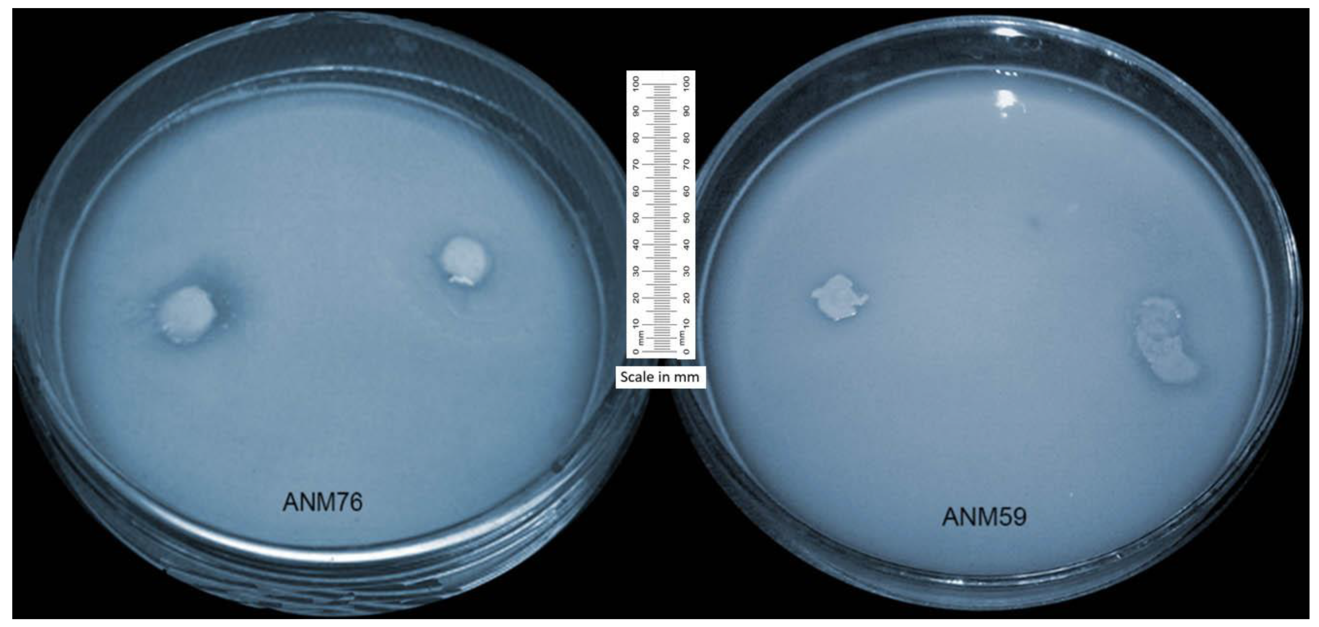
Figure 1. In vitro solubilization of inorganic phosphorus by endophytic strains ANM59 and ANM76 after 48 h of incubation at 28 ± 1 ºC. Halo zone around bacterial colonies indicated solubilization of phosphorus. Halo zone and bacterial colonies zones were calibrated through millimetre (mm) scale.
Agronomy 09 00621 g001
Figure 2. Comparison of shoot growth of chickpea inoculated with endophytic strains grown under the jar trial.
Agronomy 09 00621 g002
Figure 3. Comparison of growth of selected chickpea roots inoculated with endophytic strains grown under the jar trial.
Agronomy 09 00621 g003
Figure 4. Neighbour-joining phylogenetic tree produced using multiple alignment of 16S rRNA gene sequence of Paenibacillus polymyxa ANM59 with those of other bacterial strains found in the GeneBank database.
Agronomy 09 00621 g004
Figure 5. Neighbour-joining phylogenetic tree produced using multiple alignment of 16S rRNA gene sequence of Paenibacillus sp. ANM76 with those of other bacterial strains found in the GeneBank database.
Agronomy 09 00621 g005
Table 1. Plant growth promoting characteristics in terms of production of indole-3-acetic acid (IAA), phosphate solubilization, colloidal chitin solubilization and root colonization ability by tested endophytic strains from chickpea.
StrainsProduction of IAA (mg L−1) aPhosphate Solubilization bColloidal Chitin Solubilization cRoot Colonization d
Without L-Tryptophan With L-TryptophanP Halo Zone (mm)PSE (%)PSIColloidal Halo Zone (mm)CSE (%)CSI
ANM94.78 ± 0.0036.79 ± 0.005 ND eNDND9.67 ± 0.666145± 3.3162.45 ± 0.019NS f
ANM123.67 ± 0.0024.41 ± 0.003NDNDNDNDNDNDNS
ANM164.21 ± 0.0046.00 ± 0.00118.3 ± 0.333106 ± 2.9762.06 ± 0.029NDNDND4.23 × 105
ANM264.02 ± 0.0024.91 ± 0.007NDNDNDNDNDNDNS
ANM394.13 ± 0.0054.98 ± 0.00832.0 ± 0.523128 ± 5.4562.28 ± 0.05516.66 ± 0.511314 ± 1.8524.14 ± 0.0185.06 × 105
ANM583.56 ± 0.0064.24 ± 0.002NDNDNDNDNDNDNS
ANM594.27 ± 0.0065.26 ± 0.00121.0 ± 0.333 105 ± 2.2532.05 ± 0.036NDNDND4.44 × 105
ANM634.44 ± 0.0036.39 ± 0.008 29.67 ± 0.856203 ± 4.5563.03 ± 0.03313.33 ± 0.432211 ± 1.2013.11 ± 0.0424.78 × 105
ANM663.78 ± 0.0084.57 ± 0.011NDNDNDNDNDNDNS
ANM694.38 ± 0.0075.19 ± 0.01233.00 ± 0.533165 ± 8.335 2.65 ± 0.0149.67 ± 0.333147 ± 1.9152.47 ± 0.0724.12 × 105
ANM734.29 ± 0.0105.15 ± 0.00820.30 ± 0.765105 ± 9.8822.05 ± 0.01814.33 ± 0.778172 ± 4.1672.72 ± 0.2565.17 × 105
ANM753.57 ± 0.0064.24 ± 0.002NDNDNDNDNDNDNS
ANM765.24 ± 0.0037.99 ± 0.00438.33 ± 0.344311± 3.3164.11 ± 0.0058.67 ± 0.667144 ± 6.2332.44 ± 0.03295.45 × 105
ANM773.68 ± 0.0094.36 ± 0.009NDNDND14.00 ± 0.856193 ± 2.2312.93 ± 0.015NS
ANM785.31 ± 0.0028.13 ± 0.005NDNDND17.33 ± 0.333308 ± 7.3254.08 ± 0.045NS
ANM795.33 ± 0.0088.18 ± 0.001NDNDND7.33 ± 0.667122 ± 1.742 2.22 ± 0.082NS
ANM813.97 ± 0.0034.49 ± 0.007NDNDND12.33 ± 0.556186 ± 2.4452.86 ± 0.051NS
ANM825.22 ± 0.0087.91 ± 0.005NDNDND12.67 ± 0.856126 ± 4.6672.26 ± 0.027NS
ANM1003.87 ± 0.0094.58 ± 0.006NDNDNDNDNDNDNS
a Indole acetic acid (IAA) was estimated by observing optical density at 600 nm both in presence and absence of L-tryptophan; values are mean of three replicate ± standard error; b diameter of halo zones developed by endophytic strains was observed after 48 h of incubations and solubilisation efficiency and index were calculated; P = phosphorus, PSE = phosphorus solubilization efficiency, PSI = phosphorus solubilization index; means are average of three replications ± standard error; c yeast extract mannitol agar amended with colloidal chitin was centrally inoculated with respective endophytic strains and clearing zone was observed; CSE = chitin solubilization efficiency, CSI = chitin solubilization index; values are mean of three replicate ± standard error; d root colonization ability of endophytic strains was observed by colony forming unit (CFU) method from root of inoculated chickpea grown for one week; e ND stands for not detected; f NS stands for not studied.
Table 2. Plant growth promoting characteristics of endophytic strains from chickpea.
Strains aSolubilize ZnCatalase ActivityOxidase ActivityUrease ActivityChitinase ActivityStarch HydrolysisNH3 Prod.Acid Prod.Glucose UtilizationSucrose UtilizationProduce CO2Produce H2SGram Staining
ANM9b+ c++++++++−ve d
ANM12++++−ve
ANM16+++++++−ve
ANM26++++++++−ve
ANM39+++++++−ve
ANM58+++++++−ve
ANM59+++++++−ve
ANM63+++++−ve
ANM66+++++−ve
ANM69++++ ++−ve
ANM73+++++++−ve
ANM75+++++++−ve
ANM76+++++++−ve
ANM77+++++−ve
ANM78++++++−ve
ANM79+++++++−ve
ANM81++++−ve
ANM82+++++−ve
ANM100+++++−ve
a Authenticated endophytic strains were screened for plant growth promoting characteristics and in vitro tests were repeated twice for confirmation of results with three replicates each time; b symbol − represents the absence of the traits; c symbol + represents the presence of the traits; d symbol −ve represents the Gram negative bacterial strains.
Table 3. Growth of endophytic strains under various salinity and pH levels and heavy metals stress.
StrainsSalinity Tolerance apH bHeavy Metals c
1.6 dS m−1 (Normal)4.0 dS m−18.0 dS m−112.0 dS m−14.05.06.89.010.0HgZn CuCr
ANM92.38 ± 0.013 d2.15 ± 0.0071.53 ± 0.0091.46 ± 0.0050.20 ± 0.0050.29 ± 0.0070.79 ± 0.0010.21 ± 0.0060.17 ± 0.003++ e++g
ANM122.01 ± 0.0011.56 ± 0.0021.58 ± 0.0051.33 ± 0.0070.21 ± 0.0070.31 ± 0.0040.69 ± 0.0050.26 ± 0.0050.20 ± 0.004+++
ANM161.95 ± 0.0031.59 ± 0.0051.84 ± 0.0081.79 ± 0.0100.37 ± 0.0030.38 ± 0.0050.78 ± 0.0030.31 ± 0.0080.18 ± 0.002+++ f
ANM261.51 ± 0.0151.72 ± 0.0061.69 ± 0.0091.33 ± 0.0050.22 ± 0.0010.35 ± 0.0150.66 ± 0.0090.33 ± 0.0090.21 ± 0.003+++++
ANM391.84 ± 0.0231.16 ± 0.0021.23 ± 0.0111.21 ± 0.0060.16 ± 0.0120.29 ± 0.0030.65 ± 0.0050.27 ± 0.0020.15 ± 0.006+++
ANM581.77 ± 0.0170.59 ± 0.0010.89 ± 0.0050.54 ± 0.0080.13 ± 0.0070.33 ± 0.0010.73 ± 0.0080.22 ± 0.0070.11 ± 0.006+++
ANM592.37 ± 0.0112.08 ± 0.0021.59 ± 0.0061.48 ± 0.0070.18 ± 0.0050.28 ± 0.0060.72 ± 0.0030.27 ± 0.0040.14 ± 0.004+++++
ANM632.53 ± 0.0051.75 ± 0.0261.79 ± 0.0041.61 ± 0.0130.14 ± 0.0040.27 ± 0.0030.64 ± 0.0060.25 ± 0.0110.11 ± 0.003++++
ANM662.39 ± 0.0041.43 ± 0.0021.11 ± 0.0051.43 ± 0.0080.14 ± 0.0060.21 ± 0.0070.77 ± 0.0130.20 ± 0.0070.11 ± 0.002++++
ANM691.67 ± 0.0061.42 ± 0.0031.30 ± 0.0071.32 ± 0.0020.19 ± 0.0040.21 ± 0.0040.69 ± 0.0020.23 ± 0.0030.18 ± 0.005++++
ANM732.04 ± 0.0021.77 ± 0.0011.46 ± 0.0041.16 ± 0.0070.17 ± 0.0030.33 ± 0.0080.70 ± 0.0010.30 ± 0.0060.15 ± 0.002+++
ANM752.13 ± 0.0061.91 ± 0.0041.34 ± 0.0011.17 ± 0.0050.17 ± 0.0080.24 ± 0.0030.65 ± 0.0020.23 ± 0.0010.16 ± 0.013+++
ANM762.43 ± 0.0021.92 ± 0.0021.93 ± 0.0071.94 ± 0.0050.19 ± 0.0090.35 ± 0.0070.68 ± 0.0040.29 ± 0.0080.18 ± 0.001++
ANM771.97 ± 0.0061.68 ± 0.0031.37 ± 0.0091.15 ± 0.0060.14 ± 0.0140.28 ± 0.0010.71 ± 0.0070.24 ± 0.0060.12 ± 0.009+++
ANM781.65 ± 0.0201.33 ± 0.0051.22 ± 0.0081.11 ± 0.0090.20 ± 0.0010.26 ± 0.0030.66 ± 0.0020.23 ± 0.0020.25 ± 0.001++++
ANM791.83 ± 0.0121.86 ± 0.0091.54 ± 0.0061.33 ± 0.0100.10 ± 0.0030.17 ± 0.0100.64 ± 0.0080.17 ± 0.0070.11 ± 0.009+++
ANM812.09 ± 0.0151.53 ± 0.0061.39 ± 0.0041.21 ± 0.0070.16 ± 0.0070.20 ± 0.0050.63 ± 0.0110.18 ± 0.0080.15 ± 0.003++
ANM822.15 ± 0.0161.85 ± 0.0111.93 ± 0.0081.44 ± 0.0120.14 ± 0.0010.20 ± 0.0070.71 ± 0.0030.19 ± 0.0140.13 ± 0.008++++
ANM1002.53 ± 0.0022.38 ± 0.0050.84 ± 0.0051.90 ± 0.0030.16 ± 0.0050.28 ± 0.0030.73 ± 0.0070.24 ± 0.0010.15 ± 0.006+++++
a Endophytic strains were inoculated in yeast extract mannitol broth amended with various salinity levels. Optical density (OD) was observed at 540 nm after 72 h of incubation; b pH levels were maintained through 1 M HCl solution in yeast extract mannitol broth and after 24 h of incubation optical density was observed at 600 nm; c target heavy metal was added in yeast extract mannitol agar and incubated for 72 h to observed bacterial growth; d values represent the average ± standard error of three replicates; e symbol ++ represent vigorous growth; f symbol + represent presence of growth; g symbol − represent absence of growth.
Table 4. Endophytic strains promoted growth and nodulation in chickpea under the jar trial.
Endophytic Strains *Shoot Length (cm)Root Length (cm)Shoot Fresh Biomass (g)Root Fresh Biomass (g)Shoot Dry Biomass (g)Root Dry Biomass (g)Number of NodulesNodules Fresh Weight (g) Nodules Dry Weight (g)
Control13.83 ± 1.763 d15.80 ± 0.878 d1.11 ± 0.058 d0.86 ± 0.002 b0.27 ± 0.012 f0.20 ± 0.005 b0.00 ± 0.000 d0.00 ± 0.000 f0.00 ± 0.000 f
ANM1623.83 ± 1.333 ab25.43 ± 0.240 bc1.39 ± 0.058 cd1.19 ± 0.028 ab0.35 ± 0.023 e0.25 ± 0.023 ab5.33 ± 0.853 abc0.07 ± 0.009 cd0.04 ± 0.008 cd
ANM3918.27 ± 1.790 bcd29.60 ± 0.416 ab 1.71 ± 0.115 abc1.40 ± 0.114 a0.43 ± 0.012 bc0.34 ± 0.017 a6.33 ± 0.667 ab0.12 ± 0.006 b0.08 ± 0.003 b
ANM5925.50 ± 1.857 a21.13 ± 0.862 c1.59 ± 0.153 bc1.29 ± 0.115 ab0.37 ± 0.015 de0.29 ± 0.012 ab5.67 ± 0.882 abc0.08 ± 0.005 c0.05 ± 0.005 c
ANM6321.67 ± 1.301 abc28.60 ± 0.954 ab1.55 ± 0.109 c1.37 ± 0.173 ab0.38 ± 0.029 cde0.31 ± 0.014 b4.67 ± 0.899 bc0.07 ± 0.003 d0.03 ± 0.003 de
ANM6922.20 ± 1.595 abc23.13 ± 0.960 c 1.65 ± 0.079 abc1.22 ± 0.058 ab0.41 ± 0.005 cd0.27 ± 0.017 ab3.67 ± 0.333 c0.05 ± 0.003 e0.02 ± 0.005 e
ANM7317.50 ± 1.153 cd30.37 ± 0.829 a 1.87 ± 0.051 ab1.47 ± 0.017 a0.48 ± 0.024 ab0.35 ± 0.029 a7.00 ± 0.577 ab0.14 ± 0.005 a0.09 ± 0.004 ab
ANM7625.87 ± 1.718 a31.60 ± 0.896 a 1.97 ± 0.155 a1.56 ± 0.141 a0.50 ± 0.021 a0.37 ± 0.108 a7.33 ± 0.882 a0.15 ± 0.005 a0.10 ± 0.005 a
LSD (p ≤ 0.05) **5.7634.3400.31460.50970.0600.1302.4500.0200.020
* Tested endophytic strains were selected on the basis of phosphate solubilization and evaluated for growth promotion and nodule induction in chickpea; values are mean of three plants in a single jar in triplicate ± standard error; means sharing same letter do not differ significantly; ** LSD = least significant difference at 5% level of significance.
Table 5. Identification of endophytic strains ANM59 and ANM76 from chickpea by 16S rRNA partial gene sequencing.
Endophytic StrainsIdentified SpeciesAccession NumberClosely Related OrganismBPSimilarity
ANM59Paenibacillus polymyxaKX788870Paenibacillus polymyxa719 bp99%
ANM76Paenibacillus sp.KX788871Paenibacillus sp.629 bp86.9%