Article

The Cellular Innate Immune Response of the Invasive Pest Insect Drosophila suzukii against Pseudomonas entomophila Involves the Release of Extracellular Traps

1
Department Pests and Vector Insect Control, Fraunhofer Institute for Molecular Biology and Applied Ecology, Ohlebergsweg 12, D-35394 Giessen, Germany
2
Institute for Insect Biotechnology, Justus Liebig University, Heinrich Buff Ring 26-32, D-35392 Giessen, Germany
3
Institute of Parasitology, Justus Liebig University, Schubert Strasse 81, D-35392 Giessen, Germany
4
Department of Internal Medicine (Pulmonology), University of Giessen and Marburg Lung Center (UGMLC), Member of the German Center for Lung Research (DZL), Aulweg 123, D-35394 Giessen, Germany
5
Institute of Anatomy and Cell Biology, Justus Liebig University, Aulweg 123, D-35392 Giessen, Germany
*
Authors to whom correspondence should be addressed.
These authors contributed equally to this study.
Cells 2021, 10(12), 3320; https://doi.org/10.3390/cells10123320
Submission received: 13 October 2021 / Revised: 17 November 2021 / Accepted: 25 November 2021 / Published: 26 November 2021
(This article belongs to the Special Issue State-of-Art in Innate Immunity)

Abstract

:
Drosophila suzukii is a neobiotic invasive pest that causes extensive damage to fruit crops worldwide. The biological control of this species has been unsuccessful thus far, in part because of its robust cellular innate immune system, including the activity of professional phagocytes known as hemocytes and plasmatocytes. The in vitro cultivation of primary hemocytes isolated from D. suzukii third-instar larvae is a valuable tool for the investigation of hemocyte-derived effector mechanisms against pathogens such as wasp parasitoid larvae, bacteria, fungi and viruses. Here, we describe the morphological characteristics of D. suzukii hemocytes and evaluate early innate immune responses, including extracellular traps released against the entomopathogen Pseudomonas entomophila and lipopolysaccharides. We show for the first time that D. suzukii plasmatocytes cast extracellular traps to combat P. entomophila, along with other cell-mediated reactions, such as phagocytosis and the formation of filopodia.

1. Introduction

Drosophila suzukii Matsumura (Diptera: Drosophilidae), also known as the spotted wing Drosophila, is a neobiotic invasive pest native to Asia that has spread all over the world and now infests a broad range of fruit crops [1,2,3,4]. Female flies are equipped with a serrated ovipositor that can penetrate intact fruit skins [5]. Eggs are laid inside intact fruits, protecting the developing larvae from topical pesticides [6,7]. The high reproduction rate and rapid life cycle of D. suzukii pose a serious economic threat to fruit and wine production. A natural innate resistance to pathogenic stressors appears to reflect the high hemocyte count of infected individuals and efficient hemocyte recruitment to infection sites [8,9,10]. Hemocytes mediate diverse innate defense mechanisms, such as phagocytosis, degranulation, nodulation and encapsulation, as part of the arthropod innate immune system [11,12].
Hematopoiesis in Drosophila species produces two hemocyte populations, one originating from the head mesoderm during early embryogenesis and the other arising later from the mesodermal lymph glands [12]. Differentiation of the embryonic hemocytes into lamellocytes, crystal cells and plasmatocytes occurs during the final stage of embryogenesis. The lamellocytes are an independent hemocyte lineage maintained in low numbers, but the population expands significantly in response to parasitoid wasp invasion [13]. These large flat cells encapsulate invading organisms that are too large to be phagocytosed [14]. Crystal cells are involved in the melanization of pathogens and also produce free radicals, such as reactive oxygen species (ROS) [14]. Plasmatocytes are small, spherical cells capable of phagocytosis. They originate in the procephalic mesoderm and migrate to colonize the entire embryo, making up the majority of all hemocytes in vivo [14,15,16,17,18,19,20]. Plasmatocytes act as macrophages by recognizing and eliminating microorganisms and apoptotic cells [19,20,21].
Hemocytes have been studied extensively in the model organism Drosophila melanogaster [13]. However, much less is known about these cells in D. suzukii. Similarities to mammalian leukocytes, such as neutrophils, suggest a conserved set of functions (and consequences of dysfunction) [20,21,22,23]. In vertebrates, polymorphonuclear neutrophils (PMNs) are the first leukocytes to arrive at an infection site, where they facilitate the removal of pathogens not only by phagocytosis, ROS production and degranulation, but also by NETosis, the release of neutrophil extracellular traps (NETs) [23]. These extracellular webs are composed mainly of DNA decorated with nuclear histones (H1A, H2A/H2B, H3 and H4) and various antimicrobial molecules [24,25,26]. The release of extracellular traps is not limited to PMNs, but also occurs as a highly conserved mechanism in other vertebrate nucleated immune cells (e.g., monocytes, macrophages, eosinophils and mast cells), as well as their invertebrate counterparts [27,28,29,30,31,32]. In insects, for example, extracellular traps are produced by hemocytes in the larvae of the greater wax moth (Galleria mellonella) [32].
The systemic and oral infection of Drosophila by the entomopathogenic bacteria Pseudomonas entomophila has been shown to be a well-suited model system for the analysis of the insects’ humoral and cellular immune response mechanisms [33]. Assuming the bacterial infection would similarly activate defense responses in D. suzukii hemolymph, we investigated the ability of different hemocytes to cast extracellular traps following exposure to live P. entomophila cells or lipopolysaccharides (LPSs). We found that the coculture of P. entomophila with D. suzukii plasmatocytes not only triggered the extrusion of extracellular traps, but also resulted in firm bacterial entrapment. Primary cultures of D. suzukii plasmatocytes therefore provide a useful in vitro model for the analysis of insect innate immunity, particularly the formation of extracellular traps.

2. Materials and Methods

2.1. Preparation of Drosophila suzukii Fly Stocks

Flies were maintained at 26 °C and 60% humidity with a 12 h photoperiod. They were reared on a soybean and cornmeal medium comprising 10.8% (w/v) soybean and cornmeal mix, 0.8% (w/v) agar, 8% (w/v) malt, 2.2% (w/v) molasses, 1% (w/v) nipagin and 0.625% propionic acid. To avoid contamination, the food was cooked using a MediaClave 10 media sterilizer (WVR International). Before the experiments, the stock was tested for pathogens as previously described [34], including a panel of viruses that commonly infect D. suzukii [35].

2.2. Hemocyte Collection and Identification

Third-instar larvae (L3) were washed up to 10 times in distilled water to remove debris. Then, larvae were immobilized and dissected as described by Tracy et al. [36]. The latter protocol was followed for hemocyte isolation with slight modifications: hemocytes were isolated directly in Nunc Lab-Tek II chamber slides (Thermo Fisher Scientific, Schwerte, Germany) containing Grace’s insect medium (Thermo Fisher Scientific, Schwerte, Germany) supplemented with 0.1% (w/v) phenylthiourea (Merck, Darmstadt, Germany) and 10% fetal bovine serum (Merck, Darmstadt, Germany). Up to 100 larvae per well/condition were required to recover 5000 hemocytes. The hemocytes were allowed to attach to the surface of the chamber for at least 30 min and were then washed several times with sterile PBS to prevent cross-contamination [36]. For morphological characterization, isolated hemocytes were fixed in 4% paraformaldehyde (PFA) for 5 min at room temperature and stained with 1% toluidine blue (Merck, Darmstadt, Germany), and at least 100 hemocytes per sample were counted under a Leica DM4 B microscope (Leica Microsystems, Wetzlar, Germany).

2.3. Cell Viability Assay

Hemocytes (n = 100) were resuspended in imaging medium, which is Grace’s insect medium containing Hoechst (diluted 1:1000) to label DNA and SYTOX Green (diluted 1:2000), to label dead cells. For 3D holotomography, hemocytes in imaging medium were seeded into 35 mm low-rimmed tissue culture µ-dishes (Ibidi®, Gewerbehof, Germany) and allowed to settle for 10–15 min. Images were acquired using a 3D Cell Explorer-fluo microscope (Nanolive®, Tolochenaz, Switzerland) equipped with 60× magnification (λ = 520 nm, sample exposure 0.2 mW/mm2) and a depth of field of 30 µm and an Ibidi® top-stage chamber (Ibidi®, Gewerbehof, Germany ) to keep the temperature stable (RT). At the end of the experiment, images were analyzed using Steve® software v.2.6 (Nanolive®, Tolochenaz, Switzerland) to obtain refractive index (RI)-based z-stacks [37]. Further, 3D rendering and digital staining were performed based on RI values and thereafter illustrated. Additionally, each channel was exported separately using Steve® software v.2.6 (Nanolive®, Tolochenaz, Switzerland) and managed with Image J Fiji v1.7 (NIH, Bethesda, MD, USA) as described elsewhere [38,39].

2.4. Immunofluorescence Staining

Plasmatocytes were identified using an anti-NimC1 antibody mix (diluted 1:30) containing antibodies P1a and P1b [40]. Lamellocytes were identified using the L1 anti-Atilla antibody mix (diluted 1:300) containing antibodies L1a, L1b and L1c [40]. The NimC1 and L1 antibodies were kindly provided by István Andó (Biological Research Centre, Szeged, Hungary). Crystal cells were identified using antibody HC12F6 (diluted 1:30) kindly provided by Martin Speckmann and Tina Trenczek (Justus Liebig University, Giessen, Germany). Nuclear histones within hemocyte-derived extracellular traps were detected using the global anti-histone antibody MAB3422 (Merck, Darmstadt, Germany) recognizing H1, H2A/H2B, H3 and H4 (diluted 1:1000).
Samples were washed in sterile PBS, fixed with 4% PFA for 5 min and blocked for 5 min in sterile PBS containing 2% bovine serum albumin (Merck, Darmstadt, Germany) and 0.1% Triton X-100 (Merck, Darmstadt, Germany). After incubation with the primary antibodies described above at room temperature for 1 h, binding was detected with a goat anti-mouse Alexa Fluor 555 secondary antibody (Thermo Fisher Scientific, Schwerte, Germany, diluted 1:500) at RT for 1 h. The samples were then washed in PBS and mounted in Fluoromount-G anti-fading medium (Thermo Fisher Scientific, Schwerte, Germany) for analysis by confocal microscopy on an LSM 710 instrument (Zeiss, Oberkochen, Germany) with 63× magnification and a numerical aperture of 1.2 µm. Each experiment was repeated three times (using 100 larvae per condition; obtaining n ≈ 5000 hemocytes). Imaging processing was performed in Image J Fiji v1.7 using merged channels plugins and restricting to minor adjustment of brightness and contrast.

2.5. Detection of Plasmatocyte Phagocytosis and Extracellular Traps

Phagocytosis by activated hemocytes was induced in vitro by exposure to a P. entomophila strain (OD 600 nm = 0.1) expressing green fluorescent protein (GFP), kindly provided by Bruno Lemaitre (École Polytechnique Fédérale de Lausanne, France) [41]. Different ODs were previously tested for the scope of this experiment; however, this led to an excess of background. To this end, OD 600 nm = 0.1 was shown to trigger the desired immune responses without interfering with the imaging background. The formation of filopodia was induced by adding 500 mg/mL LPS (Merck, Darmstadt, Germany) [42]. Bacteria or LPS were added to Grace’s insect medium containing 0.001% Hoechst and were incubated at room temperature for 1 h. The medium was then removed and hemocyte monolayers were gently washed with PBS before fixing with 4% PFA for 10 min. Plasmatocyte immunostaining was carried out as described above. Actin was stained using Texas Red-X phalloidin (Thermo Fisher Scientific, diluted 1:500) at room temperature for 90 min. For the visualization of extracellular DNA filaments, cells were stained with DAPI for 5 min. Bacteria were identified by visualizing GFP expression. Each experiment was repeated three times (using 100 larvae per condition; obtaining n ≈ 5000 hemocytes) and a representative image was chosen. Imaging processing was performed in Image J Fiji v1.7 using merged channels plugins and restricting to minor adjustment of brightness and contrast.

2.6. Scanning Electron Microscopy (SEM)

Plasmatocytes from D. suzukii were co-cultivated with GFP+ P. entomophila (OD 600 nm = 0.1) for 1 h on 10 mm coverslips (Thermo Fisher Scientific, Schwerte, Germany) pre-coated with 0.01% poly-L-lysine (Merck, Darmstadt, Germany) for 15 min at RT. The cells were then fixed in 2.5% glutaraldehyde (Merck, Darmstadt, Germany), post-fixed in 1% osmium tetroxide (Merck, Darmstadt, Germany) and washed in distilled water before dehydration and critical point drying with CO2. Finally, the cells were gold coated by sputtering and viewed on a Philips XL30 scanning electron microscope (Institute of Anatomy and Cell Biology, Justus Liebig University, Giessen, Germany). Each experiment was repeated three times (using 100 larvae per condition; obtaining n ≈ 5000 hemocytes) and a representative image was chosen.

3. Results

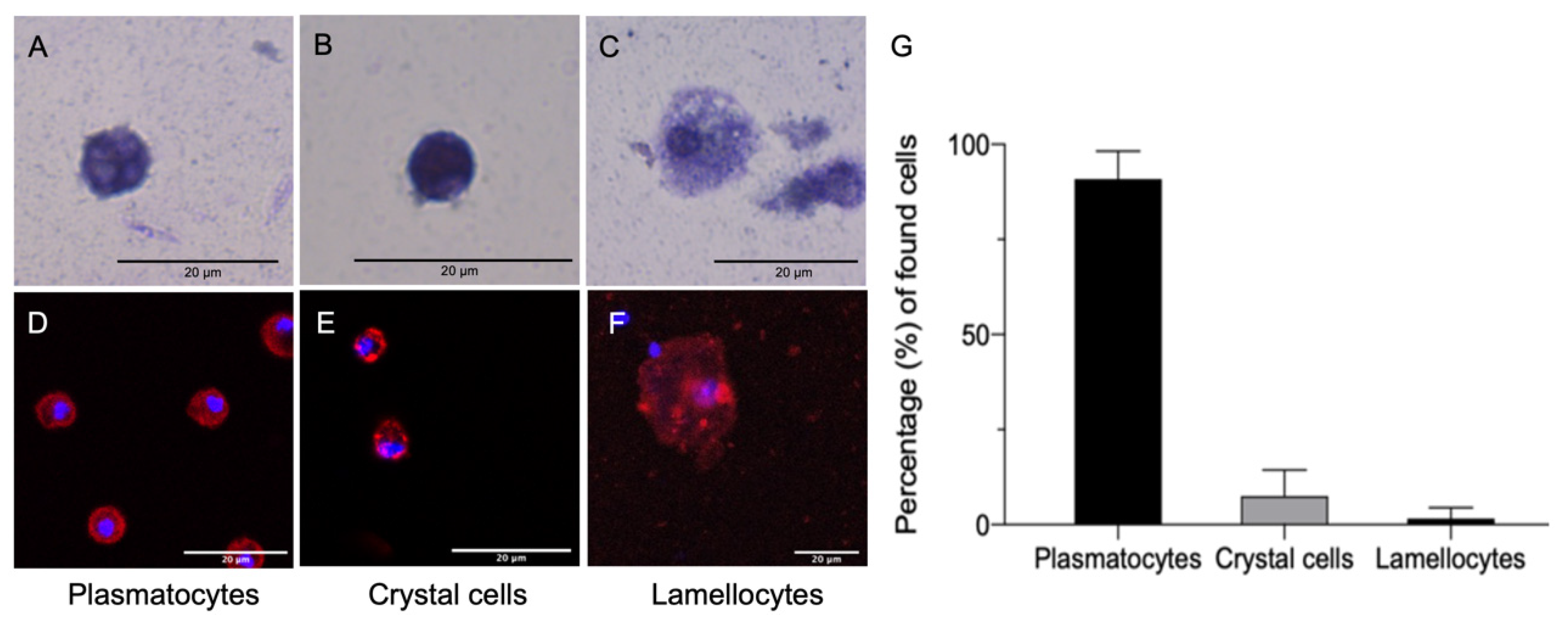
3.1. Characterization of D. suzukii Larval Hemocytes

Hemocytes isolated from D. suzukii L3 larvae ranged in diameter from 10 to 50 µm. The isolated cells were classified by their morphology after toluidine blue staining (Figure 1A–C) followed by staining with hemocyte-specific antibodies (Figure 1D–F). Round granular cells, presenting an average diameter of 9.69 ± 2.96 µm and ranging from 5.14 to 14.70 µm (n = 100) (Figure 1A), that were stained by the NimC1/P1 antibody (Figure 1D), were identified as plasmatocytes. The crystal cells were similar in size (average diameter of 9.66 ± 2.07 µm, ranging from 5.82 µm to 13.41 µm; n = 100) to the plasmatocytes, but were stained darkly with toluidine blue due to the presence of crystals in the cytoplasm (Figure 1B). They were also stained by the C1-specific antibody, which reacts with prophenoloxidase 2 (PPO2) in Drosophila spp. [32] (Figure 1E). The lamellocytes ranged in morphology from oval to elongated forms (average diameter of 28.24 ± 9.66 µm, ranging from 17.89 µm to 47.89 µm; n = 100) with a dark nucleus (Figure 1C), but they could be identified by staining with the L1 Atilla-specific antibody (Figure 1F). The most abundant cells (Figure 1G) were plasmatocytes (89.9%), followed by crystal cells (7.5%) and lamellocytes (2.6%).

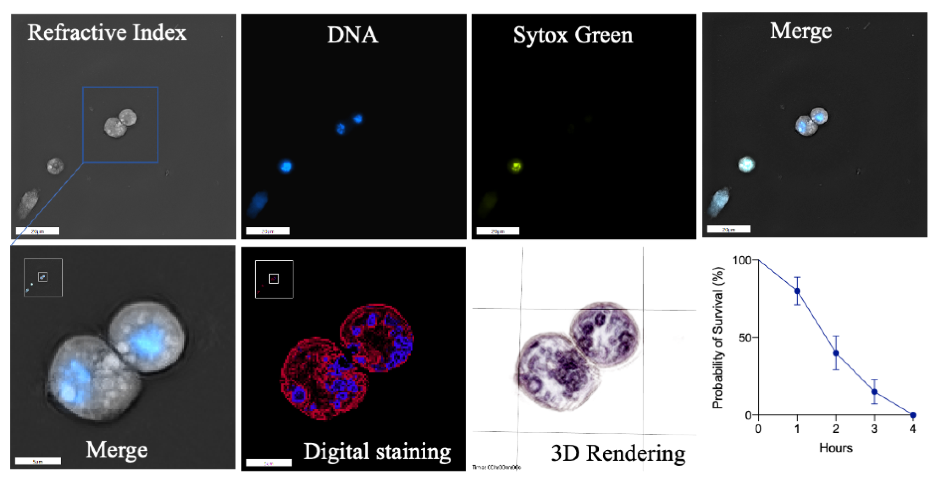
3.2. Viability of Freshly Isolated D. suzukii Larval Plasmatocytes

The viability of plasmatocytes from D. suzukii L3 larvae was assessed by 3D holotomography [38]. Freshly isolated cells were incubated in an imaging medium at room temperature until more than 90% of the cells were dead. At the beginning of the incubation period, plasmatocytes were generally rounded (Figure 2, RI), with a central nucleus stained with Hoechst (Figure 2, DNA). Visible granules surrounding the nucleus registered higher RI values than the rest of the cell contents (Figure 2, zoomed images). After 2 h of isolation, circa 50% of the cells remained viable (Figure 2, absence of SYTOX Green staining) but 90% of the cells were stained with SYTOX Green after 4 h (Figure 2, survival rate), indicating cell death, even though the cells maintained their shapes. Accordingly, all subsequent experiments were limited to 2 h post-isolation to ensure that most of the freshly isolated plasmatocytes were viable.

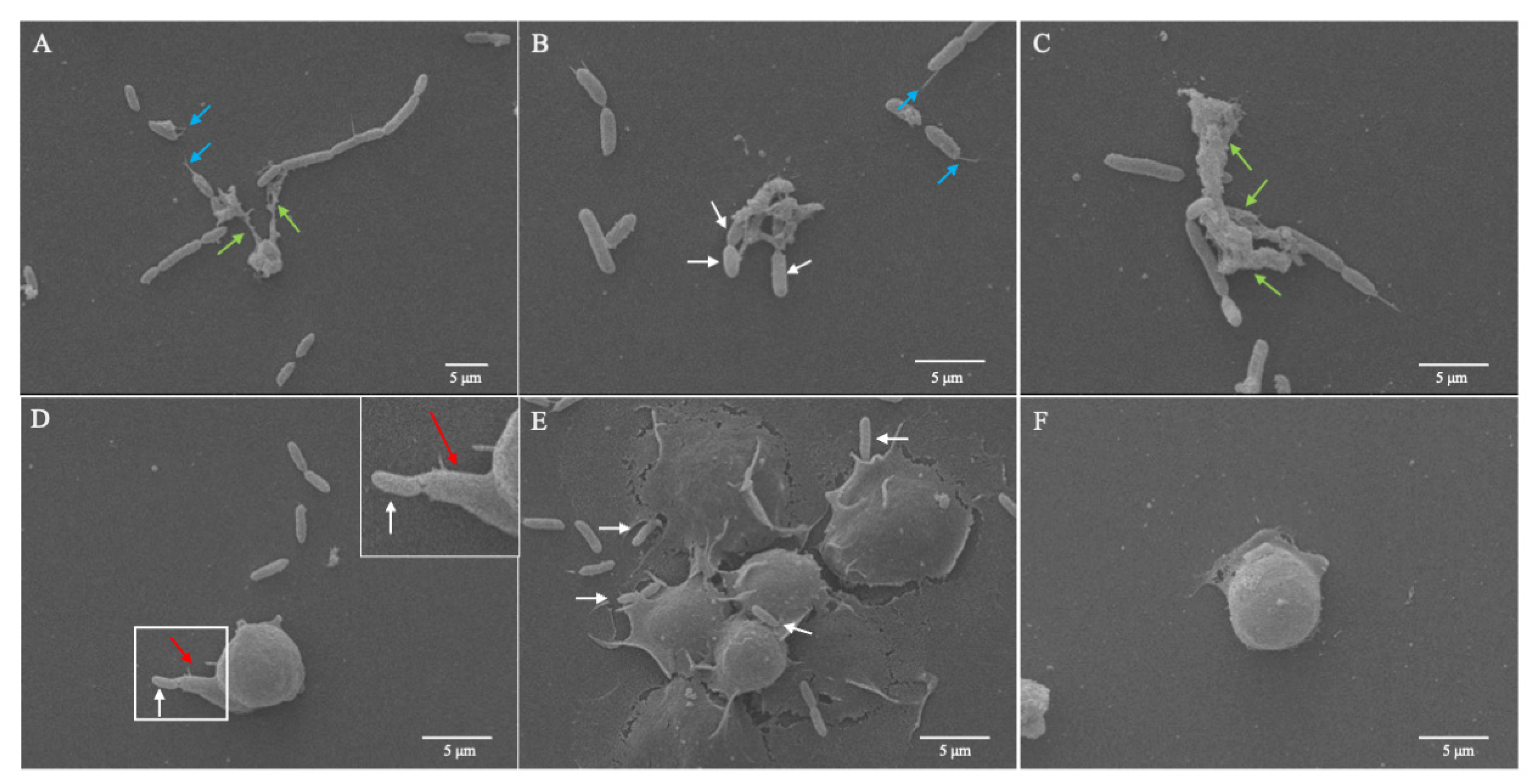
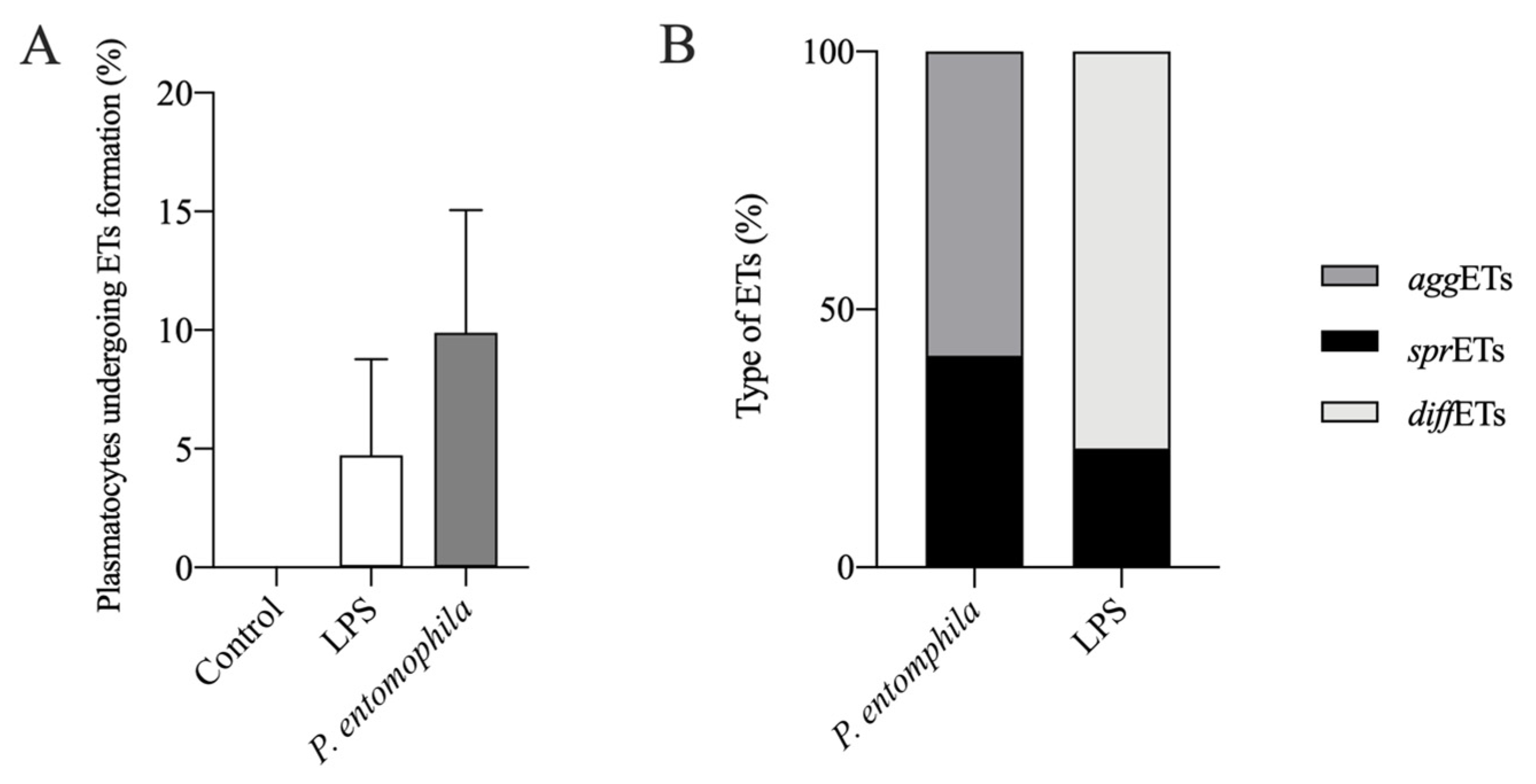
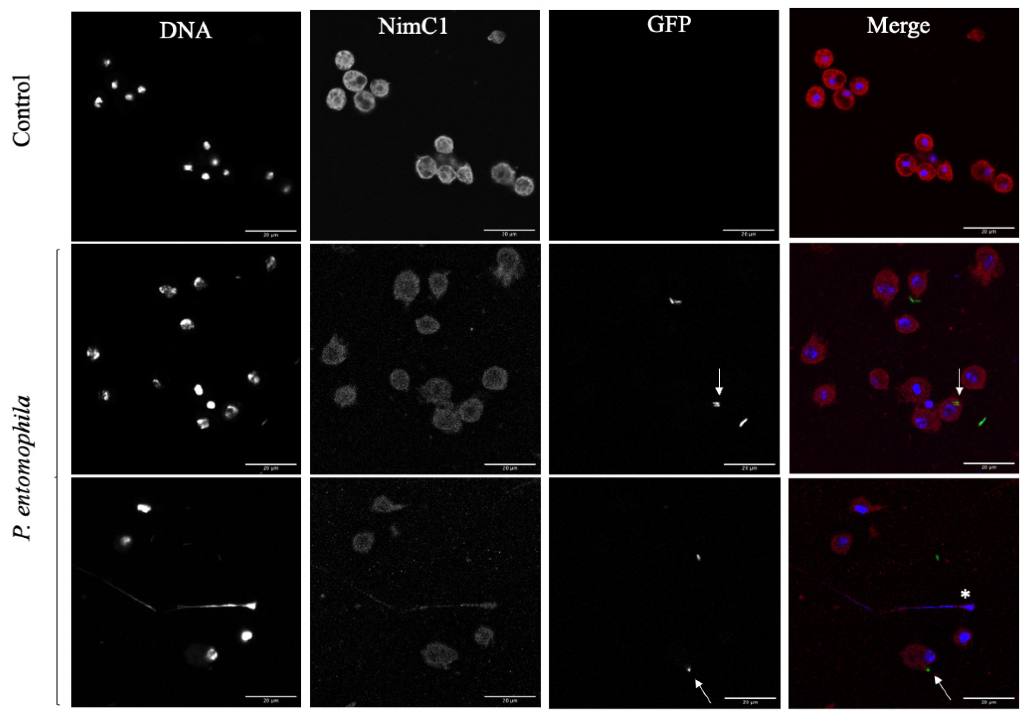
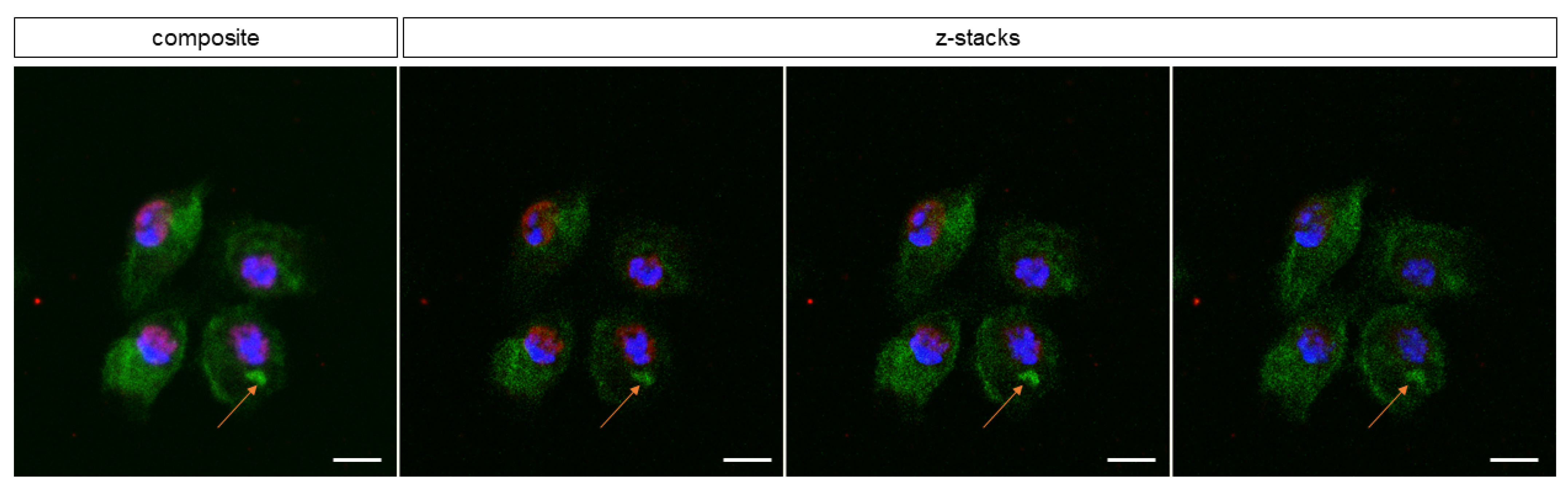
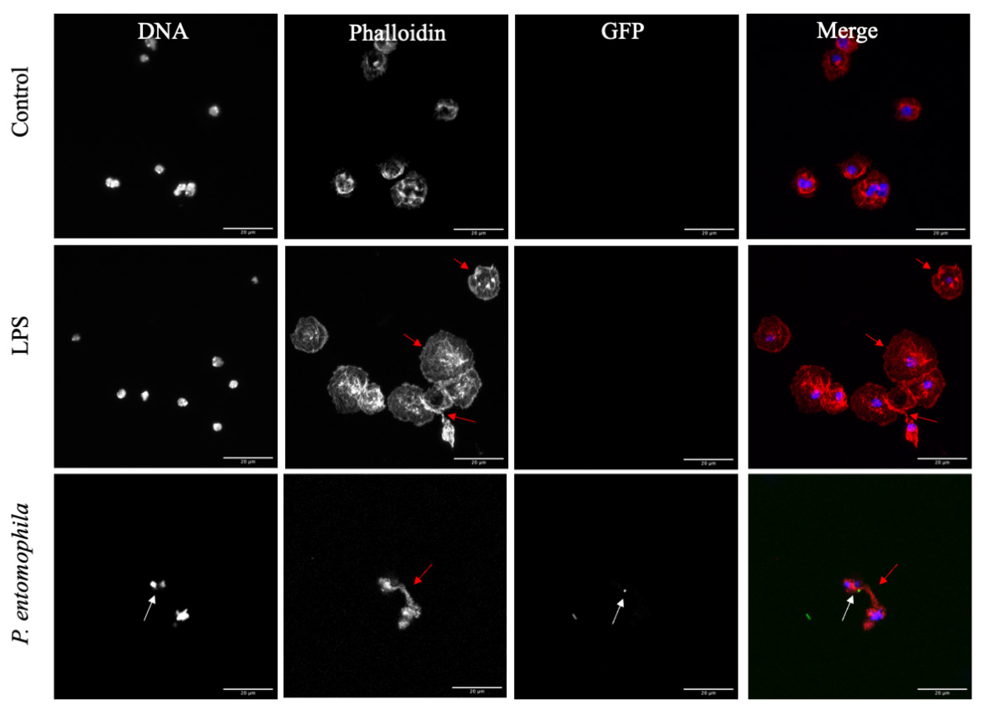
3.3. Response of D. suzukii L3 Plasmatocytes to P. entomophila

The exposure of D. suzukii plasmatocytes to a GFP+ P. entomophila strain triggered the formation of extracellular trap structures with a range of phenotypes (Figure 3), as described for other species elsewhere [42,43,44]. Electron microscopy confirmed that D. suzukii plasmatocytes react against P. entomophila, not only by casting extracellular traps, but also by forming filopodia (Figure 3).
The plasmatocytes were able to cast short spread extracellular traps (sprETs) that captured the cocultured bacteria (Figure 3A, green arrows). Some filigree filaments (Figure 3A,B, blue arrows) were also attached to bacterial cells, but not to hemocytes. These were probably extracellular DNA filaments derived from sprETs that were damaged by the evasion attempts of bacteria or by experimental handling. We also observed the presence of so-called aggregated extracellular traps (aggETs) in response to GFP+ P. entomophila (Figure 3B,C). These formed large meshes of extracellular fibers containing many immune cells releasing individual extracellular traps (Figure 3C, green arrows) and were able to trap several bacteria at once (Figure 3B, white arrows).
To confirm that D. suzukii plasmatocytes cast extracellular traps, we stained the typical components of such structures in immunofluorescence assays. After challenging plasmatocytes with P. entomophila or 500 mg/mL LPS, the formation of extracellular traps was confirmed by the co-localization of extracellular DNA and histones (Figure 4). Interestingly, LPS induced short sprETs (Figure 4, blue arrow) and diffuse extracellular traps (diffETs), the latter characterized by nuclear expansion and thus cell expansion with histone redistribution (Figure 4, red arrows). We observed the same response to three different concentrations of LPS (100, 250 and 500 µg/mL). In contrast, P. entomophila induced sprETs (Figure 4, blue arrows) and aggETs (Figure 4, green arrows), both of which were shown to entrap the GFP+ bacteria (Figure 4, white arrows), confirming the SEM data.
The percentage of plasmatocytes that produced extracellular traps was calculated after coculture with P. entomophila or stimulation with LPS (Figure 5). Approximately 9% of plasmatocytes produced extracellular traps in response to P. entomophila, whereas only 3.6% produced extracellular traps in response to stimulation with LPS, although the difference was not statistically significant (Figure 5A). Additionally, different ETs were displayed when cells were stimulated after each condition (Figure 5B). In response to P. entomophila, 41% of the displayed ETs represented sprETs, whereas 59% displayed aggETs (Figure 5B). In addition, 77% of the LPS-stimulated plasmatocytes displayed diffETs whereas 23% displayed sprETs.
To confirm that the observed immunoreactive behavior was cast by D. suzukii plasmatocytes, plasmatocyte-specific anti-NimC1 antibody staining was used. Positive stained cells were the ones releasing extracellular traps (Figure 6, white asterisk). The D. suzukii plasmatocytes also engulfed P. entomophila by phagocytosis (white arrows). One hour after the challenge, 9.86% of the plasmatocytes were shown to engage in the phagocytosis of P. entomophila (Figure 7, orange arrow).
In addition to phagocytosis and extracellular traps, we also observed the formation of filopodia as a third effector mechanism against pathogenic bacteria (Figure 3D, red arrow), followed by the adhesion of bacteria to these structures (Figure 8). Texas Red-X phalloidin staining (Figure 8, phalloidin) confirmed the actin-dependent formation filopodia in response to the bacteria and LPS, resulting in the presentation of rounded to slightly elongated plasmatocytes.

4. Discussion

In insects and other invertebrates, hemocytes are the first line of defense, eliminating pathogens upon first encounter in vivo and therefore fulfilling a similar role to professional phagocytes (PMNs, monocytes and macrophages) in mammals. We characterized the hemocyte population of the invasive pest insect D. suzukii to add to the understanding of diverse effector mechanisms as part of the early innate immune response, which has not been reported for other drosophilid species thus far. We observed phagocytosis and the formation of filipodia by D. suzukii plasmatocytes exposed to P. entomophila or LPS, but also the formation of extracellular traps (ETosis), highlighting the importance of this widespread cellular immune defense mechanism in eukaryotes [23,45]. ETosis has been reported in other insects [33,46], and also in other invertebrates, such as oysters [47,48], mussels [46] and slugs [29], and the mechanism appears highly conserved. The traps are composed of extracellular DNA decorated with nuclear histones (chromatin), combined with lactoferrin, pentraxin, myeloperoxidase, elastase, gelatinase and cathelicidin, among other antimicrobial molecules [22,23,24,26,40]. Interestingly, we observed the presence of multiple histones (H1, H2A/H2B, H3 and H4), whereas one recent study in the cockroach Periplaneta americana only identified H1/DNA complexes in extracellular traps [42], with only one study in slugs supporting our findings [29]. However, our results align with the mechanism of ETosis in mammals, where histones are one of the main components of extracellular traps both in vitro and in vivo [46]. The formation of extracellular traps by drosophilid hemocytes has not been reported before, and the absence of nucleic acid clots in the hemolymph of D. melanogaster was reported following a challenge with Escherichia coli JM109 [49]. The nature of the pathogenic stimulus may determine the mode of cellular defense. Here, we used P. entomophila, a bacterial pathogen that has already been shown to cause systemic infections that induce a range of immune responses in drosophilids [50,51]. The bacteria can be highly pathogenic when flies receive inoculum sizes sufficient to disrupt the gut epithelium and enter the hemolymph, which brings them into contact with hemocytes [50,51].
We characterized the morphology of D. suzukii hemocytes in detail and observed similar characteristics to the closely related fly D. melanogaster [13]. We were able to distinguish between lamellocytes, crystal cells and plasmatocytes using both morphological criteria and specific immunostaining, which allowed us to demonstrate the unique defense mechanisms of the plasmatocyte population. The revelation that such cells can form filopodia and extracellular traps in response to P. entomophila supports findings in other insects [42]. The initial detection of pathogenic bacteria is facilitated by the recognition of pathogen-associated molecular patterns (PAMPs) such as LPS. Our results suggest that PAMPs alone, which bind pathogen recognition receptors (PRRs) on the surface of plasmatocytes, are sufficient for inducing the formation of filopodia and the release of extracellular traps. Similar (dose-dependent) effects have been described in the cockroach P. americana [42] when hemocytes are stimulated with delipidated LPS. Naturally, hemocyte PRRs sense and respond differently to LPS or infections with live bacteria [52], highlighting the importance of adjusting the bacterial titer in order to study humoral and cellular immune responses. However, future studies should address the effect of delipidated LPS and/or other bacterial strains on the D. suzukii cellular response. The potential role of hemocyte “trained immunity” after a primary infection with P. entomophila is also an interesting topic because this form of innate immune memory has been well described in mammals and, despite the long-believed lack of “immune priming” capacities in invertebrates, more recent studies indicate its presence also in insects [53,54,55,56].
Interestingly, D. suzukii plasmatocytes-derived ETosis revealed up to three different types of extracellular traps, namely sprETs, aggETs and diffETs, as previously described for other hosts and cells [29,43]. Exposure to bacteria or LPS induced the release of sprETs, whereas diffETs were only observed following a challenge with LPS. In addition, aggETs were cast after P. entompohila stimulus. Neutrophils are known to discriminate between LPS and bacterial infection, releasing NETs that differ in structure and activity [57]. In mammals, it is likely that sprNETs and diffNETs are preliminary structures, whereas aggNETs are later and more mature forms [43]. Further experiments with longer exposure times would be necessary in determining whether a similar temporal profile exists in insects with the presence of even larger or a higher number of aggETs. It would also be interesting to investigate whether insect aggETs inhibit inflammation by degrading cytokines and chemokines as they do in mammals [44].
D. suzukii robust cellular immune responses might have facilitated its rapid worldwide spread by allowing it to overcome pathogen infections in newly colonized environments [55,56,57]. Such immunological diversity can help invasive species deal with unfamiliar pathogens [10,11,12,13,14,15,16,17,18,19,20,21,22,23,24,25,26,27,28,29,30,31,32,33,34,35,36,37,38,39,40,41,42,43,44,45,46,47,49,50,51,52,53,54,55,56,57,58], as recently shown for the invasive harlequin ladybird Harmonia axyridis [48,55,56,57,58,59,60,61]. We therefore propose that extracellular traps are a key component of the D. suzukii cellular innate immune response against pathogenic bacteria. The similarity between vertebrate and invertebrate cellular immunity highlights the evolutionary conservation of this ancient mechanism.

Author Contributions

Conceptualization, C.H., L.M.R.S., A.V., K.-Z.L., T.C. and S.T.; validation, C.H., L.M.R.S., A.V., K.-Z.L., T.C. and S.T.; investigation, C.H., L.M.R.S., D.P.-B., U.G., T.C. and S.T.; writing—original draft preparation, L.M.R.S., D.P.-B., T.C. and S.T.; writing—review and editing, C.H., L.M.R.S., A.V., K.-Z.L., A.T., T.C. and S.T.; funding acquisition A.V. All authors have read and agreed to the published version of the manuscript.

Funding

This work was funded by the excellence initiative of the Hessian Ministry of Science, Higher Education and the Arts (HMWK) supporting the LOEWE Centre for Insect Biotechnology and Bioresources.

Institutional Review Board Statement

Not applicable.

Informed Consent Statement

Not applicable.

Data Availability Statement

Not applicable.

Acknowledgments

We thank Anika Seipp (Institute of Anatomy and Cell Biology, JLU Giessen, Germany) for technical support with electron microscopy, and Henrike Schmidtberg for technical support with hemolymph extraction and processing. We also thank István Andó (Biological Research Centre, Szeged, Hungary) and Martin Speckmann (Justus Liebig University, Giessen, Germany) for the hemocyte-specific antibodies, and Richard M. Twyman for professional editing of the manuscript.

Conflicts of Interest

The authors declare no conflict of interest.

References

  1. Calabria, G.; Máca, J.; Serra, L.; Pascual, M. First Records of the Potential Pest Species Drosophila suzukii (Diptera: Drosophilidae) in Europe. J. Appl. Entomol. 2012, 136, 139–147. [Google Scholar] [CrossRef]
  2. Walsh, D.B.; Bolda, M.P.; Goodhue, R.E.; Dreves, A.J.; Lee, J.; Bruck, D.J.; Walton, V.M.; O’Neal, S.D.; Zalom, F.G. Drosophila suzukii (Diptera: Drosophilidae): Invasive Pest of Ripening Soft Fruit Expanding Its Geographic Range and Damage Potential. J. Integr. Pest Manag. 2011, 2, G1–G7. [Google Scholar] [CrossRef]
  3. Andreazza, F.; Bernardi, D.; Dos Santos, R.S.S.; Garcia, F.R.M.; Oliveira, E.E.; Botton, M.; Nava, D.E. Drosophila suzukii in Southern Neotropical Region: Current Status and Future Perspectives. Neotrop. Entomol. 2017, 46, 591–605. [Google Scholar] [CrossRef] [PubMed]
  4. Ørsted, I.V.; Ørsted, M. Species Distribution Models of the Spotted Wing Drosophila (Drosophila suzukii, Diptera: Drosophilidae) in Its Native and Invasive Range Reveal an Ecological Niche Shift. J. Appl. Ecol. 2019, 56, 423–435. [Google Scholar] [CrossRef]
  5. Atallah, J.; Teixeira, L.; Salazar, R.; Zaragoza, G.; Kopp, A. The Making of a Pest: The Evolution of a Fruit-Penetrating Ovipositor in Drosophila suzukii and Related Species. Proc. R. Soc. B Biol. Sci. 2014, 281, 20132840. [Google Scholar] [CrossRef] [PubMed] [Green Version]
  6. Tochen, S.; Dalton, D.T.; Wiman, N.; Hamm, C.; Shearer, P.W.; Walton, V.M. Temperature-Related Development and Population Parameters for Drosophila suzukii (Diptera: Drosophilidae) on Cherry and Blueberry. Environ. Entomol. 2014, 43, 501–510. [Google Scholar] [CrossRef] [PubMed] [Green Version]
  7. Wiman, N.G.; Walton, V.M.; Dalton, D.T.; Anfora, G.; Burrack, H.J.; Chiu, J.C.; Daane, K.M.; Grassi, A.; Miller, B.; Tochen, S.; et al. Integrating Temperature-Dependent Life Table Data into a Matrix Projection Model for Drosophila suzukii Population Estimation. PLoS ONE 2014, 9, e106909. [Google Scholar] [CrossRef]
  8. Kacsoh, B.Z.; Schlenke, T.A. High Hemocyte Load Is Associated with Increased Resistance against Parasitoids in Drosophila suzukii, a Relative of D. melanogaster. PLoS ONE 2012, 7, e34721. [Google Scholar] [CrossRef]
  9. Poyet, M.; Havard, S.; Prevost, G.; Chabrerie, O.; Doury, G.; Gibert, P.; Eslin, P. Resistance of Drosophila suzukii to the Larval Parasitoids Leptopilina heterotoma and Asobara japonica Is Related to Haemocyte Load. Physiol. Entomol. 2013, 38, 45–53. [Google Scholar] [CrossRef]
  10. Pech, L.L.; Strand, M.R. Granular Cells Are Required for Encapsulation of Foreign Targets by Insect Haemocytes. J. Cell Sci. 1996, 109, 2053–2060. [Google Scholar] [CrossRef]
  11. Schmidt, O.; Theopold, U.; Strand, M. Innate Immunity and Its Evasion and Suppression by Hymenopteran Endoparasitoids. BioEssays 2001, 23, 344–351. [Google Scholar] [CrossRef]
  12. Traver, D.; Zon, L.I. Walking the Walk: Migration and Other Common Themes in Blood and Vascular Development. Cell 2002, 108, 731–734. [Google Scholar] [CrossRef] [Green Version]
  13. Banerjee, U.; Girard, J.R.; Goins, L.M.; Spratford, C.M. Drosophila as a Genetic Model for Hematopoiesis. Genetics 2019, 211, 367–417. [Google Scholar] [CrossRef] [Green Version]
  14. Meister, M.; Lagueux, M. Drosophila Blood Cells. Cell. Microbiol. 2003, 5, 573–580. [Google Scholar] [CrossRef]
  15. Lebestky, T.; Chang, T.; Hartenstein, V.; Banerjee, U. Specification of Drosophila Hematopoietic Lineage by Conserved Transcription Factors. Science 2000, 288, 146–149. [Google Scholar] [CrossRef] [PubMed]
  16. Lavine, M.D.; Strand, M.R. Insect Hemocytes and Their Role in Immunity. Insect Biochem. Mol. Biol. 2002, 32, 1295–1309. [Google Scholar] [CrossRef]
  17. Tepass, U.; Fessler, L.I.; Aziz, A.; Hartenstein, V. Embryonic Origin of Hemocytes and Their Relationship to Cell Death in Drosophila. Development 1994, 120, 1829–1837. [Google Scholar] [CrossRef] [PubMed]
  18. Franc, N.C.; Heitzler, P.; Ezekowitz, R.A.B.; White, K. Requirement for Croquemort in Phagocytosis of Apoptotic Cells in Drosophila. Science 1999, 284, 1991–1994. [Google Scholar] [CrossRef] [PubMed]
  19. Müller, U.; Vogel, P.; Alber, G.; Schaub, G.A. The Innate Immune System of Mammals and Insects. Trends Innate Immun. 2008, 15, 21–44. [Google Scholar] [CrossRef]
  20. Franc, N.C.; Dimarcq, J.L.; Lagueux, M.; Hoffman, J.A.; Ezekowitz, R.A.B. Croquemort, A Novel Drosophila Hemocyte/Macrophage Receptor That Recognizes Apoptotic Cells. Immunity 1996, 4, 431–443. [Google Scholar] [CrossRef] [Green Version]
  21. Villagra-Blanco, R.; Silva, L.M.R.; Conejeros, I.; Taubert, A.; Hermosilla, C. Pinniped- and Cetacean-Derived ETosis Contributes to Combating Emerging Apicomplexan Parasites (Toxoplasma gondii, Neospora caninum) Circulating in Marine Environments. Biology 2019, 8, 12. [Google Scholar] [CrossRef] [PubMed] [Green Version]
  22. Neumann, A.; Brogden, G.; von Köckritz-Blickwede, M. Extracellular Traps: An Ancient Weapon of Multiple Kingdoms. Biology 2020, 9, 34. [Google Scholar] [CrossRef] [PubMed] [Green Version]
  23. Nathan, C. Neutrophils and Immunity: Challenges and Opportunities. Nat. Rev. Immunol. 2006, 6, 173–182. [Google Scholar] [CrossRef] [PubMed]
  24. Urban, C.F.; Ermert, D.; Schmid, M.; Abu-Abed, U.; Goosmann, C.; Nacken, W.; Brinkmann, V.; Jungblut, P.R.; Zychlinsky, A. Neutrophil Extracellular Traps Contain Calprotectin, a Cytosolic Protein Complex Involved in Host Defense against Candida albicans. PLoS Pathog. 2009, 5, e1000639. [Google Scholar] [CrossRef] [Green Version]
  25. Parker, H.; Albrett, A.M.; Kettle, A.J.; Winterbourn, C.C. Myeloperoxidase Associated with Neutrophil Extracellular Traps Is Active and Mediates Bacterial Killing in the Presence of Hydrogen Peroxide. J. Leukoc. Biol. 2012, 91, 369–376. [Google Scholar] [CrossRef]
  26. Webster, S.J.; Daigneault, M.; Bewley, M.A.; Preston, J.A.; Marriott, H.M.; Walmsley, S.R.; Read, R.C.; Whyte, M.K.B.; Dockrell, D.H. Distinct Cell Death Programs in Monocytes Regulate Innate Responses Following Challenge with Common Causes of Invasive Bacterial Disease. J. Immunol. 2010, 185, 2968–2979. [Google Scholar] [CrossRef] [Green Version]
  27. Bartneck, M.; Keul, H.A.; Zwadlo-Klarwasser, G.; Groll, J. Phagocytosis Independent Extracellular Nanoparticle Clearance by Human Immune Cells. Nano Lett. 2010, 10, 59–63. [Google Scholar] [CrossRef]
  28. Palić, D.; Ostojić, J.; Andreasen, C.B.; Roth, J.A. Fish Cast NETs: Neutrophil Extracellular Traps Are Released from Fish Neutrophils. Dev. Comp. Immunol. 2007, 31, 805–816. [Google Scholar] [CrossRef]
  29. Lange, M.K.; Penagos-Tabares, F.; Muñoz-Caro, T.; Gärtner, U.; Mejer, H.; Schaper, R.; Hermosilla, C.; Taubert, A. Gastropod-Derived Haemocyte Extracellular Traps Entrap Metastrongyloid Larval Stages of Angiostrongylus vasorum, Aelurostrongylus abstrusus and Troglostrongylus brevior. Parasites Vectors 2017, 10, 50. [Google Scholar] [CrossRef] [Green Version]
  30. Ng, T.H.; Chang, S.-H.; Wu, M.-H.; Wang, H.-C. Shrimp Hemocytes Release Extracellular Traps That Kill Bacteria. Dev. Comp. Immunol. 2013, 41, 644–651. [Google Scholar] [CrossRef]
  31. Silva, L.M.R.; Muñoz-Caro, T.; Burgos, R.A.; Hidalgo, M.A.; Taubert, A.; Hermosilla, C. Far beyond Phagocytosis: Phagocyte-Derived Extracellular Traps Act Efficiently against Protozoan Parasites In Vitro and In Vivo. Mediat. Inflamm. 2016, 2016, 5898074. [Google Scholar] [CrossRef] [Green Version]
  32. Altincicek, B.; Stötzel, S.; Wygrecka, M.; Preissner, K.T.; Vilcinskas, A. Host-Derived Extracellular Nucleic Acids Enhance Innate Immune Responses, Induce Coagulation, and Prolong Survival upon Infection in Insects. J. Immunol. 2008, 181, 2705–2712. [Google Scholar] [CrossRef] [PubMed] [Green Version]
  33. Vodovar, N.; Vinals, M.; Liehl, P.; Basset, A.; Degrouard, J.; Spellman, P.; Boccard, F.; Lemaitre, B. Drosophila Host Defense after Oral Infection by an Entomopathogenic Pseudomonas Species. Proc. Natl. Acad. Sci. USA 2005, 102, 11414–11419. [Google Scholar] [CrossRef] [PubMed] [Green Version]
  34. Merkling, S.H.; van Rij, R.P. Analysis of Resistance and Tolerance to Virus Infection in Drosophila. Nat. Protoc. 2015, 10, 1084–1097. [Google Scholar] [CrossRef] [PubMed]
  35. Medd, N.C.; Fellous, S.; Waldron, F.M.; Xuéreb, A.; Nakai, M.; Cross, J.V.; Obbard, D.J. The Virome of Drosophila suzukii, an Invasive Pest of Soft Fruit. Virus Evol. 2018, 4, vey009. [Google Scholar] [CrossRef]
  36. Tracy, C.; Krämer, H. Isolation and Infection of Drosophila Primary Hemocytes. Bio-Protoc. 2017, 7, e2300. [Google Scholar] [CrossRef] [Green Version]
  37. Sandoz, P.A.; Tremblay, C.; Equis, S.; Pop, S.; Pollaro, L.; Cotte, Y.; van der Goot, F.G.; Frechin, M. Label Free 3D Analysis of Organelles in Living Cells by Refractive Index Shows Pre-Mitotic Organelle Spinning in Mammalian Stem Cells. bioRxiv 2018, 407239. [Google Scholar] [CrossRef] [Green Version]
  38. Velásquez, Z.D.; López-Osorio, S.; Mazurek, S.; Hermosilla, C.; Taubert, A. Eimeria Bovis Macromeront Formation Induces Glycolytic Responses and Mitochondrial Changes in Primary Host Endothelial Cells. Front. Cell. Infect. Microbiol. 2021, 11, 635. [Google Scholar] [CrossRef]
  39. Larrazabal, C.; Hermosilla, C.; Taubert, A.; Conejeros, I. 3D Holotomographic Monitoring of Ca++ Dynamics during Ionophore-Induced Neospora caninum Tachyzoite Egress from Primary Bovine Host Endothelial Cells. Parasitol. Res. 2021. [Google Scholar] [CrossRef]
  40. Kurucz, E.; Váczi, B.; Márkus, R.; Laurinyecz, B.; Vilmos, P.; Zsámboki, J.; Csorba, K.; Gateff, E.; Hultmark, D.; Andó, I. Definition of Drosophila Hemocyte Subsets by Cell-Type Specific Antigens. Acta Biol. Hung. 2007, 58, 95–111. [Google Scholar] [CrossRef] [Green Version]
  41. Mulet, M.; Gomila, M.; Lemaitre, B.; Lalucat, J.; García-Valdés, E. Taxonomic Characterisation of Pseudomonas Strain L48 and Formal Proposal of Pseudomonas entomophila Sp. Nov. Syst. Appl. Microbiol. 2012, 35, 145–149. [Google Scholar] [CrossRef] [PubMed] [Green Version]
  42. Nascimento, M.T.C.; Silva, K.P.; Garcia, M.C.F.; Medeiros, M.N.; Machado, E.A.; Nascimento, S.B.; Saraiva, E.M. DNA Extracellular Traps Are Part of the Immune Repertoire of Periplaneta americana. Dev. Comp. Immunol. 2018, 84, 62–70. [Google Scholar] [CrossRef] [PubMed]
  43. Muñoz-Caro, T.; Rubio, R.M.C.; Silva, L.M.R.; Magdowski, G.; Gärtner, U.; McNeilly, T.N.; Taubert, A.; Hermosilla, C. Leucocyte-Derived Extracellular Trap Formation Significantly Contributes to Haemonchus contortus Larval Entrapment. Parasites Vectors 2015, 8, 607. [Google Scholar] [CrossRef] [PubMed] [Green Version]
  44. Schauer, C.; Janko, C.; Munoz, L.E.; Zhao, Y.; Kienhöfer, D.; Frey, B.; Lell, M.; Manger, B.; Rech, J.; Naschberger, E.; et al. Aggregated Neutrophil Extracellular Traps Limit Inflammation by Degrading Cytokines and Chemokines. Nat. Med. 2014, 20, 511–517. [Google Scholar] [CrossRef]
  45. Hakkim, A.; Fuchs, T.A.; Martinez, N.E.; Hess, S.; Prinz, H.; Zychlinsky, A.; Waldmann, H. Activation of the Raf-MEK-ERK Pathway Is Required for Neutrophil Extracellular Trap Formation. Nat. Chem. Biol. 2011, 7, 75–77. [Google Scholar] [CrossRef]
  46. Brinkmann, V.; Reichard, U.; Goosmann, C.; Fauler, B.; Uhlemann, Y.; Weiss, D.S.; Weinrauch, Y.; Zychlinsky, A. Neutrophil Extracellular Traps Kill Bacteria. Science 2004, 303, 1532–1535. [Google Scholar] [CrossRef]
  47. Zhou, E.; Silva, L.; Conejeros, I.; Velásquez, Z.; Hirz, M.; Gärtner, U.; Jacquiet, P.; Taubert, A.; Hermosilla, C. Besnoitia besnoiti bradyzoite stages induce suicidal- and rapid vital-NETosis. Parasitology 2020, 147, 401–409. [Google Scholar] [CrossRef]
  48. Beckert, A.; Wiesner, J.; Baumann, A.; Pöppel, A.-K.; Vogel, H.; Vilcinskas, A. Two C-Type Lysozymes Boost the Innate Immune System of the Invasive Ladybird Harmonia axyridis. Dev. Comp. Immunol. 2015, 49, 303–312. [Google Scholar] [CrossRef]
  49. Bidla, G.; Lindgren, M.; Theopold, U.; Dushay, M.S. Hemolymph Coagulation and Phenoloxidase in Drosophila Larvae. Dev. Comp. Immunol. 2005, 29, 669–679. [Google Scholar] [CrossRef]
  50. Hiebert, N.; Carrau, T.; Bartling, M.; Vilcinskas, A.; Lee, K.-Z. Identification of Entomopathogenic Bacteria Associated with the Invasive Pest Drosophila suzukii in Infested Areas of Germany. J. Invertebr. Pathol. 2020, 173, 107389. [Google Scholar] [CrossRef]
  51. Dieppois, G.; Opota, O.; Lalucat, J.; Lemaitre, B. Pseudomonas entomophila: A Versatile Bacterium with Entomopathogenic Properties. In Pseudomonas: New Aspects of Pseudomonas Biology; Ramos, J.-L., Goldberg, J.B., Filloux, A., Eds.; Springer: Dordrecht, The Netherlands, 2015; Volume 7, pp. 25–49. ISBN 978-94-017-9555-5. [Google Scholar]
  52. Johansson, K.C.; Metzendorf, C.; Söderhäll, K. Microarray Analysis of Immune Challenged Drosophila Hemocytes. Exp. Cell Res. 2005, 305, 145–155. [Google Scholar] [CrossRef]
  53. Frangou, E.; Vassilopoulos, D.; Boletis, J.; Boumpas, D.T. An Emerging Role of Neutrophils and NETosis in Chronic Inflammation and Fibrosis in Systemic Lupus Erythematosus (SLE) and ANCA-Associated Vasculitides (AAV): Implications for the Pathogenesis and Treatment. Autoimmun. Rev. 2019, 18, 751–760. [Google Scholar] [CrossRef] [PubMed]
  54. Mondotte, J.A.; Gausson, V.; Frangeul, L.; Blanc, H.; Lambrechts, L.; Saleh, M.-C. Immune Priming and Clearance of Orally Acquired RNA Viruses in Drosophila. Nat. Microbiol. 2018, 3, 1394–1403. [Google Scholar] [CrossRef] [PubMed]
  55. Mondotte, J.A.; Gausson, V.; Frangeul, L.; Suzuki, Y.; Vazeille, M.; Mongelli, V.; Blanc, H.; Failloux, A.-B.; Saleh, M.-C. Evidence For Long-Lasting Transgenerational Antiviral Immunity in Insects. Cell Rep. 2020, 33, 108506. [Google Scholar] [CrossRef] [PubMed]
  56. Cooper, D.; Eleftherianos, I. Memory and Specificity in the Insect Immune System: Current Perspectives and Future Challenges. Front. Immunol. 2017, 8, 539. [Google Scholar] [CrossRef]
  57. Pieterse, E.; Rother, N.; Yanginlar, C.; Hilbrands, L.B.; van der Vlag, J. Neutrophils Discriminate between Lipopolysaccharides of Different Bacterial Sources and Selectively Release Neutrophil Extracellular Traps. Front. Immunol. 2016, 7, 484. [Google Scholar] [CrossRef] [Green Version]
  58. Gegner, T.; Schmidtberg, H.; Vogel, H.; Vilcinskas, A. Population-Specific Expression of Antimicrobial Peptides Conferring Pathogen Resistance in the Invasive Ladybird Harmonia axyridis. Sci. Rep. 2018, 8, 3600. [Google Scholar] [CrossRef]
  59. Vilcinskas, A.; Stoecker, K.; Schmidtberg, H.; Röhrich, C.R.; Vogel, H. Invasive Harlequin Ladybird Carries Biological Weapons against Native Competitors. Science 2013, 340, 862–863. [Google Scholar] [CrossRef]
  60. Röhrich, C.R.; Ngwa, C.J.; Wiesner, J.; Schmidtberg, H.; Degenkolb, T.; Kollewe, C.; Fischer, R.; Pradel, G.; Vilcinskas, A. Harmonine, a Defence Compound from the Harlequin Ladybird, Inhibits Mycobacterial Growth and Demonstrates Multi-Stage Antimalarial Activity. Biol. Lett. 2012, 8, 308–311. [Google Scholar] [CrossRef]
  61. Lee, K.A.; Klasing, K.C. A Role for Immunology in Invasion Biology. Trends Ecol. Evol. 2004, 19, 523–529. [Google Scholar] [CrossRef]
Figure 1. Characterization of hemocytes from Drosophila suzukii L3 larvae. Cells isolated from the hemolymph of D. suzukii L3 larvae were fixed with 4% paraformaldehyde for 5 min at room temperature and stained with 1% toluidine blue (AC) followed by immunofluorescence staining (DF). Plasmatocytes (A), identified by staining with the anti-NimC1 antibody (D), were rounded cells with central nuclei. Crystal cells (B), identified by staining with antibody HC12F6 (E), were rounded cells containing crystals that were stained densely with toluidine blue. Lamellocytes (C), identified by staining with the L1 anti-Atilla antibody (F), were oval or elongated cells. Nuclei were counterstained with DAPI (DF, blue). Cell population analysis (G) revealed plasmatocytes to be the most abundant cells (89.9%), followed by crystal cells (7.5%) and lamellocytes (2.6%). Scale bar = 20 μm.
Cells 10 03320 g001
Figure 2. Plasmatocyte viability assay and characterization by 3D holotomography. Plasmatocytes were mostly rounded cells (RI) with a central nucleus (DNA) and perinuclear granules (zoomed images on bottom row). SYTOX Green staining, which is specific for dead cells, showed that circa half of the cells remained viable for at least 2 h but that 90% were dead after 4 h. Scale bar = 5 μm.
Cells 10 03320 g002
Figure 3. SEM images of extracellular traps formed by Drosophila suzukii plasmatocytes in response to Pseudomonas entomophila. The isolated plasmatocytes were incubated with P. entomophila (AE) or PBS (control, F) at room temperature for 1 h. The plasmatocytes cast short spread extracellular traps (sprETs) to entrap P. entomophila (A, green arrows). Filigree filaments (A,B, blue arrows) from disrupted extracellular DNA filaments also attached to P. entomophila. So-called aggregated extracellular traps (aggETs) were observed when cells were challenged with P. entomophila (B,C), resulting in massive clusters of mesh fibers containing many immune cells (C, green arrows) and bacteria (B, white arrows). The formation of filopodia (D) was observed after further incubation, and P. entomophila was found stuck to these structures (D,E, white arrows). Scale bar = 5 μm.
Cells 10 03320 g003
Figure 4. Characteristics of extracellular traps formed by Drosophila suzukii plasmatocytes in response to Pseudomonas entomophila and lipopolysaccharides (LPS). Isolated plasmatocytes were incubated with GFP+ P. entomophila, LPS or PBS as a control for 1 h before fixing with 4% PFA for 5 min at room temperature and staining with Hoechst (blue). Histones (H1, H2A, H2B, H3 and H4) were then detected with the monoclonal antibody MAB3422 followed by staining with the goat anti-mouse IgG Alexa Fluor 555 (red) and nuclear counterstaining with DAPI (blue). One hour after the challenge, two different phenotypes were observed. LPS induced spread extracellular traps (sprETs, blue arrows), as well as diffuse extracellular traps (diffETs, red arrows), whereas P. entomophila (white arrows) induced sprETs (blue arrows) and aggregated extracellular traps (aggETs, green arrows). Scale bar = 20 μm.
Cells 10 03320 g004
Figure 5. Quantification of extracellular traps induced by P. entomophila or LPS. (A) We compared the proportion of plasmatocytes that cast extracellular traps in response to GFP+ P. entomophila and LPS. P. entomophila induced higher ET formation than LPS when compared to control (no ET formation). (B) The three different ETs phenotypes were quantified and LPS induced sprETs, as well as diffETs (in greater percentage), whereas P. entomophila induced sprETs and aggETs, the latter in higher proportion.
Cells 10 03320 g005
Figure 6. Drosophila suzukii plasmatocytes cast extracellular traps and engulf Pseudomonas entomophila by phagocytosis. Plasmatocytes were challenged with GFP+ P. entomophila and stained with the plasmatocyte-specific antibody anti-NimC1 to confirm their identify. We observed the formation of extracellular traps (white asterisk, lower row) but also the phagocytosis of P. entomophila (white arrows, middle and lower rows), indicating that plasmatocytes respond to the challenge by deploying multiple defense mechanisms. Scale bar = 20 μm.
Cells 10 03320 g006
Figure 7. Drosophila suzukii plasmatocytes phagocyte Pseudomonas entomophila. After plasmatocytes were challenged with GFP+ P. entomophila, bacteria were observed intracellularly near to the nucleus of plasmatocytes (orange arrows), showing that plasmatocytes use multiple defense mechanisms, including phagocytosis. Nucleus was stained with DAPI (blue), histones in red and bacteria in green. Auto-fluorescence of the cell is observed. Scale bar = 10 μm.
Cells 10 03320 g007
Figure 8. Formation of filopodia by D. suzukii plasmatocytes. Isolated plasmatocytes were incubated with GFP+ P. entomophila, 500 µg/mL LPS or PBS as a control for 1 h before fixing in 4% paraformaldehyde for 5 min at room temperature and staining for DNA with Hoechst (blue) and for actin with Texas Red-X phalloidin (Phalloidin, red). Filopodia (red arrows) formed in response to both LPS and P. entomophila (white arrows), resulting in the presentation of rounded to slightly elongated cells (arrow). Scale bar = 20 μm.
Cells 10 03320 g008
Publisher’s Note: MDPI stays neutral with regard to jurisdictional claims in published maps and institutional affiliations.

