Review

Redox Regulation of Soluble Epoxide Hydrolase—Implications for Cardiovascular Health and Disease

Clinical Pharmacology & Precision Medicine, William Harvey Research Institute, Queen Mary University of London, London EC1M 6BQ, UK
*
Authors to whom correspondence should be addressed.
Cells 2022, 11(12), 1932; https://doi.org/10.3390/cells11121932
Submission received: 25 May 2022 / Revised: 10 June 2022 / Accepted: 13 June 2022 / Published: 15 June 2022
(This article belongs to the Special Issue Altered Cellular Communication in Cardiac Diseases)

Abstract

:
Cell responses to changes in their redox state are significantly mediated by reversible oxido-reductive post-translational modifications of proteins, potentially altering their activities or interactions. These modifications are important for the homeostatic responses of cells to environmental changes that alter their redox state. Such redox regulatory mechanisms not only operate to maintain health, but can become dysregulated and contribute to pathophysiology. In this review, we focus on the redox control of soluble epoxide hydrolase (sEH), which is widely expressed, including in blood vessels and cardiomyocytes. We review the different types of oxidative modifications that regulate sEH and how they may alter cardiovascular physiology and affect disease progression during stress.

1. Introduction

Multiple post-translational modifications (PTMs) can modify proteins and alter their stability or conformation to regulate their interactions and functional activities. Prominent examples include acetylation, phosphorylation, palmitoylation and glycosylation, together with a substantive array of oxidative PTMs (oxPTMs). Collectively, these modifications orchestrate homeostatic responses of cells; however, these mechanisms of control may be dysregulated and contribute to disease pathogenesis, including those of the cardiovascular system.
Changes in the abundance of reactive oxygen species (ROS), reactive nitrogen species (RNS) or cellular reducing biomolecules are altered during health and disease. Proteins sense these perturbations in cellular redox, with ROS or RNS reacting with amino acids to introduce oxPTMs, which can couple to a functional change that may alter physiology [1]. Significant attention has been given to the regulatory importance of the modification of cysteine, methionine and tyrosine residues in proteins, perhaps because of their greater propensity to forming oxPTMs.
Tyrosine residues can be oxidized to dityrosine and can also react with peroxynitrite to form 3-nitrotyrosine [2], which can mimic regulatory tyrosine phosphorylation [3]. Methionine residues are reversibly oxidized to methionine sulfoxide, and then further irreversibly oxidized to the sulfone [4]. The thiol groups in cysteine residues are especially sensitive to oxidation. Depending on the species and concentration of the oxidant, cysteine thiols can undergo a variety of different oxidative modifications. For example, cysteine thiols can undergo persulfidation (also known as S-sulfhydration), S-nitrosylation, S-sulfenylation or S-glutathionylation, following reactions with hydrogen sulfide, reactive derivatives of nitric oxide (so called NOx species), hydrogen peroxide or derivatives of glutathione (e.g., GSSG, GSNO), respectively [5,6,7]. Other, more stable oxidation states that cysteine residues can form include inter-protein and intra-protein disulfide bonds, as well as over oxidation reactions to sulfinic and sulfonic acids [5,6], as shown in Figure 1. Electrophilic lipids, such as 15-deoxy-Δ-prostaglandin (15d-PGJ2) and 10-nitro-oleic acid (10-NO2-OA), which we discuss in detail below, can also covalently adduct to cysteine thiols via a Michael addition reaction [8].
Multitudes of proteins are now recognised to be regulated by various oxPTMs [9,10,11,12], which can impact cellular homeostasis and disease pathogenesis. Although not discussed in this focused review, how different oxPTMs influence physiology and pathogenesis has been extensively considered elsewhere [13,14,15,16]. Mass spectrometry-based proteomics has enabled the identification and quantification of oxPTMs, which in turn has led to a greater understanding of the redox control of many different proteins and the consequences for physiology or disease progression. More recently, a large-scale study quantitatively mapped the human and mouse cysteine redox proteome in vivo [17]. This OxiMouse project identified redox networks within tissues and provided a compendium of modified proteins and mapped their often multiple sites of oxidation that in many cases demonstrated significant modification stoichiometry. It is increasingly evident that redox control of proteins is comparable with the regulation afforded by other modifications such as phosphorylation. The focus of this review is how different oxPTMs regulate the activity of soluble epoxide hydrolase (sEH), as well as discussing how these modifications affect cardiovascular physiology during health and disease.

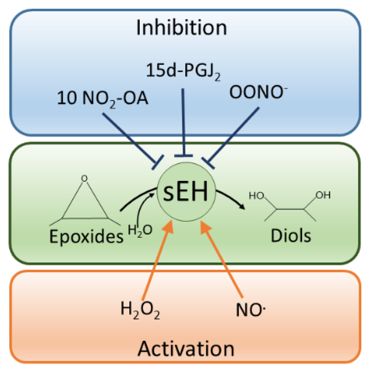
2. Soluble Epoxide Hydrolase

sEH is ubiquitously expressed, including in cardiovascular-relevant tissues such as endothelial or vascular smooth muscle cells as well as cardiomyocytes, where it is an important modulator of arterial and cardiac functions [18,19,20,21,22,23,24,25]. sEH is also a susceptibility factor for human heart failure, with polymorphisms that enhance hydrolase activity increasing cardiovascular risk [26]. Conversely, inhibitors (or transgenic knock-outs) of sEH offer a broad spectrum of cardiovascular protection, including the blockade of smooth muscle proliferation [27], reduction of atherosclerosis and hypertension [18,23,28,29,30,31], prevention and regression of cardiac hypertrophy and HF [26,32,33], and fibrosis [34]. sEH is homodimeric and arranged in an anti-parallel manner, comprising an N-terminal lipid phosphatase domain and a C-terminal epoxide hydrolase domain (Figure 2A) [19]. The N-terminal domain is capable of dephosphorylating lipids such as lysophosphatidic acids (LPA) [35,36] and sphingosine-1-phosphate (S1P). The hydrolase domain catalyzes the hydrolysis of epoxy-fatty acids (EpFAs) to their corresponding diols, as shown in Figure 2 [37,38].
The active site of the hydrolase domain, as identified through biochemical and structural analysis, is located at the base of a 25 Å deep L-shaped cavity [39], where a triad of amino acids, namely Asp335, Asp496 and His524, catalyse the epoxide hydrolysis reaction [40,41]. The mechanism involves two steps, an initial ester formation between the EET and Asp335 of the enzyme followed by the water-mediated release of the diol product that is catalysed by Asp496/His524. Two tyrosines, specifically 383 and 466, provide support by establishing hydrogen bonds with the oxygen of the epoxy ring, thereby positioning it for the nucleophilic attack by the proximal aspartate [38]. These two tyrosine residues can be nitrated to inhibit the hydrolase and will be discussed in detail below.
The most widely studied substrates of sEH are epoxyeicosatrienoic acids (EETs); however, it can also hydrolyse epoxydocosapentaenoic acids (EpDPEs), α- and γ-epoxyoctadecadienoic acids (α/γ-EpODEs) and epoxyoctadecaenoic acids (EpOMEs). EETs, of which there are four regioisomers cis 5,6-EET, 8,9-EET, 11,12-EET and 14,15-EET, are derived from arachidonic acid. EETs have a wide range of biological activities, including dilation of systemic or coronary arteries or, conversely, constricting those of the pulmonary system. EETs are broadly pro-angiogenic, anti-inflammatory and limit platelet aggregation as well as promoting fibrinolysis. Finally, EETs also enhance cardiomyocyte contractility [42,43,44,45,46,47,48,49]. Therefore, inhibiting sEH pharmacologically elevates its substrates such as EETs, allowing them to exert their beneficial actions. Indeed, inhibiting sEH has identified the hydrolase as an important modulator in cardiovascular and renal health, with additional roles in nociception [19,50,51].

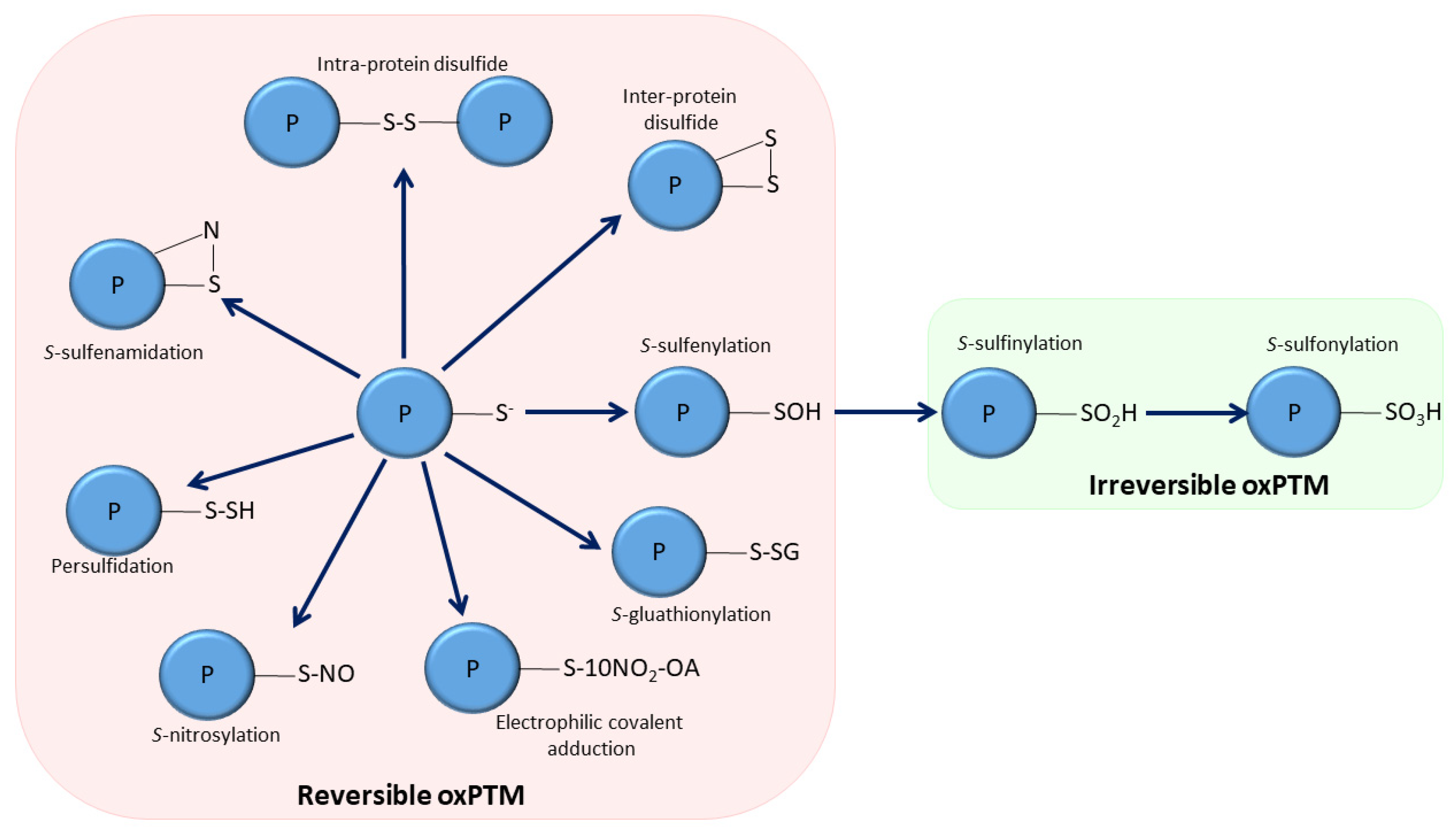
3. Oxidative Post-Translational Modifications of sEH

Until 2009, little was known about how sEH activity was regulated, and it was thought to be principally determined by its expression abundance [52]. However, it is now apparent that a number of different oxPTMs, which are discussed below, as are their effects on cardiovascular health and disease (summarized in Table 1), regulate this hydrolase.

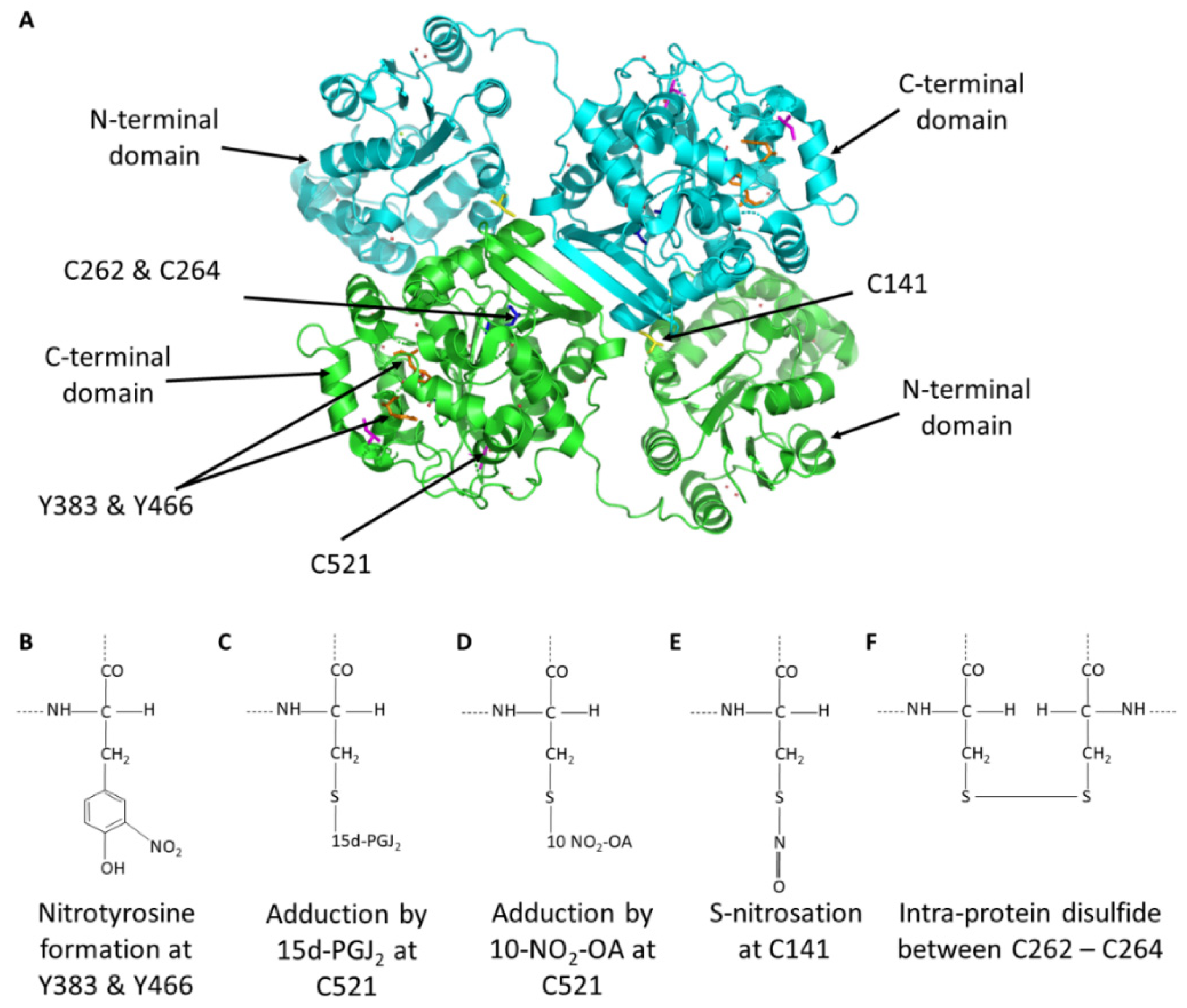
3.1. Tyrosine Nitration

Barbosa-Sicard et al. showed that sEH is inhibited by tyrosine nitration (Figure 2B), providing perhaps the first evidence that sEH could be oxidized by a PTM. Y383 and Y466, key amino acids for enzymatic sEH activity, were nitrated by peroxynitrite (ONOO) or the ONOO generator 3-morpholinosydnonimine (SIN-1) [53]. Tyrosine nitration at either of these sites resulted in hydrolase inhibition. Streptozotocin, a compound used to induce diabetes but interestingly also contains an N-nitroso moiety that may donate nitric oxide, was found to increase tyrosine nitration. Traditionally, tyrosine nitration has been associated with damage and disease. This may be due to the widely held ‘belief’ that oxidants are principally harmful, and indeed this may be the case with peroxynitrite because its high reactivity provides potential for non-selective oxidations. However, in this scenario, inhibiting sEH by tyrosine nitration would be expected to increase cardioprotective EETs, which may mitigate against dysfunction arising from other damaging modifications.

3.2. Adduction of Electrophilic Lipids to sEH

We found 15d-PGJ2 adducts to and inhibits the activity of sEH (Figure 2C) [54]. 15d-PGJ2 is an endogenously generated electrophile that contains two α,β-unsaturated carbonyls that enable it to react with nucleophilic thiol moieties in proteins via Michael addition reactions (Figure 2C). As C521 (mouse protein sequence numbering) is conserved and proximal to the catalytic triad, as discussed above, it was identified as a likely candidate to mediate the electrophilic lipid and subsequent inhibition. Although other targets of 15d-PGJ2 have been identified, with perhaps the best characterised being the nuclear peroxisome proliferator-activated receptor γ [55], we demonstrated a causal link between this electrophilic lipid and its ability to adductively inhibit sEH to induce coronary vessel vasodilation [54]. This electrophilic mechanism of inhibition also contributed to coronary artery hypoxic vasodilation [54]. Subsequently, other electrophilic lipids, such as 10-NO2-OA, were also found to adduct to sEH C521 to inhibit hydrolase activity (Figure 2D) [56,57]. 10-NO2-OA is known to protect against myocardial infarction, preserve left ventricular function [58], dilate arteries [59], attenuate platelet activation and reduce inflammation via cyclic guanosine monophosphate (cGMP)-independent mechanisms [60,61]. Perhaps these protective actions, such as the reduction in blood pressure and reducing myocardial infarct size, may also be explained by sEH inhibition. Given the complexities of establishing if the blood pressure lowering effects of 10-NO2-OA were due to sEH inhibition, a transgenic knock-in mouse in which C521 was systemically replaced with a serine was generated. This enabled investigations to determine whether electrophilic lipids exert their blood pressure lowering actions causally by inhibiting sEH. These knock-in mice were studied in the context of angiotensin II-induced hypertension, measuring their blood pressure lowering response to 10-NO2-OA compared to wild-type controls. The nitro-fatty acid inhibited sEH in wild-type mice in which EETs concomitantly accumulated and so explains the blood pressure-lowering observed. This lowering of blood pressure was completely absent in the knock-in mice that lack the cysteine to which the nitro-lipid adducts to cause hydrolase inhibition. Interestingly, inhibition of sEH by 10-NO2-OA highlighted a mechanism to account for the protection from hypertension afforded by the Mediterranean diet. Paradoxically, people who consume this diet, which is rich in fats and therefore is anticipated to promote cardiovascular disease, are in fact protected from such conditions [62,63]. The formation of nitrated fatty acids such as 10-NO2-OA are promoted under acidic conditions in the stomach following consumption of green leafy vegetables with nitrite and unsaturated fats that are key components in the Mediterranean diet [64,65,66]. Subsequent studies identified C423 as an additional site for electrophilic lipid adduction [57]. Although this cysteine is not present in the murine orthologue, adduction of a lipid electrophile to this site in recombinant human sEH was inhibitory. However, this inhibitory mechanism has yet to be substantiated in cells or in vivo. Electrophile adduction at C423 is notable because it is distal to the active site and can therefore be described as allosteric. Therefore, targeting C423 may be useful in the development of allosteric sEH inhibitors [57]. C521 is located at the exit of the hydrophobic tunnel and therefore may not be completely allosteric due to its proximity to the active site. However, the mechanism of inhibition at C521 could potentially be exploited by the development of novel electrophilic drugs to combat cardiovascular disease. Collectively, our work clearly demonstrates that the enzymes’ activity can be inhibited by electrophilic lipids.

3.3. S-Nitrosation of sEH

In 2016, Ding et al. showed sEH is activated by S-nitrosation (Figure 2E) [67]. During myocardial reperfusion, sEH was activated and lead to a loss of cardioprotective EETs. A trans-nitrosating nitric oxide donor, namely nitrosocysteine (CysNO), recapitulated this activation of sEH in vitro (Figure 2E). C141 was identified as the main site of S-nitrosation, although a number of different cysteine residues were also found to be modified in this way, including C264, which will be considered further below. Interestingly, C141 is located in the N-terminal domain of sEH, however due to the domain swapped anti-parallel architecture of sEH, a SNO modification at this site perhaps stabilises bonds in the hydrolase domain of the opposite monomer, leading to enhanced hydrolase activity. S-Nitrosoglutathione (GSNO) is another nitric oxide donor that might be expected to activate sEH in a similar manner as described here by CysNO. These authors also reported that GSNO similarly activated sEH, whereas we found that it did not do this [54]. It is possible that GSNO may induces S-glutathionylation of sEH and the effect of this disulfide modification on activity remains unclear.

3.4. Intra-Disulfide Formation in sEH

In 2021, we described a mechanism by which sEH can be activated by oxidative intra-disulfide formation (Figure 2F) [68]. Oxidation induced either by exogenously applied hydrogen peroxide or the pro-oxidant vasopressor hormone angiotensin II increased sEH activity by catalyzing a disulfide between Cys262 and C264 (mouse protein sequence numbering), as depicted in Figure 2D. This oxidative activation increased the hydrolysis of vasodilatory EETs. Indeed, following angiotensin II treatment, the EET/DHET ratio decreased, consistent with and potentially explaining, at least in part, the pressor action of angiotensin II. This is notable, as we have previously shown that S-nitrosated proteins generically transition to form a disulfide bond [69]; hence, it is likely that the S-nitrosation of C264 observed by Ding et al. induced the activating intra-disulfide with C262.

4. Redox Regulation of sEH in Blood Vessels

Decreasing sEH activity, either pharmacologically or genetically, has illustrated a broad role for this hydrolase and its EET substrates in vascular homeostasis. EETs are angiogenic, anti-inflammatory, and suppress platelet aggregation [45,47,70,71]. Regulation of angiogenesis is critical to the maintenance of vascular and myocardial health, with EETs able to increase endothelial cell proliferation to promote such vascularization [45,72,73]. On this basis, inhibiting sEH, because of the angiogenic roles of EETs, promotes neovascularization in ischemic tissue. Interestingly, the redox status of the cell affects angiogenesis [74,75]. Consequently, oxidative modifications that inhibit sEH, may be expected to promote angiogenesis. Although not directly investigated in the study reporting that tyrosine nitration of sEH is inhibitory [53], it is conceivable that this oxPTM enables nitrosative signaling to be coupled with angiogenesis. Our group demonstrated, albeit in a cancer model, that sEH inhibition induced by nitro-oleate adduction to C521 promoted angiogenesis [76]. Indeed, this electrophilic inhibition of sEH increased cell proliferation and migration in wild-type (WT) mice more than in C521S knock-in mice lacking the critical cysteine that renders the hydrolase electrophile-resistant. In direct contrast to sEH inhibition promoting angiogenesis, increasing hydrolase activity is anticipated to reduce it. Therefore, following S-nitrosation or disulfide bond formation, namely the scenarios mentioned above that activate sEH, a decrease in cell proliferation and migration may be expected. However, treatment with a diol restored blood flow in sEH null mice to levels observed in WT mice during hind limb ischemia [77]. Similarly, sEH null mice displayed attenuated sprouting angiogenesis in their retinas, which could again be rescued by the application of the diols [78]. Therefore, in these scenarios, increasing sEH activity by either S-nitrosation or disulfide formation may be beneficial. While it is clear that sEH plays a role in angiogenesis, further research is required to elucidate how sEH is regulated to elicit its opposing angiogenic effects in different tissues and the precise roles redox regulation play in specific cells or scenarios.
The anti-inflammatory actions of EETs also contribute to vascular homeostasis. 11–12 EET decreased the number of adherent and rolling mononuclear cells in mice following TNF-injections [79]. Similarly, sEH null mice or in AUDA-BE treated mice had an attenuated inflammatory response to lipopolysaccharide compared to WTs [80]. Likewise, sEH inhibition not only decreased infiltration of macrophages in angiotensin II-induced or deoxycorticosterone (DOCA) salt hypertension, but also attenuated renal inflammation [81,82]. Despite evidence that sEH inhibition is anti-inflammatory, more research is needed to establish whether the sEH redox state is modulated in such scenarios, which is likely given the roles of oxidants in inflammation [83]. As tyrosine nitration and electrophilic lipid adduction both inhibit sEH, it is conceivable that each of these mechanisms increase EETs and their anti-inflammatory actions—a prospect that warrants further investigations.
EETs are vasodilators that induce vascular smooth muscle cell (VSMC) hyperpolarization and the activation of large-conductance calcium activated potassium channels (KCa) to cause arterial dilation [47,84]. Some evidence indicates that EETs can also activate Ca2+-induced intermediate-conductance (IKCa) and small-conductance (SKCa) potassium channels that may also participate in cell hyperpolarization [85], which spreads via gap junctions [47]. EETs can also activate transient receptor potential vanilloid channel 4 on VSMCs [86]. EETs released by endothelial cells are also thought to activate a yet unidentified EET receptor on VSMCs, leading to adenylate cyclase and protein kinase A activation, activating BKCa and ATP-sensitive potassium (KATP) channels to induce hyperpolarization and vasodilation [19]. Based on the vasodilatory actions of ETTS, inhibiting the hydrolase either through pharmacological or genetic manipulation decreased blood pressure in several different animal models of hypertension [30,31,81,87,88]. As discussed above, we found that the electrophilic lipid NO2-OA, covalently adducted to sEH at C521 to inhibit it, increased EETs/DHETs, thereby lowering blood pressure in angiotensin II-induced hypertensive mice. This study clearly demonstrates a role for the redox control of sEH in blood pressure regulation and the pathogenesis of hypertension. It is plausible that other mechanisms of inhibitory sEH oxidation, such as tyrosine nitration, may also couple to vasodilation. On the other hand, oxidative modifications that activate the enzyme are anticipated to have the opposite effect and induce vasoconstriction instead due to the increased hydrolysis of vasodilatory EETs. Interestingly, the vasopressor angiotensin II not only increases cellular H2O2 [89,90] but also decreases EETs in mice, consistent with sEH being oxidized in this scenario [68].

5. The Role of Redox Regulation of sEH in the Heart

Myocardial sEH has been significantly investigated in terms of cardiac pathophysiology, with relatively few studies investigating its role in physiological functions during health. However, similar to the dilation of the systemic vasculature, EETs can also relax the coronary vasculature by activating KCa channels [91]. Our work with 15d-PGJ2 also demonstrated that EETs relax the coronary vasculature, whereby the electrophilic lipid induced a prominent and sustained vasodilation in ex vivo perfused rat hearts. This coronary vasodilation was due to 15d-PGJ2 adducting to C521 and inhibiting sEH, a mechanism that contributed to coronary hypoxic vasodilation [54].
In myocytes, 5,6-EET or 11,12-EET increased cell shortening and intracellular calcium concentrations [92]. Other studies have since demonstrated that EETs can inhibit cardiac myocytes Na+ channels [93], activate L-type Ca2+ channels [94], as well as increase KATP channel openings [95]. Despite these effects on cardiac myocyte ion channel activity, very few investigations have explored EETs action on normal cardiac function, perhaps because of the difficulty of functionally separating the contractile actions of EETs from their coronary vasodilatory actions [47]. Further research is required to establish if there is a role for redox control of sEH in these processes. However, because oxPTMs that inhibit sEH will likely increase EETs, they may reasonably be anticipated to increase cardiac myocyte contraction.
The role of myocardial sEH in cardioprotection has been widely studied. Hearts were protected from cardiac hypertrophy as well as myocardial ischemia and reperfusion injury when the hydrolase was absent in sEH null mice or was pharmacologically inhibited in WTs [33,88,96]. Cardioprotection has been attributed to the EETs that are increased when sEH is absent in knockout animals or inhibited pharmacologically. In terms of redox regulation of sEH and cardioprotection, electrophilic lipid adduction to C521 in sEH alleviated cardiac hypertrophy induced by angiotensin II [56]. Although this protection from hypertrophic growth is likely explained by the blood pressure-lowering action of the lipid, a contribution from the inhibition of cardiomyocyte sEH is also plausible. Our observation is consistent with other studies showing that classical sEH inhibitors not only prevented maladaptive growth during cardiac pressure load in rodents, but in some cases reversed established hypertrophy [33,96].
Administering 10-NO2-OA to hearts prior to ischemia and reperfusion protected them from loss of contractile function [97], which is likely to be partially mediated by it inhibiting sEH and so increasing EETs that have established cytoprotective actions. Although sEH inhibition via either electrophilic lipid adduction or tyrosine nitration may be anticipated to protect against myocardial ischemia and reperfusion injury, further research is required to determine this.
Interestingly, following angiotensin II-induced cardiac hypertrophy or ischemia and reperfusion injury, sEH activity is increased [33,98]. This may be expected as sEH expression is increased upon angiotensin II treatment [98], as well as after ischemic injury [99]. However, this increased expression did not fully explain the increase in hydrolase activity, consistent with the prospect of oxidative activation [67,68]. During hypertrophy or ischemia and reperfusion, ROS are increased as NADPH oxidases and xanthine oxidase are stimulated, producing superoxide and H2O2 [90,100,101]. These oxidants may account for the increased hydrolase activity observed, considering that we have demonstrated that peroxide molecules activate sEH [68].

6. Perspectives and Future Directions

It notable that the Oximouse compendium shows that a number of cysteine residues in sEH, both in the human and mouse proteome, are susceptible to oxidation, and these are altered during aging in various different tissues [17]. The Oximouse dataset demonstrated that there is not an overall increase in protein oxidation with age, but instead that the redox networks are remodeled [17]. In fact when looking at sEH in this dataset, it is apparent that some cysteines become more oxidized during aging, whereas others are concomitantly reduced. Although the number of these cysteines have previously been identified as sites of oxidation, including C521, C141 and C423, further research is required to elucidate the functional significance of the other sites identified.
For a protein to be considered truly redox regulated, the oxPTMs need to be able to be reversed as this enables dynamic, two-way control. In terms of the oxPTM in sEH discussed above, one wonders about their reversibility and if they are enzymatically reversed. In the case the intra-protein disulfide in sEH, we provided evidence that thioredoxin reduces the disulfide back to the basal reduced state [68]. S-nitrosylated proteins can supposedly be reversed by thioredoxin [102], however this has yet to be determined specifically for sEH. Although not conclusive, as its identity remains elusive, there is evidence for a cellular denitrase that reverses protein tyrosine nitration [103]. Such a denitrase may reverse the inhibitory modification at Y383 and Y466. Formation of the covalent protein modification with either 15d-PGJ2 or NO2-OA and sEH was thought to be irreversible, with proteolysis being the main mechanism of reversal. However, there is now evidence that electrophilic nitroalkylation is reversible and may be mediated by GSH [104].
Interestingly, the studies describing covalent adduction of endogenous electrophilic lipids offers some hope for the generation of a novel class of sEH inhibitors which have advantages over classical inhibitors [105]. The sites of this endogenous inhibitory mechanism may be exploited for the design of novel drugs, which may be used to target hypertension and other cardiovascular dysfunction. Covalent compounds have traditionally been avoided in the development of therapies, as they were thought to non-selectively conjugate with many different proteins. However, this view has been challenged by studies showing that covalent compounds can offer selectivity and potency benefits over traditional drugs [106,107,108,109]. Indeed, there are a number of widely used drugs, such as penicillin and aspirin [107], which mediate their therapeutic actions via covalent adduction to target proteins.
Previously, antioxidants have been tested as a therapy for cardiovascular diseases in an attempt to combat excessive oxidant production. However, despite multiple clinical trials assessing their efficacy, the outcomes have been disappointing, with no cardiovascular protection observed, and in some instances they were even harmful [110,111]. However, these studies failed to consider the homeostatic and adaptive oxidant signaling that may be lost with antioxidant interventions. Therefore, by understanding specific actions of different ROS or RNS, it may be possible to target precise oxidation events with antioxidants. Interestingly, certain antioxidants can paradoxically oxidize proteins. For example, the antioxidant resveratrol can convert to a reactive quinone and then oxidize Protein Kinase G Iα [112]. How antioxidants influence sEH activity is worthy of exploration.
This review has primarily focused on the hydrolase domain of sEH. However, as mentioned above, sEH also contains an N-terminal lipid phosphatase domain [113,114] which can dephosphorylate LPA [35,36] and S1P. As LPA and S1P are important modulators of vascular homeostasis, the redox regulation of the N-terminal phosphatase may also play a role in the context of cardiovascular health and disease. Interestingly, Oximouse compendium indicates that the cysteine residues within the N-terminal domain can be redox modulated [17]. Likewise, there are different isoforms of epoxide hydrolase, including microsomal epoxide hydrolase, epoxide hydrolase 3 and epoxide hydrolase 4. Although minimally expressed in cardiac cells, it would be interesting to determine if they are redox controlled.

7. Closing Thoughts and Considerations

It is now abundantly evident that many proteins are regulated by oxPTMs that couple changes in cellular redox status to functional responses of proteins and the cells they are found in. Over the last 20 years or so, we have increasingly changed our view of the role of oxidants in cells and tissues. Although they have important roles in dysregulation and disease pathogenesis, there is a plethora of evidence for their regulatory roles during health and adaptive changes that limit disease progression. It is evident that the redox regulation of cardiac and vascular sEH is highly intricate, and much remains to be understood. Importantly, redox control of the hydrolase contributes both to protective as well as maladaptive pathways, and this likely depends upon the specific ROS source and their concentration and locality to sEH, as shown in Figure 3.

Author Contributions

R.C. and P.E. wrote this article and agreed to the published version of the manuscript. All authors have read and agreed to the published version of the manuscript.

Funding

R.C. is supported by a British Heart Foundation Intermediate Fellowship (FS/17/36/32874). P.E. is supported by The Barts Charity Cardiovascular Programme Award G00913 and programme grants from the British Heart Foundation and the Medical Research Council.

Institutional Review Board Statement

Not applicable.

Informed Consent Statement

Not applicable.

Data Availability Statement

Not applicable.

Conflicts of Interest

The authors declare that they have no conflict of interest.

References

  1. Davies, M.J. Protein Oxidation and Peroxidation. Biochem. J. 2016, 473, 805–825. [Google Scholar] [CrossRef] [PubMed] [Green Version]
  2. Radi, R. Protein Tyrosine Nitration: Biochemical Mech anisms and Structural Basis of Functional Effects. Acc. Chem. Res. 2013, 46, 550–559. [Google Scholar] [CrossRef] [PubMed] [Green Version]
  3. Mallozzi, C.; Di Stasi, A.M.M.; Minetti, M. Nitrotyrosine Mimics Phosphotyrosine Binding to the Sh2 Domain of the Src Family Tyrosine Kinase Lyn. FEBS Lett. 2001, 503, 189–195. [Google Scholar] [CrossRef] [Green Version]
  4. Hoshi, T.; Heinemann, S. Regulation of Cell Function by Methionine Oxidation and Reduction. J. Physiol. 2001, 531 Pt 1, 1–11. [Google Scholar] [CrossRef]
  5. Reddie, K.G.; Carroll, K.S. Carroll. Expanding the Functional Diversity of Proteins through Cysteine Oxidation. Curr. Opin. Chem. Biol. 2008, 12, 746–754. [Google Scholar] [CrossRef]
  6. Rudyk, O.; Eaton, P. Biochemical Methods for Monitoring Protein Thiol Redox States in Biological Systems. Redox Biol. 2014, 2, 803–813. [Google Scholar] [CrossRef] [Green Version]
  7. Bibli, S.I.; Fleming, I. Oxidative Post-Translational Modifications: A Focus on Cysteine S-Sulfhydration and the Regulation of Endothelial Fitness. Antioxid. Redox Signal. 2021, 35, 1494–1514. [Google Scholar] [CrossRef]
  8. Schopfer, F.J.; Cipollina, C.; Freeman, B.A. Freeman. Formation and Signaling Actions of Electrophilic Lipids. Chem. Rev. 2011, 111, 5997–6021. [Google Scholar] [CrossRef] [Green Version]
  9. Burgoyne, J.R.; Madhani, M.; Cuello, F.; Charles, R.L.; Brennan, J.P.; Schroder, E.; Browning, D.D.; Eaton, P. Cysteine Redox Sensor in Pkgia Enables Oxidant-Induced Activation. Science 2007, 317, 1393–1397. [Google Scholar] [CrossRef]
  10. Brennan, J.P.; Bardswell, S.C.; Burgoyne, J.; Fuller, W.; Schröder, E.; Wait, R.; Begum, S.; Kentish, J.C.; Eaton, P. Oxidant-Induced Activation of Type I Protein Kinase a Is Mediated by Ri Subunit Interprotein Disulfide Bond Formation. J. Biol. Chem. 2006, 281, 21827–21836. [Google Scholar] [CrossRef] [Green Version]
  11. Maller, C.; Schröder, E.; Eaton, P. Glyceraldehyde 3-Phosphate Dehydrogenase Is Unlikely to Mediate Hydrogen Peroxide Signaling: Studies with a Novel Anti-Dimedone Sulfenic Acid Antibody. Antioxid. Redox Signal. 2011, 14, 49–60. [Google Scholar] [CrossRef] [PubMed]
  12. Salmeen, A.; Andersen, J.N.; Myers, M.P.; Meng, T.-C.; Hinks, J.A.; Tonks, N.K.; Barford, D. Redox Regulation of Protein Tyrosine Phosphatase 1b Involves a Sulphenyl-Amide Intermediate. Nature 2003, 423, 769–773. [Google Scholar] [CrossRef] [PubMed]
  13. Burgoyne, J.R.; Mongue-Din, H.; Eaton, P.; Shah, A.M. Redox Signaling in Cardiac Physiology and Pathology. Circ. Res. 2012, 111, 1091–1116. [Google Scholar] [CrossRef] [PubMed]
  14. Charles, R.L.; Eaton, P. Redox Signalling in Cardiovascular Disease. Proteom.-Clin. Appl. 2008, 2, 823–836. [Google Scholar] [CrossRef]
  15. Benhar, M. Oxidants, Antioxidants and Thiol Redox Switches in the Control of Regulated Cell Death Pathways. Antioxidants 2020, 9, 309. [Google Scholar] [CrossRef] [Green Version]
  16. Paulsen, C.E.; Carroll, K.S. Cysteine-Mediated Redox Signaling: Chemistry, Biology, and Tools for Discovery. Chem. Rev. 2013, 7, 4633–4679. [Google Scholar] [CrossRef]
  17. Xiao, H.; Jedrychowski, M.P.; Schweppe, D.K.; Huttlin, E.L.; Yu, Q.; Heppner, D.E.; Li, J.; Long, J.; Mills, E.L.; Szpyt, J.; et al. A Quantitative Tissue-Specific Landscape of Protein Redox Regulation during Aging. Cell 2020, 180, 968–983.e24. [Google Scholar] [CrossRef]
  18. Revermann, M. Pharmacological inhibition of the soluble epoxide hydrolase—from mouse to man. Curr. Opin. Pharmacol. 2010, 10, 173–178. [Google Scholar] [CrossRef]
  19. Imig, J.D.; Hammock, B.D. Soluble epoxide hydrolase as a therapeutic target for cardiovascular diseases. Nat. Rev. Drug Discov. 2009, 8, 794–805. [Google Scholar] [CrossRef] [Green Version]
  20. Chiamvimonvat, N.; Ho, C.-M.; Tsai, H.-J.; Hammock, B.D. The Soluble Epoxide Hydrolase as a Pharmaceutical Target for Hypertension. J. Cardiovasc. Pharmacol. 2007, 50, 225–237. [Google Scholar] [CrossRef] [Green Version]
  21. Marino, J.P., Jr. Soluble Epoxide Hydrolase, a Target with Multiple Opportunities for Cardiovascular Drug Discovery. Curr. Top. Med. Chem. 2009, 9, 452–463. [Google Scholar] [CrossRef] [PubMed]
  22. Imig, J.D. Cardiovascular Therapeutic Aspects of Soluble Epoxide Hydrolase Inhibitors. Cardiovasc. Drug Rev. 2006, 24, 169–188. [Google Scholar] [CrossRef] [PubMed]
  23. Imig, J.D.; Zhao, X.; Zaharis, C.Z.; Olearczyk, J.J.; Pollock, D.M.; Newman, J.; Kim, I.-H.; Watanabe, T.; Hammock, B.D. An Orally Active Epoxide Hydrolase Inhibitor Lowers Blood Pressure and Provides Renal Protection in Salt-Sensitive Hypertension. Hypertension 2005, 46, 975–981. [Google Scholar] [CrossRef] [Green Version]
  24. Gross, G.J.; Nithipatikom, K. Soluble epoxide hydrolase: A new target for cardioprotection. Curr. Opin. Investig. Drugs 2009, 10, 253–258. [Google Scholar] [PubMed]
  25. Fang, X. Soluble Epoxide Hydrolase: A Novel Target for the Treatment of Hypertension. Recent Patents Cardiovasc. Drug Discov. 2006, 1, 67–72. [Google Scholar] [CrossRef] [PubMed]
  26. Monti, J.; Fischer, J.; Paskas, S.; Heinig, M.; Schulz, H.; Gosele, C.; Heuser, A.; Fischer, R.; Schmidt, C.; Schirdewan, A.; et al. Soluble epoxide hydrolase is a susceptibility factor for heart failure in a rat model of human disease. Nat. Genet. 2008, 40, 529–537. [Google Scholar] [CrossRef] [PubMed]
  27. Davis, B.B.; Morisseau, C.; Newman, J.W.; Pedersen, T.L.; Hammock, B.D.; Weiss, R.H. Attenuation of Vascular Smooth Muscle Cell Proliferation by 1-Cyclohexyl-3-dodecyl Urea Is Independent of Soluble Epoxide Hydrolase Inhibition. J. Pharmacol. Exp. Ther. 2006, 316, 815–821. [Google Scholar] [CrossRef] [Green Version]
  28. Ulu, A.; Davis, B.B.; Tsai, H.-J.; Kim, I.-H.; Morisseau, C.; Inceoglu, B.; Fiehn, O.; Hammock, B.D.; Weiss, R.H. Soluble Epoxide Hydrolase Inhibitors Reduce the Development of Atherosclerosis in Apolipoprotein E-Knockout Mouse Model. J. Cardiovasc. Pharmacol. 2009, 52, 314–323. [Google Scholar] [CrossRef] [Green Version]
  29. Olearczyk, J.J.; Quigley, J.E.; Mitchell, B.C.; Yamamoto, T.; Kim, I.-H.; Newman, J.W.; Luria, A.; Hammock, B.D.; Imig, J.D. Administration of a substituted adamantyl urea inhibitor of soluble epoxide hydrolase protects the kidney from damage in hypertensive Goto–Kakizaki rats. Clin. Sci. 2008, 116, 61–70. [Google Scholar] [CrossRef] [Green Version]
  30. Jung, O.; Brandes, R.P.; Kim, I.-H.; Schweda, F.; Schmidt, R.; Hammock, B.D.; Busse, R.; Fleming, I. Soluble Epoxide Hydrolase Is a Main Effector of Angiotensin II–Induced Hypertension. Hypertension 2005, 45, 759–765. [Google Scholar] [CrossRef] [Green Version]
  31. Imig, J.D.; Zhao, X.; Capdevila, J.H.; Morisseau, C.; Hammock, B.D. Soluble epoxide hydrolase inhibition lowers arterial blood pressure in angiotensin II hypertension. Hypertension 2002, 39, 690–694. [Google Scholar] [CrossRef] [PubMed] [Green Version]
  32. Harris, T.R.; Li, N.; Nipavan, C.; Hammock, B.D. The Potential of Soluble Epoxide Hydrolase Inhibition in the Treatment of Cardiac Hypertrophy. Congest. Hear. Fail. 2008, 14, 219–224. [Google Scholar] [CrossRef] [PubMed]
  33. Ai, D.; Pang, W.; Li, N.; Xu, M.; Jones, P.D.; Yang, J.; Zhang, Y.; Chiamvimonvat, N.; Shyy, J.Y.-J.; Hammock, B.D.; et al. Soluble epoxide hydrolase plays an essential role in angiotensin II-induced cardiac hypertrophy. Proc. Natl. Acad. Sci. USA 2009, 106, 564–569. [Google Scholar] [CrossRef] [PubMed] [Green Version]
  34. Sirish, P.; Li, N.; Liu, J.-Y.; Lee, K.S.S.; Hwang, S.H.; Qiu, H.; Zhao, C.; Ma, S.M.; López, J.E.; Hammock, B.D.; et al. Unique mechanistic insights into the beneficial effects of soluble epoxide hydrolase inhibitors in the prevention of cardiac fibrosis. Proc. Natl. Acad. Sci. USA 2013, 110, 5618–5623. [Google Scholar] [CrossRef] [Green Version]
  35. Morisseau, C.; Schebb, N.H.; Dong, H.; Ulu, A.; Aronov, P.A.; Hammock, B.D. Role of soluble epoxide hydrolase phosphatase activity in the metabolism of lysophosphatidic acids. Biochem. Biophys. Res. Commun. 2012, 419, 796–800. [Google Scholar] [CrossRef] [Green Version]
  36. Oguro, A.; Imaoka, S. Lysophosphatidic acids are new substrates for the phosphatase domain of soluble epoxide hydrolase. J. Lipid Res. 2012, 53, 505–512. [Google Scholar] [CrossRef] [Green Version]
  37. Arand, M.; Cronin, A.; Adamska, M.; Oesch, F. Epoxide Hydrolases: Structure, Function, Mechanism, and Assay. Methods Enzym. 2005, 400, 569–588. [Google Scholar]
  38. Newman, J.W.; Morisseau, C.; Hammock, B.D. Epoxide hydrolases: Their roles and interactions with lipid metabolism. Prog. Lipid Res. 2005, 44, 1–51. [Google Scholar] [CrossRef]
  39. Argiriadi, M.A.; Morisseau, C.; Goodrow, M.H.; Dowdy, D.L.; Hammock, B.D.; Christianson, D.W. Binding of Alkylurea Inhibitors to Epoxide Hydrolase Implicates Active Site Tyrosines in Substrate Activation. J. Biol. Chem. 2000, 275, 15265–15270. [Google Scholar] [CrossRef] [Green Version]
  40. Gomez, G.A.; Morisseau, C.; Hammock, A.B.D.; Christianson, D.W. Structure of Human Epoxide Hydrolase Reveals Mechanistic Inferences on Bifunctional Catalysis in Epoxide and Phosphate Ester Hydrolysis. Biochemistry 2004, 43, 4716–4723. [Google Scholar] [CrossRef]
  41. Gomez, G.A.; Morisseau, C.; Hammock, B.D.; Christianson, D.W. Human Soluble Epoxide Hydrolase: Structural Basis of Inhibition by 4-(3-Cyclohexylureido)-Carboxylic Acids. Protein. Sci. 2006, 15, 58–64. [Google Scholar] [CrossRef] [PubMed]
  42. Campbell, W.B.; Deeter, C.; Gauthier, K.M.; Ingraham, R.H.; Falck, J.R.; Li, P.L. 14,15-Dihydroxyeicosatrienoic Acid Relaxes Bovine Coronary Arteries by Activation of K(Ca) Channels. Am. J. Physiol. Heart Circ. Physiol. 2002, 282, H1656–H1664. [Google Scholar] [CrossRef] [PubMed] [Green Version]
  43. Campbell, W.B.; Gebremedhin, D.; Pratt, P.F.; Harder, D.R. Identification of Epoxyeicosatrienoic Acids as Endothelium-Derived Hyperpolarizing Factors. Circ. Res. 1996, 78, 415–423. [Google Scholar] [CrossRef] [PubMed]
  44. Campbell, W.B.; Harder, D.R. Endothelium-derived hyperpolarizing factors and vascular cytochrome P450 metabolites of arachidonic acid in the regulation of tone. Circ. Res. 1999, 84, 484–488. [Google Scholar] [CrossRef] [Green Version]
  45. Fleming, I. Discreet Regulators of Homeostasis: Epoxyeicosatrienoic Acids, Cytochrome P450 Epoxygenases and Vascular Inflammation. Trends Pharmacol. Sci. 2007, 28, 448–452. [Google Scholar] [CrossRef]
  46. Luria, A.; Weldon, S.M.; Kabcenell, A.K.; Ingraham, R.H.; Matera, D.; Jiang, H.; Gill, R.; Morisseau, C.; Newman, J.; Hammock, B.D. Compensatory Mechanism for Homeostatic Blood Pressure Regulation in Ephx2 Gene-disrupted Mice. J. Biol. Chem. 2007, 282, 2891–2898. [Google Scholar] [CrossRef] [Green Version]
  47. Imig, J.D. Epoxides and Soluble Epoxide Hydrolase in Cardiovascular Physiology. Physiol. Rev. 2012, 92, 101–130. [Google Scholar] [CrossRef] [Green Version]
  48. Yang, L.; Mäki-Petäjä, K.; Cheriyan, J.; McEniery, C.; Wilkinson, I.B. The role of epoxyeicosatrienoic acids in the cardiovascular system. Br. J. Clin. Pharmacol. 2015, 80, 28–44. [Google Scholar] [CrossRef] [Green Version]
  49. Node, K.; Ruan, X.-L.; Dai, J.; Yang, S.-X.; Graham, L.; Zeldin, D.C.; Liao, J.K. Activation of Galpha S Mediates Induction of Tissue-Type Plasminogen Activator Gene Transcription by Epoxyeicosatrienoic Acids. J. Biol. Chem. 2001, 276, 15983–15989. [Google Scholar] [CrossRef] [Green Version]
  50. Inceoglu, B.; Jinks, S.L.; Ulu, A.; Hegedus, C.M.; Georgi, K.; Schmelzer, K.R.; Wagner, K.; Jones, P.D.; Morisseau, C.; Hammock, B.D. Soluble epoxide hydrolase and epoxyeicosatrienoic acids modulate two distinct analgesic pathways. Proc. Natl. Acad. Sci. USA 2008, 105, 18901–18906. [Google Scholar] [CrossRef] [Green Version]
  51. Imig, J.D. Epoxide hydrolase and epoxygenase metabolites as therapeutic targets for renal diseases. Am. J. Physiol. Physiol. 2005, 289, F496–F503. [Google Scholar] [CrossRef] [PubMed] [Green Version]
  52. Harris, T.R.; Hammock, B.D. Soluble Epoxide Hydrolase: Gene Structure, Expression and Deletion. Gene 2013, 526, 61–74. [Google Scholar] [CrossRef] [PubMed] [Green Version]
  53. Barbosa-Sicard, E.; Frömel, T.; Keserü, B.; Brandes, R.P.; Morisseau, C.; Hammock, B.D.; Braun, T.; Krüger, M.; Fleming, I. Inhibition of the Soluble Epoxide Hydrolase by Tyrosine Nitration. J. Biol. Chem. 2009, 284, 28156–28163. [Google Scholar] [CrossRef] [PubMed] [Green Version]
  54. Charles, R.L.; Burgoyne, J.R.; Mayr, M.; Weldon, S.M.; Hubner, N.; Dong, H.; Morisseau, C.; Hammock, B.D.; Landar, A.; Eaton, P. Redox Regulation of Soluble Epoxide Hydrolase by 15-Deoxy-Delta-Prostaglandin J2 Controls Coronary Hypoxic Vasodilation. Circ. Res. 2011, 108, 324–334. [Google Scholar] [CrossRef] [Green Version]
  55. Shibata, T. 15-Deoxy-Delta(1)(2),(1)(4)-Prostaglandin J(2) as an Electrophilic Mediator. Biosci. Biotechnol. Biochem. 2015, 79, 1044–1049. [Google Scholar] [CrossRef] [Green Version]
  56. Charles, R.L.; Rudyk, O.; Prysyazhna, O.; Kamynina, A.; Yang, J.; Morisseau, C.; Hammock, B.D.; Freeman, B.A.; Eaton, P. Protection from hypertension in mice by the Mediterranean diet is mediated by nitro fatty acid inhibition of soluble epoxide hydrolase. Proc. Natl. Acad. Sci. USA 2014, 111, 8167–8172. [Google Scholar] [CrossRef] [Green Version]
  57. Abis, G.; Charles, R.L.; Kopec, J.; Yue, W.W.; Atkinson, R.A.; Bui, T.T.T.; Lynham, S.; Popova, S.; Sun, Y.-B.; Fraternali, F.; et al. 15-deoxy-Δ12,14-Prostaglandin J2 inhibits human soluble epoxide hydrolase by a dual orthosteric and allosteric mechanism. Commun. Biol. 2019, 2, 188. [Google Scholar] [CrossRef]
  58. Rudolph, V.; Rudolph, T.K.; Schopfer, F.J.; Bonacci, G.; Woodcock, S.R.; Cole, M.P.; Baker, P.R.; Ramani, R.; Freeman, B.A. Endogenous generation and protective effects of nitro-fatty acids in a murine model of focal cardiac ischaemia and reperfusion. Cardiovasc. Res. 2010, 85, 155–166. [Google Scholar] [CrossRef] [Green Version]
  59. Zhang, J.; Luis, V.; Lin, C.; Fan, Z.; Milton, H.; Zhu, T.; Chen, S.; Chen, M.; Cole, P.; Francisco, J.; et al. Nitro-Oleic Acid Inhibits Angiotensin Ii–Induced Hypertension. Circ. Res. 2010, 107, 540–548. [Google Scholar] [CrossRef] [Green Version]
  60. Coles, B.; Bloodsworth, A.; Clark, S.R.; Lewis, M.J.; Cross, A.R.; Freeman, B.A.; O’Donnell, V.B. Nitrolinoleate Inhibits Superoxide Generation, Degranulation, and Integrin Expression by Human Neutrophils: Novel Antiinflammatory Properties of Nitric Oxide–Derived Reactive Species in Vascular Cells. Circ. Res. 2002, 91, 375–381. [Google Scholar] [CrossRef] [Green Version]
  61. Coles, B.; Bloodsworth, A.; Eiserich, J.P.; Coffey, M.J.; McLoughlin, R.; Giddings, J.C.; Lewis, M.J.; Haslam, R.J.; Freeman, B.A.; O’Donnell, V.B. Nitrolinoleate Inhibits Platelet Activation by Attenuating Calcium Mobilization and Inducing Phosphorylation of Vasodilator-stimulated Phosphoprotein through Elevation of cAMP. J. Biol. Chem. 2002, 277, 5832–5840. [Google Scholar] [CrossRef] [PubMed] [Green Version]
  62. Covas, M.-I. Olive oil and the cardiovascular system. Pharmacol. Res. 2007, 55, 175–186. [Google Scholar] [CrossRef] [PubMed]
  63. Toledo, E.; Hu, F.B.; Estruch, R.; Buil-Cosiales, P.; Corella, D.; Salas-Salvad, Ó.J.; Covas, M.; Arós, F.; Gómez-Gracia, E.; Fiol, M.; et al. Effect of the Mediterranean Diet on Blood Pressure in the Predimed Trial: Results from a Randomized Controlled Trial. BMC Med. 2013, 11, 207. [Google Scholar] [CrossRef] [PubMed] [Green Version]
  64. Napolitano, A.; Panzella, L.; Savarese, M.; Sacchi, R.; Giudicianni, I.; Paolillo, L.; D’Ischia, M. Acid-Induced Structural Modifications of Unsaturated Fatty Acids and Phenolic Olive Oil Constituents by Nitrite Ions: A Chemical Assessment. Chem. Res. Toxicol. 2004, 17, 1329–1337. [Google Scholar] [CrossRef] [PubMed]
  65. Villacorta, L.; Chang, L.; Salvatore, S.; Ichikawa, T.; Zhang, J.; Petrovic-Djergovic, D.; Jia, L.; Carlsen, H.; Schopfer, F.J.; Freeman, B.A.; et al. Electrophilic nitro-fatty acids inhibit vascular inflammation by disrupting LPS-dependent TLR4 signalling in lipid rafts. Cardiovasc. Res. 2013, 98, 116–124. [Google Scholar] [CrossRef]
  66. Weitzberg, E.; Lundberg, J.O. Novel Aspects of Dietary Nitrate and Human Health. Annu. Rev. Nutr. 2013, 33, 129–159. [Google Scholar] [CrossRef]
  67. Ding, Y.; Li, Y.; Zhang, X.; He, J.; Lu, D.; Fang, X.; Wang, Y.; Wang, J.; Zhang, Y.; Qiao, X.; et al. Soluble epoxide hydrolase activation by S -nitrosation contributes to cardiac ischemia–reperfusion injury. J. Mol. Cell. Cardiol. 2017, 110, 70–79. [Google Scholar] [CrossRef]
  68. Charles, R.L.; Abis, G.; Fernandez, B.F.; Guttzeit, S.; Buccafusca, R.; Conte, M.R.; Eaton, P. A thiol redox sensor in soluble epoxide hydrolase enables oxidative activation by intra-protein disulfide bond formation. Redox Biol. 2021, 46, 102107. [Google Scholar] [CrossRef]
  69. Wolhuter, K.; Whitwell, H.J.; Switzer, C.H.; Burgoyne, J.R.; Timms, J.F.; Eaton, P. Evidence against Stable Protein S-Nitrosylation as a Widespread Mechanism of Post-translational Regulation. Mol. Cell 2018, 69, 438–450.e5. [Google Scholar] [CrossRef]
  70. Heizer, M.L.; McKinney, J.S.; Ellis, E.F. 14,15-Epoxyeicosatrienoic acid inhibits platelet aggregation in mouse cerebral arterioles. Stroke 1991, 22, 1389–1393. [Google Scholar] [CrossRef] [Green Version]
  71. Fitzpatrick, F.A.; Ennis, M.D.; Baze, M.E.; Wynalda, M.A.; McGee, J.E.; Liggett, W.F. Inhibition of Cyclooxygenase Activity and Platelet Aggregation by Epoxyeicosatrienoic Acids. Influence of Stereochemistry. J. Biol. Chem. 1986, 261, 15334–15338. [Google Scholar] [CrossRef]
  72. Potente, M.; Fisslthaler, B.; Busse, R.; Fleming, I. 11,12-Epoxyeicosatrienoic Acid-Induced Inhibition of Foxo Factors Promotes Endothelial Proliferation by Down-Regulating P27kip1. J. Biol. Chem. 2003, 278, 29619–29625. [Google Scholar] [CrossRef] [PubMed] [Green Version]
  73. Potente, M.; Michaelis, U.R.; Fisslthaler, B.; Busse, R.; Fleming, I. Cytochrome P450 2c9-Induced Endothelial Cell Proliferation Involves Induction of Mitogen-Activated Protein (Map) Kinase Phosphatase-1, Inhibition of the C-Jun N-Terminal Kinase, and up-Regulation of Cyclin D1. J. Biol. Chem. 2002, 277, 15671–15676. [Google Scholar] [CrossRef] [PubMed] [Green Version]
  74. Bir, S.C.; Kolluru, G.K.; Fang, K.; Kevil, C.G. Redox balance dynamically regulates vascular growth and remodeling. Semin. Cell Dev. Biol. 2012, 23, 745–757. [Google Scholar] [CrossRef] [Green Version]
  75. Zhang, M.; Brewer, A.C.; Schröder, K.; Santos, C.X.C.; Grieve, D.J.; Wang, M.; Anilkumar, N.; Yu, B.; Dong, X.; Walker, S.J.; et al. NADPH oxidase-4 mediates protection against chronic load-induced stress in mouse hearts by enhancing angiogenesis. Proc. Natl. Acad. Sci. USA 2010, 107, 18121–18126. [Google Scholar] [CrossRef] [Green Version]
  76. Cho, H.J.; Switzer, C.H.; Kamynina, A.; Charles, R.; Rudyk, O.; Ng, T.; Burgoyne, J.R.; Eaton, P. Complex Interrelationships between Nitro-Alkene-Dependent Inhibition of Soluble Epoxide Hydrolase, Inflammation and Tumor Growth. Redox Biol. 2020, 29, 101405. [Google Scholar] [CrossRef]
  77. Frömel, T.; Jungblut, B.; Hu, J.; Trouvain, C.; Barbosa-Sicard, E.; Popp, R.; Liebner, S.; Dimmeler, S.; Hammock, B.D.; Fleming, I. Soluble epoxide hydrolase regulates hematopoietic progenitor cell function via generation of fatty acid diols. Proc. Natl. Acad. Sci. USA 2012, 109, 9995–10000. [Google Scholar] [CrossRef] [Green Version]
  78. Hu, J.; Popp, R.; Fromel, T.; Ehling, M.; Awwad, K.; Adams, R.H.; Hammes, H.P.; Fleming, I. Muller Glia Cells Regulate Notch Signaling and Retinal Angiogenesis Via the Generation of 19,20-Dihydroxydocosapentaenoic Acid. J. Exp. Med. 2014, 211, 281–295. [Google Scholar] [CrossRef] [Green Version]
  79. Node, K.; Huo, Y.; Ruan, X.; Yang, B.; Spiecker, M.; Ley, K.; Zeldin, D.C.; Liao, J.K. Anti-inflammatory Properties of Cytochrome P450 Epoxygenase-Derived Eicosanoids. Science 1999, 285, 1276–1279. [Google Scholar] [CrossRef] [Green Version]
  80. Inceoglu, B.; Jinks, S.L.; Schmelzer, K.R.; Waite, T.; Kim, I.H.; Hammock, B.D. Inhibition of soluble epoxide hydrolase reduces LPS-induced thermal hyperalgesia and mechanical allodynia in a rat model of inflammatory pain. Life Sci. 2006, 79, 2311–2319. [Google Scholar] [CrossRef] [Green Version]
  81. Manhiani, M.; Quigley, J.E.; Knight, S.F.; Tasoobshirazi, S.; Moore, T.; Brands, M.W.; Hammock, B.D.; Imig, J.D. Soluble epoxide hydrolase gene deletion attenuates renal injury and inflammation with DOCA-salt hypertension. Am. J. Physiol. Physiol. 2009, 297, F740–F748. [Google Scholar] [CrossRef] [PubMed] [Green Version]
  82. Zhao, X.; Yamamoto, T.; Newman, J.W.; Kim, I.-H.; Watanabe, T.; Hammock, B.D.; Stewart, J.; Pollock, J.S.; Pollock, D.M.; Imig, J.D. Soluble epoxide hydrolase inhibition protects the kidney from hypertension-induced damage. J. Am. Soc. Nephrol. 2004, 15, 1244–1253. [Google Scholar] [PubMed]
  83. Mittal, M.; Siddiqui, M.R.; Tran, K.; Reddy, S.P.; Malik, A.B. Reactive Oxygen Species in Inflammation and Tissue Injury. Antioxid. Redox Signal. 2014, 20, 1126–1167. [Google Scholar] [CrossRef] [PubMed] [Green Version]
  84. Sudhahar, V.; Shaw, S.; Imig, J.D. Epoxyeicosatrienoic Acid Analogs and Vascular Function. Curr. Med. Chem. 2010, 17, 1181–1190. [Google Scholar] [CrossRef] [PubMed]
  85. Fleming, I.; Rueben, A.; Popp, R.; Fisslthaler, B.; Schrodt, S.; Sander, A.; Haendeler, J.; Falck, J.R.; Morisseau, C.; Hammock, B.D.; et al. Epoxyeicosatrienoic Acids Regulate Trp Channel—Dependent Ca2+ Signaling and Hyperpolarization in Endothelial Cells. Arter. Thromb. Vasc. Biol. 2007, 27, 2612–2618. [Google Scholar] [CrossRef] [PubMed] [Green Version]
  86. Watanabe, H.; Vriens, J.; Prenen, J.; Droogmans, G.; Voets, T.; Nilius, B. Anandamide and arachidonic acid use epoxyeicosatrienoic acids to activate TRPV4 channels. Nature 2003, 424, 434–438. [Google Scholar] [CrossRef]
  87. Makita, K.; Takahashi, K.; Karara, A.; Jacobson, H.R.; Falck, J.; Capdevila, J.H. Experimental and/or genetically controlled alterations of the renal microsomal cytochrome P450 epoxygenase induce hypertension in rats fed a high salt diet. J. Clin. Investig. 1994, 94, 2414–2420. [Google Scholar] [CrossRef]
  88. Loch, D.; Hoey, A.; Morisseau, C.; Hammock, B.O.; Brown, L. Prevention of Hypertension in Doca-Salt Rats by an Inhibitor of Soluble Epoxide Hydrolase. Cell Biochem. Biophys. 2007, 47, 87–98. [Google Scholar] [CrossRef]
  89. Welch, W.J. Angiotensin Ii-Dependent Superoxide: Effects on Hypertension and Vascular Dysfunction. Hypertension 2008, 52, 51–56. [Google Scholar] [CrossRef] [Green Version]
  90. Griendling, K.K.; Sorescu, D.; Ushio-Fukai, M. Nad(P)H Oxidase: Role in Cardiovascular Biology and Disease. Circ. Res. 2000, 86, 494–501. [Google Scholar] [CrossRef] [Green Version]
  91. Gauthier, K.M.; Edwards, E.M.; Falck, J.R.; Reddy, D.S.; Campbell, W.B. 14,15-Epoxyeicosatrienoic Acid Represents a Transferable Endothelium-Dependent Relaxing Factor in Bovine Coronary Arteries. Hypertension 2005, 45, 666–671. [Google Scholar] [CrossRef] [PubMed] [Green Version]
  92. Moffat, M.P.; Ward, C.A.; Bend, J.R.; Mock, T.; Farhangkhoee, P.; Karmazyn, M. Effects of epoxyeicosatrienoic acids on isolated hearts and ventricular myocytes. Am. J. Physiol. Circ. Physiol. 1993, 264 Pt 2, H1154–H1160. [Google Scholar] [CrossRef] [PubMed]
  93. Lee, H.; Lu, T.; Weintraub, N.; VanRollins, M.; Spector, A.A.; Shibata, E. Effects of epoxyeicosatrienoic acids on the cardiac sodium channels in isolated rat ventricular myocytes. J. Physiol. 1999, 519, 153–168. [Google Scholar] [CrossRef] [PubMed]
  94. Xiao, Y.F.; Huang, L.; Morgan, J.P. Cytochrome P450: A novel system modulating Ca2+ channels and contraction in mammalian heart cells. J. Physiol. 1998, 508, 777–792. [Google Scholar] [CrossRef]
  95. Lu, T.; Ye, D.; Wang, X.; Seubert, J.M.; Graves, J.P.; Bradbury, J.A.; Zeldin, D.C.; Lee, H.C. Cardiac and Vascular Katp Channels in Rats Are Activated by Endogenous Epoxyeicosatrienoic Acids through Different Mechanisms. J. Physiol. 2006, 575 Pt 2, 627–644. [Google Scholar] [CrossRef]
  96. Xu, D.; Li, N.; He, Y.; Timofeyev, V.; Lu, L.; Tsai, H.-J.; Kim, I.-H.; Tuteja, D.; Mateo, R.K.P.; Singapuri, A.; et al. Prevention and reversal of cardiac hypertrophy by soluble epoxide hydrolase inhibitors. Proc. Natl. Acad. Sci. USA 2006, 103, 18733–18738. [Google Scholar] [CrossRef] [Green Version]
  97. Nadtochiy, S.M.; Zhu, Q.; Urciuoli, W.; Rafikov, R.; Black, S.M.; Brookes, P.S. Nitroalkenes Confer Acute Cardioprotection via Adenine Nucleotide Translocase 1. J. Biol. Chem. 2012, 287, 3573–3580. [Google Scholar] [CrossRef] [Green Version]
  98. Sun, W.-J.; Xu, D.-G.; Hu, H.-L.; Zou, M.-J.; Du, J.-F.; Wang, J.-F.; Cai, X.; Wang, J.-X.; Ai, H.-S. Construction and characterization of soluble HLA-A*0201-PR1 complex. Zhongguo Shi Yan Xue Ye Xue Za Zhi 2007, 15, 352–356. [Google Scholar]
  99. Jamieson, K.; Darwesh, A.; Sosnowski, D.; Zhang, H.; Shah, S.; Zhabyeyev, P.; Yang, J.; Hammock, B.; Edin, M.; Zeldin, D.; et al. Soluble Epoxide Hydrolase in Aged Female Mice and Human Explanted Hearts Following Ischemic Injury. Int. J. Mol. Sci. 2021, 22, 1691. [Google Scholar] [CrossRef]
  100. Nishino, T.; Okamoto, K.; Eger, B.T.; Pai, E.F.; Nishino, T. Mammalian xanthine oxidoreductase-mechanism of transition from xanthine dehydrogenase to xanthine oxidase. FEBS J. 2008, 275, 3278–3289. [Google Scholar] [CrossRef]
  101. Li, J.-M.; Gall, N.P.; Grieve, D.J.; Chen, M.; Shah, A.M. Activation of NADPH Oxidase During Progression of Cardiac Hypertrophy to Failure. Hypertension 2002, 40, 477–484. [Google Scholar] [CrossRef] [PubMed] [Green Version]
  102. Benhar, M.; Forrester, M.T.; Hess, D.T.; Stamler, J.S. Regulated Protein Denitrosylation by Cytosolic and Mitochondrial Thioredoxins. Science 2008, 320, 1050–1054. [Google Scholar] [CrossRef] [PubMed] [Green Version]
  103. Irie, Y.; Saeki, M.; Kamisaki, Y.; Martin, E.; Murad, F. Histone H1.2 is a substrate for denitrase, an activity that reduces nitrotyrosine immunoreactivity in proteins. Proc. Natl. Acad. Sci. USA 2003, 100, 5634–5639. [Google Scholar] [CrossRef] [Green Version]
  104. Batthyány, C.; Schopfer, F.J.; Baker, P.R.S.; Durán, R.; Baker, L.M.S.; Huang, Y.; Cerveñansky, C.; Branchaud, B.P.; Freeman, B.A. Reversible Post-translational Modification of Proteins by Nitrated Fatty Acids in Vivo. J. Biol. Chem. 2006, 281, 20450–20463. [Google Scholar] [CrossRef] [PubMed] [Green Version]
  105. Abis, G.S.A.; Stockmann, H.B.A.C.; Bonjer, H.J.; van Veenendaal, N.; van Doorn-Schepens, M.L.M.; Budding, A.; Wilschut, J.; van Egmond, M.; Oosterling, S.J.; de Lange, E.S.M.; et al. Randomized clinical trial of selective decontamination of the digestive tract in elective colorectal cancer surgery (SELECT trial). Br. J. Surg. 2019, 106, 355–363. [Google Scholar] [CrossRef] [PubMed]
  106. Bradshaw, J.M.; McFarland, J.M.; Paavilainen, V.O.; Bisconte, A.; Tam, D.; Phan, V.T.; Romanov, S.; Finkle, D.; Shu, J.; Patel, V.; et al. Prolonged and tunable residence time using reversible covalent kinase inhibitors. Nat. Chem. Biol. 2015, 11, 525–531. [Google Scholar] [CrossRef] [PubMed] [Green Version]
  107. Gonzalez-Bello, C. Designing Irreversible Inhibitors—Worth the Effort? Chem. Med. Chem. 2016, 11, 22–30. [Google Scholar] [CrossRef]
  108. Singh, J.; Petter, R.C.; Baillie, T.A.; Whitty, A. The resurgence of covalent drugs. Nat. Rev. Drug Discov. 2011, 10, 307–317. [Google Scholar] [CrossRef]
  109. Smith, A.J.T.; Zhang, X.; Leach, A.G.; Houk, K.N. Beyond Picomolar Affinities: Quantitative Aspects of Noncovalent and Covalent Binding of Drugs to Proteins. J. Med. Chem. 2009, 52, 225–233. [Google Scholar] [CrossRef] [Green Version]
  110. Steinhubl, S.R. Why Have Antioxidants Failed in Clinical Trials? Am. J. Cardiol. 2008, 101, S14–S19. [Google Scholar] [CrossRef]
  111. Vivekananthan, D.P.; Penn, M.S.; Sapp, S.K.; Hsu, A.; Topol, E.J. Use of Antioxidant Vitamins for the Prevention of Cardiovascular Disease: Meta-Analysis of Randomised Trials. Lancet 2003, 361, 2017–2023. [Google Scholar] [CrossRef]
  112. Prysyazhna, O.; Wolhuter, K.; Switzer, C.; Santos, C.; Yang, X.; Lynham, S.; Shah, A.M.; Eaton, P.; Burgoyne, J.R. Blood Pressure-Lowering by the Antioxidant Resveratrol Is Counterintuitively Mediated by Oxidation of Cgmp-Dependent Protein Kinase. Circulation 2019, 140, 126–137. [Google Scholar] [CrossRef] [PubMed]
  113. Cronin, A.; Sherry, M.; Heike, D.; Shirli, H.; Ingrid, F.; Beate, F.; Franz, O.; Michael, A. The N-Terminal Domain of Mammalian Soluble Epoxide Hydrolase Is a Phosphatase. Proc. Natl. Acad. Sci. USA 2003, 100, 1552–1557. [Google Scholar] [CrossRef] [PubMed] [Green Version]
  114. Newman, J.W.; Morisseau, C.; Harris, T.R.; Hammock, B.D. The soluble epoxide hydrolase encoded by EPXH2 is a bifunctional enzyme with novel lipid phosphate phosphatase activity. Proc. Natl. Acad. Sci. USA 2003, 100, 1558–1563. [Google Scholar] [CrossRef] [PubMed] [Green Version]
Figure 1. Summary of the oxPTMs formed in protein thiols. Thiolate anions can react with a variety of reactive oxygen or nitrogen species to form reversible (intra-protein disulfides, inter-protein disulfides, S-sulfenylation, S-nitrosylation, S-glutathionylation, persulfidation, S-sulfenamidation, electrophilic covalent adduction) and irreversible hyper-oxidized (S-sulfinylation, S-sulfonylation) redox states.
Cells 11 01932 g001
Figure 2. sEH structure (A) Domain swapped homo-dimeric structure of sEH. (BF) Overview of various oxidative modifications of sEH and the sites at which they occur.
Cells 11 01932 g002
Figure 3. sEH activity is regulated by diverse redox mechanisms. Different reactive oxygen species or reactive nitrogen species can differentially modify sEH to alter its activity. For example, the lipid electrophiles 10-NO2-OA and 15d-PGJ2 as well as peroxynitrite inhibit the hydrolase, whereas hydrogen peroxide or NO stimulate it.
Cells 11 01932 g003
Table 1. Summary of different oxPTM that modulate sEH activity and their influences on cardiovascular health and disease.
oxPTMsEH ActivityPotential and Known Effects on Cardiovascular Function
Tryosine nitrationInhibitedAngiogenic
Anti-inflammatory
Cardioprotective
Anti-hypertensive
Coronary vasodilatory
Increase myocyte contraction
Electrophilic lipid adductionInhibitedAngiogenic
Anti-inflammatory
Cardioprotective
Anti-hypertensive
Coronary vasodilatory
Increase myocyte contraction
S-nitrosationActivatedAnti-angiogenic
Vasoconstriction
Hypertrophic
Increased ischemic & reperfusion injury
Intra-disulfide FormationActivatedAnti-angiogenic
Vasoconstriction
Hypertrophic
Increased ischemic & reperfusion injury
Publisher’s Note: MDPI stays neutral with regard to jurisdictional claims in published maps and institutional affiliations.

