Review

Regulation of cGAS Activity and Downstream Signaling

Department of Pharmacology and Regenerative Medicine, College of Medicine, University of Illinois, 835 S Wolcott Avenue, Chicago, IL 60612, USA
*
Author to whom correspondence should be addressed.
These authors contributed equally to this work.
Cells 2022, 11(18), 2812; https://doi.org/10.3390/cells11182812
Submission received: 19 July 2022 / Revised: 31 August 2022 / Accepted: 5 September 2022 / Published: 8 September 2022

Abstract

:
Cyclic GMP-AMP synthase (cGAS) is a predominant and ubiquitously expressed cytosolic onfirmedDNA sensor that activates innate immune responses by producing a second messenger, cyclic GMP-AMP (cGAMP), and the stimulator of interferon genes (STING). cGAS contains a highly disordered N-terminus, which can sense genomic/chromatin DNA, while the C terminal of cGAS binds dsDNA liberated from various sources, including mitochondria, pathogens, and dead cells. Furthermore, cGAS cellular localization dictates its response to foreign versus self-DNA. Recent evidence has also highlighted the importance of dsDNA-induced post-translational modifications of cGAS in modulating inflammatory responses. This review summarizes and analyzes cGAS activity regulation based on structure, sub-cellular localization, post-translational mechanisms, and Ca2+ signaling. We also discussed the role of cGAS activation in different diseases and clinical outcomes.

1. Introduction

Pathogenic invaders, including bacteria and viruses, and genetic mutations associated with immunogenic diseases continuously challenge host cell homeostasis [1].A defining feature of innate immunity by the infected cell is to liberate DNA/RNA to induce the cell death pathway through the pattern recognition receptors (PRR) [2,3]. Sun et al. initially identified cyclic GMP–AMP synthase (cGAS) as one of the most crucial cytosolic DNA sensors [4]. Since then, our knowledge of how the liberated DNA/RNA in the host cell provokes an immune response, particularly for the cGAS and its effector, stimulator of interferon genes (STING), has burgeoned. cGAS is now one of the most intensely studied areas of basic and clinical research leading to publications of several excellent reviews [5,6]. As cGAS is one of the most rapidly diverging genes in the mammalian genome [7,8], more understanding of cGAS function and downstream signaling is required.
To date, our understanding of cGAS structure comes from inactive human enzymes [9,10,11], mouse cGAS (mcGAS), and mammalian homologs [11,12,13,14,15]. We discussed the variability in cGAS sequence within different species and how it dictates cGAS overall function.
Another rapidly emerging and debatable area in cGAS biology is its sub-cellular localization. The cytosolic cGAS protein pool regulates immune signaling against aberrant dsDNA in the cytosol while the nuclear cGAS pool is inactive [16,17]. These findings suggest that cGAS enzymatic activity is an exclusive function of cytosolic cGAS. We have attempted to decode the functional and physiological aspects of cGAS in the different cellular compartments and the potential upstream cellular stress signals which can influence cGAS sub-cellular localization.
Recent reviews discussed the role of Ca2+ signaling in initiating STING-mediated innate immune response [18]. However, how Ca2+ signaling intercepts with cGAS activation is still unclear. Thus, we have leaped through published and unpublished data to interlink Ca2+ signaling with cGAS activation.
Herein we describe the current understanding of the cGAS activity regulation based on (1) structure, (2) sub-cellular localization, (3) post-translational mechanisms, and (4) Ca2+ signaling. We also discuss the role of cGAS activation in different diseases and clinical outcomes.

2. cGAS

Cyclic guanosine monophosphate-adenosine monophosphate (cGAMP) synthase (cGAS), otherwise known as C6ORF150 or Mab-21 domain having 1 (MB21D1), belongs to the nucleotidyltransferases family. cGAS was first identified almost a decade ago by Sun et al. [4]. cGAS activates immune response when it binds dsDNA liberated from various sources such as mitochondrial DNA, micronuclei, self-DNA, viral DNA, bacterial DNA, or the DNA from dying cells [4,11,14,19,20]. In addition to cGAS, several other potential DNA sensors, like DAI, RNA polymerase III, IFI16, DDX41, and DNA helicases, also induce type 1 interferon [21,22,23,24,25,26,27,28]. However, except for cGAS, none of these sensors have been acknowledged universally [29].
cGAS binds dsDNA in a length-dependent manner to influence immune responses [13,30]. While the shorter DNA (~20 bp) can bind cGAS, it cannot dimerize and efficiently activate cGAS. The longer dsDNA of >45 bp can dimerize cGAS and form a ladder-like network, leading to maximum enzyme activity [24,31,32,33]. Mechanisms by which dsDNA activates cGAS remain to parse out. However, dsDNA-mediated conformational changes in cGAS activate the enzymes leading to the synthesis of cGAMP [4,14,19,34]. The cGAMP binds STING [34,35,36], which in turn triggers activation of the interferon regulatory factor 3 (IRF3) and nuclear factor-kB (NF-kB) transcription factors [4,20,34]. Activated IRF3 and NFkB induce inflammation through generating type I interferon IFN-β [34]. Thus, dsDNA binding to cGAS and generation of cGAMP induces inflammatory signaling by activating STING.

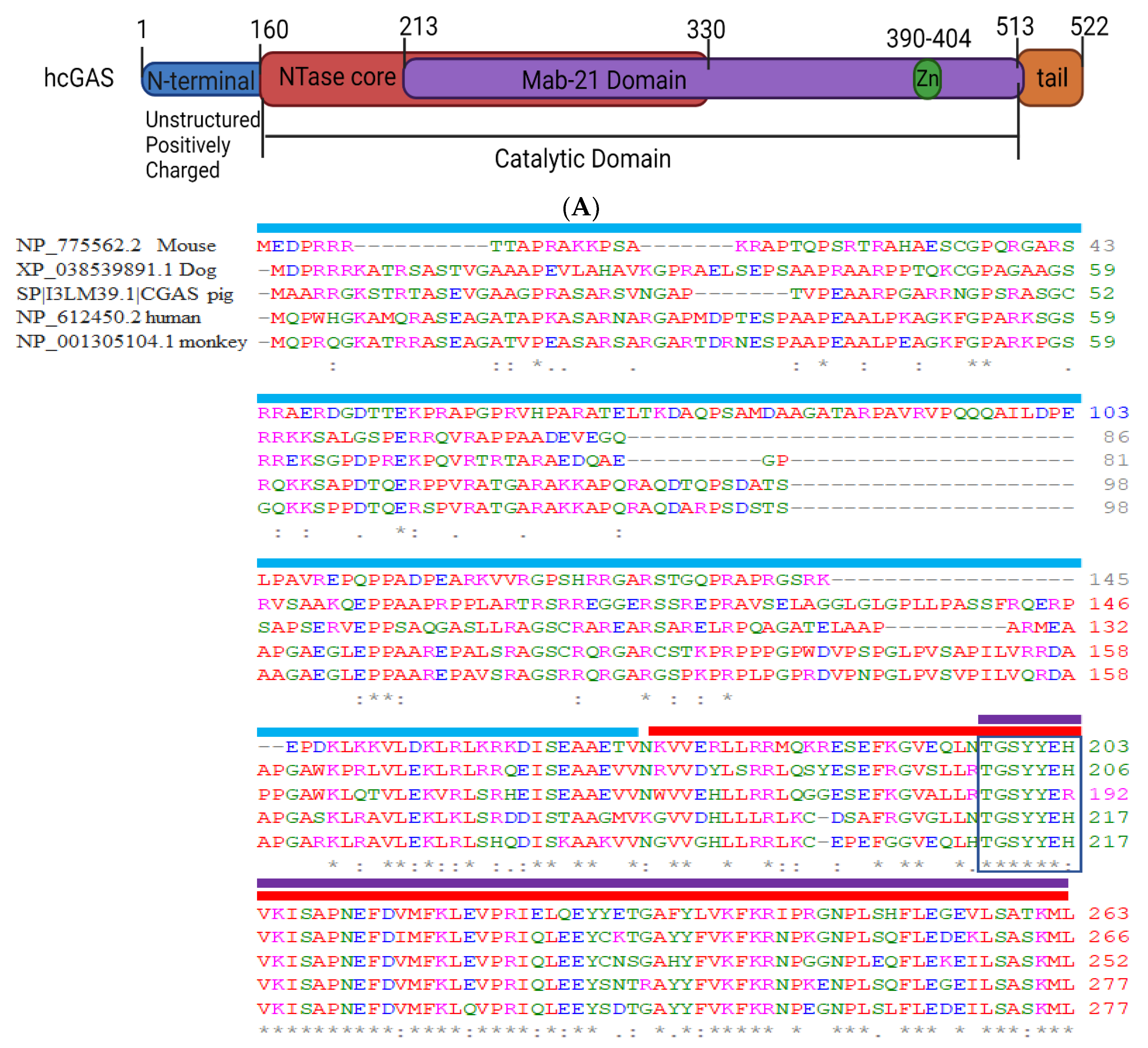
2.1. Structural Domain of cGAS

cGAS is ubiquitous but rapidly diverging [7,8]. We analyzed cGAS protein sequences from five common vertebrates (>NP_612450.2 cGAS-HUMAN, >NP_775562.2 cGAS-MOUSE, >NP_001305104.1 cGAS-MONKEY, >sp|I3LM39.1|CGAS-PIG, >XP_038539891.1 cGAS-DOG) using BlastP (Basic Local Alignment Search Tool, NCBI, USA) and found considerable differences in the sequence homology (Figure 1). The h-cGAS shares < 60% amino acid identity with the mouse (m)-cGAS [37] while 70–80% similarity with other vertebrates. Multiple sequence alignment for the corresponding, truncated amino acid sequences illustrated the highly conserved regions throughout the homologs (Figure 1).
The alignment of the amino acid sequence of cGAS within different mammalian species further suggested that the cGAS N-terminal domain (~160-amino-acids) is highly diverse and disordered. Therefore, the structural and functional characterization of the N-terminal domain of cGAS is highly unpredictable (Figure 1). Recent studies have shown that the serine (13, 37, 64, 129, 143) residue in cGAS-N terminus sense genomic/chromatin DNA but not mitochondrial DNA [38,39]. Unphosphorylated cGAS-N-terminus seem to form multivalent interactions with the nuclear chromatin DNA. However, hyperphosphorylation of serine and threonine residues (Ser13, Ser37, Ser64, Thr69, Thr91, Ser116, Ser129, and Ser143) within N-terminus blocked chromatin DNA sensing in mitotic cells [39]. Evidence indicates that the N-terminus of cGAS in humans has a phosphoinositide-binding domain and interacts selectively with phosphatidyl inositol (4,5) bisphosphate (PIP2) in the plasma membrane, and this interaction restrains cGAS activity from self-DNA [40]. Consistently, cGAS deprived of N-terminus (δN-cGAS) localizes to the cytosolic and nuclear compartments [40]. Thus, the non-enzymatic N-terminal domain can dictate cGAS localization in the cytosol, or on centromeres leading to activation of nuclear cGAS [41]. However, Zhou et al., showed that two unique substitutions K187 and L195 in the N-terminus of hcGAS are responsible for human-specific control of 2′3′ cGAMP synthesis [42].
The C terminal catalytic domain of cGAS consists of two structurally strong-conserved motifs, which include the nucleotidyltransferase (NTase) core domain (160–330) and a Mab21 domain (213–513) [11,13]. The NTase domain harbors the conserved acidic residues, which function as nucleic acid sensors but are indispensable for cGAS enzyme activity [4,43]. The Mab 21 domain of cGAS is the catalytic domain and harbors the conserved Zn finger motif (H390-C405), which has a weaker binding affinity for DNA than NTase but is vital for cGAS enzymatic activity and downstream innate-immune signaling activation [11,13]. Substitutions to the zinc coordination motif (H390A, C396A, C397A, C404A) abolish cGAS activity when exposed to ds DNA [38].
Free cGAS (not bound to dsDNA) does not have a suitably structured catalytic site [9,13,14]. The binding of dsDNA (length > 45 b) between two cGAS monomers creates a structured catalytic site [11,12,44], leading to cGAS activation [13,14]. While amino acids required for cGAS activity are conserved within species, h-cGAS exhibit severely reduced levels of cGAMP synthesis because of enhanced selectivity to DNA, with activity increasing with dsDNA of >45 base pairs [24,32]. h-cGAS, therefore, balances cGAS sensitivity to pathogen-or stress-derived DNA while allowing accurate tolerance of self-DNA. Thus, N and C terminus control cGAS activity upon binding dsDNA.

2.2. cGAS Localization

The subcellular localization of cGAS is still a matter of debate because cGAS localizes in the plasma membrane, cytosol and nucleus [40,41,45,46]. Cytosolic cGAS may be beneficial for triggering genotoxic stimulation with the DNA that needs restraining in the nucleus [40,41]. Plasma membrane-bound cGAS is translocated to the cytosol upon stimulation with exogenous DNA [40]. The association of cGAS with the plasma membrane is mainly due to electrostatic interactions between the positively charged N-terminal domain of cGAS (residues 1 to 160) and the negatively charged PIP2 [40]. The unstructured positively charged N-terminal domain of cGAS can bind the extracellular dsDNA, but there is a lack of enough evidence of activation of cGAS after binding of DNA to the N-terminal domain [39] (Figure 2). However, cGAS lacking N-terminal domain strongly induces interferon responses to genotoxic stress but weaker responses to any bacterial or viral infection [40] (Figure 2). Gentili et al., 2019, have shown that nuclear cGAS is required for constitutive expression of interferon stimulating genes (ISGs), suggesting that a basal level of nucleic acids activates the sensor in the absence of microbial infection or apparent damage [41,47] (Figure 2). Thus, the constitutive induction of beneficial (early) type I IFN response confers antiviral protection to the host cells [48,49,50,51]. Also, cGAS can be activated differentially during mitotic errors and later contributes to birth defects, aging, carcinogenesis, and cancer therapy [52,53]. While mitotic error occurs at a low frequency in normal cells, a much higher frequency occurs in conditions, such as chemotherapeutics which can lead to cGAS activation via forming micronuclei and chromatin bridges [54,55]. Thus, the localization of cGAS at different compartment of the cells mediate specific activity and the ability to respond distinctly between foreign and self-DNA.

2.3. cGAS Droplet Formation

DNA arising in the cytoplasm binds cGAS and drives the formation of cytoplasmic liquid phase condensation called “droplets or foci” [56]. The DNA and RNA can also form liquid droplets. However, only DNA binding will induce a conformational change to activate cGAS [57]. While the molecular mechanism and functional effect of such cGAS-DNA droplet formation are poorly understood, these foci lack a membrane and can fuse to form larger foci, a feature of liquid drop without a membrane [58]. These droplets function as microreactors in which the enzyme (cGAS) and reactants (DNA) are concentrated in the presence of ATP and GTP to enhance the production of a product (cGAMP) (Figure 2). This mechanism allows cGAS to detect the presence of DNA in the cytoplasm above a certain threshold due to the multivalent interaction of cGAS positive charge with negatively charged DNA. However, cGAS-DNA interaction is vulnerable to cytoplasmic salt concentrations, which may prevent spurious activation of cGAS by self-DNA below a certain threshold. Du and Chen [56] showed that Zn+ ions substantially enhance cGAS phase separation with DNA and its enzymatic activity at physiological salt concentrations. Free zinc ions are stored mainly in organelles such as the mitochondria and the endoplasmic reticulum [59], indicating that Zn+ delivery to the cytosol may be critical for regulating cGAS activity in cells. Further characterization of the dynamics and composition of the cGAS condensates should provide deeper insights into the mechanism by which Zn+ levels in mitochondria or ER contribute to regulating cGAS activity while simultaneously avoiding autoimmune reactions to self-tissue. Altogether, cGAS droplet formation explains the cytosolic regulation of cGAS after sensing DNA and reveals a mechanism that enables the cellular balance between tolerance and innate immune activation.

3. Regulation of cGAS Activity

cGAS serves as a node for regulating diverse regulatory functions in response to various cellular processes including autophagy, apoptosis, inflammation, tissue fibrosis, pathogenic infections (bacteria or virus), tumorigenesis, and cancer [19,53,60,61,62,63]. Dysregulation of cGAS results in intrusion of cellular homeostasis during viral or bacterial infections, creating a tumor-prone microenvironment by promoting evasion of immune surveillance. On the other hand, hyperactivated cGAS signals autoimmune disease but may serve as subsidiary cancer therapy. Thus, the tight regulation of cGAS signaling is highly required to maintain cellular homeostasis, preventing thereby autoimmune and pathological angiogenesis.

3.1. Transcriptional Regulation of cGAS

The transcriptional regulation of cGAS activation is not a well-explored area. cGAS promoter contains three IFN-sensitive response elements (ISREs) and one STAT1 binding site that can potentially induce cGAS synthesis by type 1-IFN [57]. As h-cGAS and m-cGAS are <60% homologous, cGAS can be synthesized differentially in different species. Chen et al., characterized the molecular mechanisms controlling the cGAS transcriptional activity [64]. By a series of 5′ deletion and promoter constructions, they found that the region (−414 to +76 relatives to the transcription start site) was sufficient for promoter activity [65]. Mutation of SP1 and CREB binding sites in this promoter region led to an apparent reduction of the cGAS promoter activity [65]. Moreover, elevated expression of microRNA (miR)-93/25 observed in breast cancer facilitates tumor evasion from immune surveillance and destruction partially through downregulating cGAS expression. Thus, miR, SP1 and CREB may control cGAS synthesis and inflammatory signaling.

3.2. Translational Regulations of cGAS Activation

Posttranslational modifications such as phosphorylation, ubiquitination, sumoylation, and acetylation play a pivotal role in cGAS signaling regulation, dimerization, and transcriptional regulation, ultimately regulating a plethora of biological processes such as cell survival and DNA damage (DDR) in response to genomic insult (Figure 3).

3.2.1. Phosphorylation

cGAS contains many serine/threonine residues within its catalytic sites and C-terminus [66]. Phosphorylation of cGAS by serine kinases such as serine/threonine-protein kinase (AKT), cyclin-dependent kinase-1 (CDK1), and Aurora kinase B (AKB) has been shown to repress cGAS activity and thereby immune response [39,67,68]. Seo et al. showed that HSV-1 infection on 293T cells leads to AKT activation which in turn phosphorylated cGAS at S291 (m-cGAS) and S305 (h-cGAS) [67]. cGAS phosphorylation at S291/S301 impaired cGAS generation of cGAMP and thereby the production of type 1 IFN [67]. In another study, HSV-1 infection of human primary fibroblasts and HEK293T cells caused cGAS phosphorylation at multiple serine sites (Ser37, Ser116, Ser201, Ser221, Ser263) which also suppressed cGAS activity during their post-translational modification, however, the signaling behind the phosphorylation is not yet clearly understood [66]. Zhong et al., showed similar suppression of cGAS activity after phosphorylation at S305 residue by CDK1-cyclin B kinase complex in mitotic cells [68]. Li et al., recently demonstrated that hyperphosphorylation of cGAS N-terminus at serine and threonine residues (Ser13, Ser37, Ser64, Thr69, Thr91, Ser116, Ser129, and Ser143) by AKB blocked chromatin DNA sensing in mitotic cells [39]. Thus, it is plausible that AKT, CDK1, and AKB govern cGAS phosphorylation to control cGAS activation during critical phases of the cell cycle like DNA replication (S) and mitotic division (M). However, cGAS phosphorylation by these kinases was reversed either by protein phosphatase 1 (PP1) [68] or PP6 [69] reinstating cGAS DNA-sensing ability.
Interestingly, cGAS also contains several tyrosine residues of unknown functions. A study showed that the phosphorylation of Tyr215 in hcGAS by BLK (B lymphocyte kinase) retained cGAS in the cytoplasm for cytosolic DNA sensing [60]. Intriguingly, we showed that synthetic dsDNA as well DNA isolated from Pseudomonas aeruginous and HSV-1 infection of vascular cells induced cGAS phosphorylation at Y215 and Y242 residues by the tyrosine kinase, cSRC (data not published). We further showed that cGAS phosphorylation at Y215/Y242 promoted cGAS-DNA liquid phase condensation and cGAS activation since transduction of mutated cGAS in endothelial cells inhibited these events (unpublished). Thus, serine and tyrosine phosphorylation of cGAS seems to have an antagonistic role in regulating cGAS activity.

3.2.2. Ubiquitination

Several ubiquitin ligases are shown to regulate cGAS activity. The E3 ligase, TRIM56, monoubiquitinated cGAS at Lys335 which resulted in a marked increase in its dimerization, DNA-binding activity, and cGAMP production [70]. TRIM41 also promoted cGAS monoubiquitination using biochemical assays [71]. Wang et al., reported that ER ubiquitin ligase, RNF185, specifically catalyzed the K27-linked poly-ubiquitination of cGAS at K-384 and promoted its enzymatic activity [72]. The patients suffering from Systemic Lupus Erythematosus (SLE), has constitutive activation of cGAMP-STING signaling [73], and displayed elevated expression of RNF185 mRNA [72]. Whether increased RNF185 mRNA was the factor in increasing cGAS-STING signaling is unclear. It was reported that upon DNA virus infection, USP14 was recruited by TRIM14 to remove K48-linked ubiquitin chains at the Lys414 site in h-cGAS, leading to cGAS stabilization to promote antiviral innate immunity [34]. Similarly, USP27X [74,75] and USP29 [51] have also been reported to stabilize cGAS proteins by cleaving K48-linked poly-ubiquitin chains from cGAS, and both serve as positive regulators in activating innate immunity to fight against DNA viral infection. Thus, deubiquitinase and ubiquitinase have additional control on cGAS activation.

3.2.3. SUMOylation

Like ubiquitination, TRIM38 maintains SUMOylation of m-cGAS at Lys217 and Lys464 residues (corresponding to Lys231 and Lys479 in hcGAS), which prevented K48-linked cGAS polyubiquitination and degradation [75]. On the other hand, SENP7 alleviates SUMO-mediated suppression of cytosolic DNA sensing by removing cGAS SUMOylation [76].

3.2.4. Acetylation

The acetylation of cGAS can regulate cGAS activity either positively or negatively depending upon the acetylation sites. The protein acetylation occurs on lysine residues by the action of acetyl-transferases and deacetylases [77]. Dai et al., found acetylation of hcGAS at Lys384, Lys394, and Lys414 residues at resting state which restrained cGAS activity [78]. However, deacetylation of these sites upon DNA challenge enabled cGAS activation [78]. Thus, this group introduced a promising role of aspirin, which could directly acetylate cGAS to inhibit cGAS activation, as a therapeutic strategy in treating autoimmune diseases, such as Aicardi–Goutieres syndrome (AGS) [66,78,79]. Recently, the N-terminal domain of human cGAS was reported to be acetylated by lysine acetyltransferase, KAT5, at Lys47/Lys56/Lys62/Lys83 residues. These acetylation events increased cGAS binding with DNA and activity in response to DNA challenge [79]. Further functional validation suggests that acetylation at Lys414 suppresses, while acetylation at Lys198 promotes h-cGAS activation. Interestingly, h-cGAS-Lys198 acetylation was found to be decreased by quantitative proteomics upon infection by either HSV-1 or HCMV (human cytomegalovirus), suggesting that these DNA viruses might evade innate-immune surveillance by hijacking acetylation of cGAS at these sites [66].
Thus, phosphorylation, ubiquitination, SUMOylation, and acetylation all control cGAS activity. How these posttranslational modifications of cGAS are regulated at different stages of infection and whether ubiquitination precede acetylation or vice versa may provide an approach to specifically target cGAS activity while preserving host-defense.

4. cGAS Activation of STING

cGAS-induced cGAMP binds STING (stimulator of interferon genes) in the ER (endoplasmic reticulum) membrane to induce inflammatory signaling [46,67] (Figure 4). STING is a transmembrane homodimer that upon being activated phosphorylates itself and translocates to the Golgi apparatus where it binds to and activates TANK-binding kinase 1 (TBK1) and IFN regulatory factor 3 (IRF3) via a phosphorylation-dependent mechanism, leading to the generation of type 1 IFN [34,80].

4.1. Role of Gasdermin D

Gasdermin D (GSDMD) forms ~21 nm diameter oligomeric pores in the plasma membrane which induces cell death (pyroptosis) [81]. Studies showed that caspases (caspase-1 and -4/-5/-11) cleaved human and mouse GSDMD after Asp275 and Asp276 respectively, generating an N-terminal cleavage product (GSDMD-N) [82]. GSMD-N triggered pyroptosis and the release of inflammatory cytokines IL-1β and IL-18 and downstream signaling [83]. Interestingly, Huang et al., showed that GSDMD-N could form pores in the mitochondria to release mitochondrial DNA (mtDNA) that activates the cGAS-STING pathway to suppress cellular proliferation [84]. A recent study also showed that PIP2 content in the membrane retains GSDMD pores predominantly in the open state [85]. Since PIP2 induces cGAS translocation [40] it is possible that membrane PIP2 levels control both GSDMD and cGAS activation and thereby STING signaling (Figure 4). Thus, GSDMD and cGAS may operate in parallel to trigger STING signaling [86].

4.2. Role of Ca2+ Signaling

ER also forms the hub for regulating a rise in intracellular Ca2+ signaling. An increase in cytosolic Ca2+ is required for several primary cellular processes including apoptosis, proliferation, cellular motility, differentiation and exocytosis, and innate immunity [87,88,89]. Interestingly, Ca2+ signaling seems to have a dual role in regulating cGAS-STING signaling since STIM1 (stromal interaction molecule 1), an established sensor of Ca2+ in ER physically interacts with STING via its EF-hand domain and inhibits STING activity by both cGAMP and cGAMP independent STING activation [90]. Similarly, in systemic lupus erythematosus (SLE) hyperactivation of cGAS-STING signaling in T cells also required suppression of Ca2+ homeostasis, but whether STIM1 was involved is not clear [91]. In contrast, the DNA-dependent activator of IFN-regulatory factors (DAI), which functions as a secondary cytoplasmic DNA sensor to cGAS, induces NF-κB and IRF3 in a calcium-dependent manner [18,92]. Likewise, the quenching of cytosolic Ca2+ by BAPTA-AM (a Ca2+ chelator) or CGP37157 (which inhibits the mitochondrial sodium-Ca2+ pump) dampened DAI-induced autoimmune response in macrophages [86].
STIM1 is a single-pass transmembrane protein and binds Ca2+ in the lumen of ER in several cell types such as T cells and endothelial cells. STIM1 organizes as puncta following ER-depletion and binds to plasmalemmal Ca2+ channels such as ORAI1 and TRPC1/TRPC4 at ER-PM junctions to mediate the influx of Ca2+ from the extracellular space into the cell by a process referred to as store-operated Ca2+ entry (SOCE) [93,94,95]. STIM1 inhibits STING in macrophages by anchoring it to the ER, which inhibits its translocation to the Golgi and thus halts STING-mediated IFN-β production. However, we showed that in alveolar macrophages, the most sentinel cells in the tissue, STIM1 is dispensable since endotoxin stimulated Ca2+ influx in a STIM independent but in transient receptor vanilloid potential (TRPV4) channel-dependent manner [96].
Ca2+ is known to activate calcium/calmodulin-dependent protein kinase II (CAMKII), which may alter cGAS activation of STING. CAMKII can bypass cGAS activity by activating STING via inducing the activity of AMP-activated protein kinase (AMPK) [97]. AMPK has been shown to induce STING signaling through repressing ULK1 [98,99]. ULK1 phosphorylates and negatively regulates cGAS-STING signaling [99] (Figure 4). However, ionomycin-induced saturating levels of cytosolic Ca2+ inhibited STING activity indicating that an optimal Ca2+ influx may be required for STING function [100]. PIP2 can suppress cGAS activity through AKT phosphorylation of cGAS [67]. Thus, there is a need to tease out whether Ca2+ influx promotes or dampens cGAS activity.
In mouse embryonic fibroblasts and human Jurkat T cells, loss of STING leads to elevated Ca2+ influx [101]. While in the absence of STING, STIM1 became enriched at ER-PM sites and this likely led to the dysregulated induction of store-operated Ca2+ entry. Thus, it is possible that the STIM1-STING binding under resting conditions reciprocally regulates the pool of STIM1 protein to maintain Ca2+ homeostasis. These findings also suggest that during STING activation, STIM1 may be free to translocate and interact with cGAS or ORAI1, resulting in a transient cytosolic Ca2+ influx. Future work studying this relationship will be important in enhancing our understanding of the interconnecting pathways linking innate immune signaling and Ca2+ homeostasis. However, the activation of cGAS-STING signaling during calcium influx and calcium-related signaling is yet to be explored.

5. cGAS-STING Pathway and Pathologies

cGAS-STING signaling is expressed in various immune cells, including macrophages, dendritic cells, T cells, epithelial cells, endothelial cells, and even some specific fibroblasts (Barber, 2015). cGAS-STING signaling is emerging as a significant regulator of pathogenic infection, development of cancer or autoimmune diseases, or inflammatory disorders. Below we summarize cGAS-STING role in regulating these diseases.

5.1. Acute Lung Injury (ALI) and COVID-19

ALI is a devastating inflammatory condition of the lungs that is characterized by disruption of alveolar-capillary junctions leading to an uncontrolled influx of proteins rich fluid and blood cells into the alveolar space [102,103,104,105]. Neutrophils and tissue-resident macrophages propagate inflammatory responses by releasing various inflammatory cytokines and chemoattractants. However, these cells also seem to have a resolving role in ALI [106]. Upon sensing pathogens macrophages orchestrate the inflammatory signaling by releasing inflammatory cytokines leading to the influx of activated neutrophils [107]. Later these macrophages clean up the debris by a process called efferocytosis to resolve the lung inflammation [108,109]. Thus, based on their activation lung macrophages can acquire inflammatory (M1) or anti-inflammatory (M2) lineage [110,111]. The alveolar macrophages (AM), the major immune cells in the lungs, are the first offender of invading pathogens. Another subset of macrophages in the lungs are interstitial (IM) and monocytes-derived macrophages (MoMacs). While the role of IM remains unclear, MoMacs compensate for the dying AM and serve as an anti-inflammatory and pro-resolving macrophage.
IFN-β is one of the important cytokines in macrophages induced by viruses as well as bacteria [112,113]. IFN-β can be generated either by non-canonical (inducible signaling pathway) or canonical (signaling which is constitutively active) pathway and seem to dictate the outcome of ALI and patient mortality from COVID-19 as well as autoimmune diseases such as Aicardi-Goutieres syndrome (AGS), Systemic lupus erythematosus (SLE), and STING-associated vasculopathy with onset in infancy (SAVI), etc [114,115,116,117,118,119]. The initial induction of type I IFNs limits virus propagation, however, a sustained increase in IFN-β level in the later stage of the infection is associated with aberrant inflammation. Studies showed that type I &type III IFNs significantly precipitated the SARS-CoV-2 replication and pathological responses associated with SARS-CoV-2 and a crucial component of a skin lesion in these patients [120,121,122,123].
We showed that conditional ablation of MoMacs during injury augmented the activation of cGAS-STING signaling by LPS and Pseudomonas aeruginous (PA) [103]. In WT mice LPS and PA resulted in reversible generation IFNβ leading to resolution of ALI. However, ablation of MoMacs during injury increased the production of secondary messenger, cGAMP in the lungs leading to persistent STING signaling and lung injury [103]. While the detailed mechanism remains to be parsed out, these authors showed that MoMacs inhibit cGAS-STING signaling by generating a lipid mediator, sphingosine 1 phosphate. Consistently, the adoptive transfer of MoMacs in mice lacking endogenous MoMacs reduced IFN-β generation and resolved ALI.
In another study, Hong et al. showed that accumulated mtDNA can stimulate cGAS in endothelial cells leading to the expression of type I IFNs and cell death [84]. They showed that LPS activated gasdermin D, which formed mitochondrial pores to enhance the influx of mtDNA into the cytosol of endothelial cells [84]. mtDNA was recognized by the DNA sensor cGAS leading to the generation of the second messenger cGAMP, which suppressed endothelial cell proliferation by downregulating the signaling induced by the transcription factor, YAP1. Studies have also shown that in idiopathic pulmonary fibrosis patients (mt DNA) can be accumulated in type II alveolar epithelial cells due to ER stress and PINK 1 deficiency [124,125,126,127]. In COPD patients increased level of cell-free DNA in the buccal cell was associated with apoptotic or necrotic cell death [127,128]. It is possible therefore that mtDNA accumulation in Type II cells can exacerbate lung damage as these cells generate Type I epithelial cells to repair lung injury.
Using a lung-on-chip model, Domizio et al., showed that upon infection with SARS-CoV-2, macrophages released mitochondrial DNA which activated cGAS–STING signaling in endothelial cells leading to their death and irreversible IFN-β production [121]. Pharmacological inhibition of STING reduced severe lung inflammation induced by SARS-CoV-2 and improved disease outcome. In terms of preventive measures during viral infection, early induction of IFN-β before the high viral load was found to be protective, whereas late induction resulted in increased inflammation and lethal pneumonia [15,129].
Thus, these findings raise the possibility that cGAS-STING signaling in lung cells can exacerbate ALI, and therefore cell therapy (CD11b+-Mɸ) or STING inhibitors are valuable targets for the treatment of ALI pathology in inflammatory conditions

5.2. Adaptive Immunity

Immune system checks on the invading microbes by activating innate or adoptive immunity. As the name suggests innate immunity is antigen and memory-independent, the non-specific first line of defense dependent on pattern recognition receptors (PRRs) (discussed above). In contrast, adaptive immunity is antigen-dependent, specific for the pathogenic antigens, and has immunological memory to kill the pathogens if attacked by the same pathogens. Macrophages and T lymphocytes are the most representative cells of innate immunity and adaptive immunity, respectively. Macrophages, as major professional antigen-presenting cells (APCs), are best known for their ability to prime the host defense by engulfing foreign pathogens [130]. The DNA released through pathogens or endogenous necrotic/apoptotic cells activates the cGAS-STING signal pathway in macrophages, leading to the release of cytokines including IFN-β, which leads to the ani-viral response, to cope with various stresses and maintain homeostasis [131]. Over activation of the cGAS-STING signaling in macrophages can thereby lead to auto-immune diseases [80,132]. For instance, STING-associated vasculopathy in infancy (SAVI), Systemic Lupus Erythematosus (SLE), and Aicardi-Goutières syndrome (AGS) are auto-immune diseases caused by the overproduction of type-I interferon. In SAVI, mutation of STING (R281Q) in patients results in the gain of function of STING causing long-lasting IFN-β production and severe autoinflammatory diseases [133]. AGS is characterized by inherited encephalopathy and leads to severe mental and physical problems, that affect newborn infants. AGS occurs due to a genetic mutation in DNase II, Trex1 (also known as DNase III), or RNaseH2 [134,135,136]. However, in AGS, the mutation in DNAses leads to the amassing of cytosolic DNA resulting in the overactivation of cGAS-STING signaling to generate IFN-β [136,137]. Similarly, in SLE increased IFN-β generation affects various cells and organs such as blood, lung, interstitial, kidney, and skin [138,139,140]. Thus, the cGAS-STING signaling pathway in macrophages and T cell may serve as a connecting link between innate immunity and adaptive immunity [141].

5.3. Cancer

cGAS-STING signaling plays a crucial role in regulating various cancer subtypes. Suppression of STING activation in pancreatic cancer cells enhanced tumor size by reducing the infiltration of immune cells [142,143]. In mouse model of the breast cancer, cGAMP reduced tumor burden [144] and liposomal delivery of cGAMP also activated STING in the macrophages [145]. In syngeneic ovarian cancer (ID8) and colon cancer (CT26), poly (ADPribose) polymerase inhibitors (PARPi) increased IRF3 and STING activity [146]. Grabosch et al. found that cisplatin reduced ovarian cancer growth by increasing type I IFN production and inflammation [147]. Xie et al., 2019 found that tumor-associated mutations, G303E and K432T in h-cGAS [148] induced uterine endometrioid carcinoma by suppressing cGAMP generation [149].
Mechanistically, in the tumor microenvironment, STING is widely expressed in multiple cell types such as dendritic cells, myeloid cells, and endothelial cells [150,151,152] and serves as an important pathway for tumor-associated antigen presentation and regression of the tumor in mouse tumor models [153]. Unlike normal cells, cancer cells possess unstable genomes which undergo chromosome mis segregation during mitosis. The consequence of such segregation is the defective generation of micronuclei and their rupture that releases their genomic contents into the cytosol, resulting in activation of the cGAS-STING pathway [132,154,155,156]. Thus, mtDNA from ruptured micronuclei but not cytosolic DNA stimulates the cGAS-STING pathway in cancer. Oxidative stress in cancer cells is the primary mechanism to increase the porosity of the mitochondrial membrane leading to the export of mtDNA in the cytosol [84,157,158]. Indeed, the tumor cells are deprived of mtDNA and import it through exosomal transfer from host cells to restore mitochondrial function which enhances metastatic potential [23,159]. Thus, activation of cGAS-STING, through mtDNA and/or dsDNA, in cancer cells may serve as a checkpoint to the initial progression of neoplasm by the secretion of Type 1 IFNs inflammatory genes. Alternatively, it leads to the release of various other pro-inflammatory cytokines, chemokines, proteases, and growth factors. Collectively these various factors are called senescence-associated secretory phenotype (SASP) and have played a major role in the restriction of tumorigenesis [160,161,162]. Therefore, the STING activation in cancer cells suppresses its growth and/or recruits immune cells to clear cancer cells [160,163,164].
Antigen-presenting cells (APCs), such as dendritic cells (DCs) and macrophages, are thought to clear necrotic malignant cells [165,166]. DNA from tumor cells is also inserted into the immune cells such as macrophages and dendritic cells, however, the mechanism of DNA insertion inside the immune cells is not known [153]. Some studies suggest that ATP-gated P2X7 receptors are involved in the delivery of DNA to tumor- associate-macrophages (TAMs) to activate STING signaling in immune cells and enhance the tumor-antigen presentation into the CD8 T cells for their proliferation and cytotoxicity activity [153,167,168]. Activation of STING-signaling in TAM is associated with the polarization of TAM into the M1 stage and suppressing its tumor progressing activity. cGAMP generated into the tumor cells is transported into the host immune cells by SLC19A1, a folate transporter [169], which in turn activates STING-IRF3 signaling in immune cells to induce its tumoricidal activity such as in NK-cells [149].
Studies by various group showed that direct injection of STING agonist into the tumor microenvironment also decrease the suppressive Foxp3+ Treg protein in association with CD8+ T cell activation. DCs in the tumor microenvironment mostly respond to the tumor cells generated cGAMP to activate STING signaling. However, other cells may also respond such as fibroblast cells to inhibit tumor growth [167].
Taken together, cGAS-STING activation in the tumor microenvironment is important for anti-tumor activity and its regression. However, uncontrolled activation of the cGAS-STING pathway can promote angiogenesis due to excessive release of type I IFNs, chemokines, and growth factors [170].

5.4. Role of cGAS in Other Diseases

cGAS activation also causes other diseases, including neurological and macular degeneration and liver and renal diseases. Several excellent reviews discussed these topics [171,172,173,174,175,176,177,178]. Therefore, we discussed these diseases briefly in the following section
In the brain, microglia are the principal cells that generate cGAS-STING dependent type-I interferon [172].
However, excessive cGAS activation leads to microglial pyroptosis after viral infection, trauma, stroke, and subarachnoid hemorrhage causing chronic neurodegenerative diseases [172,177]. Moreover, aging is a crucial factor in precipitating neurological disorders via augmenting cGAS-STING signaling and generation of aged diploid fibroblast cells [172,179,180,181]. In Alzheimer’s patients, oxidative stress induced by the accumulation of tau and beta-amyloid protein aggregates in the neurons leads to cell death and activation of cGAS-STING signaling [178,180,182,183].
In the liver, malfunctioning of kupffer cells or monocytes-derived macrophages and hepatocytes is linked with mtDNA release and cGAS activity causes various liver diseases including hepatic ischemia-reperfusion injury, and hepatic cancer [171,173,184,185]. In this context, Luther’s lab showed that connexin-32 expression in hepatic parenchyma is a significant regulator of cGAS-induced type-I interferon synthesis in alcoholic liver diseases [186]. Iron accumulation in hepatic cells and metabolic stress also activated cGAS-STING signaling resulting in an inflammatory liver [187].
Persistent cGAS-STING activation is also a leading cause of kidney injuries such as glomerulonephritis, vasculitis, and chronic kidney injury that can culminate in kidney fibrosis [176,188]. Cell death occurring from post-drug-induced nephrotoxicity, diabetic nephropathy, and ischemia-induced renal injury also activated cGAS-STING activation in the renal tubules [178]. Thus, initial insults in various tissues such as microglia, hepatocytes, and renal tubules can cause neurological, liver, and kidney diseases which can be aggravated further by aging.

6. Clinical Aspect of cGAS

cGAS-STING signaling is an intense area of potential therapeutics. The available cGAS inhibitors suppress signaling either by binding to the cGAS active site or competing with dsDNA. ODN-A151, a small molecule inhibitor of cGAS, competes with dsDNA [189]. Aspirin, an existing non-steroidal anti-inflammatory drug, targets cGAS by acetylating the enzyme at Lys384, Lys394, and Lys494 [5,78]. Other anti-malaria drugs, such as quinine, chloroquine, and hydroxychloroquine, inhibit cGAS activity by binding cGAS at R342 and K372 of the DNA binding site [5,190].
Sorafenib, an FDA-approved antitumor drug for hepatocellular carcinoma, a small molecule inhibitor of multiple kinases, inhibits the cGAS-STING signaling induced by RLR-MAVS pathways [191]. Other inhibitors of cGAS include tetrazolo[1,5-a] pyrimidine-7-ol, pyrazolopyrimidinones, and tricyclic benzo-fluoropyrimidine compounds [192,193]. Suramin is being touted in a clinical trial for inhibiting the cGAS activation in human autoimmune disease [194]. RU.521 inhibited macrophage-cGAS signaling in a mouse model of AGS [195,196], but this compound failed in clinical studies due to its very low efficacy against h-cGAS [196]. Chu et al., identified perillaldehyde (PAH), a natural monoterpenoid derived from Perilla frutescens, as an effective inhibitor of cGAS signaling in the preclinical AGS model [197]. Whether PAH is effective in inhibiting cGAS signaling in AGS patient remain unclear.

7. Conclusions

We discussed the cGAS-STING signaling as a fundamental process regulating cell response to dsDNA. We also assessed the requirements of an unstructured N terminal and highly organized C-terminal of cGAS in regulating its catalytic activity. Further, we learned that the subcellular localization of cGAS specifically dictates the ability to respond distinctly between foreign and self-DNA. We also described the role of cGAS-DNA induced liquid phase condensation to explain the cytosolic regulation of cGAS after sensing DNA, which reveals a mechanism that enables the cellular balance between tolerance and innate immune activation. We also discussed the tight regulation of cGAS signaling to maintain cellular homeostasis and the prevention of autoimmune and pathological angiogenesis. The post-translational regulation of cGAS during any pathogenic insult may either activate or deregulate the cGAS-STING signaling post-infection. cGAS regulation by PIP2 at the membrane is crucial for cGAS sensing of self-DNA. PIP2 is a major regulator of Ca2+ signaling through the ion channels, and the endoplasmic reticulum (ER) stores linked Ca2+ -sensor, STIM1. STIM1-mediated Ca2+ entry is known to be involved in regulating both innate and adaptive immunity. However, studies indicate that STIM1 can regulate STING signaling independent of Ca2+. We also discussed the role of cGAS in various inflammatory disease conditions. The COVID-19 outbreak caused by the newly described SARS-CoV-2-RNA virus caused devastating effects worldwide. Interestingly, the signaling output of SARS-CoV-2 is the transcriptional up-regulation of type-I IFNs and other IRF3 target genes, such as ISGs.

8. Future Directions

cGAS activity regulates various inflammatory diseases and cancer. Because cGAS cannot discriminate between self and non-self-DNA, a question emerges whether cGAS sensing of self-DNA can be used as a biomarker to identify the initial stages of inflammatory and autoimmune disease. COVID-19 infection causes massive death of lung cells such as macrophages and epithelial cells that may induce self-DNA accumulation. Whether assessing DNA concentration in the bronchoalveolar lavage can be used as a measure of viral load and cGAS activity needs to be seen. Thus, advancing research in these directions may lead to early diagnosis and treatment of life-threatening vascular and lung inflammatory diseases.
We described the role of serine versus tyrosine kinases in regulating cGAS activity. These studies raised a fundamental question of how post-translationally modified cGAS contribute to pathological processes. Aurora kinase B and cSrc are involved in various cancers and other human diseases. A question is whether these enzymes contribute to pathological processes through regulating cGAS modifications. Another interesting question is whether protein tyrosine phosphatases such as PP1 and PP6 function as an inducer of tyrosine phosphorylation of cGAS to promote downstream signaling. In this context, studies need to address whether protein tyrosine phosphatase such as PTP and serine-threonine kinases such as PP1 and PP6 plays a dominant role in regulating the outcome of cGAS downstream signaling. The acetylation and ubiquitination regulate cGAS activity, but whether ubiquitination precedes acetylation remains elusive.
Membrane PIP2 regulates cGAS sensing of self-DNA. STIM1-mediated Ca2+ entry regulates both innate and adaptive immunity. However, studies indicate that STIM1 can regulate STING signaling independent of Ca2+. Thus, exploring the mechanism by which PIP, cGAS, and STIM1 are inked, and whether this pathway impacts cGAS-mediated self and foreign DNA are needed.

Author Contributions

B.J., J.C.J. and D.M. conceptualized the manuscript. B.J., J.C.J. and D.M.: edited the manuscript. B.J., J.C.J. and D.M. wrote and reviewed the manuscript. All authors have read and agreed to the published version of the manuscript.

Funding

This review was funded by National Institutes of Health, USA grants P01-HL 151327, HL-137179-01, HL-155941 and HL-084153.

Institutional Review Board Statement

Not applicable.

Informed Consent Statement

Not applicable.

Data Availability Statement

Not applicable.

Acknowledgments

This work was supported by the National Institutes of Health, USA grants P01-HL 151327, HL-137179-01, HL-155941 and HL-084153.

Conflicts of Interest

The authors declare no conflict of interest.

References

  1. Ahn, J.; Barber, G.N. Self-DNA, STING-dependent signaling and the origins of autoinflammatory disease. Curr. Opin. Immunol. 2014, 31, 121–126. [Google Scholar] [CrossRef] [PubMed]
  2. Janeway, C.A., Jr.; Medzhitov, R. Innate immune recognition. Annu. Rev. Immunol. 2002, 20, 197–216. [Google Scholar] [CrossRef] [PubMed]
  3. Kawai, T.; Akira, S. The role of pattern-recognition receptors in innate immunity: Update on Toll-like receptors. Nat. Immunol. 2010, 11, 373–384. [Google Scholar] [CrossRef] [PubMed]
  4. Sun, L.; Wu, J.; Du, F.; Chen, X.; Chen, Z.J. Cyclic GMP-AMP synthase is a cytosolic DNA sensor that activates the type I interferon pathway. Science 2013, 339, 786–791. [Google Scholar] [CrossRef] [PubMed]
  5. Li, Q.; Tian, S.; Liang, J.; Fan, J.; Lai, J.; Chen, Q. Therapeutic Development by Targeting the cGAS-STING Pathway in Autoimmune Disease and Cancer. Front. Pharmacol. 2021, 12, 779425. [Google Scholar] [CrossRef]
  6. Hopfner, K.P.; Hornung, V. Molecular mechanisms and cellular functions of cGAS-STING signalling. Nat. Rev. Mol. Cell Biol. 2020, 21, 501–521. [Google Scholar] [CrossRef]
  7. George, R.D.; McVicker, G.; Diederich, R.; Ng, S.B.; MacKenzie, A.P.; Swanson, W.J.; Shendure, J.; Thomas, J.H. Trans genomic capture and sequencing of primate exomes Rev..eals new targets of positive selection. Genome. Res. 2011, 21, 1686–1694. [Google Scholar] [CrossRef]
  8. Hancks, D.C.; Hartley, M.K.; Hagan, C.; Clark, N.L.; Elde, N.C. Overlapping Patterns of Rapid Evolution in the Nucleic Acid Sensors cGAS and OAS1 Suggest a Common Mechanism of Pathogen Antagonism and Escape. PLoS Genet. 2015, 11, e1005203. [Google Scholar] [CrossRef]
  9. Kranzusch, P.J.; Lee, A.S.; Berger, J.M.; Doudna, J.A. Structure of human cGAS reveals a conserved family of second-messenger enzymes in innate immunity. Cell Rep. 2013, 3, 1362–1368. [Google Scholar] [CrossRef] [PubMed]
  10. Zhang, X.; Wu, J.; Du, F.; Xu, H.; Sun, L.; Chen, Z.; Brautigam, C.A.; Zhang, X.; Chen, Z.J. The cytosolic DNA sensor cGAS forms an oligomeric complex with DNA and undergoes switch-like conformational changes in the activation loop. Cell Rep. 2014, 6, 421–430. [Google Scholar] [CrossRef] [Green Version]
  11. Li, X.; Shu, C.; Yi, G.; Chaton, C.T.; Shelton, C.L.; Diao, J.; Zuo, X.; Kao, C.C.; Herr, A.B.; Li, P. Cyclic GMP-AMP synthase is activated by double-stranded DNA-induced oligomerization. Immunity 2013, 39, 1019–1031. [Google Scholar] [CrossRef] [PubMed]
  12. Andreeva, L.; Hiller, B.; Kostrewa, D.; Lassig, C.; de Oliveira Mann, C.C.; Drexler, D.J.; Maiser, A.; Gaidt, M.; Leonhardt, H.; Hornung, V.; et al. cGAS senses long and HMGB/TFAM-bound U-turn DNA by forming protein-DNA ladders. Nature 2017, 549, 394–398. [Google Scholar] [CrossRef] [PubMed]
  13. Civril, F.; Deimling, T.; de Oliveira Mann, C.C.; Ablasser, A.; Moldt, M.; Witte, G.; Hornung, V.; Hopfner, K.P. Structural mechanism of cytosolic DNA sensing by cGAS. Nature 2013, 498, 332–337. [Google Scholar] [CrossRef] [PubMed]
  14. Gao, D.; Wu, J.; Wu, Y.T.; Du, F.; Aroh, C.; Yan, N.; Sun, L.; Chen, Z.J. Cyclic GMP-AMP synthase is an innate immune sensor of HIV and other retroviruses. Science 2013, 341, 903–906. [Google Scholar] [CrossRef] [PubMed]
  15. Zhou, J.; Chu, H.; Li, C.; Wong, B.H.-Y.; Cheng, Z.-S.; Poon, V.K.-M.; Sun, T.; Lau, C.C.-Y.; Wong, K.K.-Y.; Chan, J.Y.-W.; et al. Active replication of Middle East respiratory syndrome coronavirus and aberrant induction of inflammatory cytokines and chemokines in human macrophages: Implications for pathogenesis. J. Infect. Dis. 2014, 209, 1331–1342. [Google Scholar] [CrossRef]
  16. de Oliveira Mann, C.C.; Hopfner, K.P. Nuclear cGAS: Guard or prisoner? EMBO J. 2021, 40, e108293. [Google Scholar] [CrossRef]
  17. Lahaye, X.; Gentili, M.; Silvin, A.; Conrad, C.; Picard, L.; Jouve, M.; Zueva, E.; Maurin, M.; Nadalin, F.; Knott, G.J.; et al. NONO Detects the Nuclear HIV Capsid to Promote cGAS-Mediated Innate Immune Activation. Cell 2018, 175, 488–501.e422. [Google Scholar] [CrossRef]
  18. Mathavarajah, S.; Salsman, J.; Dellaire, G. An emerging role for calcium signalling in innate and autoimmunity via the cGAS-STING axis. Cytokine Growth Factor Rev. 2019, 50, 43–51. [Google Scholar] [CrossRef]
  19. Ablasser, A.; Schmid-Burgk, J.L.; Hemmerling, I.; Horvath, G.L.; Schmidt, T.; Latz, E.; Hornung, V. Cell intrinsic immunity spreads to bystander cells via the intercellular transfer of cGAMP. Nature 2013, 503, 530–534. [Google Scholar] [CrossRef]
  20. Wu, J.; Sun, L.; Chen, X.; Du, F.; Shi, H.; Chen, C.; Chen, Z.J. Cyclic GMP-AMP is an endogenous second messenger in innate immune signaling by cytosolic DNA. Science 2013, 339, 826–830. [Google Scholar] [CrossRef] [Green Version]
  21. Lund, J.; Sato, A.; Akira, S.; Medzhitov, R.; Iwasaki, A. Toll-like receptor 9-mediated recognition of Herpes simplex virus-2 by plasmacytoid dendritic cells. J. Exp. Med. 2003, 198, 513–520. [Google Scholar] [CrossRef] [PubMed]
  22. Horan, K.A.; Hansen, K.; Jakobsen, M.R.; Holm, C.K.; Søby, S.; Unterholzner, L.; Thompson, M.; West, J.A.; Iversen, M.B.; Rasmussen, S.B.; et al. Proteasomal degradation of herpes simplex virus capsids in macrophages releases DNA to the cytosol for recognition by DNA sensors. J. Immunol. 2013, 190, 2311–2319. [Google Scholar] [CrossRef] [PubMed]
  23. Tan, X.; Sun, L.; Chen, J.; Chen, Z.J. Detection of Microbial Infections Through Innate Immune Sensing of Nucleic Acids. Annu. Rev. Microbiol. 2018, 72, 447–478. [Google Scholar] [CrossRef] [PubMed]
  24. Unterholzner, L.; Keating, S.E.; Baran, M.; Horan, K.A.; Jensen, S.B.; Sharma, S.; Sirois, C.M.; Jin, T.; Latz, E.; Xiao, T.S.; et al. IFI16 is an innate immune sensor for intracellular DNA. Nat. Immunol. 2010, 11, 997–1004. [Google Scholar] [CrossRef]
  25. Kerur, N.; Veettil, M.V.; Sharma-Walia, N.; Bottero, V.; Sadagopan, S.; Otageri, P.; Chandran, B. IFI16 acts as a nuclear pathogen sensor to induce the inflammasome in response to Kaposi Sarcoma-associated herpesvirus infection. Cell Host Microbe. 2011, 9, 363–375. [Google Scholar] [CrossRef]
  26. Zhang, Z.; Yuan, B.; Bao, M.; Lu, N.; Kim, T.; Liu, Y.J. The helicase DDX41 senses intracellular DNA mediated by the adaptor STING in dendritic cells. Nat. Immunol. 2011, 12, 959–965. [Google Scholar] [CrossRef]
  27. Chiu, Y.H.; Macmillan, J.B.; Chen, Z.J. RNA polymerase III detects cytosolic DNA and induces type I interferons through the RIG-I pathway. Cell 2009, 138, 576–591. [Google Scholar] [CrossRef]
  28. Takaoka, A.; Wang, Z.; Choi, M.K.; Yanai, H.; Negishi, H.; Ban, T.; Lu, Y.; Miyagishi, M.; Kodama, T.; Honda, K.; et al. DAI (DLM-1/ZBP1) is a cytosolic DNA sensor and an activator of innate immune response. Nature 2007, 448, 501–505. [Google Scholar] [CrossRef]
  29. Keating, S.E.; Baran, M.; Bowie, A.G. Cytosolic DNA sensors regulating type I interferon induction. Trends Immunol. 2011, 32, 574–581. [Google Scholar] [CrossRef]
  30. Yu, L.; Liu, P. Cytosolic DNA sensing by cGAS: Regulation, function, and human diseases. Signal Transduct Target 2021, 6, 170. [Google Scholar] [CrossRef]
  31. Karayel, E.; Burckstummer, T.; Bilban, M.; Durnberger, G.; Weitzer, S.; Martinez, J.; Superti-Furga, G. The TLR-independent DNA recognition pathway in murine macrophages: Ligand features and molecular sigNature. Eur. J. Immunol. 2009, 39, 1929–1936. [Google Scholar] [CrossRef] [PubMed]
  32. Luecke, S.; Holleufer, A.; Christensen, M.H.; Jonsson, K.L.; Boni, G.A.; Sorensen, L.K.; Johannsen, M.; Jakobsen, M.R.; Hartmann, R.; Paludan, S.R. cGAS is activated by DNA in a length-dependent manner. EMBO Rep. 2017, 18, 1707–1715. [Google Scholar] [CrossRef] [PubMed]
  33. Stetson, D.B.; Medzhitov, R. Recognition of cytosolic DNA activates an IRF3-dependent innate immune response. Immunity 2006, 24, 93–103. [Google Scholar] [CrossRef] [PubMed]
  34. Chen, M.; Meng, Q.; Qin, Y.; Liang, P.; Tan, P.; He, L.; Zhou, Y.; Chen, Y.; Huang, J.; Wang, R.F.; et al. TRIM14 Inhibits cGAS Degradation Mediated by Selective Autophagy Receptor p62 to Promote Innate Immune Responses. Mol. Cell 2016, 64, 105–119. [Google Scholar] [CrossRef]
  35. Ishikawa, H.; Barber, G.N. STING is an endoplasmic reticulum adaptor that facilitates innate immune signalling. Nature 2008, 455, 674–678. [Google Scholar] [CrossRef]
  36. Kranzusch, P.J.; Wilson, S.C.; Lee, A.S.; Berger, J.M.; Doudna, J.A.; Vance, R.E. Ancient Origin of cGAS-STING reveals Mechanism of Universal 2’, 3’ cGAMP Signaling. Mol. Cell 2015, 59, 891–903. [Google Scholar] [CrossRef]
  37. Wang, X.; Zhang, H.; Li, W. DNA-binding mechanisms of human and mouse cGAS: A comparative MD and MM/GBSA study. Phys. Chem. Chem. Phys. 2020, 22, 26390–26401. [Google Scholar] [CrossRef]
  38. Kranzusch, P.J.; Vance, R.E. cGAS dimerization entangles DNA recognition. Immunity 2013, 39, 992–994. [Google Scholar] [CrossRef] [PubMed]
  39. Li, T.; Huang, T.; Du, M.; Chen, X.; Du, F.; Ren, J.; Chen, Z.J. Phosphorylation and chromatin tethering prevent cGAS activation during mitosis. Science 2021, 371, 1204. [Google Scholar] [CrossRef]
  40. Barnett, K.C.; Coronas-Serna, J.M.; Zhou, W.; Ernandes, M.J.; Cao, A.; Kranzusch, P.J.; Kagan, J.C. Phosphoinositide Interactions Position cGAS at the Plasma Membrane to Ensure Efficient Distinction between Self- and Viral DNA. Cell 2019, 176, 1432–1446.e1411. [Google Scholar] [CrossRef] [Green Version]
  41. Gentili, M.; Lahaye, X.; Nadalin, F.; Nader, G.P.F.; Puig Lombardi, E.; Herve, S.; De Silva, N.S.; Rookhuizen, D.C.; Zueva, E.; Goudot, C.; et al. The N-Terminal Domain of cGAS Determines Preferential Association with Centromeric DNA and Innate Immune Activation in the Nucleus. Cell Rep. 2019, 26, 2377–2393.e2313. [Google Scholar] [CrossRef] [PubMed]
  42. Zhou, W.; Whiteley, A.T.; de Oliveira Mann, C.C.; Morehouse, B.R.; Nowak, R.P.; Fischer, E.S.; Gray, N.S.; Mekalanos, J.J.; Kranzusch, P.J. Structure of the Human cGAS-DNA Complex reveals Enhanced Control of Immune Surveillance. Cell 2018, 174, 300–311.e311. [Google Scholar] [CrossRef] [PubMed]
  43. Kranzusch, P.J. cGAS and CD-NTase enzymes: Structure, mechanism, and evolution. Curr. Opin. Struct. Biol. 2019, 59, 178–187. [Google Scholar] [CrossRef]
  44. Hooy, R.M.; Sohn, J. The allosteric activation of cGAS underpins its dynamic signaling landscape. eLife 2018, 7, e39984. [Google Scholar] [CrossRef]
  45. Gekara, N.O.; Jiang, H. The innate immune DNA sensor cGAS: A membrane, cytosolic, or nuclear protein? Sci. Signal. 2019, 12, eaax3521. [Google Scholar] [CrossRef]
  46. Zierhut, C.; Yamaguchi, N.; Paredes, M.; Luo, J.-D.; Carroll, T.; Funabiki, H. The cytoplasmic DNA sensor cGAS promotes mitotic cell death. Cell 2019, 178, 302–315.e323. [Google Scholar] [CrossRef]
  47. Schoggins, J.W.; MacDuff, D.A.; Imanaka, N.; Gainey, M.D.; Shrestha, B.; Eitson, J.L.; Mar, K.B.; Richardson, R.B.; Ratushny, A.V.; Litvak, V. Pan-viral specificity of IFN-induced genes reveals new roles for cGAS in innate immunity. Nature 2014, 505, 691–695. [Google Scholar] [CrossRef]
  48. Mazaleuskaya, L.; Veltrop, R.; Ikpeze, N.; Martin-Garcia, J.; Navas-Martin, S. Protective role of Toll-like receptor 3-induced type I interferon in murine coronavirus infection of macrophages. Viruses 2012, 4, 901–923. [Google Scholar] [CrossRef]
  49. van der Made, C.I.; Hoischen, A.; Netea, M.G.; van de Veerdonk, F.L. Primary immunodeficiencies in cytosolic pattern-recognition receptor pathways: Toward host-directed treatment strategies. Immunol. Ogical. Rev. 2020, 297, 247–272. [Google Scholar] [CrossRef] [PubMed]
  50. Yang, L.; Wang, J.; Hui, P.; Yarovinsky, T.O.; Badeti, S.; Pham, K.; Liu, C. Potential role of IFN-α in COVID-19 patients and its underlying treatment options. Appl. Microbiol. Biotechnol. 2021, 105, 4005–4015. [Google Scholar] [CrossRef]
  51. Karayel, E. Zhang, Q.; Tang, Z.; An, R.; Ye, L.; Zhong, B. USP29 maintains the stability of cGAS and promotes cellular antiviral responses and autoimmunity. Cell Res. 2020, 30, 914–927. [Google Scholar]
  52. Du, J.-M.; Qian, M.-J.; Yuan, T.; Chen, R.-H.; He, Q.-J.; Yang, B.; Ling, Q.; Zhu, H. cGAS and cancer therapy: A double-edged sword. Acta. Pharmacol. Sin. 2022, 43, 2202–2211. [Google Scholar] [CrossRef] [PubMed]
  53. Li, T.; Chen, Z.J. The cGAS-cGAMP-STING pathway connects DNA damage to inflammation, senescence, and cancer. J. Exp. Med. 2018, 215, 1287–1299. [Google Scholar] [CrossRef] [PubMed]
  54. Fenech, M.; Morley, A.A. Measurement of micronuclei in lymphocytes. Mutat. Res. Environ. Mutag. Relat. Subjects. 1985, 147, 29–36. [Google Scholar] [CrossRef]
  55. Nelson, B.C.; Dizdaroglu, M. Implications of DNA damage and DNA repair on human diseases. Mutagenesis 2020, 35, 1–3. [Google Scholar] [CrossRef]
  56. Du, M.; Chen, Z.J. DNA-induced liquid phase condensation of cGAS activates innate immune signaling. Science 2018, 361, 704–709. [Google Scholar] [CrossRef]
  57. Ma, Z.; Jacobs, S.R.; West, J.A.; Stopford, C.; Zhang, Z.; Davis, Z.; Barber, G.N.; Glaunsinger, B.A.; Dittmer, D.P.; Damania, B. Modulation of the cGAS-STING DNA sensing pathway by gammaherpesviruses. Proc. Natl. Acad. Sci. USA 2015, 112, E4306–E4315. [Google Scholar] [CrossRef]
  58. Su, Q.; Mehta, S.; Zhang, J. Liquid-liquid phase separation: Orchestrating cell signaling through time and space. Mol. Cell 2021, 81, 4137–4146. [Google Scholar] [CrossRef]
  59. Lu, Q.; Haragopal, H.; Slepchenko, K.G.; Stork, C.; Li, Y.V. Intracellular zinc distribution in mitochondria, ER and the Golgi apparatus. Int. J. Physiol. Pathophysiol. Pharmacol. 2016, 8, 35. [Google Scholar]
  60. Liu, H.; Zhang, H.; Wu, X.; Ma, D.; Wu, J.; Wang, L.; Jiang, Y.; Fei, Y.; Zhu, C.; Tan, R.; et al. Nuclear cGAS suppresses DNA repair and promotes tumorigenesis. Nature 2018, 563, 131–136. [Google Scholar] [CrossRef]
  61. Wu, J.; Chen, Y.J.; Dobbs, N.; Sakai, T.; Liou, J.; Miner, J.J.; Yan, N. STING-mediated disruption of calcium homeostasis chronically activates ER stress and primes T cell death. J. Exp. Med. 2019, 216, 867–883. [Google Scholar] [CrossRef]
  62. Ablasser, A.; Chen, Z.J. cGAS in action: Expanding roles in immunity and inflammation. Science 2019, 363, eaat8657. [Google Scholar] [CrossRef] [PubMed]
  63. Liang, Q.; Seo, G.J.; Choi, Y.J.; Kwak, M.-J.; Ge, J.; Rodgers, M.A.; Shi, M.; Leslie, B.J.; Hopfner, K.-P.; Ha, T. Crosstalk between the cGAS DNA sensor and Beclin-1 autophagy protein shapes innate antimicrobial immune responses. Cell Host Microbe. 2014, 15, 228–238. [Google Scholar] [CrossRef] [PubMed]
  64. Chen, H.-Y.; Pang, X.-Y.; Xu, Y.-Y.; Zhou, G.-P.; Xu, H.-G. Transcriptional regulation of human cyclic GMP-AMP synthase gene. Cell. Signal. 2019, 62, 109355. [Google Scholar] [CrossRef] [PubMed]
  65. Wu, M.-Z.; Cheng, W.-C.; Chen, S.-F.; Nieh, S.; O’Connor, C.; Liu, C.-L.; Tsai, W.-W.; Wu, C.-J.; Martin, L.; Lin, Y.-S. miR-25/93 mediates hypoxia-induced immunosuppression by repressing cGAS. Nat. Cell Biol. 2017, 19, 1286–1296. [Google Scholar] [CrossRef] [PubMed]
  66. Song, B.; Greco, T.M.; Lum, K.K.; Taber, C.E.; Cristea, I.M. The DNA Sensor cGAS is Decorated by Acetylation and Phosphorylation Modifications in the Context of Immune Signaling. Mol. Cell Proteom. 2020, 19, 1193–1208. [Google Scholar] [CrossRef]
  67. Seo, G.J.; Yang, A.; Tan, B.; Kim, S.; Liang, Q.; Choi, Y.; Yuan, W.; Feng, P.; Park, H.S.; Jung, J.U. Akt Kinase-Mediated Checkpoint of cGAS DNA Sensing Pathway. Cell Rep. 2015, 13, 440–449. [Google Scholar] [CrossRef]
  68. Zhong, L.; Hu, M.M.; Bian, L.J.; Liu, Y.; Chen, Q.; Shu, H.B. Phosphorylation of cGAS by CDK1 impairs self-DNA sensing in mitosis. Cell Discov. 2020, 6, 26. [Google Scholar] [CrossRef]
  69. Li, M.; Shu, H.B. Dephosphorylation of cGAS by PPP6C impairs its substrate binding activity and innate antiviral response. Protein Cell 2020, 11, 584–599. [Google Scholar] [CrossRef]
  70. Seo, G.J.; Kim, C.; Shin, W.J.; Sklan, E.H.; Eoh, H.; Jung, J.U. TRIM56-mediated monoubiquitination of cGAS for cytosolic DNA sensing. Nat. Commun. 2018, 9, 613. [Google Scholar] [CrossRef]
  71. Liu, Z.S.; Zhang, Z.Y.; Cai, H.; Zhao, M.; Mao, J.; Dai, J.; Xia, T.; Zhang, X.M.; Li, T. RINCK-mediated monoubiquitination of cGAS promotes antiviral innate immune responses. Cell Biosci. 2018, 8, 35. [Google Scholar] [CrossRef] [PubMed]
  72. Wang, Q.; Huang, L.; Hong, Z.; Lv, Z.; Mao, Z.; Tang, Y.; Kong, X.; Li, S.; Cui, Y.; Liu, H.; et al. The E3 ubiquitin ligase RNF185 facilitates the cGAS-mediated innate immune response. PLoS Pathog. 2017, 13, e1006264. [Google Scholar] [CrossRef]
  73. Kato, Y.; Park, J.; Takamatsu, H.; Konaka, H.; Aoki, W.; Aburaya, S.; Ueda, M.; Nishide, M.; Koyama, S.; Hayama, Y.; et al. Apoptosis-derived membrane vesicles drive the cGAS-STING pathway and enhance type I IFN production in systemic lupus erythematosus. Ann. Rheum. Dis. 2018, 77, 1507–1515. [Google Scholar] [CrossRef] [PubMed]
  74. Guo, Y.; Jiang, F.; Kong, L.; Li, B.; Yang, Y.; Zhang, L.; Liu, B.; Zheng, Y.; Gao, C. Cutting Edge: USP27X DeubiquitiNat.es and Stabilizes the DNA Sensor cGAS to Regulate Cytosolic DNA-Mediated Signaling. J. Immunol. 2019, 203, 2049–2054. [Google Scholar] [CrossRef] [PubMed]
  75. Hu, M.M.; Yang, Q.; Xie, X.Q.; Liao, C.Y.; Lin, H.; Liu, T.T.; Yin, L.; Shu, H.B. Sumoylation Promotes the Stability of the DNA Sensor cGAS and the Adaptor STING to Regulate the Kinetics of Response to DNA Virus. Immunity 2016, 45, 555–569. [Google Scholar] [CrossRef]
  76. Cui, Y.; Yu, H.; Zheng, X.; Peng, R.; Wang, Q.; Zhou, Y.; Wang, R.; Wang, J.; Qu, B.; Shen, N.; et al. SENP7 Potentiates cGAS Activation by Relieving SUMO-Mediated Inhibition of Cytosolic DNA Sensing. PLoS Pathog. 2017, 13, e1006156. [Google Scholar] [CrossRef]
  77. Narita, T.; Weinert, B.T.; Choudhary, C. Functions and mechanisms of non-histone protein acetylation. Nat. Rev. Mol. Cell Biol. 2019, 20, 156–174. [Google Scholar] [CrossRef]
  78. Dai, J.; Huang, Y.J.; He, X.; Zhao, M.; Wang, X.; Liu, Z.S.; Xue, W.; Cai, H.; Zhan, X.Y.; Huang, S.Y.; et al. Acetylation Blocks cGAS Activity and Inhibits Self-DNA-Induced Autoimmunity. Cell 2019, 176, 1447–1460.e1414. [Google Scholar] [CrossRef]
  79. Song, Z.M.; Lin, H.; Yi, X.M.; Guo, W.; Hu, M.M.; Shu, H.B. KAT5 acetylates cGAS to promote innate immune response to DNA virus. Proc. Natl Acad. Sci. USA 2020, 117, 21568–21575. [Google Scholar] [CrossRef]
  80. Barber, G.N. STING: Infection, inflammation and cancer. Nat. Rev. Immunol. 2015, 15, 760–770. [Google Scholar] [CrossRef]
  81. Place, D.E.; Kanneganti, T.-D. Cell death–mediated cytokine release and its therapeutic implications. J. Exp. Med. 2019, 216, 1474–1486. [Google Scholar] [CrossRef]
  82. Kuang, S.; Zheng, J.; Yang, H.; Li, S.; Duan, S.; Shen, Y.; Ji, C.; Gan, J.; Xu, X.-W.; Li, J. Structure insight of GSDMD reveals the basis of GSDMD autoinhibition in cell pyroptosis. Proc. Natl. Acad. Sci. USA 2017, 114, 10642–10647. [Google Scholar] [CrossRef] [PubMed] [Green Version]
  83. Liu, X.; Zhang, Z.; Ruan, J.; Pan, Y.; Magupalli, V.G.; Wu, H.; Lieberman, J. Inflammasome-activated gasdermin D causes pyroptosis by forming membrane pores. Nature 2016, 535, 153–158. [Google Scholar] [CrossRef]
  84. Huang, L.S.; Hong, Z.; Wu, W.; Xiong, S.; Zhong, M.; Gao, X.; Rehman, J.; Malik, A.B. mtDNA activates cGAS signaling and suppresses the YAP-mediated endothelial cell proliferation program to promote inflammatory injury. Immunity 2020, 52, 475–486.e475. [Google Scholar] [CrossRef]
  85. Díaz-García, E.; García-Tovar, S.; Alfaro, E.; Jaureguizar, A.; Casitas, R.; Sánchez-Sánchez, B.; Zamarrón, E.; Fernández-Lahera, J.; López-Collazo, E.; Cubillos-Zapata, C. Inflammasome activation: A keystone of proinflammatory response in obstructive sleep apnea. Am. J. Respir. Crit. Care Med. 2022, 205, 1337–1348. [Google Scholar] [CrossRef] [PubMed]
  86. Zhang, W.; Zhou, Q.; Xu, W.; Cai, Y.; Yin, Z.; Gao, X.; Xiong, S. DNA-dependent activator of interferon-regulatory factors (DAI) promotes lupus nephritis by activating the calcium pathway. J. Biol. Chem. 2013, 288, 13534–13550. [Google Scholar] [CrossRef]
  87. Berridge, M.J.; Lipp, P.; Bootman, M.D. The versatility and universality of calcium signalling. Nat. Rev. Mol. Cell. Biol. 2000, 1, 11–21. [Google Scholar] [CrossRef] [PubMed]
  88. Wang, L.; Tassiulas, I.; Park-Min, K.H.; Reid, A.C.; Gil-Henn, H.; Schlessinger, J.; Baron, R.; Zhang, J.J.; Ivashkiv, L.B. “Tuning” of type I interferon-induced Jak-STAT1 signaling by calcium-dependent kinases in macrophages. Nat. Immunol. 2008, 9, 186–193. [Google Scholar] [CrossRef] [PubMed]
  89. Tano, J.Y.; Vazquez, G. Requirement for non-regulated, constitutive calcium influx in macrophage survival signaling. Biochem. Biophys. Res. Commun. 2011, 407, 432–437. [Google Scholar] [CrossRef]
  90. Srikanth, S.; Woo, J.S.; Wu, B.; El-Sherbiny, Y.M.; Leung, J.; Chupradit, K.; Rice, L.; Seo, G.J.; Calmettes, G.; Ramakrishna, C. The Ca2+ sensor STIM1 regulates the type I interferon response by retaining the signaling adaptor STING at the endoplasmic reticulum. Nat. Immunol. 2019, 20, 152–162. [Google Scholar] [CrossRef]
  91. Kyttaris, V.C.; Zhang, Z.; Kampagianni, O.; Tsokos, G.C. Calcium signaling in systemic lupus erythematosus T cells: A treatment target. Arthritis Rheumatism. 2011, 63, 2058–2066. [Google Scholar] [CrossRef] [PubMed]
  92. Smith, S.; Jefferies, C. Role of DNA/RNA sensors and contribution to autoimmunity. Cytokine Growth Factor Rev. 2014, 25, 745–757. [Google Scholar] [CrossRef] [PubMed]
  93. Srivastava, N.; Tauseef, M.; Amin, R.; Joshi, B.; Joshi, J.C.; Kini, V.; Klomp, J.; Li, W.; Knezevic, N.; Barbera, N.; et al. Noncanonical function of long myosin light chain kinase in increasing ER-PM junctions and augmentation of SOCE. FASEB J. 2020, 34, 12805–12819. [Google Scholar] [CrossRef] [PubMed]
  94. Yazbeck, P.; Tauseef, M.; Kruse, K.; Amin, M.R.; Sheikh, R.; Feske, S.; Komarova, Y.; Mehta, D. STIM1 Phosphorylation at Y361 Recruits Orai1 to STIM1 Puncta and Induces Ca(2+) Entry. Sci. Rep. 2017, 7, 42758. [Google Scholar] [CrossRef]
  95. Soni, D.; Regmi, S.C.; Wang, D.-M.; DebRoy, A.; Zhao, Y.-Y.; Vogel, S.M.; Malik, A.B.; Tiruppathi, C. Pyk2 phosphorylation of VE-PTP downstream of STIM1-induced Ca2+ entry regulates disassembly of adherens junctions. Am. J. Physiol. -Lung Cell. Mol. Physiol. 2017, 312, L1003–L1017. [Google Scholar] [CrossRef]
  96. Rayees, S.; Joshi, J.C.; Tauseef, M.; Anwar, M.; Baweja, S.; Rochford, I.; Joshi, B.; Hollenberg, M.D.; Reddy, S.P.; Mehta, D. PAR2-Mediated cAMP Generation Suppresses TRPV4-Dependent Ca(2+) Signaling in Alveolar Macrophages to Resolve TLR4-Induced Inflammation. Cell Rep. 2019, 27, 793–805.e794. [Google Scholar] [CrossRef]
  97. Hurley, R.L.; Anderson, K.A.; Franzone, J.M.; Kemp, B.E.; Means, A.R.; Witters, L.A. The Ca2+/calmodulin-dependent protein kinase kinases are AMP-activated protein kinase kinases. J. Biol. Chem. 2005, 280, 29060–29066. [Google Scholar] [CrossRef]
  98. Prantner, D.; Darville, T.; Nagarajan, U.M. Stimulator of IFN gene is critical for induction of IFN-beta during Chlamydia muridarum infection. J. Immunol. 2010, 184, 2551–2560. [Google Scholar] [CrossRef]
  99. Konno, H.; Konno, K.; Barber, G.N. Cyclic dinucleotides trigger ULK1 (ATG1) phosphorylation of STING to prevent sustained innate immune signaling. Cell 2013, 155, 688–698. [Google Scholar] [CrossRef]
  100. Kwon, D.; Sesaki, H.; Kang, S.J. Intracellular calcium is a rheostat for the STING signaling pathway. Biochem. Biophys. Res. Commun. 2018, 500, 497–503. [Google Scholar] [CrossRef]
  101. Son, A.; de Jesus, A.A.; Schwartz, D.M. STIM1 holds a STING in its (N-terminal) tail. Cell Calcium 2019, 80, 192–193. [Google Scholar] [CrossRef]
  102. Matthay, M.A.; Zemans, R.L. The acute respiratory distress syndrome: Pathogenesis and treatment. Annu. Rev. Pathol. 2011, 6, 147–163. [Google Scholar] [CrossRef] [Green Version]
  103. Joshi, J.C.; Joshi, B.; Rochford, I.; Rayees, S.; Akhter, M.Z.; Baweja, S.; Chava, K.R.; Tauseef, M.; Abdelkarim, H.; Natarajan, V.; et al. SPHK2-Generated S1P in CD11b(+) Macrophages Blocks STING to Suppress the Inflammatory Function of Alveolar Macrophages. Cell Rep. 2020, 30, 4096–4109.e4095. [Google Scholar] [CrossRef] [PubMed]
  104. Bellani, G.; Laffey, J.G.; Pham, T.; Fan, E.; Brochard, L.; Esteban, A.; Gattinoni, L.; van Haren, F.; Larsson, A.; McAuley, D.F.; et al. Epidemiology, Patterns of Care, and Mortality for Patients with Acute Respiratory Distress Syndrome in Intensive Care Units in 50 Countries. JAMA 2016, 315, 788–800. [Google Scholar] [CrossRef] [PubMed]
  105. Rochford, I.; Joshi, J.C.; Rayees, S.; Anwar, M.; Akhter, M.Z.; Yalagala, L.; Banerjee, S.; Mehta, D. Evidence for reprogramming of monocytes into reparative alveolar macrophages in vivo by targeting PDE4b. Am. J. Physiol. Lung Cell Mol. Physiol. 2021, 321, L686–L702. [Google Scholar] [CrossRef] [PubMed]
  106. Bachmaier, K.; Stuart, A.; Singh, A.; Mukhopadhyay, A.; Chakraborty, S.; Hong, Z.; Wang, L.; Tsukasaki, Y.; Maienschein-Cline, M.; Ganesh, B.B.; et al. Albumin Nanoparticle Endocytosing Subset of Neutrophils for Precision Therapeutic Targeting of Inflammatory Tissue Injury. ACS Nano 2022, 16, 4084–4101. [Google Scholar] [CrossRef]
  107. Leseigneur, C.; Le-Bury, P.; Pizarro-Cerda, J.; Dussurget, O. Emerging Evasion Mechanisms of Macrophage Defenses by Pathogenic Bacteria. Front. Cell Infect. Microbiol. 2020, 10, 577559. [Google Scholar] [CrossRef]
  108. Korns, D.; Frasch, S.C.; Fernandez-Boyanapalli, R.; Henson, P.M.; Bratton, D.L. Modulation of macrophage efferocytosis in inflammation. Front. Immunol. 2011, 2, 57. [Google Scholar] [CrossRef]
  109. Elliott, M.R.; Koster, K.M.; Murphy, P.S. Efferocytosis Signaling in the Regulation of Macrophage Inflammatory Responses. J. Immunol. 2017, 198, 1387–1394. [Google Scholar] [CrossRef]
  110. Funes, S.C.; Rios, M.; Escobar-Vera, J.; Kalergis, A.M. Implications of macrophage polarization in autoimmunity. Immunol. Ogy. 2018, 154, 186–195. [Google Scholar] [CrossRef]
  111. Mosser, D.M.; Edwards, J.P. Exploring the full spectrum of macrophage activation. Nat. Rev. Immunol. 2008, 8, 958–969. [Google Scholar] [CrossRef]
  112. Galbraith, M.D.; Kinning, K.T.; Sullivan, K.D.; Araya, P.; Smith, K.P.; Granrath, R.E.; Shaw, J.R.; Baxter, R.; Jordan, K.R.; Russell, S.; et al. Specialized interferon action in COVID-19. Proc. Natl. Acad Sci. USA 2022, 119, e2116730119. [Google Scholar] [CrossRef] [PubMed]
  113. Decout, A.; Katz, J.D.; Venkatraman, S.; Ablasser, A. The cGAS-STING pathway as a therapeutic target in inflammatory diseases. Nat. Rev. Immunol. 2021, 21, 548–569. [Google Scholar] [CrossRef] [PubMed]
  114. Lucas, C.; Wong, P.; Klein, J.; Castro, T.B.R.; Silva, J.; Sundaram, M.; Ellingson, M.K.; Mao, T.; Oh, J.E.; Israelow, B.; et al. Longitudinal analyses reveal immunological misfiring in severe COVID-19. Nature 2020, 584, 463–469. [Google Scholar] [CrossRef] [PubMed]
  115. Nienhold, R.; Ciani, Y.; Koelzer, V.H.; Tzankov, A.; Haslbauer, J.D.; Menter, T.; Schwab, N.; Henkel, M.; Frank, A.; Zsikla, V.; et al. Two distinct immunopathological profiles in autopsy lungs of COVID-19. Nat. Commun. 2020, 11, 5086. [Google Scholar] [CrossRef]
  116. Park, A.; Iwasaki, A. Type I and Type III Interferons—Induction, Signaling, Evasion, and Application to Combat COVID-19. Cell Host Microbe. 2020, 27, 870–878. [Google Scholar] [CrossRef]
  117. Hadjadj, J.; Yatim, N.; Barnabei, L.; Corneau, A.; Boussier, J.; Smith, N.; Pere, H.; Charbit, B.; Bondet, V.; Chenevier-Gobeaux, C.; et al. Impaired type I interferon activity and inflammatory responses in severe COVID-19 patients. Science 2020, 369, 718–724. [Google Scholar] [CrossRef]
  118. Schulte-Schrepping, J.; Reusch, N.; Paclik, D.; Bassler, K.; Schlickeiser, S.; Zhang, B.; Kramer, B.; Krammer, T.; Brumhard, S.; Bonaguro, L.; et al. Severe COVID-19 Is Marked by a Dysregulated Myeloid Cell Compartment. Cell 2020, 182, 1419–1440.e1423. [Google Scholar] [CrossRef]
  119. Rice, G.I.; Forte, G.M.; Szynkiewicz, M.; Chase, D.S.; Aeby, A.; Abdel-Hamid, M.S.; Ackroyd, S.; Allcock, R.; Bailey, K.M.; Balottin, U.; et al. Assessment of interferon-related biomarkers in Aicardi-Goutieres syndrome associated with mutations in TREX1, RNASEH2A, RNASEH2B, RNASEH2C, SAMHD1, and ADAR: A case-control study. Lancet. Neurol. 2013, 12, 1159–1169. [Google Scholar] [CrossRef]
  120. Israelow, B.; Song, E.; Mao, T.; Lu, P.; Meir, A.; Liu, F.; Alfajaro, M.M.; Wei, J.; Dong, H.; Homer, R.J. Mouse model of SARS-CoV-2 Reveals inflammatory role of type I interferon signaling. J. Exp. Med. 2020, 217, e20201241. [Google Scholar] [CrossRef]
  121. Domizio, J.D.; Gulen, M.F.; Saidoune, F.; Thacker, V.V.; Yatim, A.; Sharma, K.; Nass, T.; Guenova, E.; Schaller, M.; Conrad, C. The cGAS–STING pathway drives type I IFN immunopathology in COVID-19. Nature 2022, 603, 145–151. [Google Scholar] [CrossRef] [PubMed]
  122. Heuberger, J.; Trimpert, J.; Vladimirova, D.; Goosmann, C.; Lin, M.; Schmuck, R.; Mollenkopf, H.J.; Brinkmann, V.; Tacke, F.; Osterrieder, N. Epithelial response to IFN-γ promotes SARS-CoV-2 infection. EMBO Mol. Med. 2021, 13, e13191. [Google Scholar] [CrossRef] [PubMed]
  123. Akamatsu, H.; Toi, Y.; Hayashi, H.; Fujimoto, D.; Tachihara, M.; Furuya, N.; Otani, S.; Shimizu, J.; Katakami, N.; Azuma, K. Efficacy of Osimertinib Plus Bevacizumab vs Osimertinib in Patients With EGFR T790M–Mutated Non–Small Cell Lung Cancer PRev..iously Treated With Epidermal Growth Factor Receptor–Tyrosine Kinase Inhibitor: West Japan Oncology Group 8715L Phase 2 Randomized Clinical Trial. JAMA Oncol. 2021, 7, 386–394. [Google Scholar] [PubMed]
  124. Bueno, M.; Zank, D.; Buendia-Roldan, I.; Fiedler, K.; Mays, B.G.; Alvarez, D.; Sembrat, J.; Kimball, B.; Bullock, J.K.; Martin, J.L.; et al. PINK1 attenuates mtDNA release in alveolar epithelial cells and TLR9 mediated profibrotic responses. PLoS ONE 2019, 14, e0218003. [Google Scholar] [CrossRef]
  125. Schuliga, M.; Pechkovsky, D.V.; Read, J.; Waters, D.W.; Blokland, K.E.C.; Reid, A.T.; Hogaboam, C.M.; Khalil, N.; Burgess, J.K.; Prele, C.M.; et al. Mitochondrial dysfunction contributes to the senescent phenotype of IPF lung fibroblasts. J. Cell. Mol. Med. 2018, 22, 5847–5861. [Google Scholar] [CrossRef]
  126. Schuliga, M.; Read, J.; Blokland, K.E.C.; Waters, D.W.; Burgess, J.; Prele, C.; Mutsaers, S.E.; Jaffar, J.; Westall, G.; Reid, A.; et al. Self DNA perpetuates IPF lung fibroblast senescence in a cGAS-dependent manner. Clin. Sci. (Lond.) 2020, 134, 889–905. [Google Scholar] [CrossRef]
  127. da Silva, A.L.; Bresciani, M.J.; Karnopp, T.E.; Weber, A.F.; Ellwanger, J.H.; Henriques, J.A.; Valim, A.R.; Possuelo, L.G. DNA damage and cellular abnormalities in tuberculosis, lung cancer and chronic obstructive pulmonary disease. Multidiscip. Respir. Med. 2015, 10, 38. [Google Scholar] [CrossRef]
  128. Maluf, S.W.; Mergener, M.; Dalcanale, L.; Costa, C.C.; Pollo, T.; Kayser, M.; da Silva, L.B.; Pra, D.; Teixeira, P.J.Z. DNA damage in peripheral blood of patients with chronic obstructive pulmonary disease (COPD). Mutat. Res. 2007, 626, 180–184. [Google Scholar] [CrossRef]
  129. Channappanavar, R.; Fehr, A.R.; Zheng, J.; Wohlford-Lenane, C.; Abrahante, J.E.; Mack, M.; Sompallae, R.; McCray, P.B.; Meyerholz, D.K.; Perlman, S. IFN-I response timing relative to virus replication determines MERS coronavirus infection outcomes. J. Clin. Investig. 2019, 129, 3625–3639. [Google Scholar] [CrossRef]
  130. Guerriero, J.L. Macrophages: Their Untold Story in T Cell Activation and Function. Int. Rev. Cell Mol. Biol. 2019, 342, 73–93. [Google Scholar]
  131. Bergsmedh, A.; Szeles, A.; Henriksson, M.; Bratt, A.; Folkman, M.J.; Spetz, A.L.; Holmgren, L. Horizontal transfer of oncogenes by uptake of apoptotic bodies. Proc. Natl. Acad. Sci. USA 2001, 98, 6407–6411. [Google Scholar] [CrossRef] [PubMed]
  132. Kwon, J.; Bakhoum, S.F. The Cytosolic DNA-Sensing cGAS-STING Pathway in Cancer. Cancer Discov. 2020, 10, 26–39. [Google Scholar] [CrossRef]
  133. Wang, Y.; Wang, F.; Zhang, X. STING-associated vasculopathy with onset in infancy: A familial case series report and literature Review. Ann. Transl. Med. 2021, 9, 176. [Google Scholar] [CrossRef] [PubMed]
  134. Cuadrado, E.; Michailidou, I.; van Bodegraven, E.J.; Jansen, M.H.; Sluijs, J.A.; Geerts, D.; Couraud, P.O.; De Filippis, L.; Vescovi, A.L.; Kuijpers, T.W.; et al. Phenotypic variation in Aicardi-Goutieres syndrome explained by cell-specific IFN-stimulated gene response and cytokine release. J. Immunol. 2015, 194, 3623–3633. [Google Scholar] [CrossRef] [PubMed]
  135. Crow, Y.J.; Hayward, B.E.; Parmar, R.; Robins, P.; Leitch, A.; Ali, M.; Black, D.N.; van Bokhoven, H.; Brunner, H.G.; Hamel, B.C.; et al. Mutations in the gene encoding the 3’-5’ DNA exonuclease TREX1 cause Aicardi-Goutieres syndrome at the AGS1 locus. Nat. Genet. 2006, 38, 917–920. [Google Scholar] [CrossRef]
  136. Yan, N. Immune Diseases Associated with TREX1 and STING Dysfunction. J. Interferon Cytokine Res. 2017, 37, 198–206. [Google Scholar] [CrossRef]
  137. Stetson, D.B.; Ko, J.S.; Heidmann, T.; Medzhitov, R. Trex1 prevents cell-intrinsic initiation of autoimmunity. Cell 2008, 134, 587–598. [Google Scholar] [CrossRef]
  138. Lundgren, M.C.; Molitor, J.A.; Spilseth, B.; Adeyi, O. A Fatal Case of Diffuse Alveolar Hemorrhage in the Setting of Systemic Lupus Erythematosus: A Case Report and Review of Noninfectious Causes of Acute Pulmonary Hemorrhage in Adults. Case Rep. Rheumatol. 2021, 2021, 6620701. [Google Scholar] [CrossRef]
  139. Hepburn, A.L.; Narat, S.; Mason, J.C. The management of peripheral blood cytopenias in systemic lupus erythematosus. Rheumatology 2010, 49, 2243–2254. [Google Scholar] [CrossRef]
  140. Knight, J.S.; Subramanian, V.; O’Dell, A.A.; Yalavarthi, S.; Zhao, W.; Smith, C.K.; Hodgin, J.B.; Thompson, P.R.; Kaplan, M.J. Peptidylarginine deiminase inhibition disrupts NET formation and protects against kidney, skin and vascular disease in lupus-prone MRL/lpr mice. Ann. Rheum. Dis. 2015, 74, 2199–2206. [Google Scholar] [CrossRef]
  141. Zheng, J.; Mo, J.; Zhu, T.; Zhuo, W.; Yi, Y.; Hu, S.; Yin, J.; Zhang, W.; Zhou, H.; Liu, Z. Comprehensive elaboration of the cGAS-STING signaling axis in cancer development and immunotherapy. Mol. Cancer 2020, 19, 133. [Google Scholar] [CrossRef] [PubMed]
  142. Takashima, K.; Takeda, Y.; Oshiumi, H.; Shime, H.; Okabe, M.; Ikawa, M.; Matsumoto, M.; Seya, T. STING in tumor and host cells cooperatively work for NK cell-mediated tumor growth retardation. Biochem. Biophys. Res. Commun. 2016, 478, 1764–1771. [Google Scholar] [CrossRef] [PubMed]
  143. Ho, S.S.W.; Zhang, W.Y.L.; Tan, N.Y.J.; Khatoo, M.; Suter, M.A.; Tripathi, S.; Cheung, F.S.G.; Lim, W.K.; Tan, P.H.; Ngeow, J.; et al. The DNA Structure-Specific Endonuclease MUS81 Mediates DNA Sensor STING-Dependent Host Rejection of Prostate Cancer Cells. Immunity 2016, 44, 1177–1189. [Google Scholar] [CrossRef] [Green Version]
  144. Chen, Q.; Boire, A.; Jin, X.; Valiente, M.; Er, E.E.; Lopez-Soto, A.; Jacob, L.S.; Patwa, R.; Shah, H.; Xu, K.; et al. Carcinoma-astrocyte gap junctions promote brain metastasis by cGAMP transfer. Nature 2016, 533, 493–498. [Google Scholar] [CrossRef] [PubMed]
  145. Petrovic, M.; Borchard, G.; Jordan, O. Considerations for the delivery of STING ligands in cancer immunotherapy. J. Control. Release 2021, 339, 235–247. [Google Scholar] [CrossRef] [PubMed]
  146. Shen, J.; Zhao, W.; Ju, Z.; Wang, L.; Peng, Y.; Labrie, M.; Yap, T.A.; Mills, G.B.; Peng, G. PARPi Triggers the STING-Dependent Immune Response and Enhances the Therapeutic Efficacy of Immune Checkpoint Blockade Independent of BRCAness. Cancer Res. 2019, 79, 311–319. [Google Scholar] [CrossRef]
  147. Grabosch, S.; Bulatovic, M.; Zeng, F.; Ma, T.; Zhang, L.; Ross, M.; Brozick, J.; Fang, Y.; Tseng, G.; Kim, E.; et al. Cisplatin-induced immune modulation in ovarian cancer mouse models with distinct inflammation profiles. Oncogene 2019, 38, 2380–2393. [Google Scholar] [CrossRef]
  148. Xie, W.; Lama, L.; Adura, C.; Tomita, D.; Glickman, J.F.; Tuschl, T.; Patel, D.J. Human cGAS catalytic domain has an additional DNA-binding interface that enhances enzymatic activity and liquid-phase condensation. Proc. Natl. Acad. Sci. USA 2019, 116, 11946–11955. [Google Scholar] [CrossRef]
  149. Marcus, A.; Mao, A.J.; Lensink-Vasan, M.; Wang, L.; Vance, R.E.; Raulet, D.H. Tumor-Derived cGAMP Triggers a STING-Mediated Interferon Response in Non-tumor Cells to Activate the NK Cell Response. Immunity 2018, 49, 754–763.e754. [Google Scholar] [CrossRef]
  150. Ohkuri, T.; Ghosh, A.; Kosaka, A.; Zhu, J.; Ikeura, M.; David, M.; Watkins, S.C.; Sarkar, S.N.; Okada, H. STING contributes to antiglioma immunity via triggering type I IFN signals in the tumor microenvironment. Cancer Immunol. Res. 2014, 2, 1199–1208. [Google Scholar] [CrossRef]
  151. Klarquist, J.; Hennies, C.M.; Lehn, M.A.; Reboulet, R.A.; Feau, S.; Janssen, E.M. STING-mediated DNA sensing promotes antitumor and autoimmune responses to dying cells. J. Immunol. 2014, 193, 6124–6134. [Google Scholar] [CrossRef] [PubMed]
  152. Demaria, O.; De Gassart, A.; Coso, S.; Gestermann, N.; Di Domizio, J.; Flatz, L.; Gaide, O.; Michielin, O.; Hwu, P.; Petrova, T.V.; et al. STING activation of tumor endothelial cells initiates spontaneous and therapeutic antitumor immunity. Proc. Natl. Acad. Sci. USA 2015, 112, 15408–15413. [Google Scholar] [CrossRef] [PubMed]
  153. Woo, S.R.; Fuertes, M.B.; Corrales, L.; Spranger, S.; Furdyna, M.J.; Leung, M.Y.; Duggan, R.; Wang, Y.; Barber, G.N.; Fitzgerald, K.A.; et al. STING-dependent cytosolic DNA sensing mediates innate immune recognition of immunogenic tumors. Immunity 2014, 41, 830–842. [Google Scholar] [CrossRef] [PubMed] [Green Version]
  154. Crasta, K.; Ganem, N.J.; Dagher, R.; Lantermann, A.B.; Ivanova, E.V.; Pan, Y.; Nezi, L.; Protopopov, A.; Chowdhury, D.; Pellman, D. DNA breaks and chromosome pulverization from errors in mitosis. Nature 2012, 482, 53–58. [Google Scholar] [CrossRef]
  155. Harding, S.M.; Benci, J.L.; Irianto, J.; Discher, D.E.; Minn, A.J.; Greenberg, R.A. Mitotic progression following DNA damage enables pattern recognition within micronuclei. Nature 2017, 548, 466–470. [Google Scholar] [CrossRef]
  156. Mackenzie, K.J.; Carroll, P.; Martin, C.-A.; Murina, O.; Fluteau, A.; Simpson, D.J.; Olova, N.; Sutcliffe, H.; Rainger, J.K.; Leitch, A.; et al. cGAS surveillance of micronuclei links genome instability to innate immunity. Nature 2017, 548, 461–465. [Google Scholar] [CrossRef]
  157. Kitajima, S.; Ivanova, E.; Guo, S.; Yoshida, R.; Campisi, M.; Sundararaman, S.K.; Tange, S.; Mitsuishi, Y.; Thai, T.C.; Masuda, S.; et al. Suppression of STING Associated with LKB1 Loss in KRAS-Driven Lung Cancer. Cancer Discov. 2019, 9, 34–45. [Google Scholar] [CrossRef]
  158. Platnich, J.M.; Chung, H.; Lau, A.; Sandall, C.F.; Bondzi-Simpson, A.; Chen, H.M.; Komada, T.; Trotman-Grant, A.C.; Brandelli, J.R.; Chun, J.; et al. Shiga Toxin/Lipopolysaccharide Activates Caspase-4 and Gasdermin D to Trigger Mitochondrial Reactive Oxygen Species Upstream of the NLRP3 Inflammasome. Cell Rep. 2018, 25, 1525–1536.e1527. [Google Scholar] [CrossRef]
  159. Sansone, P.; Savini, C.; Kurelac, I.; Chang, Q.; Amato, L.B.; Strillacci, A.; Stepanova, A.; Iommarini, L.; Mastroleo, C.; Daly, L.; et al. Packaging and transfer of mitochondrial DNA via exosomes regulate escape from dormancy in hormonal therapy-resistant breast cancer. Proc. Natl Acad. Sci. USA 2017, 114, E9066–E9075. [Google Scholar] [CrossRef]
  160. Dou, Z.; Ghosh, K.; Vizioli, M.G.; Zhu, J.; Sen, P.; Wangensteen, K.J.; Simithy, J.; Lan, Y.; Lin, Y.; Zhou, Z.; et al. Cytoplasmic chromatin triggers inflammation in senescence and cancer. Nature 2017, 550, 402–406. [Google Scholar] [CrossRef]
  161. Yang, H.; Wang, H.; Ren, J.; Chen, Q.; Chen, Z.J. cGAS is essential for cellular senescence. Proc. Natl. Acad. Sci. USA 2017, 114, E4612–E4620. [Google Scholar] [CrossRef] [PubMed]
  162. Coppe, J.P.; Patil, C.K.; Rodier, F.; Sun, Y.; Munoz, D.P.; Goldstein, J.; Nelson, P.S.; Desprez, P.Y.; Campisi, J. Senescence-associated secretory phenotypes reveal cell-nonautonomous functions of oncogenic RAS and the p53 tumor suppressor. PLoS Biol. 2008, 6, 2853–2868. [Google Scholar] [CrossRef] [PubMed]
  163. Gluck, S.; Guey, B.; Gulen, M.F.; Wolter, K.; Kang, T.W.; Schmacke, N.A.; Bridgeman, A.; Rehwinkel, J.; Zender, L.; Ablasser, A. Innate immune sensing of cytosolic chromatin fragments through cGAS promotes senescence. Nat. Cell Biol. 2017, 19, 1061–1070. [Google Scholar] [CrossRef] [PubMed]
  164. Ranoa, D.R.E.; Widau, R.C.; Mallon, S.; Parekh, A.D.; Nicolae, C.M.; Huang, X.; Bolt, M.J.; Arina, A.; Parry, R.; Kron, S.J.; et al. STING Promotes Homeostasis via Regulation of Cell Proliferation and Chromosomal Stability. Cancer Res. 2019, 79, 1465–1479. [Google Scholar] [CrossRef]
  165. Sauter, B.; Albert, M.L.; Francisco, L.; Larsson, M.; Somersan, S.; Bhardwaj, N. Consequences of cell death: Exposure to necrotic tumor cells, but not primary tissue cells or apoptotic cells, induces the maturation of immunostimulatory dendritic cells. J. Exp. Med. 2000, 191, 423–434. [Google Scholar] [CrossRef]
  166. Smyth, M.J.; Godfrey, D.I.; Trapani, J.A. A fresh look at tumor immunosurveillance and immunotherapy. Nat. Immunol. 2001, 2, 293–299. [Google Scholar] [CrossRef]
  167. Deng, L.; Liang, H.; Xu, M.; Yang, X.; Burnette, B.; Arina, A.; Li, X.D.; Mauceri, H.; Beckett, M.; Darga, T.; et al. STING-Dependent Cytosolic DNA Sensing Promotes Radiation-Induced Type I Interferon-Dependent Antitumor Immunity in Immunogenic Tumors. Immunity 2014, 41, 843–852. [Google Scholar] [CrossRef]
  168. Diamond, M.S.; Kinder, M.; Matsushita, H.; Mashayekhi, M.; Dunn, G.P.; Archambault, J.M.; Lee, H.; Arthur, C.D.; White, J.M.; Kalinke, U.; et al. Type I interferon is selectively required by dendritic cells for immune rejection of tumors. J. Exp. Med. 2011, 208, 1989–2003. [Google Scholar] [CrossRef]
  169. Ritchie, C.; Cordova, A.F.; Hess, G.T.; Bassik, M.C.; Li, L. SLC19A1 Is an Importer of the Immunotransmitter cGAMP. Mol. Cell 2019, 75, 372–381.e375. [Google Scholar] [CrossRef]
  170. Nowarski, R.; Gagliani, N.; Huber, S.; Flavell, R.A. Innate immune cells in inflammation and cancer. Cancer Immunol. Res. 2013, 1, 77–84. [Google Scholar] [CrossRef]
  171. Bao, T.; Liu, J.; Leng, J.; Cai, L. The cGAS-STING pathway: More than fighting against viruses and cancer. Cell Biosci. 2021, 11, 209. [Google Scholar] [CrossRef] [PubMed]
  172. Fryer, A.L.; Abdullah, A.; Taylor, J.M.; Crack, P.J. The Complexity of the cGAS-STING Pathway in CNS Pathologies. Front. Neurosci. 2021, 15, 621501. [Google Scholar] [CrossRef] [PubMed]
  173. Chen, B.; Rao, X.; Wang, X.; Luo, Z.; Wang, J.; Sheng, S.; Liu, Y.; Zhang, N.; Jin, S.; Chen, H.; et al. cGAS-STING Signaling Pathway and Liver Disease: From Basic Research to Clinical Practice. Front. Pharm. 2021, 12, 719644. [Google Scholar] [CrossRef] [PubMed]
  174. Chen, R.; Du, J.; Zhu, H.; Ling, Q. The role of cGAS-STING signalling in liver diseases. JHEP Rep. 2021, 3, 100324. [Google Scholar] [CrossRef] [PubMed]
  175. Jauhari, A.; Baranov, S.V.; Suofu, Y.; Kim, J.; Singh, T.; Yablonska, S.; Li, F.; Wang, X.; Oberly, P.; Minnigh, M.B.; et al. Melatonin inhibits cytosolic mitochondrial DNA—induced neuroinflammatory signaling in accelerated aging and neurodegeneration. J. Clin. Invest. 2020, 130, 3124–3136. [Google Scholar] [CrossRef]
  176. Bai, J.; Liu, F. cGASSTING signaling and function in metabolism and kidney diseases. J. Mol. Cell Biol. 2021, 13, 728–738. [Google Scholar] [CrossRef]
  177. Ding, R.; Li, H.; Liu, Y.; Ou, W.; Zhang, X.; Chai, H.; Huang, X.; Yang, W.; Wang, Q. Activating cGAS-STING axis contributes to neuroinflammation in CVST mouse model and induces inflammasome activation and microglia pyroptosis. J. Neuroinflamm. 2022, 19, 137. [Google Scholar] [CrossRef]
  178. Skopelja-Gardner, S.; An, J.; Elkon, K.B. Role of the cGAS-STING pathway in systemic and organ-specific diseases. Nat. Rev. Nephrol. 2022, 18, 558–572. [Google Scholar] [CrossRef]
  179. Barrett, J.P.; Knoblach, S.M.; Bhattacharya, S.; Gordish-Dressman, H.; Stoica, B.A.; Loane, D.J. Traumatic Brain Injury Induces cGAS Activation and Type I Interferon Signaling in Aged Mice. Front. Immunol. 2021, 12, 710608. [Google Scholar] [CrossRef]
  180. Larrick, J.W.; Mendelsohn, A.R. Modulation of cGAS-STING Pathway by Nicotinamide Riboside in Alzheimer’s Disease. Rejuven. Res. 2021, 24, 397–402. [Google Scholar] [CrossRef]
  181. Li, F.; Wang, N.; Zheng, Y.; Luo, Y.; Zhang, Y. cGAS- Stimulator of Interferon Genes Signaling in Central Nervous System Disorders. Aging. Dis. 2021, 12, 1658–1674. [Google Scholar] [CrossRef] [PubMed]
  182. Jin, M.; Shiwaku, H.; Tanaka, H.; Obita, T.; Ohuchi, S.; Yoshioka, Y.; Jin, X.; Kondo, K.; Fujita, K.; Homma, H.; et al. activates microglia via the PQBP1-cGAS-STING pathway to promote brain inflammation. Nat. Commun. 2021, 12, 6565. [Google Scholar] [CrossRef] [PubMed]
  183. Paul, B.D.; Snyder, S.H.; Bohr, V.A. Signaling by cGAS-STING in Neurodegeneration, Neuroinflammation, and Aging. Trends Neurosci. 2021, 44, 83–96. [Google Scholar] [CrossRef]
  184. Xu, D.; Tian, Y.; Xia, Q.; Ke, B. The cGAS-STING Pathway: Novel Perspectives in Liver Diseases. Front. Immunol. 2021, 12, 682736. [Google Scholar] [CrossRef] [PubMed]
  185. Wang, X.; Rao, H.; Zhao, J.; Wee, A.; Li, X.; Fei, R.; Huang, R.; Wu, C.; Liu, F.; Wei, L. STING expression in monocyte-derived macrophages is associated with the progression of liver inflammation and fibrosis in patients with nonalcoholic fatty liver disease. Lab. Invest. 2020, 100, 542–552. [Google Scholar] [CrossRef]
  186. Luther, J.; Khan, S.; Gala, M.K.; Kedrin, D.; Sridharan, G.; Goodman, R.P.; Garber, J.J.; Masia, R.; Diagacomo, E.; Adams, D.; et al. Hepatic gap junctions amplify alcohol liver injury by propagating cGAS-mediated IRF3 activation. Proc. Natl. Acad. Sci. USA 2020, 117, 11667–11673. [Google Scholar] [CrossRef]
  187. Li, H.; Hu, L.; Wang, L.; Wang, Y.; Shao, M.; Chen, Y.; Wu, W.; Wang, L. Iron Activates cGAS-STING Signaling and Promotes Hepatic Inflammation. J. Agric. Food Chem. 2022, 70, 2211–2220. [Google Scholar] [CrossRef] [PubMed]
  188. Chung, K.W.; Dhillon, P.; Huang, S.; Sheng, X.; Shrestha, R.; Qiu, C.; Kaufman, B.A.; Park, J.; Pei, L.; Baur, J.; et al. Mitochondrial Damage and Activation of the STING Pathway Lead to Renal Inflammation and Fibrosis. Cell Metab. 2019, 30, 784–799.e785. [Google Scholar] [CrossRef] [PubMed]
  189. Steinhagen, F.; Zillinger, T.; Peukert, K.; Fox, M.; Thudium, M.; Barchet, W.; Putensen, C.; Klinman, D.; Latz, E.; Bode, C. Suppressive oligodeoxynucleotides containing TTAGGG motifs inhibit cGAS activation in human monocytes. Eur. J. Immunol. 2018, 48, 605–611. [Google Scholar] [CrossRef]
  190. An, J.; Woodward, J.J.; Lai, W.; Minie, M.; Sun, X.; Tanaka, L.; Snyder, J.M.; Sasaki, T.; Elkon, K.B. Inhibition of Cyclic GMP-AMP Synthase Using a Novel Antimalarial Drug Derivative in Trex1-Deficient Mice. Arthritis Rheumatol. 2018, 70, 1807–1819. [Google Scholar] [CrossRef]
  191. Huang, Y.; Liang, W.; Li, K.; Liao, X.; Chen, J.; Qiu, X.; Liu, K.; Qiu, D.; Qin, Y. Sorafenib suppresses the activation of type I interferon pathway induced by RLR-MAVS and cGAS-STING signaling. Biochem. Biophys. Res. Commun. 2022, 623, 181–188. [Google Scholar] [CrossRef] [PubMed]
  192. Hall, J.; Brault, A.; Vincent, F.; Weng, S.; Wang, H.; Dumlao, D.; Aulabaugh, A.; Aivazian, D.; Castro, D.; Chen, M.; et al. Discovery of PF-06928215 as a high affinity inhibitor of cGAS enabled by a novel fluorescence polarization assay. PLoS ONE 2017, 12, e0184843. [Google Scholar]
  193. Hong, Z.; Mei, J.; Guo, H.; Zhu, J.; Wang, C. Intervention of cGASSTING signaling in sterile inflammatory diseases. J. Mol. Cell Biol. 2022, 14, mjac005. [Google Scholar] [CrossRef] [PubMed]
  194. Wang, M.; Sooreshjani, M.A.; Mikek, C.; Opoku-Temeng, C.; Sintim, H.O. Suramin potently inhibits cGAMP synthase, cGAS, in THP1 cells to modulate IFN-beta levels. Future Med. Chem. 2018, 10, 1301–1317. [Google Scholar] [CrossRef] [PubMed] [Green Version]
  195. Vincent, J.; Adura, C.; Gao, P.; Luz, A.; Lama, L.; Asano, Y.; Okamoto, R.; Imaeda, T.; Aida, J.; Rothamel, K.; et al. Small molecule inhibition of cGAS reduces interferon expression in primary macrophages from autoimmune mice. Nat. Commun. 2017, 8, 750. [Google Scholar] [CrossRef]
  196. Lama, L.; Adura, C.; Xie, W.; Tomita, D.; Kamei, T.; Kuryavyi, V.; Gogakos, T.; Steinberg, J.I.; Miller, M.; Ramos-Espiritu, L.; et al. Development of human cGAS-specific small-molecule inhibitors for repression of dsDNA-triggered interferon expression. Nat. Commun. 2019, 10, 2261. [Google Scholar] [CrossRef]
  197. Chu, L.; Li, C.; Li, Y.; Yu, Q.; Yu, H.; Li, C.; Meng, W.; Zhu, J.; Wang, Q.; Wang, C.; et al. Perillaldehyde Inhibition of cGAS Reduces dsDNA-Induced Interferon Response. Front. Immunol. 2021, 12, 655637. [Google Scholar] [CrossRef]
Figure 1. (A) Sequence structure of cGAS showing different domains. (B) Multiple sequence alignment of truncated amino acid sequences of (>NP_612450.2 cGAS-HUMAN, >NP_775562.2 cGAS-MOUSE, >NP_001305104.1 cGAS-MONKEY, >sp|I3LM39.1|CGAS-PIG, >XP_038539891.1 cGAS-DOG using Clustal OMEGA. cGAS protein sequence alignment from different mammalian species showed highly disorganized N terminal (~1–160 aa) whereas the C terminal of cGAS (161–370 aa) is more than 70% conserved among different mammalian species. The Blue box indicate the most conserved DNA binding region and the green box indicate the conserved Zinc Finger motif.
Cells 11 02812 g001aCells 11 02812 g001b
Figure 2. cGAS localization and signaling. cGAS is present in all the cellular compartments of the cell (membrane, cytosol, and nucleus). The positively charge N-terminal of cGAS binds with the negatively charged PIP2 present in the plasma membrane. Upon sensing dsDNA from pathogens or mt DNA excreting to the cytosol by forming a pore in mitochondria by GSDMD or ds DNA liberating from apoptotic cell or ds DNA from micronuclei or during chromatin repair during mitosis, cGAS dimerize which result in the generation of cGAMP. cGAMP binds to STING. STING autophosphorylates and translocates to the Golgi, where it phosphorylates TBK. p-TBK phosphorylate IRF3 which after phosphorylation enters the nucleus and results in the generation of IFN-β.
Cells 11 02812 g002
Figure 3. Post-translational regulation of cGAS. cGAS is regulated by various posttranslational modifications in response to dsDNA. The major post-translational modifications in the cells are monoubiquitination, polyubiquitination, phosphorylation, acetylation, and deamidation. This figure illustrates major post-translational modifications occurring on cGAS proteins.
Cells 11 02812 g003
Figure 4. Indirect role of calcium. Calcium/calmodulin-dependent protein kinase II (CAMKII) may alter cGAS activation of STING. CAMKII induces phosphorylation of AMP-activated protein kinase (AMPK). Phosphorylation of AMPK induces STING signaling through repressing ULK1 by its phosphorylation. ULK1 phosphorylates and negatively regulates cGAS-STING signaling. Indirect activation of AKT via PI3K can also suppress cGAS activity through AKT phosphorylation of cGAS at serine residue (s305/s291). Direct role of calcium. LPS induces TLR4 signaling resulting in an increase in intracellular Ca2+. LPS also induce GSDMD cleavage, which in turn induces pore formation in the mitochondria releasing mitochondrial DNA (Mt DNA) in the cytosol. Mt DNA activates cGAS dimerization. This results in the generation of secondary messenger cGAMP and thus activation of cGAS-STING signaling.
Cells 11 02812 g004
Publisher’s Note: MDPI stays neutral with regard to jurisdictional claims in published maps and institutional affiliations.

