Next Issue
Volume 11, October-1
Previous Issue
Volume 11, September-1


Cells, Volume 11, Issue 18 (September-2 2022) – 158 articles

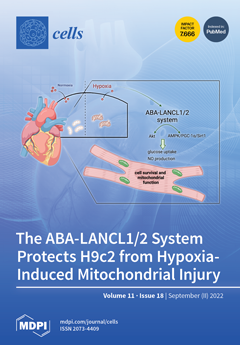
Cover Story (view full-size image): Abscisic acid (ABA) regulates plant responses to stress, partly via NO. In H9c2, hypoxia induced ABA release, and ABA stimulated NO production. ABA increased the survival of H9c2 to hypoxia, and L-NAME, an inhibitor of NO synthase (NOS), abrogated this effect. ABA also increased glucose uptake and NADPH levels and increased the phosphorylation of Akt, AMPK and eNOS. Overexpression or silencing of LANCL1/2 increased or decreased, respectively, the transcription, expression and phosphorylation of AMPK, Akt and eNOS; transcription of NAMPT, Sirt1 and the arginine transporter. The mitochondrial proton gradient and cell vitality increased in LANCL1/2-overexpressing vs. -silenced cells after hypoxia/reoxygenation, and L-NAME abrogated this difference. These results implicate the ABA-LANCL1/2 system in NO-mediated cardiomyocyte protection against hypoxia. View this paper
  • Issues are regarded as officially published after their release is announced to the table of contents alert mailing list.
  • You may sign up for e-mail alerts to receive table of contents of newly released issues.
  • PDF is the official format for papers published in both, html and pdf forms. To view the papers in pdf format, click on the "PDF Full-text" link, and use the free Adobe Reader to open them.
Order results
Result details
Section
Select all
Export citation of selected articles as:
22 pages, 1884 KiB  
Article
Retinoids Promote Mouse Bone Marrow-Derived Macrophage Differentiation and Efferocytosis via Upregulating Bone Morphogenetic Protein-2 and Smad3
by Éva Fige, Zsolt Sarang, László Sós and Zsuzsa Szondy
Cells 2022, 11(18), 2928; https://doi.org/10.3390/cells11182928 - 19 Sep 2022
Cited by 6 | Viewed by 3538
Abstract
Clearance of apoptotic cells by bone marrow-derived macrophages differentiated from monocytes plays a central role in the resolution of inflammation, as the conversion of pro-inflammatory M1 macrophages to M2 macrophages that mediate the resolution process occurs during efferocytosis. Thus, proper efferocytosis is a [...] Read more.
Clearance of apoptotic cells by bone marrow-derived macrophages differentiated from monocytes plays a central role in the resolution of inflammation, as the conversion of pro-inflammatory M1 macrophages to M2 macrophages that mediate the resolution process occurs during efferocytosis. Thus, proper efferocytosis is a prerequisite for proper resolution of inflammation, and failure in efferocytosis is associated with the development of chronic inflammatory diseases. Previous studies from our laboratory have shown that (13R)-all-trans-13,14-dihydroretinol (DHR), the product of retinol saturase, acting from day 4 of monocyte differentiation enhances the efferocytosis capacity of the resulted macrophages. Loss of retinol saturase in mice leads to impaired efferocytosis, and to development of autoimmunity. In the present paper, we report that in differentiating monocytes DHR, retinol, and all-trans retinoic acid all act directly on retinoic acid receptors and enhance the clearance of apoptotic cells by upregulating the expression of several efferocytosis-related genes. The effect of retinoids seems to be mediated by bone morphogenetic protein (BMP)-2, and the Smad3 transcription factor. In addition, retinoids also upregulate the expression of the vitamin D receptor and that of vascular endothelial growth factor A, indicating that altogether retinoids promote the generation of a pro-reparative M2 macrophage population during monocyte differentiation. Full article
Show Figures

Figure 1

16 pages, 3683 KiB  
Article
Isoalantolactone (IAL) Regulates Neuro-Inflammation and Neuronal Apoptosis to Curb Pathology of Parkinson’s Disease
by Dewei He, Yanting Liu, Jie Li, Hefei Wang, Bojian Ye, Yuan He, Zhe Li, Xiyu Gao, Shoupeng Fu and Dianfeng Liu
Cells 2022, 11(18), 2927; https://doi.org/10.3390/cells11182927 - 19 Sep 2022
Cited by 5 | Viewed by 2780
Abstract
Parkinson’s disease (PD) is a neurodegenerative disease in which neuronal apoptosis and associated inflammation are involved in its pathogenesis. However, there is still no specific treatment that can stop PD progression. Isoalantolactone (IAL) plays a role in many inflammation-related diseases. However, its effect [...] Read more.
Parkinson’s disease (PD) is a neurodegenerative disease in which neuronal apoptosis and associated inflammation are involved in its pathogenesis. However, there is still no specific treatment that can stop PD progression. Isoalantolactone (IAL) plays a role in many inflammation-related diseases. However, its effect and mechanism in PD remain unclear. In this study, results showed that IAL administration ameliorated 1-methyl-4-phenyl-1, 2, 3, 6-tetrahydropyridine (MPTP)-induced PD-related pathological impairment and decreased motor activity in mice. Results from in vitro mechanistic studies showed that IAL regulated apoptosis-related proteins by activating the AKT/Nrf2 pathway, thereby suppressing the apoptosis of SN4741 cells induced by N-methyl-4-phenylpyridinium Iodide (MPP+). On the other hand, IAL inhibited LPS-induced release of pro-inflammatory mediators in BV2 cells by activating the AKT/Nrf2/HO-1 pathway and inhibiting the NF-κB pathway. In addition, IAL protected SN4741 from microglial activation-mediated neurotoxicity. Taken together, these results highlight the beneficial role of IAL as a novel therapy and potential PD drug due to its pharmacological profile. Full article
(This article belongs to the Special Issue The Function of Astrocytes in Neurodegenerative Diseases)
Show Figures

Figure 1

20 pages, 20183 KiB  
Article
MARCKS as a Potential Therapeutic Target in Inflammatory Breast Cancer
by Maroua Manai, Ines ELBini-Dhouib, Pascal Finetti, Haifa Bichiou, Carolina Reduzzi, Dorra Aissaoui, Naziha Ben-Hamida, Emilie Agavnian, Najet Srairi-Abid, Marc Lopez, Fatma Amri, Lamia Guizani-Tabbane, Khaled Rahal, Karima Mrad, Mohamed Manai, Daniel Birnbaum, Emilie Mamessier, Massimo Cristofanilli, Hamouda Boussen, Maher Kharrat, Raoudha Doghri and François Bertucciadd Show full author list remove Hide full author list
Cells 2022, 11(18), 2926; https://doi.org/10.3390/cells11182926 - 19 Sep 2022
Cited by 3 | Viewed by 3479
Abstract
Inflammatory breast cancer (IBC) is the most pro-metastatic form of breast cancer (BC). We previously demonstrated that protein overexpression of Myristoylated Alanine-Rich C Kinase Substrate (MARCKS) protein was associated with shorter survival in IBC patients. MARCKS has been associated with the PI3K/AKT pathway. [...] Read more.
Inflammatory breast cancer (IBC) is the most pro-metastatic form of breast cancer (BC). We previously demonstrated that protein overexpression of Myristoylated Alanine-Rich C Kinase Substrate (MARCKS) protein was associated with shorter survival in IBC patients. MARCKS has been associated with the PI3K/AKT pathway. MARCKS inhibitors are in development. Our objective was to investigate MARCKS, expressed preferentially in IBC that non-IBC (nIBC), as a novel potential therapeutic target for IBC. The biologic activity of MPS, a MARCKS peptide inhibitor, on cell proliferation, migration, invasion, and mammosphere formation was evaluated in IBC (SUM149 and SUM190) and nIBC (MDA-MB-231 and MCF7) cell lines, as well as its effects on protein expression in the PTEN/AKT and MAPK pathways. The prognostic relevance of MARCKS and phosphatase and tensin homolog (PTEN) protein expression as a surrogate marker of metastasis-free survival (MFS) was evaluated by immunohistochemistry (IHC) in a retrospective series of archival tumor samples derived from 180 IBC patients and 355 nIBC patients. In vitro MPS impaired cell proliferation, migration and invasion, and mammosphere formation in IBC cells. MARCKS inhibition upregulated PTEN and downregulated pAKT and pMAPK expression in IBC cells, but not in nIBC cells. By IHC, MARCKS expression and PTEN expression were negatively correlated in IBC samples and were associated with shorter MFS and longer MFS, respectively, in multivariate analysis. The combination of MARCKS-/PTEN+ protein status was associated with longer MFS in IBC patient only (p = 8.7 × 10⁻³), and mirrored the molecular profile (MARCKS-downregulated/PTEN-upregulated) of MPS-treated IBC cell lines. In conclusion, our results uncover a functional role of MARCKS implicated in IBC aggressiveness. Associated with the good-prognosis value of the MARCKS-/PTEN+ protein status that mirrors the molecular profile of MPS-treated IBC cell lines, our results suggest that MARCKS could be a potential therapeutic target in patients with MARCKS-positive IBC. Future preclinical studies using a larger panel of IBC cell lines, animal models and analysis of a larger series of clinical samples are warranted in order to validate our results. Full article
Show Figures

Graphical abstract

34 pages, 6242 KiB  
Article
Proteomics Identifies Substrates and a Novel Component in hSnd2-Dependent ER Protein Targeting
by Andrea Tirincsi, Sarah O’Keefe, Duy Nguyen, Mark Sicking, Johanna Dudek, Friedrich Förster, Martin Jung, Drazena Hadzibeganovic, Volkhard Helms, Stephen High, Richard Zimmermann and Sven Lang
Cells 2022, 11(18), 2925; https://doi.org/10.3390/cells11182925 - 19 Sep 2022
Cited by 3 | Viewed by 3000
Abstract
Importing proteins into the endoplasmic reticulum (ER) is essential for about 30% of the human proteome. It involves the targeting of precursor proteins to the ER and their insertion into or translocation across the ER membrane. Furthermore, it relies on signals in the [...] Read more.
Importing proteins into the endoplasmic reticulum (ER) is essential for about 30% of the human proteome. It involves the targeting of precursor proteins to the ER and their insertion into or translocation across the ER membrane. Furthermore, it relies on signals in the precursor polypeptides and components, which read the signals and facilitate their targeting to a protein-conducting channel in the ER membrane, the Sec61 complex. Compared to the SRP- and TRC-dependent pathways, little is known about the SRP-independent/SND pathway. Our aim was to identify additional components and characterize the client spectrum of the human SND pathway. The established strategy of combining the depletion of the central hSnd2 component from HeLa cells with proteomic and differential protein abundance analysis was used. The SRP and TRC targeting pathways were analyzed in comparison. TMEM109 was characterized as hSnd3. Unlike SRP but similar to TRC, the SND clients are predominantly membrane proteins with N-terminal, central, or C-terminal targeting signals. Full article
(This article belongs to the Section Intracellular and Plasma Membranes)
Show Figures

Figure 1

16 pages, 5394 KiB  
Article
Define the Two Molecular Subtypes of Epithelioid Malignant Pleural Mesothelioma
by Umair Ali Khan Saddozai, Fengling Wang, Saadullah Khattak, Muhammad Usman Akbar, Muhammad Badar, Nazeer Hussain Khan, Lu Zhang, Wan Zhu, Longxiang Xie, Yongqiang Li, Xinying Ji and Xiangqian Guo
Cells 2022, 11(18), 2924; https://doi.org/10.3390/cells11182924 - 19 Sep 2022
Cited by 4 | Viewed by 3987
Abstract
Malignant pleural mesothelioma (MPM) is a fatal disease of respiratory system. Despite the availability of invasive biomarkers with promising results, there are still significant diagnostic and therapeutic challenges in the treatment of MPM. One of three main mesothelioma cell types, epithelioid mesothelioma makes [...] Read more.
Malignant pleural mesothelioma (MPM) is a fatal disease of respiratory system. Despite the availability of invasive biomarkers with promising results, there are still significant diagnostic and therapeutic challenges in the treatment of MPM. One of three main mesothelioma cell types, epithelioid mesothelioma makes up approximately 70% of all mesothelioma cases. Different observational findings are under process, but the molecular heterogeneity and pathogenesis of epithelioid malignant pleural mesothelioma (eMPM) are still not well understood. Through molecular analysis, expression profiling data were used to determine the possibility and optimal number of eMPM molecular subtypes. Next, clinicopathological characteristics and different molecular pathways of each subtype were analyzed to prospect the clinical applications and advanced mechanisms of eMPM. In this study, we identified two distinct epithelioid malignant pleural mesothelioma subtypes with distinct gene expression patterns. Subtype I eMPMs were involved in steroid hormone biosynthesis, porphyrin and chlorophyll metabolism, and drug metabolism, while subtype II eMPMs were involved in rational metabolism, tyrosine metabolism, and chemical carcinogenesis pathways. Additionally, we identified potential subtype-specific therapeutic targets, including CCNE1, EPHA3, RNF43, ROS1, and RSPO2 for subtype I and CDKN2A and RET for subtype II. Considering the need for potent diagnostic and therapeutic biomarkers for eMPM, we are anticipating that our findings will help both in exploring underlying mechanisms in the development of eMPM and in designing targeted therapy for eMPM. Full article
(This article belongs to the Special Issue Molecular and Cellular Mechanisms of Lung Cancers)
Show Figures

Figure 1

21 pages, 3676 KiB  
Article
Macrophage-Secreted CSF1 Transmits a Calorie Restriction-Induced Self-Renewal Signal to Mammary Epithelial Stem Cells
by Anna Kosenko, Tomer Meir Salame, Gilgi Friedlander and Itamar Barash
Cells 2022, 11(18), 2923; https://doi.org/10.3390/cells11182923 - 19 Sep 2022
Cited by 2 | Viewed by 2162
Abstract
Calorie restriction enhances stem cell self-renewal in various tissues, including the mammary gland. We hypothesized that similar to their intestinal counterparts, mammary epithelial stem cells are insulated from sensing changes in energy supply, depending instead on niche signaling. The latter was investigated by [...] Read more.
Calorie restriction enhances stem cell self-renewal in various tissues, including the mammary gland. We hypothesized that similar to their intestinal counterparts, mammary epithelial stem cells are insulated from sensing changes in energy supply, depending instead on niche signaling. The latter was investigated by subjecting cultures of mammary epithelial stem cells for 8 days to in vivo paracrine calorie-restriction signals collected from a 4-day-conditioned medium of individual mammary cell populations. Conditioned medium from calorie-restricted non-epithelial cells induced latent cell propagation and mammosphere formation—established markers of stem cell self-renewal. Combined RNA-Seq, immunohistochemistry and immunofluorescence analyses of the non-epithelial population identified macrophages and secreted CSF1 as the energy sensor and paracrine signal, respectively. Calorie restriction-induced pStat6 expression in macrophages suggested that skewing to the M2 phenotype contributes to the sensing mechanism. Enhancing CSF1 signaling with recombinant protein and interrupting the interaction with its highly expressed receptor in the epithelial stem cells by neutralizing antibodies were both affected stem cell self-renewal. In conclusion, combined in vivo, in vitro and in silico studies identified macrophages and secreted CSF1 as the energy sensor and paracrine transmitter, respectively, of the calorie restriction-induced effect on mammary stem cell self-renewal. Full article
(This article belongs to the Topic Stem Cell Differentiation and Applications)
Show Figures

Figure 1

19 pages, 1445 KiB  
Article
Multi-Strain Probiotic Mixture Affects Brain Morphology and Resting State Brain Function in Healthy Subjects: An RCT
by Julia Rode, Hanna M. T. Edebol Carlman, Julia König, Ashley N. Hutchinson, Per Thunberg, Jonas Persson and Robert J. Brummer
Cells 2022, 11(18), 2922; https://doi.org/10.3390/cells11182922 - 19 Sep 2022
Cited by 18 | Viewed by 7234
Abstract
Probiotics can alter brain function via the gut–brain axis. We investigated the effect of a probiotic mixture containing Bifidobacterium longum, Lactobacillus helveticus and Lactiplantibacillus plantarum. In a randomized, placebo-controlled, double-blinded crossover design, 22 healthy subjects (6 m/16 f; 24.2 ± 3.4 [...] Read more.
Probiotics can alter brain function via the gut–brain axis. We investigated the effect of a probiotic mixture containing Bifidobacterium longum, Lactobacillus helveticus and Lactiplantibacillus plantarum. In a randomized, placebo-controlled, double-blinded crossover design, 22 healthy subjects (6 m/16 f; 24.2 ± 3.4 years) underwent four-week intervention periods with probiotics and placebo, separated by a four-week washout period. Voxel-based morphometry indicated that the probiotic intervention affected the gray matter volume of a cluster covering the left supramarginal gyrus and superior parietal lobule (p < 0.0001), two regions that were also among those with an altered resting state functional connectivity. Probiotic intervention resulted in significant (FDR < 0.05) functional connectivity changes between regions within the default mode, salience, frontoparietal as well as the language network and several regions located outside these networks. Psychological symptoms trended towards improvement after probiotic intervention, i.e., the total score of the Hospital Anxiety and Depression Scale (p = 0.056) and its depression sub-score (p = 0.093), as well as sleep patterns (p = 0.058). The probiotic intervention evoked distinct changes in brain morphology and resting state brain function alongside slight improvements of psycho(bio)logical markers of the gut–brain axis. The combination of those parameters may provide new insights into the modes of action by which gut microbiota can affect gut–brain communication and hence brain function. Full article
(This article belongs to the Special Issue Gut Microbiota in Nutrition and Health)
Show Figures

Figure 1

13 pages, 630 KiB  
Review
Sirtuin 1 in Host Defense during Infection
by Jin Kyung Kim, Prashanta Silwal and Eun-Kyeong Jo
Cells 2022, 11(18), 2921; https://doi.org/10.3390/cells11182921 - 19 Sep 2022
Cited by 9 | Viewed by 5250
Abstract
Sirtuins (SIRTs) are members of the class III histone deacetylase family and epigenetically control multiple target genes to modulate diverse biological responses in cells. Among the SIRTs, SIRT1 is the most well-studied, with a role in the modulation of immune and inflammatory responses [...] Read more.
Sirtuins (SIRTs) are members of the class III histone deacetylase family and epigenetically control multiple target genes to modulate diverse biological responses in cells. Among the SIRTs, SIRT1 is the most well-studied, with a role in the modulation of immune and inflammatory responses following infection. The functions of SIRT1 include orchestrating immune, inflammatory, metabolic, and autophagic responses, all of which are required in establishing and controlling host defenses during infection. In this review, we summarize recent information on the roles of SIRT1 and its regulatory mechanisms during bacterial, viral, and parasitic infections. We also discuss several SIRT1 modulators, as potential antimicrobial treatments. Understanding the function of SIRT1 in balancing immune homeostasis will contribute to the development of new therapeutics for the treatment of infection and inflammatory disease. Full article
Show Figures

Figure 1

19 pages, 6671 KiB  
Review
Emerging Roles of miRNA, lncRNA, circRNA, and Their Cross-Talk in Pituitary Adenoma
by Wentao Wu, Lei Cao, Yanfei Jia, Youchao Xiao, Xu Zhang and Songbai Gui
Cells 2022, 11(18), 2920; https://doi.org/10.3390/cells11182920 - 19 Sep 2022
Cited by 11 | Viewed by 3199
Abstract
Pituitary adenoma (PA) is a common intracranial tumor without specific biomarkers for diagnosis and treatment. Non-coding RNAs (ncRNAs), including microRNAs (miRNA), long non-coding RNA (lncRNA), and circular RNA (circRNA), regulate a variety of cellular processes, such as cell proliferation, differentiation, and apoptosis. Increasing [...] Read more.
Pituitary adenoma (PA) is a common intracranial tumor without specific biomarkers for diagnosis and treatment. Non-coding RNAs (ncRNAs), including microRNAs (miRNA), long non-coding RNA (lncRNA), and circular RNA (circRNA), regulate a variety of cellular processes, such as cell proliferation, differentiation, and apoptosis. Increasing studies have shown that the dysregulation of ncRNAs, especially the cross-talk between lncRNA/circRNA and miRNA, is related to the pathogenesis, diagnosis, and prognosis of PA. Therefore, ncRNAs can be considered as promising biomarkers for PA. In this review, we summarize the roles of ncRNAs from different specimens (i.e., tissues, biofluids, cells, and exosomes) in multiple subtypes of PA and highlight important advances in understanding the contribution of the cross-talk between ncRNAs (e.g., competing endogenous RNAs) to PA disease. Full article
(This article belongs to the Special Issue Non-coding RNAs and Neurological Diseases 2022)
Show Figures

Figure 1

19 pages, 3349 KiB  
Article
Whole-Transcriptome Sequencing Analyses of Nuclear Antixoxidant-1 in Endothelial Cells: Role in Inflammation and Atherosclerosis
by Varadarajan Sudhahar, Yang Shi, Jack H. Kaplan, Masuko Ushio-Fukai and Tohru Fukai
Cells 2022, 11(18), 2919; https://doi.org/10.3390/cells11182919 - 18 Sep 2022
Cited by 5 | Viewed by 3064
Abstract
Inflammation, oxidative stress, and copper (Cu) play an important role in cardiovascular disease, including atherosclerosis. We previously reported that cytosolic Cu chaperone antioxidant-1 (Atox1) translocates to the nucleus in response to inflammatory cytokines or exogenous Cu and that Atox1 is localized at the [...] Read more.
Inflammation, oxidative stress, and copper (Cu) play an important role in cardiovascular disease, including atherosclerosis. We previously reported that cytosolic Cu chaperone antioxidant-1 (Atox1) translocates to the nucleus in response to inflammatory cytokines or exogenous Cu and that Atox1 is localized at the nucleus in the endothelium of inflamed atherosclerotic aorta. However, the roles of nuclear Atox1 and their function are poorly understood. Here we showed that Atox1 deficiency in ApoE−/− mice with a Western diet exhibited a significant reduction of atherosclerotic lesion formation. In vitro, adenovirus-mediated overexpression of nuclear-targeted Atox1 (Ad-Atox1-NLS) in cultured human endothelial cells (ECs) increased monocyte adhesion and reactive oxygen species (ROS) production compared to control cells (Ad-null). To address the underlying mechanisms, we performed genome-wide mapping of Atox1-regulated targets in ECs, using an unbiased systemic approach integrating sequencing data. Combination of ChIP-Seq and RNA-Seq analyses in ECs transfected with Ad-Atox1-NLS or Ad-null identified 1387 differentially expressed genes (DEG). Motif enrichment assay and KEGG pathway enrichment analysis revealed that 248 differentially expressed genes, including inflammatory and angiogenic genes, were regulated by Atox1-NLS, which was then confirmed by real-time qPCR. Among these genes, functional analysis of inflammatory responses identified CD137, CSF1, and IL5RA as new nuclear Atox1-targeted inflammatory genes, while CD137 is also a key regulator of Atox1-NLS-induced ROS production. These findings uncover new nuclear Atox1 downstream targets involved in inflammation and ROS production and provide insights into the nuclear Atox1 as a potential therapeutic target for the treatment of inflammatory diseases such as atherosclerosis. Full article
(This article belongs to the Collection Cellular and Molecular Mechanisms of Atherosclerosis)
Show Figures

Figure 1

18 pages, 4412 KiB  
Review
Purkinje Cell Patterning—Insights from Single-Cell Sequencing
by Elizabeth J. Apsley and Esther B. E. Becker
Cells 2022, 11(18), 2918; https://doi.org/10.3390/cells11182918 - 18 Sep 2022
Cited by 4 | Viewed by 6125
Abstract
Despite their homogeneous appearance, Purkinje cells are remarkably diverse with respect to their molecular phenotypes, physiological properties, afferent and efferent connectivity, as well as their vulnerability to insults. Heterogeneity in Purkinje cells arises early in development, with molecularly distinct embryonic cell clusters present [...] Read more.
Despite their homogeneous appearance, Purkinje cells are remarkably diverse with respect to their molecular phenotypes, physiological properties, afferent and efferent connectivity, as well as their vulnerability to insults. Heterogeneity in Purkinje cells arises early in development, with molecularly distinct embryonic cell clusters present soon after Purkinje cell specification. Traditional methods have characterized cerebellar development and cell types, including Purkinje cell subtypes, based on knowledge of selected markers. However, recent single-cell RNA sequencing studies provide vastly increased resolution of the whole cerebellar transcriptome. Here we draw together the results of multiple single-cell transcriptomic studies in developing and adult cerebellum in both mouse and human. We describe how this detailed transcriptomic data has increased our understanding of the intricate development and function of Purkinje cells and provides first clues into features specific to human cerebellar development. Full article
(This article belongs to the Special Issue Cerebellar Development in Health and Disease)
Show Figures

Figure 1

22 pages, 2509 KiB  
Review
Diabetic Proteinuria Revisited: Updated Physiologic Perspectives
by Samuel N. Heyman, Itamar Raz, Jamie P. Dwyer, Roni Weinberg Sibony, Julia B. Lewis and Zaid Abassi
Cells 2022, 11(18), 2917; https://doi.org/10.3390/cells11182917 - 18 Sep 2022
Cited by 27 | Viewed by 7231
Abstract
Albuminuria, a hallmark of diabetic nephropathy, reflects not only injury and dysfunction of the filtration apparatus, but is also affected by altered glomerular hemodynamics and hyperfiltration, as well as by the inability of renal tubular cells to fully retrieve filtered albumin. Albuminuria further [...] Read more.
Albuminuria, a hallmark of diabetic nephropathy, reflects not only injury and dysfunction of the filtration apparatus, but is also affected by altered glomerular hemodynamics and hyperfiltration, as well as by the inability of renal tubular cells to fully retrieve filtered albumin. Albuminuria further plays a role in the progression of diabetic nephropathy, and the suppression of glomerular albumin leak is a key factor in its prevention. Although microalbuminuria is a classic manifestation of diabetic nephropathy, often progressing to macroalbuminuria or overt proteinuria over time, it does not always precede renal function loss in diabetes. The various components leading to diabetic albuminuria and their associations are herein reviewed, and the physiologic rationale and efficacy of therapeutic interventions that reduce glomerular hyperfiltration and proteinuria are discussed. With these perspectives, we propose that these measures should be initiated early, before microalbuminuria develops, as substantial renal injury may already be present in the absence of proteinuria. We further advocate that the inhibition of the renin–angiotensin axis or of sodium–glucose co-transport likely permits the administration of a normal recommended or even high-protein diet, highly desirable for sarcopenic diabetic patients. Full article
(This article belongs to the Special Issue Recent Advances in Development and Progression of Kidney Diseases)
Show Figures

Figure 1

14 pages, 1984 KiB  
Article
Mast Cell Proteases Promote Diverse Effects on the Plasminogen Activation System and Wound Healing in A549 Alveolar Epithelial Cells
by Sofia Mogren, Frida Berlin, Lykke Eskilsson, Nicole Van Der Burg, Ellen Tufvesson and Cecilia K. Andersson
Cells 2022, 11(18), 2916; https://doi.org/10.3390/cells11182916 - 18 Sep 2022
Cited by 1 | Viewed by 2847
Abstract
Tissue damage, epithelial alterations, and intraepithelial presence of mast cells (MCs) are characteristics of asthma pathogenesis. Increased alveolar infiltration of MC populations has also been identified as a feature of asthma and other chronic respiratory diseases. The asthma associated receptor, urokinase plasminogen activator [...] Read more.
Tissue damage, epithelial alterations, and intraepithelial presence of mast cells (MCs) are characteristics of asthma pathogenesis. Increased alveolar infiltration of MC populations has also been identified as a feature of asthma and other chronic respiratory diseases. The asthma associated receptor, urokinase plasminogen activator receptor (uPAR), has been shown to regulate bronchial epithelial repair responses. However, the impact of MC tryptase and chymase on functional properties and expression of uPAR in alveolar epithelial cells have not been fully investigated. Alveolar epithelial cell migration and wound healing were investigated using holographic live cell imaging of A549 cells in a wound scratch model post stimulation with tryptase or chymase. The expression of uPAR was investigated on the protein and gene level from cellular supernatants and in bronchoalveolar lavage fluid fractions from allergic asthmatics. We found that tryptase improved wound healing capacity, cellular migration and membrane bound uPAR expression. Chymase reduced gap closure capacity, cellular migration and membrane bound uPAR expression but increased soluble uPAR release. Our data suggest a dual regulatory response from the MC proteases in events related to uPAR expression and wound healing which could be important features in asthmatic disease. Full article
(This article belongs to the Collection Mast Cells in Health and Diseases)
Show Figures

Figure 1

22 pages, 1771 KiB  
Review
Disturbed Cardiac Metabolism Triggers Atrial Arrhythmogenesis in Diabetes Mellitus: Energy Substrate Alternate as a Potential Therapeutic Intervention
by Baigalmaa Lkhagva, Ting-Wei Lee, Yung-Kuo Lin, Yao-Chang Chen, Cheng-Chih Chung, Satoshi Higa and Yi-Jen Chen
Cells 2022, 11(18), 2915; https://doi.org/10.3390/cells11182915 - 18 Sep 2022
Cited by 6 | Viewed by 3189
Abstract
Atrial fibrillation (AF) is the most common type of sustained arrhythmia in diabetes mellitus (DM). Its morbidity and mortality rates are high, and its prevalence will increase as the population ages. Despite expanding knowledge on the pathophysiological mechanisms of AF, current pharmacological interventions [...] Read more.
Atrial fibrillation (AF) is the most common type of sustained arrhythmia in diabetes mellitus (DM). Its morbidity and mortality rates are high, and its prevalence will increase as the population ages. Despite expanding knowledge on the pathophysiological mechanisms of AF, current pharmacological interventions remain unsatisfactory; therefore, novel findings on the underlying mechanism are required. A growing body of evidence suggests that an altered energy metabolism is closely related to atrial arrhythmogenesis, and this finding engenders novel insights into the pathogenesis of the pathophysiology of AF. In this review, we provide comprehensive information on the mechanistic insights into the cardiac energy metabolic changes, altered substrate oxidation rates, and mitochondrial dysfunctions involved in atrial arrhythmogenesis, and suggest a promising advanced new therapeutic approach to treat patients with AF. Full article
(This article belongs to the Section Intracellular and Plasma Membranes)
Show Figures

Figure 1

18 pages, 2562 KiB  
Article
Ablation of Sphingosine Kinase 1 Protects Cornea from Neovascularization in a Mouse Corneal Injury Model
by Joseph L. Wilkerson, Sandip K. Basu, Megan A. Stiles, Amanda Prislovsky, Richard C. Grambergs, Sarah E. Nicholas, Dimitrios Karamichos, Jeremy C. Allegood, Richard L. Proia and Nawajes Mandal
Cells 2022, 11(18), 2914; https://doi.org/10.3390/cells11182914 - 17 Sep 2022
Cited by 4 | Viewed by 2254
Abstract
The purpose of this study was to investigate the role of sphingosine kinase 1 (SphK1), which generates sphingosine-1-phosphate (S1P), in corneal neovascularization (NV). Wild-type (WT) and Sphk1 knockout (Sphk1/) mice received corneal alkali-burn treatment to induce corneal [...] Read more.
The purpose of this study was to investigate the role of sphingosine kinase 1 (SphK1), which generates sphingosine-1-phosphate (S1P), in corneal neovascularization (NV). Wild-type (WT) and Sphk1 knockout (Sphk1/) mice received corneal alkali-burn treatment to induce corneal NV by placing a 2 mm round piece of Whatman No. 1 filter paper soaked in 1N NaOH on the center of the cornea for 20 s. Corneal sphingolipid species were extracted and identified using liquid chromatography/mass spectrometry (LC/MS). The total number of tip cells and those positive for ethynyl deoxy uridine (EdU) were quantified. Immunocytochemistry was done to examine whether pericytes were present on newly forming blood vessels. Cytokine signaling and angiogenic markers were compared between the two groups using multiplex assays. Data were analyzed using appropriate statistical tests. Here, we show that ablation of SphK1 can significantly reduce NV invasion in the cornea following injury. Corneal sphingolipid analysis showed that total levels of ceramides, monohexosyl ceramides (HexCer), and sphingomyelin were significantly elevated in Sphk/ corneas compared to WT corneas, with a comparable level of sphingosine among the two genotypes. The numbers of total and proliferating endothelial tip cells were also lower in the Sphk1/ corneas following injury. This study underscores the role of S1P in post-injury corneal NV and raises further questions about the roles played by ceramide, HexCer, and sphingomyelin in regulating corneal NV. Further studies are needed to unravel the role played by bioactive sphingolipids in maintenance of corneal transparency and clear vision. Full article
(This article belongs to the Special Issue Sphingolipid Signaling and Human Disease 2022)
Show Figures

Figure 1

26 pages, 731 KiB  
Review
Small Extracellular Vesicles and Their Involvement in Cancer Resistance: An Up-to-Date Review
by Artur Słomka, Miroslaw Kornek and William C. Cho
Cells 2022, 11(18), 2913; https://doi.org/10.3390/cells11182913 - 17 Sep 2022
Cited by 22 | Viewed by 3202
Abstract
In recent years, tremendous progress has been made in understanding the roles of extracellular vesicles (EVs) in cancer. Thanks to advancements in molecular biology, it has been found that the fraction of EVs called exosomes or small EVs (sEVs) modulates the sensitivity of [...] Read more.
In recent years, tremendous progress has been made in understanding the roles of extracellular vesicles (EVs) in cancer. Thanks to advancements in molecular biology, it has been found that the fraction of EVs called exosomes or small EVs (sEVs) modulates the sensitivity of cancer cells to chemotherapeutic agents by delivering molecularly active non-coding RNAs (ncRNAs). An in-depth analysis shows that two main molecular mechanisms are involved in exosomal modified chemoresistance: (1) translational repression of anti-oncogenes by exosomal microRNAs (miRs) and (2) lack of translational repression of oncogenes by sponging of miRs through long non-coding RNAs (lncRNAs) and circular RNAs (circRNAs). At the cellular level, these processes increase the proliferation and survival of cancer cells and improve their ability to metastasize and resist apoptosis. In addition, studies in animal models have shown enhancing tumor size under the influence of exosomal ncRNAs. Ultimately, exosomal ncRNAs are responsible for clinically significant chemotherapy failures in patients with different types of cancer. Preliminary data have also revealed that exosomal ncRNAs can overcome chemotherapeutic agent resistance, but the results are thoroughly fragmented. This review presents how exosomes modulate the response of cancer cells to chemotherapeutic agents. Understanding how exosomes interfere with chemoresistance may become a milestone in developing new therapeutic options, but more data are still required. Full article
Show Figures

Figure 1

14 pages, 2903 KiB  
Article
SARS-CoV-2 Infection of Airway Epithelium Triggers Pulmonary Endothelial Cell Activation and Senescence Associated with Type I IFN Production
by Veronica Bordoni, Davide Mariotti, Giulia Matusali, Francesca Colavita, Eleonora Cimini, Giuseppe Ippolito and Chiara Agrati
Cells 2022, 11(18), 2912; https://doi.org/10.3390/cells11182912 - 17 Sep 2022
Cited by 8 | Viewed by 2276
Abstract
Airway epithelial cells represent the main target of SARS-CoV-2 replication but several pieces of evidence suggest that endothelial cells (ECs), lining pulmonary blood vessels, are key players in lung injury in COVID-19 patients. Although in vivo evidence of SARS-CoV-2 affecting the vascular endothelium [...] Read more.
Airway epithelial cells represent the main target of SARS-CoV-2 replication but several pieces of evidence suggest that endothelial cells (ECs), lining pulmonary blood vessels, are key players in lung injury in COVID-19 patients. Although in vivo evidence of SARS-CoV-2 affecting the vascular endothelium exists, in vitro data are limited. In the present study, we set up an organotypic model to dissect the crosstalk between airway epithelium and pulmonary endothelial cells during SARS-CoV-2 infection. We showed that SARS-CoV-2 infected airway epithelium triggers the induction of endothelial adhesion molecules in ECs, suggesting a bystander effect of dangerous soluble signals from the infected epithelium. The endothelial activation was correlated with inflammatory cytokines (IL-1β, IL-6, IL-8) and with the viral replication in the airway epithelium. Interestingly, SARS-CoV-2 infection determined a modulation of endothelial p21, which could be partially reversed by inhibiting IFN-β production from ECs when co-cultured with HAE. Altogether, we demonstrated that SARS-CoV-2 infected epithelium triggers activation/senescence processes in ECs involving type I IFN-β production, suggesting possible antiviral/damage mechanisms occurring in the endothelium. Full article
(This article belongs to the Section Cellular Immunology)
Show Figures

Figure 1

14 pages, 2557 KiB  
Article
Low-Ammonium Environment Increases the Nutrient Exchange between Diatom–Diazotroph Association Cells and Facilitates Photosynthesis and N2 Fixation—a Mechanistic Modeling Analysis
by Meng Gao, Gabrielle Armin and Keisuke Inomura
Cells 2022, 11(18), 2911; https://doi.org/10.3390/cells11182911 - 17 Sep 2022
Cited by 2 | Viewed by 1995
Abstract
Diatom–diazotroph associations (DDAs) are one of the most important symbiotic dinitrogen (N2) fixing groups in the oligotrophic ocean. Despite their capability to fix N2, ammonium (NH4+) remains a key nitrogen (N) source for DDAs, and the [...] Read more.
Diatom–diazotroph associations (DDAs) are one of the most important symbiotic dinitrogen (N2) fixing groups in the oligotrophic ocean. Despite their capability to fix N2, ammonium (NH4+) remains a key nitrogen (N) source for DDAs, and the effect of NH4+ on their metabolism remains elusive. Here, we developed a coarse-grained, cellular model of the DDA with NH4+ uptake and quantified how the level of extracellular NH4+ influences metabolism and nutrient exchange within the symbiosis. The model shows that, under a fixed growth rate, an increased NH4+ concentration may lower the required level of N2 fixation and photosynthesis, and decrease carbon (C) and N exchange. A low-NH4+ environment leads to more C and N in nutrient exchange and more fixed N2 to support a higher growth rate. With higher growth rates, nutrient exchange and metabolism increased. Our study shows a strong effect of NH4+ on metabolic processes within DDAs, and thus highlights the importance of in situ measurement of NH4+ concentrations. Full article
(This article belongs to the Section Plant, Algae and Fungi Cell Biology)
Show Figures

Figure 1

39 pages, 1954 KiB  
Review
Ionic Plasticity: Common Mechanistic Underpinnings of Pathology in Spinal Cord Injury and the Brain
by Kelsey E. Hudson and James W. Grau
Cells 2022, 11(18), 2910; https://doi.org/10.3390/cells11182910 - 17 Sep 2022
Cited by 5 | Viewed by 3181
Abstract
The neurotransmitter GABA is normally characterized as having an inhibitory effect on neural activity in the adult central nervous system (CNS), which quells over-excitation and limits neural plasticity. Spinal cord injury (SCI) can bring about a modification that weakens the inhibitory effect of [...] Read more.
The neurotransmitter GABA is normally characterized as having an inhibitory effect on neural activity in the adult central nervous system (CNS), which quells over-excitation and limits neural plasticity. Spinal cord injury (SCI) can bring about a modification that weakens the inhibitory effect of GABA in the central gray caudal to injury. This change is linked to the downregulation of the potassium/chloride cotransporter (KCC2) and the consequent rise in intracellular Cl in the postsynaptic neuron. As the intracellular concentration increases, the inward flow of Cl through an ionotropic GABA-A receptor is reduced, which decreases its hyperpolarizing (inhibitory) effect, a modulatory effect known as ionic plasticity. The loss of GABA-dependent inhibition enables a state of over-excitation within the spinal cord that fosters aberrant motor activity (spasticity) and chronic pain. A downregulation of KCC2 also contributes to the development of a number of brain-dependent pathologies linked to states of neural over-excitation, including epilepsy, addiction, and developmental disorders, along with other diseases such as hypertension, asthma, and irritable bowel syndrome. Pharmacological treatments that target ionic plasticity have been shown to bring therapeutic benefits. Full article
(This article belongs to the Collection Cell Biology of Spinal Cord Injury and Repair)
Show Figures

Figure 1

16 pages, 1861 KiB  
Article
L-Ornithine L-Aspartate Restores Mitochondrial Function and Modulates Intracellular Calcium Homeostasis in Parkinson’s Disease Models
by Maria Josè Sisalli, Salvatore Della Notte, Agnese Secondo, Carmelo Ventra, Lucio Annunziato and Antonella Scorziello
Cells 2022, 11(18), 2909; https://doi.org/10.3390/cells11182909 - 17 Sep 2022
Cited by 7 | Viewed by 3023
Abstract
The altered crosstalk between mitochondrial dysfunction, intracellular Ca2+ homeostasis, and oxidative stress has a central role in the dopaminergic neurodegeneration. In the present study, we investigated the hypothesis that pharmacological strategies able to improve mitochondrial functions might prevent neuronal dysfunction in in [...] Read more.
The altered crosstalk between mitochondrial dysfunction, intracellular Ca2+ homeostasis, and oxidative stress has a central role in the dopaminergic neurodegeneration. In the present study, we investigated the hypothesis that pharmacological strategies able to improve mitochondrial functions might prevent neuronal dysfunction in in vitro models of Parkinson’s disease. To this aim, the attention was focused on the amino acid ornithine due to its ability to cross the blood–brain barrier, to selectively reach and penetrate the mitochondria through the ornithine transporter 1, and to control mitochondrial function. To pursue this issue, experiments were performed in human neuroblastoma cells SH-SY5Y treated with rotenone and 6-hydroxydopamine to investigate the pharmacological profile of the compound L-Ornithine-L-Aspartate (LOLA) as a new potential therapeutic strategy to prevent dopaminergic neurons’ death. In these models, confocal microscopy experiments with fluorescent dyes measuring mitochondrial calcium content, mitochondrial membrane potential, and mitochondrial ROS production, demonstrated that LOLA improved mitochondrial functions. Moreover, by increasing NCXs expression and activity, LOLA also reduced cytosolic [Ca2+] thanks to its ability to modulate NO production. Collectively, these results indicate that LOLA, by interfering with those mitochondrial mechanisms related to ROS and RNS production, promotes mitochondrial functional recovery, thus confirming the tight relationship existing between cytosolic ionic homeostasis and cellular metabolism depending on the type of insult applied. Full article
(This article belongs to the Section Mitochondria)
Show Figures

Figure 1

40 pages, 1282 KiB  
Review
Neuroinflammation and Parkinson’s Disease—From Neurodegeneration to Therapeutic Opportunities
by Bruna Araújo, Rita Caridade-Silva, Carla Soares-Guedes, Joana Martins-Macedo, Eduardo D. Gomes, Susana Monteiro and Fábio G. Teixeira
Cells 2022, 11(18), 2908; https://doi.org/10.3390/cells11182908 - 17 Sep 2022
Cited by 51 | Viewed by 6101
Abstract
Parkinson’s disease (PD) is the second most prevalent neurodegenerative disorder worldwide. Clinically, it is characterized by a progressive degeneration of dopaminergic neurons (DAn), resulting in severe motor complications. Preclinical and clinical studies have indicated that neuroinflammation can play a role in PD pathophysiology, [...] Read more.
Parkinson’s disease (PD) is the second most prevalent neurodegenerative disorder worldwide. Clinically, it is characterized by a progressive degeneration of dopaminergic neurons (DAn), resulting in severe motor complications. Preclinical and clinical studies have indicated that neuroinflammation can play a role in PD pathophysiology, being associated with its onset and progression. Nevertheless, several key points concerning the neuroinflammatory process in PD remain to be answered. Bearing this in mind, in the present review, we cover the impact of neuroinflammation on PD by exploring the role of inflammatory cells (i.e., microglia and astrocytes) and the interconnections between the brain and the peripheral system. Furthermore, we discuss both the innate and adaptive immune responses regarding PD pathology and explore the gut–brain axis communication and its influence on the progression of the disease. Full article
(This article belongs to the Special Issue Stem Cells, Metabolism and Neurodegenerative Diseases)
Show Figures

Figure 1

20 pages, 793 KiB  
Review
Transient Receptor Potential (TRP) Channels in Airway Toxicity and Disease: An Update
by Isabel Müller, Philipp Alt, Suhasini Rajan, Lena Schaller, Fabienne Geiger and Alexander Dietrich
Cells 2022, 11(18), 2907; https://doi.org/10.3390/cells11182907 - 17 Sep 2022
Cited by 19 | Viewed by 4500
Abstract
Our respiratory system is exposed to toxicants and pathogens from both sides: the airways and the vasculature. While tracheal, bronchial and alveolar epithelial cells form a natural barrier in the airways, endothelial cells protect the lung from perfused toxic compounds, particulate matter and [...] Read more.
Our respiratory system is exposed to toxicants and pathogens from both sides: the airways and the vasculature. While tracheal, bronchial and alveolar epithelial cells form a natural barrier in the airways, endothelial cells protect the lung from perfused toxic compounds, particulate matter and invading microorganism in the vascular system. Damages induce inflammation by our immune response and wound healing by (myo)fibroblast proliferation. Members of the transient receptor potential (TRP) superfamily of ion channel are expressed in many cells of the respiratory tract and serve multiple functions in physiology and pathophysiology. TRP expression patterns in non-neuronal cells with a focus on TRPA1, TRPC6, TRPM2, TRPM5, TRPM7, TRPV2, TRPV4 and TRPV6 channels are presented, and their roles in barrier function, immune regulation and phagocytosis are summarized. Moreover, TRP channels as future pharmacological targets in chronic obstructive pulmonary disease (COPD), asthma, cystic and pulmonary fibrosis as well as lung edema are discussed. Full article
(This article belongs to the Special Issue Molecular and Cellular Mechanism of Airway Diseases)
Show Figures

Figure 1

14 pages, 2394 KiB  
Article
Impact of Mycobacterium tuberculosis Infection on Human B Cell Compartment and Antibody Responses
by Marco P. La Manna, Mojtaba Shekarkar-Azgomi, Giusto D. Badami, Bartolo Tamburini, Costanza Dieli, Paola Di Carlo, Teresa Fasciana, Vito Marcianò, Bruna Lo Sasso, Rosaria V. Giglio, Anna Giammanco, Marcello Ciaccio, Francesco Dieli and Nadia Caccamo
Cells 2022, 11(18), 2906; https://doi.org/10.3390/cells11182906 - 17 Sep 2022
Cited by 7 | Viewed by 2624
Abstract
Tuberculosis (TB) remains one of the most important health challenges worldwide. Control of the TB epidemic has not yet been achieved because of the lack of an effective vaccine and rapid and sensitive diagnostic approaches, as well as the emergence of drug-resistant forms [...] Read more.
Tuberculosis (TB) remains one of the most important health challenges worldwide. Control of the TB epidemic has not yet been achieved because of the lack of an effective vaccine and rapid and sensitive diagnostic approaches, as well as the emergence of drug-resistant forms of M. tuberculosis. Cellular immunity has a pivotal role against M. tuberculosis infection, but the role of humoral immunity is still controversial. We analyzed the frequency, absolute counts, and phenotypic and functional subsets of B lymphocytes in the peripheral blood of patients with active TB and subjects with latent infection compared to healthy donors. Moreover, we analyzed serum levels of total Ig and their IgA, IgM, and IgG isotypes and the titers of preexisting antibodies against a pool of common viral pathogens. FlowCT and unsupervised clusterization analysis show that patients with active TB and LTBI subjects have modest non-significant reduction in the numbers of circulating B lymphocytes as compared to healthy donors. Moreover, LTBI subjects had high percentages of atypical B cell population and lower percentages of naive and switched memory B cells. These findings were supported by gene expression and GSEA analysis. Moreover, there were no differences between active TB patients, LTBI subjects and HD, either in serum levels of total Ig isotypes or in preexisting IgG antibody titers, to ten different antigens from eight common pathogenic viruses, clearly demonstrating that either active or latent M. tuberculosis infection preserves the antibody production capacity of long-lived plasma cells. Thus, our results agree with previous studies reporting unaltered B cell frequencies in the blood of active TB patients and LTBI individuals as compared to healthy controls. Full article
(This article belongs to the Section Cellular Immunology)
Show Figures

Figure 1

27 pages, 3490 KiB  
Article
Human Stem Cell-Derived TRPV1-Positive Sensory Neurons: A New Tool to Study Mechanisms of Sensitization
by Katrin Schrenk-Siemens, Jörg Pohle, Charlotte Rostock, Muad Abd El Hay, Ruby M. Lam, Marcin Szczot, Shiying Lu, Alexander T. Chesler and Jan Siemens
Cells 2022, 11(18), 2905; https://doi.org/10.3390/cells11182905 - 17 Sep 2022
Cited by 7 | Viewed by 3834
Abstract
Somatosensation, the detection and transduction of external and internal stimuli such as temperature or mechanical force, is vital to sustaining our bodily integrity. But still, some of the mechanisms of distinct stimuli detection and transduction are not entirely understood, especially when noxious perception [...] Read more.
Somatosensation, the detection and transduction of external and internal stimuli such as temperature or mechanical force, is vital to sustaining our bodily integrity. But still, some of the mechanisms of distinct stimuli detection and transduction are not entirely understood, especially when noxious perception turns into chronic pain. Over the past decade major progress has increased our understanding in areas such as mechanotransduction or sensory neuron classification. However, it is in particular the access to human pluripotent stem cells and the possibility of generating and studying human sensory neurons that has enriched the somatosensory research field. Based on our previous work, we describe here the generation of human stem cell-derived nociceptor-like cells. We show that by varying the differentiation strategy, we can produce different nociceptive subpopulations with different responsiveness to nociceptive stimuli such as capsaicin. Functional as well as deep sequencing analysis demonstrated that one protocol in particular allowed the generation of a mechano-nociceptive sensory neuron population, homogeneously expressing TRPV1. Accordingly, we find the cells to homogenously respond to capsaicin, to become sensitized upon inflammatory stimuli, and to respond to temperature stimulation. The efficient and homogenous generation of these neurons make them an ideal translational tool to study mechanisms of sensitization, also in the context of chronic pain. Full article
(This article belongs to the Special Issue Cellular and Molecular Mechanisms Underlying Pain Chronicity)
Show Figures

Figure 1

21 pages, 3667 KiB  
Article
Gene Profiling of a 3D Psoriatic Skin Model Enriched in T Cells: Downregulation of PTPRM Promotes Keratinocyte Proliferation through Excessive ERK1/2 Signaling
by Geneviève Rioux, Florence Turgeon, Gaëtan Le-Bel, Camille Grenier, Sylvain L. Guérin and Roxane Pouliot
Cells 2022, 11(18), 2904; https://doi.org/10.3390/cells11182904 - 16 Sep 2022
Cited by 5 | Viewed by 2906
Abstract
Psoriasis is a complex, immune-mediated skin disease involving a wide range of epithelial and immune cells. The underlying mechanisms that govern the epidermal defects and immunological dysfunction observed in this condition remain largely unknown. In recent years, the emergence of new, more sophisticated [...] Read more.
Psoriasis is a complex, immune-mediated skin disease involving a wide range of epithelial and immune cells. The underlying mechanisms that govern the epidermal defects and immunological dysfunction observed in this condition remain largely unknown. In recent years, the emergence of new, more sophisticated models has allowed the evolution of our knowledge of the pathogenesis of psoriasis. The development of psoriatic skin biomaterials that more closely mimic native psoriatic skin provides advanced preclinical models that will prove relevant in predicting clinical outcomes. In this study, we used a tissue-engineered, two-layered (dermis and epidermis) human skin substitute enriched in T cells as a biomaterial to study both the cellular and molecular mechanisms involved in psoriasis’ pathogenesis. Gene profiling on microarrays revealed significant changes in the profile of genes expressed by the psoriatic skin substitutes compared with the healthy ones. Two genes, namely, PTPRM and NELL2, whose products influence the ERK1/2 signaling pathway have been identified as being deregulated in psoriatic substitutes. Deregulation of these genes supports excessive activation of the ERK1/2 pathway in psoriatic skin substitutes. Most importantly, electrophoresis mobility shift assays provided evidence that the DNA-binding properties of two downstream nuclear targets of ERK1/2, both the NF-κB and Sp1 transcription factors, are increased under psoriatic conditions. Moreover, the results obtained with the inhibition of RSK, a downstream effector of ERK1/2, supported the therapeutic potential of inhibiting this signaling pathway for psoriasis treatment. In conclusion, this two-layered human psoriatic skin substitute enriched in T cells may prove particularly useful in deciphering the mechanistic details of psoriatic pathogenesis and provide a relevant biomaterial for the study of potential therapeutic targets. Full article
(This article belongs to the Special Issue Cellular Mechanisms of Skin Diseases)
Show Figures

Graphical abstract

14 pages, 24658 KiB  
Hypothesis
Elucidation of Prebiotics, Probiotics, Postbiotics, and Target from Gut Microbiota to Alleviate Obesity via Network Pharmacology Study
by Ki-Kwang Oh, Haripriya Gupta, Byeong-Hyun Min, Raja Ganesan, Satya Priya Sharma, Sung-Min Won, Jin-Ju Jeong, Su-Been Lee, Min-Gi Cha, Goo-Hyun Kwon, Min-Kyo Jeong, Ji-Ye Hyun, Jung-A Eom, Hee-Jin Park, Sang-Jun Yoon, Mi-Ran Choi, Dong Joon Kim and Ki-Tae Suk
Cells 2022, 11(18), 2903; https://doi.org/10.3390/cells11182903 - 16 Sep 2022
Cited by 11 | Viewed by 5139
Abstract
The metabolites produced by the gut microbiota have been reported as crucial agents against obesity; however, their key targets have not been revealed completely in complex microbiome systems. Hence, the aim of this study was to decipher promising prebiotics, probiotics, postbiotics, and more [...] Read more.
The metabolites produced by the gut microbiota have been reported as crucial agents against obesity; however, their key targets have not been revealed completely in complex microbiome systems. Hence, the aim of this study was to decipher promising prebiotics, probiotics, postbiotics, and more importantly, key target(s) via a network pharmacology approach. First, we retrieved the metabolites related to gut microbes from the gutMGene database. Then, we performed a meta-analysis to identify metabolite-related targets via the similarity ensemble approach (SEA) and SwissTargetPrediction (STP), and obesity-related targets were identified by DisGeNET and OMIM databases. After selecting the overlapping targets, we adopted topological analysis to identify core targets against obesity. Furthermore, we employed the integrated networks to microbiota–substrate–metabolite–target (MSMT) via R Package. Finally, we performed a molecular docking test (MDT) to verify the binding affinity between metabolite(s) and target(s) with the Autodock 1.5.6 tool. Based on holistic viewpoints, we performed a filtering step to discover the core targets through topological analysis. Then, we implemented protein–protein interaction (PPI) networks with 342 overlapping target, another subnetwork was constructed with the top 30% degree centrality (DC), and the final core networks were obtained after screening the top 30% betweenness centrality (BC). The final core targets were IL6, AKT1, and ALB. We showed that the three core targets interacted with three other components via the MSMT network in alleviating obesity, i.e., four microbiota, two substrates, and six metabolites. The MDT confirmed that equol (postbiotics) converted from isoflavone (prebiotics) via Lactobacillus paracasei JS1 (probiotics) can bind the most stably on IL6 (target) compared with the other four metabolites (3-indolepropionic acid, trimethylamine oxide, butyrate, and acetate). In this study, we demonstrated that the promising substate (prebiotics), microbe (probiotics), metabolite (postbiotics), and target are suitable for obsesity treatment, providing a microbiome basis for further research. Full article
(This article belongs to the Special Issue Host–Microbiome Interactions in Metabolic Health)
Show Figures

Figure 1

13 pages, 3666 KiB  
Article
Highly Efficient Genome Editing Using Geminivirus-Based CRISPR/Cas9 System in Cotton Plant
by Bo Li, Chunyang Fu, Jiawei Zhou, Fengjiao Hui, Qiongqiong Wang, Fuqiu Wang, Guanying Wang, Zhongping Xu, Lianlian Che, Daojun Yuan, Yanqin Wang, Xianlong Zhang and Shuangxia Jin
Cells 2022, 11(18), 2902; https://doi.org/10.3390/cells11182902 - 16 Sep 2022
Cited by 16 | Viewed by 4232
Abstract
Upland cotton (Gossypium hirsutum), an allotetraploid, contains At- and Dt- subgenome and most genes have multiple homologous copies, which pose a huge challenge to investigate genes’ function due to the functional redundancy. Therefore, it is of great significance to establish effective [...] Read more.
Upland cotton (Gossypium hirsutum), an allotetraploid, contains At- and Dt- subgenome and most genes have multiple homologous copies, which pose a huge challenge to investigate genes’ function due to the functional redundancy. Therefore, it is of great significance to establish effective techniques for the functional genomics in cotton. In this study, we tested two novel genome editing vectors and compared them with the CRISPR/Cas9 system (pRGEB32-GhU6.7) developed in our laboratory previously. In the first new vector, the sgRNA transcription unite was constructed into the replicon (LIR-Donor-SIR-Rep-LIR) of the bean yellow dwarf virus (BeYDV) and named as pBeYDV-Cas9-KO and in the second vector, the ubiquitin promoter that drives Cas9 protein was replaced with a constitutive CaMV 35S promoter and defined as pRGEB32-35S. The results from transgenic cotton calli/plants revealed that pBeYDV-Cas9-KO vector showed the highest editing efficiency of GhCLA1 in At and Dt subgenomes edited simultaneously up to 73.3% compared to the 44.6% of pRGEB32-GhU6.7 and 51.2% of pRGEB32-35S. The editing efficiency of GhCLA1 in At and Dt subgenome by pBeYDV-Cas9-KO was 85.7% and 97.2%, respectively, whereas the efficiency by pRGEB32-GhU6.7 and pRGEB32-35S vectors was 67.7%, 86.5%, 84%, and 87.2%, respectively. The editing profile of pBeYDV-Cas9-KO was mainly composed of fragment deletion, accounting for 84.0% and ranging 1–10 bp in length. The main editing sites are located at positions 11–17 upstream of PAM site. The off-target effects were not detected in all potential off-target sites. Taken together, the pBeYDV-Cas9-KO system has high editing efficiency and specificity with wide editing range than the traditional CRISPR/Cas9 system, which provides a powerful tool for cotton functional genomics research and molecular breeding. Full article
(This article belongs to the Special Issue Plant Genome Editing: State-of-the-Art and Perspectives in China)
Show Figures

Figure 1

20 pages, 2579 KiB  
Article
Dysregulated Neutrophil Phenotype and Function in Hospitalised Non-ICU COVID-19 Pneumonia
by Kylie B. R. Belchamber, Onn S. Thein, Jon Hazeldine, Frances S. Grudzinska, Aduragbemi A. Faniyi, Michael J. Hughes, Alice E. Jasper, Kay Por Yip, Louise E. Crowley, Sebastian T. Lugg, Elizabeth Sapey, Dhruv Parekh, David R. Thickett and Aaron Scott
Cells 2022, 11(18), 2901; https://doi.org/10.3390/cells11182901 - 16 Sep 2022
Cited by 11 | Viewed by 3981
Abstract
Rationale: Infection with the SARS-CoV2 virus is associated with elevated neutrophil counts. Evidence of neutrophil dysfunction in COVID-19 is based on transcriptomics or single functional assays. Cell functions are interwoven pathways, and understanding the effect across the spectrum of neutrophil function may [...] Read more.
Rationale: Infection with the SARS-CoV2 virus is associated with elevated neutrophil counts. Evidence of neutrophil dysfunction in COVID-19 is based on transcriptomics or single functional assays. Cell functions are interwoven pathways, and understanding the effect across the spectrum of neutrophil function may identify therapeutic targets. Objectives: Examine neutrophil phenotype and function in 41 hospitalised, non-ICU COVID-19 patients versus 23 age-matched controls (AMC) and 26 community acquired pneumonia patients (CAP). Methods: Isolated neutrophils underwent ex vivo analyses for migration, bacterial phagocytosis, ROS generation, NETosis and receptor expression. Circulating DNAse 1 activity, levels of cfDNA, MPO, VEGF, IL-6 and sTNFRI were measured and correlated to clinical outcome. Serial sampling on day three to five post hospitalization were also measured. The effect of ex vivo PI3K inhibition was measured in a further cohort of 18 COVID-19 patients. Results: Compared to AMC and CAP, COVID-19 neutrophils demonstrated elevated transmigration (p = 0.0397) and NETosis (p = 0.0332), and impaired phagocytosis (p = 0.0036) associated with impaired ROS generation (p < 0.0001). The percentage of CD54+ neutrophils (p < 0.001) was significantly increased, while surface expression of CD11b (p = 0.0014) and PD-L1 (p = 0.006) were significantly decreased in COVID-19. COVID-19 and CAP patients showed increased systemic markers of NETosis including increased cfDNA (p = 0.0396) and impaired DNAse activity (p < 0.0001). The ex vivo inhibition of PI3K γ and δ reduced NET release by COVID-19 neutrophils (p = 0.0129). Conclusions: COVID-19 is associated with neutrophil dysfunction across all main effector functions, with altered phenotype, elevated migration and NETosis, and impaired antimicrobial responses. These changes highlight that targeting neutrophil function may help modulate COVID-19 severity. Full article
(This article belongs to the Special Issue New Insights into Neutrophil Biology)
Show Figures

Figure 1

19 pages, 4749 KiB  
Article
Rat Hepatic Stellate Cell Line CFSC-2G: Genetic Markers and Short Tandem Repeat Profile Useful for Cell Line Authentication
by Indrajit Nanda, Sarah K. Schröder, Claus Steinlein, Thomas Haaf, Eva M. Buhl, Domink G. Grimm and Ralf Weiskirchen
Cells 2022, 11(18), 2900; https://doi.org/10.3390/cells11182900 - 16 Sep 2022
Cited by 3 | Viewed by 2331
Abstract
Hepatic stellate cells (HSCs) are also known as lipocytes, fat-storing cells, perisinusoidal cells, or Ito cells. These liver-specific mesenchymal cells represent about 5% to 8% of all liver cells, playing a key role in maintaining the microenvironment of the hepatic sinusoid. Upon chronic [...] Read more.
Hepatic stellate cells (HSCs) are also known as lipocytes, fat-storing cells, perisinusoidal cells, or Ito cells. These liver-specific mesenchymal cells represent about 5% to 8% of all liver cells, playing a key role in maintaining the microenvironment of the hepatic sinusoid. Upon chronic liver injury or in primary culture, these cells become activated and transdifferentiate into a contractile phenotype, i.e., the myofibroblast, capable of producing and secreting large quantities of extracellular matrix compounds. Based on their central role in the initiation and progression of chronic liver diseases, cultured HSCs are valuable in vitro tools to study molecular and cellular aspects of liver diseases. However, the isolation of these cells requires special equipment, trained personnel, and in some cases needs approval from respective authorities. To overcome these limitations, several immortalized HSC lines were established. One of these cell lines is CFSC, which was originally established from cirrhotic rat livers induced by carbon tetrachloride. First introduced in 1991, this cell line and derivatives thereof (i.e., CFSC-2G, CFSC-3H, CFSC-5H, and CFSC-8B) are now used in many laboratories as an established in vitro HSC model. We here describe molecular features that are suitable for cell authentication. Importantly, chromosome banding and multicolor spectral karyotyping (SKY) analysis demonstrate that the CFSC-2G genome has accumulated extensive chromosome rearrangements and most chromosomes exist in multiple copies producing a pseudo-triploid karyotype. Furthermore, our study documents a defined short tandem repeat (STR) profile including 31 species-specific markers, and a list of genes expressed in CFSC-2G established by bulk mRNA next-generation sequencing (NGS). Full article
Show Figures

Graphical abstract

19 pages, 2060 KiB  
Review
The “Yin and Yang” of Unfolded Protein Response in Cancer and Immunogenic Cell Death
by Nicole Rufo, Yihan Yang, Steven De Vleeschouwer and Patrizia Agostinis
Cells 2022, 11(18), 2899; https://doi.org/10.3390/cells11182899 - 16 Sep 2022
Cited by 9 | Viewed by 4129
Abstract
Physiological and pathological burdens that perturb endoplasmic reticulum homeostasis activate the unfolded protein response (UPR), a conserved cytosol-to-nucleus signaling pathway that aims to reinstate the vital biosynthetic and secretory capacity of the ER. Disrupted ER homeostasis, causing maladaptive UPR signaling, is an emerging [...] Read more.
Physiological and pathological burdens that perturb endoplasmic reticulum homeostasis activate the unfolded protein response (UPR), a conserved cytosol-to-nucleus signaling pathway that aims to reinstate the vital biosynthetic and secretory capacity of the ER. Disrupted ER homeostasis, causing maladaptive UPR signaling, is an emerging trait of cancer cells. Maladaptive UPR sustains oncogene-driven reprogramming of proteostasis and metabolism and fosters proinflammatory pathways promoting tissue repair and protumorigenic immune responses. However, when cancer cells are exposed to conditions causing irreparable ER homeostasis, such as those elicited by anticancer therapies, the UPR switches from a survival to a cell death program. This lethal ER stress response can elicit immunogenic cell death (ICD), a form of cell death with proinflammatory traits favoring antitumor immune responses. How UPR-driven pathways transit from a protective to a killing modality with favorable immunogenic and proinflammatory output remains unresolved. Here, we discuss key aspects of the functional dichotomy of UPR in cancer cells and how this signal can be harnessed for therapeutic benefit in the context of ICD, especially from the aspect of inflammation aroused by the UPR. Full article
(This article belongs to the Special Issue Immunogenic Cell Stress and Death)
Show Figures

Figure 1

Previous Issue
Back to TopTop