Article

Transcriptomic Analysis in the Hippocampus and Retina of Tg2576 AD Mice Reveals Defective Mitochondrial Oxidative Phosphorylation and Recovery by Tau 12A12mAb Treatment

by
Giovanna Morello
1,
Maria Guarnaccia
1,
Valentina La Cognata
1,
Valentina Latina
2,
Pietro Calissano
2,
Giuseppina Amadoro
2,3 and
Sebastiano Cavallaro
1,*
1
Institute for Biomedical Research and Innovation, National Research Council (CNR-IRIB), Via Paolo Gaifami, 18, 95126 Catania, Italy
2
European Brain Research Institute (EBRI), Viale Regina Elena 295, 00161 Rome, Italy
3
Institute of Translational Pharmacology (IFT), National Research Council (CNR), Via Fosso del Cavaliere 100, 00133 Rome, Italy
*
Author to whom correspondence should be addressed.
Cells 2023, 12(18), 2254; https://doi.org/10.3390/cells12182254
Submission received: 27 June 2023 / Revised: 31 August 2023 / Accepted: 8 September 2023 / Published: 12 September 2023
(This article belongs to the Special Issue Recent Research on the Role of Mitochondria in Neurodegeneration)

Abstract

:
Increasing evidence implicates decreased energy metabolism and mitochondrial dysfunctions among the earliest pathogenic events of Alzheimer’s disease (AD). However, the molecular mechanisms underlying bioenergetic dysfunctions in AD remain, to date, largely unknown. In this work, we analyzed transcriptomic changes occurring in the hippocampus and retina of a Tg2576 AD mouse model and wild-type controls, evaluating their functional implications by gene set enrichment analysis. The results revealed that oxidative phosphorylation and mitochondrial-related pathways are significantly down-regulated in both tissues of Tg2576 mice, supporting the role of these processes in the pathogenesis of AD. In addition, we also analyzed transcriptomic changes occurring in Tg2576 mice treated with the 12A12 monoclonal antibody that neutralizes an AD-relevant tau-derived neurotoxic peptide in vivo. Our analysis showed that the mitochondrial alterations observed in AD mice were significantly reverted by treatment with 12A12mAb, supporting bioenergetic pathways as key mediators of its in vivo neuroprotective and anti-amyloidogenic effects. This study provides, for the first time, a comprehensive characterization of molecular events underlying the disrupted mitochondrial bioenergetics in AD pathology, laying the foundation for the future development of diagnostic and therapeutic tools.

1. Introduction

Alzheimer’s disease (AD) is a progressive debilitating neurological condition characterized by memory deficits and cognitive and behavioral impairment. AD incidence, which increases with aging, is the most common and well-known form of dementia in adults, accounting for 60–80% of all cases [1]. Amyloid-positive plaque deposition, tau-laden neurofibrillary tangles and synaptic and neuronal dysfunction are the core neuropathological hallmarks of AD and the disease is preceded by a pre-symptomatic stage that can last for years, during which time the clinical symptoms are undetectable [2]. Despite the amyloid cascade hypothesis dominating the field for years, clinical efforts to target this hallmark have not had robust beneficial effects in preventing or clearly ameliorating AD progression, leading to research exploring other potential mechanisms that could explain or contribute to disease pathogenesis [3,4]. Among others, bioenergetic failure and mitochondrial dysfunction are emerging as two of the factors that may actively contribute to AD onset and progression [5,6,7,8,9].
Mitochondria are intracellular organelles with key roles covering a multitude of metabolic and signaling pathways, including energy metabolism, apoptotic signaling, regulation of innate and adaptive immunity, and the control of second messenger levels, such as calcium and reactive oxygen species (ROS) [5,6]. Neurons critically depend on mitochondrial oxidative phosphorylation to meet energy demands for establishing membrane excitability and executing the complex processes of neurotransmission and plasticity [10]. Therefore, it is not surprising that disturbances in the mitochondrial function in areas highly associated with cognitive function, such as the hippocampus and cortex, may inevitably disturb neuronal function, sensitizing cells to neurotoxic insults and initiating cell death, all significant phenomena associated with the pathophysiology of AD [11,12,13].
A large body of research has shown a consistent and progressive reduction in brain bioenergetics (glucose hypometabolism and mitochondrial dysfunction) in AD patients as well as in multiple preclinical in vitro and in vivo AD models [14,15,16,17,18,19,20]. In particular, increased aerobic glycolysis, decreased respiratory capacity, increased mitochondrial fragmentation, defective oxidative phosphorylation, Ca2+ dyshomeostasis and increased ROS production were reported in the AD context, establishing that these alterations occur early in the course of disease before the onset of significant plaque pathology, suggesting their potential causative role in disease pathogenesis [7,20,21,22,23,24].
In recent years, our research group has extensively evidenced mitochondrial dysfunction not only in hippocampal circuitries but also in the retina of Tg2576 mice—a transgenic model overexpressing a mutant form of APP (isoform 695) with the Swedish mutation (KM670/671NL)—suggesting the intriguing possibility of developing easily measurable retinal biomarkers for early-stage AD diagnosis [25,26,27,28,29,30].
Mounting evidence has also demonstrated that pathological tau directly and/or indirectly impairs mitochondrial structural and functional homeostasis [31,32,33,34]. In this framework, we reported that an NH2-derived 20–22 kDa tau fragment (i.e., NH2htau) exerts a “gain-of-function” deleterious action into neurons by directly impairing mitochondrial oxidative phosphorylation [25]. Furthermore, we have recently demonstrated that the neuroprotective effect exerted in Tg2576 by the in vivo administration of the monoclonal antibody 12A12 (12A12mAb) [35], which specifically neutralizes the neurotoxic NH2htau peptide both in the hippocampus and retina, involves, at least in part, metabolic parameters associated with mitochondrial impairment [36].
Despite this growing body of evidence supporting the importance of mitochondrial dysfunction in the pathogenesis of AD, most previous studies were focused on dissecting the alterations of single proteins or genes, leaving the systems biology of the underlying crucial molecular processes largely unexplored. In order to gain additional insights into the molecular mechanisms leading to bioenergetic dysfunctions in AD, here we analyzed transcriptomes of both hippocampal and retinal specimens in parallel from a well-established preclinical AD animal model, Tg2576, and age-matched wild-type (wt) mice, and evaluated the corresponding functional alterations via a Gene Set Enrichment Analysis (GSEA). Compared to traditional single-gene methods based on the identification of differentially expressed genes (DEGs), the GSEA strategy has been extensively validated as a method to identify patterns in gene expression with robust relevance in biological functions that are driven by a large number of genes with moderate but coordinate changes [37,38]. Finally, we also analyzed transcriptomic changes occurring in the hippocampus and retina of Tg2576 mice following treatment with the 12A12mAb tau antibody in order to elucidate the role of mitochondrial-function-associated genes in mediating its beneficial effects in vivo. We are confident that the bulk of data obtained with such a parallel approach could give for the first time novel translational insights into the complex genetic syndrome that characterizes AD.

2. Materials and Methods

2.1. Animals, Ethical Approval and Tissue Collection

All animal procedures and experiments were conducted according to the ARRIVE guidelines and were performed in accordance with the ethical guidelines established by the European Council Directive (Directive 2010/63/EU of 22 September 2010) regarding the care and use of animals for experimental procedures. Experimental approval was obtained from the Italian Ministry of Health (protocol # 1038-2020-PR). This study was carried out according to the 3R principles: Replacement, Reduction and Refinement.
Details regarding animal resources and experimental designs can be found in [36]. Briefly, Tg2576 transgenic mice of both genders (Tg-AD) and their age-matched wt littermates were used at 6 months of age (n = 8–10 per group) in the immunization regimen. Tg2576 mice overexpress the human amyloid precursor protein (hAPP), with the Swedish (K670N/M671L) mutation [39], which causes an increase in amyloid β (Aβ) production [40]. Genotyping was carried out to confirm the presence of the human mutant APP DNA sequence by PCR. Mice were randomized into three experimental groups: (1) wt mice treated with a saline vehicle; (2) Tg2576 mice treated with a saline vehicle and (3) Tg2576 mice treated with the N-terminal tau 12A12 monoclonal antibody (Tg-AD + mAb; 30 μg/dose). Animals from all experimental groups were sacrificed by cervical dislocation and perfused transcardially with ice-cold phosphate-buffered saline (PBS), their brains and eyes were collected, their hippocampi and retinas were dissected and immediately frozen on dry ice and stored at −80 °C until use.

2.2. RNA Extraction and Sequencing

High-quality RNA was extracted from the hippocampi and retinas of wild-type (n = 5 hippocampus; n = 4 retina), Tg-AD (n = 6 hippocampus; n = 5 retina) and Tg-AD + mAb (n = 6 hippocampus; n = 5 retina) mice using Trizol reagent (Sigma-Aldrich, St. Louis, MO, USA) according to the manufacturer’s protocol. The quantity and quality of the extracted RNA were checked by using an Agilent 4200 Tape Station using Agilent High Sensitivity RNA Screen Tape (Agilent Technologies, Santa Clara, CA, USA). Libraries were prepared using a whole transcriptome RNA sequencing methodology, the Ion AmpliSeq Transcriptome Mouse Gene Expression Panel, Chef-Ready Kit on Ion Chef (Thermo Fisher Scientific, Waltham, MA, USA) according to the manufacturer’s instructions. The Ion AmpliSeq Transcriptome Mouse Gene Expression Panel, Chef-Ready Kit allows simultaneous gene expression measurements of >90% representation of mouse RefSeq genes (20,767 well-annotated RefSeq genes + 3163 coding and noncoding transcripts with provisional annotations). Briefly, 10 ng of total RNA was reverse transcribed using a SuperScript VILO cDNA Synthesis Kit (Thermo Fisher Scientific, Waltham, MA, USA) before library preparation via the Ion Chef™ instrument (Thermo Fisher Scientific, Waltham, MA, USA). The resulting cDNA was amplified to prepare barcoded libraries using the Ion Code PCR Plate and the Ion AmpliSeq Transcriptome Mouse Gene Expression Core Panel and Chef-Ready Kit according to the manufacturer’s instructions. Barcoded libraries were quantified using the Ion Library TaqManTM Quantitation Kit (Thermo Fisher Scientific, Waltham, MA, USA) and were combined to a final concentration of 100 pM and used to prepare template-positive Ion Sphere Particles to load on Ion 540 Chips using the Ion 540 Kit-Chef. Sequencing was performed on an Ion S5 sequencer with Torrent Suite Software v6 (Thermo Fisher Scientific, Waltham, MA, USA). Complete high throughput sequencing (RNA-seq) data were deposited to NCBI’s Gene Expression Omnibus (GEO) with the accession number GSE223593.

2.3. Transcriptomic Data Analysis

Sequencing data were mapped and analyzed by the Torrent Suite Software version 5.4.0 and the AmpliSeq RNA plugin (Thermo Fisher Scientific, Waltham, MA, USA). Normalized counts (reads per million) were evaluated by the Transcriptome Analysis Console Software (version 4.0.2; Thermo Fisher Scientific, Waltham, MA, USA).
Gene Set Enrichment Analysis (GSEA) was used for functional interpretation of transcriptomic data. This computational method is particularly useful in identifying changes in biological functions that are driven by a large number of genes with moderate changes in comparison to a single-gene analysis. Enrichment analysis was performed using GSEA software (v. 4.2.3) and the Molecular Signatures Database (MSigDB, v2022.1, https://www.gsea-msigdb.org/gsea/msigdb, accessed on 20 April 2023) collection of annotated gene sets from various online pathway databases, gene ontology, gene sets, the biomedical literature and contributions from domain experts [41]. Mouse gene symbols were collapsed to Human Genome Organization (HUGO) symbols with the GSEA tool. GSEA was performed by using normalized expression values and the following default parameters: 1000 gene set permutations, weighted enrichment statistics, gene set size between 15 and 500 and the Signal2Noise metric for ranking genes. The false discovery rate (FDR) was estimated to control the false positive finding of a given normalized enrichment score (NES) by comparing the tails of the observed and null distributions derived from. The NES score indicated whether the genes were mostly up- or down-regulated in a given gene set. A leading-edge analysis was used to identify the core set genes that chiefly account for the enrichment signal [38].

2.4. Validation by Reverse Transcription-Quantitative Real-Time Polymerase Chain Reaction (RT-qPCR)

Quantitative real-time PCR (qRT-PCR) was used to validate the gene expression results obtained from the RNA-seq analysis. Seven representative core set genes, including Atp6v1h, Eci1, Hspa, Mrpl15, Ndufa9, Ndufb2 and Rhot1, were selected for RT-qPCR validation. Primers used in this study were designed using the Primer-BLAST design tool of the NCBI database (https://www.ncbi.nlm.nih.gov/tools/primer-blast, accessed on 30 May 2023). About 500 ng of total RNA from samples obtained as described above were retrotranscribed to cDNA using a QuantiTect Reverse Transcription kit (Qiagen, Hilden, Germany). Real-time PCR amplification was performed in triplicate using an AriaMx Real-Time PCR System (Agilent Technologies, Santa Clara, CA, USA) by using the Brilliant III Ultra-Fast SYBR® Green qPCR Master Mix (Agilent Technologies, Santa Clara, CA, USA), according to manual’s instructions. Expression levels were normalized by using the β-actin (Actb) gene as an internal control. The relative expression was calculated by the 2−∆∆Ct method. Used primers are displayed in Supplementary Table S1.

3. Results

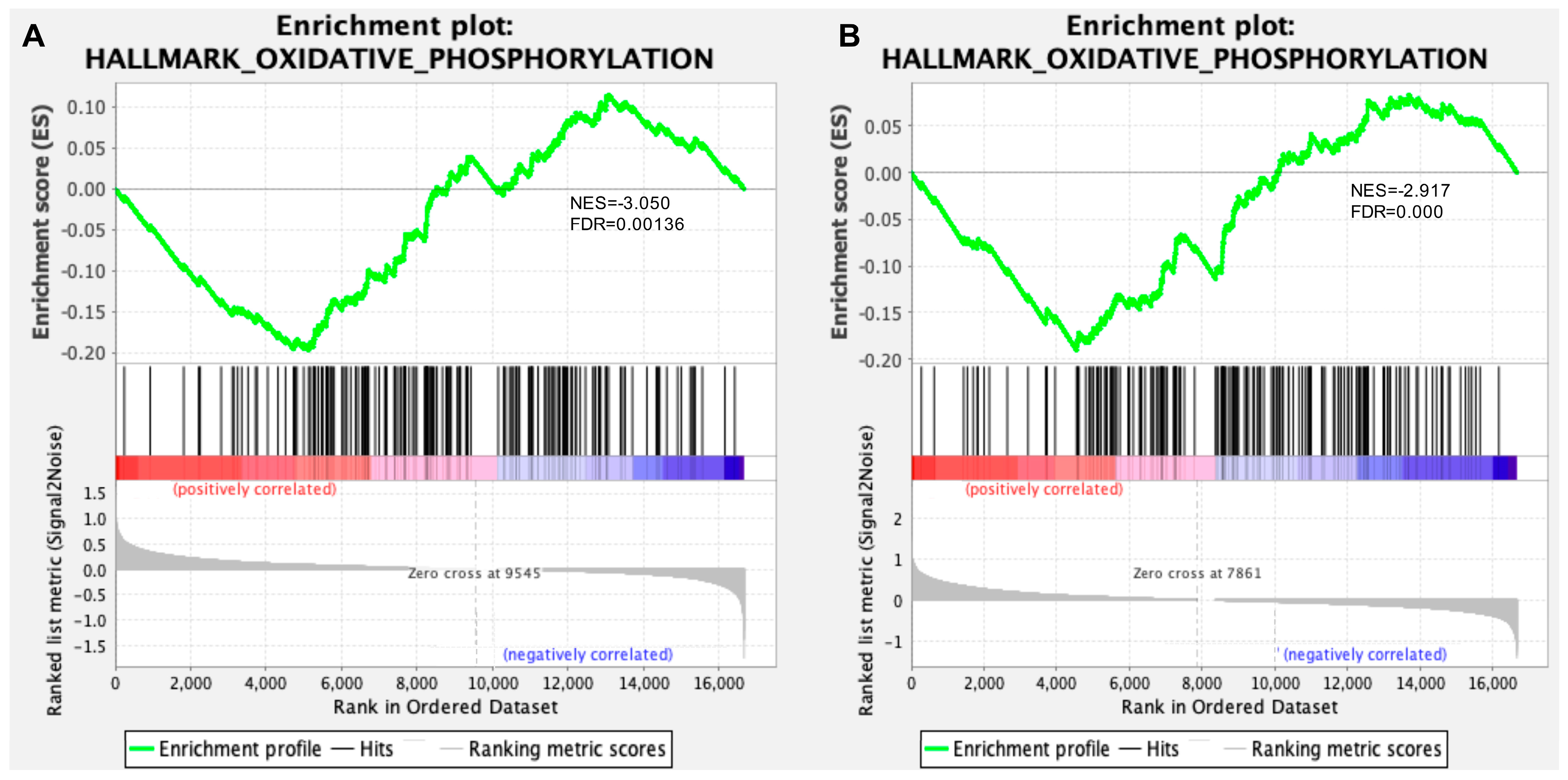
3.1. Transcriptomic Analysis of Hippocampus and Retina from Symptomatic Tg2576 AD Mice Revealed Defective Mitochondrial Bioenergetics

To gain a mechanistic understanding of bioenergetic dysfunctions in AD from a systems biology perspective, we performed RNA sequencing on hippocampal and retinal specimens from Tg2576 mice at 6 months of age compared with their wt littermates, followed by GSEA analysis to functionally interpret gene expression data. Sequencing metrics for each sample (such as depth of coverage and on-target rate) are reported in Supplementary Table S2.
Compared to a DEG-based functional analysis that depends heavily on an arbitrary cutoff to identify significant genes, with a consequent possible underestimation of the biological importance of genes outside of the DEG selection criteria, the GSEA approach uses all of the information about the genes in an experiment to investigate the underlying biological mechanisms. As expected, GSEA results revealed the “hallmark_oxidative_phosphorylation” gene set as the most statistically significant down-regulated gene set in both the hippocampus and retina of Tg2576 AD mice in comparison to their wt littermates (Figure 1, Supplementary Tables S3 and S4). This gene set included genes involved in the regulation of mitochondrial transport and membrane permeability, as well as in various energy metabolism pathways, including lipid oxidation, pyruvate metabolism, mitochondrial electron transport chain and the ATP metabolic process.
A leading-edge subset analysis was then performed to identify the core set of genes that accounts for the gene set enrichment signal. Of the 176 genes associated with oxidative phosphorylation/mitochondrial function, 69 genes were down-regulated in the hippocampus and 106 were down-regulated in the retina of Tg2576 AD in comparison with wt counterparts (Figure 2, Supplementary Figures S1 and S2). Of note, a group of 25 genes were commonly down-regulated between the two tissues and included regulators of the mitochondrial organization and transport (i.e., Rhot1, Rhot2, Grpel1, Slc25a20, Immt), fatty acid β-oxidation and pyruvate/lactate metabolism (i.e., Eci1, Ldha, Sdha, Lrpprc, Hadha) and the electron transport chain and ATP metabolic process (i.e., Cox4i1, Cox7a2, Ndufs1, Ndufs2, Ndufa5, Ndufb2, Ndufa9) (Figure 2).

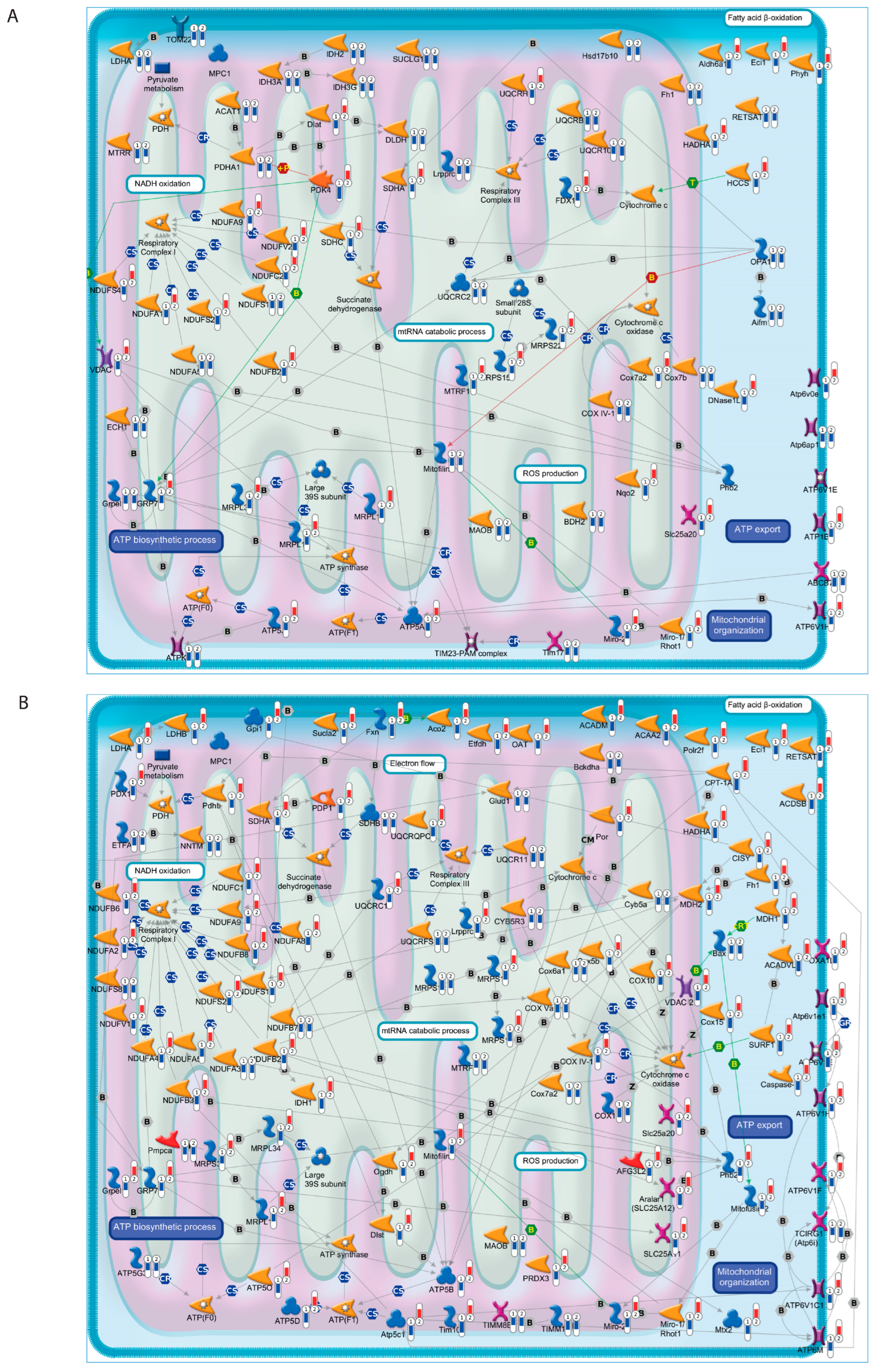
3.2. Defective Mitochondrial Bioenergetics Are Recovered by 12A12mAb-Mediated Neuroprotection in Both the Hippocampus and Retina of the AD Mouse Model

We have previously demonstrated that in vivo administration of the monoclonal antibody 12A12 (12A12mAb), which specifically neutralizes the neurotoxic NH2htau peptide, relieves behavioral and neuropathological hallmarks associated with animals’ AD phenotype [42] and normalizes the basal level of L-lactate [36], which may accumulate along the glycolytic route in response to mitochondrial impairment. To investigate the effects of 12A12mAb on mitochondrial bioenergetics, we performed RNA sequencing followed by GSEA analysis on hippocampal and retinal specimens from Tg2576 mice at 6 months of age after immunization with 12A12mAb and compared these with their not-immunized counterpart and wt littermates.
In line with the toxic action of the NH2htau peptide on mitochondria functions, we found that the expression levels of the majority of core set genes associated with oxidative phosphorylation/mitochondrial function genes (37/69 in hippocampus and 81/106 in retina) were markedly reverted in the Tg2576 AD mouse model following 12A12mAb treatment (Figure 2 and Supplementary Figure S1). Within this context, interesting examples are the coordinated down-regulated expression of a large number of genes that encode the mitochondrial respiratory chain complexes I (Ndufa1, Ndufa2, Ndufa3, Ndufa5, Ndufa8, Ndufa9, Ndufb2, Ndufb3, Ndufb7, Ndufc2, Ndufs1, Ndufs2, Ndufs3, Ndufs4, Ndufs8, Ndufs7, Ndufv1, Ndufv2), II (Sdha, Sdhb, Sdhc), III (Uqcrc1, Uqcrc2, Uqcr10, Uqcr11, Uqcrfs1, Uqcrb, Uqcrh, Uqcrq), IV (Cox4i1, Cox5a, Cox5b, Cox6a1, Cox7a2, Cox7b, Cox10, Cox11, Cox15) and V (Atp5a1, Atp5b, Atp1b1, Atp5c1, Atp5d, Atp5g3, Atp5j, Atp5j2, Atp5o) in the hippocampus and/or retina of Tg2576 AD mice whose alterations were recovered following in vivo administration of 12A12mAb (Figure 2, Supplementary Figures S1 and S2). Several genes encoding proteins involved in the maintenance of mitochondrial membrane potential (Atp6v0e, Atp6v1e1, Atp6v1c1, Atp6v1d, Atp6v1f, Atp6v1h, Atp6v1h, Atp6ap1, Oxa1l, Slc25a3, Slc25a12, Slc25a20, Slc25a20, Slc25a11, Timm8b, Timm10, Timm17a, Tomm22, Timm13, Tcirg1), as well as in the mtRNA stabilizing protein (Lrpprc) and mitochondrial redox state (Nnt), were also down-regulated in Tg2576 mice and their expression was strongly rescued by 12A12mAb immunization (Figure 2, Supplementary Figures S1 and S2). A group of seven core set genes (Atp6v1h, Eci1, Hspa, Mrpl15, Ndufa9, Ndufb2, Rhot1), showing an increased expression following 12A12mAb treatment in both the retina and hippocampus, were selected for RNA-seq transcription validation using qRT-PCR (Supplementary Figure S3). The relative expression patterns of the tested core set genes were found to be positively correlated with the fold change variations obtained from the RNA-seq results (Supplementary Figure S4).

4. Discussion

Although multiple lines of evidence implicate energy deficiency and mitochondrial dysfunction as early pathomechanisms in AD, the molecular mechanisms leading to mitochondrial failure are still to be further investigated and understood [6,7,8,9,19,20,21,23,26,27,29,31,32,33,34,43]. The purpose of this study was to provide a systems biology characterization of the molecular mechanisms underlying bioenergetic dysfunctions in AD. To address this, we analyzed transcriptomes (RNA-seq data) of the hippocampus and retina of Tg2576 AD mice in comparison with their wt littermates, assessing the functional relevance of their gene expression changes in the context of mitochondrial bioenergetic processes.
In line with findings that emerged from our previous work [36], our transcriptomic analysis revealed a comprehensive decrease in the expression of genes involved in mitochondrial/oxidative phosphorylation pathways both in the hippocampus and retina of Tg2576 AD mice (Figure 1), further sustaining the essential role of mitochondria bioenergetic dysfunction in the pathogenesis of AD and supporting its potential utility as a disease biomarker.
Among the core set genes of the mitochondrial oxidative phosphorylation pathway, we observed the coordinated downregulation of genes encoding components of mitochondrial pyruvate metabolism/catabolism, such as pyruvate dehydrogenase complex (Pdha1, Pdhb, Pdk4, Pdp1, Pdhx, Dlat, Dldh, Dlst, Acat1), succinate dehydrogenase (Sdha, Sdhb, Sdhc), lactate dehydrogenase (Ldha, Ldhb) and the mitochondrial fatty acid β-oxidation pathway (Aldh6a1, Eci1, Hsd17b10, Hadha, Retsat, Acadsb, Acadm, Acadvl, Acca2, Aco2, Cpt1a, Oat, Etfdh, Bckdha) (Figure 2). Significant reductions in the expression and activity of pyruvate dehydrogenase complex and lactate dehydrogenase were previously reported in post mortem brain tissues from Alzheimer’s patients, as well as in animal models of AD, demonstrating how the accumulation of lactate due to these metabolic defects may impair neuronal bioenergetics and result in cognitive decline and neurodegeneration [7,44,45,46,47,48,49,50,51,52,53]. In this regard, our previous functional and metabolic findings highlight a general decline in glucose utilization and elevated levels of L-lactate both in hippocampal and, even more, retinal homogenates from Tg2576 AD mice in comparison with their wild-type littermates [36].
A decreased expression of several components of the mitochondrial respiratory chain complexes I (Ndufa1, Ndufa2, Ndufa3, Ndufa5, Ndufa8, Ndufa9, Ndufb2, Ndufb3, Ndufb7, Ndufc2, Ndufs1, Ndufs2, Ndufs3, Ndufs4, Ndufs8, Ndufs7, Ndufv1, Ndufv2), II (Sdha, Sdhb, Sdhc), III (Uqcrc1, Uqcrc2, Uqcr10, Uqcr11, Uqcrfs1, Uqcrb, Uqcrh, Uqcrq), IV (Cox4i1, Cox5a, Cox5b, Cox6a1, Cox7a2, Cox7b, Cox10, Cox11, Cox15) and V (Atp5a1, Atp5b, Atp1b1, Atp5c1, Atp5d, Atp5g3, Atp5j, Atp5j2, Atp5o) was also found in the hippocampus and/or retina of Tg2576 AD mice. Differential expression of these mitochondrial complexes has been previously reported in brain and peripheral tissues of AD patients and animal models, and their functional defects were proposed as a driver of AD pathology by determining a decreased cellular energy production, an increased ROS level and a depolarized mitochondrial membrane, which in turn cause a dramatic effect on ATP production and protein transport across mitochondrial membranes [54,55,56,57,58,59,60]. Dysregulation of mitochondrial redox states in AD also emerged by the decreased expression of nicotinamide nucleotide transhydrogenase (Nnt) in the retina of Tg2576 mice (Figure 2). Nnt encodes a key anti-oxidative enzyme that regenerates NADPH from NADH, and mutations or deletions in this gene impair mitochondrial function, causing a shift to an oxidized redox state that is deleterious in AD [61,62].
As previously demonstrated, Aβ deposition promotes dysfunction of mitochondria also by altering membrane permeability, reducing membrane potential and mtRNA stability and increasing ROS generation, eventually leading to nerve cell death [63,64,65,66]. In agreement with these findings, we observed a reduced expression in Tg2576 mice of several genes encoding proteins involved in the maintenance of mitochondrial membrane potential (Atp6v0e, Atp6v1e1, Atp6v1c1, Atp6v1d, Atp6v1f, Atp6v1h, Atp6v1h, Atp6ap1, Oxa1l, Slc25a3, Slc25a12, Slc25a20, Slc25a20, Slc25a11, Timm8b, Timm10, Timm17a, Tomm22, Timm13, Tcirg1), as well as in mitochondrial transport and organization (Lrpprc, Rhot1, Rhot2) (Figure 2). Consistent with our findings, deficiency of the vacuolar (H+) ATPase (V-ATPase), a large multi-subunit complex of ATP-driven proton pumps, was detected in AD patients and animal models and associated with defective cognitive functions by triggering defects in oxidative phosphorylation, autophagy and axonal transport and synaptic transmission alterations [65,67,68,69,70,71,72,73]. Although the role of the SLC25 family of mitochondrial transporters in the brain is fairly unknown, a reduced expression of some of its members was detected in the brain of AD patients and associated with tau hyperphosphorylation and Aβ deposition [74,75,76]. A significant reduction in LRPPRC, a known mtRNA-stabilizing protein, was found in AD brain samples, while its knockdown causes a general decrease in mitochondrial mRNA levels, impaired translation and a general decrease in respiratory chain complexes [77,78]. The outer mitochondrial membrane proteins Miro1/2 (encoded by the Rhot1 and Rhot genes, respectively) play a vital role in the maintenance of mitochondrial dynamics, calcium homeostasis, axonal transport and cellular energy generation, and their involvement in the pathogenesis of AD has recently been proposed [79,80,81,82].
With the aim to validate our results, we applied a GSEA analysis to an independent data set deposited in GEO (accession number: GSE31624) that includes gene expression profiles in the hippocampus of 5-month-old heterozygous Tg2576 mice and wt littermates. This analysis confirmed “oxidative phosphorylation” as the most down-regulated gene set in the hippocampus of early symptomatic Tg2576 mice, further supporting the relevance of this mechanism in AD pathology (Supplementary Table S5). In addition, we also compared our gene expression data with those detected in the cerebral cortex of Tg2576 mice long before (2 months old), immediately before (5 months old) and after (18 months old) the appearance of amyloid pathology and cognitive impairment in order to establish whether emerging mitochondrial dysfunction can be detected early in the course of AD and represent a valid disease biomarker [24]. To this end, our comparative analysis identified some of our core set genes (Atp6ap1, Atp6v1c1, Cox4i1, Cox7a2, Cs, Gpi1, Sucla2, Uqcrc1) were significantly down-regulated in the cerebral cortex of Tg2576 mice during the earliest pre-symptomatic stages, supporting their potential role in early-stage AD diagnosis [24].
Besides providing new insights into the importance of mitochondrial injury in the etiology and pathogenesis of AD, our results also offer interesting clues on the possibility of targeting these mitochondrial defects to counteract neurodegeneration in AD. In our previous work [36], we showed that the neuroprotective effect exerted in Tg2576 by the in vivo administration of 12A12mAb, a monoclonal antibody that neutralizes the neurotoxic NH2htau peptide, is associated with the mitigation of mitochondrial energetic dyshomeostasis, both in the hippocampus and retina. In line with these results, we found that the expression levels of the majority of core set genes (retina: 76%; hippocampus: 53%) were reverted to their normal level after treatment with the 12A12mAb tau antibody (Figure 2 and Supplementary Figure S1), further supporting mitochondrial metabolism as a key mechanism involved in 12A12mAb-mediated neuroprotection. Concerning the mechanistic link between the administration of 12A12mAb targeting the NH2htau peptide and the transcriptomic changes in the gene expression of mitochondrial function, it is noteworthy that a sizeable amount of tau, under physiological conditions, binds chromatin and nucleic acids. However, it seems that pathological (hyperphosphorylated and/or truncated) tau species accumulate in the nucleus of neurons, locally exerting a harmful effect that further accelerates neurodegeneration [83,84].

5. Conclusions

Our findings, for the first time, offer a systems biology perspective of disrupted mitochondrial bioenergetics in AD pathology and its recovery following 12A12mAb treatment, highlighting its potential utility for the development of useful early biomarkers and future disease-modifying therapies for AD.

Supplementary Materials

The following supporting information can be downloaded at: https://www.mdpi.com/article/10.3390/cells12182254/s1. Figure S1. Heat maps showing the expression levels (fold change) of the core set genes in both the hippocampus and retina under various experimental comparisons. Figure S2. Heatmaps showing the expression levels (log2 of counts normalized by RPM) of core set genes in the hippocampus and retina of each sample. Table S1. Primers used for real-time RT-PCR. Figure S3. Validation of RNA sequencing by RT-qPCR. Figure S4. Comparison of RNA-Seq analysis with RT-qPCR validation assays. Table S2. Sequencing coverage metrics for each sample. Table S3. Gene set enrichment analysis (GSEA) results of transcriptome data in the hippocampus and retina of Tg2576 vs. wt mice. Table S4. Detailed results of the leading-edge set analysis in GSEA for the enriched gene set “OXIDATIVE_PHOSPHORYLATION” in the hippocampus and retina of Tg2576 vs. wt mice. Table S5. Gene set enrichment analysis (GSEA) results of transcriptome data in the hippocampus of 5-month-old heterozygous Tg2576 mice and wt littermates (GSE31624).

Author Contributions

Conceptualization, S.C., G.A. and P.C.; methodology, G.A., V.L.C., M.G., V.L. and G.M.; software, G.M.; formal analysis, G.M.; resources, P.C., S.C. and G.A.; data curation, G.M.; writing—original draft preparation, G.M.; writing—review and editing, G.M., S.C., V.L.C., M.G., G.A., V.L. and P.C.; supervision, S.C.; project administration, S.C.; funding acquisition, S.C. All authors have read and agreed to the published version of the manuscript.

Funding

This research was funded by project “An integrated multi-omics approach to study neurodegeneration” (DSB.AD007.304). G.A. was supported by Regione Lazio, POR FESR Lazio 2014–2020. “Progetti di Gruppi di Ricerca 2020” (Determinazione dirigenziale n.G08487 del 19 July 2020) Project T0002E0001 G04014_13_04_2021 and Alzheimer’s Association Research Grant—Proposal ID: 971925 and Ministry of Health Project RF-2021-12374301 and PNRR (National Plan for Recovery and Resilience Next Generation EU)—PE Neuroscience project-MNESYS “A multiscale integrated approach to the study of the nervous system in health and disease”. This work was also supported (in part) by Fondo Ordinario Enti (FOE D.M865/2019) funds in the framework of a collaboration agreement between the Italian National Research Council and EBRI.

Institutional Review Board Statement

The animal study protocol was approved by the Italian Ministry of Health (protocol # 1038-2020-PR).

Informed Consent Statement

Not applicable.

Data Availability Statement

The data that support the findings of this study have been submitted to the NCBI Gene Expression Omnibus (GEO) with the accession number GSE223593.

Acknowledgments

The authors gratefully acknowledge Cristina Calì, Alfia Corsino, Maria Patrizia D’Angelo and Francesco Marino for their administrative and technical support.

Conflicts of Interest

The authors declare no conflict of interest.

References

  1. Alzheimer’s Association. 2015 Alzheimer’s disease facts and figures. Alzheimer’s Dement. 2015, 11, 332–384. [Google Scholar] [CrossRef]
  2. Murphy, M.P.; Levine, H. Alzheimer’s Disease and the Amyloid-β Peptide. J. Alzheimer’s Dis. 2010, 19, 311–323. [Google Scholar] [CrossRef]
  3. Kocahan, S.; Doǧan, Z. Mechanisms of Alzheimer’s Disease Pathogenesis and Prevention: The Brain, Neural Pathology, N-methyl-D-aspartate Receptors, Tau Protein and Other Risk Factors. Clin. Psychopharmacol. Neurosci. 2017, 15, 1–8. [Google Scholar] [CrossRef] [PubMed]
  4. Musiek, E.S.; Holtzman, D.M. Three dimensions of the amyloid hypothesis: Time, space and “wingmen”. Nat. Neurosci. 2015, 18, 800–806. [Google Scholar] [CrossRef] [PubMed]
  5. Austad, S.N.; Ballinger, S.; Buford, T.W.; Carter, C.S.; Smith, D.L.; Darley-Usmar, V.; Zhang, J. Targeting whole body metabolism and mitochondrial bioenergetics in the drug development for Alzheimer’s disease. Acta Pharm. Sin. B 2022, 12, 511–531. [Google Scholar] [CrossRef] [PubMed]
  6. Wang, W.; Zhao, F.; Ma, X.; Perry, G.; Zhu, X. Mitochondria dysfunction in the pathogenesis of Alzheimer’s disease: Recent advances. Mol. Neurodegener. 2020, 15, 30. [Google Scholar] [CrossRef]
  7. Yao, J.; Irwin, R.W.; Zhao, L.; Nilsen, J.; Hamilton, R.T.; Brinton, R.D. Mitochondrial bioenergetic deficit precedes Alzheimer’s pathology in female mouse model of Alzheimer’s disease. Proc. Natl. Acad. Sci. USA 2009, 106, 14670–14675. [Google Scholar] [CrossRef]
  8. Swerdlow, R.H. The Mitochondrial Hypothesis: Dysfunction, Bioenergetic Defects, and the Metabolic Link to Alzheimer’s Disease. Int. Rev. Neurobiol. 2020, 154, 207–233. [Google Scholar] [CrossRef] [PubMed]
  9. Cenini, G.; Voos, W. Mitochondria as potential targets in Alzheimer disease therapy: An update. Front. Pharmacol. 2019, 10, 902. [Google Scholar] [CrossRef] [PubMed]
  10. Kann, O.; Kovács, R. Mitochondria and neuronal activity. Am. J. Physiol.-Cell Physiol. 2007, 292, 641–657. [Google Scholar] [CrossRef] [PubMed]
  11. Trigo, D.; Avelar, C.; Fernandes, M.; Sá, J.; da Cruz e Silva, O. Mitochondria, energy, and metabolism in neuronal health and disease. FEBS Lett. 2022, 596, 1095–1110. [Google Scholar] [CrossRef] [PubMed]
  12. Lezi, E.; Swerdlow, R.H. Mitochondria in Neurodegeneration. Adv. Exp. Med. Biol. 2012, 942, 269–286. [Google Scholar] [CrossRef]
  13. Jadiya, P.; Garbincius, J.F.; Elrod, J.W. Reappraisal of metabolic dysfunction in neurodegeneration: Focus on mitochondrial function and calcium signaling. Acta Neuropathol. Commun. 2021, 9, 124. [Google Scholar] [CrossRef] [PubMed]
  14. Marmolejo-Garza, A.; Medeiros-Furquim, T.; Rao, R.; Eggen, B.J.; Boddeke, E.; Dolga, A.M. Transcriptomic and epigenomic landscapes of Alzheimer’s disease evidence mitochondrial-related pathways. Biochim. Biophys. Acta-Mol. Cell Res. 2022, 1869, 119326. [Google Scholar] [CrossRef]
  15. Harerimana, N.V.; Paliwali, D.; Romero-Molina, C.; Bennett, D.A.; Pa, J.; Goate, A.; Swerdlow, R.H.; Andrews, S.J. The role of mitochondrial genome abundance in Alzheimer’s disease. Alzheimer’s Dement. 2023, 19, 2069–2083. [Google Scholar] [CrossRef]
  16. Mani, S.; Swargiary, G.; Singh, M.; Agarwal, S.; Dey, A.; Ojha, S.; Jha, N.K. Mitochondrial defects: An emerging theranostic avenue towards Alzheimer’s associated dysregulations. Life Sci. 2021, 285, 119985. [Google Scholar] [CrossRef]
  17. Piras, I.S.; Krate, J.; Delvaux, E.; Nolz, J.; Mastroeni, D.F.; Persico, A.M.; Jepsen, W.M.; Beach, T.G.; Huentelman, M.J.; Coleman, P.D.; et al. Transcriptome Changes in the Alzheimer’s Disease Middle Temporal Gyrus: Importance of RNA Metabolism and Mitochondria-Associated Membrane Genes. J. Alzheimer’s Dis. 2019, 70, 691–713. [Google Scholar] [CrossRef]
  18. Mastroeni, D.; Khdour, O.M.; Delvaux, E.; Nolz, J.; Olsen, G.; Berchtold, N.; Cotman, C.; Hecht, S.M.; Coleman, P.D. Nuclear but not mitochondrial-encoded oxidative phosphorylation genes are altered in aging, mild cognitive impairment, and Alzheimer’s disease. Alzheimer’s Dement. 2017, 13, 510–519. [Google Scholar] [CrossRef]
  19. Manczak, M.; Anekonda, T.S.; Henson, E.; Park, B.S.; Quinn, J.; Reddy, P.H. Mitochondria are a direct site of Aβ accumulation in Alzheimer’s disease neurons: Implications for free radical generation and oxidative damage in disease progression. Hum. Mol. Genet. 2006, 15, 1437–1449. [Google Scholar] [CrossRef]
  20. Reddy, P.H.; Tripathi, R.; Troung, Q.; Tirumala, K.; Reddy, T.P.; Anekonda, V.; Shirendeb, U.P.; Calkins, M.J.; Reddy, A.P.; Mao, P.; et al. Abnormal mitochondrial dynamics and synaptic degeneration as early events in Alzheimer’s disease: Implications to mitochondria-targeted antioxidant therapeutics. Biochim. Biophys. Acta-Mol. Basis Dis. 2012, 1822, 639–649. [Google Scholar] [CrossRef] [PubMed]
  21. Lunnon, K.; Keohane, A.; Pidsley, R.; Newhouse, S.; Riddoch-Contreras, J.; Thubron, E.B.; Devall, M.; Soininen, H.; Kłoszewska, I.; Mecocci, P.; et al. Mitochondrial genes are altered in blood early in Alzheimer’s disease. Neurobiol. Aging 2017, 53, 36–47. [Google Scholar] [CrossRef] [PubMed]
  22. Gillardon, F.; Rist, W.; Kussmaul, L.; Vogel, J.; Berg, M.; Danzer, K.; Kraut, N.; Hengerer, B. Proteomic and functional alterations in brain mitochondria from Tg2576 mice occur before amyloid plaque deposition. Proteomics 2007, 7, 605–616. [Google Scholar] [CrossRef]
  23. Varghese, M.; Zhao, W.; Wang, J.; Cheng, A.; Qian, X.; Chaudhry, A.; Ho, L.; Pasinetti, G.M. Mitochondrial bioenergetics is defective in presymptomatic Tg2576 AD mice. Transl. Neurosci. 2011, 2, 1–5. [Google Scholar] [CrossRef]
  24. Reddy, P.H.; McWeeney, S.; Park, B.S.; Manczak, M.; Gutala, R.V.; Partovi, D.; Jung, Y.; Yau, V.; Searles, R.; Mori, M.; et al. Gene expression profiles of transcripts in amyloid precursor protein transgenic mice: Up-regulation of mitochondrial metabolism and apoptotic genes is an early cellular change in Alzheimer’s disease. Hum. Mol. Genet. 2004, 13, 1225–1240. [Google Scholar] [CrossRef]
  25. Amadoro, G.; Latina, V.; Corsetti, V.; Calissano, P. N-terminal tau truncation in the pathogenesis of Alzheimer’s disease (AD): Developing a novel diagnostic and therapeutic approach. Biochim. Biophys. Acta-Mol. Basis Dis. 2020, 1866, 165584. [Google Scholar] [CrossRef]
  26. Amadoro, G.; Corsetti, V.; Atlante, A.; Florenzano, F.; Capsoni, S.; Bussani, R.; Mercanti, D.; Calissano, P. Interaction between NH(2)-tau fragment and Aβ in Alzheimer’s disease mitochondria contributes to the synaptic deterioration. Neurobiol. Aging 2012, 33, 833.e1–833.e25. [Google Scholar] [CrossRef]
  27. Amadoro, G.; Corsetti, V.; Stringaro, A.; Colone, M.; D’aguanno, S.; Meli, G.; Ciotti, M.; Sancesario, G.; Cattaneo, A.; Bussani, R.; et al. A NH2 Tau Fragment Targets Neuronal Mitochondria at AD Synapses: Possible Implications for Neurodegeneration. J. Alzheimer’s Dis. 2010, 21, 445–470. [Google Scholar] [CrossRef]
  28. Latina, V.; Giacovazzo, G.; Cordella, F.; Balzamino, B.O.; Micera, A.; Varano, M.; Marchetti, C.; Malerba, F.; Florio, R.; Ercole, B.B.; et al. Systemic delivery of a specific antibody targeting the pathological N-terminal truncated tau peptide reduces retinal degeneration in a mouse model of Alzheimer’s Disease. Acta Neuropathol. Commun. 2021, 9, 38. [Google Scholar] [CrossRef]
  29. Atlante, A.; Amadoro, G.; Bobba, A.; de Bari, L.; Corsetti, V.; Pappalardo, G.; Marra, E.; Calissano, P.; Passarella, S. A peptide containing residues 26–44 of tau protein impairs mitochondrial oxidative phosphorylation acting at the level of the adenine nucleotide translocator. Biochim. Biophys. Acta-Bioenerg. 2008, 1777, 1289–1300. [Google Scholar] [CrossRef] [PubMed]
  30. Corsetti, V.; Florenzano, F.; Atlante, A.; Bobba, A.; Ciotti, M.T.; Natale, F.; Della Valle, F.; Borreca, A.; Manca, A.; Meli, G.; et al. NH2-truncated human tau induces deregulated mitophagy in neurons by aberrant recruitment of Parkin and UCHL-1: Implications in Alzheimer’s disease. Hum. Mol. Genet. 2015, 24, 3058–3081. [Google Scholar] [CrossRef]
  31. Szabo, L.; Eckert, A.; Grimm, A. Insights into Disease-Associated Tau Impact on Mitochondria. Int. J. Mol. Sci. 2020, 21, 6344. [Google Scholar] [CrossRef] [PubMed]
  32. Pérez, M.J.; Jara, C.; Quintanilla, R.A. Contribution of Tau Pathology to Mitochondrial Impairment in Neurodegeneration. Front. Neurosci. 2018, 12, 441. [Google Scholar] [CrossRef] [PubMed]
  33. Cheng, Y.; Bai, F. The association of tau with mitochondrial dysfunction in Alzheimer’s disease. Front. Neurosci. 2018, 12, 163. [Google Scholar] [CrossRef]
  34. Torres, A.; Rivera, B.; Polanco, C.; Jara, C.; Tapia-Rojas, C. Phosphorylated tau as a toxic agent in synaptic mitochondria: Implications in aging and Alzheimer’s disease. Neural Regen. Res. 2022, 17, 1645–1651. [Google Scholar] [CrossRef] [PubMed]
  35. Amadoro, G.; Latina, V.; Calissano, P. A long story for a short peptide: Therapeutic efficacy of a cleavage-specific tau antibody. Neural Regen. Res. 2021, 16, 2417–2419. [Google Scholar] [CrossRef]
  36. Latina, V.; Atlante, A.; Malerba, F.; La Regina, F.; Balzamino, B.O.; Micera, A.; Pignataro, A.; Stigliano, E.; Cavallaro, S.; Calissano, P.; et al. The Cleavage-Specific Tau 12A12mAb Exerts an Anti-Amyloidogenic Action by Modulating the Endocytic and Bioenergetic Pathways in Alzheimer’s Disease Mouse Model. Int. J. Mol. Sci. 2023, 24, 9683. [Google Scholar] [CrossRef]
  37. Mootha, V.K.; Lindgren, C.M.; Eriksson, K.F.; Subramanian, A.; Sihag, S.; Lehar, J.; Puigserver, P.; Carlsson, E.; Ridderstråle, M.; Laurila, E.; et al. PGC-1α-responsive genes involved in oxidative phosphorylation are coordinately downregulated in human diabetes. Nat. Genet. 2003, 34, 267–273. [Google Scholar] [CrossRef]
  38. Subramanian, A.; Tamayo, P.; Mootha, V.K.; Mukherjee, S.; Ebert, B.L.; Gillette, M.A.; Paulovich, A.; Pomeroy, S.L.; Golub, T.R.; Lander, E.S.; et al. Gene set enrichment analysis: A knowledge-based approach for interpreting genome-wide expression profiles. Proc. Natl. Acad. Sci. USA 2005, 102, 15545–15550. [Google Scholar] [CrossRef] [PubMed]
  39. Hsiao, K.; Chapman, P.; Nilsen, S.; Eckman, C.; Harigaya, Y.; Younkin, S.; Yang, F.; Cole, G. Correlative Memory Deficits, Aβ Elevation, and Amyloid Plaques in Transgenic Mice. Science 1996, 274, 99–103. [Google Scholar] [CrossRef]
  40. Sasaguri, H.; Nilsson, P.; Hashimoto, S.; Nagata, K.; Saito, T.; De Strooper, B.; Hardy, J.; Vassar, R.; Winblad, B.; Saido, T.C. APP mouse models for Alzheimer’s disease preclinical studies. EMBO J. 2017, 36, 2473–2487. [Google Scholar] [CrossRef] [PubMed]
  41. Liberzon, A.; Birger, C.; Thorvaldsdóttir, H.; Ghandi, M.; Mesirov, J.P.; Tamayo, P. The Molecular Signatures Database (MSigDB) hallmark gene set collection. Cell Syst. 2015, 1, 417–425. [Google Scholar] [CrossRef]
  42. Corsetti, V.; Borreca, A.; Latina, V.; Giacovazzo, G.; Pignataro, A.; Krashia, P.; Natale, F.; Cocco, S.; Rinaudo, M.; Malerba, F.; et al. Passive immunotherapy for N-truncated tau ameliorates the cognitive deficits in two mouse Alzheimer’s disease models. Brain Commun. 2020, 2, fcaa039. [Google Scholar] [CrossRef]
  43. Misrani, A.; Tabassum, S.; Yang, L. Mitochondrial Dysfunction and Oxidative Stress in Alzheimer’s Disease. Front. Aging Neurosci. 2021, 13, 617588. [Google Scholar] [CrossRef]
  44. Dong, Y.; Brewer, G.J. Global Metabolic Shifts in Age and Alzheimer’s Disease Mouse Brains Pivot at NAD+/NADH Redox Sites. J. Alzheimer’s Dis. 2019, 71, 119–140. [Google Scholar] [CrossRef]
  45. Newington, J.T.; Rappon, T.; Albers, S.; Wong, D.Y.; Rylett, R.J.; Cumming, R.C. Overexpression of Pyruvate Dehydrogenase Kinase 1 and Lactate Dehydrogenase A in Nerve Cells Confers Resistance to Amyloid and Other Toxins by Decreasing Mitochondrial Respiration and Reactive Oxygen Species Production. J. Biol. Chem. 2012, 287, 37245–37258. [Google Scholar] [CrossRef] [PubMed]
  46. Perry, E.K.; Perry, R.H.; Tomlinson, B.E.; Blessed, G.; Gibson, P.H. Coenzyme a-acetylating enzymes in Alzheimer’s disease: Possible cholinergic ‘compartment’ of pyruvate dehydrogenase. Neurosci. Lett. 1980, 18, 105–110. [Google Scholar] [CrossRef] [PubMed]
  47. Rossi, A.; Rigotto, G.; Valente, G.; Giorgio, V.; Basso, E.; Filadi, R.; Pizzo, P. Defective Mitochondrial Pyruvate Flux Affects Cell Bioenergetics in Alzheimer’s Disease-Related Models. Cell Rep. 2020, 30, 2332–2348.e10. [Google Scholar] [CrossRef]
  48. Harris, R.A.; Tindale, L.; Lone, A.; Singh, O.; Macauley, S.L.; Stanley, M.; Holtzman, D.M.; Bartha, R.; Cumming, R.C. Aerobic Glycolysis in the Frontal Cortex Correlates with Memory Performance in Wild-Type Mice But Not the APP/PS1 Mouse Model of Cerebral Amyloidosis. J. Neurosci. 2016, 36, 1871–1878. [Google Scholar] [CrossRef] [PubMed]
  49. Ryu, W.I.; Bormann, M.K.; Shen, M.; Kim, D.; Forester, B.; Park, Y.; So, J.; Seo, H.; Sonntag, K.C.; Cohen, B.M. Brain cells derived from Alzheimer’s disease patients have multiple specific innate abnormalities in energy metabolism. Mol. Psychiatry 2021, 26, 5702–5714. [Google Scholar] [CrossRef]
  50. Park, J.S.; Saeed, K.; Jo, M.H.; Kim, M.W.; Lee, H.J.; Park, C.B.; Lee, G.; Kim, M.O. LDHB Deficiency Promotes Mitochondrial Dysfunction Mediated Oxidative Stress and Neurodegeneration in Adult Mouse Brain. Antioxidants 2022, 11, 261. [Google Scholar] [CrossRef]
  51. Chen, W.; Sun, X.; Zhan, L.; Zhou, W.; Bi, T. Conditional Knockout of Pdha1 in Mouse Hippocampus Impairs Cognitive Function: The Possible Involvement of Lactate. Front. Neurosci. 2021, 15, 767560. [Google Scholar] [CrossRef]
  52. Stacpoole, P.W. The pyruvate dehydrogenase complex as a therapeutic target for age-related diseases. Aging Cell 2012, 11, 371–377. [Google Scholar] [CrossRef]
  53. Rex Sheu, K.-F.; Kim, Y.-T.; Blass, J.P.; Weksler, M.E. An immunochemical study of the pyruvate dehydrogenase deficit in Alzheimer’s disease brain. Ann. Neurol. 1985, 17, 444–449. [Google Scholar] [CrossRef]
  54. Adav, S.S.; Park, J.E.; Sze, S.K. Quantitative profiling brain proteomes revealed mitochondrial dysfunction in Alzheimer’s disease. Mol. Brain 2019, 12, 8. [Google Scholar] [CrossRef]
  55. Patro, S.; Ratna, S.; Yamamoto, H.A.; Ebenezer, A.T.; Ferguson, D.S.; Kaur, A.; McIntyre, B.C.; Snow, R.; Solesio, M.E. ATP Synthase and Mitochondrial Bioenergetics Dysfunction in Alzheimer’s Disease. Int. J. Mol. Sci. 2021, 22, 11185. [Google Scholar] [CrossRef]
  56. Trumpff, C.; Owusu-Ansah, E.; Klein, H.-U.; Lee, A.; Petyuk, V.; Wingo, T.S.; Wingo, A.P.; Thambisetty, M.; Ferrucci, L.; Seyfried, N.T.; et al. Mitochondrial Respiratory Chain Protein Co-Regulation in the Human Brain. Heliyon 2022, 8, e09353. [Google Scholar] [CrossRef]
  57. Galindo, M.F.; Ikuta, I.; Zhu, X.; Casadesus, G.; Jordán, J. Mitochondrial biology in Alzheimer’s disease pathogenesis. J. Neurochem. 2010, 114, 933–945. [Google Scholar] [CrossRef] [PubMed]
  58. Fišar, Z.; Hroudová, J.; Zvěřová, M.; Jirák, R.; Vaněčková, M. Disruption of mitochondrial respiration and the monoamine neurotransmitter system in Alzheimer’s disease. Res. Sq. 2022; preprint. [Google Scholar] [CrossRef]
  59. Parihar, M.S.; Brewer, G.J. Mitoenergetic failure in Alzheimer disease. Am. J. Physiol.-Cell Physiol. 2007, 292, 8–23. [Google Scholar] [CrossRef] [PubMed]
  60. Parker, W.D.; Parks, J.; Filley, C.M.; Kleinschmidt-Demasters, B.K. Electron transport chain defects in Alzheimer’s disease brain. Neurology 1994, 44, 1090. [Google Scholar] [CrossRef] [PubMed]
  61. Ghosh, D.; Levault, K.R.; Brewer, G.J. Relative importance of redox buffers GSH and NAD(P)H in age-related neurodegeneration and Alzheimer disease-like mouse neurons. Aging Cell 2014, 13, 631–640. [Google Scholar] [CrossRef]
  62. Yin, F.; Sancheti, H.; Cadenas, E. Silencing of nicotinamide nucleotide transhydrogenase impairs cellular redox homeostasis and energy metabolism in PC12 cells. Biochim. Biophys. Acta-Bioenerg. 2012, 1817, 401–409. [Google Scholar] [CrossRef]
  63. Eckert, A.; Schmitt, K.; Götz, J. Mitochondrial dysfunction—The beginning of the end in Alzheimer’s disease? Separate and synergistic modes of tau and amyloid-toxicity. Alzheimer’s Res. Ther. 2011, 3, 15. [Google Scholar] [CrossRef] [PubMed]
  64. Onyango, I.G.; Dennis, J.; Khan, S.M. Mitochondrial Dysfunction in Alzheimer’s Disease and the Rationale for Bioenergetics Based Therapies. Aging Dis. 2016, 7, 201–214. [Google Scholar] [CrossRef] [PubMed]
  65. Zhao, Z.W.; Yuan, Z.; Zhao, D.Q.; Wang, Z.W.; Zhu, F.Q.; Luan, P. The effect of V-ATPase function defects in pathogenesis of Alzheimer’s disease. CNS Neurosci. Ther. 2018, 24, 837–840. [Google Scholar] [CrossRef]
  66. Reiss, A.B.; Ahmed, S.; Dayaramani, C.; Glass, A.D.; Gomolin, I.H.; Pinkhasov, A.; Stecker, M.M.; Wisniewski, T.; De Leon, J. The role of mitochondrial dysfunction in Alzheimer’s disease: A potential pathway to treatment. Exp. Gerontol. 2022, 164, 111828. [Google Scholar] [CrossRef]
  67. Collins, M.P.; Forgac, M. Regulation and function of V-ATPases in physiology and disease. Biochim. Biophys. Acta-Biomembr. 2020, 1862, 183341. [Google Scholar] [CrossRef]
  68. Santos-Pereira, C.; Rodrigues, L.R.; Côrte-Real, M. Emerging insights on the role of V-ATPase in human diseases: Therapeutic challenges and opportunities. Med. Res. Rev. 2021, 41, 1927–1964. [Google Scholar] [CrossRef]
  69. Eaton, A.F.; Merkulova, M.; Brown, D. The H+-ATPase (V-ATPase): From proton pump to signaling complex in health and disease. Am. J. Physiol.-Cell Physiol. 2021, 320, C392–C414. [Google Scholar] [CrossRef]
  70. Williamson, W.R.; Hiesinger, P.R. On the role of v-ATPase V0a1–dependent degradation in Alzheimer Disease. Commun. Integr. Biol. 2010, 3, 604–607. [Google Scholar] [CrossRef]
  71. Zhou, Z.; Bai, J.; Zhong, S.; Zhang, R.; Kang, K.; Zhang, X.; Xu, Y.; Zhao, C.; Zhao, M. Downregulation of ATP6V1A Involved in Alzheimer’s Disease via Synaptic Vesicle Cycle, Phagosome, and Oxidative Phosphorylation. Oxid. Med. Cell. Longev. 2021, 2021, 5555634. [Google Scholar] [CrossRef] [PubMed]
  72. Kim, S.-H.; Cho, Y.-S.; Kim, Y.; Park, J.; Yoo, S.-M.; Gwak, J.; Kim, Y.; Gwon, Y.; Kam, T.-i.; Jung, Y.-K. Endolysosomal impairment by binding of amyloid beta or MAPT/Tau to V-ATPase and rescue via the HYAL-CD44 axis in Alzheimer disease. Autophagy 2023, 19, 2318–2337. [Google Scholar] [CrossRef] [PubMed]
  73. Song, Q.; Meng, B.; Xu, H.; Mao, Z. The emerging roles of vacuolar-type ATPase-dependent Lysosomal acidification in neurodegenerative diseases. Transl. Neurodegener. 2020, 9, 17. [Google Scholar] [CrossRef]
  74. Haitina, T.; Lindblom, J.; Renström, T.; Fredriksson, R. Fourteen novel human members of mitochondrial solute carrier family 25 (SLC25) widely expressed in the central nervous system. Genomics 2006, 88, 779–790. [Google Scholar] [CrossRef] [PubMed]
  75. Hawking, Z.L. Alzheimer’s disease: The role of mitochondrial dysfunction and potential new therapies. Biosci. Horizons Int. J. Student Res. 2016, 9, hzw014. [Google Scholar] [CrossRef]
  76. Hu, C.; Tao, L.; Cao, X.; Chen, L. The solute carrier transporters and the brain: Physiological and pharmacological implications. Asian J. Pharm. Sci. 2020, 15, 131–144. [Google Scholar] [CrossRef]
  77. Sasarman, F.; Brunel-Guitton, C.; Antonicka, H.; Wai, T.; Shoubridge, E.A.; Allen, B.; Burelle, Y.; Charron, G.; Coderre, L.; DesRosiers, C.; et al. A Highlights from MBoC Selection: LRPPRC and SLIRP Interact in a Ribonucleoprotein Complex That Regulates Posttranscriptional Gene Expression in Mitochondria. Mol. Biol. Cell 2010, 21, 1315–1434. [Google Scholar] [CrossRef]
  78. Bennett, J.P.; Keeney, P.M. Alzheimer’s and Parkinson’s brain tissues have reduced expression of genes for mtDNA OXPHOS Proteins, mitobiogenesis regulator PGC-1α protein and mtRNA stabilizing protein LRPPRC (LRP130). Mitochondrion 2020, 53, 154–157. [Google Scholar] [CrossRef]
  79. Panchal, K.; Tiwari, A.K. Miro, a Rho GTPase genetically interacts with Alzheimer’s disease-associated genes (Tau, Aβ42 and Appl) in Drosophila melanogaster. Biol. Open 2020, 9, bio049569. [Google Scholar] [CrossRef]
  80. Panchal, K.; Tiwari, A.K. Miro (Mitochondrial Rho GTPase), a key player of mitochondrial axonal transport and mitochondrial dynamics in neurodegenerative diseases. Mitochondrion 2021, 56, 118–135. [Google Scholar] [CrossRef]
  81. Iijima-Ando, K.; Sekiya, M.; Maruko-Otake, A.; Ohtake, Y.; Suzuki, E.; Lu, B.; Iijima, K.M. Loss of Axonal Mitochondria Promotes Tau-Mediated Neurodegeneration and Alzheimer’s Disease–Related Tau Phosphorylation Via PAR-1. PLoS Genet. 2012, 8, e1002918. [Google Scholar] [CrossRef]
  82. Kay, L.; Pienaar, I.S.; Cooray, R.; Black, G.; Soundararajan, M. Understanding Miro GTPases: Implications in the Treatment of Neurodegenerative Disorders. Mol. Neurobiol. 2018, 55, 7352–7365. [Google Scholar] [CrossRef] [PubMed]
  83. Maina, M.B.; Al-Hilaly, Y.K.; Serpell, L.C. Nuclear Tau and Its Potential Role in Alzheimer’s Disease. Biomolecules 2016, 6, 9. [Google Scholar] [CrossRef] [PubMed]
  84. Antón-Fernández, A.; Vallés-Saiz, L.; Avila, J.; Hernández, F. Neuronal nuclear tau and neurodegeneration. Neuroscience 2023, 518, 178–184. [Google Scholar] [CrossRef] [PubMed]
Figure 1. Gene Set Enrichment Analysis (GSEA) supports defective mitochondrial oxidative phosphorylation as a key pathological hallmark of AD. GSEA plots showing enrichment of the “hallmark_oxidative_phosphorylation” gene set in the hippocampus (A) and retina (B) of in comparison to wt controls. The top portion of each GSEA plot shows the running enrichment score for validated genes as it moves down the ranked list. The bottom portion of each plot shows the value of ranking matrices as it moves down the list of ranked genes. The colored horizontal bar indicates a shift from positively correlated genes (red) to negatively correlated genes (blue). The normalized enrichment scores (NES) and false discovery rate q value (FDR q) are indicated. Further details on GSEA plots can be found at Broad Institute web site (http://www.broadinstitute.org/gsea/index.jsp, accessed on 20 April 2023).
Cells 12 02254 g001
Figure 2. Mitochondrial-related core set genes down-regulated in Tg2576 AD mice contribute to 12A12mAb-mediated neuroprotection. Representative functional illustrations of core set genes that chiefly contribute to the down-regulation of the “hallmark_oxidative_phosphorylation” gene set in the (A) hippocampus and (B) retina of Tg2576 AD mice in comparison to wt controls. The pathway map was created using MetaCore Pathway Map Creator tool (GeneGo). Expression levels (fold change) of core set genes in Tg2576 mice vs. wt (thermometer #1) and Tg2576 + mAb vs. Tg2576 mice (thermometer #2) are presented on the map as ‘thermometer-like’ figures (red for up-regulation, blue for down-regulation) and visualized as heatmaps in Supplementary Figures S1 and S2. Further explanations are provided at https://portal.genego.com/legends/MetaCoreQuickReferenceGuide.pdf, accessed on 2 May 2023.
Cells 12 02254 g002
Disclaimer/Publisher’s Note: The statements, opinions and data contained in all publications are solely those of the individual author(s) and contributor(s) and not of MDPI and/or the editor(s). MDPI and/or the editor(s) disclaim responsibility for any injury to people or property resulting from any ideas, methods, instructions or products referred to in the content.

