Article

Denosumab and Zoledronic Acid Differently Affect Circulating Immune Subsets: A Possible Role in the Onset of MRONJ

1
Bone and Dental Bioengineering Laboratory, CIR-Dental School, Department of Surgical Sciences, University of Turin, Via Nizza 230, 10126 Turin, Italy
2
Institute of Sciences and Technologies for Sustainable Energy and Mobility, National Council of Research, 10135 Turin, Italy
3
DIMEAS, Politecnico di Torino, 10129 Turin, Italy
4
CIR-Dental School, Città della Scienza e della Salute, 10126 Turin, Italy
*
Author to whom correspondence should be addressed.
These authors contributed equally to this work.
Cells 2023, 12(20), 2430; https://doi.org/10.3390/cells12202430
Submission received: 8 September 2023 / Revised: 29 September 2023 / Accepted: 8 October 2023 / Published: 11 October 2023

Abstract

:
This work investigated whether the anti-resorptive drugs (ARDs) zoledronic acid (Zol) and denosumab (Dmab) affect differently the levels of circulating immune cell subsets, possibly predicting the risk of developing medication-related ONJ (MRONJ) during the first 18 months of treatment. Blood samples were collected from 10 bone metastatic breast cancer patients receiving cyclin inhibitors at 0, 6, 12, and 18 months from the beginning of Dmab or Zol treatment. Eight breast cancer patients already diagnosed with MRONJ and treated with cyclin inhibitors and ARDs were in the control group. PBMCs were isolated; the trend of circulating immune subsets during the ARD treatment was monitored, and 12 pro-inflammatory cytokines were analyzed in sera using flow cytometry. In Dmab-treated patients, activated T cells were stable or increased, as were the levels of IL-12, TNF-α, GM-CSF, IL-5, and IL-10, sustaining them. In Zol-treated patients, CD8+T cells decreased, and the level of IFN-γ was undetectable. γδT cells were not altered in Dmab-treated patients, while they dramatically decreased in Zol-treated patients. In the MRONJ control group, Zol-ONJ patients showed a reduction in activated T cells and γδT cells compared to Dmab-ONJ patients. Dmab was less immunosuppressive than Zol, not affecting γδT cells and increasing activated T cells.

1. Introduction

Medication-related ONJ (MRONJ) is a multifactorial disease [1,2], occurring especially in the jaws, often at teeth extraction sites, in patients exposed to anti-resorptive drugs (ARDs) [3,4], and in a smaller measure to cyclin inhibitors and antiangiogenic drugs [5]. Indeed, recently, it has also been found that there is a correlation between MRONJ and antiangiogenic drugs, which induce microcirculation dysfunction, which contributes, in turn, to MRONJ [6,7]. Another risk factor contributing to ONJ onset is represented by the mechanical loading to which jaws are subjected, leading to a high bone turnover rate [8,9].
The role of bacterial infections in the pathogenesis of ONJ is also a debated point; indeed, commensal oral microbiota can be more or less promoting MRONJ [10,11,12,13]. At the early stage of MRONJ development, the commensal microbiome plays a protective role, but long-term treatment with antibiotics reduces or depletes it, favoring MRONJ. Thus, Ewald et al. conclude that a balance between reducing pathological bacteria and preserving the indigenous microbiome is a relevant factor in MRONJ onset [10].
The causal role of ARDs has been proven, and particularly, the time of exposure, cumulative dose, and administration intensity increase MRONJ risk [5,14,15,16]. Cancer patients with bone metastases who are treated with ARDs such as bisphosphonates (BPs) and the receptor activator of nuclear factor κB ligand (RANKL) antibody (Dmab) are more susceptible to developing MRONJ [6,14]. ARDs inhibit osteoclast (OC) function with a consequent suppression of bone turnover, normally due to the balanced activities of OC and osteoblast (OB) [17]. Both N-BPs and Dmab affect the immune system of MRONJ patients [18,19,20] by hindering their capability to respond properly to immunological stress [18,21,22]. According to Soma et al., the administration of N-BPs is associated with a cytokine storm showing a strong inflammatory response, which can be prevented using anti-inflammatory cytokines such as anti-tumor necrosis factor-α (TNFα) and anti-interleukin 6 (IL-6) [3]. Indeed, in a murine model, the administration of either anti-inflammatory or antibiotic drugs significantly blocked Zol-induced ONJ following tooth extraction [23]. The role of different immune cell subsets such as γδ T cells [19], T helper 17 (Th17), T regulatory (T reg) cells [24], natural killer (NK) cells [25], dendritic cells (DCs) [26], neutrophils [27], and macrophages [24,28] has been proven in the pathogenesis of MRONJ. Nonetheless, most of the studies concern the effect of Zol on different immune cell subsets, and only a few works are focused on Dmab action, as we recently reviewed [29].
A fundamental role in the pathogenesis of MRONJ is played by myeloid cells in the oral barrier tissue, where they are recruited, and they guide wound healing in the injured barrier. Zol directly affects myeloid cells, reducing the healing process, as demonstrated in a mouse model where Zol administration, followed by tooth extraction, caused osteonecrotic lesions that were dependent on an increased localization of Ly6G+/Gr1+ myeloid cells in the oral barrier [30].
Zol also modulates γδ T cells, inducing the accumulation of isopentenyl pyrophosphate (IPP), a metabolite in the mevalonate pathway of the cholesterol metabolism [31,32], which initially stimulates γδ T cells but later induces their reduction [33]. N-BPs also induce a high release of reactive oxygen species from neutrophils, which leads to the loss of γδ T cells [27]. In a mouse model, Zol modulates the Treg/Th17 ratio, increasing Th17 and reducing Treg cells, which leads to an inflammatory condition promoting ONJ [24].
In the present work, we studied a small cohort of bone metastatic breast cancer patients who started ARD treatment for bone lesions to shed light on the effects of ARDs on circulating immune cell subsets and on their general activation state during the first 18 months of treatment. We monitored helper T cells (CD3+CD4+Th), cytotoxic T cells (CD3+CD8+Tc), γδ T cells, and osteoclast precursors (OCPs), pursuing a twofold aim: (a) to understand whether Zol and Dmab could differently affect the level and the activation state of these different immune cell subsets and (b) to evaluate whether these changes could be a predictive measure of the risk of developing MRONJ in the first 18 months of treatment.

2. Material and Methods

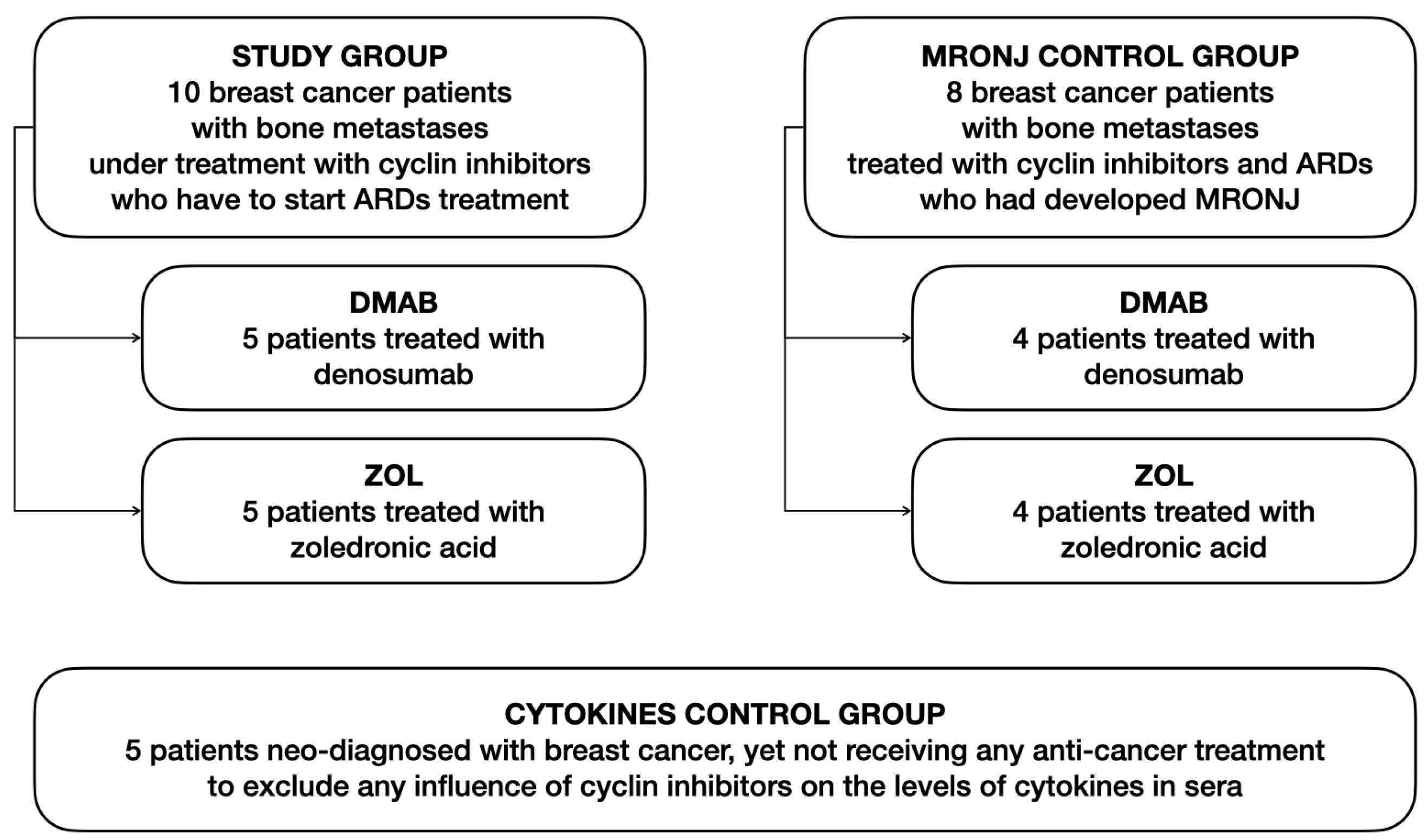
2.1. Patients and Study Design

Ethical approval for the present study was requested and obtained from the Institutional Review Board of the Città della Salute e della Scienza di Torino (IRB number: ID-552651). All patients signed their written informed consent to participate in this prospective study at the Oral Surgery Department of the Dental School of Turin. The characteristics of patients are reported in Figure 1. The study group was composed of 10 patients affected by breast cancer treated with cyclin inhibitors who developed bone metastases (58.5 ± 11.2; mean age ± SD), not assuming glucocorticoids or other drugs affected the immune system or bone turnover. Before starting ARDs to treat bone metastases, these patients were subjected to routine oral cavity examinations to exclude the potential evident risks of developing osteonecrosis soon after ARD intake. Five patients were treated with Zol and 5 with Dmab, and their blood samples were taken at 0 (T0), 6 (T6), 12 (T2), and 18 (T18) months to perform analysis on peripheral blood mononuclear cells (PBMCs) and sera. At every time point, patients were interviewed, and their clinical records were assessed to ensure that the anticancer treatment conditions remained unaltered for a long time. Two control groups were utilized, that is, (a) 8 patients with neo-diagnosis of MRONJ [4 patients with Dmab-induced ONJ (29 ± 11.3, mean months from the onset of ONJ ± SD) and 4 with Zol-induced ONJ (34 ± 16.2)] to compare their PBMCs with the study group’s ones and (b) 5 patients with neo-diagnosis of breast cancer, who had not received any treatment, to exclude that the cyclin inhibitor treatment affects the levels of cytokines in sera.

2.2. Flow Cytometry

At 0, 6, 12, and 18 months, PBMCs were collected, and the following immune subsets of cells were analyzed: CD4+ Th, CD8+ Tc, γδ T cells, and OCPs. On Th and Tc cells, we also evaluated the expression of two activation markers, namely, CD69 and CD25. We used the anti-human antibodies CD3 FITC, CD4 APC, PeRCP CD8 PerCP, γδ TCR PE, CD11b APC, CD51-61 PE (Miltenyi Biotec, Bergisch Gladbach, Germany), CD25 FITC, CD69 PE (BioLegend, SanDiego, CA, USA), and CD14 FITC (Thermo Fisher Scientific, Waltham, MA, USA), and related isotype and unstained controls. Appropriate controls were used to determine optimal voltage settings and electronic subtraction for the spectral fluorescence overlap correction. Samples were analyzed by the MACsQuant10 instrument and elaborated by MACs quantify software (Miltenyi Biotec, Bergisch Gladbach, Germany). Data represent a percentage of positive cells, determined by subtracting the percentage value of the appropriate isotype and unstained controls from each sample.

2.3. Cell Cultures

PBMCs were isolated from patients’ peripheral blood after centrifugation over a density gradient, Lymphoprep (Nycomed Pharma, Zürich, Switzerland). Cells were cultured in α-MEM supplemented with FBS, 100 IU/mL penicillin, and 100 μg/mL streptomycin and then counted and plated (Thermo Fisher Scientific, Waltham, MA, USA). To obtain fully differentiated human OCs, PBMCs were cultured for 15 days. As a positive control, PBMCs were also cultured in the presence of recombinant human M-CSF (25 ng/mL) and RANKL (30 ng/mL) (Peprotech, London, UK). Culture medium was refreshed every 5 days, either supplemented or not, with the factors reported above. At the end of the culture period, mature OCs were stained by the tartrate-resistant acid phosphatase (TRAP) kit (Cosmo Bio Co., Ltd. Tokyo, Japan) and identified as TRAP-positive, multinucleated cells containing three or more nuclei. OCs were counted using a microscope, and the presence of spontaneous osteoclastogenesis was considered significant when the cut-off number of 70 cells was reached or overcome, according to previously published works (16).

2.4. Cytokine Dosage

We collected patients’ sera from blood samples after centrifugation at 3000 rpm for 10 min at each time point (0, 6, 12, and 18 months), and we analyzed 12 pro-inflammatory cytokines according to the MACSPlex Cytokine 12 Kit (Miltenyi Biotec, Bergisch Gladbach, Germany), which is designed for determining concentrations of multiple soluble analytes (IL-2, IL-4, IL-5, IL-6, IL-9, IL-10, IL-12, IL-17, IFN-α, IFN-γ, TNF-α, and GM-CSF) in a single sample. The analysis was based on MACSPlex capture beads, which were a cocktail of different bead populations labeled with various fluorocromes. Each bead population was coated with a specific antibody, reacting with one of the analytes within the sample. A standard curve was obtained starting from the dilution of the standard concentrate provided with the kit, and it was utilized to quantify the analytes in the tested sera samples. Indeed, samples containing unknown levels of analytes were incubated with antibody-coated MACSPlex capture beads to promote binding with specific antibodies. Then a detection reagent containing a cocktail of fluorescent conjugated antibodies specific to the analytes was added. The different fluorescences corresponding to the analytes were identified using standard flow cytometry techniques. Samples were analyzed by the MACsQuant10 instrument and elaborated by the MACs quantify software (Miltenyi Biotec, Bergisch Gladbach, Germany).

2.5. Statistical Analyses

STATA software (version 17.0; StataCorp, College Station, TX, USA) was used for statistical analysis. Data were subjected to a Student’s t-test to evaluate statistically significant differences between the variables in the denosumab and zoledronate groups. A one-way ANOVA with Dunnet’s multiple comparison test was performed to assess differences in the serum values of the 12 cytokines in the two groups at different observational times. Statistical significance was considered at p < 0.05.

3. Results

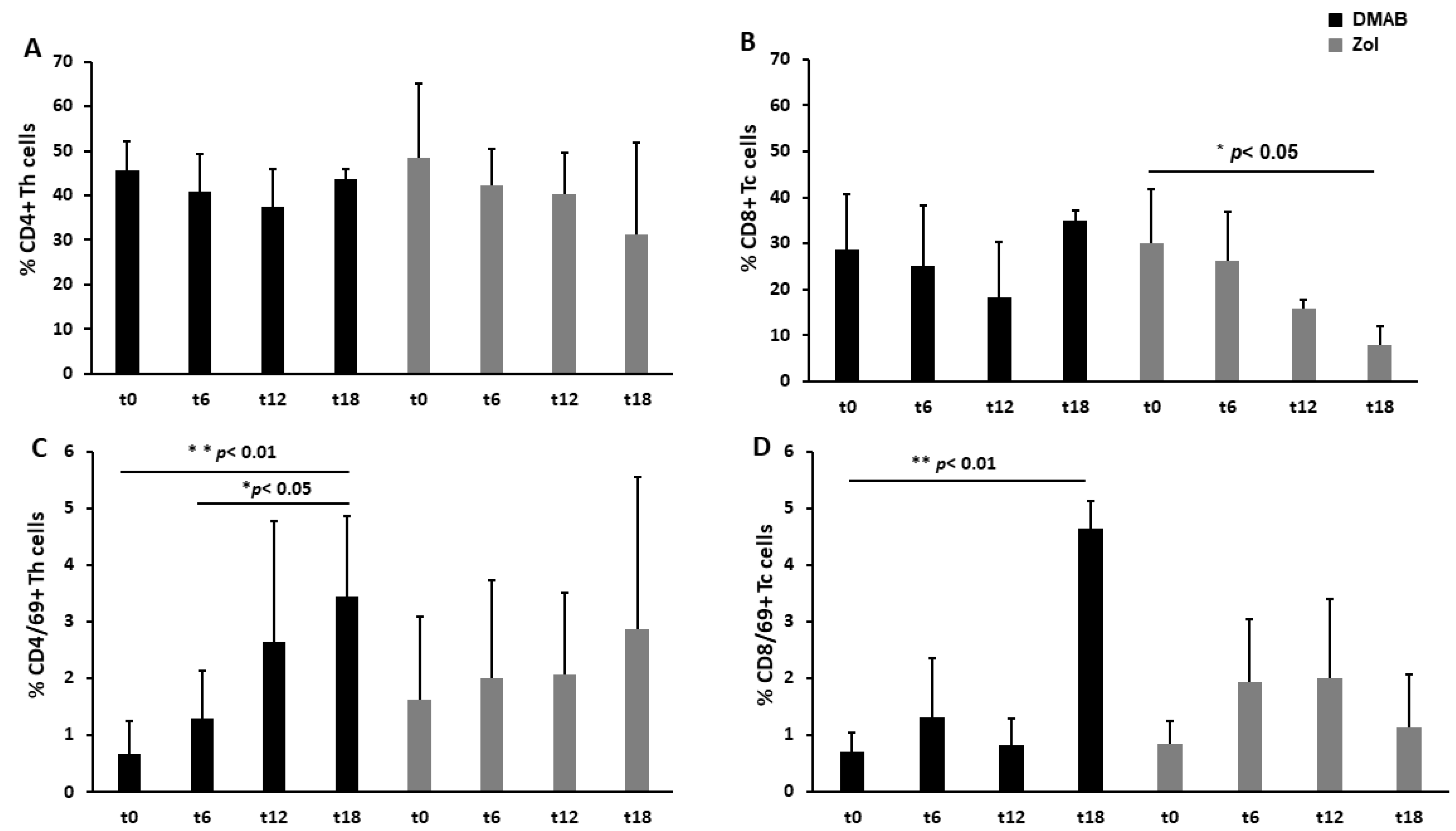
3.1. Dmab-Treated Patients Show an Increased Level of CD69, a T Cell Activation Marker

The analysis of CD4+ T cells showed that their level did not significantly change during the time or due to the different anti-resorptive treatments (Figure 2A). Conversely, cytotoxic CD8+ T cells remained stable in Dmab-treated patients, while they decreased in Zol-treated patients, with a significant difference at T18 compared to T0 (patients before starting the ARD treatment) (Figure 2B). Looking at the expression of activation markers, we observed in Dmab-treated patients a significant increase in CD4/CD69+ cells (3.4 ± 1.4 vs. 0.7 ± 0.6; 3.4 ± 1.4 vs. 1.3 ± 0.8% of positive cells ± SD, ** p < 0.01 and * p < 0.05) at T18 compared to T0 and T6, respectively (Figure 2C). CD8+ Tc cells expressed significantly more CD69 at T18 than at T0 (4.6 ± 0.5 vs. 0.7 ± 0.3, p < 0.01) (Figure 2D).
We also evaluated the expression of CD25 on both CD4+ and CD8+ T cells, which was not significantly different in the two groups at any time (Figure S1).
To investigate whether the above-described trends of T cell subsets and their different behaviors according to ZOL and Dmab treatment were comparable or not with the status of T cells in patients who developed MRONJ, we performed the same analysis on the latter. In the group of patients with MRONJ induced by Zol, we observed a higher level of CD4+ and CD8+ cells and a decreased level of CD4/CD69+ T cells compared to patients with Dmab-induced ONJ, p < 0.05 (Figure 3).

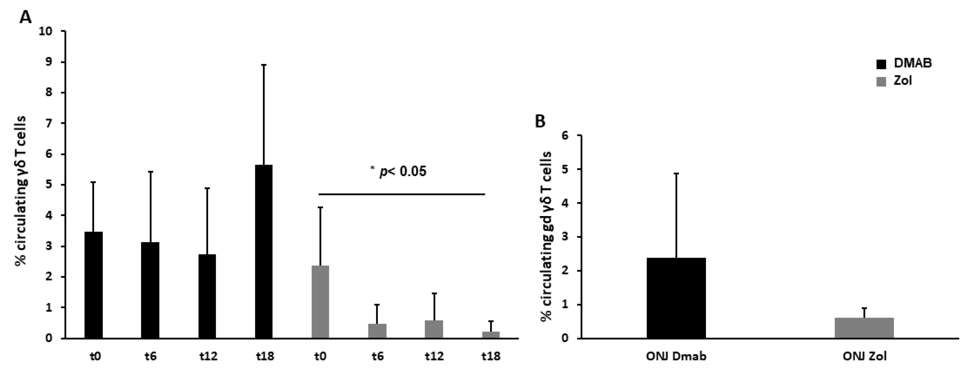
3.2. Dmab-Treated Patients Show a Normal Level of Circulating γδ T Cells

We monitored γδ T cells due to their reported role in maintaining bone oral cavity homeostasis, observing that, in Dmab-treated patients, circulating γδ T cells remained constant for 12 months, and then an increasing trend was evident at T18. γδ T cell level dramatically decreased at T6 after starting Zol treatment (2.4 ± 1.9 vs. 0.5 ± 0.6, p < 0.05) and remained constantly low during the rest of the observational period (Figure 4A). A lower level of γδ T cells was also detected in patients who developed Zol-induced ONJ compared to patients with Dmab-induced ONJ (Figure 4B).

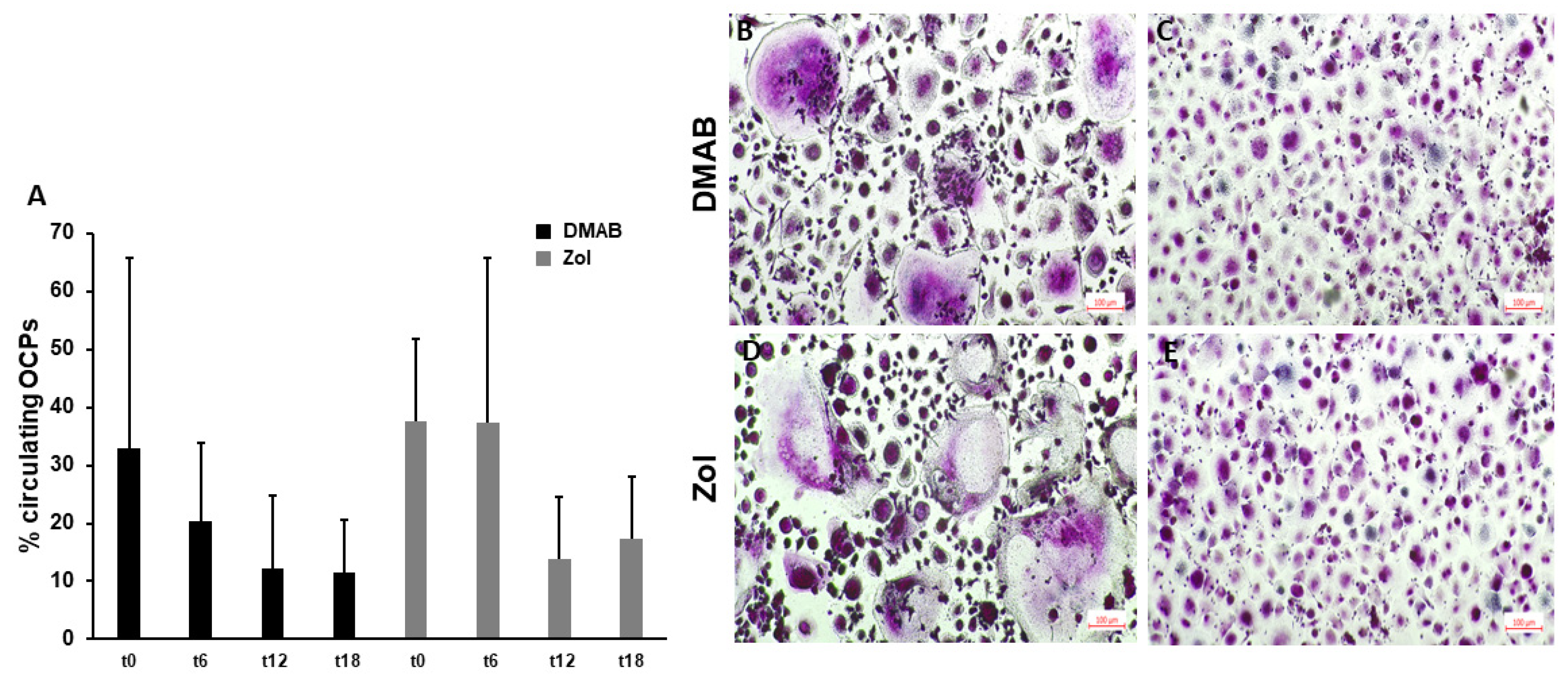
3.3. Circulating OCPs and OC Formation Decrease after 12 Months of Treatment with ARDs

The level of circulating OCPs is generally associated with the OC number and activity; thus, it is augmented in diseases characterized by increased bone resorption, such as bone metastases. For both Dmab- and Zol-treated patients, circulating OCPs were stable for the first 6 months, while a decrease occurred later at T12 (Figure 5A). To verify whether ARD treatment may affect OC formation, we performed an osteoclastogenesis in vitro assay, showing that at T12, the number of TRAP+ OCs was reduced compared to that at T0 (75 ± 31 vs. 112 ± 46, mean OC number ± SD). Even though the difference was not significant, the morphology showed large and multinucleated TRAP+ OCs at T0 (Figure 5B,D) and small TRAP+ OCs (with no more than 3 or 4 nuclei) at T12 in both the Zol- and Dmab-treated patients (Figure 5C,E).

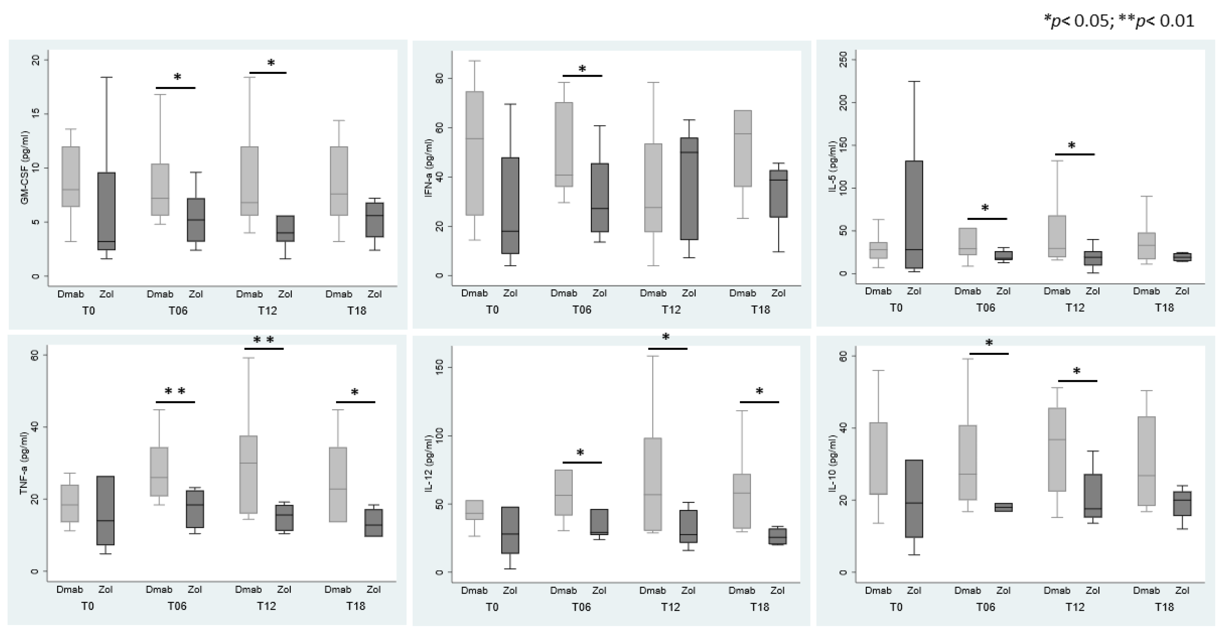
3.4. Cytokines Modulation in Sera According to ARDs

The level of pro-inflammatory cytokines was detected in the sera of patients with a neo-diagnosis of cancer (control group) before they started anticancer treatment to exclude bias in the cytokine levels dependent on therapy. We could not detect any significant difference in circulating cytokines among patients in the control group and our series of Dmab- or Zol-treated patients (Figure S2). By analyzing the single group of treatment (i.e., Dmab and Zol), we did not detect any variation in cytokine level during the monitoring time, except for IFN-γ, which became undetectable in Zol-treated patients at T12 and T18 (Table 1).
Conversely, we detected differences between the two groups of patients at specific time points. IFN-α was higher in Dmab-treated patients than in Zol ones at T6; GM-CSF, IL-5, and IL-10 were significantly increased at T6 and T12; and IL-12 and TNF-α were higher at T6, T12, and T18 (Figure 6). IL-4, IL-6, and IL-17 did not show significant variations between the two groups. IL-2 and IL-9 were completely nondetectable (Table 1).

4. Discussion

The role of immune responses and inflammation in the onset and/or progression of MRONJ has become more and more evident since the first papers on the matter [34,35]. Here, in 10 bone metastatic breast cancer patients treated with either Dmab or Zol, we investigated and compared the possible effects elicited on different circulating immune subsets to evaluate whether any changes in circulating immune subsets take place during the first 18 months of ARD treatment and whether they could be predictive of MRONJ development.
During the observational period, we did not register any significant variation in the level of circulating CD4+ Th cells with both the ARDs, although differences were detected for cytokines regulating Th cells. In particular, the serum levels of TNF-α and IL-12 were higher in Dmab-treated patients than in Zol ones. Notably, a main effect of IL-12 is the induction of IFN-γ, which, in turn, stimulates Th1 cells endowed with anti-inflammatory and anti-tumor functions [36,37]. In Dmab-treated patients, the levels of IFN-γ remained stable, as did the Th cells. Conversely, in Zol-treated patients, who also had lower IL-12 levels, IFN-γ decreased dramatically and became undetectable at T12 and T18. Moreover, we detected a significant increase in GM-CSF, IL-5, and IL-10 levels in the sera of Dmab-treated patients at T6 and T12. According to the literature, Zol and the RANKL inhibitor OPG blocked GM-CSF-induced tumor growth in bone, but this did not affect the capability of GM-CSF to reverse chemotherapy-induced leukopenia [38]. Thus, in Dmab-treated patients, the increase in GM-CSF could help maintain the correct pool of leukocytes and could also depend on activated Th1, as previously reported [39]. Furthermore, GM-CSF promotes the differentiation of myeloid cells, which are also the main producers of IL-10, an anti-inflammatory cytokine [40].
In the study group, Zol may be responsible for the reduction in circulating CD8+Tc cells, since we observed that their level progressively decreased in circulation until 18 months, while in Dmab-treated patients, they remained stable. Conversely, in Zol-induced ONJ patients, CD8+Tc cells significantly increased compared to those in Dmab-ONJ patients, but they did not express activation markers, thus suggesting an immunosuppressive action of Zol on this cell subset. Looking at the expression of the activating marker CD69 on CD4+ Th cells, we observed that Dmab induced a progressive increase in CD69+ Th cells during the time, while in CD69+ Tc cells, the increment was significant only at T18. This observation may be of interest in light of the increase in CD69+ Th cells that was detected in Dmab-induced ONJ patients, suggesting perhaps a contributing role of Dmab treatment in maintaining such an increase in these subjects. On the contrary, Zol did not affect CD69+ Th cells, suggesting that Dmab might allow the maintenance of circulating activated immune subsets, which deserves further investigation. Indeed, CD69 is not only an activation marker but also regulates the retention of cells on which it is expressed, and it is involved in the pathogenesis of inflammatory disorders, irrespective of the type of inflammation; thus, activated T cells are recruited into the inflamed tissue through the interaction of CD69 with its ligands [41,42].
The role of γδ T cells in the pathogenesis of MRONJ is debated, even though γδ T cells are known to be fundamental for fracture healing, to which tooth extraction can be compared. An increased level of γδ T cells in MRONJ patients’ tissue and in murine models has been reported [43]. Both γδ T null and wild-type mice treated with Zol developed osteonecrosis, while after injecting human γδ T cells into Zol-immunodeficient (Rag2−/−) mice, osteonecrosis was not induced [19]. Looking at the mechanisms underlying MRONJ, it has been shown that activated γδ T cells produced Semaforin-4D (Sema4D), which is implicated in the pathogenesis of T cell-mediated inflammatory diseases [44], and it stimulates pro-inflammatory cytokine production [43]. Here, we reported a drop in the number of γδ T cells after 6 months of Zol treatment, according to the literature data [33,45]. This drop can be explained by supposing that N-BPs cause the accumulation of a metabolite in the mevalonate pathway, isopentenyl pyrophosphate (IPP) [31,32,46], which is an antigen identified by γδ T cells that initially stimulates them but later induces their reduction through a mechanism of activation-induced cell death commonly used by the immune system to maintain homeostasis [33,47,48]. Another hypothesis explains the loss of γδ T cells with the high release of reactive oxygen species from neutrophils taking up N-BPs, which results in γδ T cell suppression [22,27]. Interestingly, our Dmab-treated patients did not show a decrease in γδ T cells differently from Zol ones, and looking at Dmab-induced ONJ patients, they had higher levels of γδ T cells compared to Zol-induced ONJ ones, suggesting that Dmab and Zol act in different ways on γδ T cells. Dmab does not interfere with the mevalonate pathway, but it is an anti-RANKL, so it can act on T cell activation and on thymic selection during the development of γδ T cell progenitors [49]. Kalyan et al. hypothesized that Dmab lowered the immune defenses by reducing T cell development [50]. Indeed, the RANK-RANKL system is a fundamental bridge between the immune system and bone [51,52], and a gene expression analysis of MRONJ patients deficient for γδ T cells showed a low expression of immune factors such as RANKL, RANK, TNF-α, and GM-CSF [21].
In our series, only one Dmab patient experienced MRONJ after 18 months of treatment, but his immune subsets remained stable, suggesting that the state of circulating cells likely does not reflect the oral cavity damage induced by MRONJ. Altogether, our data suggest that γδ T cells contribute to ONJ pathogenesis, but they do not mediate the core mechanism for ONJ onset, consistent with Park et al. [19].
Since γδ T cells are activated by OC deriving from peripheral blood monocytes [53], we assessed the level of circulating OCPs, showing that they were stable for the first 6 months, while a decrease started later at 12 months for both the ARDs. We also evaluated the capability of OCPs to differentiate spontaneously into OCs, which is a characteristic of pathologies associated with increased bone resorption, such as bone metastases [54,55]. As expected, we observed marked spontaneous osteoclastogenesis in patients before starting ARDs, but after 12 months, the number of OCs reduced, confirming that ARDs were effective in blocking OC activity according to the reduction in circulating OCPs.
This study has some limitations, such as the small number of enrolled patients, mainly due to the fact that cancer patients are often subjected to changes or therapy adjustments, and thus, it is difficult to obtain a clean series of patients. Moreover, we did not investigate the gut microbiota of breast cancer patients, even though accumulating evidence suggests that it can trigger both innate and adaptive immune responses through immunomodulatory cytokines and chemokines affecting tumor progression and response to anticancer therapies [56,57]. Indeed, cyclin inhibitor treatment efficacy can be improved by probiotics [57], and both dysbiosis and estrogen inhibition are associated with a reduction in γδ T cells in terms of number and activity. Thus, we cannot exclude the involvement of dysfunction in the gut microbiota in our cohort. In our clinical experience, Dmab-treated patients develop osteonecrosis before Zol ones, as is also evident by the different mean times of MRONJ development, dependent on Dmab or Zol treatment, observed in our MRONJ control group. In the present research, only one patient developed MRONJ, and the onset occurred at T18, a time that is consistent with the range covered by Dmab-induced MRONJ patients. Another interesting clinical observation is that patients treated with Dmab usually display less severe osteonecrotic features with faster healing lesions than Zol ones. We can speculate that this could depend on the lower immunosuppressive state induced by Dmab compared to Zol, also considering that Zol-treated patients completely lose the γδ T cell subset, which is paramount for the maintenance of oral cavity bone homeostasis.

5. Conclusions

MRONJ occurs commonly after dental extraction, which requires a healing process similar to bone regeneration. A coupled process of bone resorption and formation is also physiologically regulated by the immune system and guarantees effective bone remodeling. Thus, treatments that interfere with bone remodeling, affecting the immune–bone interface, should be carefully evaluated. ARDs can affect circulating immune cell subsets, altering their activation state and promoting an inflammatory microenvironment responsible for immunosuppression, which eventually does not sustain the correct bone remodeling. According to our results, ARDs differ in their capacity to modulate the immune system, potentially explaining the different clinical evolution of ONJ induced by Dmab or Zol. Indeed, in our clinical observation, patients treated with Dmab usually display lesions characterized by less severe osteonecrotic features with faster healing lesions than Zol ones, which might depend on the maintenance of a more active state of the immune system in Dmab-treated patients.

Supplementary Materials

The following supporting information can be downloaded at: https://www.mdpi.com/article/10.3390/cells12202430/s1, Figure S1: CD25 expression on CD4+ Th cells and CD8+ Tc cells. Figure S2: Cytokine levels in the control group and Dmab- or Zol-treated patients are comparable.

Author Contributions

Conceptualization, I.R., L.P. and F.M.; methodology, I.R. and R.P.; software, R.P., G.B. and L.P.; validation, I.R., L.P. and F.M.; formal analysis, I.R., I.B., F.E. and R.P.; investigation, L.P., G.B., I.B. and I.R.; resources, I.R. and F.M.; data curation, I.R. and L.P.; writing—original draft preparation, I.R. and L.P.; writing—review and editing, I.R., L.P. and F.M.; visualization, I.R., L.P, F.E. and F.M.; supervision, I.R. and F.M.; project administration, I.R. and F.M.; funding acquisition, I.R. and F.M. All authors have read and agreed to the published version of the manuscript.

Funding

This research was funded by CRT Foundations (CRT2020), Fondazione Ricerca Molinette ONLUS, and PRIN (CONCERTO) Prot. 2020BN5ZW9.

Institutional Review Board Statement

The study was conducted in accordance with the Declaration of Helsinki and approved by the Institutional Review Board of A.O.U. Città della Salute e della Scienza di Torino (protocol code ID-552651).

Informed Consent Statement

Informed consent was obtained from all subjects involved in the study.

Data Availability Statement

The data presented in this study are available on request from the corresponding author. The data are not publicly available due to privacy.

Conflicts of Interest

The authors declare no conflict of interest.

References

  1. Ruggiero, S.L.; Dodson, T.B.; Aghaloo, T.; Carlson, E.R.; Ward, B.B.; Kademani, D. American Association of Oral and Maxillofacial Surgeons’ Position Paper on Medication-Related Osteonecrosis of the Jaws-2022 Update. J. Oral Maxillofac. Surg. 2022, 80, 920–943. [Google Scholar] [CrossRef] [PubMed]
  2. Reid, I.R. Osteonecrosis of the jaw: Who gets it, and why? Bone 2009, 44, 4–10. [Google Scholar] [CrossRef] [PubMed]
  3. Soma, T.; Iwasaki, R.; Sato, Y.; Kobayashi, T.; Nakamura, S.; Kaneko, Y.; Ito, E.; Okada, H.; Watanabe, H.; Miyamoto, K.; et al. Tooth extraction in mice administered zoledronate increases inflammatory cytokine levels and promotes osteonecrosis of the jaw. J. Bone Miner. Metab. 2021, 39, 372–384. [Google Scholar] [CrossRef] [PubMed]
  4. Marx, R.E.; Sawatari, Y.; Fortin, M.; Broumand, V. Bisphosphonate-induced exposed bone (osteonecrosis/osteopetrosis) of the jaws: Risk factors, recognition, prevention, and treatment. J. Oral Maxillofac. Surg. 2005, 63, 1567–1575. [Google Scholar] [CrossRef] [PubMed]
  5. Fusco, V.; Santini, D.; Armento, G.; Tonini, G.; Campisi, G. Osteonecrosis of jaw beyond antiresorptive (bone-targeted) agents: New horizons in oncology. Expert. Opin. Drug Saf. 2016, 15, 925–935. [Google Scholar] [CrossRef]
  6. Eguia, A.; Bagan-Debon, L.; Cardona, F. Review and update on drugs related to the development of osteonecrosis of the jaw. Med. Oral Patol. Oral Cir. Bucal 2020, 25, e71–e83. [Google Scholar] [CrossRef]
  7. Papadopoulou, E.; Vardas, E.; Tziveleka, S.; Georgaki, M.; Kouri, M.; Katoumas, K.; Piperi, E.; Nikitakis, N.G. Oral Side Effects in Patients with Metastatic Renal Cell Carcinoma Receiving the Antiangiogenic Agent Pazopanib-Report of Three Cases. Dent. J. 2022, 10, 232. [Google Scholar] [CrossRef]
  8. Huja, S.S.; Fernandez, S.A.; Hill, K.J.; Li, Y. Remodeling dynamics in the alveolar process in skeletally mature dogs. Anat. Rec. A Discov. Mol. Cell Evol. Biol. 2006, 288, 1243–1249. [Google Scholar] [CrossRef]
  9. Allen, M.R.; Burr, D.B. The pathogenesis of bisphosphonate-related osteonecrosis of the jaw: So many hypotheses, so few data. J. Oral Maxillofac. Surg. 2009, 67, 61–70. [Google Scholar] [CrossRef]
  10. Ewald, F.; Wuesthoff, F.; Koehnke, R.; Friedrich, R.E.; Gosau, M.; Smeets, R.; Rohde, H.; Assaf, A.T. Retrospective analysis of bacterial colonization of necrotic bone and antibiotic resistance in 98 patients with medication-related osteonecrosis of the jaw (MRONJ). Clin. Oral Investig. 2021, 25, 2801–2809. [Google Scholar] [CrossRef]
  11. Nair, S.P.; Meghji, S.; Wilson, M.; Reddi, K.; White, P.; Henderson, B. Bacterially induced bone destruction: Mechanisms and misconceptions. Infect. Immun. 1996, 64, 2371–2380. [Google Scholar] [CrossRef] [PubMed]
  12. Meghji, S.; Crean, S.J.; Hill, P.A.; Sheikh, M.; Nair, S.P.; Heron, K.; Henderson, B.; Mawer, E.B.; Harris, M. Surface-associated protein from Staphylococcus aureus stimulates osteoclastogenesis: Possible role in S. aureus-induced bone pathology. Br. J. Rheumatol. 1998, 37, 1095–1101. [Google Scholar] [CrossRef] [PubMed]
  13. Du, W.; Yang, M.; Kim, T.; Kim, S.; Williams, D.W.; Esmaeili, M.; Hong, C.; Shin, K.H.; Kang, M.K.; Park, N.H.; et al. Indigenous microbiota protects development of medication-related osteonecrosis induced by periapical disease in mice. Int. J. Oral Sci. 2022, 14, 16. [Google Scholar] [CrossRef] [PubMed]
  14. Nicolatou-Galitis, O.; Schiodt, M.; Mendes, R.A.; Ripamonti, C.; Hope, S.; Drudge-Coates, L.; Niepel, D.; Van den Wyngaert, T. Medication-related osteonecrosis of the jaw: Definition and best practice for prevention, diagnosis, and treatment. Oral Surg. Oral Med. Oral Pathol. Oral Radiol. 2019, 127, 117–135. [Google Scholar] [CrossRef] [PubMed]
  15. Messer, J.G.; Mendieta Calle, J.L.; Jiron, J.M.; Castillo, E.J.; Van Poznak, C.; Bhattacharyya, N.; Kimmel, D.B.; Aguirre, J.I. Zoledronic acid increases the prevalence of medication-related osteonecrosis of the jaw in a dose dependent manner in rice rats (Oryzomys palustris) with localized periodontitis. Bone 2018, 108, 79–88. [Google Scholar] [CrossRef] [PubMed]
  16. Peng, J.; Wang, H.; Liu, Z.; Xu, Z.L.; Wang, M.X.; Chen, Q.M.; Wu, M.L.; Ren, X.L.; Liang, Q.H.; Liu, F.P.; et al. Real-world study of antiresorptive-related osteonecrosis of jaw based on the US food and drug administration adverse event reporting system database. Front. Pharmacol. 2022, 13, 1017391. [Google Scholar] [CrossRef]
  17. Hanley, D.A.; Adachi, J.D.; Bell, A.; Brown, V. Denosumab: Mechanism of action and clinical outcomes. Int. J. Clin. Pract. 2012, 66, 1139–1146. [Google Scholar] [CrossRef]
  18. Francisconi, C.F.; Vieira, A.E.; Azevedo, M.C.S.; Tabanez, A.P.; Fonseca, A.C.; Trombone, A.P.F.; Letra, A.; Silva, R.M.; Sfeir, C.S.; Little, S.R.; et al. RANKL Triggers Treg-Mediated Immunoregulation in Inflammatory Osteolysis. J. Dent. Res. 2018, 97, 917–927. [Google Scholar] [CrossRef]
  19. Park, S.; Kanayama, K.; Kaur, K.; Tseng, H.C.; Banankhah, S.; Quje, D.T.; Sayre, J.W.; Jewett, A.; Nishimura, I. Osteonecrosis of the jaw developed in mice: Disease variants regulated by gammadelta T cells in oral mucosal barrier immunity. J. Biol. Chem. 2015, 290, 17349–17366. [Google Scholar] [CrossRef]
  20. Zhu, W.; Xu, R.; Du, J.; Fu, Y.; Li, S.; Zhang, P.; Liu, L.; Jiang, H. Zoledronic acid promotes TLR-4-mediated M1 macrophage polarization in bisphosphonate-related osteonecrosis of the jaw. FASEB J. 2019, 33, 5208–5219. [Google Scholar] [CrossRef]
  21. Kalyan, S.; Wang, J.; Quabius, E.S.; Huck, J.; Wiltfang, J.; Baines, J.F.; Kabelitz, D. Systemic immunity shapes the oral microbiome and susceptibility to bisphosphonate-associated osteonecrosis of the jaw. J. Transl. Med. 2015, 13, 212. [Google Scholar] [CrossRef] [PubMed]
  22. Kalyan, S.; Kabelitz, D. Defining the nature of human gammadelta T cells: A biographical sketch of the highly empathetic. Cell Mol. Immunol. 2013, 10, 21–29. [Google Scholar] [CrossRef]
  23. Soma, T.; Iwasaki, R.; Sato, Y.; Kobayashi, T.; Ito, E.; Matsumoto, T.; Kimura, A.; Miyamoto, K.; Matsumoto, M.; Nakamura, M.; et al. Osteonecrosis development by tooth extraction in zoledronate treated mice is inhibited by active vitamin D analogues, anti-inflammatory agents or antibiotics. Sci. Rep. 2022, 12, 19. [Google Scholar] [CrossRef]
  24. Zhang, Q.; Atsuta, I.; Liu, S.; Chen, C.; Shi, S.; Shi, S.; Le, A.D. IL-17-mediated M1/M2 macrophage alteration contributes to pathogenesis of bisphosphonate-related osteonecrosis of the jaws. Clin. Cancer Res. 2013, 19, 3176–3188. [Google Scholar] [CrossRef] [PubMed]
  25. Kaur, K.; Sun, Y.; Kanayama, K.; Morinaga, K.; Hokugo, A.; Nishimura, I.; Jewett, A. Augmentation of IFN-gamma by bone marrow derived immune cells in the presence of severe suppression of IFN-gamma in gingivae induced by zoledronic acid and denosumab in Hu-BLT mice model of ONJ. Front. Endocrinol. 2023, 14, 1111627. [Google Scholar] [CrossRef]
  26. Elsayed, R.; Kurago, Z.; Cutler, C.W.; Arce, R.M.; Gerber, J.; Celis, E.; Sultan, H.; Elashiry, M.; Meghil, M.; Sun, C.; et al. Role of dendritic cell-mediated immune response in oral homeostasis: A new mechanism of osteonecrosis of the jaw. FASEB J. 2020, 34, 2595–2608. [Google Scholar] [CrossRef]
  27. Hagelauer, N.; Pabst, A.M.; Ziebart, T.; Ulbrich, H.; Walter, C. In vitro effects of bisphosphonates on chemotaxis, phagocytosis, and oxidative burst of neutrophil granulocytes. Clin. Oral Investig. 2015, 19, 139–148. [Google Scholar] [CrossRef]
  28. Gkouveris, I.; Soundia, A.; Gouveris, P.; Zouki, D.; Hadaya, D.; Tetradis, S. Macrophage Involvement in Medication-Related Osteonecrosis of the Jaw (MRONJ): A Comprehensive, Short Review. Cancers 2022, 14, 330. [Google Scholar] [CrossRef]
  29. Roato, I.; Mauceri, R.; Notaro, V.; Genova, T.; Fusco, V.; Mussano, F. Immune Dysfunction in Medication-Related Osteonecrosis of the Jaw. Int. J. Mol. Sci. 2023, 24, 7948. [Google Scholar] [CrossRef]
  30. Prager, I.; Watzl, C. Mechanisms of natural killer cell-mediated cellular cytotoxicity. J. Leukoc. Biol. 2019, 105, 1319–1329. [Google Scholar] [CrossRef] [PubMed]
  31. Idrees, A.S.; Sugie, T.; Inoue, C.; Murata-Hirai, K.; Okamura, H.; Morita, C.T.; Minato, N.; Toi, M.; Tanaka, Y. Comparison of gammadelta T cell responses and farnesyl diphosphate synthase inhibition in tumor cells pretreated with zoledronic acid. Cancer Sci. 2013, 104, 536–542. [Google Scholar] [CrossRef]
  32. Roelofs, A.J.; Jauhiainen, M.; Monkkonen, H.; Rogers, M.J.; Monkkonen, J.; Thompson, K. Peripheral blood monocytes are responsible for gammadelta T cell activation induced by zoledronic acid through accumulation of IPP/DMAPP. Br. J. Haematol. 2009, 144, 245–250. [Google Scholar] [CrossRef]
  33. Rossini, M.; Adami, S.; Viapiana, O.; Fracassi, E.; Ortolani, R.; Vella, A.; Zanotti, R.; Tripi, G.; Idolazzi, L.; Gatti, D. Long-term effects of amino-bisphosphonates on circulating gammadelta T cells. Calcif. Tissue Int. 2012, 91, 395–399. [Google Scholar] [CrossRef] [PubMed]
  34. Salesi, N.; Pistilli, R.; Marcelli, V.; Govoni, F.A.; Bozza, F.; Bossone, G.; Venturelli, V.; Di Cocco, B.; Pacetti, U.; Ciorra, A.; et al. Bisphosphonates and oral cavity avascular bone necrosis: A review of twelve cases. Anticancer Res. 2006, 26, 3111–3115. [Google Scholar] [CrossRef] [PubMed]
  35. Kikuiri, T.; Kim, I.; Yamaza, T.; Akiyama, K.; Zhang, Q.; Li, Y.; Chen, C.; Chen, W.; Wang, S.; Le, A.D.; et al. Cell-based immunotherapy with mesenchymal stem cells cures bisphosphonate-related osteonecrosis of the jaw-like disease in mice. J. Bone Miner. Res. 2010, 25, 1668–1679. [Google Scholar] [CrossRef] [PubMed]
  36. Trinchieri, G. Interleukin-12 and the regulation of innate resistance and adaptive immunity. Nat. Rev. Immunol. 2003, 3, 133–146. [Google Scholar] [CrossRef]
  37. Ullrich, K.A.; Schulze, L.L.; Paap, E.M.; Muller, T.M.; Neurath, M.F.; Zundler, S. Immunology of IL-12: An update on functional activities and implications for disease. EXCLI J. 2020, 19, 1563–1589. [Google Scholar] [CrossRef]
  38. Dai, J.; Lu, Y.; Yu, C.; Keller, J.M.; Mizokami, A.; Zhang, J.; Keller, E.T. Reversal of chemotherapy-induced leukopenia using granulocyte macrophage colony-stimulating factor promotes bone metastasis that can be blocked with osteoclast inhibitors. Cancer Res. 2010, 70, 5014–5023. [Google Scholar] [CrossRef]
  39. Abiko, Y.; Saitoh, M.; Nishimura, M.; Yamazaki, M.; Sawamura, D.; Kaku, T. Role of beta-defensins in oral epithelial health and disease. Med. Mol. Morphol. 2007, 40, 179–184. [Google Scholar] [CrossRef]
  40. Saraiva, M.; O’Garra, A. The regulation of IL-10 production by immune cells. Nat. Rev. Immunol. 2010, 10, 170–181. [Google Scholar] [CrossRef]
  41. Kimura, M.Y.; Koyama-Nasu, R.; Yagi, R.; Nakayama, T. A new therapeutic target: The CD69-Myl9 system in immune responses. Semin. Immunopathol. 2019, 41, 349–358. [Google Scholar] [CrossRef]
  42. Koyama-Nasu, R.; Wang, Y.; Hasegawa, I.; Endo, Y.; Nakayama, T.; Kimura, M.Y. The cellular and molecular basis of CD69 function in anti-tumor immunity. Int. Immunol. 2022, 34, 555–561. [Google Scholar] [CrossRef]
  43. Movila, A.; Mawardi, H.; Nishimura, K.; Kiyama, T.; Egashira, K.; Kim, J.Y.; Villa, A.; Sasaki, H.; Woo, S.B.; Kawai, T. Possible pathogenic engagement of soluble Semaphorin 4D produced by gammadeltaT cells in medication-related osteonecrosis of the jaw (MRONJ). Biochem. Biophys. Res. Commun. 2016, 480, 42–47. [Google Scholar] [CrossRef]
  44. Maleki, K.T.; Cornillet, M.; Bjorkstrom, N.K. Soluble SEMA4D/CD100: A novel immunoregulator in infectious and inflammatory diseases. Clin. Immunol. 2016, 163, 52–59. [Google Scholar] [CrossRef] [PubMed]
  45. Walsh, M.; White, G.; Romeril, K.; Buyck, H.; Stephens, M.; Brooks, C.; Weinkove, R. Innate-like T cell profile in myeloma: Severe deficiency of Vgamma9Vdelta2 T cells in aminobisphosphonate-treated patients. Leuk. Lymphoma 2016, 57, 977–980. [Google Scholar] [CrossRef] [PubMed]
  46. Gober, H.J.; Kistowska, M.; Angman, L.; Jeno, P.; Mori, L.; De Libero, G. Human T cell receptor gammadelta cells recognize endogenous mevalonate metabolites in tumor cells. J. Exp. Med. 2003, 197, 163–168. [Google Scholar] [CrossRef] [PubMed]
  47. Gan, Y.H.; Wallace, M.; Malkovsky, M. Fas-dependent, activation-induced cell death of gammadelta cells. J. Biol. Regul. Homeost. Agents 2001, 15, 277–285. [Google Scholar] [PubMed]
  48. Green, D.R.; Droin, N.; Pinkoski, M. Activation-induced cell death in T cells. Immunol. Rev. 2003, 193, 70–81. [Google Scholar] [CrossRef] [PubMed]
  49. Roberts, N.A.; White, A.J.; Jenkinson, W.E.; Turchinovich, G.; Nakamura, K.; Withers, D.R.; McConnell, F.M.; Desanti, G.E.; Benezech, C.; Parnell, S.M.; et al. Rank signaling links the development of invariant gammadelta T cell progenitors and Aire(+) medullary epithelium. Immunity 2012, 36, 427–437. [Google Scholar] [CrossRef]
  50. Kalyan, S. It May Seem Inflammatory, but Some T Cells Are Innately Healing to the Bone. J. Bone Miner. Res. 2016, 31, 1997–2000. [Google Scholar] [CrossRef]
  51. Wada, T.; Nakashima, T.; Hiroshi, N.; Penninger, J.M. RANKL-RANK signaling in osteoclastogenesis and bone disease. Trends Mol. Med. 2006, 12, 17–25. [Google Scholar] [CrossRef] [PubMed]
  52. Wong, B.R.; Josien, R.; Lee, S.Y.; Sauter, B.; Li, H.L.; Steinman, R.M.; Choi, Y. TRANCE (tumor necrosis factor [TNF]-related activation-induced cytokine), a new TNF family member predominantly expressed in T cells, is a dendritic cell-specific survival factor. J. Exp. Med. 1997, 186, 2075–2080. [Google Scholar] [CrossRef] [PubMed]
  53. Pappalardo, A.; Thompson, K. Novel immunostimulatory effects of osteoclasts and macrophages on human gammadelta T cells. Bone 2015, 71, 180–188. [Google Scholar] [CrossRef] [PubMed]
  54. Roato, I.; Grano, M.; Brunetti, G.; Colucci, S.; Mussa, A.; Bertetto, O.; Ferracini, R. Mechanisms of spontaneous osteoclastogenesis in cancer with bone involvement. FASEB J. 2005, 19, 228–230. [Google Scholar] [CrossRef]
  55. Roato, I.; Gorassini, E.; Buffoni, L.; Lyberis, P.; Ruffini, E.; Bonello, L.; Baldi, I.; Ciuffreda, L.; Mussa, A.; Ferracini, R. Spontaneous osteoclastogenesis is a predictive factor for bone metastases from non-small cell lung cancer. Lung Cancer 2008, 61, 109–116. [Google Scholar] [CrossRef]
  56. Gopalakrishnan, V.; Helmink, B.A.; Spencer, C.N.; Reuben, A.; Wargo, J.A. The Influence of the Gut Microbiome on Cancer, Immunity, and Cancer Immunotherapy. Cancer Cell 2018, 33, 570–580. [Google Scholar] [CrossRef]
  57. Panebianco, C.; Andriulli, A.; Pazienza, V. Pharmacomicrobiomics: Exploiting the drug-microbiota interactions in anticancer therapies. Microbiome 2018, 6, 92. [Google Scholar] [CrossRef]
Disclaimer/Publisher’s Note: The statements, opinions, and data contained in all publications are solely those of the individual author(s) and contributor(s) and not of MDPI and/or the editor(s). MDPI and/or the editor(s) disclaim responsibility for any injury to people or property resulting from any ideas, methods, instructions, or products referred to in the content.
Figure 1. Diagram depicting the study design and patient groups involved in the study.
Cells 12 02430 g001
Figure 2. Circulating T cell phenotypes in the two groups of study. CD4+ Th cells were stable in both groups of treatment and at different time points (A). CD8+ Tc significantly decreased in Zol-treated patients at 18 months, p < 0.05 (B). The subsets of activated CD69+ cells progressively increased in Th and Tc cells of Dmab-treated patients at 18 months (C,D).
Cells 12 02430 g002
Figure 3. Circulating T cell subsets in MRONJ patients. Circulating Th CD4+ and Tc CD8+ cells were increased in Zol-treated patients compared to Dmab ones (p < 0.05). Conversely, activated CD4/CD69+ T cells were significantly decreased only in Zol-treated patients (p < 0.05).
Cells 12 02430 g003
Figure 4. Dmab and Zol effects on γδ T cells. In Dmab-treated patients, the level of circulating γδ T cells remained constant with an increasing trend at 18 months, while it decreased soon after Zol treatment (A). In the group of patients with ONJ, γδ T cells were lower in patients with Zol-induced ONJ (B).
Cells 12 02430 g004
Figure 5. Circulating OCPs and OC formation. The number of circulating OCPs decreased during treatment with ARDs, without significant differences (A). Osteoclastogenesis in vitro at T0 (B,D) was reduced, with fewer large multinucleated TRAP+ OCs (C,E) after 12 months of treatment with both ARDs.
Cells 12 02430 g005
Figure 6. Cytokine expression in sera. Graphs show the serum levels of cytokines, which were significantly increased in Dmab-treated patients compared to Zol ones at the different time points.
Cells 12 02430 g006
Table 1. Cytokines serum levels. The mean ± SD (pg/mL) of the serum levels of cytokines analyzed in the patient’s sera at the different time points are reported.
DMABZOL
T0T6T12T18T0T6T12T18
GM-CSF8.6 ± 1.38.6 ± 1.48.6 ± 1.48.4 ± 1.86.8 ± 2.05.5 ± 1.14.0 ± 0.65.2 ± 1.1
IFNα51.4 ± 9.850.0 ± 6.235.8 ± 7.659.9 ± 13.429.7 ± 7.932.0 ± 7.440.1 ± 9.533.2 ± 8.0
IFNγ13.4 ± 7.032.9 ± 13.441.9 ± 14.724.3 ± 20.7114.9 ± 55.110.4 ± 7.2not detectablenot detectable
IL-4395.8 ± 43.1419.8 ± 55.1419.0 ± 53.0467.2 ± 70.9328.6 ± 34.7412.1 ± 25.7446.9 ± 24.4380.6 ± 43.9
IL-529.6 ± 6.145.0 ± 12.149.8 ± 12.838.8 ± 11.669.9 ± 33.820.3 ± 2.719.2 ± 5.519.4 ± 2.7
IL-69.8 ± 0.910.6 ± 1.011.8 ± 2.39.6 ± 2.58.1 ± 1.310.0 ± 1.89.5 ± 1.410.2 ± 2.0
IL-1030.5 ± 4.932.8 ± 4.934.0 ± 4.230.4 ± 5.932.6 ± 10.820.1 ± 4.520.8 ± 3.219.0 ± 2.5
IL-1252.5 ± 9.069.2 ± 13.573.6 ± 16.261.3 ± 13.372.2 ± 32.638.7 ± 8.131.6 ± 5.726.2 ± 3.4
IL-17A17.9 ± 4.928.3 ± 5.627.3 ± 7.129.6 ± 13.535.2 ± 11.519.7 ± 5.713.2 ± 3.116.4 ± 6.1
TNFα21.8 ± 3.727.8 ± 2.829.6 ± 4.425.3 ± 5.123.8 ± 7.717.5 ± 2.215.1 ± 1.513.4 ± 2.2
Disclaimer/Publisher’s Note: The statements, opinions and data contained in all publications are solely those of the individual author(s) and contributor(s) and not of MDPI and/or the editor(s). MDPI and/or the editor(s) disclaim responsibility for any injury to people or property resulting from any ideas, methods, instructions or products referred to in the content.

