Review

Chemistry of Hydrogen Sulfide—Pathological and Physiological Functions in Mammalian Cells

by
Celia María Curieses Andrés
1,
José Manuel Pérez de la Lastra
2,*,
Celia Andrés Juan
3,
Francisco J. Plou
4 and
Eduardo Pérez-Lebeña
5
1
Hospital Clínico Universitario of Valladolid, Avenida de Ramón y Cajal, 3, 47003 Valladolid, Spain
2
Institute of Natural Products and Agrobiology, CSIC-Spanish Research Council, Avda. Astrofísico Fco. Sánchez, 3, 38206 La Laguna, Spain
3
Cinquima Institute and Department of Organic Chemistry, Faculty of Sciences, Valladolid University, Paseo de Belén, 7, 47011 Valladolid, Spain
4
Institute of Catalysis and Petrochemistry, CSIC-Spanish Research Council, 28049 Madrid, Spain
5
Sistemas de Biotecnología y Recursos Naturales, 47625 Valladolid, Spain
*
Author to whom correspondence should be addressed.
Cells 2023, 12(23), 2684; https://doi.org/10.3390/cells12232684
Submission received: 18 September 2023 / Revised: 2 November 2023 / Accepted: 20 November 2023 / Published: 22 November 2023

Abstract

:
Hydrogen sulfide (H2S) was recognized as a gaseous signaling molecule, similar to nitric oxide (-NO) and carbon monoxide (CO). The aim of this review is to provide an overview of the formation of hydrogen sulfide (H2S) in the human body. H2S is synthesized by enzymatic processes involving cysteine and several enzymes, including cystathionine-β-synthase (CBS), cystathionine-γ-lyase (CSE), cysteine aminotransferase (CAT), 3-mercaptopyruvate sulfurtransferase (3MST) and D-amino acid oxidase (DAO). The physiological and pathological effects of hydrogen sulfide (H2S) on various systems in the human body have led to extensive research efforts to develop appropriate methods to deliver H2S under conditions that mimic physiological settings and respond to various stimuli. These functions span a wide spectrum, ranging from effects on the endocrine system and cellular lifespan to protection of liver and kidney function. The exact physiological and hazardous thresholds of hydrogen sulfide (H2S) in the human body are currently not well understood and need to be researched in depth. This article provides an overview of the physiological significance of H2S in the human body. It highlights the various sources of H2S production in different situations and examines existing techniques for detecting this gas.

1. Introduction

Hydrogen sulfide (H2S) [1] is currently considered a physiological modulator in mammals. It can be formed in mammalian cells and is an important gasotransmitter that plays important physiological and pathophysiological roles. H2S is the most recently discovered endogenous gasotransmitter and, like NO and CO, crosses cell membranes without a specific transporter [2]. H2S can be generated by various processes and can undergo chemical reactions with metal centers and oxidized thiol compounds, especially asymmetric disulfides (R1SSR2). Initially considered a toxic molecule, it is now recognized that mammalian cells are endowed with enzymatic pathways for H2S production. The identification of H2S as a gasotransmitter has led to a renewed interest during the last two decades, focusing on (i) creating chemical tools for its physiological detection, (ii) determining its signaling functions in various organs and systems in the plant and animal kingdoms and (iii) exploiting its biological signaling capacity for therapeutic benefits.
In the late 20th century, H2S was discovered to be an endogenously produced gas in tissues. It is synthesized from the amino acid L-cysteine, D-cysteine, homocysteine, cystathione and 3-mercaptopyruvate by a series of enzymes whose expression and molecular regulation in various tissues have been characterized by several groups. It is an endogenous mediator of inflammation [3] and a potential cytoprotective compound [4]. At that time, H2S was found to be generated enzymatically, and it was included in the family of gaseous transmitters (also due to its chemical properties). NO, CO and H2S are part of the group of gasotransmitters (Figure 1).
The expression of H2S-catabolic catabolic enzymes varies in numerous physiological systems, including the endocrine, neuronal, cardiovascular, respiratory, immunological, reproductive, renal, hepatic and gastrointestinal systems. Various enzymes play a role in modulating the activities of different systems by regulating the generation of H2S. Pathological conditions such as atherosclerosis, hypertension, heart failure, cirrhosis, inflammation, asthma, sepsis, diabetes, erectile dysfunction and neurodegenerative diseases arise because of alterations in H2S metabolism [5,6,7].
During the First World War, the British army used H2S as a chemical weapon. However, its suitability as a combat gas was compromised by its flammability and unique odor, which could serve as an indicator of the gas’s location to the enemy [8]. Today it is known to act as a second messenger and is associated with important functions at the level of blood vessel walls [9]. The properties attributed to it are antioxidant, anti-atherogenic, anti-apoptosis and anti-inflammatory properties, as well as anti-proliferative, neuromodulatory and cytoprotective properties [10].

2. H2S Chemistry

H2S is a gas at room temperature, heavier than air, colorless with a characteristic rotten egg odor and toxic [11,12,13]. Until the mid-1990s, it was considered mainly toxic to humans and harmful to the environment. The substance is moderately soluble in water, with a solubility of 2.77 volumes in 1 volume of water at a temperature of 20 °C. Moreover, it can be effectively eliminated from an aqueous solution by complete dissolution during boiling.
A sulfur atom is larger than an oxygen atom (covalent radius 105 vs. 66) and has a comparatively lower electronegativity value of 2.5 on the Pauling scale, while oxygen (O) has a higher electronegativity value of 3.5. In addition, sulfur is characterized by a higher degree of polarizability. Consequently, the dipole moment of H2S is comparatively smaller than that of water, being 0.97 D and 1.85 D, respectively, and intermolecular interactions are weaker. It has a similar structure to water, but since sulfur has lower electronegativity values than oxygen (S = 2.5 and O = 3.5, according to the Pauling scale), H2S is less polar than water [14]. Compared to water, this difference has an impact on the inability to form hydrogen bridges. H2S can diffuse freely through the hydrophobic core of biological membranes [15]. The sulfur in H2S has the electronic configuration 1s2, 2s2 2p6, 3s2 and 3p6 and borrows an electron from each hydrogen to complete its valence shell (Figure 2).
H2S is considered a toxic substance when inhaled in its pure form or even when diluted in a ratio of 1 part gas to 200 parts air. Bird species are very sensitive to H2S, rendering them susceptible to mortality even at a dilute concentration of 1 part per 1500 in atmospheric air. H2S has significant inhibitory effects on specific enzymes and processes related to oxidative phosphorylation, ultimately leading to cell suffocation. Most people perceive hydrogen sulfide through the sense of smell when the concentration is above 5 parts per billion (ppb). Concentrations of 20 to 50 parts per million (ppm) have been found to cause irritation to both the eyes and the respiratory system.
Short-term inhalation of 100 to 250 parts per million (ppm) can cause adverse effects such as incoordination and cognitive and motor impairment. At concentrations of about 150 to 200 parts per million (ppm), individuals may suffer from olfactory fatigue or anosmia, resulting in an inability to detect the characteristic odor of H2S. Inhalation of a concentration of 500 parts per million (ppm) for a period of 30 min may result in the development of pulmonary edema and pneumonia.
Concentrations greater than 600 parts per million (ppm) have the potential to cause death within a short 30 min period, primarily due to paralysis of the respiratory system. A concentration of 800 ppm is considered immediately fatal to humans. H2S has a comparatively high solubility in water, 110 millimoles per atmosphere at standard room temperature [16,17].
H2S is the smallest and simplest inorganic biologically relevant thiol with the maximal reduced state of sulfur atom (−2) [18]. H2S has only oxidizing properties and does not have the ability to act as an oxidant. H2S is a reducing species, with a reduction potential of −280 mV (pH 7.0 vs. standard hydrogen electrode) for the two-electron redox pair HS/S0 and −230 mV for H2S/S0, close to the values for the glutathione/glutathione disulfide (GSSG/GSH) and cystine/cysteine redox couples [19]. The first evidence for the physiological role of H2S was its detection in brain tissues of different species [20].
H2S serves as a stimulator of electron transport in mammalian mitochondria by acting as an electron donor (the electron acceptor is oxidized CoQ), involving sulfur-quinone oxidoreductase (SQR) and leading to the production of GSSH-glutathione persulfide or thiosulfate and reduced CoQ [21].
The H2S-derived effect on electron transport processes is a double-edged sword: a low amount can stimulate respiration, while a higher dose will lead to complete inhibition of respiration by inhibiting the heme-containing proteins. It is still questionable whether H2S has any impact on the electron transport chain via enrichment of the reduced CoQ pool, particularly under normoxic (in vivo) or supra-normoxic conditions (21% O2, in cell incubators [22,23].

3. Pathways of H2S Production

3.1. Environmental (Anthropogenic and Non-Anthropogenic) Sources

Non-anthropogenic environmental sources of H2S are wetlands, volcanic eruptions and emissions, geothermal activity, thermal upwellings and sulfide-metabolizing bacteria. Anthropogenic emissions of H2S are paper factories, tanneries, mining, oil refineries, combustion and sewage dumps.

3.2. Endogenous Sources of H2S in Mammalian Cells

In 1996, it was shown that cystathionine-β-synthase can produce H2S in the brain and that H2S facilitates the induction of hippocampal long-term potentiation by enhancing NMDA receptor activity [24]. In 1997, another H2S-producing enzyme, cystathionine-γ-lyase, was found to be expressed in the thoracic aorta, portal vein and ileum, and it was found that H2S relaxes these tissues [25]. Based on these findings, H2S may be a neuromodulator as well as a smooth muscle relaxant, in addition to functioning as a signaling molecule and as a cytoprotectant [4]. H2S protects neurons from oxidative stress by restoring depleted glutathione levels. A third H2S-producing enzyme, 3-mercaptopyruvate sulfurtransferase (3MST), is expressed in neurons, the vascular endothelium [26] and the mitochondria of almost all cells [27]. In addition to restoring glutathione levels, the H2S produced by 3MST, which is mainly located in the mitochondria, reduces ROS generated in these organelles [24].
The biosynthetic pathways leading to H2S production rely on the activity of three enzymes: cystathionine-γ-lyase (CSE), cystathionine-β-synthase (CBS) and 3MST, which synthesize H2S in vivo (CSE from L-cysteine, CBS from homocysteine and 3MST from mercaptopyruvate) [28]. CBS is the main H2S generator in the brain, while CSE generates H2S in peripheral tissues, and both enzymes are present in both the central nervous system and peripheral tissues. The third enzyme involved in H2S production, 3MST, also functions in the brain [29].
Selenium-Binding Protein 1 (SELENBP1) supports H2S biosynthesis and adipogenesis. The cellular regulatory functions of SELENBP1 are not fully understood [30]. Subsequently, another pathway was documented in which H2S is produced using D-cysteine as a substrate in conjunction with the enzymes D-amino acid oxidase and 3MST [31,32]. The gut microbiota also produces H2S, which has a significant impact on the physiological processes of both the host organism and the microbial community. H2S has the potential to be released in a living organism. There are two major sources that have the ability to release H2S. The first is the labile acid pool, which includes proteins with iron–sulfur groups. The second source is the sulfane sulfur pool, which functions in the presence of reducing agents [33]. In addition to an immediate release of H2S from the producing enzymes, H2S can be stored as sulfane sulfur (any sulfur atom bound to another sulfur atom and an ionizable hydrogen).
There are at least two mechanisms for H2S release [33]:
(i)
H2S is released immediately after its production by enzymes;
(ii)
H2S is stored and released in response to a physiological signal.
And there are two forms of sulfur deposition in cells [34,35]:
  • Acidic conditions release H2S from acid-labile sulfur. Acid-labile sulfur is mainly sulfur atoms in iron–sulfur complexes, which play a key role in a wide range of redox reactions in respiratory chain enzymes in mitochondria. The critical pH below which H2S is released from labile sulfur to acid is 5.4 [36]. Because the mitochondrial pH is between 7 and 8, which is higher than the critical pH, acid-labile sulfur may not release H2S under physiological conditions.
  • Another form of storage is called sulfane sulfur, which is localized in the cytoplasm, releasing H2S under reducing conditions [37]. This includes compounds such as polysulfides, thiosulfates, polyethionates, thiosulfonates, bisorganyl-polysulfanes or monoarylthiosulfonates and elemental sulfur. Sulfane sulfur compounds such as polysulfides release H2S under reducing conditions, suggesting that the cellular redox state is important in regulating their bioavailability [38]. The activity of reducing substances increases under alkaline conditions, and H2S can be released from sulfane sulfur when intracellular conditions become alkaline. In addition, free H2S can be incorporated into proteins as sulfane sulfur, where its divalent sulfur form binds only elemental sulfur, persulfides and polysulfides [39].
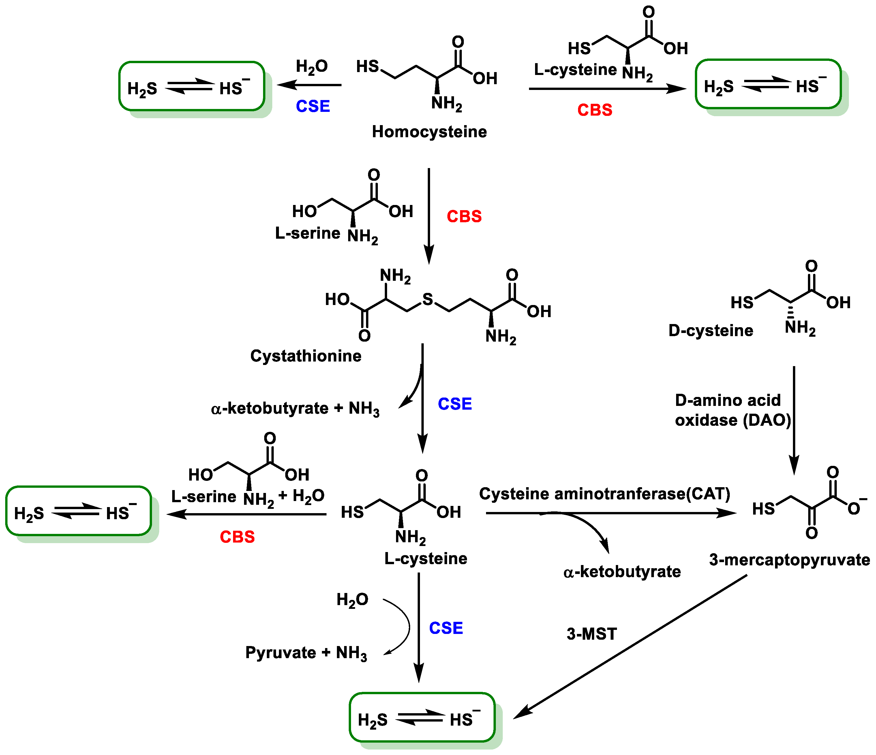
H2S is produced in almost all mammalian cell types. In humans, there are at least two sources of H2S generation: (i) enzymatic synthesis in tissues and (ii) metabolism by bacteria in the digestive tract [40]. Erythrocytes are also capable of reducing elemental sulfur to HS in a non-enzymatic manner [41].
Non-enzymatic H2S production occurs via glucose, glutathione, organic and inorganic polysulfides and elemental sulfur. The reduction of elemental sulfur using NADPH or the oxidation of glucose in the phosphogluconate pathway also produces H2S in a non-enzymatic form [41].
The non-enzymatic form, although less important, comes from the reduction of elemental sulfur to H2S using reductants that come from glucose oxidation, such as lactate, or others such as nicotinamide adenine dinucleotide hydrogen (NADH), nicotinamide adenine dinucleotide phosphate (NADPH) and glutathione (Figure 3).
The non-enzymatic pathway involves the production of NADPH as a by-product of glycolysis to drive the reduction of glutathione disulfide (GSSG) to liberate H2S from sulfane sulfur compounds [42].
Non-enzymatic H2S production in eukaryotes requires various forms of sulfane such as thiosulfate, thiocysteine, polysulfides, persulfides, thiosulfonate and elemental sulfur to release H2S in the presence of reactive molecules [43]. The oxidation of glucose in glycolysis generates reducing equivalents, such as NADPH, which are used for the reduction of glutathione disulfide to glutathione. In addition, this process leads to the release of H2S from the above-mentioned sulfur-containing compounds [41].
Among the enzymes involved in sulfide synthesis, cystathionine-β-synthase, cystathionine-γ-lyase and mercaptopyruvatesulfur transferase are part of the transsulfuration pathway, the latter involved in cysteine catabolism (Figure 4).
The enzymatic pathway involves the production of H2S from L-cysteine, which is an amino acid synthesized by the organism. In mammals, the enzymatic synthesis of H2S involves two pyridoxal-5′-phosphate-dependent enzymes located in the cytosol: cystathionine β-synthase (CBS) and cystathionine γ-lyase (CSE) [44]. A third H2S-producing enzyme, 3MST, is expressed in neurons and the vascular endothelium. In addition, 3MST and cysteine aminotransferase (CAT) contribute to H2S biosynthesis [45]. The third enzyme mentioned, 3MST, unlike the other two which are only in the cytosol, is found in both cytosol and mitochondria [46] and catalyzes the anaerobic pathway of cysteine conversion together with cysteine aminotransferase, transporting sulfide ions to thiol (-SH) groups and forming H2S. This enzyme is localized in the kidney, liver, heart, lungs, thymus, testis, brain and erythrocytes [41,47] (Figure 5).
Cystathionine-β-synthase (CBS) is the predominant enzyme in the production of H2S in the brain and nervous system; is present in liver, kidney, ileum, uterus, placenta and pancreatic islets [46]; belongs to the family of carbon-oxygen-lyases; and is a hydrolase as it catalyzes the cleavage of a C-O bond in serine, with the loss of a water molecule. Human CBS is a homopolymer and uses pyridoxal-5′-phosphate (PLP) as a cofactor, and each monomer contains a non-catalytic heme cofactor [44]. CBS can produce H2S by three distinct pathways:
(1)
Hydrolysis of cysteine to form serine and H2S.
(2)
Condensation of cysteine and homocysteine to generate cystathionine and H2S.
(3)
Condensation of two cysteine molecules to form lanthionine and H2S [48].
Cystathionine-γ-lyase (CSE) is capable of producing H2S via both cysteine (70%) and homocysteine (30%) and is expressed in liver, kidney, ileum, uterus, brain, pancreatic islets and placenta, as well as being characteristic of the vascular system and its smooth muscle [49]. CSE is a PLP-using enzyme, and its catalytic mechanism is very similar to that of CBS. It is a homotetramer [50].
H2S can also be generated from L-methionine via transsulfuration [51]. Homocysteine, a risk factor for cardiovascular and neurocognitive diseases, is converted to H2S, a cardiovascular and neuronal protector, via the transsulfuration pathway or the release of a persulfide (RS-SH) by thiol–disulfide exchange [51].
Mercaptopyruvatesulfurtransferase (3MST) belongs to the family of sulfurtransferases, which catalyze the transfer of a sulfur from a sulfur donor to a thiophilic acceptor by the formation of a protein cysteine persulfur intermediate [52]. The sulfur atom donor is 3-mercaptopyruvate, and the kinetically favored acceptor under physiological conditions is thioredoxin [53] (Figure 6).
The enzyme known as cysteine aminotransferase (CAT) facilitates the transamination process of cysteine and α-ketoglutarate, leading to the production of mercaptopyruvate and glutamate, respectively. Subsequently, another enzyme called mercaptopyruvate sulfurtransferase (MST) catalyzes the transfer of sulfur from 3-MP to a specific active cysteine site. This transfer leads to the formation of pyruvate and a persulfide compound bound to MST. The persulfide bound to MST then reacts with thioredoxin and eventually produces H2S [53].
A synthesis pathway has been described from D-cysteine, which by action of the enzyme D-amino acid oxidase (DAO) produces 3-mercaptopyruvate, which serves as a substrate for 3MST [54] (Figure 7).
In contrast to the L-cysteine system, the D-cysteine-dependent metabolic pathway functions mainly in the cerebellum and kidney, and it is approximately 80 times more efficient than CAT/3MST in H2S production in these tissues [32,55].

3.3. Microbial Synthesis of H2S

Some intestinal bacteria use sulfate as a final acceptor in their respiratory chain and produce H2S, implying that there is an additional source of sulfide in this tissue, in addition to the basal production in the intestinal cells from CBS and CSE enzymes. This results in the organs of the digestive tract, mainly the intestinal epithelium, being exposed to high concentrations of the gas [56,57]. However, the amount of H2S produced by the microbiota is variable and may be related to diet and the type of microbiota present in the gut [58]. H2S has a major influence on digestive health and can be generated from the degradation of cysteine, as well as through the dissimilatory reduction of sulfate [59] (Figure 8).
Sulfate reduction, depending on the micro-organism, can take place in two ways: assimilatory and dissimilatory [60,61]. In dissimilatory sulfate reduction, H2S is produced, and in the assimilatory form, the terminal product is cysteine. The above pathways show clear differences, which are mainly characterized by the role of dissimilatory sulfate in energy production and the function of sulfate as an electron acceptor. In the process of assimilatory sulfate reduction, sulfate serves as a substrate for the production of amino acids.
Sulfate-reducing bacteria are responsible for generating 50% of the H2S found in colonocytes, while the other 50% corresponds to the action of CBS and CSE enzymes [62].

3.4. Production in Laboratory or Industrially

In ambient conditions, hydrogen gas (H2) and the element sulfur (S) show no reactivity. However, when temperatures above room temperature are reached, these elements initiate a chemical reaction, with the optimal temperature being 310 °C.
However, since this process is very slow, alternative methods of extraction are used, such as the one shown in the figure below. Metal sulfides, including iron sulfide, react with acids, such as hydrochloric acid, in a dilute solution (Figure 9).
In this way, H2S gas is obtained and, due to its toxicity, must be collected safely.

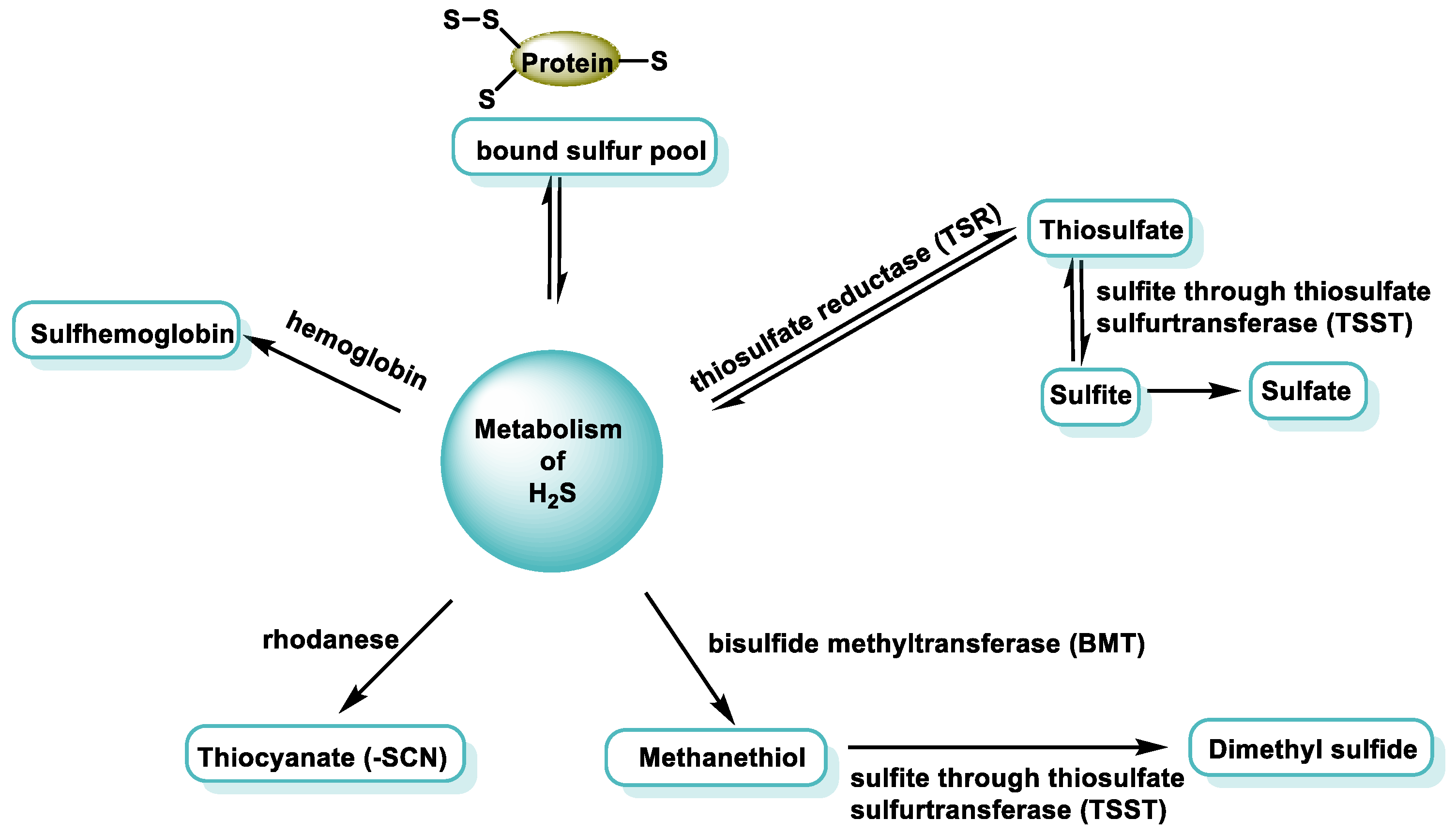
4. H2S Consumption Routes

There are five ways of eliminating H2S in the body (Figure 10).
It is important to control the concentration of H2S in mammalian tissues, which is maintained at nanomolar levels [63]. The concentration of H2S varies between different tissues, and the general agreement is that its concentration should be present in nanomolar concentration, as higher concentrations can inhibit metal-containing proteins. However, methods for the detection of H2S are currently being developed.
H2S only takes on an important signaling function at low concentrations. At higher concentrations, however, it has the potential to have a toxic effect by inhibiting cell respiration through the inhibition of cytochrome c oxidase (CcOX) [64]. To prevent toxicity, it is necessary to regulate the bioavailability of H2S [65].

4.1. Mitochondrial Pathway of Sulfur Oxidation

In the body, H2S is broken down by the mitochondrial enzyme system, currently designated as the “sulfuroxidising unit” or “sulfuroxidising pathway” [66], which transforms sulfur into sulfate and thiosulfate.
Four enzymes are involved in the oxidation pathway: (i) One is sulfur quinone oxidoreductase (SQR), which catalyzes the transfer of two electrons from sulfur to oxidized coenzyme Q. The sulfur is oxidized and transferred to an acceptor molecule (GSH or sulfite). (ii) Others are sulfur transferase (rhodanase) and (iii) oxidases such as persulfide dioxygenase (PDO) and sulfite oxidase (Figure 11). Sulfate and thiosulfate are the main products of the mammalian mitochondrial sulfide oxidation pathway and are safer than sulfide [14].
According to the current knowledge, SQR is the ancient enzyme (from algae and bacteria) initially used instead of Complex I to provide the electrons from H2S to the rest of the primitive ETC process, mostly consisting in its beginning of SQR and the primitive version of Complex IV and Complex V [67]. Mitochondria have the main intracellular machinery responsible for the uptake of H2S and its oxidation into inert metabolites: sulfite and sulfate [68].
Globins and other proteins, including catalase and superoxide dismutase, can serve as catalysts for the oxidation of H2S to polysulfide. This process involves the use of O2 or H2O2 as an electron acceptor [65,69,70,71,72].

4.2. Oxidation of Sulfides by Hemoproteins

Sulfur can react with metal centers through covalent bonding, redox interaction or coordination. This is described as another possible mechanism of sulfide detoxification in red blood cells. As there are no mitochondria in red blood cells, oxidation of sulfur by the route described above is not possible [73].
In methemoglobin, iron is present as Fe(III), which can react reversibly with H2S to form an intermediate MetHb-Fe(III)-SH2. H2S can dissociate and subsequently replenish iron heme through a reversible mechanism. Alternatively, it has the potential to convert ferrous heme into its ferrous counterpart. This redox process would be favored by the loss of a proton from H2S bound to the heme. Methehemoglobin binds to H2S, and the end result is oxidation to a mixture of thiosulfate and hydropolysulfides (Figure 12).
Like human Hb and Mb, there is a set of enzymes such as globins, lactoperoxidases and catalase that react with H2S to form sulfemo analog derivatives, where a sulfur atom covalently bonds to one of the pyrroles of the porphyrin ring [74].

5. H2S Reactivity

H2S has strong nucleophilic properties that allow it to react with electrophilic species, whereas sulfur is a good reductant and will react with oxidants. Sulfur has a variable oxidation state, which can range from −2 to +6, and, in its lowest oxidation state, −2, is a good reductant. This means that it can give up electrons to other atoms, reducing its oxidation state. The sulfur atom from H2S is also a good reductant because sulfur has an oxidation state of −2. When H2S reacts with an oxidant, the sulfur gives up electrons to the oxidant, reducing its oxidation state. It is, therefore, a good reductant acting as a general sulfur atom or as a sulfur atom from H2S.
These properties have been used to make probes to determine the amount of H2S generated. As shown in Figure 13, sulfur can act as a double nucleophile, and this is an important difference from cysteine.

5.1. In Aqueous Solution

H2S is a weak acid that possesses two ionizable protons [75] (Figure 14).
The limited ionization of the first proton can be derived from the initial ionization constant. The ionization of the second proton is minimal, but solutions of hydrogen sulfide contain a certain amount of the sulfur anion S2− [76].
Under physiological conditions, at pH 7.4, the monoanionic species (HS) will predominate by 71.5%, while the remaining 28.5% will be in the protonated form (H2S [65]; under these conditions, the concentration of the dianionic species (S2) is considered negligible.
In the alkaline mitochondrial matrix, which is characterized by a pH of 8.0, about 92% of the sulfur species present are in the form of HS, while the remaining 8% are due to H2S. HS is a prominent sulfur species that exerts a strong attraction to cell nuclei by binding to specific metal centers found in some molecules, such as the oxygen binding site of hemoglobin [76]. The enormous biological potential of H2S is attributed to this phenomenon. In the acidic environment of lysosomes, which is characterized by a pH of 4.7, most H2S is present in its H2S form, which accounts for over 99% of the compound. In this state, H2S has low polarity, allowing it to move freely, diffuse and accumulate in an aqueous or hydrophobic medium, such as biological membranes. About 40% of the sulfides present in the body are in the form of H2S, while the remaining portion is present as hydrogen sulfide anion (HS). There is a tiny and potentially insignificant amount of S2−. The fundamental difference of H2S from the other gasotransmitters is its ability to dissociate in a solution.

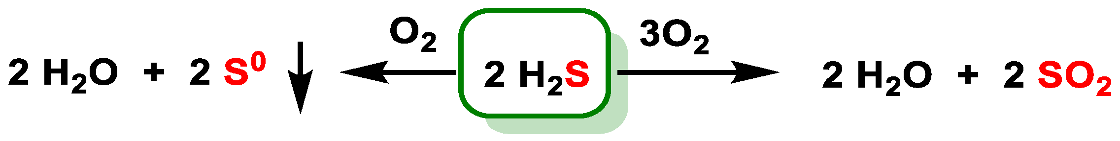
5.2. Reaction with Oxygen

H2S interacts with the oxygen present in the atmosphere, leading to the following chemical reactions (Figure 15).

5.3. Basic Chemical Reactivity of H2S

The basic chemical reactivity of H2S is summarized in Figure 16.
The formation of hydrogen halides and sulfur takes place in the presence of chlorine Cl2, bromine Br2 and iodine I2 [77]. H2S reacts with metals by forming metal sulfide [78]. H2S and SO2 are present in volcanic gases and react with each other to form solid sulfur [79]. H2S is not very stable; it decomposes easily upon heating [80].

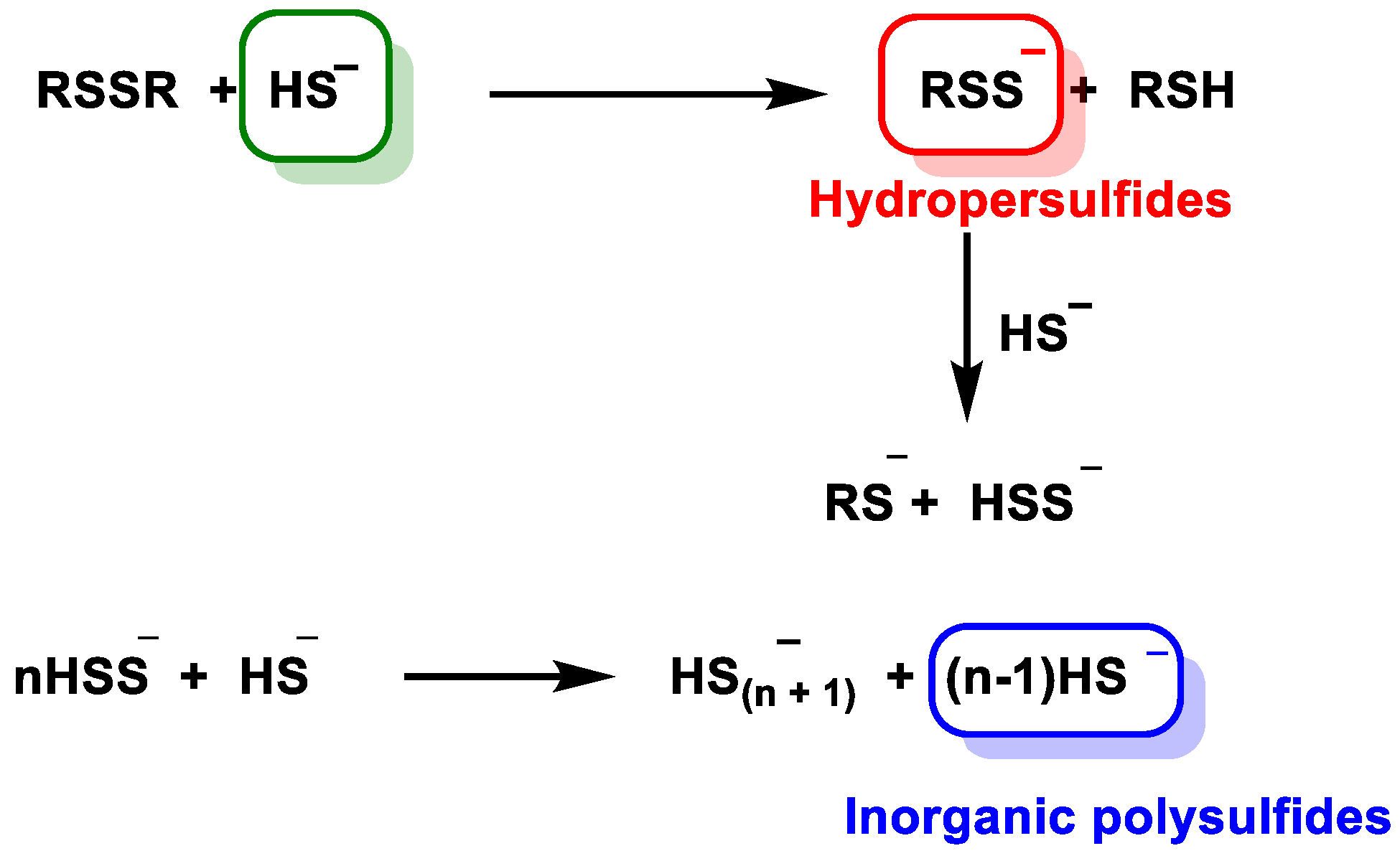
5.4. Reaction with Disulfide

Kinetic studies showed that the reaction of disulfide reduction by HS occurs through multistep equilibria with the formation of disulfide, hydropersulfide and inorganic polysulfides [81]. The mechanism presented in the figure indicates that inorganic polysulfides (HSn) are formed in parallel with hydrosulfides (RSS) (Figure 17).
Inorganic polysulfides have been identified as potential intermediates in the transition pathway from sulfhydryl (SH) to persulfide (SSH) species and are thought to play a role in thiol-based redox signaling mechanisms.

6. Biological Functions of H2S

Endogenous H2S refers to the natural presence of this compound in the body because of regular metabolic processes in humans, animals and other organisms. Research conducted from the early 2000s to the present has revealed that endogenous H2S plays a critical role in regulating specific systems and processes in living organisms [82]. H2S has a high affinity for lipids, which makes it extremely lipophilic and facilitates its penetration of cell membranes, allowing it to enter various cell types. H2S plays a central role in the regulation of various physiological and pathological processes [83,84]. The potential consequences of decreasing H2S levels in the human body include the development and progression of various health conditions, including hypertension, atherosclerosis, gastrointestinal ulcers, cirrhosis of the liver, diabetes, inflammation, Alzheimer’s disease, cancer and other diseases [85]. The accompanying diagram provides a comprehensive representation of the biological mechanisms in human physiology that are controlled by endogenous H2S or show a response to pharmacological intervention with H2S or its derivatives (Figure 18).
Among the variety of effects attributed to H2S, we can mention its participation in cardiovascular processes (in vasodilation, one of the first effects described), the central nervous system, the gastrointestinal system, the endocrine system, cytoprotection, etc. [86,87,88,89] (Figure 19).

6.1. Cardiovascular System

H2S plays a central role in modulating and regulating various signaling pathways involved in metabolism, cardiac function and cellular viability in mammalian organisms [90], and it has a significant effect on the cardiovascular system, blood vessels and blood components. Both mitochondrial activity and cellular metabolism are affected [21].
H2S has been found to be associated with hypertension, atherosclerosis and myocardial damage in the cardiovascular system. The potential efficacy of this compound in the treatment of hypertension may be due to its ability to induce vasodilation. Relaxation of the rat thoracic aorta, portal vein and mesenteric artery by H2S has been demonstrated, suggesting that hydrogen sulfide plays an important role in regulating contractility and blood pressure. In contrast, another study suggests that H2S exhibits vasoconstrictor properties at low concentrations, possibly through a mechanism that inhibits the activity of nitric oxide (-NO), a molecule also involved in contractility [91,92]. H2S may help patients recover from myocardial injury, particularly ischemia–reperfusion injury. Numerous cardiovascular diseases have been associated with H2S, suggesting a potentially broad applicability of H2S in the context of heart disease [90].
H2S exerts several effects on the cardiovascular system. These include attenuating ischemia–reperfusion injury to cardiac tissue, facilitating angiogenesis, relaxing smooth muscle cells and regulating blood pressure [93].

6.2. Gastrointestinal System

H2S is known to have a significant effect on reducing gastric mucosal damage and has the potential to act as a crucial mediator of gastrointestinal motility.
Insulin secretion and diabetes mellitus may be affected by H2S because the pancreas is among the targets of H2S [94,95,96]. In the pancreas, CSE is the main enzyme that converts cysteine to H2S. H2S concentration is elevated in response to the presence of pancreatitis, which is attributed to its proinflammatory effect. H2S administration contributes to chloride secretion, which aggravates certain types of gastritis [97]. H2S concentration increases when abdominal sepsis or endotoxemia occurs [98].
H2S has a protective anti-inflammatory effect on the gastrointestinal system in some types of gastritis and colitis [99,100,101]. H2S elicits both proinflammatory and anti-inflammatory responses in various models of inflammation [102]. The synthesis of H2S is markedly increased in colon ulcers, resulting in accelerated restoration of epithelial barrier integrity and healing of injured tissues [103].

6.3. Respiratory System

A study that investigated the relationship between H2S and pulmonary hypertension represents a pioneering achievement in the field of pathophysiology and H2S on a global scale [104]. In recent years, numerous studies have been conducted to investigate the involvement of H2S in the development of pulmonary hypertension. These studies have primarily focused on the administration of H2S to animal models suffering from chronic hypoxia.
H2S is implicated in the pathogenesis and therapeutic interventions of chronic obstructive pulmonary disease, as endogenous H2S is involved in the regulation of physiological functions of the respiratory system and pathophysiological alterations, such as chronic obstructive pulmonary disease, asthma, pulmonary fibrosis and hypoxia-induced pulmonary hypertension [105].

6.4. Nervous System

In addition, H2S exerts its action on important functions of the central nervous system and provides neuroprotective protection against oxidative stress. There is a belief that it has potentially protective properties against neurodegenerative diseases such as Parkinson’s disease, Alzheimer’s disease and Huntington’s disease [106]; spinocerebellar ataxia; and traumatic brain injury. It has been published that H2S levels are reduced in the brains of patients with Alzheimer’s disease compared to healthy individuals [107]. In the central nervous system, both sides of H2S are observed: it ameliorates ischemic lesions, but it leads to the aggravation of stroke.

6.5. Endocrine System

The variation in H2S concentration is related to a variety of endocrine disorders [108]. Understanding the effect of H2S concentration on the endocrine system is useful for the treatment of hypertension, diabetes and other diseases. H2S can affect the secretion of many hormones and participate in the onset and development of endocrine diseases. H2S can regulate hormone secretion through antioxidant stress and regulation of ion channels and protect endocrine organs. H2S can regulate glucose and fat metabolism through the pancreas, liver, adipose tissue and skeletal muscle [109].

6.6. Visual System

H2S protects retinal photoreceptor cells from light-induced degeneration. Deficiency of H2S or its substrates was found to be associated with ectopialentis, myopia, cataracts, optic atrophy and retinal detachment [110,111,112].

6.7. Age-Related Diseases

H2S is a reducing agent and can be metabolized by various oxidants in the human body. It counteracts oxidative species, such as reactive oxygen species (ROS and reactive nitrogen species (RNS), in the human body. The activation of antioxidant enzymes serves to limit the reactions of free radicals and thus protects against the harmful effects of aging [113].
H2S has several cytoprotective and physiological functions related to age-related diseases. Most notably, it serves as a potent antioxidant and gasotransmitter. Oxidative stress plays an important role in the development and progression of many age-related diseases [114].

6.8. H2S in Cancer

It is documented that several types of cancer such as colon, breast, ovarian and prostate cancers have higher levels of CBS, CSE or 3MST enzymes or synthesize greater amounts of H2S compared to adjacent non-tumor tissue [115,116,117,118].
The outcome of treating cancer cells with H2S donors depends on the concentration/dose, time, cell type and drug used. The effects of both natural and synthetic donors vary from potent cancer suppressors to promoters [40,119]. The administration of H2S donors to various cancer cell lines has been shown to produce cell death, with this effect being dependent on increasing concentrations of H2S. This indicates that H2S donors could represent a therapeutic potential as anticancer drugs. The combination of non-steroidal anti-inflammatory drugs with slow-release H2S donors has been shown to effectively inhibit the growth of human colon, mammary, pancreatic, prostate, lung and bone marrow cancer cells by promoting cell apoptosis via the activation of p38 MAPK [120].

6.9. H2S and Antimicrobial Resistance

The prevalence of antimicrobial resistance (AMR) is increasing and represents a major public health challenge [121]. Similar to mammalian organisms, bacterial cells also have three enzymes involved in the synthesis of H2S [122]. These enzymes are known as cystathionine-γ-lyase (CSE), cystathionine-β-synthetase (CBS) and 3-mercaptopyruvate sulfurtransferase (3MST) [123]. Bacteria have been shown to produce H2S as a cytoprotective agent in response to host-induced stress, such as oxidative stress and antibiotics [124]. Endogenously produced H2S stimulates ROS-scavenging enzymes and interferes with the Fenton reaction, reducing the amount of ROS produced by cells and promoting antibiotic tolerance [125]. Antibiotics such as quinolones, beta-lactams and aminoglycosides are more effective against bacterial pathogens in vitro and in mouse models when combined with small compounds that block a bacterial enzyme involved in the formation of hydrogen sulfide [126]. Therefore, it has been hypothesized that this messenger is a fundamental anti-antibiotic defense mechanism in bacteria [126,127]. In biofilms, persisting cells had significantly higher H2S content than active cells, supporting the notion that H2S is a critical component in bacterial biofilm formation [128]. However, not all significant pathogenic bacteria encode the H2S biosynthetic pathway. Pathogenic Acinetobacter baumannii bacteria do not produce endogenous H2S. By manipulating the sulfide content of A. baumannii with a H2S-releasing chemical, researchers showed that exogenous-H2S-sensitized A. baumannii was able to reverse acquired resistance to gentamicin [129]. It appears that the presence of exogenous H2S triggered a disruption of redox and energy balance that ultimately led to increased susceptibility to the lethal effects of antibiotics [126]. Therefore, it was hypothesized that H2S can be used as an antibiotic-potentiating and resistance-converting agent in bacteria that do not produce it themselves [129]. The recognition of H2S biogenesis is a promising focus for the development of antibacterial adjuvants to combat tolerance and resistance [130]. Influencing hydrogen sulfide-based defenses is a largely unexplored alternative to conventional antibiotic discovery.

7. Mechanisms of Action of H2S and Molecular Targets

The mechanisms through which H2S exerts its effects are not yet fully understood. However, there is sufficient consensus regarding four mechanisms of action or molecular targets (Figure 20).
H2S is of great importance as a secondary messenger that binds to specific target proteins to facilitate signal transduction, especially in the field of mammalian physiology. It is now known that the signal transduction function of H2S occurs through at least three main mechanisms: (i) interactions with metal centers [131], (ii) scavenging of ROS and RNS [132], (iii) S-persulfidation [6] and (iv) effects on ion channels.

7.1. Effect of H2S on Ion Channels

There is abundant literature on the effect of H2S on ion channels. For example, H2S opens ATP-dependent potassium channels [133,134] and modulates various types of calcium [135,136,137] and chloride channels [138].
Ion channels are proteins that form pores in the membranes of cells and organelles, having the function of regulating the flow of ions through them. H2S can act directly or indirectly on the channels. ATP-dependent potassium channels (K+ATP) are the most studied in terms of their interaction with H2S.
K+ATP is a hetero-octamer consisting of four pore-forming subunits, Kir6.x, and four regulatory subunits, SURx, and their activity is inhibited by binding to ATP [139] (Figure 21).
Cysteine 43 of the Kir6.1 subunit is the target of H2S persulfuration. The interaction of the gas with the protein was accompanied by decreased ATP binding and increased binding of phosphatidyl inositol diphosphate, suggesting that the effect of the interaction with H2S decreases the affinity of the protein for ATP, thus activating the channel. Other authors showed with targeted mutagenesis experiments that the effect of H2S was only observed when the two subunits, Kir6.1 and rvSUR1, were present, but not with Kir6.1 alone [139]. They also identified cysteine residues in the regulatory subunit, cysteine 6 and cysteine 26, necessary for the effect.
H2S is unable to regulate the function of K+ATP channels directly [140]. It has been observed that the cardioprotective effects of H2S partially disappear when K+ATP channels are chemically blocked [141].

7.2. Direct Reaction of H2S with ROS and RNS

There is conflicting evidence for the influence of H2S on various organisms, mainly due to its toxic nature and its ability to scavenge reactive oxygen species (ROS) and thus mitigate oxidative stress [7,20]. Hydrogen peroxide, peroxynitrite, hypochlorite and the superoxide radical anion are examples of ROS and reactive nitrogen species (RNS) that can react with H2S. This means that it can inhibit the harmful effects of ROS and/or RNS on organic substances.
Recently, there has been increased interest in the study of H2S due to its toxic nature and its ability to protect bacteria from the harmful effects of oxidative stress induced by antibiotic therapy [142]. The process has the ability to render the redox centers of metalloenzymes inactive [143,144,145,146], causing DNA damage [147] and protein denaturation by disrupting disulfide bonds [148].

7.2.1. Non-Radical Species (Two-Electron Oxidation)

H2S reacts with two-electron oxidants such as hydrogen peroxide, peroxonitrous acid and hypochlorous acid and chloramines to transform into sulfenic acid (HSOH) which is an unstable intermediate (Figure 22).
The primary outcome of the chemical reaction between H2S and hydrogen peroxide (H2O2) is the formation of hydroxylthiol (HSOH). The final product consists largely of polysulfides, elemental sulfur and, in the case of excess oxidant, sulfate, and it depends on the initial ratios of hydrogen peroxide and H2S.
The direct reaction between peroxynitrous acid and HS involves a nucleophilic substitution of HS, leading to the formation of HSOH and NO2 as starting products. In the presence of an excess amount of H2S, HSOH further reacts with a second HS, leading to the formation of HSS/HSSH and other compounds.
It is likely that the interaction between hypochlorous acid and HS occurs through the formation of HSCl, which is subsequently subjected to rapid hydrolysis to produce HSOH. Chloramines, particularly RHNCl and R2NCl, exhibit lower reactivity but higher selectivity as oxidants compared to hypochlorous acid, as indicated in Table 1.
HSOH is the main product. Polysulfides, elemental sulfur and sulfate can all be produced by this reaction, but the exact composition of the final product depends on the ratio of hydrogen peroxide to hydrogen sulfide. It is noteworthy that the system exhibits the typical properties of a chemical oscillator. The oxidation of H2S can lead to the formation of a very reactive reductant: sulfoxylic acid. In many respects, H2S exhibits a reactivity profile similar to cysteine. It is a powerful nucleophile and, like thiols, can react with electrophiles as well as oxidants.
The oxidation process of H2S can lead to the formation of many compounds in which the sulfur atom can have oxidation states from −2 to +6. The oxidation products include several chemical compounds such as sulfate (SO42−), sulfite (SO32−), thiosulfate (S2O32−), persulfides (RSS), organic and inorganic polysulfides and elemental sulfur (Sn).
The oxidation pathways of thiols lead to the formation of sulfenic acids as transient intermediates. The main mechanism by which they are degraded is the formation of disulfide bonds in response to the presence of another thiol, and they are widely recognized for their instability [153,154].

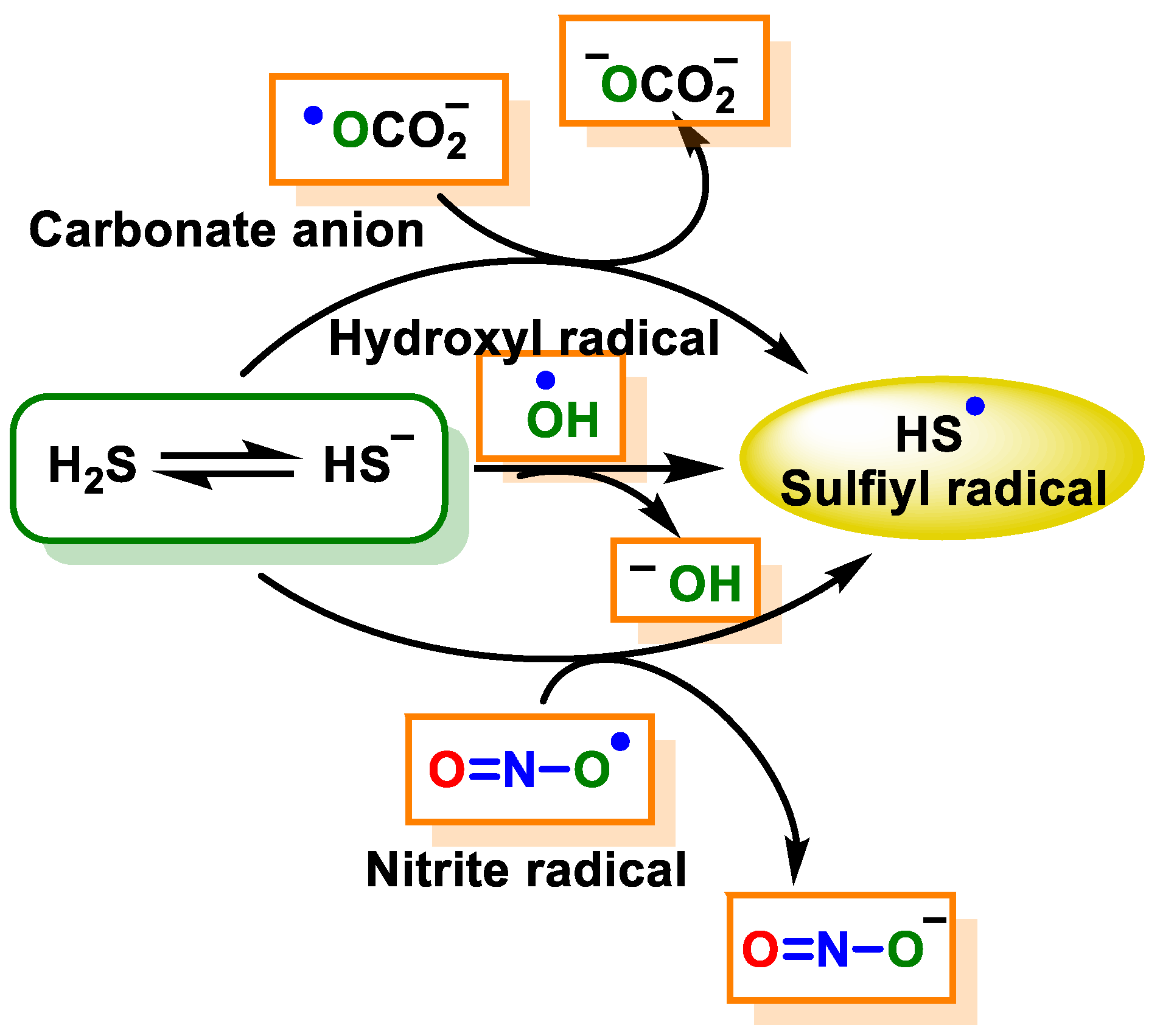
7.2.2. Radical Species (One-Electron Oxidation)

H2S can be oxidized to HS/S•− by a limited number of strong one-electron oxidizing agents, namely the hydroxyl radical (HO), the carbonate radical (CO3•−), nitrogen dioxide (NO2) and the peroxidase compounds oxoferryl I and II.
The initial product of the one-electron oxidation of H2S is the sulfyl radical (HS/S) (Figure 23).
This particular radical exhibits oxidizing properties and is capable of undergoing reactions with a number of electron donors or hydrogen atoms. The compound is capable of undergoing a reaction with a secondary radical HS/S•−, forming HSSH/HSS [155], or it can react with HS to form HSS•2−, which is a reducing radical that has the ability to undergo a reaction with oxygen that leads to the formation of the superoxide radical. HS/S•− can [155] also react with oxygen to form SO2•−, which again can react with oxygen to form a superoxide radical [156]. The one-electron oxidation of H2S has the ability to trigger oxygen-dependent free radical chain reactions that can lead to an amplification of the original oxidation species (Figure 24).
In addition to ROS and RNS, it is also necessary to consider reactive sulfur species, including hydropersulfides, polysulfides and H2S. The concept of RSS was postulated in 2001 [157], RSS that are produced under oxidative stress include RS, sulfenic acids (RSOH), disulfides (RSSR), thiosulfinate (RS(O)SR), thiosulfonate (RS(O)2SR) and S-nitrosothiols (SNTs), the products of cysteine transformations, H2S and sulfane sulfur-containing compounds. SSRs that are produced under physiological conditions (without oxidative stress) are referred to in the scientific literature as “the first class of SSRs”. The “second class of SSRs”, on the other hand, “refers to species that are formed by the initial action of oxidative stress” [158].
H2S plays an important role in combating oxidative species such as ROS and RNS in the body. Filipovic, in 2012 [159], studied the reaction of H2S with peroxynitrite in vitro and in different cell models. The results showed that H2S can remove peroxynitrite with a second-order rate constant and that the reaction does not proceed through radicals. In this reaction, a new product is formed, which was characterized by spectral and computational studies as HSNO2 (thionitrate), mostly as sulfinyl nitrite HS(O)NO.
The ability of HS(O)NO to function as a nitric oxide (NO) donor in response to pH and its ability to release NO in cellular environments have been successfully demonstrated. Therefore, H2S removal plays a role in modulating the chemical and biological effects of peroxynitrite. This process effectively suppresses the pro-apoptotic, oxidative and nitratative properties associated with peroxynitrite (Figure 25).

8. Persulfidation or S-Sulfhydration of Protein Thiols

Sulfhydration of cysteine residues and nitration of tyrosine are H2S-induced post-translational modifications induced by H2S and RNS, respectively [160]. H2S is an important biological messenger molecule that transmits signals through the formation of persulfide bonds (SSH) in proteins or low-molecular-weight thiols [1,161,162]. S-Persulfidation is the process in which a thiol (R-SH) is converted into a perthiol (R-SSH). The modification of thiols to form persulfides is one of the mechanisms by which sulfide exerts signaling functions. Protein modification by persulfurization of cysteine residues is able to modulate the activity of different proteins. The formation of persulfides is associated with the body’s sulfur reserve [1]. The direct reaction of H2S with protein cysteines does not take place; for this reaction to occur, the presence of an oxidant is required. The proposed mechanism for the formation of persulfides is the reaction of sulfur with oxidized cysteines such as sulfenic acid (RSOH) or disulfide (RSSR) [163]. Persulfides can also be formed via radicals, through the reaction of the sulfhydryl radical (HS•−) with the RS, although the low concentration of these species means that this reaction is of little biological relevance [148,164]. HS•− can also react with a non-radical thiol to generate the radical anion RSSH•−, which gives up its unpaired electron to molecular oxygen to give the persulfide and superoxide radical anion [165] (Figure 26).
Persulfides are unstable and have an electrophilic character. They also retain the nucleophilic character of the original thiol, or even enhance it due to the presence of an adjacent sulfur containing unshared electron pairs, i.e., the α-effect [166,167,168]. It has been proposed that these compounds are responsible for the biological effects initially assigned to H2S [169].
One of the ways in which H2S functions as a messenger molecule is by sulfhydration of reactive cysteine residues of target proteins in a manner analogous to protein nitrosylation [1,28].
Due to a decrease in pKa and an increase in nucleophilicity of perthiols compared to thiols, S-persulfidation can affect the biological activity of proteins [170]. For example, the enzyme glyceraldehyde-3-phosphate dehydrogenase (GAPDH), which is primarily involved in glycolysis and gluconeogenesis, undergoes an activity shift after persulfidation to prevent cell death [170]. Persulfidation at KATP channels is a factor contributing to vasodilation caused by H2S [171].
The reaction of persulfides with cyanide gives thiols and thiocyanate (Figure 27).
The cyanolysis reaction is a common reaction involving hydrosulfides and other sulfur compounds. This reaction serves as a reliable method to confirm the presence of the -SSH group on a protein. In addition, this reaction has characteristic properties of hydrosulfides and can be used for the detection of persulfides. The reaction between thiocyanate and ferric ions leads to the formation of a red complex that exhibits absorption at a wavelength of 460 nm. This complex can be accurately measured and quantified by spectrophotometric methods [172] (Figure 28).

9. Detection of H2S

The two deprotonated H2S species (HS and S2−) absorb in UV at 230 nm with molar extinction coefficients of 8 × 10³ and 4.6 × 10³ M−1 cm−1, respectively, at 25 °C, so the concentration of the predominant species (HS) could be measured by absorbance [173]. In practice, their oxidation products generate interferences.
Several research groups have focused their efforts on the development of H2S detection probes. Classical instrumental methods for H2S detection include the following: (i) colorimetric and electrochemical assays, (ii) gas chromatography and (iii) sulfide precipitation [38,75,140,174,175]. These techniques often require complex sample processing. The results of these methods may differ due to the high reactivity of H2S [63,163,176]. The high sensitivity of fluorescence-based assays suggests that they could be potentially valuable in this area. However, the limited number of fluorescence techniques available today for the detection of H2S poses a challenge when it comes to monitoring this gas in biological samples in real time [75,177]. A major challenge is to develop molecular probes that can detect aqueous sulfides (H2S and HS at neuronal pH) in the presence of other thiols found inside most cells.
Numerous H2S detection techniques have been described, including spectrophotometric methods, where sulfide can be monitored by the formation of lead sulfide or methylene blue at 390 or 670 nm, respectively; fluorimetric methods, using fluorescein mercuric acetate; and polarographic methods with sulfide-specific electrodes, as well as by liquid or gas chromatography [178,179].
Another methodology used is the trapping of sulfide on ZnS particles by a reaction with zinc acetate. This technique is used in conjunction with other methods [14].
Classical iodometric titrations are used to prepare a standard solution. H2S is first immobilized in zinc acetate to reduce its dispersion and then reacts with an excess of iodine in an acidic environment. The remaining iodine is subjected to titration with sodium thiosulfate, with starch added as an indicator (Figure 29).
However, this method leads to errors due to the presence of other reductants. Numerous research groups have focused primarily on the development of probes for the detection of H2S. The approaches currently described for the detection of sulfur are usually based on its nucleophilicity or its reductive capacity, both of which are common to other thiols (glutathione and protein thiols) in biological studies and can easily mask the signal corresponding to sulfur. Figure 30 shows the most commonly used methods for H2S detection.

9.1. Lead Acetate

The enzymatic synthesis of hydrogen sulfide can be traced back to a simple approach involving the use of lead acetate and the determination of the formation of lead sulfide (Figure 31), which is insoluble and can be detected by increasing turbidity at 390 nm.
When using this approach to calculate H2S concentrations, it is required to compare the results with a calibration curve created with already-known lead sulfide concentrations [180]. This approach provides semi-quantitative data and has relatively low sensitivity.

9.2. Methylene Blue Method

Methylene blue is formed when an oxidizing agent, usually ferric iron, reacts with H2Saq and N,N-dimethyl-p-phenylenediamine (N,N-dimethylbenzene-1,4-diamine) in an acidic environment (Figure 32).
The methylene blue formation reaction can be used to determine the H2S concentration. The concentration of methylene blue is determined at a wavelength of 670 nm and then compared with calibration curves generated with samples of known H2S concentrations subjected to comparable processing procedures [179,181]. This method also has a number of disadvantages.

9.3. Monobromobimane Derivatization

H2S undergoes a nucleophilic substitution reaction with monobromobimane, resulting in the formation of a bimane-substituted thiol compound. This biman-substituted thiol can further react with a second monobromobiman molecule, leading to the formation of dibiman sulfide. The fluorescence of dibiman sulfide can be observed upon its separation by high-performance liquid chromatography (HPLC) or mass spectrometry. This method has found wide use recently; however, the reaction rate is relatively slow (K ≈ 10 M−1 s−1 at pH 8) [182] (Figure 33).

9.4. Methods Based on the Reducing Capacity of H2S

The sensitivity of fluorescent probes can be quite high. Certain probes, which have been described in detail, use nitro or azide derivatives of rhodamine, dansyl, coumarins or naphthylamides, which can be reduced by H2S to produce fluorescent amines (Figure 34).
In chemical synthesis, H2S is frequently used for the reduction of azido groups (N3) [183] and aromatic nitro groups [184] to aniline derivatives due to its strong reducing agent properties.
By attaching an N3 group to an SF1 rhodamine core, the research team led by Chang was able to produce a selective probe for the detection of H2S. Such probes exhibit strong selectivity for H2S over oxygen and nitrogen [185], two other reactive sulfur species that are physiologically important, and the fluorescent signal is released upon reduction to the amine. Using a similar strategy, Wang’s lab has developed a sulfonyl azidedansyl derivative, whose electrical and fluorescent properties are due to the different electronegativity of the azide and amine groups. H2S could be captured with a fluorescent probe to detect and visualize hydrogen sulfide. However, its slow kinetics are a disadvantage.
To track changes in mitochondrial H2S content in living organisms, Mike Murphy’s team synthesized and characterized MitoA [186]. MitoA consists of an aryl azide coupled with a lipophilic triphenylphosphonium cation (TPP). In living organisms, the TPP cation causes MitoA to accumulate in the mitochondrial structures of the cell. The arylazide group forms MitoN, an arylamine species, when it interacts with H2S (Figure 35).
Therefore, the extent to which MitoA is converted to MitoN serves as an indicator of the amount of H2S in the mitochondria of a living organism. Detection and quantification of these chemicals in tissues can be performed with high sensitivity by liquid chromatography–tandem mass spectrometry (LC-MS/MS), using deuterated internal standards for accurate measurement.
Certain electrochemical techniques utilize the inherent reducing capability of sulfide as a fundamental principle. A polarographic technique based on the oxidation reaction between sulfide and ferricyanide can be used [175].

9.5. Methods Based on Nucleophilicity

H2S is considered a nucleophile that usually occurs as HS under physiological pH conditions. Consequently, it exhibits stronger nucleophilic activity than various other thiols in the cellular environment, which are predominantly present in their protonated form (RSH). The observed difference indicates specific advantages in terms of the nucleophilicity of sulfur compared to thiols. For the selective detection of H2S, it is crucial to distinguish H2S from other nucleophilic compounds present in biological systems, especially thiols such as cysteine and glutathione. In a theoretical context, it is plausible to classify H2S as an unmodified thiol that has the potential to perform two nucleophilic attacks. In contrast, other thiols, such as cysteine, are capable of performing only a single nucleophilic attack. In view of these considerations, probes containing electrophilic functions capable of transforming in the presence of H2S have been described.
HS is known for its strong nucleophilic properties, which allow it to readily bind to electrophilic sites in luminescent compounds such as cyanine dyes. This approach has been used in the development of radiometric H2S sensors, where fluorescence emission is altered by disrupting an extended pi system [187,188,189].
This review introduces a novel radiometric fluorescence probe called CouMC, which is described in a recently published research article. The functionality of this probe is based on the process of selective nucleophilic addition of HS to a merocyanine derivative in a near-neutral pH environment. In addition to its ability to rapidly and specifically detect H2S, this probe also shows potential for the selective visualization of H2S in the mitochondria of living cells (Figure 36).
To construct the CouMC probe, an ethylene group was used to link a coumarin fluorophore to an indole block. By aligning with the electrically positive benzopyrilium moiety of the fluorescent probe, H2S can be distinguished from biothiols and other biological products. This allows the probe to detect H2S based on a flavilium derivative.
The sensitivity of fluorescent probes can be of a considerable order of magnitude. The Xian research group postulated that the introduction of a probe with two electrophilic cores could potentially lead to selectivity for H2S, prompting them to investigate this particular property. The synthesis of a fluorescein ester of thiosalicylic acid was achieved by introducing a thiopyridyl disulfide functionality into the thiol group. This result was achieved by a synthesis process. Active disulfides can undergo disulfide exchange reactions with thiols and H2S. It is important to note that further rearrangement can only take place with the disulfide intermediate generated from H2S. The rearrangement of the ester occurs because of an intramolecular nucleophilic attack on the carboxyl carbon atom, resulting in the release of benzodithiolone and a fluorophore [75] (Figure 37).
Aldehydes and acrylates are two examples of electrophilic centers that have been used in the development of probes. Hemithioacetal is formed by the addition of hydrogen ions to the aldehyde compound. Subsequently, in a neutral pH aqueous solution, the nucleophilic behavior of S is observed; S undergoes an intramolecular Michael addition, specifically adding to the beta position of the α,β-unsaturated ester. The process described above leads to a cyclic thioacetal compound. The results of this research represent a significant advance for Chuan’s research team, as they have effectively visualized the enzymatic production of H2S in living cell systems [190] (Figure 38).

9.6. Methods Based on the Ability to Bind Metal Cations

Another property that can be exploited for the detection of H2S is its remarkable affinity for metals. Novel probes consisting of a fluorophore coupled with Cu2+ have been developed. The precipitation of CuS and subsequent increase in fluorescence are the result of H2S binding to the copper ion [191] (Figure 39).

10. H2S Donors

There is an urgent need for the development of novel chemical tools that facilitate the controlled release of H2S to study its biological activities for research purposes and potential therapeutic applications. This need arises from the frequently observed low concentrations of endogenous H2S, which pose a challenge in studying its biological effects. Different categories of hydrogen sulfide donors can release hydrogen sulfide at different rates. Since a synthetic source of H2S is needed, chemists have developed chemical tools to analyze H2S in biological contexts.
Given the problems associated with the direct administration of H2S gas in various biological contexts, inorganic sulfide salts such as sodium (NaSH) and sodium sulfide (Na2S) are often used as alternative sulfide sources. Potential problems associated with these salts include their susceptibility to oxidation, their volatility and/or shortened effective residence time, and the rapid release of hydrogen sulfide upon dissolution, which can be challenging in some contexts [192]. To address this problem, researchers have developed and studied small H2S donors that release H2S at a controlled rate, like enzymatic H2S synthesis [193]. Currently, there is a wide range of chemicals that are commonly used as donors of H2S. These compounds include aryl isothiocyanates [194], phosphinodithioates [195], thioacids [196]/amides [197], dithiolethiones [198], thiolysis of protected trisulfides [199] and persulfides [200]. In addition, the emission of carbonyl sulfide (COS) from thiocarbamates, followed by its conversion to H2S by the action of carbonate anhydrase [201], is also considered to be a component of these donors.

10.1. Sulfur Salts

Inorganic salts, such as sodium acid sulfide (NaHS) and sodium sulfide (Na2S), were historically the first compounds used and are still the most widely used to generate H2S. These salts are very soluble in water and upon dissolution release a large amount of H2S quickly, and for this reason, they are called fast-releasing donors (Figure 40).
More recently, slow-releasing donors that gradually release small amounts of H2S have been developed. These are usually organic compounds, and their effect more closely resembles a physiological situation, where the gas is released at a low but constant rate [202]. One of the most widely used slow releasers is GYYY4137 [195]. In addition, peptide-based releasers capable of releasing H2S in a more controlled manner are being explored [203].

10.2. Natural Donors

The human body is able to convert sulfur compounds contained in some foods such as garlic, onions, mushrooms and selected edible legumes and fruits into hydrogen sulfide through chemical or enzymatic processes. The compounds isolated from natural products are polysulfides substituted with allyl residues. The garlic derivatives diallyl sulfide, diallyl disulfide and diallyl trisulfide (DAS, DADS and DATS) are capable of releasing H2S in the presence of GSH.
There has long been speculation about the possible health benefits associated with the consumption of garlic.
The major bioactive compound in freshly pressed garlic and certain other Allium plants is allicin [204]. Allicin is a naturally occurring compound that is chemically synthesized during the mechanical crushing of a garlic clove [205]. The compound exhibits inherent instability and is converted into many secondary metabolites that possess bioactive properties. Allicin and its secondary metabolites are a class of organosulfur compounds (OSCs) that exhibit various physiological functions, such as anticancer properties, activity against pathogenic organisms, modulation of the gut microbiota and antioxidant and anti-inflammatory effects. Their effects on pathogenic organisms include antibacterial, antifungal, antiviral and antiparasitic activities [206] (Figure 41). Numerous studies have demonstrated the following beneficial effects of garlic on cardiovascular health: (i) reducing blood pressure, (ii) reducing blood cholesterol levels and aggregation of platelets and (iii) reducing oxidative stress. S-allyl-l-cysteine (SAC) is proposed to be responsible for the cardioprotective effects of garlic.
When garlic cloves are crushed or chopped, the enzyme allinase, which is stored in the vacuoles, is released. When it comes into contact with the cytosolic allicin, it converts it into a series of thiosulfinates, of which allicin is the best known (Figure 42).
The chemical compound allicin exhibits a high degree of instability, as it is susceptible to decomposition and subsequent rearrangement, which leads to the formation of various organosulfur compounds (Figure 43).
Ajoene is a colorless liquid containing a sulfoxide group and a disulfide group and is found as a mixture of up to four stereoisomers and two geometric isomers (E-Z) due to the chirality of sulfoxide (R-S). This chemical that gives garlic its distinctive aroma and flavor is released when the cloves are mechanically crushed or cut. To synthesize ajoene, allicin is dissolved in various solvents, such as edible oils. It has antioxidant properties and thus prevents the formation of superoxides. In addition, it shows antithrombotic and antiviral effects, especially against vaccinia, herpes simplex, rhinovirus, human parainfluenza virus and vesicular stomatitis virus. In addition, ajoene has been observed to have an inhibitory effect on integrin-mediated viral mechanisms in the context of HIV infections. In addition, this molecule has shown antibacterial and antifungal properties, particularly against Candida albicans and Tinea pedis infections.
The chemical composition of the garlic juice produced by the mechanical crushing of the garlic cloves differs significantly from that of the intact garlic clove, particularly with regard to the content of organosulfur components. Unprocessed garlic consists mainly of sulfur derivatives (Figure 44).
The proposed mechanism of H2S release from DADS by GSH attack of sulfur to form an allyl perthiol is shown in Figure 45. Similarly, red blood cells rapidly released H2S from diallyl disulfide (DADS) under oxygen deprivation and in the presence of glutathione [199].
The identification of DATS signifies the early recognition of a garlic-derived chemical that possesses vasoactive properties as well as a marked ability to selectively inhibit the proliferation of cancer cells [207]. Garlic-derived chemical compounds are commonly known as precursors of H2S during the absorption and metabolization process in the circulatory system.
The antiviral potential of garlic and its CSOs has been demonstrated in both in vivo and in vitro studies against a variety of viruses, including those belonging to the families Adenoviridae, Arteriviridae, Coronaviridae, Flaviviridae, Flaviviridae, Herpesviridae, Orthomyxoviridae, Picornaviridae, Paramyxoviridae, Poxviridae, Rhabdoviridae and Retroviridae. Blocking viral entry and fusion into host cells; inhibiting viral RNA polymerase, reverse transcriptase and viral replication; and enhancing the host immune response are the primary mechanisms by which garlic and its CSOs exert their antiviral activity. Garlic and its CSOs were also responsible for enhancing the host immune response. Further research is needed to better understand the properties of garlic and its active compounds, known as CSOs, in relation to their role in antiviral therapy. This means that additional research needs to be conducted focusing on the pharmacokinetics and clinical aspects of the therapeutic effect of garlic.
Other natural sulfur compounds extracted from other plants are isothiocyanates (or sulforaphane), present in cabbages such as broccoli and cauliflower, and erucin, present in seeds and leaves of rocket [101,208], (Figure 46).

10.3. Synthetic H2S Donors
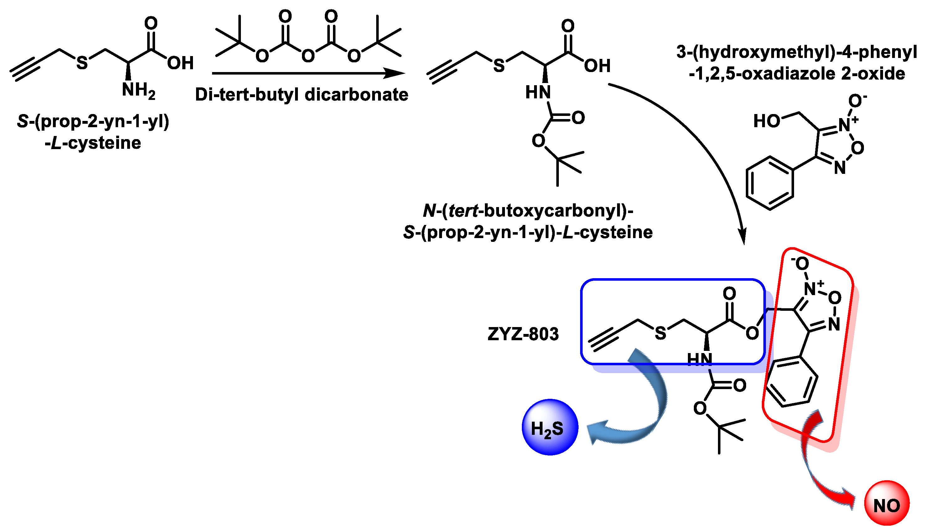
Organic hydrogen sulfide donors, unlike inorganic sulfide salts, are capable of producing a continuous and regulated release of hydrogen sulfide in concentrations comparable to endogenous conditions. Many different types of organic H2S donors have been developed, and the methods by which they produce H2S are as diverse as possible. To advance the development of novel H2S-releasing donors, scientists have begun to alter the chemical compositions of extensively characterized substances known for their sulfur-releasing properties. Examples include donors activated in response to hydrolysis; to endogenous species such as thiols, ROS and enzymes; and to external stimuli such as photoactivation and bioorthogonal chemistry. Another possibility for the release of H2S is the use of the catalyzed hydrolysis of carbonyl sulfide (COS) by carbonic anhydrase. Figure 47 shows the published H2S donors, grouped according to their activation mechanism [209,210,211].

10.4. H2S and NO Hybrid Synthetic Donor

It is synthesized from S-(prop-2-yn-1-yl)-L-cysteine by reaction with di-tert-butyl dicarbonate and subsequent reaction of N-(tert-butoxycarbonyl)-S-(prop-2-yn-1-yl)-L-cysteine with 3-(hydroxymethyl)-4-phenyl-1,2,5-oxadiazole 2-oxide (Figure 48).
ZYZ-803 is a synthetic H2S and NO donor, in vitro and in vivo, and its potency appears to be higher than that of the H2S and/or NO donor. ZYZ-803 also stimulates H2S production from CSE and NO production from eNOS [212,213].
Despite the remarkable development and study of H2S donors, there is an absence of compounds that can address all the requirements for the perfect H2S donor in clinical studies.

10.5. Mitochondrial H2S Donors

AP39 [214], AP123 [215] and MitoPerSulf [216] as new donors of slowly released hydrogen sulfide to the mitochondria are shown in Figure 49.
H2S donors with a slow mitochondrial release rate, such as AP39 and AP123, are >1000 times more potent than Na2S against hyperglycemia-induced oxidant production and also have a beneficial effect on cellular bioenergetics in endothelial cells [215].
The thiocarbonyl group of the 1,2-dithiole-3-thione of AP39 is hydrolyzed to form the corresponding 1,2-dithiole-3-one (RT01) and releases H2S. RT01 undergoes further hydrolysis to release H2S and generate a series of unknown products. The mechanism of H2S release by AP123 involves the hydrolysis of the thiocarbonyl group of the thiobenzamide to form the corresponding amide and releases H2S (Figure 50).
MitoPerSulf is rapidly taken up by the mitochondria, where it reacts with endogenous thiols to generate a persulfur intermediate that releases H2S. MitoPerSulf reacts with thiols such as GSH to rapidly form persulfide, MitoNAP-SSH and GSCOPh. In the presence of excess GSH, GSSH forms H2S by forming GSSG which can react with MitoNAP-SH to form MitoNAP-SSG (Figure 51).
The facilitation of the internalization of MitoPerSulf into the mitochondria is enhanced by the incorporation of the triphenylphosphonium group (TPP), which has a special affinity for the mitochondria. Upon entry into the intended environment, the benzoyl thioester is cleaved due to its interaction with the thiols, resulting in the formation of the labile persulfide compound known as MitoNAP-SSH. The mitochondrial thiols play a crucial role in the persulfidation processes associated with each persulfide. The previously described persulfides show a remarkable propensity to generate hydrogen sulfide and to form disulfides when they interact with other thiols.

11. Hydrogen Sulfide as a Therapeutic Agent

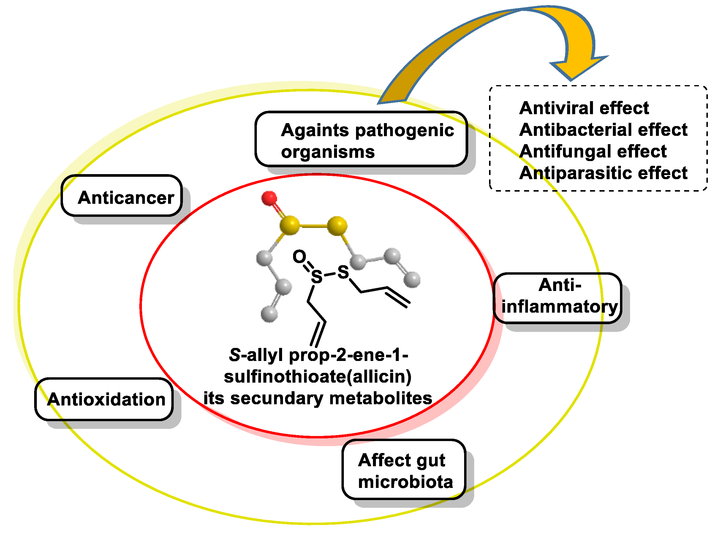
11.1. Garlic Source of H2S Supplements

Garlic has a unique aroma and is a popular ingredient in many dishes. It contains several bioactive components such as polysaccharides, organic sulfides, saponins and phenolic chemicals. Garlic contains organic sulfides such as allicin, alliin, diallyl sulfide, diallyl disulfide, diallyl trisulfide, ajoene and S-allyl cysteine, which are its main bioactive constituents. The bioactive components of garlic have numerous biological effects, such as reducing inflammation, protecting the cardiovascular system, fighting cancer, preventing diabetes, reducing obesity, protecting the nervous system, preventing kidney damage and fighting bacteria and fungi. Through a mechanism involving the reduction of thiols in or on the cell membrane, human red blood cells or rat aortic rings can convert garlic-derived organic polysulfides into hydrogen sulfide [199].
The formation of H2S during the decomposition of organic polysulfides is enhanced by the presence of allylic substituents and an increasing number of sulfur atoms attached to the molecule. In view of the new information obtained, it is conceivable that changes can be made to the compound S-allylcysteine (SAC) found in garlic. The result was a novel molecule called S-propargyl cysteine (SPRC), which is capable of releasing H2S. It was successfully synthesized and proved to be resistant to oxidation in the presence of air [217]. The administration of SPRC (ip) showed a reduction in cognitive impairment caused by intracerebroventricular injection of LPS in rats. The administration of SPRC effectively reduced the lipopolysaccharide (LPS)-induced decrease in H2S levels in the hippocampus of rats.
The proven anticancer effect of allicin and its secondary metabolites suggests promising prospects, even if the practical application of this compound as a pharmaceutical drug is still a long way off. Further experiments are needed to improve the effectiveness of this treatment in fighting cancers of the digestive tract. Garlic is characterized by its non-toxicity or relatively low toxicity. Further studies are needed to explore the potential effects of fermentation or heat treatment of garlic, as these processes may affect the physiological functions and safety aspects of garlic. Additional clinical studies are needed to validate the putative therapeutic benefits of garlic in human cohorts, with particular attention to thoroughly assessing any adverse effects and ensuring overall safety.

11.2. Sulfur Drugs and Their Therapeutic Potential

Functional groups produced from sulfur exhibit a wide range of pharmacological properties and serve as valuable resources for the development of new therapeutic drugs. Pharmaceutical compounds containing sulfur in their chemical composition have the potential to either (i) release H2S or (ii) affect its endogenous synthesis. The ability of pharmaceutical compounds to release H2S has the potential to increase their biological efficacy and provide additional therapeutic benefits, but it can also lead to unfavorable outcomes. The action of sulfur groups and the emission of H2S from the active ingredient may affect the pharmacological activity of routinely used drugs.
Drugs with various therapeutic purposes, such as antihypertensive drugs, antibacterial agents, analgesics, anticancer drugs and anti-inflammatory compounds, may contain detectable amounts of sulfur (Figure 52).
It is assumed that the activities of these compounds are related to their ability to release hydrogen sulfide [218]. Drugs that are capable of releasing H2S have been synthesized as a strategy to enhance their effects or to reduce the adverse effects of treatments. For example, a H2S-releasing derivative of aspirin was effective in protecting the stomach mucosa against gastric damage from regular aspirin in rats [219]. Figure 53 shows the H2S-releasing aspirin derivative (ACS14) and aspirin.
It has been postulated that H2S in combination with non-steroidal anti-inflammatory drugs (NSAIDs) may act as a mediator inducing an anti-inflammatory response. Studies have shown that non-steroidal anti-inflammatory drugs (NSAIDs) that release H2S have increased efficacy and/or improved safety characteristics. Recent evidence has shown that gaseous mediators can improve blood circulation; attenuate oxidative stress; protect gastrointestinal mucosa from damage; enhance the anti-inflammatory properties of non-steroidal anti-inflammatory drugs (NSAIDs), and promote the resolution of inflammation, angiogenesis and epithelialization [220].
A diclofenac derivative showed a greater anti-inflammatory effect than diclofenac [220]. H2S-releasing l-DOPA derivatives (ACS83, ACS84, ACS85 and ACS86) (Figure 54) were synthesized and studied for the treatment of Parkinson’s disease [221].

11.3. Balneotherapy and H2S

Balneotherapy in sulfurous waters, although effective on its own, is even more effective when applied in conjunction with a pelotherapy treatment with peloids with sulfurous components or with hydro-hypnotherapy with sulfurous water. Pelotherapy, in chronic rheumatic pathology, consists of local application by means of plasters on the affected areas, or the immersion of the whole body in the peloid at a high temperature (39–48 °C), for 20–30 min, during 12–15 days of treatment. In natural antioxidant peloids (NAPs), a special clay is combined with sulfurous mineral medicinal water (AMmS), rich in H2S (13 mg/L), which is used in rheumatological, vascular and dermatological treatments, with excellent medical results, especially in the elderly. Sludges made with AMmS, with a recognized antioxidant capacity, due to the effective presence of H2S, are those that have been shown to have the greatest therapeutic capacity.
Studies carried out solely with sulfurous waters in balneotherapy, pelotherapy and drinking water in patients with osteoarthritis/arthrosis (OA) reveal that these waters increase the levels of H2S in plasma after the three types of treatment together [222] and that in general they have an antioxidant effect [223,224] and improve patient mobility and overall quality of life.

12. Conclusions

The identification of the endogenous synthesis of H2S in mammals and the observation that small amounts of H2S have modulatory effects led to the notion that H2S plays a regulatory role by influencing biological processes through its interaction with specific target proteins [152,225].
The cellular production of small amounts of H2S has several signaling functions. While high levels of H2S are extremely toxic, enzymes in the body can detoxify it by oxidation to harmless sulfate.
H2S has attracted considerable interest as a potential pharmacological target and as an important endogenous gas mediator. Drugs that can release H2S slowly are being designed to act beneficially on a variety of diseases. The efficacy, safety and mechanism of action are not fully understood.
Several research papers have addressed the therapeutic properties of externally administered H2S, suggesting that the use of H2S donor medications may be beneficial for specific pathological conditions. For example, the administration of exogenous H2S drugs was found to improve blood vessel function in animal models of diabetes, suggesting a potential therapeutic benefit for diabetic patients. It has been observed that exogenous H2S administration promotes angiogenesis, the process of blood vessel formation. This finding suggests that H2S may be a potential therapeutic intervention in chronic ischemic diseases.
In further research, it is essential to comprehensively evaluate other biological functions of garlic while isolating and identifying the specific chemicals contained in garlic. Further research should be conducted to further explore the underlying mechanisms by which garlic exerts its effects. In addition, more clinical studies should be carried out to confirm the positive effects of garlic on human health. The risks and negative consequences of garlic consumption should be emphasized.

Author Contributions

Conceptualization, C.M.C.A. and C.A.J.; investigation, C.M.C.A. and C.A.J.; writing—review and editing, C.M.C.A., C.A.J., J.M.P.d.l.L., E.P.-L. and F.J.P.; supervision, C.M.C.A., C.A.J. and J.M.P.d.l.L. All authors have read and agreed to the published version of the manuscript.

Funding

This research was funded by Agencia Canaria de Investigación, Innovación y Sociedad de la Información (ACIISI) del Gobierno de Canarias, Project ProID2020010134, Caja Canarias, Project 2019SP43, the Spanish Ministry of Economy and Competitiveness (Grant PID2019-105838RB-C31) and the State Plan for Scientific, Technical Research and Innovation 2021–2023 from the Spanish Ministry of Science and Innovation (project PLEC2022-009507).

Conflicts of Interest

Eduardo Perez Lebeña is an employee of Sistemas de Biotecnología y Recursos Naturales. The other authors declare no conflict of interest. The funders had no role in the design of the study; in the collection, analyses or interpretation of data; in the writing of the manuscript; or in the decision to publish the results.

References

  1. Mustafa, A.K.; Gadalla, M.M.; Sen, N.; Kim, S.; Mu, W.; Gazi, S.K.; Barrow, R.K.; Yang, G.; Wang, R.; Snyder, S.H. H2S signals through protein S-sulfhydration. Sci. Signal. 2009, 2, ra72. [Google Scholar] [CrossRef] [PubMed]
  2. Mathai, J.C.; Missner, A.; Kügler, P.; Saparov, S.M.; Zeidel, M.L.; Lee, J.K.; Pohl, P. No facilitator required for membrane transport of hydrogen sulfide. Proc. Natl. Acad. Sci. USA 2009, 106, 16633–16638. [Google Scholar] [CrossRef] [PubMed]
  3. Whiteman, M.; Winyard, P.G. Hydrogen sulfide and inflammation: The good, the bad, the ugly and the promising. Expert. Rev. Clin. Pharmacol. 2011, 4, 13–32. [Google Scholar] [CrossRef] [PubMed]
  4. Kimura, H.; Shibuya, N.; Kimura, Y. Hydrogen sulfide is a signaling molecule and a cytoprotectant. Antioxid. Redox Signal. 2012, 17, 45–57. [Google Scholar] [CrossRef] [PubMed]
  5. Szabó, C. Hydrogen sulphide and its therapeutic potential. Nat. Rev. Drug Discov. 2007, 6, 917–935. [Google Scholar] [CrossRef] [PubMed]
  6. Li, L.; Rose, P.; Moore, P.K. Hydrogen sulfide and cell signaling. Annu. Rev. Pharmacol. Toxicol. 2011, 51, 169–187. [Google Scholar] [CrossRef] [PubMed]
  7. Wang, R. Physiological implications of hydrogen sulfide: A whiff exploration that blossomed. Physiol. Rev. 2012, 92, 791–896. [Google Scholar] [CrossRef] [PubMed]
  8. Foulkes, M.-G.C. “GAS!”—The Story of the Special Brigade; Andrews UK Limited: Luton, UK, 2012. [Google Scholar]
  9. Yang, G.; Wang, R. H2S and Blood Vessels: An Overview. Handb. Exp. Pharmacol. 2015, 230, 85–110. [Google Scholar] [CrossRef]
  10. Rubio-Ruiz, M.E.; Pérez-Torres, I.; Soto, M.E.; Pastelín, G.; Guarner-Lans, V. Aging in blood vessels. Medicinal agents FOR systemic arterial hypertension in the elderly. Ageing Res. Rev. 2014, 18, 132–147. [Google Scholar] [CrossRef]
  11. Beauchamp, R.O., Jr.; Bus, J.S.; Popp, J.A.; Boreiko, C.J.; Andjelkovich, D.A. A critical review of the literature on hydrogen sulfide toxicity. Crit. Rev. Toxicol. 1984, 13, 25–97. [Google Scholar] [CrossRef]
  12. Reiffenstein, R.J.; Hulbert, W.C.; Roth, S.H. Toxicology of hydrogen sulfide. Annu. Rev. Pharmacol. Toxicol. 1992, 32, 109–134. [Google Scholar] [CrossRef] [PubMed]
  13. Marshall, T.; Dorman, D.; Gardner, D.; Adeshina, F. Provisional advisory levels (PALs) for hydrogen sulfide (H2S). Inhal. Toxicol. 2009, 21, 56–72. [Google Scholar] [CrossRef] [PubMed]
  14. Filipovic, M.R.; Zivanovic, J.; Alvarez, B.; Banerjee, R. Chemical Biology of H2S Signaling through Persulfidation. Chem. Rev. 2018, 118, 1253–1337. [Google Scholar] [CrossRef] [PubMed]
  15. Riahi, S.; Rowley, C.N. Why can hydrogen sulfide permeate cell membranes? J. Am. Chem. Soc. 2014, 136, 15111–15113. [Google Scholar] [CrossRef] [PubMed]
  16. Millero, F.J. The thermodynamics and kinetics of the hydrogen sulfide system in natural waters. Mar. Chem. 1986, 18, 121–147. [Google Scholar] [CrossRef]
  17. Fogg, P.G. Hydrogen Sulfide, Deuterium Sulfide & Hydrogen Selenide; Elsevier: Amsterdam, The Netherlands, 2013; Volume 32. [Google Scholar]
  18. Haouzi, P. Ventilatory and metabolic effects of exogenous hydrogen sulfide. Respir. Physiol. Neurobiol. 2012, 184, 170–177. [Google Scholar] [CrossRef] [PubMed]
  19. Li, Q.; Lancaster, J.R. Chemical foundations of hydrogen sulfide biology. Nitric Oxide 2013, 35, 21–34. [Google Scholar] [CrossRef]
  20. Kimura, H. Production and physiological effects of hydrogen sulfide. Antioxid. Redox Signal. 2014, 20, 783–793. [Google Scholar] [CrossRef]
  21. Paul, B.D.; Snyder, S.H.; Kashfi, K. Effects of hydrogen sulfide on mitochondrial function and cellular bioenergetics. Redox Biol. 2021, 38, 101772. [Google Scholar] [CrossRef]
  22. Hanna, D.; Kumar, R.; Banerjee, R. A Metabolic Paradigm for Hydrogen Sulfide Signaling via Electron Transport Chain Plasticity. Antioxid. Redox Signal. 2023, 38, 57–67. [Google Scholar] [CrossRef]
  23. Szabo, C. A timeline of hydrogen sulfide (H2S) research: From environmental toxin to biological mediator. Biochem. Pharmacol. 2018, 149, 5–19. [Google Scholar] [CrossRef] [PubMed]
  24. Kimura, H. Hydrogen sulfide: Its production and functions. Exp. Physiol. 2011, 96, 833–835. [Google Scholar] [CrossRef] [PubMed]
  25. Hosoki, R.; Matsuki, N.; Kimura, H. The possible role of hydrogen sulfide as an endogenous smooth muscle relaxant in synergy with nitric oxide. Biochem. Biophys. Res. Commun. 1997, 237, 527–531. [Google Scholar] [CrossRef] [PubMed]
  26. Shibuya, N.; Mikami, Y.; Kimura, Y.; Nagahara, N.; Kimura, H. Vascular endothelium expresses 3-mercaptopyruvate sulfurtransferase and produces hydrogen sulfide. J. Biochem. 2009, 146, 623–626. [Google Scholar] [CrossRef]
  27. Murphy, B.; Bhattacharya, R.; Mukherjee, P. Hydrogen sulfide signaling in mitochondria and disease. FASEB J. 2019, 33, 13098–13125. [Google Scholar] [CrossRef] [PubMed]
  28. Paul, B.D.; Snyder, S.H. H2S signalling through protein sulfhydration and beyond. Nat. Rev. Mol. Cell Biol. 2012, 13, 499–507. [Google Scholar] [CrossRef] [PubMed]
  29. Shibuya, N.; Tanaka, M.; Yoshida, M.; Ogasawara, Y.; Togawa, T.; Ishii, K.; Kimura, H. 3-Mercaptopyruvate sulfurtransferase produces hydrogen sulfide and bound sulfane sulfur in the brain. Antioxid. Redox Signal. 2009, 11, 703–714. [Google Scholar] [CrossRef] [PubMed]
  30. Randi, E.B.; Casili, G.; Jacquemai, S.; Szabo, C. Selenium-Binding Protein 1 (SELENBP1) Supports Hydrogen Sulfide Biosynthesis and Adipogenesis. Antioxidants 2021, 10, 361. [Google Scholar] [CrossRef] [PubMed]
  31. Shibuya, N.; Kimura, H. Production of hydrogen sulfide from d-cysteine and its therapeutic potential. Front. Endocrinol. 2013, 4, 87. [Google Scholar] [CrossRef]
  32. Shibuya, N.; Koike, S.; Tanaka, M.; Ishigami-Yuasa, M.; Kimura, Y.; Ogasawara, Y.; Fukui, K.; Nagahara, N.; Kimura, H. A novel pathway for the production of hydrogen sulfide from D-cysteine in mammalian cells. Nat. Commun. 2013, 4, 1366. [Google Scholar] [CrossRef]
  33. Kimura, H. Hydrogen sulfide: Its production, release and functions. Amino Acids 2011, 41, 113–121. [Google Scholar] [CrossRef] [PubMed]
  34. Ogasawara, Y.; Ishii, K.; Togawa, T.; Tanabe, S. Determination of bound sulfur in serum by gas dialysis/high-performance liquid chromatography. Anal. Biochem. 1993, 215, 73–81. [Google Scholar] [CrossRef]
  35. Toohey, J.I. Sulphane sulphur in biological systems: A possible regulatory role. Biochem. J. 1989, 264, 625–632. [Google Scholar] [CrossRef] [PubMed]
  36. Ishigami, M.; Hiraki, K.; Umemura, K.; Ogasawara, Y.; Ishii, K.; Kimura, H. A source of hydrogen sulfide and a mechanism of its release in the brain. Antioxid. Redox Signal. 2009, 11, 205–214. [Google Scholar] [CrossRef]
  37. Ogasawara, Y.; Isoda, S.; Tanabe, S. Tissue and subcellular distribution of bound and acid-labile sulfur, and the enzymic capacity for sulfide production in the rat. Biol. Pharm. Bull. 1994, 17, 1535–1542. [Google Scholar] [CrossRef] [PubMed]
  38. Ubuka, T. Assay methods and biological roles of labile sulfur in animal tissues. J. Chromatogr. B Anal. Technol. Biomed. Life Sci. 2002, 781, 227–249. [Google Scholar] [CrossRef] [PubMed]
  39. Westley, A.M.; Westley, J. Biological sulfane sulfur. Anal. Biochem. 1991, 195, 63–67. [Google Scholar] [CrossRef] [PubMed]
  40. Szabo, C. Gasotransmitters in cancer: From pathophysiology to experimental therapy. Nat. Rev. Drug Discov. 2016, 15, 185–203. [Google Scholar] [CrossRef] [PubMed]
  41. Searcy, D.G.; Lee, S.H. Sulfur reduction by human erythrocytes. J. Exp. Zool. 1998, 282, 310–322. [Google Scholar] [CrossRef]
  42. Mantle, D.; Yang, G. Hydrogen sulfide and metal interaction: The pathophysiological implications. Mol. Cell Biochem. 2022, 477, 2235–2248. [Google Scholar] [CrossRef] [PubMed]
  43. Kabil, O.; Banerjee, R. Enzymology of H2S biogenesis, decay and signaling. Antioxid. Redox Signal. 2014, 20, 770–782. [Google Scholar] [CrossRef] [PubMed]
  44. Meier, M.; Janosik, M.; Kery, V.; Kraus, J.P.; Burkhard, P. Structure of human cystathionine beta-synthase: A unique pyridoxal 5′-phosphate-dependent heme protein. EMBO J. 2001, 20, 3910–3916. [Google Scholar] [CrossRef] [PubMed]
  45. Stipanuk, M.H. Sulfur amino acid metabolism: Pathways for production and removal of homocysteine and cysteine. Annu. Rev. Nutr. 2004, 24, 539–577. [Google Scholar] [CrossRef] [PubMed]
  46. Iciek, M.; Włodek, L. Biosynthesis and biological properties of compounds containing highly reactive, reduced sulfane sulfur. Pol. J. Pharmacol. 2001, 53, 215–225. [Google Scholar] [PubMed]
  47. Zheng, M.; Zeng, Q.; Shi, X.Q.; Zhao, J.; Tang, C.S.; Sun, N.L.; Geng, B. Erythrocytic or serum hydrogen sulfide association with hypertension development in untreated essential hypertension. Chin. Med. J. 2011, 124, 3693–3701. [Google Scholar] [PubMed]
  48. Singh, S.; Banerjee, R. PLP-dependent H2S biogenesis. Biochim. Biophys. Acta 2011, 1814, 1518–1527. [Google Scholar] [CrossRef] [PubMed]
  49. Wen, Y.D.; Wang, H.; Zhu, Y.Z. The Drug Developments of Hydrogen Sulfide on Cardiovascular Disease. Oxid. Med. Cell Longev. 2018, 2018, 4010395. [Google Scholar] [CrossRef]
  50. Zuhra, K.; Augsburger, F.; Majtan, T.; Szabo, C. Cystathionine-β-Synthase: Molecular Regulation and Pharmacological Inhibition. Biomolecules 2020, 10, 697. [Google Scholar] [CrossRef]
  51. Bearden, S.E.; Beard, R.S., Jr.; Pfau, J.C. Extracellular transsulfuration generates hydrogen sulfide from homocysteine and protects endothelium from redox stress. Am. J. Physiol. Heart Circ. Physiol. 2010, 299, H1568–H1576. [Google Scholar] [CrossRef]
  52. Rao, S.P.; Dobariya, P.; Bellamkonda, H.; More, S.S. Role of 3-Mercaptopyruvate Sulfurtransferase (3-MST) in Physiology and Disease. Antioxidants 2023, 12, 603. [Google Scholar] [CrossRef]
  53. Yadav, P.K.; Yamada, K.; Chiku, T.; Koutmos, M.; Banerjee, R. Structure and kinetic analysis of H2S production by human mercaptopyruvate sulfurtransferase. J. Biol. Chem. 2013, 288, 20002–20013. [Google Scholar] [CrossRef] [PubMed]
  54. Hipólito, A.; Nunes, S.C.; Vicente, J.B.; Serpa, J. Cysteine Aminotransferase (CAT): A Pivotal Sponsor in Metabolic Remodeling and an Ally of 3-Mercaptopyruvate Sulfurtransferase (MST) in Cancer. Molecules 2020, 25, 3984. [Google Scholar] [CrossRef] [PubMed]
  55. Kimura, H. Metabolic turnover of hydrogen sulfide. Front. Physiol. 2012, 3, 101. [Google Scholar] [CrossRef] [PubMed]
  56. Magee, E.A.; Richardson, C.J.; Hughes, R.; Cummings, J.H. Contribution of dietary protein to sulfide production in the large intestine: An in vitro and a controlled feeding study in humans. Am. J. Clin. Nutr. 2000, 72, 1488–1494. [Google Scholar] [CrossRef] [PubMed]
  57. Levitt, M.D.; Springfield, J.; Furne, J.; Koenig, T.; Suarez, F.L. Physiology of sulfide in the rat colon: Use of bismuth to assess colonic sulfide production. J. Appl. Physiol. 2002, 92, 1655–1660. [Google Scholar] [CrossRef] [PubMed]
  58. Yao, C.K.; Rotbart, A.; Ou, J.Z.; Kalantar-Zadeh, K.; Muir, J.G.; Gibson, P.R. Modulation of colonic hydrogen sulfide production by diet and mesalazine utilizing a novel gas-profiling technology. Gut Microbes 2018, 9, 510–522. [Google Scholar] [CrossRef] [PubMed]
  59. Singh, S.B.; Lin, H.C. Hydrogen Sulfide in Physiology and Diseases of the Digestive Tract. Microorganisms 2015, 3, 866–889. [Google Scholar] [CrossRef] [PubMed]
  60. Barton, L.L.; Fauque, G.D. Biochemistry, physiology and biotechnology of sulfate-reducing bacteria. Adv. Appl. Microbiol. 2009, 68, 41–98. [Google Scholar] [CrossRef]
  61. Carbonero, F.; Benefiel, A.C.; Alizadeh-Ghamsari, A.H.; Gaskins, H.R. Microbial pathways in colonic sulfur metabolism and links with health and disease. Front. Physiol. 2012, 3, 448. [Google Scholar] [CrossRef]
  62. Bryan, N.S.; Lefer, D.J. Update on Gaseous Signaling Molecules Nitric Oxide and Hydrogen Sulfide: Strategies to Capture their Functional Activity for Human Therapeutics. Mol. Pharmacol. 2019, 96, 109–114. [Google Scholar] [CrossRef]
  63. Furne, J.; Saeed, A.; Levitt, M.D. Whole tissue hydrogen sulfide concentrations are orders of magnitude lower than presently accepted values. Am. J. Physiol. Regul. Integr. Comp. Physiol. 2008, 295, R1479–R1485. [Google Scholar] [CrossRef] [PubMed]
  64. Petersen, L.C. The effect of inhibitors on the oxygen kinetics of cytochrome c oxidase. Biochim. Biophys. Acta BBA Bioenerg. 1977, 460, 299–307. [Google Scholar] [CrossRef] [PubMed]
  65. Giuffrè, A.; Vicente, J.B. Hydrogen Sulfide Biochemistry and Interplay with Other Gaseous Mediators in Mammalian Physiology. Oxidative Med. Cell. Longev. 2018, 2018, 6290931. [Google Scholar] [CrossRef] [PubMed]
  66. Lagoutte, E.; Mimoun, S.; Andriamihaja, M.; Chaumontet, C.; Blachier, F.; Bouillaud, F. Oxidation of hydrogen sulfide remains a priority in mammalian cells and causes reverse electron transfer in colonocytes. Biochim. Biophys. Acta BBA Bioenerg. 2010, 1797, 1500–1511. [Google Scholar] [CrossRef] [PubMed]
  67. Theissen, U.; Hoffmeister, M.; Grieshaber, M.; Martin, W. Single eubacterial origin of eukaryotic sulfide: Quinone oxidoreductase, a mitochondrial enzyme conserved from the early evolution of eukaryotes during anoxic and sulfidic times. Mol. Biol. Evol. 2003, 20, 1564–1574. [Google Scholar] [CrossRef] [PubMed]
  68. Borisov, V.B.; Forte, E. Impact of Hydrogen Sulfide on Mitochondrial and Bacterial Bioenergetics. Int. J. Mol. Sci. 2021, 22, 2688. [Google Scholar] [CrossRef]
  69. Olson, K.R.; Gao, Y.; Arif, F.; Arora, K.; Patel, S.; DeLeon, E.R.; Sutton, T.R.; Feelisch, M.; Cortese-Krott, M.M.; Straub, K.D. Metabolism of hydrogen sulfide (H2S) and Production of Reactive Sulfur Species (RSS) by superoxide dismutase. Redox Biol. 2018, 15, 74–85. [Google Scholar] [CrossRef]
  70. Olson, K.R.; Gao, Y.; DeLeon, E.R.; Arif, M.; Arif, F.; Arora, N.; Straub, K.D. Catalase as a sulfide-sulfur oxido-reductase: An ancient (and modern?) regulator of reactive sulfur species (RSS). Redox Biol. 2017, 12, 325–339. [Google Scholar] [CrossRef]
  71. Searcy, D.G. HS-:O2 oxidoreductase activity of Cu, Zn superoxide dismutase. Arch. Biochem. Biophys. 1996, 334, 50–58. [Google Scholar] [CrossRef]
  72. Searcy, D.G.; Whitehead, J.P.; Maroney, M.J. Interaction of Cu, Zn superoxide dismutase with hydrogen sulfide. Arch. Biochem. Biophys. 1995, 318, 251–263. [Google Scholar] [CrossRef]
  73. Vitvitsky, V.; Yadav, P.K.; Kurthen, A.; Banerjee, R. Sulfide oxidation by a noncanonical pathway in red blood cells generates thiosulfate and polysulfides. J. Biol. Chem. 2015, 290, 8310–8320. [Google Scholar] [CrossRef] [PubMed]
  74. Ríos-González, B.B.; Román-Morales, E.M.; Pietri, R.; López-Garriga, J. Hydrogen sulfide activation in hemeproteins: The sulfheme scenario. J. Inorg. Biochem. 2014, 133, 78–86. [Google Scholar] [CrossRef] [PubMed]
  75. Liu, C.; Pan, J.; Li, S.; Zhao, Y.; Wu, L.Y.; Berkman, C.E.; Whorton, A.R.; Xian, M. Capture and visualization of hydrogen sulfide by a fluorescent probe. Angew. Chem. Int. Ed. Engl. 2011, 50, 10327–10329. [Google Scholar] [CrossRef] [PubMed]
  76. Martelli, A.; Testai, L.; Breschi, M.C.; Blandizzi, C.; Virdis, A.; Taddei, S.; Calderone, V. Hydrogen sulphide: Novel opportunity for drug discovery. Med. Res. Rev. 2012, 32, 1093–1130. [Google Scholar] [CrossRef] [PubMed]
  77. Cavallo, G.; Metrangolo, P.; Milani, R.; Pilati, T.; Priimagi, A.; Resnati, G.; Terraneo, G. The Halogen Bond. Chem. Rev. 2016, 116, 2478–2601. [Google Scholar] [CrossRef] [PubMed]
  78. Hartle, M.D.; Sommer, S.K.; Dietrich, S.R.; Pluth, M.D. Chemically reversible reactions of hydrogen sulfide with metal phthalocyanines. Inorg. Chem. 2014, 53, 7800–7802. [Google Scholar] [CrossRef]
  79. Aiuppa, A.; Federico, C.; Giudice, G.; Gurrieri, S.; Valenza, M. Hydrothermal buffering of the SO2/H2S ratio in volcanic gases: Evidence from La Fossa Crater fumarolic field, Vulcano Island. Geophys. Res. Lett. 2006, 33, 21. [Google Scholar] [CrossRef]
  80. Jiang, G.; Yang, Z.; Zhang, F.; Li, G.; Wei, Z.; Niu, B.; Zhao, M.; Zhang, Z.; Hao, Z. Thermal Decomposition of H2S at Low Temperature on Mo-Containing Catalysts Derived from MAlO (M = Mg, Co, and Ni) Layered Double Hydroxides. Ind. Eng. Chem. Res. 2023, 62, 7224–7234. [Google Scholar] [CrossRef]
  81. Vasas, A.; Dóka, É.; Fábián, I.; Nagy, P. Kinetic and thermodynamic studies on the disulfide-bond reducing potential of hydrogen sulfide. Nitric Oxide 2015, 46, 93–101. [Google Scholar] [CrossRef]
  82. Dilek, N.; Papapetropoulos, A.; Toliver-Kinsky, T.; Szabo, C. Hydrogen sulfide: An endogenous regulator of the immune system. Pharmacol. Res. 2020, 161, 105119. [Google Scholar] [CrossRef]
  83. Vandiver, M.; Snyder, S.H. Hydrogen sulfide: A gasotransmitter of clinical relevance. J. Mol. Med. 2012, 90, 255–263. [Google Scholar] [CrossRef]
  84. Kolluru, G.K.; Shen, X.; Bir, S.C.; Kevil, C.G. Hydrogen sulfide chemical biology: Pathophysiological roles and detection. Nitric Oxide 2013, 35, 5–20. [Google Scholar] [CrossRef] [PubMed]
  85. Zheng, Q.; Pan, L.; Ji, Y. H2S protects against diabetes-accelerated atherosclerosis by preventing the activation of NLRP3 inflammasome. J. Biomed. Res. 2019, 34, 94–102. [Google Scholar] [CrossRef] [PubMed]
  86. Parsanathan, R.; Jain, S.K. Hydrogen sulfide regulates circadian-clock genes in C2C12 myotubes and the muscle of high-fat-diet-fed mice. Arch. Biochem. Biophys. 2019, 672, 108054. [Google Scholar] [CrossRef] [PubMed]
  87. Szabo, C.; Papapetropoulos, A. International Union of Basic and Clinical Pharmacology. CII: Pharmacological Modulation of H2S Levels: H2S Donors and H2S Biosynthesis Inhibitors. Pharmacol. Rev. 2017, 69, 497–564. [Google Scholar] [CrossRef] [PubMed]
  88. Kimura, H.; Nagai, Y.; Umemura, K.; Kimura, Y. Physiological roles of hydrogen sulfide: Synaptic modulation, neuroprotection, and smooth muscle relaxation. Antioxid. Redox Signal. 2005, 7, 795–803. [Google Scholar] [CrossRef] [PubMed]
  89. Iciek, M.; Bilska-Wilkosz, A.; Kozdrowicki, M.; Górny, M. Reactive sulfur species and their significance in health and disease. Biosci. Rep. 2022, 42, BSR20221006. [Google Scholar] [CrossRef] [PubMed]
  90. Kolluru, G.K.; Shackelford, R.E.; Shen, X.; Dominic, P.; Kevil, C.G. Sulfide regulation of cardiovascular function in health and disease. Nat. Rev. Cardiol. 2023, 20, 109–125. [Google Scholar] [CrossRef] [PubMed]
  91. Rossoni, G.; Manfredi, B.; Tazzari, V.; Sparatore, A.; Trivulzio, S.; Del Soldato, P.; Berti, F. Activity of a new hydrogen sulfide-releasing aspirin (ACS14) on pathological cardiovascular alterations induced by glutathione depletion in rats. Eur. J. Pharmacol. 2010, 648, 139–145. [Google Scholar] [CrossRef]
  92. Li, Y.F.; Xiao, C.S.; Hui, R.T. Calcium sulfide (CaS), a donor of hydrogen sulfide (H2S): A new antihypertensive drug? Med. Hypotheses 2009, 73, 445–447. [Google Scholar] [CrossRef]
  93. Shen, Y.; Shen, Z.; Luo, S.; Guo, W.; Zhu, Y.Z. The Cardioprotective Effects of Hydrogen Sulfide in Heart Diseases: From Molecular Mechanisms to Therapeutic Potential. Oxidative Med. Cell. Longev. 2015, 2015, 925167. [Google Scholar] [CrossRef] [PubMed]
  94. Tamizhselvi, R.; Koh, Y.H.; Sun, J.; Zhang, H.; Bhatia, M. Hydrogen sulfide induces ICAM-1 expression and neutrophil adhesion to caerulein-treated pancreatic acinar cells through NF-kappaB and Src-family kinases pathway. Exp. Cell Res. 2010, 316, 1625–1636. [Google Scholar] [CrossRef] [PubMed]
  95. Morselli-Labate, A.M.; Fantini, L.; Pezzilli, R. Hydrogen sulfide, nitric oxide and a molecular mass 66 u substance in the exhaled breath of chronic pancreatitis patients. Pancreatology 2007, 7, 497–504. [Google Scholar] [CrossRef] [PubMed]
  96. Bhatia, M. H2S and substance P in inflammation. Methods Enzymol. 2015, 555, 195–205. [Google Scholar] [CrossRef] [PubMed]
  97. Linden, D.R.; Levitt, M.D.; Farrugia, G.; Szurszewski, J.H. Endogenous production of H2S in the gastrointestinal tract: Still in search of a physiologic function. Antioxid. Redox Signal. 2010, 12, 1135–1146. [Google Scholar] [CrossRef] [PubMed]
  98. Ganster, F.; Burban, M.; de la Bourdonnaye, M.; Fizanne, L.; Douay, O.; Loufrani, L.; Mercat, A.; Calès, P.; Radermacher, P.; Henrion, D.; et al. Effects of hydrogen sulfide on hemodynamics, inflammatory response and oxidative stress during resuscitated hemorrhagic shock in rats. Crit. Care 2010, 14, R165. [Google Scholar] [CrossRef] [PubMed]
  99. Wallace, J.L.; Vong, L.; McKnight, W.; Dicay, M.; Martin, G.R. Endogenous and exogenous hydrogen sulfide promotes resolution of colitis in rats. Gastroenterology 2009, 137, 569–578. [Google Scholar] [CrossRef] [PubMed]
  100. Chávez-Piña, A.E.; Tapia-Alvarez, G.R.; Navarrete, A. Inhibition of endogenous hydrogen sulfide synthesis by PAG protects against ethanol-induced gastric damage in the rat. Eur. J. Pharmacol. 2010, 630, 131–136. [Google Scholar] [CrossRef]
  101. Guo, W.; Cheng, Z.-y.; Zhu, Y.-z. Hydrogen sulfide and translational medicine. Acta Pharmacol. Sin. 2013, 34, 1284–1291. [Google Scholar] [CrossRef]
  102. Bhatia, M. H2S and Inflammation: An Overview. Handb. Exp. Pharmacol. 2015, 230, 165–180. [Google Scholar] [CrossRef]
  103. Flannigan, K.L.; Ferraz, J.G.; Wang, R.; Wallace, J.L. Enhanced synthesis and diminished degradation of hydrogen sulfide in experimental colitis: A site-specific, pro-resolution mechanism. PLoS ONE 2013, 8, e71962. [Google Scholar] [CrossRef] [PubMed]
  104. Chunyu, Z.; Junbao, D.; Dingfang, B.; Hui, Y.; Xiuying, T.; Chaoshu, T. The regulatory effect of hydrogen sulfide on hypoxic pulmonary hypertension in rats. Biochem. Biophys. Res. Commun. 2003, 302, 810–816. [Google Scholar] [CrossRef] [PubMed]
  105. Khattak, S.; Zhang, Q.Q.; Sarfraz, M.; Muhammad, P.; Ngowi, E.E.; Khan, N.H.; Rauf, S.; Wang, Y.Z.; Qi, H.W.; Wang, D.; et al. The Role of Hydrogen Sulfide in Respiratory Diseases. Biomolecules 2021, 11, 682. [Google Scholar] [CrossRef] [PubMed]
  106. Sharif, A.H.; Iqbal, M.; Manhoosh, B.; Gholampoor, N.; Ma, D.; Marwah, M.; Sanchez-Aranguren, L. Hydrogen Sulphide-Based Therapeutics for Neurological Conditions: Perspectives and Challenges. Neurochem. Res. 2023, 48, 1981–1996. [Google Scholar] [CrossRef] [PubMed]
  107. Eto, K.; Asada, T.; Arima, K.; Makifuchi, T.; Kimura, H. Brain hydrogen sulfide is severely decreased in Alzheimer’s disease. Biochem. Biophys. Res. Commun. 2002, 293, 1485–1488. [Google Scholar] [CrossRef] [PubMed]
  108. Chen, H.J.; Ngowi, E.E.; Qian, L.; Li, T.; Qin, Y.Z.; Zhou, J.J.; Li, K.; Ji, X.Y.; Wu, D.D. Role of Hydrogen Sulfide in the Endocrine System. Front. Endocrinol. 2021, 12, 704620. [Google Scholar] [CrossRef] [PubMed]
  109. Zhu, X.Y.; Gu, H.; Ni, X. Hydrogen sulfide in the endocrine and reproductive systems. Expert. Rev. Clin. Pharmacol. 2011, 4, 75–82. [Google Scholar] [CrossRef]
  110. Yu, M.; Sturgill-Short, G.; Ganapathy, P.; Tawfik, A.; Peachey, N.S.; Smith, S.B. Age-related changes in visual function in cystathionine-beta-synthase mutant mice, a model of hyperhomocysteinemia. Exp. Eye Res. 2012, 96, 124–131. [Google Scholar] [CrossRef]
  111. Ganapathy, P.S.; Moister, B.; Roon, P.; Mysona, B.A.; Duplantier, J.; Dun, Y.; Moister, T.K.; Farley, M.J.; Prasad, P.D.; Liu, K.; et al. Endogenous elevation of homocysteine induces retinal neuron death in the cystathionine-beta-synthase mutant mouse. Investig. Ophthalmol. Vis. Sci. 2009, 50, 4460–4470. [Google Scholar] [CrossRef]
  112. Tawfik, A.; Markand, S.; Al-Shabrawey, M.; Mayo, J.N.; Reynolds, J.; Bearden, S.E.; Ganapathy, V.; Smith, S.B. Alterations of retinal vasculature in cystathionine-β-synthase heterozygous mice: A model of mild to moderate hyperhomocysteinemia. Am. J. Pathol. 2014, 184, 2573–2585. [Google Scholar] [CrossRef]
  113. Perridon, B.W.; Leuvenink, H.G.; Hillebrands, J.L.; van Goor, H.; Bos, E.M. The role of hydrogen sulfide in aging and age-related pathologies. Aging 2016, 8, 2264–2289. [Google Scholar] [CrossRef]
  114. Wilkie, S.E.; Borland, G.; Carter, R.N.; Morton, N.M.; Selman, C. Hydrogen sulfide in ageing, longevity and disease. Biochem. J. 2021, 478, 3485–3504. [Google Scholar] [CrossRef] [PubMed]
  115. Ngowi, E.E.; Afzal, A.; Sarfraz, M.; Khattak, S.; Zaman, S.U.; Khan, N.H.; Li, T.; Jiang, Q.Y.; Zhang, X.; Duan, S.F.; et al. Role of hydrogen sulfide donors in cancer development and progression. Int. J. Biol. Sci. 2021, 17, 73–88. [Google Scholar] [CrossRef] [PubMed]
  116. Khan, N.H.; Ngowi, E.E.; Li, Y.; Khattak, S.; Zhao, Y.; Shahid, M.; Zafar, U.; Waheed, I.; Khan, F.; Virk, R.; et al. Hydrogen sulfide donors and inhibitors in cancer research: A state-of-the-art review. Gene Protein Dis. 2022, 2, 164. [Google Scholar] [CrossRef]
  117. Fan, X.; Fei, W.; Zhang, M.; Yang, S.; Zhao, M.; Zheng, C. Nanotherapeutics for hydrogen sulfide-involved treatment: An emerging approach for cancer therapy. Nanotechnol. Rev. 2022, 11, 2320–2348. [Google Scholar] [CrossRef]
  118. Khattak, S.; Rauf, M.A.; Khan, N.H.; Zhang, Q.-Q.; Chen, H.-J.; Muhammad, P.; Ansari, M.A.; Alomary, M.N.; Jahangir, M.; Zhang, C.-Y. Hydrogen sulfide biology and its role in cancer. Molecules 2022, 27, 3389. [Google Scholar] [CrossRef] [PubMed]
  119. Hellmich, M.R.; Szabo, C. Hydrogen Sulfide and Cancer. Handb. Exp. Pharmacol. 2015, 230, 233–241. [Google Scholar] [CrossRef] [PubMed]
  120. Zhang, Y.; Tang, Z.H.; Ren, Z.; Qu, S.L.; Liu, M.H.; Liu, L.S.; Jiang, Z.S. Hydrogen sulfide, the next potent preventive and therapeutic agent in aging and age-associated diseases. Mol. Cell Biol. 2013, 33, 1104–1113. [Google Scholar] [CrossRef] [PubMed]
  121. Tang, K.W.K.; Millar, B.C.; Moore, J.E. Antimicrobial Resistance (AMR). Br. J. Biomed. Sci. 2023, 80, 11387. [Google Scholar] [CrossRef]
  122. Han, S.; Li, Y.; Gao, H. Generation and Physiology of Hydrogen Sulfide and Reactive Sulfur Species in Bacteria. Antioxidants 2022, 11, 2487. [Google Scholar] [CrossRef]
  123. Shen, X.; Carlström, M.; Borniquel, S.; Jädert, C.; Kevil, C.G.; Lundberg, J.O. Microbial regulation of host hydrogen sulfide bioavailability and metabolism. Free Radic. Biol. Med. 2013, 60, 195–200. [Google Scholar] [CrossRef] [PubMed]
  124. Walsh, B.J.; Costa, S.S.; Edmonds, K.A.; Trinidad, J.C.; Issoglio, F.M.; Brito, J.A.; Giedroc, D.P. Metabolic and Structural Insights into Hydrogen Sulfide Mis-Regulation in Enterococcus faecalis. Antioxidants 2022, 11, 1607. [Google Scholar] [CrossRef] [PubMed]
  125. Zhao, X.; Drlica, K. Reactive oxygen species and the bacterial response to lethal stress. Curr. Opin. Microbiol. 2014, 21, 1–6. [Google Scholar] [CrossRef] [PubMed]
  126. Seregina, T.A.; Lobanov, K.V.; Shakulov, R.S.; Mironov, A.S. Enhancement of the Bactericidal Effect of Antibiotics by Inhibition of Enzymes Involved in Production of Hydrogen Sulfide in Bacteria. Mol. Biol. 2022, 56, 638–648. [Google Scholar] [CrossRef] [PubMed]
  127. Li, H.; Song, X.; Liu, W.; Zhang, Y.; Guan, H.; Wu, J.; Yu, S.; Xue, W. Revealing the antibacterial power of hydrogen-releasing PdH nanohydride against drug resistant Staphylococcus aureus: An in-depth mechanism study. J. Mater. Chem. B 2023, 11, 1495–1505. [Google Scholar] [CrossRef] [PubMed]
  128. Shatalin, K.; Nuthanakanti, A.; Kaushik, A.; Shishov, D.; Peselis, A.; Shamovsky, I.; Pani, B.; Lechpammer, M.; Vasilyev, N.; Shatalina, E. Inhibitors of bacterial H2S biogenesis targeting antibiotic resistance and tolerance. Science 2021, 372, 1169–1175. [Google Scholar] [CrossRef] [PubMed]
  129. Ng, S.Y.; Ong, K.X.; Surendran, S.T.; Sinha, A.; Lai, J.J.H.; Chen, J.; Liang, J.; Tay, L.K.S.; Cui, L.; Loo, H.L.; et al. Hydrogen sulfide sensitizes Acinetobacter baumannii to killing by antibiotics. Front. Microbiol. 2020, 11, 1875. [Google Scholar] [CrossRef] [PubMed]
  130. Pal, V.K.; Bandyopadhyay, P.; Singh, A. Hydrogen sulfide in physiology and pathogenesis of bacteria and viruses. IUBMB Life 2018, 70, 393–410. [Google Scholar] [CrossRef]
  131. Pietri, R.; Román-Morales, E.; López-Garriga, J. Hydrogen sulfide and hemeproteins: Knowledge and mysteries. Antioxid. Redox Signal. 2011, 15, 393–404. [Google Scholar] [CrossRef]
  132. Spassov, S.G.; Donus, R.; Ihle, P.M.; Engelstaedter, H.; Hoetzel, A.; Faller, S. Hydrogen Sulfide Prevents Formation of Reactive Oxygen Species through PI3K/Akt Signaling and Limits Ventilator-Induced Lung Injury. Oxidative Med. Cell. Longev. 2017, 2017, 3715037. [Google Scholar] [CrossRef]
  133. Zhao, W.; Zhang, J.; Lu, Y.; Wang, R. The vasorelaxant effect of H2S as a novel endogenous gaseous K(ATP) channel opener. EMBO J. 2001, 20, 6008–6016. [Google Scholar] [CrossRef]
  134. Streeter, E.; Hart, J.; Badoer, E. An investigation of the mechanisms of hydrogen sulfide-induced vasorelaxation in rat middle cerebral arteries. Naunyn Schmiedebergs Arch. Pharmacol. 2012, 385, 991–1002. [Google Scholar] [CrossRef] [PubMed]
  135. Sun, Y.G.; Cao, Y.X.; Wang, W.W.; Ma, S.F.; Yao, T.; Zhu, Y.C. Hydrogen sulphide is an inhibitor of L-type calcium channels and mechanical contraction in rat cardiomyocytes. Cardiovasc. Res. 2008, 79, 632–641. [Google Scholar] [CrossRef] [PubMed]
  136. Pan, T.T.; Neo, K.L.; Hu, L.F.; Yong, Q.C.; Bian, J.S. H2S preconditioning-induced PKC activation regulates intracellular calcium handling in rat cardiomyocytes. Am. J. Physiol. Cell Physiol. 2008, 294, C169–C177. [Google Scholar] [CrossRef]
  137. Tian, X.Y.; Wong, W.T.; Sayed, N.; Luo, J.; Tsang, S.Y.; Bian, Z.X.; Lu, Y.; Cheang, W.S.; Yao, X.; Chen, Z.Y.; et al. NaHS relaxes rat cerebral artery in vitro via inhibition of l-type voltage-sensitive Ca2+ channel. Pharmacol. Res. 2011, 65, 239–246. [Google Scholar] [CrossRef] [PubMed]
  138. Shefa, U.; Kim, M.S.; Jeong, N.Y.; Jung, J. Antioxidant and Cell-Signaling Functions of Hydrogen Sulfide in the Central Nervous System. Oxidative Med. Cell. Longev. 2018, 2018, 1873962. [Google Scholar] [CrossRef] [PubMed]
  139. Tang, G.; Wu, L.; Wang, R. Interaction of hydrogen sulfide with ion channels. Clin. Exp. Pharmacol. Physiol. 2010, 37, 753–763. [Google Scholar] [CrossRef] [PubMed]
  140. Mustafa, A.K.; Sikka, G.; Gazi, S.K.; Steppan, J.; Jung, S.M.; Bhunia, A.K.; Barodka, V.M.; Gazi, F.K.; Barrow, R.K.; Wang, R.; et al. Hydrogen sulfide as endothelium-derived hyperpolarizing factor sulfhydrates potassium channels. Circ. Res. 2011, 109, 1259–1268. [Google Scholar] [CrossRef] [PubMed]
  141. Sivarajah, A.; McDonald, M.C.; Thiemermann, C. The production of hydrogen sulfide limits myocardial ischemia and reperfusion injury and contributes to the cardioprotective effects of preconditioning with endotoxin, but not ischemia in the rat. Shock 2006, 26, 154–161. [Google Scholar] [CrossRef]
  142. Shatalin, K.; Shatalina, E.; Mironov, A.; Nudler, E. H2S: A universal defense against antibiotics in bacteria. Science 2011, 334, 986–990. [Google Scholar] [CrossRef]
  143. Eghbal, M.A.; Pennefather, P.S.; O’Brien, P.J. H2S cytotoxicity mechanism involves reactive oxygen species formation and mitochondrial depolarisation. Toxicology 2004, 203, 69–76. [Google Scholar] [CrossRef] [PubMed]
  144. Lalucat, J.; Bennasar, A.; Bosch, R.; García-Valdés, E.; Palleroni, N.J. Biology of Pseudomonas stutzeri. Microbiol. Mol. Biol. Rev. 2006, 70, 510–547. [Google Scholar] [CrossRef] [PubMed]
  145. Caro, A.A.; Thompson, S.; Tackett, J. Increased oxidative stress and cytotoxicity by hydrogen sulfide in HepG2 cells overexpressing cytochrome P450 2E1. Cell Biol. Toxicol. 2011, 27, 439–453. [Google Scholar] [CrossRef] [PubMed]
  146. Mirzoyan, N.; Schreier, H.J. Effect of sulfide on growth of marine bacteria. Arch. Microbiol. 2014, 196, 279–287. [Google Scholar] [CrossRef] [PubMed]
  147. Attene-Ramos, M.S.; Wagner, E.D.; Plewa, M.J.; Gaskins, H.R. Evidence that hydrogen sulfide is a genotoxic agent. Mol. Cancer Res. 2006, 4, 9–14. [Google Scholar] [CrossRef] [PubMed]
  148. Cuevasanta, E.; Lange, M.; Bonanata, J.; Coitiño, E.L.; Ferrer-Sueta, G.; Filipovic, M.R.; Alvarez, B. Reaction of Hydrogen Sulfide with Disulfide and Sulfenic Acid to Form the Strongly Nucleophilic Persulfide. J. Biol. Chem. 2015, 290, 26866–26880. [Google Scholar] [CrossRef] [PubMed]
  149. Carballal, S.; Trujillo, M.; Cuevasanta, E.; Bartesaghi, S.; Möller, M.N.; Folkes, L.K.; García-Bereguiaín, M.A.; Gutiérrez-Merino, C.; Wardman, P.; Denicola, A.; et al. Reactivity of hydrogen sulfide with peroxynitrite and other oxidants of biological interest. Free Radic. Biol. Med. 2011, 50, 196–205. [Google Scholar] [CrossRef] [PubMed]
  150. Cuevasanta, E.; Zeida, A.; Carballal, S.; Wedmann, R.; Morzan, U.N.; Trujillo, M.; Radi, R.; Estrin, D.A.; Filipovic, M.R.; Alvarez, B. Insights into the mechanism of the reaction between hydrogen sulfide and peroxynitrite. Free Radic. Biol. Med. 2015, 80, 93–100. [Google Scholar] [CrossRef]
  151. Nagy, P.; Winterbourn, C.C. Rapid reaction of hydrogen sulfide with the neutrophil oxidant hypochlorous acid to generate polysulfides. Chem. Res. Toxicol. 2010, 23, 1541–1543. [Google Scholar] [CrossRef]
  152. Benchoam, D.; Cuevasanta, E.; Möller, M.N.; Alvarez, B. Hydrogen Sulfide and Persulfides Oxidation by Biologically Relevant Oxidizing Species. Antioxidants 2019, 8, 48. [Google Scholar] [CrossRef]
  153. Hoffmann, M.R. Kinetics and mechanism of oxidation of hydrogen sulfide by hydrogen peroxide in acidic solution. Environ. Sci. Technol. 1977, 11, 61–66. [Google Scholar] [CrossRef]
  154. Rabai, G.; Orban, M.; Epstein, I.R. Systematic design of chemical oscillators. 77. A model for the pH-regulated oscillatory reaction between hydrogen peroxide and sulfide ion. J. Phys. Chem. 1992, 96, 5414–5419. [Google Scholar] [CrossRef]
  155. Mills, G.; Schmidt, K.H.; Matheson, M.S.; Meisel, D. Thermal and photochemical reactions of sulfhydryl radicals. Implications for colloid photocorrosion. J. Phys. Chem. 1987, 91, 1590–1596. [Google Scholar] [CrossRef]
  156. Creutz, C.; Sutin, N. Kinetics of the reactions of sodium dithionite with dioxygen and hydrogen peroxide. Inorg. Chem. 1974, 13, 2041–2043. [Google Scholar] [CrossRef]
  157. Giles, G.I.; Tasker, K.M.; Jacob, C. Hypothesis: The role of reactive sulfur species in oxidative stress. Free Radic. Biol. Med. 2001, 31, 1279–1283. [Google Scholar] [CrossRef] [PubMed]
  158. Gruhlke, M.C.; Slusarenko, A.J. The biology of reactive sulfur species (RSS). Plant Physiol. Biochem. 2012, 59, 98–107. [Google Scholar] [CrossRef] [PubMed]
  159. Filipovic, M.R.; Miljkovic, J.; Allgäuer, A.; Chaurio, R.; Shubina, T.; Herrmann, M.; Ivanovic-Burmazovic, I. Biochemical insight into physiological effects of H2S: Reaction with peroxynitrite and formation of a new nitric oxide donor, sulfinyl nitrite. Biochem. J. 2012, 441, 609–621. [Google Scholar] [CrossRef]
  160. Kang, M.; Hashimoto, A.; Gade, A.; Akbarali, H.I. Interaction between hydrogen sulfide-induced sulfhydration and tyrosine nitration in the KATP channel complex. Am. J. Physiol. Gastrointest. Liver Physiol. 2015, 308, G532–G539. [Google Scholar] [CrossRef]
  161. Iciek, M.; Kowalczyk-Pachel, D.; Bilska-Wilkosz, A.; Kwiecień, I.; Górny, M.; Włodek, L. S-sulfhydration as a cellular redox regulation. Biosci. Rep. 2015, 36, e00304. [Google Scholar] [CrossRef]
  162. Paul, B.D.; Snyder, S.H. H2S: A Novel Gasotransmitter that Signals by Sulfhydration. Trends Biochem. Sci. 2015, 40, 687–700. [Google Scholar] [CrossRef]
  163. Kabil, O.; Banerjee, R. Redox biochemistry of hydrogen sulfide. J. Biol. Chem. 2010, 285, 21903–21907. [Google Scholar] [CrossRef] [PubMed]
  164. Cuevasanta, E.; Reyes, A.M.; Zeida, A.; Mastrogiovanni, M.; De Armas, M.I.; Radi, R.; Alvarez, B.; Trujillo, M. Kinetics of formation and reactivity of the persulfide in the one-cysteine peroxiredoxin from Mycobacterium tuberculosis. J. Biol. Chem. 2019, 294, 13593–13605. [Google Scholar] [CrossRef] [PubMed]
  165. Cuevasanta, E.; Möller, M.N.; Alvarez, B. Biological chemistry of hydrogen sulfide and persulfides. Arch. Biochem. Biophys. 2017, 617, 9–25. [Google Scholar] [CrossRef] [PubMed]
  166. Paulsen, C.E.; Carroll, K.S. Cysteine-Mediated Redox Signaling: Chemistry, Biology, and Tools for Discovery. Chem. Rev. 2013, 113, 4633–4679. [Google Scholar] [CrossRef] [PubMed]
  167. Jencks, W.P.; Carriuolo, J. Reactivity of Nucleophilic Reagents toward Esters. J. Am. Chem. Soc. 1960, 82, 1778–1786. [Google Scholar] [CrossRef]
  168. Edwards, J.O.; Pearson, R.G. The Factors Determining Nucleophilic Reactivities. J. Am. Chem. Soc. 1962, 84, 16–24. [Google Scholar] [CrossRef]
  169. Toohey, J.I. Sulfur signaling: Is the agent sulfide or sulfane? Anal. Biochem. 2011, 413, 1–7. [Google Scholar] [CrossRef] [PubMed]
  170. Ono, K.; Akaike, T.; Sawa, T.; Kumagai, Y.; Wink, D.A.; Tantillo, D.J.; Hobbs, A.J.; Nagy, P.; Xian, M.; Lin, J.; et al. Redox chemistry and chemical biology of H2S, hydropersulfides, and derived species: Implications of their possible biological activity and utility. Free Radic. Biol. Med. 2014, 77, 82–94. [Google Scholar] [CrossRef]
  171. Filipovic, M.R. Persulfidation (S-sulfhydration) and H2S. Handb. Exp. Pharmacol. 2015, 230, 29–59. [Google Scholar] [CrossRef]
  172. Wood, J.L. Sulfane sulfur. Methods Enzymol. 1987, 143, 25–29. [Google Scholar] [CrossRef]
  173. Zuman, P.; Szafranski, W. Ultraviolet spectra of hydroxide, alkoxide, and hydrogen sulfide anions. Anal. Chem. 1976, 48, 2162–2163. [Google Scholar] [CrossRef]
  174. Warenycia, M.W.; Goodwin, L.R.; Benishin, C.G.; Reiffenstein, R.J.; Francom, D.M.; Taylor, J.D.; Dieken, F.P. Acute hydrogen sulfide poisoning: Demonstration of selective uptake of sulfide by the brainstem by measurement of brain sulfide levels. Biochem. Pharmacol. 1989, 38, 973–981. [Google Scholar] [CrossRef] [PubMed]
  175. Doeller, J.E.; Isbell, T.S.; Benavides, G.; Koenitzer, J.; Patel, H.; Patel, R.P.; Lancaster, J.R., Jr.; Darley-Usmar, V.M.; Kraus, D.W. Polarographic measurement of hydrogen sulfide production and consumption by mammalian tissues. Anal. Biochem. 2005, 341, 40–51. [Google Scholar] [CrossRef] [PubMed]
  176. Olson, K.R. Is hydrogen sulfide a circulating “gasotransmitter” in vertebrate blood? Biochim. Biophys. Acta BBA Bioenerg. 2009, 1787, 856–863. [Google Scholar] [CrossRef] [PubMed]
  177. Toda, K.; Dasgupta, P.K.; Li, J.; Tarver, G.A.; Zarus, G.M. Fluorometric field instrument for continuous measurement of atmospheric hydrogen sulfide. Anal. Chem. 2001, 73, 5716–5724. [Google Scholar] [CrossRef] [PubMed]
  178. Chen, X.; Jhee, K.H.; Kruger, W.D. Production of the neuromodulator H2S by cystathionine beta-synthase via the condensation of cysteine and homocysteine. J. Biol. Chem. 2004, 279, 52082–52086. [Google Scholar] [CrossRef]
  179. Siegel, L.M. A direct microdetermination for sulfide. Anal. Biochem. 1965, 11, 126–132. [Google Scholar] [CrossRef]
  180. Hine, C.; Mitchell, J.R. Endpoint or Kinetic Measurement of Hydrogen Sulfide Production Capacity in Tissue Extracts. Bio-Protocol 2017, 7, e2382. [Google Scholar] [CrossRef]
  181. Fogo, J.K.; Popowsky, M. Spectrophotometric Determination of Hydrogen Sulfide. Anal. Chem. 1949, 21, 732–734. [Google Scholar] [CrossRef]
  182. Wintner, E.A.; Deckwerth, T.L.; Langston, W.; Bengtsson, A.; Leviten, D.; Hill, P.; Insko, M.A.; Dumpit, R.; VandenEkart, E.; Toombs, C.F.; et al. A monobromobimane-based assay to measure the pharmacokinetic profile of reactive sulphide species in blood. Br. J. Pharmacol. 2010, 160, 941–957. [Google Scholar] [CrossRef]
  183. Pang, L.-J.; Wang, D.; Zhou, J.; Zhang, L.-H.; Ye, X.-S. Synthesis of neamine-derived pseudodisaccharides by stereo- and regio-selective functional group transformations. Org. Biomol. Chem. 2009, 7, 4252–4266. [Google Scholar] [CrossRef] [PubMed]
  184. Shabib, A.; Shoman, M.; Abdelhafez, E.M.N. Current Trends and Future Perspectives of Hydrogen Sulfide Donors. J. Adv. Biomed. Pharm. Sci. 2021, 4, 231–245. [Google Scholar] [CrossRef]
  185. Lippert, A.R.; New, E.J.; Chang, C.J. Reaction-based fluorescent probes for selective imaging of hydrogen sulfide in living cells. J. Am. Chem. Soc. 2011, 133, 10078–10080. [Google Scholar] [CrossRef] [PubMed]
  186. Arndt, S.; Baeza-Garza, C.D.; Logan, A.; Rosa, T.; Wedmann, R.; Prime, T.A.; Martin, J.L.; Saeb-Parsy, K.; Krieg, T.; Filipovic, M.R.; et al. Assessment of H2S in vivo using the newly developed mitochondria-targeted mass spectrometry probe MitoA. J. Biol. Chem. 2017, 292, 7761–7773. [Google Scholar] [CrossRef] [PubMed]
  187. Chen, Y.; Zhu, C.; Yang, Z.; Chen, J.; He, Y.; Jiao, Y.; He, W.; Qiu, L.; Cen, J.; Guo, Z. A ratiometric fluorescent probe for rapid detection of hydrogen sulfide in mitochondria. Angew. Chem. Int. Ed. Engl. 2013, 52, 1688–1691. [Google Scholar] [CrossRef] [PubMed]
  188. Sun, Y.-Q.; Liu, J.; Zhang, J.; Yang, T.; Guo, W. Fluorescent probe for biological gas SO2 derivatives bisulfite and sulfite. Chem. Commun. 2013, 49, 2637–2639. [Google Scholar] [CrossRef] [PubMed]
  189. Lin, V.S.; Chen, W.; Xian, M.; Chang, C.J. Chemical probes for molecular imaging and detection of hydrogen sulfide and reactive sulfur species in biological systems. Chem. Soc. Rev. 2015, 44, 4596–4618. [Google Scholar] [CrossRef]
  190. Qian, Y.; Karpus, J.; Kabil, O.; Zhang, S.Y.; Zhu, H.L.; Banerjee, R.; Zhao, J.; He, C. Selective fluorescent probes for live-cell monitoring of sulphide. Nat. Commun. 2011, 2, 495. [Google Scholar] [CrossRef]
  191. Sasakura, K.; Hanaoka, K.; Shibuya, N.; Mikami, Y.; Kimura, Y.; Komatsu, T.; Ueno, T.; Terai, T.; Kimura, H.; Nagano, T. Development of a Highly Selective Fluorescence Probe for Hydrogen Sulfide. J. Am. Chem. Soc. 2011, 133, 18003–18005. [Google Scholar] [CrossRef]
  192. DeLeon, E.R.; Stoy, G.F.; Olson, K.R. Passive loss of hydrogen sulfide in biological experiments. Anal. Biochem. 2012, 421, 203–207. [Google Scholar] [CrossRef]
  193. Zhao, Y.; Biggs, T.D.; Xian, M. Hydrogen sulfide (H2S) releasing agents: Chemistry and biological applications. Chem. Commun. 2014, 50, 11788–11805. [Google Scholar] [CrossRef] [PubMed]
  194. Citi, V.; Martelli, A.; Gorica, E.; Brogi, S.; Testai, L.; Calderone, V. Role of hydrogen sulfide in endothelial dysfunction: Pathophysiology and therapeutic approaches. J. Adv. Res. 2021, 27, 99–113. [Google Scholar] [CrossRef] [PubMed]
  195. Li, L.; Whiteman, M.; Guan, Y.Y.; Neo, K.L.; Cheng, Y.; Lee, S.W.; Zhao, Y.; Baskar, R.; Tan, C.H.; Moore, P.K. Characterization of a novel, water-soluble hydrogen sulfide-releasing molecule (GYY4137): New insights into the biology of hydrogen sulfide. Circulation 2008, 117, 2351–2360. [Google Scholar] [CrossRef] [PubMed]
  196. Zheng, Y.; Yu, B.; Ji, K.; Pan, Z.; Chittavong, V.; Wang, B. Esterase-Sensitive Prodrugs with Tunable Release Rates and Direct Generation of Hydrogen Sulfide. Angew. Chem. Int. Ed. Engl. 2016, 55, 4514–4518. [Google Scholar] [CrossRef] [PubMed]
  197. Martelli, A.; Testai, L.; Citi, V.; Marino, A.; Pugliesi, I.; Barresi, E.; Nesi, G.; Rapposelli, S.; Taliani, S.; Da Settimo, F.; et al. Arylthioamides as H2S Donors: L-Cysteine-Activated Releasing Properties and Vascular Effects in Vitro and in Vivo. ACS Med. Chem. Lett. 2013, 4, 904–908. [Google Scholar] [CrossRef] [PubMed]
  198. Bełtowski, J.; Jamroz-Wiśniewska, A. Hydrogen Sulfide in the Adipose Tissue-Physiology, Pathology and a Target for Pharmacotherapy. Molecules 2016, 22, 63. [Google Scholar] [CrossRef] [PubMed]
  199. Benavides, G.A.; Squadrito, G.L.; Mills, R.W.; Patel, H.D.; Isbell, T.S.; Patel, R.P.; Darley-Usmar, V.M.; Doeller, J.E.; Kraus, D.W. Hydrogen sulfide mediates the vasoactivity of garlic. Proc. Natl. Acad. Sci. USA 2007, 104, 17977–17982. [Google Scholar] [CrossRef] [PubMed]
  200. Zhao, Y.; Bhushan, S.; Yang, C.; Otsuka, H.; Stein, J.D.; Pacheco, A.; Peng, B.; Devarie-Baez, N.O.; Aguilar, H.C.; Lefer, D.J.; et al. Controllable hydrogen sulfide donors and their activity against myocardial ischemia-reperfusion injury. ACS Chem. Biol. 2013, 8, 1283–1290. [Google Scholar] [CrossRef] [PubMed]
  201. Steiger, A.K.; Pardue, S.; Kevil, C.G.; Pluth, M.D. Self-Immolative Thiocarbamates Provide Access to Triggered H2S Donors and Analyte Replacement Fluorescent Probes. J. Am. Chem. Soc. 2016, 138, 7256–7259. [Google Scholar] [CrossRef]
  202. Fox, B.; Schantz, J.T.; Haigh, R.; Wood, M.E.; Moore, P.K.; Viner, N.; Spencer, J.P.; Winyard, P.G.; Whiteman, M. Inducible hydrogen sulfide synthesis in chondrocytes and mesenchymal progenitor cells: Is H2S a novel cytoprotective mediator in the inflamed joint? J. Cell. Mol. Med. 2012, 16, 896–910. [Google Scholar] [CrossRef]
  203. Ali, R.; Pal, H.A.; Hameed, R.; Nazir, A.; Verma, S. Controlled release of hydrogen sulfide significantly reduces ROS stress and increases dopamine levels in transgenic C. elegans. Chem. Commun. 2019, 55, 10142–10145. [Google Scholar] [CrossRef] [PubMed]
  204. Zhou, Y.; Li, X.; Luo, W.; Zhu, J.; Zhao, J.; Wang, M.; Sang, L.; Chang, B.; Wang, B. Allicin in Digestive System Cancer: From Biological Effects to Clinical Treatment. Front. Pharmacol. 2022, 13, 903259. [Google Scholar] [CrossRef] [PubMed]
  205. Sarvizadeh, M.; Hasanpour, O.; Naderi Ghale-Noie, Z.; Mollazadeh, S.; Rezaei, M.; Pourghadamyari, H.; Masoud Khooy, M.; Aschner, M.; Khan, H.; Rezaei, N.; et al. Allicin and Digestive System Cancers: From Chemical Structure to Its Therapeutic Opportunities. Front. Oncol. 2021, 11, 650256. [Google Scholar] [CrossRef] [PubMed]
  206. El-Saber Batiha, G.; Magdy Beshbishy, A.; Wasef, L.G.; Elewa, Y.H.A.; Al-Sagan, A.A.; Abd El-Hack, M.E.; Taha, A.E.; Abd-Elhakim, Y.M.; Prasad Devkota, H. Chemical Constituents and Pharmacological Activities of Garlic (Allium sativum L.): A Review. Nutrients 2020, 12, 872. [Google Scholar] [CrossRef] [PubMed]
  207. Jacob, C.; Anwar, A.; Burkholz, T. Perspective on recent developments on sulfur-containing agents and hydrogen sulfide signaling. Planta Med. 2008, 74, 1580–1592. [Google Scholar] [CrossRef] [PubMed]
  208. Citi, V.; Martelli, A.; Testai, L.; Marino, A.; Breschi, M.C.; Calderone, V. Hydrogen sulfide releasing capacity of natural isothiocyanates: Is it a reliable explanation for the multiple biological effects of Brassicaceae? Planta Med. 2014, 80, 610–613. [Google Scholar] [CrossRef] [PubMed]
  209. Powell, C.R.; Dillon, K.M.; Matson, J.B. A review of hydrogen sulfide (H2S) donors: Chemistry and potential therapeutic applications. Biochem. Pharmacol. 2018, 149, 110–123. [Google Scholar] [CrossRef] [PubMed]
  210. Magli, E.; Perissutti, E.; Santagada, V.; Caliendo, G.; Corvino, A.; Esposito, G.; Esposito, G.; Fiorino, F.; Migliaccio, M.; Scognamiglio, A.; et al. H2S Donors and Their Use in Medicinal Chemistry. Biomolecules 2021, 11, 1899. [Google Scholar] [CrossRef]
  211. Corvino, A.; Frecentese, F.; Magli, E.; Perissutti, E.; Santagada, V.; Scognamiglio, A.; Caliendo, G.; Fiorino, F.; Severino, B. Trends in H2S-Donors Chemistry and Their Effects in Cardiovascular Diseases. Antioxidants 2021, 10, 429. [Google Scholar] [CrossRef]
  212. Hu, Q.; Wu, D.; Ma, F.; Yang, S.; Tan, B.; Xin, H.; Gu, X.; Chen, X.; Chen, S.; Mao, Y.; et al. Novel Angiogenic Activity and Molecular Mechanisms of ZYZ-803, a Slow-Releasing Hydrogen Sulfide-Nitric Oxide Hybrid Molecule. Antioxid. Redox Signal. 2016, 25, 498–514. [Google Scholar] [CrossRef]
  213. Xiong, Y.; Chang, L.L.; Tran, B.; Dai, T.; Zhong, R.; Mao, Y.C.; Zhu, Y.Z. ZYZ-803, a novel hydrogen sulfide-nitric oxide conjugated donor, promotes angiogenesis via cross-talk between STAT3 and CaMKII. Acta Pharmacol. Sin. 2020, 41, 218–228. [Google Scholar] [CrossRef] [PubMed]
  214. Szczesny, B.; Módis, K.; Yanagi, K.; Coletta, C.; Le Trionnaire, S.; Perry, A.; Wood, M.E.; Whiteman, M.; Szabo, C. AP39, a novel mitochondria-targeted hydrogen sulfide donor, stimulates cellular bioenergetics, exerts cytoprotective effects and protects against the loss of mitochondrial DNA integrity in oxidatively stressed endothelial cells in vitro. Nitric Oxide 2014, 41, 120–130. [Google Scholar] [CrossRef] [PubMed]
  215. Gerő, D.; Torregrossa, R.; Perry, A.; Waters, A.; Le-Trionnaire, S.; Whatmore, J.L.; Wood, M.; Whiteman, M. The novel mitochondria-targeted hydrogen sulfide (H2S) donors AP123 and AP39 protect against hyperglycemic injury in microvascular endothelial cells in vitro. Pharmacol. Res. 2016, 113, 186–198. [Google Scholar] [CrossRef] [PubMed]
  216. Miljkovic, J.L.; Burger, N.; Gawel, J.M.; Mulvey, J.F.; Norman, A.A.I.; Nishimura, T.; Tsujihata, Y.; Logan, A.; Sauchanka, O.; Caldwell, S.T.; et al. Rapid and selective generation of H2S within mitochondria protects against cardiac ischemia-reperfusion injury. Redox Biol. 2022, 55, 102429. [Google Scholar] [CrossRef] [PubMed]
  217. Gong, Q.H.; Wang, Q.; Pan, L.L.; Liu, X.H.; Xin, H.; Zhu, Y.Z. S-propargyl-cysteine, a novel hydrogen sulfide-modulated agent, attenuates lipopolysaccharide-induced spatial learning and memory impairment: Involvement of TNF signaling and NF-κB pathway in rats. Brain Behav. Immun. 2011, 25, 110–119. [Google Scholar] [CrossRef] [PubMed]
  218. Zaorska, E.; Tomasova, L.; Koszelewski, D.; Ostaszewski, R.; Ufnal, M. Hydrogen Sulfide in Pharmacotherapy, Beyond the Hydrogen Sulfide-Donors. Biomolecules 2020, 10, 323. [Google Scholar] [CrossRef] [PubMed]
  219. Sparatore, A.; Perrino, E.; Tazzari, V.; Giustarini, D.; Rossi, R.; Rossoni, G.; Erdmann, K.; Schröder, H.; Del Soldato, P. Pharmacological profile of a novel H2S-releasing aspirin. Free Radic. Biol. Med. 2009, 46, 586–592. [Google Scholar] [CrossRef] [PubMed]
  220. Sulaieva, O.; Wallace, J.L. Gaseous mediator-based anti-inflammatory drugs. Curr. Opin. Pharmacol. 2015, 25, 1–6. [Google Scholar] [CrossRef]
  221. Lee, M.; Tazzari, V.; Giustarini, D.; Rossi, R.; Sparatore, A.; Del Soldato, P.; McGeer, E.; McGeer, P.L. Effects of hydrogen sulfide-releasing L-DOPA derivatives on glial activation: Potential for treating Parkinson disease. J. Biol. Chem. 2010, 285, 17318–17328. [Google Scholar] [CrossRef]
  222. Benedetti, S.; Canino, C.; Tonti, G.; Medda, V.; Calcaterra, P.; Nappi, G.; Salaffi, F.; Canestrari, F. Biomarkers of oxidation, inflammation and cartilage degradation in osteoarthritis patients undergoing sulfur-based spa therapies. Clin. Biochem. 2010, 43, 973–978. [Google Scholar] [CrossRef]
  223. Costantino, M.; Giuberti, G.; Caraglia, M.; Lombardi, A.; Misso, G.; Abbruzzese, A.; Ciani, F.; Lampa, E. Possible antioxidant role of SPA therapy with chlorine-sulphur-bicarbonate mineral water. Amino Acids 2009, 36, 161–165. [Google Scholar] [CrossRef]
  224. Benedetti, S.; Benvenuti, F.; Nappi, G.; Fortunati, N.A.; Marino, L.; Aureli, T.; De Luca, S.; Pagliarani, S.; Canestrari, F. Antioxidative effects of sulfurous mineral water: Protection against lipid and protein oxidation. Eur. J. Clin. Nutr. 2009, 63, 106–112. [Google Scholar] [CrossRef]
  225. Kabil, O.; Vitvitsky, V.; Xie, P.; Banerjee, R. The quantitative significance of the transsulfuration enzymes for H2S production in murine tissues. Antioxid. Redox Signal. 2011, 15, 363–372. [Google Scholar] [CrossRef]
Figure 1. NO, CO and H2S are gaseous signaling molecules involved in biological functions.
Cells 12 02684 g001
Figure 2. H2S molecule structural formula, molecular geometry, angle and Lewis structure.
Cells 12 02684 g002
Figure 3. Various non-enzymatic routes of H2S synthesis. In the presence of reducing equivalents such as NADPH and NADH, reactive sulfur species in persulfides, thiosulfate and polysulfides are reduced into H2S and other metabolites. GSH is glutathione and GSSG is glutathione disulfide.
Cells 12 02684 g003
Figure 4. The biosynthesis of H2S mammalian cells primarily involves cystathionine β-synthase (CBS), cystathionine-γ-lyase (CSE) and 3-mercaptopyruvate sulfurtransferase (3MST).
Cells 12 02684 g004
Figure 5. Main route of H2S synthesis mediated by the enzymes CBS, CSE and 3MST.
Cells 12 02684 g005
Figure 6. H2S synthesis via the 3MST/CAT pathway.
Cells 12 02684 g006
Figure 7. Formation of 3-mercaptopyruvate from D-cysteine.
Cells 12 02684 g007
Figure 8. Dissimilatory sulfate reduction pathway.
Cells 12 02684 g008
Figure 9. Mineral-acid-catalyzed H2S synthesis.
Cells 12 02684 g009
Figure 10. The metabolic processes involved in the breakdown and utilization of H2S. H2S is degraded by various enzymatic reactions. The enzymes rhodanese, bisulfide methyltransferase (BMT) and thiosulfate reductase (TSR) play critical roles in catalyzing the conversion of H2S to thiocyanate, methanethiol and thiosulfate, respectively. Oxidation of thiosulfate to sulfite can occur through the enzymatic action of thiosulfate sulfurtransferase (TSST), followed by further oxidation to sulfate. H2S reacts with hemoglobin, resulting in the formation of sulfohemoglobin. In addition, H2S combines with proteins present in the tissue, resulting in the formation of a bound sulfur pool.
Cells 12 02684 g010
Figure 11. The two alternative modes for sulfide oxidation.
Cells 12 02684 g011
Figure 12. Reaction mechanism postulated for the oxidation of H2S by MetHb. Methemoglobin binds to H2S and oxidizes it to a mixture of thiosulfate and hydropolysulfides.
Cells 12 02684 g012
Figure 13. HS reaction with two different nucleophiles.
Cells 12 02684 g013
Figure 14. H2S dissociation equilibria.
Cells 12 02684 g014
Figure 15. Reaction of H2S with oxygen.
Cells 12 02684 g015
Figure 16. Chemical reactivity of H2S.
Cells 12 02684 g016
Figure 17. Reaction of disulfide reduction by HS.
Cells 12 02684 g017
Figure 18. Examples of diseases related to low levels of H2S.
Cells 12 02684 g018
Figure 19. Physiological roles of H2S.
Cells 12 02684 g019
Figure 20. H2S signaling mechanisms.
Cells 12 02684 g020
Figure 21. KATP channel subunits.
Cells 12 02684 g021
Figure 22. Oxidation of H2S by H2O2, ONOOH and HOCl. Formation of sulfenic acid (HSOH).
Cells 12 02684 g022
Figure 23. Oxidation of H2S and formation of the sulfiyl radical (HS/S•−).
Cells 12 02684 g023
Figure 24. Chain reactions produced by the sulfiyl radical (HS/S•−), which is an oxidant.
Cells 12 02684 g024
Figure 25. Reaction of H2S with peroxynitrite with formation of sulfinyl nitrite.
Cells 12 02684 g025
Figure 26. Mechanisms of protein persulfidation.
Cells 12 02684 g026
Figure 27. Hydropersulfide cyanolysis reaction.
Cells 12 02684 g027
Figure 28. Persulfide detection reaction.
Cells 12 02684 g028
Figure 29. Classical iodometric titration.
Cells 12 02684 g029
Figure 30. Methods for H2S measurement.
Cells 12 02684 g030
Figure 31. Hydrogen sulfide reacts with lead acetate to form a brown solid of lead sulfide (PbS).
Cells 12 02684 g031
Figure 32. Reaction in the methylene blue method for sulfide detection.
Cells 12 02684 g032
Figure 33. Bromobrimane reaction to obtain dibiman sulfide.
Cells 12 02684 g033
Figure 34. Fluorescent probes for the detection of H2S using the reduction of azide (ac) or nitro (d) groups.
Cells 12 02684 g034
Figure 35. Reaction of MitoA with H2S to form MitoN.
Cells 12 02684 g035
Figure 36. Structures of ratiometric H2S probes that function by disrupting the conjugated p-system within a fluorophore. Also shown is the process by which these probes react with H2S.
Cells 12 02684 g036
Figure 37. Fluorescent probe for the detection of hydrogen sulfide on the basis of H2S-mediated benzodithiolone formation.
Cells 12 02684 g037
Figure 38. Fluorescent probes and reaction of methyl (E)-3-(5-(3-(3,5-difluorophenyl)-1-phenyl-1H-pyrazol-5-yl)-2-formylphenyl)acrylate with H2S.
Cells 12 02684 g038
Figure 39. Use of azamacrocyclic copper(II) ion complex chemistry to modulate fluorescence in the fluorescent probe for the detection of H2S, known as HSip-1.
Cells 12 02684 g039
Figure 40. Sodium acid sulfide and sodium sulfide spontaneously release H2S.
Cells 12 02684 g040
Figure 41. The biological functions of allicin and its secondary metabolites.
Cells 12 02684 g041
Figure 42. Enzymatic conversion of allin to allicin. Two molecules of 2-propenesulfenic acid interact to form allicin and remove water.
Cells 12 02684 g042
Figure 43. Organosulfur compounds derived from allicin.
Cells 12 02684 g043
Figure 44. The main compounds found in intact garlic cloves.
Cells 12 02684 g044
Figure 45. Proposed mechanism of H2S release from trisulfide in the presence of GSH by initial GSH attack on sulfur and a second GSH attack on α-carbon with the formation of S-allyi glutathione.
Cells 12 02684 g045
Figure 46. Natural sulfur compounds present in plants that have been associated with the formation of hydrogen sulfide.
Cells 12 02684 g046
Figure 47. Organic H2S donors and the mechanisms by which they produce H2S.
Cells 12 02684 g047
Figure 48. Chemical synthesis of ZYZ-803.
Cells 12 02684 g048
Figure 49. Mitochondrial H2S donors.
Cells 12 02684 g049
Figure 50. H2S release mechanism by AP39 and H2S release mechanism by AP123.
Cells 12 02684 g050
Figure 51. Reaction mechanism of thiol-dependent H2S release by MitoPerSulf. Reactions with GSH are shown.
Cells 12 02684 g051
Figure 52. Examples of approved and used drugs containing sulfur-containing functional groups or residues and therapeutic applications.
Cells 12 02684 g052
Figure 53. Structural formula of 2-acetoxybenzoic acid and 4-(3-thioxo-3H-1,2-dithiol-5-yl) phenyl-2-acetoxybenzoate.
Cells 12 02684 g053
Figure 54. The structure of the H2S-releasing agents derived from l-DOPA: ACS83, ACS84, ACS85 and ACS86.
Cells 12 02684 g054
Table 1. Rate constants determined for reactions of H2O2, ONOOH and HOCl with H2S.
Oxidizingk2 M−1 s−1pHTemperatureReference
Hydrogen peroxide0.737.437 °C[149]
Peroxynitrous acid6.7 × 1037.437 °C[150]
Hypochlorous acid0.8–20 × 1087.4 [151]
Taurine chloramine3 × 1027.437 °C[152]
Disclaimer/Publisher’s Note: The statements, opinions and data contained in all publications are solely those of the individual author(s) and contributor(s) and not of MDPI and/or the editor(s). MDPI and/or the editor(s) disclaim responsibility for any injury to people or property resulting from any ideas, methods, instructions or products referred to in the content.

