Review

Deciphering Acute Myeloid Leukemia Associated Transcription Factors in Human Primary CD34+ Hematopoietic Stem/Progenitor Cells

Division of Transfusion Medicine, Cell Therapeutics and Haemostaseology, LMU University Hospital, LMU Munich, 81377 Munich, Germany
*
Author to whom correspondence should be addressed.
Cells 2024, 13(1), 78; https://doi.org/10.3390/cells13010078
Submission received: 10 November 2023 / Revised: 14 December 2023 / Accepted: 22 December 2023 / Published: 29 December 2023

Abstract

:
Hemato-oncological diseases account for nearly 10% of all malignancies and can be classified into leukemia, lymphoma, myeloproliferative diseases, and myelodysplastic syndromes. The causes and prognosis of these disease entities are highly variable. Most entities are not permanently controllable and ultimately lead to the patient’s death. At the molecular level, recurrent mutations including chromosomal translocations initiate the transformation from normal stem-/progenitor cells into malignant blasts finally floating the patient’s bone marrow and blood system. In acute myeloid leukemia (AML), the so-called master transcription factors such as RUNX1, KMT2A, and HOX are frequently disrupted by chromosomal translocations, resulting in neomorphic oncogenic fusion genes. Triggering ex vivo expansion of primary human CD34+ stem/progenitor cells represents a distinct characteristic of such chimeric AML transcription factors. Regarding oncogenic mechanisms of AML, most studies focus on murine models. However, due to biological differences between mice and humans, findings are only partly transferable. This review focuses on the genetic manipulation of human CD34+ primary hematopoietic stem/progenitor cells derived from healthy donors to model acute myeloid leukemia cell growth. Analysis of defined single- or multi-hit human cellular AML models will elucidate molecular mechanisms of the development, maintenance, and potential molecular intervention strategies to counteract malignant human AML blast cell growth.

1. Introduction: AML and Driver Fusion Genes

Hematological malignancies arise from components of the blood forming system including myeloid and lymphoid compartments, but highly differ in their severity and prognosis. Depending on their location and cells of origin, these diseases can be classified into leukemia, lymphoma, or multiple myeloma. All hematological malignancies arise from uncontrolled proliferation and blocked differentiation of immature progenitor cells, floating the blood system with blast cells interfering with regular blood cell functions [1]. Among the adult population, the most common type of acute leukemia is acute myeloid leukemia (AML), where the disease originates in hematopoietic bone marrow progenitors. AML accounts for approximately 80% of all cases and has the shortest five-year survival rate of all leukemias [2]. Genetic alterations enabling the malignant transformation of the hematopoietic progenitors include over-activated proliferative signaling, blockade of differentiation and apoptosis, metabolic remodeling, drug response and evasion, replicative immortality, and genome instability propelling further gene mutations [3,4,5]. The main causes of AML are chromosomal abnormalities resulting from translocations, point mutations, or copy number alterations. Particularly, chromosomal rearrangements such as deletions, duplications, inversions, and translocations lead to malignant transformation by aberrant expression of oncogenes or tumor-specific fusion transcripts [6]. In AML it has been shown that around 13 somatic mutations must occur for full-blown leukemia, hence it is regarded as a multi-step process mostly affecting patients at older ages [7]. Originally, mutations in AML were classified in oncogenic transcription factors and activated receptor tyrosine kinases, blocking differentiation and propelling proliferation [4]. However, Other genes, such as chromatin modifiers, cohesion complex components, spliceosome genes, and tumor suppressors, are also involved.
Since the first discovery of chromosomal translocation in a patient with chronic myeloid leukemia (CML) in 1972 [8], dozens of other chromosomal translocations and corresponding oncogenic fusion-proteins have been identified among leukemic entities, including AML. Based on the chromosomal abnormalities the World Health Organization (WHO) divides AML into several groups [9], whereas the International Consensus Classification (ICC) divides AML by current diagnostic criteria, genetic abnormalities, and blast count [10]. A summary of defining genetic abnormalities associated with AML according to WHO and ICC classification is shown in Table 1.

2. Defined Chimeric Transcription Factors Expand Human CD34+ Progenitors in Ex Vivo Cultures as Single Factors

Starting from chromosomal translocations as initial genetic lesions detected in leukemia patients and thereof derived fusion oncoproteins, several research groups aimed to model oncogenic transformation of myeloid blood progenitors in vitro. To do so, such oncogenic transcription factors were introduced into human CD34+ hematopoietic stem/progenitor cells from healthy donors to study the steps of cell transformation and clonal expansion. Table 2 summarizes the oncogenes used to expand human CD34+ cells ex vivo and indicates culture conditions as well as resulting phenotypes.
The following sections detail how the various oncogenes contribute to the leukemic transformation of human CD34+ blood cell progenitors and how genetic engineering can be used to study leukemic ex vivo progenitor cell proliferation (Figure 1).

2.1. KMT2A Rearrangements (KMT2A-r)

KMT2A (Mixed Lineage Leukemia, KMT2A, and MLL) rearrangement (KMT2A-r) derived fusion proteins are strong drivers of leukemic transformation. KMT2A translocations can cause different forms of hematological malignancies including acute myeloid leukemia, different types of acute lymphocytic leukemia, myelodysplastic syndrome, and lymphoblastic lymphoma and therefore affect more than one specific cell type [21,22]. In acute myeloid leukemia, KMT2A translocations are found in about 10% of all cases and with particularly high frequencies in pediatric patients (15–20%) and infants (approximately 50%) [23]. Physiologically, the histone methyl transferase KMT2A is expressed in a broad variety of tissues and consists of several highly conserved protein domains crucial for its function. KMT2A is generally involved in epigenetic regulation of developmental and hematopoiesis-associated target genes. Homozygous knockout of KMT2A in mice was shown to be embryonically lethal, whereas heterozygous KMT2A deficient embryos displayed severe misdevelopments including growth and skeletal disorders, as well as impaired hematopoiesis [24]. The expression interrelationship between KMT2A and highly conserved homeobox genes is crucial for normal hematopoiesis and development. Studies showed that KMT2A is not necessary for initiation but for maintenance of HOX gene expression [25,26]. Direct transcriptional targets of KMT2A include the homeobox gene HOXA9 and HOX cofactor MEIS1, along with a broad network of genes up- or downregulated directly by KMT2A, orchestrating hematopoiesis [27].
Epigenetic activation or repression of associated genes is accomplished by chromatin remodeling through direct binding to promotor regions and methyl transferase activity [28,29]. In the case of chromosomal translocation, the 5′ N-terminus of KMT2A fuses to the 3′ C-terminus of more than 100 fusion partners, leading to various chimeric functional genes. Even though KMT2A can fuse to a variety of genes, over 90% of KMT2A rearrangements involve nine specific gene fusions, notably fusions to AF4 (AFF1), AF9 (MLLT3), and ENL (MLLT1) accounting for 36%, 19%, and 13% of all AML cases with KMT2A-, respectively [30].
Even though KMT2A rearrangements were studied in vitro and in vivo mainly in syngeneic murine models since the 1990s [31,32], differences between mice and humans should not be underestimated. Especially considering hematopoiesis, mouse models cannot fully reflect the human counterpart [33]. Immunophenotyping of human and murine HSPCs for example showed distinctly different expression of cell surface markers such as CD34 or FLT3, which are only expressed on human HSPCs but not in mice [34]. Further, immunological processes differ between mice and humans, which could distort the comparison of effects [35]. Considering these divergences, research on primary human cells is highly relevant but up to date only little data is available in the human context. Montes and colleagues were one of the first to investigate the oncogenic effects of KMT2A::AF4 in vitro in primary human CD34+ hematopoietic stem/progenitor cells (HSPCs) [15]. AF4 interacts with several protein complexes, such as pTEFb, DOT1L, or SL1, and thus is involved in RNA polymerase II recruitment and phosphorylation, as well as transcriptional elongation [36,37,38]. The KMT2A::AF4 in-frame fusion gene was cloned together with an EGFP reporter in a lentiviral vector backbone. Ex vivo liquid culture of KMT2A::AF4 transduced human umbilical cord blood HSPC CD34+ cells, notably without the need for additional feeder cells support, revealed phenotypical and functional differences between KMT2A::AF4 and non-transduced hCD34+ cells. Upon overexpression of KMT2A::AF4 enhanced growth, reduced apoptosis and increased clonogenic growth could be observed. Further, KMT2A::AF4 modulated HOX gene expression profiles. For instance, enhanced HOXA9 expression was measured in KMT2A::AF4+ progenitor cells. This demonstrated as a proof-of-principle that KMT2A::AF4 can immortalize human primary CD34+ HSPCs in vitro resulting in long-term ex vivo liquid cultures [15]. Besides using retroviral vectors to induce KMT2A::AF4 overexpression in CD34+ HSPCs in vitro, CRISPR-Cas9 gene-editing [39,40,41] was used to introduce the t(4;11) translocation in primary human fetal liver HSPCs (FL HSPCs) isolated from donated fetal liver tissue, to investigate the transforming capacity of KMT2A::AF4 in vitro. To induce the KMT2A::AF4 translocation, FL HSPCs were electroporated with Cas9/KMT2A- and Cas9/AF4-single guide RNA nucleotides. In accordance with the most common breakpoint regions single guide RNAs were designed to target intron 11 and intron 3 of KMT2A and AF4, respectively. Co-culture with stromal MS-5 cells was necessary for the successful expansion of transformed HSPCs, which strongly committed to the B-lineage. This presents common characteristics of acute lymphocytic leukemia and was not successful for long-term culture and expansion of genetically modified human KMT2A::AF4+ progenitors, as cell numbers declined rapidly after 7 weeks in co-culture. Nevertheless, using CRISPR-Cas9 to introduce the t(4;11) translocation in primary human HSPCs both reciprocal fusion genes, KMT2A::AF4 and AF4::KMT2A, could be expressed. This technical improvement model KMT2A::AF4 translocation in vitro is much closer compared to the retroviral fusion gene overexpression [42,43,44].
The second most common fusion partner AF9 similar to KMT2A plays a transcriptional role in embryonic development and hematopoiesis [45,46]. The t(9;11) translocation, resulting in the KMT2A::AF9 fusion gene was shown to be capable of transforming human HSPCs in vitro by several groups. Mulloy and colleagues expressed the KMT2A::AF9 cDNA using a retroviral vector construct in primary human umbilical cord blood CD34+ cells. The primary HSPCs were transformed and immortalized by KMT2A::AF9 expression. The KMT2A::AF9 long-term culture could be established without a feeder cell support [17].
Depending on the culture conditions such as cytokines, the KMT2A::AF9 expressing cells were either lineage committed or displayed a mixed-lineage phenotype [12,17]. Notably, those KMT2A::AF9 transformed HSPCs shared several characteristics with KMT2A::AF9 human primary patient samples and displayed limitless replicative potential, lineage commitment, and a very similar transcriptome. By comparing gene expression signatures of KMT2A::AF9+ cells, primary human AML samples, and RUNX1::RUNX1T1 or CBFβ MYH11 control cells, differently expressed gene sets were established. KMT2A::AF9+ cell culture samples strongly clustered with gene expression programs found in primary human AML samples and were even more closely related to the pediatric cohort rather than the adult acute myeloid leukemia [17]. More recently, genome editing was used as a non-viral-based method to introduce the KMT2A::AF9 translocation into primary human HSPCs [39,47]. Here, transcription activator-like effector nucleases (TALENs) were utilized to introduce the t(9;11) translocation into CD34+ human cord blood cells. Similar to the CRISPR-Cas9 approach for KMT2A::AF4, the TALEN approach also generated both reciprocal forms, KMT2A-AF9 and AF9::KMT2A. Despite the very low translocation frequency, KMT2A::AF9+ cells displayed monoclonal outgrowth, maintained their viability in long-term cultures, and kept proliferating and blocked maturation, resulting in an immature myelomonocytic phenotype [47].
The third most common KMT2A rearrangement in AML, t(11;19), results in the KMT2A::ENL fusion. The structure of ENL is highly similar to AF9. Physiologically, ENL acts as a histone modulator and transcriptional regulator, mostly through the highly conserved YEATS domain [48]. Precipitation and analysis of ENL-associated proteins revealed direct interaction with the histone H3K79 methyltransferase DOT1L, indicating a direct role of ENL in chromatin modification. Transcriptional elongation participation was shown by the interaction of ENL with subunits of pTEF-b and RNA Pol II CTD kinase. Further, ENL is associated with other frequent KMT2A partners such as AF4 [49]. Several research groups have demonstrated the establishment of human KMT2A::ENL+ long-term ex vivo cultures derived from primary HSPCs. Barabe and colleagues showed that retroviral expression of the KMT2A::ENL fusion gene itself is sufficient to initiate leukemogenic programs in primary human HSPCs to induce leukemia in vivo. When transplanting retrovirally transduced KMT2A::ENL+ human HSPCs directly into immunodeficient mice [50], recipient mice died within 20 weeks, showing pathologies also observed in human ALL patients. Secondary transplantation of BM from primary KMT2A::ENL injected mice led to the development of leukemia with the same phenotype as observed in the primary recipient, but with shorter latency. KMT2A::ENL+ HSPCs, cultured under myeloid conditions, induced B-ALL-like leukemia in vivo upon injection into immunodeficient mice. However, when injected after 50 days of culture, mice either developed a B-ALL, AML, or mixed phenotype leukemia. Injection of KMT2A::ENL+ cells after 70 days in ex vivo culture resulted strictly in an AML phenotype in vivo. No engraftment was observed when using KMT2A::ENL+ cells cultured for more than 90 days under myeloid conditions. These experiments demonstrate that the KMT2A::ENL fusion product can initiate both lymphoid and myeloid leukemogenesis. In addition, the observed lineage switch from ALL to AML is consistent with clinical data [51], where B-ALL is mostly found in pediatric patients with the KMT2A::ENL translocation and lineage switching is observed during the relapse [20]. Recently, we provided a detailed protocol for KMT2A::ENL-driven immortalization of human HSPCs in vitro. Using a retroviral approach to express the KMT2A::ENL fusion in human HSPCs, long-term ex vivo cultures of KMT2A::ENL+ monocytic progenitor cells could be established this way [52]. Further, several groups used TALEN and CRISPR/Cas9 genome-editing tools to induce the KMT2A::ENL translocation in primary human HSPCs. Buechele and colleagues introduced the KMT2A::ENL translocation into human cord blood HSPCs using a TALEN approach. KMT2A::ENL knock-in cells showed advanced survival and proliferation compared to control cells as well as higher clonogenic potential in CFU assay. However, engineered HSPCs were not fully transformed by the endogenous KMT2A::ENL expression, shown by proliferative exhaustion and enhanced differentiation when cultured > 4 months or further replated [53]. Similar results were obtained when using CRISPR/Cas9 to generate the t(11;19) translocation in human cord blood CD34+ HSPCs. Here KMT2A::ENL provided enhanced self-renewal and plating capacity but could not induce long-term proliferation in vitro. In contrast, transplantation of t(11;19) KMT2A::ENL+ HSPCs into immunodeficient mice led to long-term engraftment, enrichment of immature monocytic cells in the bone marrow as well as severe liver infiltration of myeloid cells, representing a malignant hematological phenotype. These findings indicate a critical role of the microenvironment and cellular context in modulating the transforming capacity of the KMT2A::ENL fusion [54] (Figure 1 and Figure 2).

2.2. CBF Rearrangements

The core binding factor (CBF) family consists of heterodimeric transcription factors with a non-DNA-binding beta chain (CBFB) and a DNA-binding alpha chain (CBFA). These proteins play an essential role in several stages of hematopoiesis [55] and are frequently involved in chromosomal translocations. The resulting fusion proteins disturb critical CBF-related functions especially cellular differentiation, thereby propelling leukemia [56]. One of the most commonly found and best-studied chromosomal translocation products associated with AML is the t(8;21) derived RUNX1::RUNX1T1 (formerly known as AML1-ETO) fusion gene. Here, the breakpoints are located in intron 5 of RUNX1, also known as core-binding factor subunit alpha-2 (CBFA2), and intron 1 of RUNX1T1, resulting in a chimeric protein containing the N-terminus of RUNX1 and almost the entire RUNX1T1 protein. The RUNX1 part comprises the runt-homology domain responsible for DNA binding but lacks the transcriptional activation domain of the full-length RUNX1 [57]. The RUNX1T1 part on the other hand contains four nervy homology regions (NHR) and is a strong transcriptional modulator. Functionally, the NHR2 region orchestrates tetramer formation, either homo- or hetero-oligomerization of RUNX1T1 and different RUNX1T1 family members such as RUNX1T12 and MTGR1 [57]. The RUNX1::RUNX1T1 fusion protein interacts with a variety of epigenetic modulators such as histone deacetylases (HDACs) or DNA methyltransferases (DNMTs) and, together with co-factors, can act as a strong transcriptional repressor, affecting inter alia myeloid transcription, tumor suppressor genes, anti-apoptotic genes, genomic stability, and self-renewal capacities [58]. In particular, RUNX1T1’s NHR2 and NHR4 interactions with the co-repressors N-CoR/SMRT and mSin3a account for the transcriptional repressor properties [59]. Besides the repressor characteristic, RUNX1::RUNX1T1 can act as a transcriptional activator by binding the co-activator p300 via RUNX1T1’s NHR1 domain [60]. This interaction leads to p300-dependent acetylation of RUNX1::RUNX1T1, which is essential for both the self-renewal-fostering effects and the property to transactivate gene expression of the proto-oncogene c-KIT [60,61]. Retroviral RUNX1::RUNX1T1 overexpression in human CD34+ cells led to an oligo- or polyclonal outgrowth and establishment of long-term ex vivo cultures in a cytokine-dependent manner without feeder cell support required (Figure 1). Transduced cells showed high self-renewal capacity for over seven months, an immature phenotype with retained multi-lineage differentiation properties, as well as a normal karyotype [12,13,62]. When injected into immunocompromised NOD/SCID mice the RUNX1::RUNX1T1 transduced cells were able to engraft but did not elicit leukemia (Figure 2). Hence, overexpressing RUNX1::RUNX1T1 in human CD34+ as a single genetic element rather models a pre-leukemic phase of AML allowing for the investigation of additional hits to progress to leukemia [12,13]. In contrast, a truncated RUNX1::RUNX1T1 isoform, generated via alternative splicing at exon 9, can elicit acute myeloid leukemia transformation in murine HSPCs [63]. The C-terminal truncated RUNX1::RUNX1T1 lacks RUNX1T1’s NHR3 and 4 domains rendering the protein a less potent repressor due to loss of interactions with NCoR/SMRT and HDACs [64]. This attenuated repressor function seems to be responsible for the enhanced leukemogenic potential as demonstrated by DeKelver and colleagues [65].
However, the in vitro proliferation rate of human CD34+ cells expressing truncated RUNX1::RUNX1T1 is not increased in comparison to full-length RUNX1::RUNX1T1. Instead, in co-culture studies full-length RUNX1::RUNX1T1 expressing cells outcompeted truncated RUNX1::RUNX1T1 expressing cells [64]. In general, both RUNX1T1’s characteristics to induce oligomerization and confer transcriptional repressor activity are essential to drive the ex vivo expansion of HSPCs. Deletion constructs of regions responsible for either function could not maintain proliferation concluding that oligomerization or transcriptional repression alone is not sufficient [62].
Inv(16) or t(16;16) represents a further chromosomal translocation involving a CBF transcription factor family member. Via the breakdown and rejoining of a segment of chromosome 16, a fusion of the CBFB gene to the MYH11 gene occurs. With mouse models heterozygous for a CBFβ::MYH11 knock-in into the endogenous CBFβ locus, it was demonstrated that the fusion protein operates as a repressor of CBF functions thereby disturbing the hematopoiesis [66]. Wunderlich and colleagues showed that retroviral overexpression of CBFβ::MYH11 in human hematopoietic CD34+ cells from healthy donors led to a sustained proliferation of transduced cells for up to seven months similar to RUNX1::RUNX1T1 overexpression. Yet, transduced cells displayed a strong myelomonocytic phenotype with a propensity to differentiate toward eosinophils in accordance with inv(16) leukemia which is associated with the AML subtype M4Eo [14].
Apart from chromosomal translocations and the generation of fusion genes, RUNX1 mutations were found in myelodysplasia, de novo, and secondary AML. Such mutations are often associated with a loss of function and result in the expansion of common myeloid and granulocyte-macrophage progenitors. One of these mutants RUNX1-S291fs300X leads to a C-terminal truncation and was used in overexpression studies in human hematopoietic stem/progenitor cells by Gerritsen and colleagues [19]. Interestingly, this mutant alone was sufficient to increase human progenitor self-renewal and enhance long-term culture-initiating cell frequency by modulating specific gene expression patterns different from RUNX1::RUNX1T1. Upregulated genes in this context include MEIS1 and ERG, which are associated with leukemogenesis.

2.3. NUP98::HOXA9 Rearrangement

Another group of AML-associated chromosomal rearrangements involves the nucleoporin 98 (NUP98) gene, fusing to over 28 different partner genes [67]. The NUP98 protein plays a major role in nuclear import and export as part of the nuclear pore complex. However, it was demonstrated that NUP98 participates in gene transcription with its amino-terminal transactivation domain [67]. Following chromosomal translocation, the fusion partners can be classified into either homeodomain-containing proteins or non-homeodomain proteins. The first category covers a variety of homeobox (HOX) genes with the fusion protein NUP98::HOXA9 generated by a t(7;11) translocation being the most common and best-studied fusion. Similarly to the other rearrangements, the t(7;11) translocation results from the fusion of the N-terminus of NUP98 and the C-terminal of HOXA9. Using the human myeloid cell line K562, it has been found that NUP98::HOXA9 acts as a transcriptional activator and is associated with enhanced proliferation and blocked differentiation [68]. In general, NUP98 fusions are associated with high expression of HOXA7, HOXA9, HOXA10, and the co-factor MEIS1 thereby deregulating normal hematopoiesis. Takeda and colleagues studied the effects of retrovirally overexpressed NUP98::HOXA9 in primary human hematopoietic progenitor cells. Hindered myeloid and erythroid differentiation and enhanced self-renewal of immature cells were observed when NUP98::HOXA9+ progenitor cells were seeded on semisolid media. In liquid cultures without feeder cell support, transduced CD34+ cells initially showed growth inhibition, most likely due to IFNβ1 up-regulation, followed by long-term proliferation of immature cells for nearly eight weeks, before cell numbers declined [69]. Similarly, Chung and colleagues observed an in vitro proliferative advantage of NUP98::HOXA9 expressing human CD34+ cells in both stromal co-cultures and without feeder cell support in cytokine-stimulated liquid cultures. These cells exhibited a myeloblast/promyelocytic phenotype with an up to 10-fold greater expansion capacity compared to empty vector-transduced control cells. The committed progenitor cells were able to form secondary and tertiary myeloid colonies highlighting their self-renewal potential. Moreover, when injected in NOD/SCID, NOD/SCID β2Mnull, and NOD/SCID γ2null mice [50], engraftment was obtained with untransduced CD34+ cells, NUP98::HOXA9-expressing and empty vector-transduced cells. Within 5 to 6 weeks a significant increase in NUP98::HOXA9 positive cells was observed emphasizing their proliferative advantage in vivo. These cells displayed a primitive myelomonocytic blast morphology [18]. Similar in vivo results were obtained with the fusion protein NUP98::HOXA10HD, where the N-terminal part of NUP98 is fused only to the homeodomain (HD) of HOXA10. However, in vitro data revealed that NUP98::HOXA10HD expression in human CD34+ cells did not result in an enhanced proliferation and self-renewal [70].

2.4. PML::RARA Rearrangement

Acute promyelocytic leukemia (APL), a subtype of AML, is defined by the translocation t(15;17) resulting in the formation of the PML::RARA fusion. Over 98% of APL patients harbor this specific translocation [71]. Here the nuclear retinoic acid receptor RARA fuses nearly entirely to PML (promyelocytic leukemia protein). RARα as a member of the retinoic acid receptor family (RARs) acts as a nuclear transcription factor that forms heterodimers with retinoid X receptors (RXR). In the absence of a ligand DNA-bound RXR/RARA acts as a transcriptional repressor by recruiting specific co-factors. Upon retinoic acid (RA) ligand binding, the complex undergoes conformational changes and can subsequently recruit coactivators, resulting in transcriptional activation of RARα target genes [72,73]. Hence RARα influences both proliferation and differentiation as well as survival and apoptosis [74]. PML is described as a tumor suppressor, mainly acting via the associated PML-nuclear bodies (PML-NBs) influencing transcriptional regulation, e.g., activation of p53 signaling, apoptosis, senescence, DNA damage response, and viral defense mechanisms via its different isoforms [75].
The fusion product PML::RARA interferes both with the RARA signaling as well as the PML-NBs. First, PML::RARA is a dominant negative toward RXR/RARA, inhibiting transcriptional activation of target genes resulting in blocked differentiation and enhanced self-renewal of promyelocytes. Second, PML::RARA modulates PML-NBs in a way that inhibits apoptosis, senescence, and DNA damage response pathways [76].
APL cells are relatively differentiated [77], which makes it unclear whether the oncogenic effect occurs on the stem cell level or further down in the hierarchy. There are indications that primitive HSPCs are not involved in the onset of APL [78]. Nevertheless, several groups retrovirally transduced human HSPCs with PML::RARA cDNA to investigate the effects of this translocation in vitro. Retrovirally transduced PML::RARA could establish a leukemic promyelocytic phenotype in peripheral blood HPC/HSC through the reprogramming towards promyelocytic differentiation followed by inhibition of further differentiation. But PML::RARA+ cells quickly stopped proliferating and long-term culture could not be established [79,80].
In sharp contrast to this, murine HSPCs retrovirally transduced with PML::RARA continued proliferating, demonstrated by high colony-forming ability with several rounds of replating [81], highlighting fundamental differences between murine and human cells.
Primary human APL samples are very difficult to engraft in immunodeficient mice, however, there are xenograft APL models in which PML::RARA was introduced into human cord blood HSPCs and transplanted into immunodeficient, humanized NOG mice [82,83]. Before transplantation, PML::RARA+ cells showed several features of APL in vitro, such as the disruption of PML-NBs and induction of promyelocytic differentiation. Further PML::RARA dramatically reduced the colony-forming capacity. In vivo PML::RARA+ cells engrafted and induced APL, which reflected the phenotype of the human disease both genetically and functionally [83].

3. “Two-Hit” Models for Human CD34+ Ex Vivo Progenitor Cell Expansion

During AML development the acquisition of multiple mutations induces full-blown leukemia. This stepwise development can be modeled in CD34+ ex vivo cultures by introducing defined oncogenes. Rizo and colleagues showed that oncogenic cooperativity between the BCR::ABL fusion and the polycomb gene BMI1 promotes long-term ex vivo proliferation and self-renewal capacity of transduced human CD34+ HSPC cells [16]. BMI1 is essential for normal HSPC maintenance and self-renewal and is downregulated during differentiation. Through its association with the Polycomb repressor complex 1, BMI1 acts as a transcriptional repressor through epigenetic modifications and regulates cell cycle progression, apoptosis, and senescence [84,85]. Enhanced expression of BMI1 was detected in a variety of cancer entities, including hematological malignancies such as CML and AML, and serves both as a biomarker and prognostic marker [86]. To show oncogenic cooperation between BMI1 and BCR::ABL, Rizo and colleagues retrovirally transduced human cord blood CD34+ cells with either BCR::ABL, BMI1, or both, and cultured the obtained cells under stromal cell support and a myeloid cytokine mix. After five weeks of culture, the cells were tested for replating capacity on stromal cells. BMI1+ cells could be replated once, but during the third replating, no self-renewal and expansion were observed. BCR::ABL+ cells on the other hand could not form any CFUs after replating. In contrast, double transduced BMI1+/BCR::ABL+ cells showed advanced growth throughout several rounds of replating and maintained their proliferative signaling for over 20 weeks of permanent ex vivo culture.
Morphologically, BMI1/BCR::ABL double positive long-term cultures showed a blast-like, myeloid precursor phenotype, consistent with their ability to establish long-term ex vivo cultures. In line with the previously described observations, the retroviral introduction of BMI1 into BCR::ABL+ primary CML patient samples enhanced self-renewal and proliferation in ex vivo cultures compared to untransduced CML cells. Finally, BCR::ABL+, BMI1+, and BCR::ABL+/BMI1+ CD34+ cells were transplanted into NOD/SCID mice to test for leukemia induction in vivo. BCR::ABL+, as well as BMI1+ cells alone, were not able to introduce leukemia within 25 weeks post-transplantation, whereas 50% of BCR::ABL+/BMI1+ mice succumbed to leukemia within weeks. Strong proliferative advantage of BCR::ABL+/BMI1+ cells was observed as the percentage of double-positive cells highly increased in the bone marrow and peripheral blood cells. Secondary transplantation showed robust engraftment and even shorter latency with a lymphoblastic phenotype [16,87].
Even though the RUNX1::RUNX1T1 fusion was shown to be sufficient to induce ex vivo expansion of transduced human CD34+ cells [13] the transforming capacity of RUNX1::RUNX1T1 can be dramatically enhanced by additional activation of the c-KIT receptor tyrosine kinase. C-KIT is widely expressed among several tissues but plays a particularly important role in hematopoiesis by controlling HSPC maintenance, proliferation, and differentiation. Upon SCF ligand binding c-KIT activates numerous downstream pathways such as the MAPK/ERK or the PI-3 kinase pathway, both associated with proliferation and survival [88]. Activated c-KIT mutations are frequently found in AML patients leading to enhanced oncogenic signaling [89]. Recently, we demonstrated that full-length or truncated RUNX1::RUNX1T1 together with the constitutively active c-KIT mutant N822K, triggered rapid outgrowth of transduced human peripheral blood CD34+ HSPCs, and established extensive long-term proliferating ex vivo cultures without requirement of feeder cell support. Whereas RUNX1::RUNX1T1 together with c-KIT(N822K) showed highly enhanced outgrowth potential, this could be not reproduced with RUNX1::RUNX1T1 or c-KIT(N822K) alone. Notably, co-expression of RUNX1::RUNX1T1 with wildtype c-KIT also could not establish enhanced outgrowth of human precursor cells. Hematopoietic progenitor cells expressing both, RUNX1::RUNX1T1 and c-KIT(N822K), showed growth advantage and enhanced differentiation blockage as well as overgrowth of RUNX1::RUNX1T1 single-positive cells in vitro. Morphologically, RUNX1::RUNX1T1 + c-KIT(N822K) cells displayed a more primitive phenotype compared to single transduced cells. Still, both populations were able to form colonies when plated on methylcellulose several months post-transduction. However, in serial replatings, only RUNX1::RUNX1T1 + c-KIT(N822K) cells continued proliferating [90]. In line with these observations, Chin and colleagues confirmed the oncogenic cooperation of RUNX1::RUNX1T1 with c-KIT(N822K) in human cord blood CD34+ cells. In their model, RUNX1::RUNX1T1 + c-KIT(N822K) also displayed growth advantage, enhanced expansion ability, and clonogenic potential compared to RUNX1::RUNX1T1 alone [91]. As mentioned, RUNX1::RUNX1T1 alone was not able to induce leukemogenesis in mice, prompting the question of additional co-activators necessary to induce leukemia in mice [92]. However, a truncated version of RUNX1::RUNX1T1 was reported to directly induce leukemia in mice [93,94]. Further, co-expression of full-length RUNX1::RUNX1T1 with activated c-KIT mutations efficiently triggered leukemia in vivo upon transplantation of transduced murine HSPCs into mice [95,96]. However, dramatically different outcomes were observed when transplanting primary human HSPCs into NSG mice. Even though co-expression of RUNX1::RUNX1T1 with c-KIT or other co-mutations led to enhanced in vitro growth of primary human cord blood cells, no induction of leukemogenesis was observed in xenograft models (Figure 2), pointing out fundamental differences between the human and murine systems [90,97]. It is therefore suggested that in the human context, more than “two hits” are necessary for full progenitor cell transformation, making the human system much more robust against oncogenic stress than the murine counterpart [12,58,98,99].

4. Synopsis

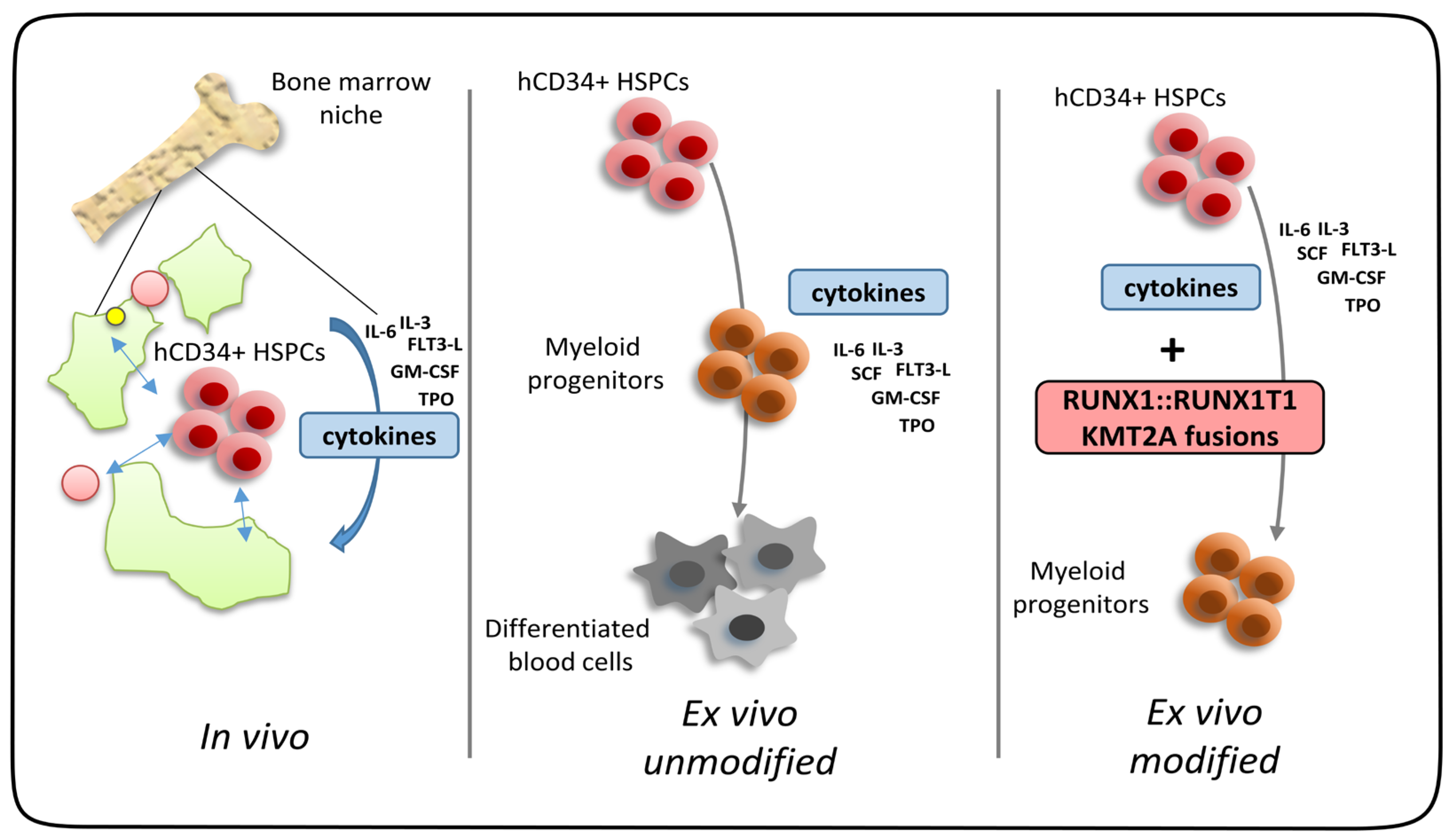
Ex vivo expansion of human CD34+ hematopoietic stem/progenitor cells is a hallmark of distinct leukemic transcription factors found in acute myeloid leukemia. Normally, stem/progenitor cells require the full support of stem cell niche factors for proper self-renewal. Cytokines alone cannot prohibit cell differentiation along the lineages. However, expression of AML-associated fusion genes in combination with cytokines allows for long-term ex vivo expansion making these cell models highly attractive to study the biology of oncogenes, as well as therapeutic avenues to block AML oncogene-dependent cell proliferation (Figure 3). As discussed above, primary human blood progenitor cells show many advantages compared to murine stem cells, especially working in the same host organism as the disease occurs. Even though primary human HSPCs are not as easily available as their murine counterpart, there are still different options for obtaining human cells. The two main resources of human cells are either remaining cells obtained from consent patient donations or samples from healthy individuals commercially available. Besides the many advantages of primary human ex vivo AML models, there are nevertheless limitations. Ex vivo cultures cannot model the complexity of the tumor microenvironment or the heterogeneity of many AML subtypes [100]. Further, the specific culture conditions can affect the phenotype and cannot represent complex physiological conditions such as oxygen levels [101]. Xenograft models, in which human leukemia cells are transplanted into immunodeficient mice, aim to overcome some of those limitations but still lack certain important features of AML. As leukemic cells are transplanted into immunodeficient mice to avoid rejection of the injected cells, there is no interaction between leukemia cells and the immune system, which can affect the disease progression and drug response [102,103,104]. Also, species-specific differences in the microenvironment, metabolism, and signaling pathways can lead to biased results [35,105]. Taken together, no artificial model system can reflect the complexity of the human organism, but ex vivo models are a very valuable tool to study human leukemic cells and should be used additionally or complementary to cell lines and in vivo models.

Author Contributions

Conceptualization, S.K.; methodology, R.W.; writing—original draft preparation, S.K. and R.W.; writing—review and editing, C.W. All authors have read and agreed to the published version of the manuscript.

Funding

This research was funded by the Wilhelm Sander-Stiftung (No. 2014.162.2, 2014.162.3, C.W.), Robert Pfleger Stiftung (No. 80768081, C.W.), Stiftung Transfusionsmedizin und Immunhämatologie (No. 80768076, C.W.), and Else Kröner-Fresenius-Stiftung (No. 2021-EKSE.92, C.W.).

Conflicts of Interest

The authors declare no conflict of interest. The funders had no role in the design of the study; in the collection, analyses, or interpretation of data; in the writing of the manuscript; or in the decision to publish the results.

References

  1. Enciso, J.; Mendoza, L.; Pelayo, R. Normal vs. Malignant hematopoiesis: The complexity of acute leukemia through systems biology. Front. Genet. 2015, 6, 290. [Google Scholar] [CrossRef] [PubMed]
  2. Shallis, R.M.; Wang, R.; Davidoff, A.; Ma, X.; Zeidan, A.M. Epidemiology of acute myeloid leukemia: Recent progress and enduring challenges. Blood Rev. 2019, 36, 70–87. [Google Scholar] [CrossRef] [PubMed]
  3. Hou, H.A.; Tien, H.F. Genomic landscape in acute myeloid leukemia and its implications in risk classification and targeted therapies. J. Biomed. Sci. 2020, 27, 81. [Google Scholar] [CrossRef] [PubMed]
  4. Kelly, L.M.; Gilliland, D.G. Genetics of myeloid leukemias. Annu. Rev. Genom. Hum. Genet. 2002, 3, 179–198. [Google Scholar] [CrossRef]
  5. Chen, S.J.; Shen, Y.; Chen, Z. A panoramic view of acute myeloid leukemia. Nat. Genet. 2013, 45, 586–587. [Google Scholar] [CrossRef] [PubMed]
  6. Padmakumar, D.; Chandraprabha, V.R.; Gopinath, P.; Vimala Devi, A.R.T.; Anitha, G.R.J.; Sreelatha, M.M.; Padmakumar, A.; Sreedharan, H. A concise review on the molecular genetics of acute myeloid leukemia. Leuk. Res. 2021, 111, 106727. [Google Scholar] [CrossRef] [PubMed]
  7. Cancer Genome Atlas Research Network; Ley, T.J.; Miller, C.; Ding, L.; Raphael, B.J.; Mungall, A.J.; Robertson, A.; Hoadley, K.; Triche, T.J., Jr.; Laird, P.W.; et al. Genomic and epigenomic landscapes of adult de novo acute myeloid leukemia. N. Engl. J. Med. 2013, 368, 2059–2074. [Google Scholar] [CrossRef]
  8. Rowley, J.D. Letter: A new consistent chromosomal abnormality in chronic myelogenous leukaemia identified by quinacrine fluorescence and Giemsa staining. Nature 1973, 243, 290–293. [Google Scholar] [CrossRef]
  9. Khoury, J.D.; Solary, E.; Abla, O.; Akkari, Y.; Alaggio, R.; Apperley, J.F.; Bejar, R.; Berti, E.; Busque, L.; Chan, J.K.C.; et al. The 5th edition of the World Health Organization Classification of Haematolymphoid Tumours: Myeloid and Histiocytic/Dendritic Neoplasms. Leukemia 2022, 36, 1703–1719. [Google Scholar] [CrossRef]
  10. Arber, D.A.; Orazi, A.; Hasserjian, R.P.; Borowitz, M.J.; Calvo, K.R.; Kvasnicka, H.M.; Wang, S.A.; Bagg, A.; Barbui, T.; Branford, S.; et al. International Consensus Classification of Myeloid Neoplasms and Acute Leukemias: Integrating morphologic, clinical, and genomic data. Blood 2022, 140, 1200–1228. [Google Scholar] [CrossRef]
  11. Huber, S.; Baer, C.; Hutter, S.; Dicker, F.; Meggendorfer, M.; Pohlkamp, C.; Kern, W.; Haferlach, T.; Haferlach, C.; Hoermann, G. AML classification in the year 2023: How to avoid a Babylonian confusion of languages. Leukemia 2023, 37, 1413–1420. [Google Scholar] [CrossRef] [PubMed]
  12. Mulloy, J.C.; Cammenga, J.; Berguido, F.J.; Wu, K.; Zhou, P.; Comenzo, R.L.; Jhanwar, S.; Moore, M.A.; Nimer, S.D. Maintaining the self-renewal and differentiation potential of human CD34+ hematopoietic cells using a single genetic element. Blood 2003, 102, 4369–4376. [Google Scholar] [CrossRef] [PubMed]
  13. Mulloy, J.C.; Cammenga, J.; MacKenzie, K.L.; Berguido, F.J.; Moore, M.A.; Nimer, S.D. The AML1-ETO fusion protein promotes the expansion of human hematopoietic stem cells. Blood 2002, 99, 15–23. [Google Scholar] [CrossRef] [PubMed]
  14. Wunderlich, M.; Krejci, O.; Wei, J.; Mulloy, J.C. Human CD34+ cells expressing the inv(16) fusion protein exhibit a myelomonocytic phenotype with greatly enhanced proliferative ability. Blood 2006, 108, 1690–1697. [Google Scholar] [CrossRef] [PubMed]
  15. Montes, R.; Ayllon, V.; Gutierrez-Aranda, I.; Prat, I.; Hernandez-Lamas, M.C.; Ponce, L.; Bresolin, S.; Te Kronnie, G.; Greaves, M.; Bueno, C.; et al. Enforced expression of MLL-AF4 fusion in cord blood CD34+ cells enhances the hematopoietic repopulating cell function and clonogenic potential but is not sufficient to initiate leukemia. Blood 2011, 117, 4746–4758. [Google Scholar] [CrossRef] [PubMed]
  16. Rizo, A.; Horton, S.J.; Olthof, S.; Dontje, B.; Ausema, A.; van Os, R.; van den Boom, V.; Vellenga, E.; de Haan, G.; Schuringa, J.J. BMI1 collaborates with BCR-ABL in leukemic transformation of human CD34+ cells. Blood 2010, 116, 4621–4630. [Google Scholar] [CrossRef]
  17. Wei, J.; Wunderlich, M.; Fox, C.; Alvarez, S.; Cigudosa, J.C.; Wilhelm, J.S.; Zheng, Y.; Cancelas, J.A.; Gu, Y.; Jansen, M.; et al. Microenvironment determines lineage fate in a human model of MLL-AF9 leukemia. Cancer Cell 2008, 13, 483–495. [Google Scholar] [CrossRef]
  18. Chung, K.Y.; Morrone, G.; Schuringa, J.J.; Plasilova, M.; Shieh, J.H.; Zhang, Y.; Zhou, P.; Moore, M.A. Enforced expression of NUP98-HOXA9 in human CD34(+) cells enhances stem cell proliferation. Cancer Res. 2006, 66, 11781–11791. [Google Scholar] [CrossRef]
  19. Gerritsen, M.; Yi, G.; Tijchon, E.; Kuster, J.; Schuringa, J.J.; Martens, J.H.A.; Vellenga, E. RUNX1 mutations enhance self-renewal and block granulocytic differentiation in human in vitro models and primary AMLs. Blood Adv. 2019, 3, 320–332. [Google Scholar] [CrossRef]
  20. Barabe, F.; Kennedy, J.A.; Hope, K.J.; Dick, J.E. Modeling the initiation and progression of human acute leukemia in mice. Science 2007, 316, 600–604. [Google Scholar] [CrossRef]
  21. Hess, J.L. MLL: A histone methyltransferase disrupted in leukemia. Trends Mol. Med. 2004, 10, 500–507. [Google Scholar] [CrossRef] [PubMed]
  22. Daser, A.; Rabbitts, T.H. The versatile mixed lineage leukaemia gene MLL and its many associations in leukaemogenesis. Semin. Cancer Biol. 2005, 15, 175–188. [Google Scholar] [CrossRef] [PubMed]
  23. Creutzig, U.; van den Heuvel-Eibrink, M.M.; Gibson, B.; Dworzak, M.N.; Adachi, S.; de Bont, E.; Harbott, J.; Hasle, H.; Johnston, D.; Kinoshita, A.; et al. Diagnosis and management of acute myeloid leukemia in children and adolescents: Recommendations from an international expert panel. Blood 2012, 120, 3187–3205. [Google Scholar] [CrossRef] [PubMed]
  24. Yu, B.D.; Hess, J.L.; Horning, S.E.; Brown, G.A.; Korsmeyer, S.J. Altered Hox expression and segmental identity in Mll-mutant mice. Nature 1995, 378, 505–508. [Google Scholar] [CrossRef] [PubMed]
  25. Yu, B.D.; Hanson, R.D.; Hess, J.L.; Horning, S.E.; Korsmeyer, S.J. MLL, a mammalian trithorax-group gene, functions as a transcriptional maintenance factor in morphogenesis. Proc. Natl. Acad. Sci. USA 1998, 95, 10632–10636. [Google Scholar] [CrossRef] [PubMed]
  26. Ernst, P.; Mabon, M.; Davidson, A.J.; Zon, L.I.; Korsmeyer, S.J. An Mll-dependent Hox program drives hematopoietic progenitor expansion. Curr. Biol. 2004, 14, 2063–2069. [Google Scholar] [CrossRef] [PubMed]
  27. Artinger, E.L.; Mishra, B.P.; Zaffuto, K.M.; Li, B.E.; Chung, E.K.; Moore, A.W.; Chen, Y.; Cheng, C.; Ernst, P. An MLL-dependent network sustains hematopoiesis. Proc. Natl. Acad. Sci. USA 2013, 110, 12000–12005. [Google Scholar] [CrossRef]
  28. Nakamura, T.; Mori, T.; Tada, S.; Krajewski, W.; Rozovskaia, T.; Wassell, R.; Dubois, G.; Mazo, A.; Croce, C.M.; Canaani, E. ALL-1 is a histone methyltransferase that assembles a supercomplex of proteins involved in transcriptional regulation. Mol. Cell 2002, 10, 1119–1128. [Google Scholar] [CrossRef]
  29. Milne, T.A.; Briggs, S.D.; Brock, H.W.; Martin, M.E.; Gibbs, D.; Allis, C.D.; Hess, J.L. MLL targets SET domain methyltransferase activity to Hox gene promoters. Mol. Cell 2002, 10, 1107–1117. [Google Scholar] [CrossRef]
  30. Meyer, C.; Larghero, P.; Almeida Lopes, B.; Burmeister, T.; Groger, D.; Sutton, R.; Venn, N.C.; Cazzaniga, G.; Corral Abascal, L.; Tsaur, G.; et al. The KMT2A recombinome of acute leukemias in 2023. Leukemia 2023, 37, 988–1005. [Google Scholar] [CrossRef]
  31. Corral, J.; Lavenir, I.; Impey, H.; Warren, A.J.; Forster, A.; Larson, T.A.; Bell, S.; McKenzie, A.N.; King, G.; Rabbitts, T.H. An Mll-AF9 fusion gene made by homologous recombination causes acute leukemia in chimeric mice: A method to create fusion oncogenes. Cell 1996, 85, 853–861. [Google Scholar] [CrossRef] [PubMed]
  32. Kohnken, R.; Porcu, P.; Mishra, A. Overview of the Use of Murine Models in Leukemia and Lymphoma Research. Front. Oncol. 2017, 7, 22. [Google Scholar] [CrossRef] [PubMed]
  33. Doulatov, S.; Notta, F.; Laurenti, E.; Dick, J.E. Hematopoiesis: A human perspective. Cell Stem Cell 2012, 10, 120–136. [Google Scholar] [CrossRef] [PubMed]
  34. Sitnicka, E.; Buza-Vidas, N.; Larsson, S.; Nygren, J.M.; Liuba, K.; Jacobsen, S.E. Human CD34+ hematopoietic stem cells capable of multilineage engrafting NOD/SCID mice express flt3: Distinct flt3 and c-kit expression and response patterns on mouse and candidate human hematopoietic stem cells. Blood 2003, 102, 881–886. [Google Scholar] [CrossRef] [PubMed]
  35. Mestas, J.; Hughes, C.C. Of mice and not men: Differences between mouse and human immunology. J. Immunol. 2004, 172, 2731–2738. [Google Scholar] [CrossRef] [PubMed]
  36. Bitoun, E.; Oliver, P.L.; Davies, K.E. The mixed-lineage leukemia fusion partner AF4 stimulates RNA polymerase II transcriptional elongation and mediates coordinated chromatin remodeling. Hum. Mol. Genet. 2007, 16, 92–106. [Google Scholar] [CrossRef] [PubMed]
  37. Yokoyama, A.; Lin, M.; Naresh, A.; Kitabayashi, I.; Cleary, M.L. A higher-order complex containing AF4 and ENL family proteins with P-TEFb facilitates oncogenic and physiologic MLL-dependent transcription. Cancer Cell 2010, 17, 198–212. [Google Scholar] [CrossRef]
  38. Peterlin, B.M.; Price, D.H. Controlling the elongation phase of transcription with P-TEFb. Mol. Cell 2006, 23, 297–305. [Google Scholar] [CrossRef]
  39. Carroll, D. Genome engineering with targetable nucleases. Annu. Rev. Biochem. 2014, 83, 409–439. [Google Scholar] [CrossRef]
  40. Ferrari, S.; Vavassori, V.; Canarutto, D.; Jacob, A.; Castiello, M.C.; Javed, A.O.; Genovese, P. Gene Editing of Hematopoietic Stem Cells: Hopes and Hurdles toward Clinical Translation. Front. Genome Ed. 2021, 3, 618378. [Google Scholar] [CrossRef]
  41. Barrangou, R.; Doudna, J.A. Applications of CRISPR technologies in research and beyond. Nat. Biotechnol. 2016, 34, 933–941. [Google Scholar] [CrossRef] [PubMed]
  42. Rice, S.; Jackson, T.; Crump, N.T.; Fordham, N.; Elliott, N.; O’Byrne, S.; Fanego, M.; Addy, D.; Crabb, T.; Dryden, C.; et al. A human fetal liver-derived infant MLL-AF4 acute lymphoblastic leukemia model reveals a distinct fetal gene expression program. Nat. Commun. 2021, 12, 6905. [Google Scholar] [CrossRef] [PubMed]
  43. Marschalek, R. The reciprocal world of MLL fusions: A personal view. Biochim. Biophys. Acta Gene Regul. Mech. 2020, 1863, 194547. [Google Scholar] [CrossRef] [PubMed]
  44. Wilhelm, A.; Marschalek, R. The role of reciprocal fusions in MLL-r acute leukemia: Studying the chromosomal translocation t(4;11). Oncogene 2021, 40, 6093–6102. [Google Scholar] [CrossRef] [PubMed]
  45. Collins, E.C.; Appert, A.; Ariza-McNaughton, L.; Pannell, R.; Yamada, Y.; Rabbitts, T.H. Mouse Af9 is a controller of embryo patterning, like Mll, whose human homologue fuses with Af9 after chromosomal translocation in leukemia. Mol. Cell. Biol. 2002, 22, 7313–7324. [Google Scholar] [CrossRef] [PubMed]
  46. Calvanese, V.; Nguyen, A.T.; Bolan, T.J.; Vavilina, A.; Su, T.; Lee, L.K.; Wang, Y.; Lay, F.D.; Magnusson, M.; Crooks, G.M.; et al. MLLT3 governs human haematopoietic stem-cell self-renewal and engraftment. Nature 2019, 576, 281–286. [Google Scholar] [CrossRef]
  47. Schneidawind, C.; Jeong, J.; Schneidawind, D.; Kim, I.S.; Duque-Afonso, J.; Wong, S.H.K.; Iwasaki, M.; Breese, E.H.; Zehnder, J.L.; Porteus, M.; et al. MLL leukemia induction by t(9;11) chromosomal translocation in human hematopoietic stem cells using genome editing. Blood Adv. 2018, 2, 832–845. [Google Scholar] [CrossRef]
  48. Zhou, J.; Ng, Y.; Chng, W.J. ENL: Structure, function, and roles in hematopoiesis and acute myeloid leukemia. Cell. Mol. Life Sci. 2018, 75, 3931–3941. [Google Scholar] [CrossRef]
  49. Mueller, D.; Bach, C.; Zeisig, D.; Garcia-Cuellar, M.P.; Monroe, S.; Sreekumar, A.; Zhou, R.; Nesvizhskii, A.; Chinnaiyan, A.; Hess, J.L.; et al. A role for the MLL fusion partner ENL in transcriptional elongation and chromatin modification. Blood 2007, 110, 4445–4454. [Google Scholar] [CrossRef]
  50. Chen, J.; Liao, S.; Xiao, Z.; Pan, Q.; Wang, X.; Shen, K.; Wang, S.; Yang, L.; Guo, F.; Liu, H.F.; et al. The development and improvement of immunodeficient mice and humanized immune system mouse models. Front. Immunol. 2022, 13, 1007579. [Google Scholar] [CrossRef]
  51. Rossi, J.G.; Bernasconi, A.R.; Alonso, C.N.; Rubio, P.L.; Gallego, M.S.; Carrara, C.A.; Guitter, M.R.; Eberle, S.E.; Cocce, M.; Zubizarreta, P.A.; et al. Lineage switch in childhood acute leukemia: An unusual event with poor outcome. Am. J. Hematol. 2012, 87, 890–897. [Google Scholar] [CrossRef] [PubMed]
  52. Windisch, R.; Kreissig, S.; Wichmann, C. Defined Human Leukemic CD34+ Liquid Cultures to Study HDAC/Transcriptional Repressor Complexes. Methods Mol. Biol. 2023, 2589, 27–49. [Google Scholar] [CrossRef] [PubMed]
  53. Buechele, C.; Breese, E.H.; Schneidawind, D.; Lin, C.H.; Jeong, J.; Duque-Afonso, J.; Wong, S.H.; Smith, K.S.; Negrin, R.S.; Porteus, M.; et al. MLL leukemia induction by genome editing of human CD34+ hematopoietic cells. Blood 2015, 126, 1683–1694. [Google Scholar] [CrossRef] [PubMed]
  54. Reimer, J.; Knoss, S.; Labuhn, M.; Charpentier, E.M.; Gohring, G.; Schlegelberger, B.; Klusmann, J.H.; Heckl, D. CRISPR-Cas9-induced t(11;19)/MLL-ENL translocations initiate leukemia in human hematopoietic progenitor cells in vivo. Haematologica 2017, 102, 1558–1566. [Google Scholar] [CrossRef] [PubMed]
  55. de Bruijn, M.F.; Speck, N.A. Core-binding factors in hematopoiesis and immune function. Oncogene 2004, 23, 4238–4248. [Google Scholar] [CrossRef] [PubMed]
  56. Miyoshi, H.; Shimizu, K.; Kozu, T.; Maseki, N.; Kaneko, Y.; Ohki, M. t(8;21) breakpoints on chromosome 21 in acute myeloid leukemia are clustered within a limited region of a single gene, AML1. Proc. Natl. Acad. Sci. USA 1991, 88, 10431–10434. [Google Scholar] [CrossRef] [PubMed]
  57. Lam, K.; Zhang, D.E. RUNX1 and RUNX1-ETO: Roles in hematopoiesis and leukemogenesis. Front. Biosci. 2012, 17, 1120–1139. [Google Scholar] [CrossRef]
  58. Lin, S.; Mulloy, J.C.; Goyama, S. RUNX1-ETO Leukemia. Adv. Exp. Med. Biol. 2017, 962, 151–173. [Google Scholar] [CrossRef]
  59. Wang, J.; Hoshino, T.; Redner, R.L.; Kajigaya, S.; Liu, J.M. ETO, fusion partner in t(8;21) acute myeloid leukemia, represses transcription by interaction with the human N-CoR/mSin3/HDAC1 complex. Proc. Natl. Acad. Sci. USA 1998, 95, 10860–10865. [Google Scholar] [CrossRef]
  60. Wang, L.; Gural, A.; Sun, X.J.; Zhao, X.; Perna, F.; Huang, G.; Hatlen, M.A.; Vu, L.; Liu, F.; Xu, H.; et al. The leukemogenicity of AML1-ETO is dependent on site-specific lysine acetylation. Science 2011, 333, 765–769. [Google Scholar] [CrossRef]
  61. Chen, G.; Liu, A.; Xu, Y.; Gao, L.; Jiang, M.; Li, Y.; Lv, N.; Zhou, L.; Wang, L.; Yu, L.; et al. The RUNX1-ETO fusion protein trans-activates c-KIT expression by recruiting histone acetyltransferase P300 on its promoter. FEBS J. 2019, 286, 901–912. [Google Scholar] [CrossRef] [PubMed]
  62. Chen-Wichmann, L.; Shvartsman, M.; Preiss, C.; Hockings, C.; Windisch, R.; Redondo Monte, E.; Leubolt, G.; Spiekermann, K.; Lausen, J.; Brendel, C.; et al. Compatibility of RUNX1/ETO fusion protein modules driving CD34+ human progenitor cell expansion. Oncogene 2019, 38, 261–272. [Google Scholar] [CrossRef] [PubMed]
  63. Yan, M.; Kanbe, E.; Peterson, L.F.; Boyapati, A.; Miao, Y.; Wang, Y.; Chen, I.M.; Chen, Z.; Rowley, J.D.; Willman, C.L.; et al. A previously unidentified alternatively spliced isoform of t(8;21) transcript promotes leukemogenesis. Nat. Med. 2006, 12, 945–949. [Google Scholar] [CrossRef] [PubMed]
  64. Link, K.A.; Lin, S.; Shrestha, M.; Bowman, M.; Wunderlich, M.; Bloomfield, C.D.; Huang, G.; Mulloy, J.C. Supraphysiologic levels of the AML1-ETO isoform AE9a are essential for transformation. Proc. Natl. Acad. Sci. USA 2016, 113, 9075–9080. [Google Scholar] [CrossRef] [PubMed]
  65. DeKelver, R.C.; Yan, M.; Ahn, E.Y.; Shia, W.J.; Speck, N.A.; Zhang, D.E. Attenuation of AML1-ETO cellular dysregulation correlates with increased leukemogenic potential. Blood 2013, 121, 3714–3717. [Google Scholar] [CrossRef] [PubMed]
  66. Castilla, L.H.; Wijmenga, C.; Wang, Q.; Stacy, T.; Speck, N.A.; Eckhaus, M.; Marin-Padilla, M.; Collins, F.S.; Wynshaw-Boris, A.; Liu, P.P. Failure of embryonic hematopoiesis and lethal hemorrhages in mouse embryos heterozygous for a knocked-in leukemia gene CBFB-MYH11. Cell 1996, 87, 687–696. [Google Scholar] [CrossRef] [PubMed]
  67. Gough, S.M.; Slape, C.I.; Aplan, P.D. NUP98 gene fusions and hematopoietic malignancies: Common themes and new biologic insights. Blood 2011, 118, 6247–6257. [Google Scholar] [CrossRef]
  68. Ghannam, G.; Takeda, A.; Camarata, T.; Moore, M.A.; Viale, A.; Yaseen, N.R. The oncogene Nup98-HOXA9 induces gene transcription in myeloid cells. J. Biol. Chem. 2004, 279, 866–875. [Google Scholar] [CrossRef]
  69. Takeda, A.; Goolsby, C.; Yaseen, N.R. NUP98-HOXA9 induces long-term proliferation and blocks differentiation of primary human CD34+ hematopoietic cells. Cancer Res. 2006, 66, 6628–6637. [Google Scholar] [CrossRef]
  70. Abraham, A.; Kim, Y.S.; Zhao, H.; Humphries, K.; Persons, D.A. Increased Engraftment of Human Short Term Repopulating Hematopoietic Cells in NOD/SCID/IL2rgammanull Mice by Lentiviral Expression of NUP98-HOXA10HD. PLoS ONE 2016, 11, e0147059. [Google Scholar] [CrossRef]
  71. Zhang, X.; Sun, J.; Yu, W.; Jin, J. Current views on the genetic landscape and management of variant acute promyelocytic leukemia. Biomark. Res. 2021, 9, 33. [Google Scholar] [CrossRef] [PubMed]
  72. Duong, V.; Rochette-Egly, C. The molecular physiology of nuclear retinoic acid receptors. From health to disease. Biochim. Biophys. Acta Mol. Basis Dis. 2011, 1812, 1023–1031. [Google Scholar] [CrossRef] [PubMed]
  73. Lavudi, K.; Nuguri, S.M.; Olverson, Z.; Dhanabalan, A.K.; Patnaik, S.; Kokkanti, R.R. Targeting the retinoic acid signaling pathway as a modern precision therapy against cancers. Front. Cell Dev. Biol. 2023, 11, 1254612. [Google Scholar] [CrossRef] [PubMed]
  74. Altucci, L.; Leibowitz, M.D.; Ogilvie, K.M.; de Lera, A.R.; Gronemeyer, H. RAR and RXR modulation in cancer and metabolic disease. Nat. Rev. Drug Discov. 2007, 6, 793–810. [Google Scholar] [CrossRef] [PubMed]
  75. Guan, D.; Kao, H.Y. The function, regulation and therapeutic implications of the tumor suppressor protein, PML. Cell Biosci. 2015, 5, 60. [Google Scholar] [CrossRef]
  76. Noguera, N.I.; Catalano, G.; Banella, C.; Divona, M.; Faraoni, I.; Ottone, T.; Arcese, W.; Voso, M.T. Acute Promyelocytic Leukemia: Update on the Mechanisms of Leukemogenesis, Resistance and on Innovative Treatment Strategies. Cancers 2019, 11, 1591. [Google Scholar] [CrossRef]
  77. Bain, B.J.; Bene, M.C. Morphological and Immunophenotypic Clues to the WHO Categories of Acute Myeloid Leukaemia. Acta Haematol. 2019, 141, 232–244. [Google Scholar] [CrossRef]
  78. Turhan, A.G.; Lemoine, F.M.; Debert, C.; Bonnet, M.L.; Baillou, C.; Picard, F.; Macintyre, E.A.; Varet, B. Highly purified primitive hematopoietic stem cells are PML-RARA negative and generate nonclonal progenitors in acute promyelocytic leukemia. Blood 1995, 85, 2154–2161. [Google Scholar] [CrossRef]
  79. Grignani, F.; Valtieri, M.; Gabbianelli, M.; Gelmetti, V.; Botta, R.; Luchetti, L.; Masella, B.; Morsilli, O.; Pelosi, E.; Samoggia, P.; et al. PML/RAR alpha fusion protein expression in normal human hematopoietic progenitors dictates myeloid commitment and the promyelocytic phenotype. Blood 2000, 96, 1531–1537. [Google Scholar] [CrossRef]
  80. Abdul-Nabi, A.M.; Yassin, E.R.; Varghese, N.; Deshmukh, H.; Yaseen, N.R. In vitro transformation of primary human CD34+ cells by AML fusion oncogenes: Early gene expression profiling reveals possible drug target in AML. PLoS ONE 2010, 5, e12464. [Google Scholar] [CrossRef]
  81. Du, C.; Redner, R.L.; Cooke, M.P.; Lavau, C. Overexpression of wild-type retinoic acid receptor alpha (RARalpha) recapitulates retinoic acid-sensitive transformation of primary myeloid progenitors by acute promyelocytic leukemia RARalpha-fusion genes. Blood 1999, 94, 793–802. [Google Scholar] [CrossRef] [PubMed]
  82. Ito, M.; Kobayashi, K.; Nakahata, T. NOD/Shi-scid IL2rgamma(null) (NOG) mice more appropriate for humanized mouse models. Curr. Top Microbiol. Immunol. 2008, 324, 53–76. [Google Scholar] [CrossRef] [PubMed]
  83. Matsushita, H.; Yahata, T.; Sheng, Y.; Nakamura, Y.; Muguruma, Y.; Matsuzawa, H.; Tanaka, M.; Hayashi, H.; Sato, T.; Damdinsuren, A.; et al. Establishment of a humanized APL model via the transplantation of PML-RARA-transduced human common myeloid progenitors into immunodeficient mice. PLoS ONE 2014, 9, e111082. [Google Scholar] [CrossRef]
  84. Lessard, J.; Sauvageau, G. Bmi-1 determines the proliferative capacity of normal and leukaemic stem cells. Nature 2003, 423, 255–260. [Google Scholar] [CrossRef] [PubMed]
  85. Hidalgo, I.; Wahlestedt, M.; Yuan, O.; Zhang, Q.; Bryder, D.; Pronk, C.J. Bmi1 induction protects hematopoietic stem cells against pronounced long-term hematopoietic stress. Exp. Hematol. 2022, 109, 35–44. [Google Scholar] [CrossRef] [PubMed]
  86. Sahasrabuddhe, A.A. BMI1: A Biomarker of Hematologic Malignancies. Biomark. Cancer 2016, 8, 65–75. [Google Scholar] [CrossRef]
  87. Rizo, A.; Dontje, B.; Vellenga, E.; de Haan, G.; Schuringa, J.J. Long-term maintenance of human hematopoietic stem/progenitor cells by expression of BMI1. Blood 2008, 111, 2621–2630. [Google Scholar] [CrossRef]
  88. Edling, C.E.; Hallberg, B. c-Kit—A hematopoietic cell essential receptor tyrosine kinase. Int. J. Biochem. Cell Biol. 2007, 39, 1995–1998. [Google Scholar] [CrossRef]
  89. Malaise, M.; Steinbach, D.; Corbacioglu, S. Clinical implications of c-Kit mutations in acute myelogenous leukemia. Curr. Hematol. Malig. Rep. 2009, 4, 77–82. [Google Scholar] [CrossRef]
  90. Wichmann, C.; Quagliano-Lo Coco, I.; Yildiz, O.; Chen-Wichmann, L.; Weber, H.; Syzonenko, T.; Doring, C.; Brendel, C.; Ponnusamy, K.; Kinner, A.; et al. Activating c-KIT mutations confer oncogenic cooperativity and rescue RUNX1/ETO-induced DNA damage and apoptosis in human primary CD34+ hematopoietic progenitors. Leukemia 2015, 29, 279–289. [Google Scholar] [CrossRef]
  91. Chin, P.S.; Assi, S.A.; Ptasinska, A.; Imperato, M.R.; Cockerill, P.N.; Bonifer, C. RUNX1/ETO and mutant KIT both contribute to programming the transcriptional and chromatin landscape in t(8;21) acute myeloid leukemia. Exp. Hematol. 2020, 92, 62–74. [Google Scholar] [CrossRef] [PubMed]
  92. Higuchi, M.; O’Brien, D.; Kumaravelu, P.; Lenny, N.; Yeoh, E.J.; Downing, J.R. Expression of a conditional AML1-ETO oncogene bypasses embryonic lethality and establishes a murine model of human t(8;21) acute myeloid leukemia. Cancer Cell 2002, 1, 63–74. [Google Scholar] [CrossRef] [PubMed]
  93. Yuan, Y.; Zhou, L.; Miyamoto, T.; Iwasaki, H.; Harakawa, N.; Hetherington, C.J.; Burel, S.A.; Lagasse, E.; Weissman, I.L.; Akashi, K.; et al. AML1-ETO expression is directly involved in the development of acute myeloid leukemia in the presence of additional mutations. Proc. Natl. Acad. Sci. USA 2001, 98, 10398–10403. [Google Scholar] [CrossRef] [PubMed]
  94. Rhoades, K.L.; Hetherington, C.J.; Harakawa, N.; Yergeau, D.A.; Zhou, L.; Liu, L.Q.; Little, M.T.; Tenen, D.G.; Zhang, D.E. Analysis of the role of AML1-ETO in leukemogenesis, using an inducible transgenic mouse model. Blood 2000, 96, 2108–2115. [Google Scholar] [CrossRef] [PubMed]
  95. Nick, H.J.; Kim, H.G.; Chang, C.W.; Harris, K.W.; Reddy, V.; Klug, C.A. Distinct classes of c-Kit-activating mutations differ in their ability to promote RUNX1-ETO-associated acute myeloid leukemia. Blood 2012, 119, 1522–1531. [Google Scholar] [CrossRef] [PubMed]
  96. Wang, Y.Y.; Zhao, L.J.; Wu, C.F.; Liu, P.; Shi, L.; Liang, Y.; Xiong, S.M.; Mi, J.Q.; Chen, Z.; Ren, R.; et al. C-KIT mutation cooperates with full-length AML1-ETO to induce acute myeloid leukemia in mice. Proc. Natl. Acad. Sci. USA 2011, 108, 2450–2455. [Google Scholar] [CrossRef]
  97. Goyama, S.; Schibler, J.; Gasilina, A.; Shrestha, M.; Lin, S.; Link, K.A.; Chen, J.; Whitman, S.P.; Bloomfield, C.D.; Nicolet, D.; et al. UBASH3B/Sts-1-CBL axis regulates myeloid proliferation in human preleukemia induced by AML1-ETO. Leukemia 2016, 30, 728–739. [Google Scholar] [CrossRef]
  98. Basecke, J.; Schwieger, M.; Griesinger, F.; Schiedlmeier, B.; Wulf, G.; Trumper, L.; Stocking, C. AML1/ETO promotes the maintenance of early hematopoietic progenitors in NOD/SCID mice but does not abrogate their lineage specific differentiation. Leuk. Lymphoma 2005, 46, 265–272. [Google Scholar] [CrossRef]
  99. Chou, F.S.; Wunderlich, M.; Griesinger, A.; Mulloy, J.C. N-Ras(G12D) induces features of stepwise transformation in preleukemic human umbilical cord blood cultures expressing the AML1-ETO fusion gene. Blood 2011, 117, 2237–2240. [Google Scholar] [CrossRef]
  100. Pinto, C.; Estrada, M.F.; Brito, C. In Vitro and Ex Vivo Models—The Tumor Microenvironment in a Flask. Adv. Exp. Med. Biol. 2020, 1219, 431–443. [Google Scholar] [CrossRef]
  101. Jagannathan, L.; Cuddapah, S.; Costa, M. Oxidative stress under ambient and physiological oxygen tension in tissue culture. Curr. Pharmacol. Rep. 2016, 2, 64–72. [Google Scholar] [CrossRef] [PubMed]
  102. Teague, R.M.; Kline, J. Immune evasion in acute myeloid leukemia: Current concepts and future directions. J. Immunother. Cancer 2013, 1, 1. [Google Scholar] [CrossRef] [PubMed]
  103. Lamble, A.J.; Lind, E.F. Targeting the Immune Microenvironment in Acute Myeloid Leukemia: A Focus on T Cell Immunity. Front. Oncol. 2018, 8, 213. [Google Scholar] [CrossRef] [PubMed]
  104. Austin, R.; Smyth, M.J.; Lane, S.W. Harnessing the immune system in acute myeloid leukaemia. Crit. Rev. Oncol. Hematol. 2016, 103, 62–77. [Google Scholar] [CrossRef]
  105. Fischer, M. Mice Are Not Humans: The Case of p53. Trends Cancer 2021, 7, 12–14. [Google Scholar] [CrossRef]
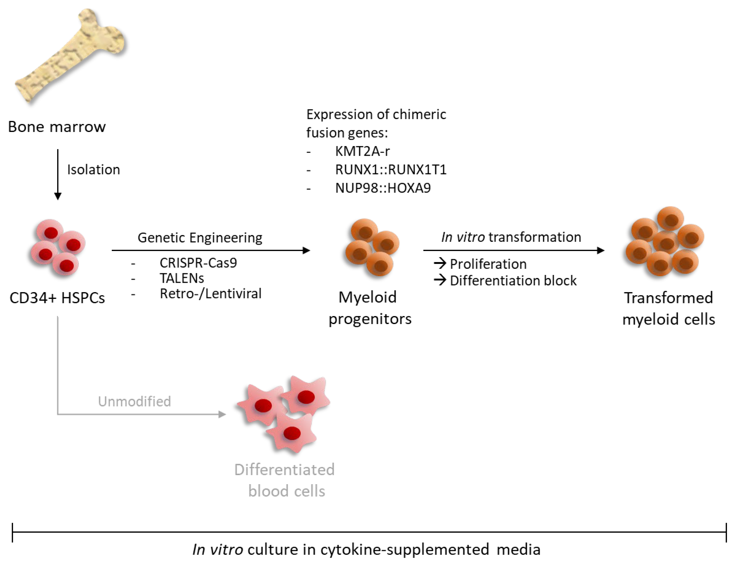
Figure 1. Modeling oncogenic transformation of myeloid blood progenitors in ex vivo cultures via genetic engineering. Human CD34+ HSPCs isolated from bone marrow or cord blood of healthy donors can be genetically manipulated to mimic leukemic transformation. Expression of leukemia-associated fusion genes can be induced either via retro- or lentiviral integration or by CRISPR-Cas9/TALENs-mediated rearrangements. In cytokine-supplemented media, continuous expression of these chimeric genes leads to enhanced proliferation and concomitant differentiation block. Unmodified CD34+ HSPCs are not able to continuously proliferate in vitro resulting in terminal differentiation.
Cells 13 00078 g001
Figure 2. Utilizing oncogenes for leukemic transformation of human myeloid progenitor cells ex vivo and in vivo. Transduction with both either KMT2A-r or RUNX1::RUNX1T1 leads to ex vivo transformation and expansion. RUNX1::RUNX1T1 primarily acts as a transcriptional repressor, while KMT2A-rearrangement-derived fusion proteins function as activators. Whereas KMT2A fusion proteins are strong independent leukemic drivers, the transforming capacity of RUNX1::RUNX1T1 must be amplified by further mutations such as c-KIT. Transplantation of transformed human cells into immune-deficient mice only leads to leukemia in the case of KMT2A-r, but not RUNX1::RUNX1T1 both with and without co-mutations.
Cells 13 00078 g002
Figure 3. AML-associated fusion genes such as RUNX1::RUNX1T1 and KMT2A-fusions can substitute for hematopoietic stem cell niche factors to allow for ex vivo progenitor cell expansion under liquid culture conditions. Detached from the bone marrow niche unmodified CD34+ cells rapidly differentiate ex vivo even in the presence of proliferation-favoring cytokines. Together with cytokines, distinct leukemic transcription factors induce niche-independent expansion of hematopoietic progenitor cells in ex vivo cultures.
Cells 13 00078 g003
Table 1. Classification of acute myeloid leukemia with defining genetic abnormalities. Adopted from [9,10,11].
Genetic AbnormalityResulting Fusion Gene/Rearrangement
t(8;21)RUNX1::RUNX1T1
t(16;16) or inv(16)CBFB::MYH11
t(6;9)DEK::NUP214
t(3;3) or inv(3)RPN1::EVI1
t(1;22)RBM15::MRTFA
t(9;22)BCR::ABL1
t(15;17)PML::RARA
t(11q23)KMT2A (MLL1, MLL)-r
t(3q26)MECOM-r
t(11p15)NUP98-r
biallelic mutations of the CEBPA gene-
with other defined genetic alterations -
Table 2. Overview of oncogenes and culture conditions for human ex vivo CD34+ liquid culture models. Genes that alone or in combination can induce human CD34+ cell expansion in ex vivo liquid cultures. For each mutated gene, the required cytokine combination and resulting cellular phenotype are listed.
PhenotypeGeneCulture ConditionsReferences
Myeloid, CD34+ subpopulation and mature cellsRUNX1::RUNX1T1SCF, IL-3, IL-6, GM-CSF, FLT3-L, erythropoietin[12,13]
CBFβ::MYH11SCF, IL-3, IL-6, FLT3-L, thrombopoietin[14]
KMT2A::AF4IL-3, SCF, FLT3-L[15]
BCR::ABL + BMI1IL-3, IL-6, G-CSF, SCF, FLT3-L
MS5 stromal feeder cells
[16]
Myeloid progenitors, no remaining CD34+ subpopulationKMT2A::AF9SCF, IL-3, IL-6, FLT3-L and TPO[17]
NUP98::HOXA9SCF, FLT3-L, thrombopoietin[18]
Immature granulocyte-macrophage progenitor-like cellsRUNX1 mutations
e.g., RUNX1-S291fs300X
IL-3, IL-6, G-CSF, SCF, FLT3-L, erythropoietin[19]
Both myeloid and lymphoid progenitorsKMT2A::ENLIL-3, SCF, FLT3-L, IL-7[20]
Disclaimer/Publisher’s Note: The statements, opinions and data contained in all publications are solely those of the individual author(s) and contributor(s) and not of MDPI and/or the editor(s). MDPI and/or the editor(s) disclaim responsibility for any injury to people or property resulting from any ideas, methods, instructions or products referred to in the content.