Article

Multiple Endocrine Neoplasia Type 1 Regulates TGFβ-Mediated Suppression of Tumor Formation and Metastasis in Melanoma

1
Cancer Research Program, Department of Medicine, Research Institute of McGill University Health Center, Montreal, QC H4A 3J1, Canada
2
Division of Medical Genetics, Fondazione IRCCS Casa Sollievo della Sofferenza, 71013 San Giovanni Rotondo, Italy
3
Endocrinology and Metabolism Unit, University-Hospital S. Maria della Misericordia, 33100 Udine, Italy
4
Endocrinology Unit, Fondazione IRCCS Casa Sollievo della Sofferenza, 71013 San Giovanni Rotondo, Italy
*
Author to whom correspondence should be addressed.
These authors contributed equally to this work.
Cells 2024, 13(11), 973; https://doi.org/10.3390/cells13110973
Submission received: 11 March 2024 / Revised: 27 May 2024 / Accepted: 28 May 2024 / Published: 4 June 2024

Abstract

:
Over the past few decades, the worldwide incidence of cutaneous melanoma, a malignant neoplasm arising from melanocytes, has been increasing markedly, leading to the highest rate of skin cancer-related deaths. While localized tumors are easily removed by excision surgery, late-stage metastatic melanomas are refractory to treatment and exhibit a poor prognosis. Consequently, unraveling the molecular mechanisms underlying melanoma tumorigenesis and metastasis is crucial for developing novel targeted therapies. We found that the multiple endocrine neoplasia type 1 (MEN1) gene product Menin is required for the transforming growth factor beta (TGFβ) signaling pathway to induce cell growth arrest and apoptosis in vitro and prevent tumorigenesis in vivo in preclinical xenograft models of melanoma. We further identified point mutations in two MEN1 family members affected by melanoma that led to proteasomal degradation of the MEN1 gene product and to a loss of TGFβ signaling. Interestingly, blocking the proteasome degradation pathway using an FDA-approved drug and RNAi targeting could efficiently restore MEN1 expression and TGFβ transcriptional responses. Together, these results provide new potential therapeutic strategies and patient stratification for the treatment of cutaneous melanoma.

1. Introduction

Cutaneous melanoma is a deadly and aggressive cancer accounting for approximately 80% of skin cancer-related deaths [1]. Globally, melanoma is the fifteenth most common cancer, with 230,000 diagnosed cases per year and 55,000 deaths. Furthermore, the incidence of melanoma has significantly increased during the past fifty years [2]. Melanoma is one of the most prevalent cancers among younger adults aged >20–35 years worldwide [3]. Based on histopathology and prognostic outcomes, melanomas are conventionally classified into four clinical stages. While patients with stage I melanoma have localized primary tumors that can be removed by surgical excision [4], stage IV patients exhibit secondary metastatic tumors to the lung, liver, bones, or brain and are refractory to traditional chemotherapy [5]. As a result, while 5-year and 15-year survival rates are very good in stage I patients (97% and 85%, respectively), they plummet to only 15% and 5%, respectively, in patients with stage IV melanoma [1,2]. In addition to UV radiation, the primary environmental factor predisposing patients to cutaneous melanoma and other genetic and molecular factors involved in the genesis of this disease have yet to be fully characterized [6]. Hence, understanding the molecular and signaling mechanisms leading to melanoma development and progression is essential for developing better targeted treatments.
Melanoma tumorigenesis results from mutations in genes implicated in the regulation of various biological processes, including cell growth and proliferation (BRAF, NRAS, NF1, PTEN, and KIT), apoptosis (TP53), and cell immortalization (hTERT) [7,8]. While mutations in the mitogenic RAS-RAF-MEK-ERK signaling pathway are very frequent, other signaling pathways, such as the Jnk/c-Jun, Wnt, NF-κB, PI3K/AKT, JAK/STAT, and TGFβ pathways, have also been implicated in the tumorigenesis process [9,10,11,12]. The TGFβ signaling pathway plays an essential role in both normal melanocytes and melanoma cells. TGFβ signals through a complex of two serine/threonine kinase receptors and intracellular Smad proteins (Smad2, 3, and 4). In melanoma, the TGFβ/Smad3 signaling pathway strongly suppresses tumorigenesis by blocking cell growth, immortalization, and cancer stem cell self-renewal activities, and by inducing cell death and autophagy [13,14,15].
Multiple endocrine neoplasia type 1 (MEN1) is an autosomal dominant disorder affecting the endocrine system and is characterized by the concomitant occurrence of tumors in the pancreas as well as in the parathyroid and anterior pituitary glands. The MEN1 gene encodes Menin, a 610-amino-acid protein that interacts with numerous protein partners, including several transcription factors [16]. Menin plays a significant role in cell-cycle regulation by inducing cyclin-dependent kinase inhibitor (CDKI) gene expression [17,18]. Interestingly, Menin was found to leverage TGFβ signaling at the transcriptional level, thus facilitating its cytostatic and differentiation functions [19,20].
Notably, non-endocrine tumors have also been reported in MEN1 patients. These include skin tumors of mesenchymal origin, such as angiofibromas, collagenomas, lipomas, and malignant melanomas [21,22,23]. Loss of heterozygosity (LOH) of chromosome 11q13 (the MEN1 gene locus) was detected in six melanoma tumors, and deletion of the MEN1 locus was found in 19 patients with sporadic metastatic melanoma. Another study suggested that multiple melanoma tumor suppressors are localized on chromosome 11q, which includes the MEN1 region [24]. These observations therefore raise the possibility of a causal association between MEN1 and melanoma. It was shown that Menin suppresses malignant phenotypes of melanoma through its involvement in PTN signaling [25]. Interestingly, a paradoxical role of MEN1 was discovered in breast cancer patients, as its high expression is correlated with poorer overall survival [26].
In this study, we identified the TGFβ/Smad3/MEN1 signaling axis as a potent tumor-suppressor pathway in cutaneous melanoma. Moreover, genetic analysis of two MEN1 family members affected by melanoma revealed the presence of specific point mutations within the MEN1 gene. We found that these point mutations induce MEN1 gene product degradation, leading to further loss of TGFβ signaling. Moreover, we showed that by targeting the co-chaperone of the proteasome degradation pathway, CHIP could restore Menin expression and TGFβ signaling in these melanoma cells. Overall, this study defines the TGFβ/MEN1 axis as a potent tumor-suppressor pathway in cutaneous melanoma and provides novel perspectives for tailor-made targeted therapies for this highly lethal malignancy.

2. Materials and Methods

2.1. Reagents

Recombinant human TGFβ (PeproTech, Saint-Laurent, QC, Canada), RPMI 1640 tissue culture medium, DMEM (HyClone Logan, South Logan, UT, USA), fetal bovine serum and penicillin/streptomycin (Gibco, Waltham, MA, USA), branched polyethyleneimine (Sigma-Aldrich, Oakville, ON, Canada), Moloney murine leukemia virus (MMLV) reverse transcriptase, and random primers (Life Science, N. St. Petersburg, FL, USA) were used. The sequence of the control siRNA was sc-37007, and that of the CHIP siRNA was sc-43555 (Santa Cruz, Santa Cruz, CA, USA). Missense mutations were generated with the Quik Change Site-Directed Mutagenesis Kit from Stratagene (La Jolla, CA, USA).

2.2. Antibodies

β-Tubulin (3F32G) (Santa Cruz); anti-Flag M2 monoclonal antibody (Sigma); MEN1 (2605) (Abcam, Cambridge, UK); P21 (C-19) (Santa Cruz, Cat#sc-397); SMAD2-3 (Santa Cruz, Cat#sc-6032); SMAD4 (Santa Cruz, Cat#sc-7966); Caspase-3 (H-277) (Santa Cruz, Cat#sc-7148); and c-myc (9E10) (Santa Cruz, Cat#sc-40)

2.3. Cell Lines

The cells were cultured at 37 °C and 5% CO2 in RPMI 1640 (BLM, WM793B, WM278, WM1232, DAUV) or DMEM (HEK293, SKMEL28, and a375m) supplemented with 10% FBS and 1% penicillin/streptomycin. Additional information is provided in Table 1.

2.4. TGFβ Treatment

Cell monolayers were grown in complete medium to 60% confluence, starved overnight in serum-free medium (0% FBS), and treated with a final concentration of 200 pM human recombinant TGFβ1 for the indicated time periods.

2.5. Quantitative Real-Time PCR

Total RNA was extracted using Trizol TM (Invitrogen, Burlington, ON, Canada). RNA was reverse transcribed using M-MLV reverse transcriptase and random primers (Invitrogen) according to the manufacturer’s protocol. cDNA amplification was performed via quantitative real-time PCR (qPCR) with SsoFast™ EvaGreen® Supermix (Bio-Rad, Mississauga, ON, Canada) using a Rotor-Gene™ 6000 Real-time Analyzer (Corbett Life Sciences, Mortlake, NSW, Australia). Human GAPDH was used as a housekeeping gene. The sequences of primers used are listed in the Table 2:

2.6. Clonogenic Assay

Melanoma cells (WM278) were grown in a 6-well plate (1000 cells) in complete RPMI medium (10% FBS). The medium was replenished after 1 week, after which the cells were fixed, stained (0.5% w/v crystal violet, 20% v/v methanol) at the endpoint and washed with PBS.

2.7. Flow Cytometry

For cell-cycle analysis, cells were stimulated with or without TGF-β (200 pM) for 24 h or 48 h in 1% FBS. The cells were washed and resuspended in PBS at 1 × 10⁶ cells/mL and fixed in ice-cold water by dropwise addition of 70% ethanol while vortexing. The mixture was incubated on ice for 30 min after fixation. For analysis, the cells were resuspended in a mixture of 50 μg/mL propidium iodide, 50 μg/mL RNase A, 10 mM HEPES (pH 7.4), 2.5 mM CaCl2 and 140 mM NaCl. The mixture was incubated for 15 min at room temperature.
For the apoptosis assay, cells were stimulated with or without TGF-β (200 pM) for 48 h in full medium. The Annexin V Kit (Santa Cruz), using an FITC-conjugated antibody and PI staining, was used according to the commercial protocol procedure.
Both cell-cycle distribution and apoptotic/pre-apoptotic/live cells were analyzed using a BD FACSCanto flow cytometer with a FACSDiva (BD Biosciences, Mississauga, ON, Canada) and FlowJo V10 software (FlowJo, LLC, Ashland, OR, USA).

2.8. Immunoblotting

Cells were lysed at 4 °C for 15 min in RIPA buffer (1 mM DTT, 1 mM EDTA, 1 mM EGTA, 150 mM NaCl, 50 mM Tris-HCl, pH 7.41% Triton X-100) supplemented with protease inhibitors (10 μg/mL aprotinin and leupeptin, 2 μg/mL pepstatin A, and 1 mM PMSF). Total lysates were immunoblotted via SDS–PAGE using specific antibodies. Immunoreactivity was visualized by chemiluminescence using Clarity™ Western ECL Substrate and detected using a ChemiDoc™ Imaging System. Densitometric analysis of protein levels was performed using Image Lab™ Software version 6.0.1 (Bio-Rad, Mississauga, ON, Canada).

2.9. Lentiviral Generation and Infection

HEK293T cells were cultured in T75 flasks to 90% confluence in complete medium and transfected with scrambled or MEN1 shRNA or the packaging plasmid pMD2. G and psPAX2 using Opti-MEM® (Invitrogen) and branched polyethyleneimine (Sigma). Melanoma cells were cultured in cell culture medium supplemented with lentivirus particles and cultured in 6-well plates until they reached 70–80% confluence. Afterwards, the cells were infected with 100 μL of lentivirus in the presence of hexadimethrine bromide and polybrene (8 μg/mL). Cells were selected with 1 μg/mL puromycin for 3 days post infection.

2.10. Generation of MEN1 CRISPR Knockout Cells

Guide RNAs (gRNAs), a nontargeting control (SCR, scramble), or a sequence targeting MEN1 or Smad2/3/4 (Table 3) were cloned and inserted into a lentiCRISPRv2 plasmid for lentiviral packaging [27]. Melanoma cells were grown in 6-well plates to 50% confluence in antibiotic-free medium and infected with 100 μL of lentivirus. For the a375m and DAUV cell lines, the cells were incubated overnight, and the medium was replenished the next day with fresh complete medium for 2 days. For BLM, WM1232, and WM278, cells were infected by spinfection (2 h, 1500 G and 33 °C), the medium was replenished immediately after centrifugation, and the cells were allowed to grow for 2 days. The pool of resistant cells that formed stable CRISPR-knockout cells was expanded in complete medium (supplemented with 10% FBS) and selected with 0.5 μg/mL (DAUV) or 1 μg/mL (a375m, BLM, WM1232 and WM278) puromycin. Before proceeding with the experiments, the knockout efficiency was verified using Western blotting.

2.11. Luciferase Assay

DAUV cells were transfected with 1.5 μg of the promoter luciferase reporter construct, 1.5 μg of the β-galactosidase (pCMV-lacZ) expression vector and 9 μg of polyethyleneimine (PEI) 25,000. The cells were serum-starved in RPMI overnight and cultured with or without TGFβ (200 pM) for 24 h. The cells were washed in PBS and lysed in 100 μL of passive lysis buffer (25 mM glycylglycine, 15 mM MgSO4, 4 mM EGTA, 1 mM DTT and 1% Triton X-100) on ice. The supernatants were collected by centrifugation (14,000 rpm, 10 min, 4 °C). Forty-five microliters of the clear cell lysates was mixed with 5 μL of cocktail buffer (30 mM ATP, 100 mM KH2PO4 pH 7.8, 100 mM MgCl2), and the luciferase activity was measured after the injection of 50 μL of 0.25 mM D-luciferin using a luminometer, where the luminescence levels were expressed as relative light units (RLUs). In parallel, 5 μL of lysate was mixed with 45 μL of ONPG (6 mg/mL) in β-Gal buffer (60 mM Na2HPO4, 40 mM NaH2PO4, 50 mM βME, 10 mM KCl, 1 mM MgCl2) and incubated at 37 °C for 1 h. The OD was measured at 420 nm, and the normalized luciferase activity of each lysate was calculated by dividing the RLU value of the luciferase activity by the corresponding β-galactosidase activity of the co-transfected β-gal vector.

2.12. Subcutaneous Tumor Xenografts

Male NSG mice were bred from mouse breeding pairs that were purchased from The Jackson Laboratory and were used for the experiments at the age of 7 weeks. The mice were housed and handled in accordance with approved guidelines of the Canadian Council on Animal Care (CCAC) under the conditions and procedures approved by the Animal Care Committee of McGill University (AUP # 7497).
For tumor xenografting, the mice were randomized into two groups that received 1 × 10⁶ MEN1 or scrambled knockout stable cells (BLM, WM1232 and WM278) per mouse via the subcutaneous route. Tumor volumes were calculated according to the formula below, and tumor growth curves were generated.
4 3 π L e n g t h 2 W i d t h 2 2
Sequence analysis of the MEN1 gene of Leukocyte DNA was performed via standard methods. Exons 2–10 of the MEN1 gene were amplified as described previously [28]. The gel-purified PCR products were directly sequenced.

2.13. Statistics

Data were collected from three or more independent experiments and are expressed as arithmetic means. All error bars are standard errors of the means (SEMs). Statistical analysis was performed using Student’s t test or one-way ANOVA to compare TGFβ-treated cells to nontreated controls (* p < 0.05, ** p < 0.01, *** p < 0.001).

3. Results

3.1. TGFβ Induces MEN1 Gene Expression in Melanoma Cells through Smad3

MEN1 patients can develop malignancies other than classical endocrine tumors, including skin tumors [29]. Previous work from our laboratory and others showed that the TGFβ signaling pathway acts as a potent tumor suppressor in melanoma [12,13,14,15]. Thus, we investigated whether MEN1 could relay some of the TGFβ tumor-suppressive response in melanoma. For this purpose, we first investigated whether TGFβ could regulate MEN1 gene expression in melanoma. A panel of human cutaneous melanoma cell lines with various pathological backgrounds was stimulated with or without TGFβ before assessing Menin mRNA and protein levels by qPCR and Western blot, respectively. As shown in Figure 1A, TGFβ significantly upregulated MEN1 at both protein (left panel) and mRNA (right panel) levels in all melanoma cell lines.
To determine whether the Smad pathway was involved in mediating the effects of TGFβ on MEN1 gene expression, we next silenced Smad2, 3, and 4 expressions in the DAUV cell line using CRISPR/Cas9 technology. Specific guide RNAs (gRNAs) targeting these genes were selected, and the efficiency of the Smad2/3/4 (Figure 1B) CRISPR-knockout (KO) efficacy was verified via Western blotting. Interestingly, knocking out Smad3 and Smad4 but not Smad2 impaired TGFβ-mediated Menin expression (Figure 1C), indicating that TGFβ-induced MEN1 gene expression is Smad3/4-specific and Smad2-independent. Taken together, these results highlight MEN1 as a novel TGFβ/Smad target in melanoma, further suggesting that MEN1 may act downstream of TGFβ in melanoma cells.

3.2. MEN1 Is Essential for Inhibiting Melanoma Cell Growth and Tumorigenesis

The TGFβ/Smad signaling pathway exerts potent tumor-suppressive effects on melanoma [15]. We thus investigated the role and contribution of MEN1 downstream of TGFβ-mediated growth inhibition. To this end, we generated MEN1 CRISPR-KOs in WM278 melanoma cells (Figure 2A) and assessed their effects on colony formation assay in vitro. As shown in Figure 2B, we found that silencing MEN1 strongly increased cell clonogenicity. These results indicate that MEN1 could have tumor-suppressive effects on melanoma. We next investigated whether the tumor-suppressive effects of the MEN1 pathway could lead to the inhibition of tumor formation in vivo using preclinical models of melanoma. For this purpose, we generated MEN1 knockout models in three different melanoma cell lines (WM1232, BLM and WM278). The KO efficacy in all cell lines was verified by Western blotting (Figure 2C). Subsequently, MEN1 KOs and scr controls were transplanted subcutaneously into immunocompromised NOD/SCID/IL2Rγ-/- (NSG) mice as previously described [15]. Interestingly, as shown in Figure 2D–F, the results showed that mice injected with MEN1 KO melanoma cells harbored significantly larger tumors than did the control animals. These results are consistent with our aforementioned in vitro data, showing that MEN1 is required for tumor growth inhibition. The significant increases in primary tumor formation observed in vivo upon MEN1 silencing further emphasize the central role of MEN1 in mediating tumor suppression in melanoma.

3.3. The TGFβ/Smad3/MEN1 Axis Is Essential for Inducing Cell-Cycle Arrest and Apoptosis in Human Melanoma Cells

TGFβ exerts its tumor-suppressive effects through regulation of the cell cycle, apoptosis, autophagy, and cell immortalization [30]. To gain further insights into the involvement of MEN1 in the cell cycle, we performed flow cytometry analysis of propidium iodide-stained scrambled and MEN1 KO WM278 cells treated or not treated with picomolar concentrations of TGFβ. The efficiency of all the KOs was tested by Western blotting (Figure 3A left panel). As shown in Figure 3A (right panel), in control cells (SCR), TGFβ significantly increased the number of cells in the G1 phase of the cell cycle, with concomitant decreases in the number of cells in the S and G2/M phases. However, in MEN1 KO cells, the TGFβ-mediated increase in cells in G1 was reduced, while no TGFβ-mediated decrease in cells in the S phase was observed compared to that in cells in the scrambled group. Moreover, no significant differences were observed between the SCR and MEN1-KO conditions. Smad3 KO was used as a positive control and nearly completely blocked the effects of TGFβ. The effects of TGFβ on cell-cycle arrest are well characterized and involve the upregulation of several cyclin-dependent kinase inhibitors, such as p21 [31], with concomitant downregulation of c-myc [32]. Thus, we next assessed the effect of TGFβ on p21 expression in MEN1 or SCR KO cells. While we found that TGFβ increased p21 gene expression in control (scr) cells, these effects were attenuated in the two MEN1 KO cell lines (Figure 3B, right panel). The efficacy of MEN1 KO was assessed by Western blotting (Figure 3B, left panel). To further assess the regulatory effect of MEN1 on p21 and c-myc expression in a more clinically relevant system, we also examined p21 and c-myc expression levels in resected tumor samples from preclinical MEN1 KO experiments (Figure 2C). Interestingly, as shown in Figure 3C, compared with control tumors, MEN1 KO tumors exhibited significantly greater levels of c-myc and no detectable p21 (scr). These data are consistent with what was observed in vitro and collectively indicate that MEN1 is required for a TGFβ-mediated increase in p21 expression, downregulation of c-myc expression, and cell-cycle arrest.
The tumor-suppressor effects of TGFβ in melanoma involve not only cell-cycle arrest but also induction of cell death by apoptosis [33]. To evaluate whether MEN1 could also play a role in apoptosis, we assessed the effects of TGFβ on apoptosis using annexin V staining. As shown in Figure 3D, TGFβ treatment of WM278 melanoma cells led to an increase in apoptotic and dead cells. However, these effects were partially reversed in MEN1 KO cells, similar to what was observed for Smad3 KO cells. By examining resected tumor samples from our in vivo transplantation experiments (Figure 2), we also found that, on average, the MEN1 KO tumor samples exhibited lower caspase 3 levels than did the control tumors (Figure 3E). Taken together, these results strongly suggest that MEN1 acts downstream of the TGFβ signaling pathway to regulate cell-cycle arrest and apoptosis in melanoma.

3.4. Identification of MEN1 Mutations in Melanoma Patients and Loss of TGFβ Responses

To gain further clinical insights into the role and contribution of MEN1 in melanoma development, we identified melanoma patients harboring MEN1 mutations. Family 1 (Figure 4A, left panel): The proband, a 61-year-old male (individual II-2), was admitted for a follow-up of pathologically diagnosed parathyroid carcinoma showing capsular invasion and infiltration into the esophagus. The proband’s serum ionized calcium concentration (iCal, mmol/liter) was 1.48 (normal range, 1.12–1.31), and her PTH level was 286 pg/mL (normal range, 10–65). At surgery, the enlarged parathyroid gland was removed. The proband was heterozygous for the recurrent missense mutation D418N in germline MEN1. This patient also developed in situ (scapula) melanoma. Assessment of first-degree relatives revealed the presence of hyperparathyroidism (hypercalcemia and hypercalciuria with high levels of PTH) in the proband’s brother (individual II-1) and daughter (individual III-1), and both were also heterozygous for the D418N mutation and developed melanomas. The proband’s brother underwent surgery for melanoma and lipoma. Family 2 (Figure 4A, right panel): The proband (individual II-1), a 34-year-old female, presented with a serum ionized calcium concentration (iCal, mmol/liter) of 1.41 (normal range, 1.12–1.31) and a PTH level of 215 pg/mL (normal range, 10–65), consistent with hyperparathyroidism. She underwent surgery for removal of the parathyroid adenoma. The proband was shown to be heterozygous for a novel germline MEN1 deletion mutation, causing a frameshift leading to a truncated Menin protein (c.628_631delACAG (p.D210Afs*18)). This change was not found in 100 MEN1 gene alleles from 50 unrelated normal individuals. The proband also developed melanoma in situ (arm), and the father of the proband (individual I-1) died from melanoma.
To study the expression and activity of the patients with MEN1 mutations, we reproduced these patients’ mutations (D418N, D210Afs*18) in wild-type (WT) MEN1 cDNA using in vitro site-directed mutagenesis. We also reproduced other well-characterized MEN1 mutations (L22R, I86F, ∆184-218, A242V) as controls. These L22R and I86F mutants were previously shown to be unstable and were used here as positive controls, while the stable mutants (∆184-218 and A242) were used as negative controls [34]. As shown in Figure 4B, while WT MEN1 and the positive controls (∆184-218 and A242V) were strongly expressed when transiently transfected into HEK293 cells, missense mutants (D418N and L22R) were expressed at much lower levels, while the frameshift mutant D210AAfs*18 was not expressed. Previous studies have shown that missense MEN1 mutants can be degraded via the ubiquitin–proteasome pathway [34,35,36]. Thus, our results suggest that the MEN1 mutation characterized in family 1 (proband individual II-1) may lead to the production of unstable MEN1 products, further leading to their rapid degradation and further loss of TGFβ transcriptional responses. For the D210Afs*18 mutant from family 2 (proband individual II-1), the frameshift mutation led to a truncated Menin product lacking more than 50% of the protein, including the nuclear localization signal sequence. As such, that product is predicted to be unstable and rapidly degraded [37] and, as a result, cannot be overexpressed (Figure 4A).
We began addressing this by first reducing MEN1 WT expression levels in WM278 melanoma cells using a shRNA-mediated knockdown (KD) strategy. As shown in Figure 4C, the efficiency of lentiviral infection and MEN1 shRNA knockdown was confirmed at both mRNA and protein levels using qPCR and Western blotting, respectively. Moreover, as shown in Figure 4C, right top panel), decreasing MEN1 expression significantly reduced the TGFβ transcriptional response to 2 different luciferase reporter constructs (CAGA and 3TPLux). To determine whether the mutant MEN1 sequence could rescue the KD phenotype, WT and the missense mutant D418N were transiently transfected into WM278 KD cells. As shown in Figure 4B (right panel, bottom), while overexpression of WT MEN1 restored TGFβ-induced luciferase activity in cells transfected with both promoter constructs, overexpression of the missense mutant D418N did not, presumably due to instability and rapid degradation. Similar results were obtained when MEN1 was knocked down in WM793B cells (Figure 4D). These results indicate that the MEN1 D418N mutation is functionally inactive in relaying TGFβ transcriptional responses.

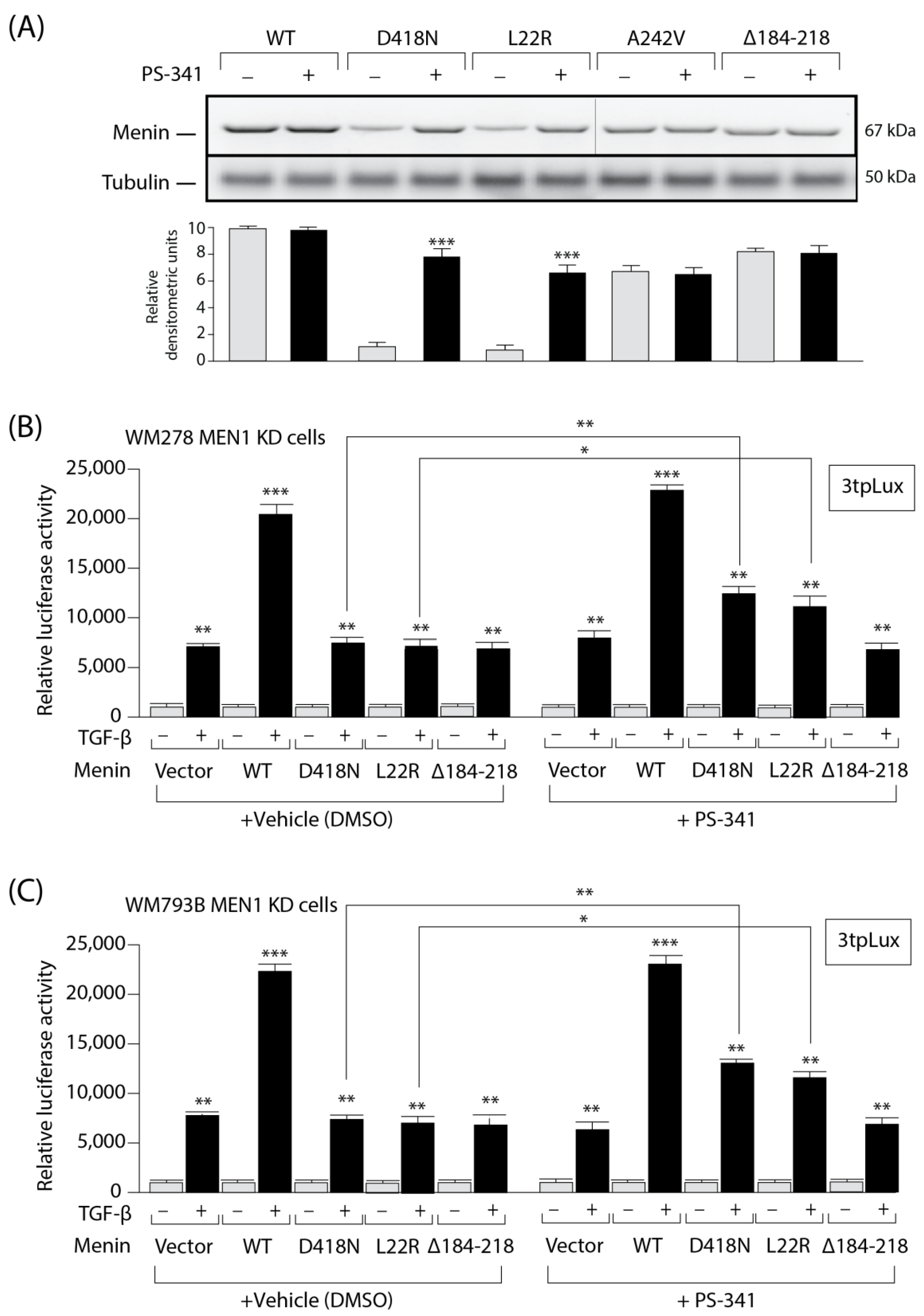
3.5. The Expression and Activity of MEN1 Missense Mutants Can Be Partially Rescued by a Proteasome Inhibitor

Having shown that the MEN1 D418N mutation failed to restore TGFβ signaling, we next investigated whether blocking its degradation could restore TGFβ responses and tumor suppression. The proteasome inhibitor PS-341 (Velcade, Bortezomib) is in clinical use for relapsed multiple myeloma and exhibits favorable selectivity toward tumors compared with normal cells [38]. As shown in Figure 5A, blocking the proteasome with the PS-341 inhibitor (for 4 h at 90 nM) partially restored the expression of the Menin missense mutant D418N and the L22R mutant, which were used here as a positive control. As expected, PS-341 had no effect on the WT or stable Menin mutants (A242V and ∆184-218).
To determine whether blocking the proteasome could offer therapeutic value for patients harboring MEN1 mutations, we examined whether the expression of the unstable MEN1 mutants and the TGFβ transcriptional response could be rescued using a proteasome inhibitor. For this purpose, we used the melanoma cell lines engineered above (Figure 5B,C; cells depleted of endogenous MEN1 or overexpressing WT or mutant MEN1). KD cells were then transfected with the 3TPLux luciferase reporter before being stimulated with TGFβ. As shown in Figure 5, in vehicle (DMSO)-treated WM278 MEN1 KD (5B) and WM793B MEN1 KD (5C) melanoma cells, the TGFβ transcriptional response was enhanced in cells overexpressing WT Menin relative to that in cells harboring the empty vector, whereas no difference was observed in cells overexpressing MEN1 mutants, confirming that the mutants failed to transmit the TGFβ responses. Interestingly, when WM278 MEN1 KD and WM793B MEN1 KD cells were treated with PS-341 for 6 hrs, the WT and the unstable MEN1 mutants (D418N and L22R) were able to partially restore TGFβ responses, consistent with the partial restoration of MEN1 mutant expression (Figure 5A). As expected, the negative control (a stable mutant [∆184-218]) had no effect. Thus, blocking the proteasome degradation pathway with a specific chemical inhibitor can restore both MEN1 expression and the TGFβ response.

3.6. The Expression and Activity of MEN1 Missense Mutants Can Be Rescued by Inhibition of the Ubiquitin Ligase CHIP

In parallel, to block the proteasome degradation pathway more specifically, we knocked down the expression of the C-terminal Hsp70 binding protein (CHIP) in melanoma cells. CHIP acts as a co-chaperone that can interact with the molecular chaperones Hsp70 and Hsp90, further leading to an imbalance of the folding-refolding machinery toward the degradation pathway [39]. Interestingly, blocking the proteasome through silencing CHIP gene expression with a specific siRNA completely restored the expression of the two MEN1 mutants (DN418 and L22R) but had no effect on the WT or stable MEN1 mutants (Figure 6A, left panel). The efficiency of the CHIP siRNA KD was verified by Western blotting (Figure 6A, right panel).
We next examined whether the missense mutation of MEN1 and the resulting TGFβ transcriptional response could be reversed by silencing CHIP expression. WM278 MEN1 KD and WM793B MEN1 KD cells were co-transfected with a scrambled (control) or CHIP-specific siRNA and the 3TPLux luciferase reporter before being stimulated with TGFβ. As shown in Figure 6B,C, only WT MEN1 could induce TGFβ-mediated luciferase activity in control WM278 MEN1 KD and WM793B MEN1 KD melanoma cells, consistent with the results observed with the proteasome inhibitor. However, both the WT and the missense MEN1 mutants (D418N and L22R) were able to almost completely restore the TGFβ response, while the negative control (∆184-218) had no effect.

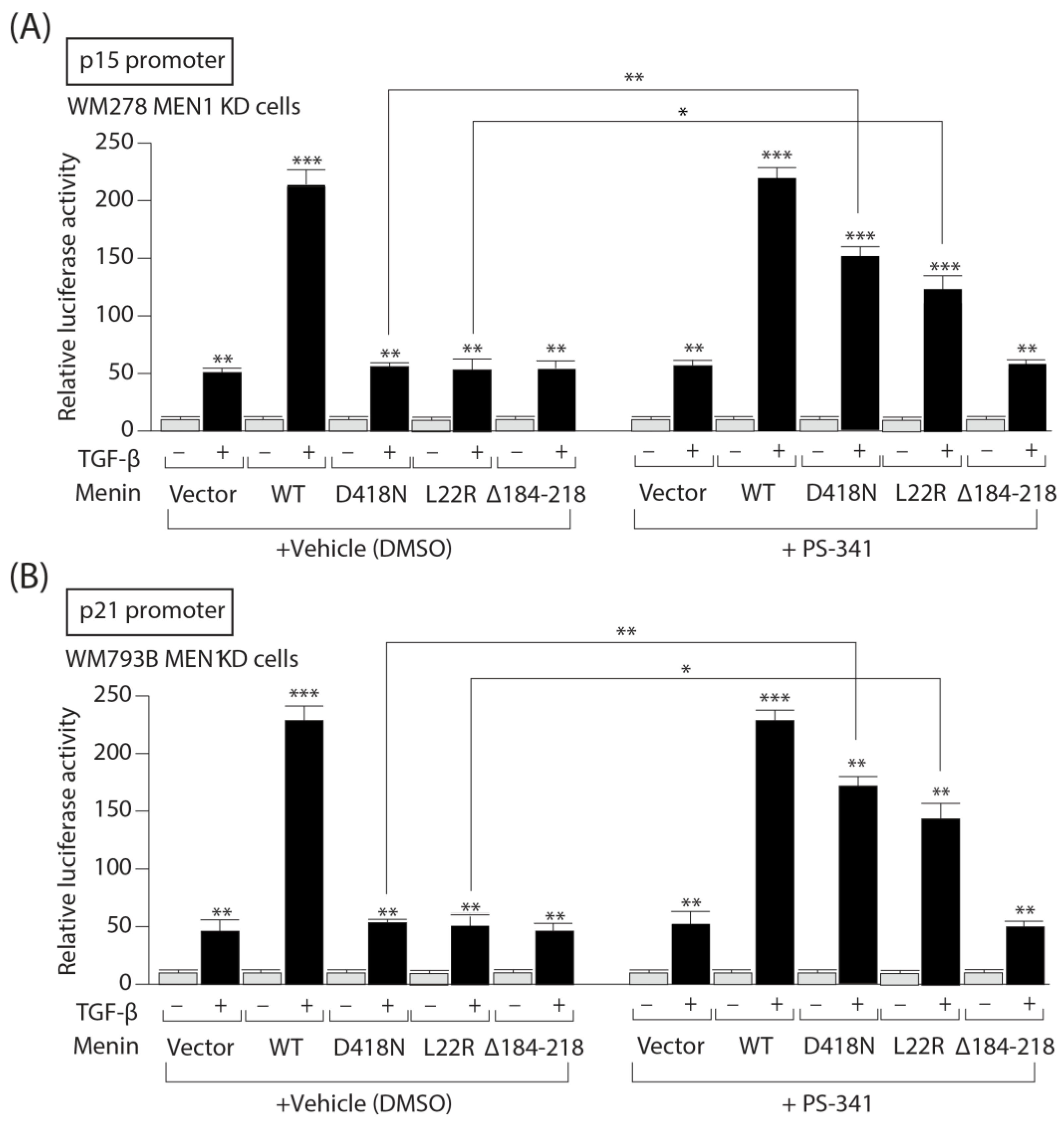
3.7. PS-341 Restores the Ability of Menin Missense Mutants to Mediate TGF-b Upregulation of the CDKI p15 and p21 Gene Promoters

Since TGF-b exerts, in part, its cytostatic actions by up-regulating the expression of CDKI genes such as p15 and p21, we examined whether treatment of Menin missense mutants with PS-341 would restore their ability to induce p15 and p21 gene promoter activities. As shown in Figure 7, in vehicle treated WM278 MEN1 KD cell, although TGF-b stimulation of luciferase activity was enhanced by Menin WT relative to vector for both promoters p15 (Figure 7A) and p21 (Figure 7B), no enhancement occurred with Menin missense mutants. However, upon treatment with PS-341, the luciferase activities of both promoters were partially restored by Menin missense mutants (D418N and L22R). The negative control (D184-218) had no effect.
Taken together, these results indicate that the loss of TGFβ tumor-suppressive responses in patients harboring MEN1 mutations leading to MEN1 degradation could be circumvented by blocking the proteasome degradation pathway, thereby offering new therapeutic opportunities for patients with melanoma.

4. Discussion

This study highlights MEN1 as a potent tumor-suppressor pathway that efficiently blocks tumorigenesis in cutaneous melanoma. Identification of specific point mutations in MEN1 family members affected by melanoma also revealed increased MEN1 gene product degradation, leading to a loss of TGFβ signaling. Using pharmacological inhibitors and RNA interference strategies, we showed that we could efficiently restore both MEN1 gene expression and TGFβ signaling in melanoma cells. Our findings indicate that the use of currently FDA-approved drugs against proteasomal degradation and/or tailor-made therapies mimicking the TGFβ/Smad3/MEN1 signaling pathway would be highly beneficial for melanoma patients, as these drugs would efficiently prevent initial tumor formation/progression and further hinder the spread of metastatic tumors to secondary organs.
Previous work highlighted MEN1 as a downstream TGFβ signaling component that regulates the growth and proliferation of pituitary adenoma cells and osteoblasts [19,40,41]. Moreover, we showed that Menin is required for activin-mediated inhibition of PRL expression in pituitary cells [42]. A screening of secreted biomarkers during macrophage/melanoma interactions revealed activin A as a highly secreted factor, but not TGFβ1-2-3. This finding highlights the role of activin A in the oncogenic activation of monocytes and macrophages in the melanoma tumor-microenvironment and the upregulation of tumor-associated genes. The differential role of activin A in melanoma is underscored with higher expression being correlated with pro-tumorigenic effect in the early phase of skin tumorigenesis [43] and worse outcome in melanoma patients [44]. The present study expands on these findings, highlighting MEN1 as a potent tumor suppressor downstream of TGFβ in nonendocrine tumors, such as melanoma. The results from our in vivo preclinical models clearly indicate that MEN1 knockout leads to increased primary melanoma tumor growth. Thus, MEN1 appears to function as a potent regulator of tumorigenesis in multiple endocrine and non-endocrine tissues, further revealing the broad range of biological processes regulated by MEN1. While TGFβ was found to play a dual role in promoting metastasis in breast cancer [44,45,46,47,48], TGFβ signaling has been shown to have antimetastatic effects on uveal melanoma [49], retinal Müller glia [50] and cutaneous melanoma. Thus, new therapeutic strategies aimed at activating the TGFβ/MEN1 signaling pathway could prove useful for melanoma patients at different stages of the disease, including primary tumor formation.
Interestingly, multiple potential melanoma tumor suppressors are localized on chromosome 11q, which includes the MEN1 region (located on chromosome 11q13) [51]. These results could suggest a possible association between MEN1 and melanoma. To confirm this, our study highlights Menin as a potential tumor suppressor in melanoma. Furthermore, we report here two families with melanoma in which at least two first-degree family members tested positive for MEN1 mutations. Interestingly, in addition to exhibiting typical MEN1 endocrine tumors, these patients also developed melanoma, suggesting that MEN1 is a strong candidate gene for familial malignant melanoma (for families in which 2 or more first-degree relatives, such as a parent, sibling, and/or child, exhibit skin cancer) [24,52]. The clinical implication is that patients should be tested for potential MEN1 gene mutations whenever 2 or more family members have developed melanoma. Moreover, our study suggested that all patients testing positive for MEN1 mutations are at risk of melanoma; because this may be a deadly disease, these patients should be monitored for melanoma. Still, a pan-cancer study reported that several mutations disabling MEN1 are somatic and that only a small fraction of germline mutation have the potential to be pathogenic and drive oncogenesis [53].
Characterization of the MEN1 gene in two families bearing melanoma highlighted specific MEN1 mutations, leading to loss of expression or increased degradation of the MEN1 gene product, further leading to a loss of TGFβ signaling. Interestingly, twenty percent of MEN1 cases involve Menin missense and small deletion or insertion mutations. The results from this study and work from others revealed that most of these mutants are expressed at markedly reduced levels relative to wild-type Menin. We found that blocking the proteasome degradation pathway with a specific proteasome inhibitor (PS-341, Velcade, or bortezomib) or with an RNA interference strategy aimed at silencing the expression of the molecular cochaperone CHIP efficiently restored MEN1 expression and the TGFβ transcriptional response. Thus, specific gene silencing has the potential to provide additional therapies to those currently available for the treatment of melanoma. The use of small chemical inhibitors also appears to be very promising for melanoma treatment. The proteasome inhibitor PS-341 (Velcade, bortezomib) used in this study is already in clinical use for relapsed multiple myeloma [38,54]. Although proteasome inhibitors have multiple effects on apoptosis and cell proliferation, the present study provides proof of principle that future exploration of their use in treating subsets of MEN1 patients is warranted.

Author Contributions

Investigation and methodology, J.B., M.G., L.C., V.G., A.S.S., A.S. and N.W.; funding acquisition, J.-J.L.; supervision, validation and project administration, S.A., D.G. and J.-J.L.; writing (original draft), J.B., D.G. and J.-J.L.; writing (review and editing), M.G., L.C., V.G., A.S.S. and A.S. All authors have read and agreed to the published version of the manuscript.

Funding

This research was funded by grants from the Canadian Institutes for Health Research (CIHR to J.-J.L.). The funders did not play a role in manuscript experimental design, data collection, data analysis and interpretation or writing of the manuscript.

Institutional Review Board Statement

All animals were housed and handled in accordance with the approved guidelines of the Canadian Council on Animal Care (CCAC) “Guide to the Care and Use of Experimental Animals”. All experiments were performed in accordance with the approved McGill University Animal Care protocol (AUP # 7497 to J.-J.L.).

Informed Consent Statement

Informed consent was obtained from each patient or subject, and the study was conducted according to a protocol that was approved by the IRCCS Casa Sollievo della Sofferenza Hospital, Research Ethics Board.

Data Availability Statement

Uncropped versions of the Western blots are available as original data files. The other datasets generated during and/or analyzed during the current study are available from the corresponding authors upon reasonable request.

Acknowledgments

The authors are grateful to Alan Spatz and Louise Larose for kindly providing the melanoma cell lines. We gratefully acknowledge the excellent contributions of Geoffrey Hendy to this work before his untimely passing.

Conflicts of Interest

The authors declare no conflicts of interest.

References

  1. Balch, C.M.; Mihm, M.C. Reply to the article “The AJCC staging proposal for cutaneous melanoma: Comments by the EORTC Melanoma Group”, by D. J. Ruiter et al. (Ann Oncol 2001; 12: 9–11). Ann. Oncol. 2002, 13, 175–176. [Google Scholar] [CrossRef]
  2. Siegel, R.L.; Miller, K.D.; Fuchs, H.E.; Jemal, A. Cancer statistics, 2022. CA Cancer J. Clin. 2022, 72, 7–33. [Google Scholar] [CrossRef]
  3. Ferlay, J.; Soerjomataram, I.; Dikshit, R.; Eser, S.; Mathers, C.; Rebelo, M.; Parkin, D.M.; Forman, D.; Bray, F. Cancer incidence and mortality worldwide: Sources, methods and major patterns in GLOBOCAN 2012. Int. J. Cancer 2015, 136, E359–E386. [Google Scholar] [CrossRef]
  4. Ross, M.I.; Gershenwald, J.E. Evidence-based treatment of early-stage melanoma. J. Surg. Oncol. 2011, 104, 341–353. [Google Scholar] [CrossRef] [PubMed]
  5. Houghton, A.N.; Polsky, D. Focus on melanoma. Cancer Cell 2002, 2, 275–278. [Google Scholar] [CrossRef]
  6. Enninga, E.A.L.; Moser, J.C.; Weaver, A.L.; Markovic, S.N.; Brewer, J.D.; Leontovich, A.A.; Hieken, T.J.; Shuster, L.; Kottschade, L.A.; Olariu, A.; et al. Survival of cutaneous melanoma based on sex, age, and stage in the United States, 1992–2011. Cancer Med. 2017, 6, 2203–2212. [Google Scholar] [CrossRef]
  7. Krauthammer, M.; Kong, Y.; Ha, B.H.; Evans, P.; Bacchiocchi, A.; McCusker, J.P.; Cheng, E.; Davis, M.J.; Goh, G.; Choi, M.; et al. Exome sequencing identifies recurrent somatic RAC1 mutations in melanoma. Nat. Genet. 2012, 44, 1006–1014. [Google Scholar] [CrossRef] [PubMed]
  8. Hodis, E.; Watson, I.R.; Kryukov, G.V.; Arold, S.T.; Imielinski, M.; Theurillat, J.P.; Nickerson, E.; Auclair, D.; Li, L.; Place, C.; et al. A landscape of driver mutations in melanoma. Cell 2012, 150, 251–263. [Google Scholar] [CrossRef]
  9. Davies, H.; Bignell, G.R.; Cox, C.; Stephens, P.; Edkins, S.; Clegg, S.; Teague, J.; Woffendin, H.; Garnett, M.J.; Bottomley, W.; et al. Mutations of the BRAF gene in human cancer. Nature 2002, 417, 949–954. [Google Scholar] [CrossRef] [PubMed]
  10. Leonardi, G.C.; Falzone, L.; Salemi, R.; Zanghì, A.; Spandidos, D.A.; Mccubrey, J.A.; Candido, S.; Libra, M. Cutaneous melanoma: From pathogenesis to therapy (Review). Int. J. Oncol. 2018, 52, 1071–1080. [Google Scholar] [CrossRef]
  11. Lopez-Bergami, P.; Fitchman, B.; Ronai, Z. Understanding signaling cascades in melanoma. Photochem. Photobiol. 2008, 84, 289–306. [Google Scholar] [CrossRef] [PubMed]
  12. Humbert, L.; Ghozlan, M.; Canaff, L.; Tian, J.; Lebrun, J.J. The leukemia inhibitory factor (LIF) and p21 mediate the TGFβ tumor suppressive effects in human cutaneous melanoma. BMC Cancer 2015, 15, 200. [Google Scholar] [CrossRef] [PubMed]
  13. Humbert, L.; Lebrun, J.J. TGF-beta inhibits human cutaneous melanoma cell migration and invasion through regulation of the plasminogen activator system. Cell. Signal. 2013, 25, 490–500. [Google Scholar] [CrossRef] [PubMed]
  14. Ramont, L.; Pasco, S.; Hornebeck, W.; Maquart, F.X.; Monboisse, J.C. Transforming growth factor-β1 inhibits tumor growth in a mouse melanoma model by down-regulating the plasminogen activation system. Exp. Cell Res. 2003, 291, 1–10. [Google Scholar] [CrossRef] [PubMed]
  15. Boudreault, J.; Wang, N.; Ghozlan, M.; Lebrun, J.-J. Transforming Growth Factor-β/Smad Signaling Inhibits Melanoma Cancer Stem Cell Self-Renewal, Tumor Formation and Metastasis. Cancers 2024, 16, 224. [Google Scholar] [CrossRef] [PubMed]
  16. Balogh, K.; Rácz, K.; Patócs, A.; Hunyady, L. Menin and its interacting proteins: Elucidation of menin function. Trends Endocrinol. Metab. 2006, 17, 357–364. [Google Scholar] [CrossRef] [PubMed]
  17. Milne, T.A.; Hughes, C.M.; Lloyd, R.; Yang, Z.; Rozenblatt-Rosen, O.; Dou, Y.; Schnepp, R.W.; Krankel, C.; LiVolsi, V.A.; Gibbs, D.; et al. Menin and MLL cooperatively regulate expression of cyclin-dependent kinase inhibitors. Proc. Natl. Acad. Sci. USA 2005, 102, 749–754. [Google Scholar] [CrossRef] [PubMed]
  18. Karnik, S.K.; Hughes, C.M.; Gu, X.; Rozenblatt-Rosen, O.; McLean, G.W.; Xiong, Y.; Meyerson, M.; Kim, S.K. Menin regulates pancreatic islet growth by promoting histone methylation and expression of genes encoding p27Kip1 and p18INK4c. Proc. Natl. Acad. Sci. USA 2005, 102, 14659–14664. [Google Scholar] [CrossRef] [PubMed]
  19. Kaji, H.; Canaff, L.; Lebrun, J.J.; Goltzman, D.; Hendy, G.N. Inactivation of menin, a Smad3-interacting protein, blocks transforming growth factor type β signaling. Proc. Natl. Acad. Sci. USA 2001, 98, 3837–3842. [Google Scholar] [CrossRef]
  20. Sowa, H.; Kaji, H.; Kitazawa, R.; Kitazawa, S.; Tsukamoto, T.; Yano, S.; Tsukada, T.; Canaff, L.; Hendy, G.N.; Sugimoto, T.; et al. Menin Inactivation Leads to Loss of Transforming Growth Factor β Inhibition of Parathyroid Cell Proliferation and Parathyroid Hormone Secretion. Cancer Res. 2004, 64, 2222–2228. [Google Scholar] [CrossRef]
  21. Thakker, R.V. Multiple endocrine neoplasia type 1. Endocrinol. Metab. Clin. N. Am. 2000, 29, 541–567. [Google Scholar] [CrossRef]
  22. Baldauf, C.; Vortmeyer, A.O.; Koch, C.A.; Sticherling, M. Combination of multiple skin malignancies with multiple endocrine neoplasia type 1: Coincidental or pathogenetically related? Dermatology 2009, 219, 365–367. [Google Scholar] [CrossRef]
  23. Nord, B.; Platz, A.; Smoczynski, K.; Kytölä, S.; Robertson, G.; Calender, A.; Murat, A.; Weintraub, D.; Burgess, J.; Edwards, M.; et al. Malignant melanoma in patients with multiple endocrine neoplasia type 1 and involvement of the MEN1 gene in sporadic melanoma. Int. J. Cancer 2000, 87, 463–467. [Google Scholar] [CrossRef]
  24. Gray-Schopfer, V.; Wellbrock, C.; Marais, R. Melanoma biology and new targeted therapy. Nature 2007, 445, 851–857. [Google Scholar] [CrossRef]
  25. Gao, S.B.; Feng, Z.J.; Xu, B.; Chen, Y.; Zheng, H.H.; Yin, P.; Hua, X.; Jin, G.H. Menin represses malignant phenotypes of melanoma through regulating multiple pathways. J. Cell. Mol. Med. 2011, 15, 2353–2363. [Google Scholar] [CrossRef] [PubMed]
  26. Massey, S.; Khan, M.A.; Rab, S.O.; Mustafa, S.; Khan, A.; Malik, Z.; Shaik, R.; Verma, M.K.; Deo, S.; Husain, S.A. Evaluating the role of MEN1 gene expression and its clinical significance in breast cancer patients. PLoS ONE 2023, 18, e0288482. [Google Scholar] [CrossRef] [PubMed]
  27. Sanjana, N.E.; Shalem, O.; Zhang, F. Imroved vectors and genome-widees for CRISPR screening. Nat. Methods 2014, 11, 783–784. [Google Scholar] [CrossRef] [PubMed]
  28. Poncin, J.; Abs, R.; Velkeniers, B.; Bonduelle, M.; Abramowicz, M.; Legros, J.J.; Verloes, A.; Meurisse, M.; Van Gaal, L.; Verellen, C.; et al. Mutation analysis of the MEN1 gene in Belgian patients with multiple endocrine neoplasia type 1 and related diseases. Hum. Mutat. 1999, 13, 54–60. [Google Scholar] [CrossRef]
  29. Ito, T.; Igarashi, H.; Uehara, H.; Berna, M.J.; Jensen, R.T. Causes of death and prognostic factors in multiple endocrine neoplasia type 1: A prospective study:Comparison of 106 men1/zollinger-ellison syndrome patients with 1613 literature men1 patients with or without pancreatic endocrine tumors. Medicine 2013, 92, 135–181. [Google Scholar] [CrossRef]
  30. Neel, J.-C.; Humbert, L.; Lebrun, J.-J. The Dual Role of TGFβ in Human Cancer: From Tumor Suppression to Cancer Metastasis. ISRN Mol. Biol. 2012, 2012, 381428. [Google Scholar] [CrossRef]
  31. Datto, M.B.; Li, Y.; Panus, J.F.; Howe, D.J.; Xiong, Y.; Wang, X.F. Transforming growth factor beta induces the cyclin-dependent kinase inhibitor p21 through a p53-independent mechanism. Proc. Natl. Acad. Sci. USA 1995, 92, 5545–5549. [Google Scholar] [CrossRef] [PubMed]
  32. Warner, B.J.; Blain, S.W.; Seoane, J.; Massagué, J. Myc Downregulation by Transforming Growth Factor β Required for Activation of the p15 Ink4b G 1 Arrest Pathway. Mol. Cell. Biol. 1999, 19, 5913–5922. [Google Scholar] [CrossRef] [PubMed]
  33. Schuster, N.; Krieglstein, K. Mechanisms of TGF-β-mediated apoptosis. Cell Tissue Res. 2002, 307, 1–14. [Google Scholar] [CrossRef] [PubMed]
  34. Canaff, L.; Vanbellinghen, J.F.; Kaji, H.; Goltzman, D.; Hendy, G.N. Impaired transforming growth factor-β (TGF-β) transcriptional activity and cell proliferation control of a menin in-frame deletion mutant associated with Multiple Endocrine Neoplasia type 1 (MEN1). J. Biol. Chem. 2012, 287, 8584–8597. [Google Scholar] [CrossRef] [PubMed]
  35. Canaff, L.; Vanbellinghen, J.F.; Kanazawa, I.; Kwak, H.; Garfield, N.; Vautour, L.; Hendy, G.N. Menin missense mutants encoded by the MEN1 gene that are targeted to the proteasome: Restoration of expression and activity by CHIP siRNA. J. Clin. Endocrinol. Metab. 2012, 97, E282–E291. [Google Scholar] [CrossRef] [PubMed]
  36. Yaguchi, H.; Ohkura, N.; Takahashi, M.; Nagamura, Y.; Kitabayashi, I.; Tsukada, T. Menin Missense Mutants Associated with Multiple Endocrine Neoplasia Type 1 Are Rapidly Degraded via the Ubiquitin-Proteasome Pathway. Mol. Cell. Biol. 2004, 24, 6569–6580. [Google Scholar] [CrossRef] [PubMed]
  37. Guru, S.C.; Goldsmith, P.K.; Lee Burns, A.; Marx, S.J.; Spiegel, A.M.; Collins, F.S.; Chandrasekharappa, S.C. Menin, the product of the MEN1 gene, is a nuclear protein. Proc. Natl. Acad. Sci. USA 1998, 95, 1630–1634. [Google Scholar] [CrossRef] [PubMed]
  38. Buac, D.; Shen, M.; Schmitt, S.; Rani Kona, F.; Deshmukh, R.; Zhang, Z.; Neslund-Dudas, C.; Mitra, B.; Dou, Q.P. From Bortezomib to other Inhibitors of the Proteasome and Beyond. Curr. Pharm. Des. 2013, 19, 4025–4038. [Google Scholar] [CrossRef] [PubMed]
  39. McDonough, H.; Patterson, C. CHIP: A link between the chaperone and proteasome systems. Cell Stress Chaperones 2003, 8, 303–308. [Google Scholar] [CrossRef]
  40. Lebrun, J.J. Activin, TGF-β and menin in pituitary tumorigenesis. Adv. Exp. Med. Biol. 2009, 668, 69–78. [Google Scholar] [CrossRef]
  41. Hendy, G.N.; Kaji, H.; Sowa, H.; Lebrun, J.-J.; Canaff, L. Menin and TGF-β Superfamily Member Signaling via the Smad Pathway in Pituitary, Parathyroid and Osteoblast. Horm. Metab. Res. 2005, 37, 375–379. [Google Scholar] [CrossRef]
  42. Lacerte, A.; Lee, E.H.; Reynaud, R.; Canaff, L.; De Guise, C.; Devost, D.; Ali, S.; Hendy, G.N.; Lebrun, J.J. Activin inhibits pituitary prolactin expression and cell growth through Smads, Pit-1 and menin. Mol. Endocrinol. 2004, 18, 1558–1569. [Google Scholar] [CrossRef]
  43. Antsiferova, M.; Piwko-Czuchra, A.; Cangkrama, M.; Wietecha, M.; Sahin, D.; Birkner, K.; Amann, V.C.; Levesque, M.; Hohl, D.; Dummer, R.; et al. Activin promotes skin carcinogenesis by attraction and reprogramming of macrophages. EMBO Mol. Med. 2017, 9, 27–45. [Google Scholar] [CrossRef]
  44. Gutiérrez-Seijo, A.; García-Martínez, E.; Barrio-Alonso, C.; Parra-Blanco, V.; Avilés-Izquierdo, J.A.; Sánchez-Mateos, P.; Samaniego, R. Activin A Sustains the Metastatic Phenotype of Tumor-Associated Macrophages and Is a Prognostic Marker in Human Cutaneous Melanoma. J. Investig. Dermatol. 2022, 142, 653–661.e2. [Google Scholar] [CrossRef]
  45. Dai, M.; Al-Odaini, A.A.; Arakelian, A.; Rabbani, S.A.; Ali, S.; Lebrun, J.J. A novel function for p21Cip1 and acetyltransferase p/CAF as critical transcriptional regulators of TGFβ-mediated breast cancer cell migration and invasion. Breast Cancer Res. 2012, 14, R127. [Google Scholar] [CrossRef]
  46. Derynck, R.; Akhurst, R.J. Differentiation plasticity regulated by TGF-β family proteins in development and disease. Nat. Cell Biol. 2007, 9, 1000–1004. [Google Scholar] [CrossRef]
  47. Thiery, J.P. Epithelial-mesenchymal transitions in development and pathologies. Curr. Opin. Cell Biol. 2003, 15, 740–746. [Google Scholar] [CrossRef]
  48. Moustakas, A.; Heldin, C.H. Signaling networks guiding epithelial-mesenchymal transitions during embryogenesis and cancer progression. Cancer Sci. 2007, 98, 1512–1520. [Google Scholar] [CrossRef]
  49. Woodward, J.K.L.; Elshaw, S.R.; Murray, A.K.; Nichols, C.E.; Cross, N.; Laws, D.; Rennie, I.G.; Sisley, K. Stimulation and inhibition of uveal melanoma invasion by HGF, GRO, IL-1α and TGF-β. Investig. Ophthalmol. Vis. Sci. 2002, 43, 3144–3152. [Google Scholar]
  50. Romo, P.; Madigan, M.C.; Provis, J.M.; Cullen, K.M. Differential effects of TGF-β and FGF-2 on in vitro proliferation and migration of primate retinal endothelial and Müller cells. Acta Ophthalmol. 2011, 89, e263–e268. [Google Scholar] [CrossRef]
  51. Agarwal, S.K. The future: Genetics advances in MEN1 therapeutic approaches and management strategies. Endocr. Relat. Cancer 2017, 24, T119–T134. [Google Scholar] [CrossRef] [PubMed]
  52. Chandrasekharappa, S.C.; Guru, S.C.; Manickam, P.; Olufemi, S.E.; Collins, F.S.; Emmert-Buck, M.R.; Debelenko, L.V.; Zhuang, Z.; Lubensky, I.A.; Liotta, L.A.; et al. Positional cloning of the gene for multiple endocrine neoplasia-type 1. Science 1997, 276, 404–406. [Google Scholar] [CrossRef] [PubMed]
  53. Nelakurti, D.D.; Pappula, A.L.; Rajasekaran, S.; Miles, W.O.; Petreaca, R.C. Comprehensive Analysis of MEN1 Mutations and Their Role in Cancer. Cancers 2020, 12, 2616. [Google Scholar] [CrossRef]
  54. Mohan, M.; Matin, A.; Davies, F.E. Update on the optimal use of Bortezomib in the treatment of multiple myeloma. Cancer Manag. Res. 2017, 9, 51–63. [Google Scholar] [CrossRef]
Figure 1. TGFβ induces MEN1 gene expression in melanoma cells through Smad3. (A) Regulation of the Menin protein (left panel) and mRNA (right panel) in human melanoma cell lines. Changes in the Menin protein and mRNA expression following TGFβ treatment. Menin and β-tubulin protein expressions were determined via Western blotting. MEN1 mRNA levels were determined by qPCR with GAPDH serving as a reference gene. (B) Generation of CRISPR/Cas9 SMAD2/3/4 and control (SCR, scrambled) knockout (KO) DAUV melanoma cell lines. The efficiency of the knockout was assessed using Western blotting. (C) MEN1 mRNA expression levels in DAUV CRISPR/Cas9 SMAD2/3/4 and control (SCR, scrambled) KO cells. MEN1 mRNA levels were determined by qPCR with GAPDH serving as a reference gene. *, p < 0.05. **, p < 0.01. ***, p < 0.001.
Cells 13 00973 g001
Figure 2. The TGFβ/Smad3/MEN1 axis is essential for inhibiting melanoma tumor formation in vivo. (A) Generation of WM278 melanoma MEN1 KO and control (SCR, scrambled) CRISPR/Cas9 knockout (KO) cell lines. The KO efficiency of MEN1 was measured by Western blotting. (B) Growth of WM278 cells cultured for 2 weeks and seeded at a low density (1000 cells/well). (C) Generation of BLM, WM278, and WM1232 melanoma CRISPR/Cas9 MEN1 KO. KO efficiency was measured by Western blotting. (DF) NSG mice were injected subcutaneously with BLM (D), WM278 (E), WM1232 (F), or scrambled (SCR) or with MEN1 knockout melanoma cells at 1 × 10⁶ cells/mouse. As shown in (DF), the left panel shows the mean tumor volumes, and the right panel shows representative images of tumors at the tumor collection endpoint. *, p < 0.05 (in red).
Cells 13 00973 g002
Figure 3. The TGFβ/Smad3/MEN1 axis is essential for inducing cell-cycle arrest in human melanoma cells. (A). (left panel) Cell-cycle distribution assessed by flow cytometry analysis of propidium iodide-stained WM278 scrambled, MEN1 KO, and SMAD3 KO cells following TGFβ treatment (24 h). (right panel) Quantification table displaying the percentage of G1, S, and G2/M phase cells. Cell-cycle experiments were performed in three biological replicates. A two-sample equal variance T-test was used to compare the unstimulated and stimulated TGFβ condition. (B) (left panel) Generation of a375m melanoma CRISPR/Cas9 MEN1 KO. (Right panel) P21 expression upon short-term TGFβ stimulation in WM278 CRISPR/Cas9 scrambled or MEN1 KOs (sg10 and sg14). Gene expression was measured by Western blotting. (C) Regulation of cell-cycle progression in tumor tissues from WM278 CRISPR/Cas9 SCR (scrambled) or MEN1 KO mice. Changes in protein levels were assessed through Western blotting. (D) (upper panel) Annexin V/PI dual staining was used to determine the percentages of live, apoptotic, and dead cells induced by TGFβ treatment. (bottom panel) Quantification table displaying the percentage of live, apoptotic, and dead cells. Cell-cycle experiments were performed in three biological replicates. (E) (upper panel) Regulation of Caspase 3 expression in tumor tissues from WM278 CRISPR/Cas9 SCR (scrambled) or MEN1 KO. Changes in protein levels were assessed through Western blotting. (bottom panel) Histogram showing the quantification by densitometry for each band relative to β-tubulin loading control. *, p < 0.05. **, p < 0.01.
Cells 13 00973 g003
Figure 4. Expression of Menin mutants in HEK293 cells and generation of stable MEN1 knockout melanoma cell lines. (A). Pedigrees of two MEN1 kindreds. (B). Flag-tagged Menin WT and mutant constructs were transfected into HEK293 cells, and after 48 h, the cell lysates were subjected to Western blot analysis with anti-Flag and anti-β-tubulin antibodies. (C). Efficiency of Menin knockdown in WM278 parental and shRNA-infected cells. Menin protein and mRNA (left panel) expression was measured via Western blot analysis and qPCR. TGFβ responsive CAGA and PAI-1 (3tpLlux) gene promoter activity in WM278 parental cells (right top panel) and WM278 MEN1 KD cells (right lower panel). The data are graphed as the arithmetic mean of relative luciferase units normalized to β-galactosidase activity. (D). Efficiency of Menin knockdown in WM793B parental and shRNA-infected cells. Menin protein and mRNA (left panel) expression was measured via Western blot analysis and qPCR. TGFβ responsive CAGA and PAI-1 gene promoter activity in WM793B parental cells (right top panel) and WM793B MEN1 KD cells (right lower panel). The data are graphed as the arithmetic mean of relative luciferase units normalized to β-galactosidase activity. *, p < 0.05. **, p < 0.01. ***, p < 0.001.
Cells 13 00973 g004
Figure 5. The expression and activity of Menin missense mutants can be rescued by the proteasome inhibitor PS-341. (A). Western blot analysis of HEK293T cells transfected with Flag-tagged WT or mutant Menin and treated (black) or not treated (gray) with PS-341 (Velcade). (B,C). TGFβ–responsive PAI-1 (3tpLux) gene promoter activity in WM278 and WM793B MEN1 KD cells treated with either vehicle or PS341. The data are graphed as the arithmetic mean of relative luciferase units normalized to β-galactosidase activity. *, p < 0.05. **, p < 0.01. ***, p < 0.001.
Cells 13 00973 g005
Figure 6. The expression and activity of Menin missense mutants can be rescued by RNAi targeting the ubiquitin ligase CHIP. (A). Western blot analysis of HEK293T cells transfected with Flag-tagged WT or mutant Menin and treated with scrambled (gray) or specific CHIP siRNA (black) (left panel). Western blot analysis of ChIP in HEK293, WM278 and WM793 cells treated with scrambled or specific CHIP siRNA (right panel). (B,C) TGFβ responsive PAI-1 (3tpLux) gene promoter activity in WM278 MEN1 KD and WM793B MEN1 KD cells transfected with scrambled siRNA or specific CHIP siRNA. The data are graphed as the arithmetic mean of relative luciferase units normalized to β-galactosidase activity. **, p < 0.01. ***, p < 0.001.
Cells 13 00973 g006
Figure 7. PS-341 restores the ability of Menin missense mutants to mediate TGF-β up-regulation of the p15 and p21 CDKI gene promoters. TGF-β responsive (A) p15 and (B) p21 gene promoter activity in WM278 MEN1 KD cells treated with either vehicle or PS-341. The data are graphed as the arithmetic mean of relative luciferase units normalized to β-galactosidase activity. *, p < 0.05. **, p < 0.01. ***, p < 0.001.
Cells 13 00973 g007
Table 1. Characteristics of cell lines.
Cell LineCell TypeOriginSex of Human DonorAgeMutations and Characteristics
WM278MelanomaPrimaryFemale62BRAF (V600E); PTEN (Deletion)
WM793BMelanomaPrimaryMale37BRAF (V600E); CDK4 (K22Q); PTEN (Deletion)
BLMMelanomaDerived from lung metastases in nude mice injected with BRO parent cell lineMale34Nras (Q61R)
WM1232MelanomaMetastaticFemaleN/ABRAF (V600E); PTEN (Deletion)
DAUVMelanomaPrimaryN/AN/ABRAF (V600E)
SKMEL-28MelanomaPrimaryMale53BRAF (V600E); CDK4 (R24C); EGFR (P753S); PTEN (T167A); TP53 (L145R); Tert (Promoter)
a375mMelanomaIsolated from a tumor from a nude mice injected with the parent a375 cell lineFemale54BRAF(V600E); CDKN2A (Deletion); Tert (Promoter)
HEK293KidneyEmbryoFemaleFetus
Table 2. Primer sequences for qPCR assay.
Gene Sequence
MEN1Forward5′-GGAAGACGACGAGGAGATCTACA-3′
MEN1Reverse5′-CAGTAGTTCAGAGGCCTTTGCGCT-3′
GAPDHForward5′-GCCTCAAGATCATCAGCAATGCCT-3′
GAPDHReverse5′-TGTGGTCATGAGTCCTTCCACGAT-3′
Table 3. gRNA sequences for molecular cloning.
Gene Sequence
MENsg1Forward5′-CACCGCACCTGCTGCGATTCTACGA-3′
MENsg1Reverse5′-AAACTCGTAGAATCGCAGCAGGTGC-3′
MEN2sg2Forward5′-CACCGACGTCGTCGATGGAGCGCAG-3′
MEN2sg2Reverse5′-AAACCTGCGCTCCATCGACGACGTC-3′
SCRsg1Forward5′-CACCGACGGAGGCTAAGCGTCGCAA-3′
SCRsg2Reverse5′-AAACTTGCGACGCTTAGCCTCCGTC-3′
SMAD2sg1Forward5′-CACCGTCCCACTGATCTATCGTATT-3′
SMAD2sg1Reverse5′-AAACAATACGATAGATCAGTGGGAC-3′
SMAD2sg2Forward5′-CACCGTGGCGGCGTGAATGGCAAGA-3′
SMAD2sg2Reverse5‘-AAACTCTTGCCATTCACGCCGCCAC-3′
SMAD3sg1Forward5′-CACCGCCCGATCGTGAAGCGCCTGC-3′
SMAD3sg1Reverse5′-AAACGCAGGCGCTTCACGATCGGGC-3′
SMAD3sg2Forward5′-CACCGTTCACGATCGGGGGAGTGAA-3′
SMAD3sg2Reverse5′-AAACTTCACTCCCCCGATCGTGAAC-3′
SMAD4sg1Forward5′-CACCGAACTCTGTACAAAGACCGCG-3′
SMAD4sg1Reverse5′-AAACCGCGGTCTTTGTACAGAGTTC-3′
Disclaimer/Publisher’s Note: The statements, opinions and data contained in all publications are solely those of the individual author(s) and contributor(s) and not of MDPI and/or the editor(s). MDPI and/or the editor(s) disclaim responsibility for any injury to people or property resulting from any ideas, methods, instructions or products referred to in the content.

