Review

Perivascular Adipose Tissue and Perivascular Adipose Tissue-Derived Extracellular Vesicles: New Insights in Vascular Disease

1
Department of Biomedical Sciences, Joan C Edwards School of Medicine, Marshall University, Huntington, WV 25755, USA
2
Department of Internal Medicine, Joan C Edwards School of Medicine, Marshall University, Huntington, WV 25755, USA
*
Author to whom correspondence should be addressed.
Cells 2024, 13(16), 1309; https://doi.org/10.3390/cells13161309 (registering DOI)
Submission received: 15 July 2024 / Revised: 29 July 2024 / Accepted: 2 August 2024 / Published: 6 August 2024

Abstract

:
Perivascular adipose tissue (PVAT) is a special deposit of fat tissue surrounding the vasculature. Previous studies suggest that PVAT modulates the vasculature function in physiological conditions and is implicated in the pathogenesis of vascular diseases. Understanding how PVAT influences vasculature function and vascular disease progression is important. Extracellular vesicles (EVs) are novel mediators of intercellular communication. EVs encapsulate molecular cargo such as proteins, lipids, and nucleic acids. EVs can influence cellular functions by transferring the carried bioactive molecules. Emerging evidence indicates that PVAT-derived EVs play an important role in vascular functions under health and disease conditions. This review will focus on the roles of PVAT and PVAT-EVs in obesity, diabetic, and metabolic syndrome-related vascular diseases, offering novel insights into therapeutic targets for vascular diseases.

1. Introduction

Perivascular adipose tissue (PVAT) is an adipose tissue deposit surrounding most of the vasculature. PVAT releases a variety of adipocytokines and chemokines [1,2,3,4]. Under physiological conditions, PVAT regulates vascular tone and reactivity by releasing various biologically active factors, adipocyte-derived relaxing factors, and perivascular-derived relaxing factors, which have anticontractile properties. However, in pathological conditions, such as obesity, diabetes, and metabolic syndrome, PVAT becomes dysfunctional. The secretory profile of PVAT changes with a reduced secretion of vaso-relaxing factors, increased vasoconstricting factors, increased immune cell infiltration, and vascular smooth muscle cell proliferation. These changes lead to PVAT inflammation, thereby detrimentally affecting vasculature function. Studies found that PVAT mass is increased in obesity in both animal models and humans [5,6,7]. In obese patients, PVAT mass correlates with visceral adipose tissue mass, a known predictor of the development of metabolic disease [6,8]. PVAT volume alone is associated with hypertension and aortic and coronary calcification [6,9]. All of these findings suggest the involvement of PVAT in vascular physiology and pathology.
Inter-cell signaling between PVAT and the vasculature remains a mystery. Recent studies have suggested that, apart from the secretory cytokines and chemokines factors, PVAT also secretes extracellular vesicles (EVs) [10,11]. EVs are cellular-derived nano-sized particles, carrying biologically active molecules that can regulate cell function. EVs can be transferred horizontally to adjacent cells (paracrine signaling) or distant cells and organs (endocrine–inter-organ signaling). They are key players in various physiological and pathological processes, ranging from cancer metastasis to cardiovascular diseases [12]. In this study, we reviewed the existing literature regarding the roles of PVAT and their derived EVs in the pathogenesis of vascular diseases. We discussed the therapeutic potential of targeting PVAT and PVAT-EVs in vascular diseases and the challenges in this field.

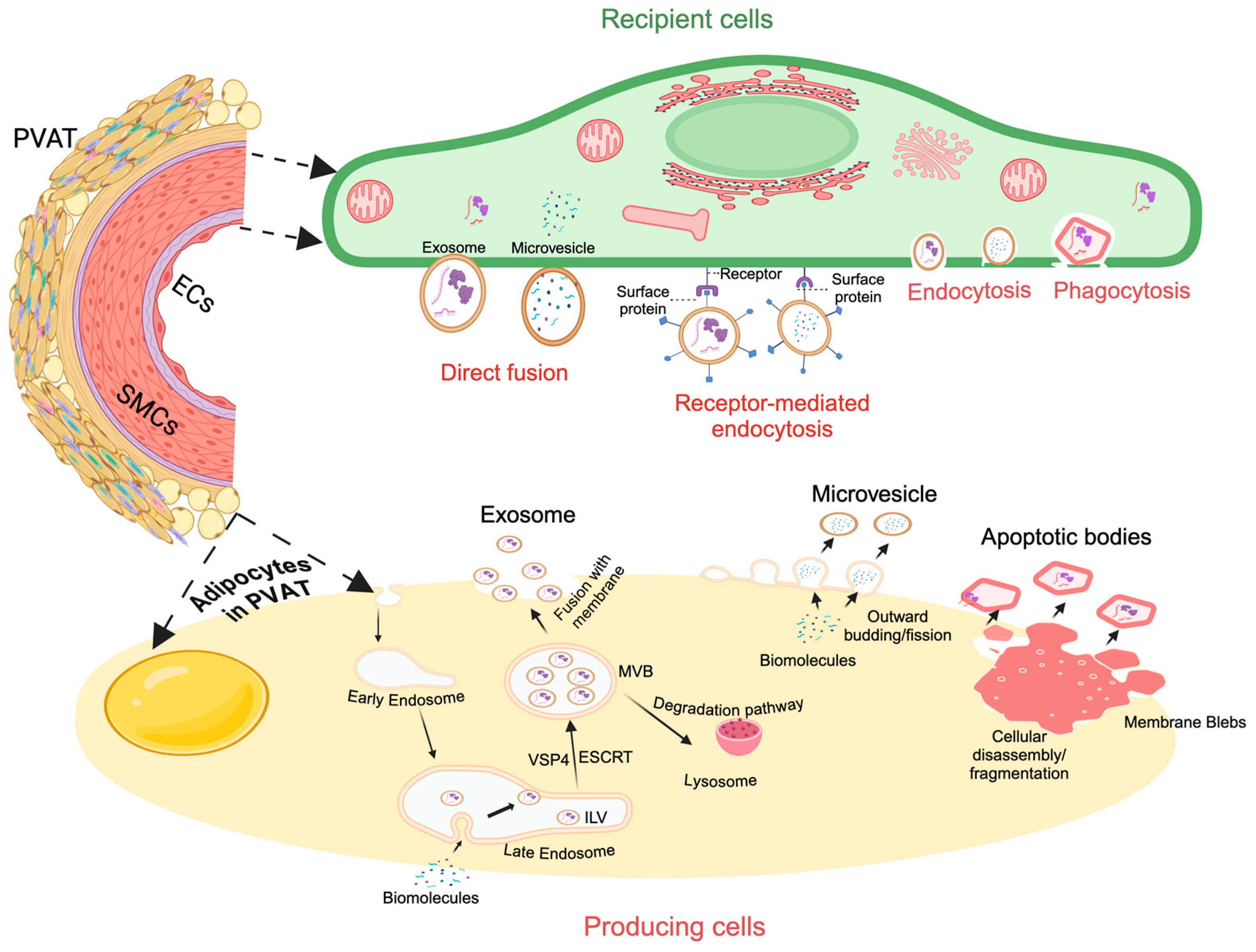
2. PVAT: Anatomy and Physiology

PVAT is a specialized type of fat tissue that adjacently surrounds blood vessels such as the aorta, coronary arteries, small and resistance vessels, and vasculature of the musculoskeletal system [13], but is absent among cerebral vessels [14]. Though PVAT is universal in that it surrounds vasculature, its volume and composition vary widely depending on physiological and pathological conditions. This demonstrates how versatile and diverse PVAT may be, and what innumerable effects it may elicit on surrounding tissue. Like most adipose tissue, PVAT could be white adipose tissue (WAT) or brown adipose tissue (BAT). For instance, the thoracic aorta is surrounded by brown PVAT while the abdominal aorta PVAT shows the WAT-like phenotype [15]. Usually, WAT performs as an energy reserve and is required for maintaining human homeostasis. WAT is regarded as the traditional “unhealthy” fat, as experimental and clinical studies have shown that it is positively related to cardiovascular diseases [16]. WAT has unilocular lipid droplets, fewer mitochondria, and higher lipid storage capacity [17]. The unique surface markers of white adipocytes include resistin, ASC-1, and FAB4 [18]. Different types of WAT include visceral adipose tissue and subcutaneous WAT. Visceral WAT has been shown to negatively impact health due to its inflammatory characteristics, which are linked to insulin resistance and cardiovascular events. On the other hand, subcutaneous WAT has a higher expression of uncoupling protein 1 (UCP-1) which reflects its ability to undergo browning [19]. Unlike WAT, BAT is considered “good” fat and is utilized for heat production and anti-pathogenic effects. BAT contains multilocular lipid droplets, a higher number of mitochondria, and thermogenic capacity due to elevated UCP1 amounts anchored in its mitochondrial inner membrane [20]. Recently, it has been made apparent that the dichotomy between WAT and BAT is not so stark. Studies show that WAT is manageable in conditions such as cold or adrenergic stimulation [21] and is capable of “beiging” or adopting traits typically characteristic of BAT such as expressing the UCP1 associated with thermogenic activity [22,23].
The main cells that compose adipose tissue are adipocytes. Besides adipocytes, preadipocytes, fibroblasts, capillary endothelial cells, macrophages, and stem cells are also present in adipose tissue. Similarly, PVAT also contains adipocytes, pre-adipocytes, immune cells, fibroblasts, endothelial cells, stem cells, etc. The variety in the cellular composition of PVAT corresponds to the diverse biomolecules it releases. PVAT can release vasodilator and anticontractile factors including adiponectin [24] and angiotensin 1–7 [25]. PVAT can also release anti-inflammatory factors (angiopoietin-like protein 2, IL-10), pro-inflammatory factors (IL-6 and TNF-a), microRNAs [26], reactive oxygen species, and metabolites [27]. It is believed that the functions of PVAT are closely related to its released factors via either paracrine or endocrine pathways. Such endocrine and paracrine effects correspond with diverse effects on vasculature [27,28]. For example, adiponectin levels are inversely proportional to visceral fat accumulation, insulin sensitivity, and risk of developing type 2 diabetes (T2D) [29]. In obese individuals, especially those with central adiposity with a higher waist-to-hip ratio, the overall adiponectin level is less when compared with non-obese individuals [30]. André Tchernof and colleagues have reported a negative correlation between adiponectin release and omental adipose tissue area/total body fat mass/omental adipocyte diameter in women undergoing elective surgery. This correlation was not present in the subcutaneous adipose tissue [31]. The possible explanation is that smaller, more insulin-sensitive omental adipocytes are more efficient in releasing adiponectin when compared with larger omental fat cells. In addition, adiponectin also plays important roles in atherosclerotic plaque formation [32], fatty acid oxidation, and glucose uptake in muscle tissue [33].
Besides regulating the vasculature dilation and contraction, PVAT has also been shown to be implicated in neointimal formation, vascular calcification, and arterial stiffness as well as the pathogenesis of aneurysm [34], although the detailed mechanisms are not fully understood. Additionally, PVAT affects muscle functions. Turaihi and colleagues found that removing PVAT from muscle blood vessels decreased glucose uptake by muscles, upregulated proteins associated with cell stress and inflammation, and downregulated essential mitochondrial proteins related to glucose metabolism [35], suggesting the roles of PVAT in the vasoreactivity in muscles. Notably, studies have found that the crosstalk between the PVAT and typical cells belonging to the vasculature is not one-sided. In times of oxidative stress and inflammation, the vascular walls may release bioactive molecules, such as lipid peroxidation products and inflammatory cytokines, which stimulate the anti-oxidative and anti-inflammatory functions of PVAT. For instance, Komuro et al. have shown that beiging of PVAT fine-tunes inflammatory response in a mouse model of endovascular injury [36]. Our recent study has revealed that exercise intervention can alleviate inflammation and oxidative stress in thoracic aorta PVAT in type 2 diabetic mice [37]. It is worthwhile to note that gender can influence PVAT functions. Clinical studies have found that perivascular and pericardial adipose tissue are increased in women after menopause, and that the volume of aortic PVAT positively correlates with the reduction in estradiol [38,39]. In addition to the abovementioned functions of PVAT, various types of cells in PVAT can release EVs. This will be the main point of discussion in this review.

3. Extracellular Vesicles (EVs)

EVs are small, membrane-bound vesicles released by cells into the extracellular environment. There are three main classifications of EVs: exosomes, microvesicles (or microparticles), and apoptotic bodies. In general, EVs, including PVAT-derived EVs (PVAT-EVs) have unique variations in properties like biogenesis, size, content, and function, which vary on their subtype, cellular origin, and cellular status. Previous studies have suggested adipose tissue-derived EVs are intercellular communicators for systemic metabolism regulation. They can also influence insulin sensitivity, inflammation, and energy homeostasis [40]. Recently, a growing body of evidence has begun to suggest that PVAT-EVs play important roles in regulating vascular homeostasis, blood endothelial and vessel functions, influence vascular tone, and metabolic inflammation, as well as being involved in the development of vascular diseases such as atherosclerosis [41,42].

3.1. PVAT-Derived Exosomes and Microvesicles: Biogenesis and Size

The biogenesis and release of PVAT-EVs share biological mechanisms similar to the general cellular EV release [43]. Exosomes are EVs typically ranging from 30 to 150 nm in diameter. The exact mechanism of exosome biogenesis from PVATs or other tissues or cells is incompletely understood. So far, most studies have shown that exosomes are formed through the endosomal membrane’s inward budding, which produces intraluminal vesicles (ILVs) within multivesicular bodies (MVBs) [44,45,46]. The MVBs subsequently fuse with the plasma membrane, releasing the intraluminal vesicles as exosomes. The endosomal sorting complex required for transport (ESCRT), which includes four protein complexes (ESCRT-0, -I, -II, and -III) and accessory proteins such as ALIX and VSP4, is one of the major biogenesis mechanisms of exosomes [47,48]. It is reported that this complex causes ILVs to be enclosed within MVBs by recognizing their ubiquitinated membrane proteins and promoting their entry into the MVBs. Studies have also proposed that ILVs may be formed through alternate mechanisms involving tetraspanin (e.g., CD9, CD63, CD81) and lipid rafts [49].
Microvesicles, called microparticles or ectosomes, are EVs typically ranging from 100 to 1000 nm in diameter [12]. The outward budding and fission of the cellular plasma membrane form microvesicles [50,51,52,53]. Studies suggest that shifts in intracellular calcium levels due to factors such as mechanical stress, inflammation, or apoptosis are one of the causes of this process. These shifts are thought to activate enzymes like calpain, responsible for cytoskeletal rearrangement, cytoskeletal protein cleavage, and membrane detachment/blebbing [50,51,52,53]. It is reported that, during membrane detachment/blebbing, enzymes like scramblases and lipases promote phospholipids, such as phosphatidylserine, on the outer leaflet of the budding membrane, inducing membrane curving and formation of vesicles. These budding vesicles are thought to take up biomolecules from the cytoplasm before being split and released from the cell membrane through the contraction of actin and myosin filaments.

3.2. PVAT-Derived Exosomes and Microvesicles: Uptake Pathways and Functions

The mechanisms of EV uptake vary depending on the specific type of EV and the recipient cell. Studies have demonstrated that multiple pathways, such as direct interaction, the fusion with the plasma membrane that is mediated by SNAREs and Rab proteins [54], cell membrane fusion [55], receptor-mediated signaling, and endocytosis, are responsible for the exosomes and microvesicles that alter the functions of recipient cells in the target tissue. For instance, during cell–cell interaction, exosomes and microvesicles fuse with target cell membranes and directly deliver their contents into the cytoplasm. During receptor-mediated signaling, EVs utilize their surface proteins to communicate with receptors on recipient cells. The endocytosis method involves exosomes and microvesicles becoming engulfed by the recipient cell membrane and brought into the cell. The transmembrane ligands on the exosome surface can bind directly with the surface receptors on recipient cells and activate downstream signaling cascades to activate target cells. To exemplify, Guan et al. have found that umbilical cord blood-derived exosomes expressing MHC-I and MHC-II stimulate T cell proliferation to produce anti-tumor activity [56]. Mesenchymal stem cells can uptake PC12-derived exosomes via clathrin-mediated endocytosis and micropinocytosis pathways [57]. Bone marrow stromal cells could uptake myeloma-derived exosomes using a caveolin-dependent pathway, associating micropinocytosis, and membrane fusion pathway [58]. All of these findings suggest that the internalization pathways for exosomes and microvesicles vary, depending on the EV subtype and target cells. Further, the EV cellular origin and cellular status also significantly affect up-taken EV and its effects. For instance, cardiac progenitor cell-secreted EVs are preferentially taken up by cardiomyocytes and endothelial cells when compared with EVs that are released by fibroblasts and bone marrow stem cells [59]. Papini et al. found that exosomes from sulforaphane pre-treated fibroblasts (an edible class I HDAC inhibitor) exhibited approximately three times more selective uptake by cardiomyocytes when compared with exosomes from untreated cells [60]. It is worthwhile to note that epigenetic food compounds such as sulforaphane may influence the acetylome of fibroblasts and cardiac progenitor cells [61], which in turn may modulate EV biogenesis and release from PVAT fibroblasts. These findings inspire investigation into PVAT-EV studies, as adipocyte progenitor cells and fibroblasts are present in PVAT [62]. Understanding whether there is a specific incorporation preference for adipose progenitor cell-derived EVs or fibroblast-derived EVs will help us manage PVAT-induced vascular diseases.
As a result of the ability to carry and transport biomolecules [12], exosomes and microvesicles have been found to participate in various biological processes. Besides removing cellular detritus, the major role of exosomes and microvesicles is the conducting of intercellular communication [63,64]. Exosomes and microvesicles, including PVAT-EVs, have been shown to deliver proteins, lipids, and nucleic acids to recipient cells, which affects target cell behavior and leads to various cascading effects. For instance, large adipocyte-derived EVs can transfer lipogenic information to stimulate lipid storage in small adipocytes [65]. PVAT-EVs from high-fat diet mice could trigger early-stage vascular remodeling in C57BL/6J mice, mainly ascribed to their carried microRNA-221–3p [66]. Adipose tissue-derived EVs from ob/ob mice have been shown to induce insulin resistance and promote pro-inflammatory cytokine secretion in control mice [10]. Zhang et al. have found that adipocyte-derived EVs from obese mice carrying microRNA-155 induced macrophage-to-pro-inflammatory phenotype change [67]. Given their role in intercellular communication, exosomes and microvesicles are shown to be implicated in various physiological and pathological processes within the body [12,63,64]. These processes include immune regulation, tumor progression, neurodegenerative disease propagation, coagulation, and tissue regeneration/repair [12,68,69]. Regarding immune regulation, research has revealed that exosomes and microvesicles can influence immunoregulation via antigen presentation and the release of immunoregulatory proteins such as cytokines [70,71]. These proteins modulate the immune activity by stimulating or suppressing it. In the context of tumor progression, studies suggest that, when exosomes and microvesicles are released by cancer cells, they convey oncogenic proteins and miRNAs that can alter and recruit local cells or cells in other regions of the body [72,73]. This enables cancer cells to create a metastasis microenvironment, which can cause effects like cell proliferation, angiogenesis, and immune system evasion. Research also reveals that cancer-derived EVs can induce immunosuppressive responses that promote tumor progression. As a result of their participation in cancer progression, exosomes and microvesicles can function as biomarkers for diagnosing and monitoring cancer or serve as therapeutic targets for cancer. Several groups have reported that exosomes and microvesicles can induce immune responses against tumors via antigen presentation and could be used as drug-delivery vehicles. In neurodegenerative disease, exosomes can propagate misfolded proteins such as amyloid-beta, tau, and alpha-synuclein [74]. These proteins promote the progression of diseases like Alzheimer’s, Parkinson’s, and Huntington’s. Exosomes have also been found to deliver inflammatory molecules such as cytokines, which induce a neuroinflammatory response when presented to neuroimmune cells like microglia. Beyond promoting neurodegenerative disease progression, exosomes have been revealed to have the potential to provide neuroprotection against neurodegenerative diseases. Research indicates that exosomes from mesenchymal stem cells can carry neuroprotective biomolecules such as microRNAs, that promote neuronal survival and repair in ischemic rats [74]. Our group has previously demonstrated that endothelial progenitor cell-derived microvesicles can elicit favorable effects in a diabetic ischemic mouse model [75]. More recently, we have found that exercise intervention can modulate the level and functions of endothelial progenitor cell-derived EVs in hypertensive conditions [76], indicating a promising therapeutic target for hypertensive-related cerebrovascular diseases such as ischemic stroke.
Microvesicles have been found to play a role in blood coagulation. Several groups have demonstrated that microvesicles from platelets carry pro-coagulant phospholipids and phosphatidylserine and tissue factor [68,69]. These molecules influence the coagulation process by promoting thrombin production and clot formation, which is important for coagulation processes such as hemostasis and thrombosis. Research also indicates that EVs may play a crucial part in tissue regeneration and repair due to their ability to carry and transport biomolecules associated with this process, such as growth factors, cytokines, and microRNAs [77]. Studies have found that EVs rich in these molecules, such as those from mesenchymal stem cells and endothelial progenitor cells, can promote regeneration and repair in damaged tissue by inducing activities, cell growth/proliferation, and cell survival [78,79,80]. They have also been found to aid regeneration and repair by regulating immune responses [77]. Increasingly, studies have indicated the effects of these EVs in improving various injuries such as wounds, heart, and liver injuries [77]. For example, exosomes from mesenchymal stem cells appear to improve cardiovascular health in rats after myocardial infarction by stimulating angiogenesis and inhibiting apoptosis [81]. Similarly, endothelial cell-derived microparticles could contribute to wound healing and tissue regeneration by promoting angiogenesis [82].
Overall, exosomes and microvesicles are EVs formed via the inward and outward budding of the plasma membrane, respectively. They are involved in various biological processes, making them important research subjects, as gaining a deeper understanding of their roles would improve their capacity for use in diagnosis and therapy.

4. Apoptotic Bodies: Biogenesis, Size, and Function

Apoptotic bodies are vesicles ranging from 500 to 2000 nm in diameter [83], and are the largest EVs in size. They form during apoptosis. The formation process begins with the initiation of apoptosis, triggered by internal stimuli (such as DNA damage) or external stimuli (such as cytokines) [84,85]. During this event, caspases, enzymes that induce the disassembly of cellular components, cause effects such as cell shrinkage, fragmentation, and membrane blebbing. As apoptosis progresses, these blebs split into membrane-bound apoptotic bodies that contain sections of the cytoplasm. Apoptotic bodies contain many cellular components, such as organelles, DNA fragments, protein fragments, and histones [12]. Research indicates that the primary role of apoptotic bodies alongside intercellular communication is preventing the release of harmful cellular contents. During apoptotic cell clearance, apoptotic bodies containing cellular materials could be ingested by phagocytes [84]. These play a major role in maintaining tissue homeostasis by safely sequestering and removing cellular debris. They are involved in various processes, such as apoptotic cell clearance, immunoregulation, and tissue remodeling [84,86,87]. Apoptotic bodies have also been found to carry immunoregulatory materials that can influence anti-inflammatory responses, immune tolerance, and immune homeostasis [87]. These findings illustrate their potential biological functions.
In summary, exosomes, microvesicles, and apoptotic bodies are PVAT-EVs with different properties such as biogenesis, size, content, and function (Figure 1). They exist in biological fluids such as blood and urine. The components and functions of EVs are distinct for each EV cell source and cellular status. Upon secretion, PVAT-EVs could mediate both paracrine and endocrine signaling by conveying their cargo, which consequently regulates the behavior of recipient cells [88]. Therefore, they have been shown to play crucial roles in a wide range of physiological and pathological processes, such as intercellular communication, immune modulation, apoptotic cell clearance, and vascular diseases, a function which has been largely ascribed to their various biomolecule cargo, including proteins, non-coding microRNAs, lipids, etc., making them important subjects for research.

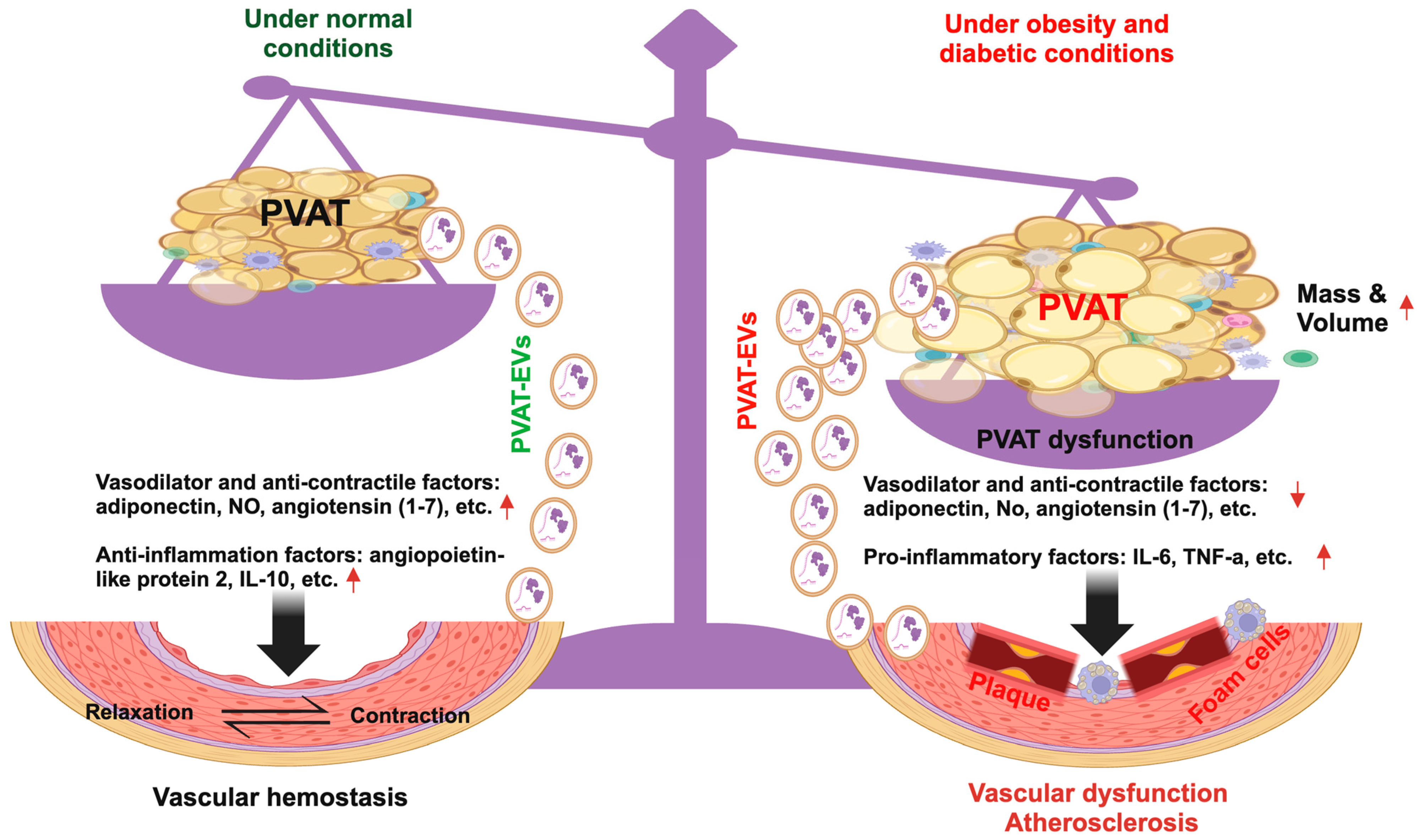
5. PVAT and PVAT-EVs in Obesity and Diabetic-Related Vascular Dysfunction

Obesity is characterized by excessive fat accumulation in adipose tissues leading to weight gain. It is a major risk factor for T2D and contributes to its severity. Diabetes mellitus is a comprehensive term for insulin-related metabolic diseases. Diabetes mellitus affects approximately ten percent of Americans [89] and about 500 million people worldwide. By 2030, this number is estimated to reach 578 million. By 2045, this proportion will reach 700 million [90]. Type 1 diabetes (T1D) is an autoimmune disorder in which beta-cell-induced destruction of insulin incites an insufficiency of the protein. It is typically unpreventable and diagnosed in adolescence [91]. T2D, in contrast, is preventable and often presents as selective insulin resistance resulting from poor diet and exercise choices. Diabetes and its comorbidities, such as obesity and dyslipidemia, induce endothelial dysfunction, inflammation, and blood coagulation. They are implicated in cardiovascular diseases (CVD) like myocardial infarction and cardiomyopathy [92]. In fact, for individuals with T1D or T2D, CVD is the primary cause of death [93]. Those with T1D are more prone to coronary artery disease and peripheral artery disease, while those with T2D are more likely to suffer from atherosclerosis and stroke [94].
Increasing evidence suggests that PVAT undergoes morphological changes in response to obesity and diabetes. A study implementing data from the Framingham Heart Study found that thoracic peri-aortic PVAT is correlated with body mass index and CVD [6]. Wang and colleagues found that the mesenteric PVAT mass increased following 10 weeks of diet-induced obesity in male C57BL6/J mice [95]. Similarly, Juha et al. observed that 8 months of a high-fat diet can increase abdominal aorta PVAT by three-fold in C57BL/6 mice [5]. On a more specific level, white PVAT around the abdominal aorta and iliac arteries increase by 1.9 and 1.7-fold respectively, following a 7-week high-fat diet on Wistar rats [96]. In a monogenic diabetic G protein-coupled estrogen receptor (GPER)-deficient mouse model, 12-month-old mice demonstrated a 3.6-fold increase of PVAT compared with wild-type control [97]. This study also identified PVAT as a novel regulator of arterial vasoconstriction, which acts through the release of adipose-derived contracting factors and likely contributes to increased vascular tone by antagonizing vasodilation. This is also mirrored in the hypertrophy of the adipocytes of abdominal PVAT in obesity conditions [98]. Increased PVAT mass around vasculature is associated with decreased insulin sensitivity [99].
Besides mass changes, PVAT undergoes functional alterations in obesity and diabetic conditions. For instance, obesity can also promote the phenotypic shift of thoracic aorta PVAT from brown to white adipocytes [100]. PVAT in aged, high-fat diet conditions demonstrates increased type 1 macrophage infiltration [101]. Such PVAT dysfunction is a major contributing factor to obesity and diabetic-associated vascular dysfunction. Several groups have observed that a high-fat diet can induce changes in PVAT functions. Bussey et al. have found that the dilation capability of PVAT, acting contrary to administered norepinephrine, is lacking in obese, high-fat diet conditions, and weight loss can reduce the inflammatory conditions of PVAT during obesity in rats [102]. Meanwhile, they found that the inflammatory factor expression was altered in obesity conditions by upregulated TNF-alpha and downregulated eNOS, the latter of which holds a protective function against oxidative stress. Chatterjee and colleagues have reported that a two-week high-fat diet can induce significant downregulations of adiponectin, PPAR-gamma, and FABP4 while upregulating leptin and MIP1-alpha and promoting inflammation in wild-type mice [103]. Almabrouk et al. found that a high-fat diet can inhibit the anticontractile activity of aortic PVAT [104]. Similarly, another study has found that nitric oxide released by PVAT, which in healthy conditions contributes to dilation of the vasculature, is remarkably decreased in obese conditions in a rat obesity model [105]. Intriguingly, the offspring of high-fat diet female mice also demonstrate decreased PVAT anticontractile activity [106]. In T2D conditions, PVAT also exhibits macrophage infiltration and changes to a vasoconstriction phenotype that aggregates endothelial dysfunction [107]. PVAT secretes less adiponectin which directly or indirectly contributes to inflammation, oxidative stress, and insulin resistance. Meijer et al. have revealed that PVAT from obese mice inhibits insulin-induced vasodilation, which could be blocked by targeting the inflammatory JNK pathway [108]. Our group has demonstrated that thoracic PVAT from diabetic mice has lower levels of adiponectin and IL-10, with increased expressions of oxidative stress, IFN-r, TNF-⍺, and IL-6 inflammatory cytokines, which influence aorta function in T2D diabetic mice [37].
The effect of clustered cardiovascular risk factors on PVAT function has also been studied. DeVallance et al. have reported that metabolic syndrome impacts thoracic aorta PVAT function, as evidenced by increased ROS production and pro-inflammatory cytokines, associated with decreased endothelial-dependent dilation function in aorta in obese Zucker rats [109]. Osaki et al. have found that endothelial-dependent relaxation is compromised in a high-fat and high-sucrose diet-induced metabolic syndrome mouse model. The impaired endothelial-dependent relaxation is caused by increased superoxide production from the aorta and PVAT [110]. Similarly, PVAT-derived oxidative stress and inflammation have been implicated in vascular dysfunction in a rat model of metabolic syndrome as reflected by increased ROS production, pro-inflammatory changes, and decreased endothelial-dependent dilation in vasculature [7]. Notably, the altered function of PVAT may be a link between obesity and hypertension, but the detailed mechanisms are incompletely understood. Marchesi and colleagues have reported that the immune cells in PVAT, such as macrophages/monocytes, can contribute to hypertension by administering oxidative stress and related inflammation. They showed that PVAT loses its ability to influence vasodilation in New Zealand obese mice that are predisposed to obesity, hyperglycemia, hyperinsulinemia, and hypertension [7]. Takemoria et al., have demonstrated that the removal of PVAT corresponds with increased blood pressure, alluding to the critical role that PVAT plays in blood pressure regulation [111]. Table 1 summarizes the potential changes of PVAT in obesity, T2D, and metabolic syndrome conditions.
Exercise is a non-pharmacological intervention approach to manage metabolic dysfunctions such as obesity and T2D. Exercise has been shown to restore TNF-⍺ levels, elicit anti-contractile effects and regulate adiponectin release [112]. The influences of exercise intervention in PVAT are multi-faceted. Beyond promoting adiponectin production by PVAT [113], exercise intervention can attenuate immune cell infiltration into PVAT, thereby improving vascular function [114]. Saxton and colleagues have found that exercise reduces PVAT inflammation and increases β3-adrenoceptor and OCT3 expressions to improve PVAT function in obesity [112]. Our group has reported that treadmill exercise can modulate macrophage phenotype, decrease superoxide production in PVAT, and increase NO levels in the aorta in T2D mice [37]. Similar findings show that exercise intervention can increase UCP-1 expression in the mesenteric artery PVAT in rats [115] and increase eNOS expression and phosphorylation in obese rats [113]. Additionally, myokines, such as FGF21 and irisin, released from skeletal muscle can regulate PVAT function via paracrine [114]. These findings suggest the potential of targeting PVAT to treat vascular complications of obesity and diabetes.
Recently, increasing evidence indicates that adipocyte-derived EVs link obesity and diabetes, and their comorbidities. EVs released from adipose tissue-derived cells in obesity or diabetic conditions, including adipocytes and macrophages, play a role in multiple manners, including regulating inflammation, insulin sensitivity, and vasculature functions. Wang et al. have reported that EVs and their cargo microRNAs mediate adipose tissue and brain inter-organ communication, inducing the synaptic damage and cognitive impairment associated with insulin resistance, and thus providing a promising strategy for pharmaceutical interventions for cognitive impairment in diabetes [116]. Ying and colleagues have revealed that obese adipose tissue macrophage-derived EVs containing microRNA-155 can modulate insulin sensitivity [40]. Furthermore, Deng et al. have revealed that intravenous administration of adipocyte-EV-induced systemic insulin resistance in lean mice, leading to altered insulin signaling in visceral adipose tissue, liver, and muscle, and enhancing the recipient animals’ inflammatory profile [10]. Camino and colleagues have reported that the EVs that are shed by pathological adipocytes could spread pathology by stimulating IL-6 and TNFα expression and promoting macrophage inflammation in recipient healthy adipocytes [117]. Circulating adipose-derived EVs have been shown to transport microRNA-99b, which is involved in regulating liver fibroblast growth factor 21, contributing to the control of metabolic homeostasis and systemic insulin resistance [26]. EVs from obese subcutaneous or obese visceral deposits contain reduced levels of vascular endothelial growth factor and matrix metalloproteinase-2, suggesting that they may have lowered pro-angiogenic potential [118]. However, EVs released from adipose tissue-derived stem cells are rich in microRNAs and can promote endothelial cells’ migration and invasion abilities [119]. Hartwig et al. have identified 897 adipokines in adipocyte-derived EVs isolated from lean or overweight women. These adipokines have been found to be strongly associated with human metabolic diseases, including metabolic diseases like diabetes mellitus, glucose metabolism disorders, and metabolic syndrome [120]. Another group has reported that subcutaneous adipocyte-derived EVs of obese patients are enriched in proteins implicated in fatty acid oxidation [121].
Li and colleagues have found that PVAT-EVs from high-fat diet mice carrying microRNA-221–3p could trigger early-stage vascular remodeling in C57BL/6J mice, suggesting the roles of PVAT-EVs in response to obesity-associated inflammation and vascular remodeling [66]. Deng et al. have demonstrated that ob/ob mouse adipose tissue-derived EVs could induce insulin resistance, promote differentiation of bone-marrow-derived monocytes into macrophage, and increase pro-inflammatory cytokine secretion in control mice [10]. Large adipocyte-released EVs can stimulate lipid storage in small adipocytes by mediating the horizontal transfer of lipogenic information to promote lipogenesis and adipocyte hypertrophy [65]. Zhang et al. have found that adipocyte-derived EVs from obese mice induce M1 macrophage phenotype through secreted microRNA-155 [67]. It has been observed that EVs secreted by obese adipose tissue stem cells contain impaired levels of microRNA-126 [122]. As exercise can restore the vasodilation functions of PVAT [37,112], the question of whether exercise could regulate the release and cargo of PVAT-EVs requires further investigation. Additionally, as discussed above, Chang and colleagues have revealed that the functions of PVATs vary from location to location, which is different from classical adipose tissue. Whether exercise intervention affects PVAT differently in different locations requires further investigation.
Overall, our knowledge of PVAT and PVAT-EVs has grown concurrently with the increasing prevalence of obesity and diabetes. Current findings (Table 2) suggest that obesity and diabetes could impact the functions of PVAT and the cargo package of PVAT-EVs. Due to the proximity of PVAT to the associated blood vessels, PVAT and PVAT-EVs could be a novel therapeutic target for interference with obesity and diabetes-related vascular diseases.

6. PVAT and PVAT-EVs in the Pathogenesis of Atherosclerosis

Atherosclerosis, a progressive cardiovascular disease, is characterized by the development of lipid-comprised and fibrous lesions and calcification of the internal walls of the vasculature. The pathogenesis of atherosclerosis is rooted in inflammation. Major risk factors of atherosclerosis include increased blood cholesterol by way of low-density lipoprotein (LDL) collection in vessels, hypertension, illicit substance misuse, and metabolic disorders such as diabetes mellitus [123]. The pathogenesis of atherosclerosis begins with stress applied to the endothelium, which serves as the barrier between vessel interiors and deeper layers, including vascular smooth muscle cells (VSMCs), adventitia, and basement membranes, which further separate vasculature from the interstitial fluid and tissue [124]. When stress is applied to endothelial cells, they can change from flat, long shapes into round shapes, which makes the walls more likely to be constricted, undergo stress, and be prone to lipid uptake and immune cell adhesion [125]. M1 macrophages can upregulate the movement of LDLs into the intima layer, and the cholesterol transported within LDLs promotes M1 macrophages to adopt the foam-cell phenotype. VSMCs can also uptake LDLs to adopt foam cell phenotypes, primarily in the coronary artery. As atherosclerosis progresses, this fatty area becomes known as a necrotic core, which becomes capped by fibers [125]. Continuing disease progression is characterized by the increased apoptosis of macrophages and VSMCs due to oxidative stress and lack of nutrients. Consequently, inflammation prevails, eventually leading to calcification of the necrotic core. If the area ruptures, thrombosis may develop, causing ischemia and damage [125].
Increasingly, studies have demonstrated that PVAT is a double-edged sword. Under physiological conditions, PVAT mainly secretes anti-inflammatory adipokines mediators such as adiponectin, omentin, fibroblast growth factor-21, nitric oxide (NO), PVAT-derived relaxing factors, and IL-10 [126]. Under disease conditions, PVAT secretes a large amount of inflammatory adipokines mediators, such as leptin, tumor necrosis factor-α (TNF-α), monocyte chemoattractant protein-1, interleukin-6 (IL-6), interleukin-1β (IL-1β), etc. [127]. PVAT could elicit favorable influences, such as anti-atherogenic effects [128], stemming from its anti-contractile and anti-inflammatory effects. The selective deletion of PVAT in mice can impair endothelial function and augment atherosclerosis [66], suggesting a protective role of healthy PVAT. On the other hand, in disease conditions like diabetes or obesity, PVAT becomes inflamed and dysfunctional and is implicated in the pathogenesis of atherosclerosis and vascular remodeling [129]. Horimatsu and colleagues have demonstrated that the transplantation of white PVAT isolated from high-fat-fed wild-type mice onto the abdominal aorta of LDL-receptor knock-out mice promotes endothelial dysfunction and atherosclerosis in the remote thoracic aorta [130], suggesting the remote effects of PVAT on the vessel. This study also supports the concept that PVAT does not require direct contact to influence the vasculature, pointing to the potential role of PVAT-EVs.
Accumulating evidence suggests the importance of adipose tissue-derived EVs in preserving cardiovascular physiology. Zhao and colleagues have reported that a period of exercise of 4 weeks can induce the enlargement of brown adipose tissue and alter the microRNA profiles of the adipose tissue released EVs. Furthermore, they have demonstrated that the adipose-tissue-released EVs could regulate cardiomyocyte survival and exhibit exercise cardioprotection in the context of myocardial ischemia/reperfusion injury [131]. Similarly, another study has shown that brown adipose tissue-derived EVs mediate the communication from brown adipose tissue to cardiac myocytes and cardiac fibroblasts. It has been shown that treatment with exosomes from brown adipocytes treated with a β3-AR agonist conferred protection against angiotensin II-induced cardiac remodeling. The authors have further pinpointed iNOS as a critical cargo component of EVs derived from beige adipocytes with β3-AR knockdown, and that it contributes to cardiac fibroblast dysfunction and cardiac remodeling [132]. Additionally, medications such as Ticagrelor, which reversibly targets ADP-mediated G protein-coupled purinergic receptor P2Y12, and has been widely used in patients with acute coronary syndrome and myocardial infarction, could modulate the functions of cardiomyocyte-derived EVs on hyperglycemic cardiomyocytes through the alleviation of oxidative and endoplasmic reticulum stress [133]. However, whether it could affect the texture and cellular proliferation of PVAT and influence the release and function of PVAT-EVs requires more investigation.
Beyond the intercellular communication with cardiomyocytes, it is speculated that the effects elicited by PVAT might be attributed to the crosstalk between PVAT-EVs and cells residing within the vasculature. Earlier studies have demonstrated that EVs travel between adipose tissue and endothelial cells. Flaherty III et al. have found that adipocyte EVs are both an alternative pathway of local lipid release and a mechanism by which parenchymal cells can modulate tissue macrophage differentiation and function [134]. Adipose-derived mesenchymal stem cell-derived exosomes could protect endothelial cells from atherosclerosis, an ability that is ascribed to their carried microRNA-342–5p [135]. Exosomes from adipose-derived stem cells can promote VEGF-C-dependent lymphangiogenesis by directly downregulating Smad7 and regulating TGF-b/Smad signaling in lymphatic endothelial cells [136]. Indeed, several studies have revealed that cardiac lymphatics play important roles in improving cardiac function [137], heart development, and ischemic cardiac disease [138]. Smad7 is one of the major conductors of stem cell cardiogenesis. Downregulation of Smad7 is associated with enhanced cardiogenesis in mouse embryonic and human mesenchymal stem cells [139]. Whether the stem and progenitor cells in PVAT may contribute to cardiogenesis by releasing exosomes that contain factors targeting Smad7 is unclear. Some studies have shown that EVs released from inflammatory adipocytes promote leukocyte attachment to vascular endothelial cells, suggesting the role of EVs in the setting of atherosclerosis [140]. Barberio et al. have found that adipocyte EVs regulate macrophage cholesterol homeostasis by increasing macrophage cholesterol efflux, thereby promoting the development of atherosclerosis [141]. Wang and colleagues have revealed that adipocyte EVs can promote atherosclerosis plaque vulnerability and atherosclerosis by inducing angiogenesis of the vasa vasorum in diabetic atheroprone mice [142]. For PVAT specifically, Mangiferin, a traditional Chinese medicine for diabetes, has been found to stimulate EV release from PVAT, which can improve endothelial cell migration, reduce apoptosis, and prevent inflammation by decreasing IL-6 and TNF-⍺ and blocking NF-κβ [143]. Liu and colleagues have found that PVAT-EVs could regulate the expression of macrophage cholesterol transporters [144]. Furthermore, they have demonstrated that PVAT-EVs can reduce macrophage foam cell formation and that the underlying mechanism is largely ascribed to the EV-carried miR-382–5p [145]. Nevertheless, Xie and colleagues have found that EVs isolated from visceral adipose tissues elicited the opposite effect. These EVs increase macrophage foam cell formation [146]. All of these findings demonstrate the potential roles of PVAT-EVs in vascular health and disease progression (Figure 2).

7. Conclusions and Perspectives

Increasing evidence indicates that PVAT plays an important role in the cardiovascular system. It is a master endocrine organ, much more than a mechanistic support to the vasculature. PVAT releases vaso-relaxing factors and anti-contractile factors to regulate the functions of endothelial and smooth muscle cells in the vasculature. It is worthwhile to note that recent studies have provided hints that PVAT can release EVs, including exosomes and microvesicles, which may either enter circulation or transfer to nearby target cells for intercellular communications. These findings open a window and shed light on the therapeutic potential of targeting PVAT and PVAT-EVs in obesity and diabetic-associated vascular diseases. However, research into PVAT-EVs is still in its infancy. There are many gaps. One of these gaps is the scarcity of studies that comprehensively explain the mechanisms and pathways through which PVAT-EVs influence the progression of different vascular diseases. On the other hand, although increasing studies show that adipose tissue-derived EV profile changes in obesity, diabetes, and vascular disorders, there is limited information on how EV biogenesis pathways are implicated in obesity and diabetic conditions. Meanwhile, how these are altered to influence the release of EVs and their cargo in individuals with obesity or diabetes is also largely unknown. Therefore, a better understanding of how EVs, including PVAT-EV biogenesis pathways, are dysregulated in obesity and diabetic conditions may allow the generation of targeted therapies to perturb EV signaling and develop new EV diagnostic tests.
Given that PVAT becomes dysfunctional in obesity and diabetic conditions, restoring PVAT function could improve cardiometabolic risk, leading to a decreased onset of vascular damage and atherosclerosis development. Physical activity has been shown to restore PVAT function in diabetic animal models. Nevertheless, the question of whether exercise can improve the level and contents of PVAT-EVs and their potential effects on vasculature has not been studied.
Finally, there is a shortage of clinical studies associated with PVAT and PVAT-EVs, as most existing studies utilize mice models. Clinical studies would allow a deeper exploration and understanding of the role of PVAT-EVs in cardiovascular disease. This would enable further research to utilize PVAT-EVs as biomarkers or as therapy for vascular disease, which is a potential future direction of PVAT-EV in vascular disease.

Funding

This work was partially supported by the pilot grant (J.W.) of the National Institute of General Medical Sciences (U54GM104942), the NASA West Virginia Space Grant Consortium (80NSSC20M0055; S.S.), and the American Heart Association (AHA) Career Development Award (935826; J.W.) and Transformational Project Award (24TPA1291189; J.W.).

Conflicts of Interest

The authors declare no conflict of interest.

References

  1. Szasz, T.; Webb, R.C. Perivascular adipose tissue: More than just structural support. Clin. Sci. 2011, 122, 1–12. [Google Scholar] [CrossRef] [PubMed]
  2. Löhn, M.; Dubrovska, G.; Lauterbach, B.; Luft, F.C.; Gollasch, M.; Sharma, A.M. Periadventitial fat releases a vascular relaxing factor. FASEB J. 2002, 16, 1057–1063. [Google Scholar] [CrossRef] [PubMed]
  3. Wang, H.; Li, J.; Wang, Z.; Tian, Y.; Li, C.; Jin, F.; Li, J.; Wang, L. Perivascular brown adipocytes-derived kynurenic acid relaxes blood vessel via endothelium PI3K-Akt-eNOS pathway. Biomed. Pharmacother. 2022, 150, 113040. [Google Scholar] [CrossRef] [PubMed]
  4. Mitidieri, E.; Turnaturi, C.; Vanacore, D.; Sorrentino, R.; Bianca, R.D.d.V. The Role of Perivascular Adipose Tissue-Derived Hydrogen Sulfide in the Control of Vascular Homeostasis. Antioxid. Redox Signal. 2022, 37, 84–97. [Google Scholar] [CrossRef] [PubMed]
  5. Ketonen, J.; Shi, J.; Martonen, E.; Mervaala, E. Periadventitial Adipose Tissue Promotes Endothelial Dysfunction via Oxidative Stress in Diet-Induced Obese C57Bl/6 Mice. Circ. J. 2010, 74, 1479–1487. [Google Scholar] [CrossRef] [PubMed]
  6. Lehman, S.J.; Massaro, J.M.; Schlett, C.L.; O’Donnell, C.J.; Hoffmann, U.; Fox, C.S. Peri-aortic fat, cardiovascular disease risk factors, and aortic calcification: The Framingham Heart Study. Atherosclerosis 2010, 210, 656–661. [Google Scholar] [CrossRef]
  7. Marchesi, C.; Ebrahimian, T.; Angulo, O.; Paradis, P.; Schiffrin, E.L. Endothelial Nitric Oxide Synthase Uncoupling and Perivascular Adipose Oxidative Stress and Inflammation Contribute to Vascular Dysfunction in a Rodent Model of Metabolic Syndrome. Hypertension 2009, 54, 1384–1392. [Google Scholar] [CrossRef]
  8. Fox, C.S.; Massaro, J.M.; Hoffmann, U.; Pou, K.M.; Maurovich-Horvat, P.; Liu, C.Y.; Vasan, R.S.; Murabito, J.M.; Meigs, J.B.; Cupples, L.A.; et al. Abdominal visceral and subcutaneous adipose tissue compartments: Association with metabolic risk factors in the Framingham Heart Study. Circulation 2007, 116, 39–48. [Google Scholar] [CrossRef]
  9. Rosito, G.A.; Massaro, J.M.; Hoffmann, U.; Ruberg, F.L.; Mahabadi, A.A.; Vasan, R.S.; O’Donnell, C.J.; Fox, C.S. Pericardial fat, visceral abdominal fat, cardiovascular disease risk factors, and vascular calcification in a community-based sample: The Framingham Heart Study. Circulation 2008, 117, 605–613. [Google Scholar] [CrossRef]
  10. Deng, Z.-B.; Poliakov, A.; Hardy, R.W.; Clements, R.; Liu, C.; Liu, Y.; Wang, J.; Xiang, X.; Zhang, S.; Zhuang, X.; et al. Adipose Tissue Exosome-Like Vesicles Mediate Activation of Macrophage-Induced Insulin Resistance. Diabetes 2009, 58, 2498–2505. [Google Scholar] [CrossRef]
  11. Ogawa, R.; Tanaka, C.; Sato, M.; Nagasaki, H.; Sugimura, K.; Okumura, K.; Nakagawa, Y.; Aoki, N. Adipocyte-derived microvesicles contain RNA that is transported into macrophages and might be secreted into blood circulation. Biochem. Biophys. Res. Commun. 2010, 398, 723–729. [Google Scholar] [CrossRef] [PubMed]
  12. Doyle, L.; Wang, M. Overview of Extracellular Vesicles, Their Origin, Composition, Purpose, and Methods for Exosome Isolation and Analysis. Cells 2019, 8, 727. [Google Scholar] [CrossRef] [PubMed]
  13. Verlohren, S.; Dubrovska, G.; Tsang, S.-Y.; Essin, K.; Luft, F.C.; Huang, Y.; Gollasch, M. Visceral Periadventitial Adipose Tissue Regulates Arterial Tone of Mesenteric Arteries. Hypertension 2004, 44, 271–276. [Google Scholar] [CrossRef] [PubMed]
  14. Gao, Y.-J. Dual Modulation of Vascular Function by Perivascular Adipose Tissue and Its Potential Correlation with Adiposity/Lipoatrophy-Related Vascular Dysfunction. Curr. Pharm. Des. 2007, 13, 2185–2192. [Google Scholar] [CrossRef] [PubMed]
  15. Padilla, J.; Jenkins, N.T.; Vieira-Potter, V.J.; Laughlin, M.H. Divergent phenotype of rat thoracic and abdominal perivascular adipose tissues. Am. J. Physiol. Integr. Comp. Physiol. 2013, 304, R543–R552. [Google Scholar] [CrossRef] [PubMed]
  16. Bastien, M.; Poirier, P.; Lemieux, I.; Després, J.-P. Overview of Epidemiology and Contribution of Obesity to Cardiovascular Disease. Prog. Cardiovasc. Dis. 2013, 56, 369–381. [Google Scholar] [CrossRef] [PubMed]
  17. Jokinen, R.; Pirnes-Karhu, S.; Pietiläinen, K.H.; Pirinen, E. Adipose tissue NAD+-homeostasis, sirtuins and poly(ADP-ribose) polymerases -important players in mitochondrial metabolism and metabolic health. Redox Biol. 2017, 12, 246–263. [Google Scholar] [CrossRef] [PubMed]
  18. Lizcano, F. The Beige Adipocyte as a Therapy for Metabolic Diseases. Int. J. Mol. Sci. 2019, 20, 5058. [Google Scholar] [CrossRef] [PubMed]
  19. Seale, P.; Conroe, H.M.; Estall, J.; Kajimura, S.; Frontini, A.; Ishibashi, J.; Cohen, P.; Cinti, S.; Spiegelman, B.M. Prdm16 determines the thermogenic program of subcutaneous white adipose tissue in mice. J. Clin. Investig. 2011, 121, 96–105. [Google Scholar] [CrossRef]
  20. Dalal, S. Lipid metabolism in cancer cachexia. Ann. Palliat. Med. 2019, 8, 13–23. [Google Scholar] [CrossRef]
  21. Altshuler-Keylin, S.; Shinoda, K.; Hasegawa, Y.; Ikeda, K.; Hong, H.; Kang, Q.; Yang, Y.; Perera, R.M.; Debnath, J.; Kajimura, S. Beige Adipocyte Maintenance Is Regulated by Autophagy-Induced Mitochondrial Clearance. Cell Metab. 2016, 24, 402–419. [Google Scholar] [CrossRef] [PubMed]
  22. Wu, J.; Boström, P.; Sparks, L.M.; Ye, L.; Choi, J.H.; Giang, A.-H.; Khandekar, M.; Virtanen, K.A.; Nuutila, P.; Schaart, G.; et al. Beige Adipocytes Are a Distinct Type of Thermogenic Fat Cell in Mouse and Human. Cell 2012, 150, 366–376. [Google Scholar] [CrossRef] [PubMed]
  23. Wang, Q.A.; Tao, C.; Jiang, L.; Shao, M.; Ye, R.; Zhu, Y.; Gordillo, R.; Ali, A.; Lian, Y.; Holland, W.L.; et al. Distinct regulatory mechanisms governing embryonic versus adult adipocyte maturation. Nat. Cell Biol. 2015, 17, 1099–1111. [Google Scholar] [CrossRef]
  24. Lynch, F.M.; Withers, S.B.; Yao, Z.; Werner, M.E.; Edwards, G.; Weston, A.H.; Heagerty, A.M. Perivascular adipose tissue-derived adiponectin activates BKCa channels to induce anticontractile responses. Am. J. Physiol.-Heart Circ. Physiol. 2013, 304, H786–H795. [Google Scholar] [CrossRef] [PubMed]
  25. Lee, R.M.; Bader, M.; Alenina, N.; Santos, R.A.; Gao, Y.-J.; Lu, C. Mas receptors in modulating relaxation induced by perivascular adipose tissue. Life Sci. 2011, 89, 467–472. [Google Scholar] [CrossRef] [PubMed]
  26. Thomou, T.; Mori, M.A.; Dreyfuss, J.M.; Konishi, M.; Sakaguchi, M.; Wolfrum, C.; Rao, T.N.; Winnay, J.N.; Garcia-Martin, R.; Grinspoon, S.K.; et al. Adipose-derived circulating miRNAs regulate gene expression in other tissues. Yearb. Paediatr. Endocrinol. 2018, 15, 11–13. [Google Scholar] [CrossRef] [PubMed]
  27. Antoniades, C.; Tousoulis, D.; Vavlukis, M.; Fleming, I.; Duncker, D.J.; Eringa, E.; Manfrini, O.; Antonopoulos, A.S.; Oikonomou, E.; Padró, T.; et al. Perivascular adipose tissue as a source of therapeutic targets and clinical biomarkers. Eur. Heart J. 2023, 44, 3827–3844. [Google Scholar] [CrossRef] [PubMed]
  28. Cheng, C.K.; Ding, H.; Jiang, M.; Yin, H.; Gollasch, M.; Huang, Y. Perivascular adipose tissue: Fine-tuner of vascular redox status and inflammation. Redox Biol. 2023, 62, 102683. [Google Scholar] [CrossRef] [PubMed]
  29. Ryo, M.; Nakamura, T.; Kihara, S.; Kumada, M.; Shibazaki, S.; Takahashi, M.; Nagai, M.; Matsuzawa, Y.; Funahashi, T. Adiponectin as a Biomarker of the Metabolic Syndrome. Circ. J. 2004, 68, 975–981. [Google Scholar] [CrossRef]
  30. Ahl, S.; Guenther, M.; Zhao, S.; James, R.; Marks, J.; Szabo, A.; Kidambi, S. Adiponectin Levels Differentiate Metabolically Healthy vs Unhealthy Among Obese and Nonobese White Individuals. J. Clin. Endocrinol. Metab. 2015, 100, 4172–4180. [Google Scholar] [CrossRef]
  31. Drolet, R.; Bélanger, C.; Fortier, M.; Huot, C.; Mailloux, J.; Légaré, D.; Tchernof, A. Fat Depot-specific Impact of Visceral Obesity on Adipocyte Adiponectin Release in Women. Obesity 2009, 17, 424–430. [Google Scholar] [CrossRef] [PubMed]
  32. Okamoto, Y.; Kihara, S.; Ouchi, N.; Nishida, M.; Arita, Y.; Kumada, M.; Ohashi, K.; Sakai, N.; Shimomura, I.; Kobayashi, H.; et al. Adiponectin Reduces Atherosclerosis in Apolipoprotein E-Deficient Mice. Circulation 2002, 106, 2767–2770. [Google Scholar] [CrossRef] [PubMed]
  33. Yamauchi, T.; Kamon, J.; Ito, Y.; Tsuchida, A.; Yokomizo, T.; Kita, S.; Sugiyama, T.; Miyagishi, M.; Hara, K.; Tsunoda, M.; et al. Cloning of adiponectin receptors that mediate antidiabetic metabolic effects. Nature 2003, 423, 762–769. [Google Scholar] [CrossRef] [PubMed]
  34. Villacorta, L.; Chang, L. The role of perivascular adipose tissue in vasoconstriction, arterial stiffness, and aneurysm. Horm. Mol. Biol. Clin. Investig. 2015, 21, 137–147. [Google Scholar] [CrossRef] [PubMed]
  35. Turaihi, A.H.; Serné, E.H.; Molthoff, C.F.; Koning, J.J.; Knol, J.; Niessen, H.W.; Goumans, M.J.T.; van Poelgeest, E.M.; Yudkin, J.S.; Smulders, Y.M.; et al. Perivascular Adipose Tissue Controls Insulin-Stimulated Perfusion, Mitochondrial Protein Expression, and Glucose Uptake in Muscle Through Adipomuscular Arterioles. Diabetes 2020, 69, 603–613. [Google Scholar] [CrossRef] [PubMed]
  36. Adachi, Y.; Ueda, K.; Nomura, S.; Ito, K.; Katoh, M.; Katagiri, M.; Yamada, S.; Hashimoto, M.; Zhai, B.; Numata, G.; et al. Beiging of perivascular adipose tissue regulates its inflammation and vascular remodeling. Nat. Commun. 2022, 13, 5117. [Google Scholar] [CrossRef] [PubMed]
  37. Wang, J.; Polaki, V.; Chen, S.; Bihl, J.C. Exercise Improves Endothelial Function Associated with Alleviated Inflammation and Oxidative Stress of Perivascular Adipose Tissue in Type 2 Diabetic Mice. Oxidative Med. Cell. Longev. 2020, 2020, 8830537. [Google Scholar] [CrossRef] [PubMed]
  38. El Khoudary, S.R.; Shields, K.J.; Janssen, I.; Hanley, C.; Budoff, M.J.; Barinas-Mitchell, E.; Everson-Rose, S.A.; Powell, L.H.; Matthews, K.A. Cardiovascular Fat, Menopause, and Sex Hormones in Women: The SWAN Cardiovascular Fat Ancillary Study. J. Clin. Endocrinol. Metab. 2015, 100, 3304–3312. [Google Scholar] [CrossRef]
  39. Victorio, J.A.; da Costa, R.M.; Tostes, R.C.; Davel, A.P. Modulation of Vascular Function by Perivascular Adipose Tissue: Sex Differences. Curr. Pharm. Des. 2020, 26, 3768–3777. [Google Scholar] [CrossRef]
  40. Ying, W.; Riopel, M.; Bandyopadhyay, G.; Dong, Y.; Birmingham, A.; Seo, J.B.; Ofrecio, J.M.; Wollam, J.; Hernandez-Carretero, A.; Fu, W.; et al. Adipose Tissue Macrophage-Derived Exosomal miRNAs Can Modulate In Vivo and In Vitro Insulin Sensitivity. Cell 2017, 171, 372–384.e12. [Google Scholar] [CrossRef]
  41. Pan, Y.; Hui, X.; Hoo, R.L.C.; Ye, D.; Chan, C.Y.C.; Feng, T.; Wang, Y.; Lam, K.S.L.; Xu, A. Adipocyte-secreted exosomal microRNA-34a inhibits M2 macrophage polarization to promote obesity-induced adipose inflammation. J. Clin. Investig. 2019, 129, 834–849. [Google Scholar] [CrossRef] [PubMed]
  42. Zhao, H.; Shang, Q.; Pan, Z.; Bai, Y.; Li, Z.; Zhang, H.; Zhang, Q.; Guo, C.; Zhang, L.; Wang, Q. Exosomes From Adipose-Derived Stem Cells Attenuate Adipose Inflammation and Obesity Through Polarizing M2 Macrophages and Beiging in White Adipose Tissue. Diabetes 2018, 67, 235–247. [Google Scholar] [CrossRef] [PubMed]
  43. Busatto, S.; Morad, G.; Guo, P.; Moses, M.A. The role of extracellular vesicles in the physiological and pathological regulation of the blood-brain barrier. FASEB BioAdv. 2021, 3, 665–675. [Google Scholar] [CrossRef] [PubMed]
  44. Hessvik, N.P.; Llorente, A. Current knowledge on exosome biogenesis and release. Cell. Mol. Life Sci. 2018, 75, 193–208. [Google Scholar] [CrossRef] [PubMed]
  45. Gangadaran, P.; Madhyastha, H.; Madhyastha, R.; Rajendran, R.L.; Nakajima, Y.; Watanabe, N.; Velikkakath, A.K.G.; Hong, C.M.; Gopi, R.V.; Muthukalianan, G.K.; et al. The emerging role of exosomes in innate immunity, diagnosis and therapy. Front. Immunol. 2023, 13, 1085057. [Google Scholar] [CrossRef] [PubMed]
  46. Raposo, G.; Stoorvogel, W. Extracellular vesicles: Exosomes, microvesicles, and friends. J. Cell Biol. 2013, 200, 373–383. [Google Scholar] [CrossRef]
  47. Ju, Y.; Bai, H.; Ren, L.; Zhang, L. The Role of Exosome and the ESCRT Pathway on Enveloped Virus Infection. Int. J. Mol. Sci. 2021, 22, 9060. [Google Scholar] [CrossRef] [PubMed]
  48. Baietti, M.F.; Zhang, Z.; Mortier, E.; Melchior, A.; Degeest, G.; Geeraerts, A.; Ivarsson, Y.; Depoortere, F.; Coomans, C.; Vermeiren, E.; et al. Syndecan-syntenin-ALIX regulates the biogenesis of exosomes. Nat. Cell Biol. 2012, 14, 677–685. [Google Scholar] [CrossRef]
  49. Colombo, M.; Raposo, G.; Théry, C. Biogenesis, secretion, and intercellular interactions of exosomes and other extracellular vesicles. Annu. Rev. Cell Dev. Biol. 2014, 30, 255–289. [Google Scholar] [CrossRef]
  50. György, B.; Szabó, T.G.; Pásztói, M.; Pál, Z.; Misják, P.; Aradi, B.; László, V.; Pállinger, É.; Pap, E.; Kittel, Á.; et al. Membrane vesicles, current state-of-the-art: Emerging role of extracellular vesicles. Cell. Mol. Life Sci. 2011, 68, 2667–2688. [Google Scholar] [CrossRef]
  51. Morel, O.; Jesel, L.; Freyssinet, J.-M.; Toti, F. Cellular Mechanisms Underlying the Formation of Circulating Microparticles. Arterioscler. Thromb. Vasc. Biol. 2011, 31, 15–26. [Google Scholar] [CrossRef] [PubMed]
  52. Muralidharan-Chari, V.; Clancy, J.W.; Sedgwick, A.; D’Souza-Schorey, C. Microvesicles: Mediators of extracellular communication during cancer progression. J. Cell Sci. 2010, 123, 1603–1611. [Google Scholar] [CrossRef] [PubMed]
  53. Zwaal, R.F.; Schroit, A.J. Pathophysiologic Implications of Membrane Phospholipid Asymmetry in Blood Cells. Blood 1997, 89, 1121–1132. [Google Scholar] [CrossRef] [PubMed]
  54. Chernomordik, L.V.; Melikyan, G.B.; Chizmadzhev, Y.A. Biomembrane fusion: A new concept derived from model studies using two interacting planar lipid bilayers. Biochim. Biophys. Acta. BBA Rev. Biomembr. 1987, 906, 309–352. [Google Scholar] [CrossRef] [PubMed]
  55. Prada, I.; Meldolesi, J. Binding and Fusion of Extracellular Vesicles to the Plasma Membrane of Their Cell Targets. Int. J. Mol. Sci. 2016, 17, 1296. [Google Scholar] [CrossRef] [PubMed]
  56. Guan, S.; Li, Q.; Liu, P.; Xuan, X.; Du, Y. Experimental immunology Umbilical cord blood-derived dendritic cells loaded with BGC823 tumor antigens and DC-derived exosomes stimulate efficient cytotoxic T-lymphocyte responses and antitumor immunity in vitro and in vivo. Cent. Eur. J. Immunol. 2014, 2, 142–151. [Google Scholar] [CrossRef] [PubMed]
  57. Tian, T.; Zhu, Y.-L.; Zhou, Y.-Y.; Liang, G.-F.; Wang, Y.-Y.; Hu, F.-H.; Xiao, Z.-D. Exosome Uptake through Clathrin-mediated Endocytosis and Macropinocytosis and Mediating miR-21 Delivery. J. Biol. Chem. 2014, 289, 22258–22267. [Google Scholar] [CrossRef] [PubMed]
  58. Zheng, Y.; Tu, C.; Zhang, J.; Wang, J. Inhibition of multiple myeloma-derived exosomes uptake suppresses the functional response in bone marrow stromal cell. Int. J. Oncol. 2019, 54, 1061–1070. [Google Scholar] [CrossRef] [PubMed]
  59. Barile, L.; Lionetti, V.; Cervio, E.; Matteucci, M.; Gherghiceanu, M.; Popescu, L.M.; Torre, T.; Siclari, F.; Moccetti, T.; Vassalli, G. Extracellular vesicles from human cardiac progenitor cells inhibit cardiomyocyte apoptosis and improve cardiac function after myocardial infarction. Cardiovasc. Res. 2014, 103, 530–541. [Google Scholar] [CrossRef]
  60. Papini, G.; Furini, G.; Matteucci, M.; Biemmi, V.; Casieri, V.; Di Lascio, N.; Milano, G.; Chincoli, L.R.; Faita, F.; Barile, L.; et al. Cardiomyocyte-targeting exosomes from sulforaphane-treated fibroblasts affords cardioprotection in infarcted rats. J. Transl. Med. 2023, 21, 313. [Google Scholar] [CrossRef]
  61. Lionetti, V.; Tuana, B.S.; Casieri, V.; Parikh, M.; Pierce, G.N. Importance of functional food compounds in cardioprotection through action on the epigenome. Eur. Heart J. 2018, 40, 575–582. [Google Scholar] [CrossRef] [PubMed]
  62. Contreras, G.A.; Thelen, K.; Ayala-Lopez, N.; Watts, S.W. The distribution and adipogenic potential of perivascular adipose tissue adipocyte progenitors is dependent on sexual dimorphism and vessel location. Physiol. Rep. 2016, 4, e12993. [Google Scholar] [CrossRef] [PubMed]
  63. Ginini, L.; Billan, S.; Fridman, E.; Gil, Z. Insight into Extracellular Vesicle-Cell Communication: From Cell Recognition to Intracellular Fate. Cells 2022, 11, 1375. [Google Scholar] [CrossRef] [PubMed]
  64. Pitt, J.M.; Kroemer, G.; Zitvogel, L. Extracellular vesicles: Masters of intercellular communication and potential clinical interventions. J. Clin. Investig. 2016, 126, 1139–1143. [Google Scholar] [CrossRef] [PubMed]
  65. Müller, G.; Schneider, M.; Biemer-Daub, G.; Wied, S. Microvesicles released from rat adipocytes and harboring glycosylphosphatidylinositol-anchored proteins transfer RNA stimulating lipid synthesis. Cell. Signal. 2011, 23, 1207–1223. [Google Scholar] [CrossRef] [PubMed]
  66. Li, X.; Ballantyne, L.L.; Yu, Y.; Funk, C.D. Perivascular adipose tissue-derived extracellular vesicle miR-221-3p mediates vascular remodeling. FASEB J. 2019, 33, 12704–12722. [Google Scholar] [CrossRef] [PubMed]
  67. Zhang, Y.; Mei, H.; Chang, X.; Chen, F.; Zhu, Y.; Han, X. Adipocyte-derived microvesicles from obese mice induce M1 macrophage phenotype through secreted miR-155. J. Mol. Cell Biol. 2016, 8, 505–517. [Google Scholar] [CrossRef] [PubMed]
  68. Guo, J.; Cui, B.; Zheng, J.; Yu, C.; Zheng, X.; Yi, L.; Zhang, S.; Wang, K. Platelet-derived microparticles and their cargos: The past, present and future. Asian J. Pharm. Sci. 2024, 19, 100907. [Google Scholar] [CrossRef] [PubMed]
  69. Owens, A.P., 3rd; Mackman, N. Microparticles in Hemostasis and Thrombosis. Circ. Res. 2011, 108, 1284–1297. [Google Scholar] [CrossRef]
  70. Essola, J.M.; Zhang, M.; Yang, H.; Li, F.; Xia, B.; Mavoungou, J.F.; Hussain, A.; Huang, Y. Exosome regulation of immune response mechanism: Pros and cons in immunotherapy. Bioact. Mater. 2024, 32, 124–146. [Google Scholar] [CrossRef]
  71. Ling, Z.L.; Combes, V.; Grau, G.E.; King, N.J.C. Microparticles as Immune Regulators in Infectious Disease ? An Opinion. Front. Immunol. 2011, 2, 67. [Google Scholar] [CrossRef] [PubMed]
  72. Kahlert, C.; Kalluri, R. Exosomes in tumor microenvironment influence cancer progression and metastasis. J. Mol. Med. 2013, 91, 431–437. [Google Scholar] [CrossRef] [PubMed]
  73. Rak, J. Extracellular Vesicles-Biomarkers and Effectors of the Cellular Interactome in Cancer. Front. Pharmacol. 2013, 4, 21. [Google Scholar] [CrossRef] [PubMed]
  74. Soria, F.N.; Pampliega, O.; Bourdenx, M.; Meissner, W.G.; Bezard, E.; Dehay, B. Exosomes, an Unmasked Culprit in Neurodegenerative Diseases. Front. Neurosci. 2017, 11, 26. [Google Scholar] [CrossRef] [PubMed]
  75. Wu, K.; Yang, Y.; Zhong, Y.; Ammar, H.M.; Zhang, P.; Guo, R.; Liu, H.; Cheng, C.F.; Koroscil, T.M.; Chen, Y.F. The effects of microvesicles on endothelial progenitor cells are compromised in type 2 diabetic patients via downregulation of the miR-126/VEGFR2 pathway. Am. J. Physiol.-Endocrinol. Metab. 2016, 310, E828–E837. [Google Scholar] [CrossRef] [PubMed]
  76. Chen, S.; Sigdel, S.; Sawant, H.; Bihl, J.; Wang, J. Exercise-Intervened Endothelial Progenitor Cell Exosomes Protect N2a Cells by Improving Mitochondrial Function. Int. J. Mol. Sci. 2024, 25, 1148. [Google Scholar] [CrossRef] [PubMed]
  77. Rani, S.; Ryan, A.E.; Griffin, M.D.; Ritter, T. Mesenchymal Stem Cell-derived Extracellular Vesicles: Toward Cell-free Therapeutic Applications. Mol. Ther. 2015, 23, 812–823. [Google Scholar] [CrossRef] [PubMed]
  78. McLaughlin, C.; Datta, P.; Singh, Y.P.; Lo, A.; Horchler, S.; Elcheva, I.A.; Ozbolat, I.T.; Ravnic, D.J.; Koduru, S.V. Mesenchymal Stem Cell-Derived Extracellular Vesicles for Therapeutic Use and in Bioengineering Applications. Cells 2022, 11, 3366. [Google Scholar] [CrossRef] [PubMed]
  79. Shekari, F.; Meyfour, A.; Davies, O.G.; Velot, É. Editorial: Mesenchymal stem cell-derived extracellular vesicles: Considerations and therapeutic applications. Front. Cell Dev. Biol. 2024, 12, 1377197. [Google Scholar] [CrossRef] [PubMed]
  80. Chen, D.-X.; Lu, C.-H.; Na, N.; Yin, R.-X.; Huang, F. Endothelial progenitor cell-derived extracellular vesicles: The world of potential prospects for the treatment of cardiovascular diseases. Cell Biosci. 2024, 14, 72. [Google Scholar] [CrossRef]
  81. Zou, L.; Ma, X.; Lin, S.; Wu, B.; Chen, Y.; Peng, C. Bone marrow mesenchymal stem cell-derived exosomes protect against myocardial infarction by promoting autophagy. Exp. Ther. Med. 2019, 18, 2574–2582. [Google Scholar] [CrossRef] [PubMed]
  82. Meziani, F.; Tesse, A.; Andriantsitohaina, R. Microparticles are vectors of paradoxical information in vascular cells including the endothelium: Role in health and diseases. Pharmacol. Rep. 2008, 60, 75–84. [Google Scholar] [PubMed]
  83. Tang, H.; Luo, H.; Zhang, Z.; Yang, D. Mesenchymal Stem Cell-Derived Apoptotic Bodies: Biological Functions and Therapeutic Potential. Cells 2022, 11, 3879. [Google Scholar] [CrossRef]
  84. Elmore, S. Apoptosis: A review of programmed cell death. Toxicol. Pathol. 2007, 35, 495–516. [Google Scholar] [CrossRef] [PubMed]
  85. Wickman, G.R.; Julian, L.; Mardilovich, K.; Schumacher, S.; Munro, J.; Rath, N.; AL Zander, S.; Mleczak, A.; Sumpton, D.; Morrice, N.; et al. Blebs produced by actin-myosin contraction during apoptosis release damage-associated molecular pattern proteins before secondary necrosis occurs. Cell Death Differ. 2013, 20, 1293–1305. [Google Scholar] [CrossRef]
  86. Arandjelovic, S.; Ravichandran, K.S. Phagocytosis of apoptotic cells in homeostasis. Nat. Immunol. 2015, 16, 907–917. [Google Scholar] [CrossRef]
  87. Trahtemberg, U.; Mevorach, D. Apoptotic Cells Induced Signaling for Immune Homeostasis in Macrophages and Dendritic Cells. Front. Immunol. 2017, 8, 1356. [Google Scholar] [CrossRef] [PubMed]
  88. Kalra, H.; Drummen, G.P.C.; Mathivanan, S. Focus on Extracellular Vesicles: Introducing the Next Small Big Thing. Int. J. Mol. Sci. 2016, 17, 170. [Google Scholar] [CrossRef] [PubMed]
  89. Schmidt, A.M. Highlighting diabetes mellitus: The epidemic continues. Arterioscler. Thromb. Vasc. Biol. 2018, 38, e1–e8. [Google Scholar] [CrossRef]
  90. Saeedi, P. A high number of people have CVD, but do not know it. Diabetes Res. Clin. Pract. 2019, 148, 270–271. [Google Scholar] [CrossRef]
  91. Petersmann, A.; Müller-Wieland, D.; Müller, U.A.; Landgraf, R.; Nauck, M.; Freckmann, G.; Heinemann, L.; Schleicher, E. Definition, Classification and Diagnosis of Diabetes Mellitus. Exp. Clin. Endocrinol. Diabetes 2019, 127, S1–S7. [Google Scholar] [CrossRef] [PubMed]
  92. Leon, B.M.; Maddox, T.M. Diabetes and cardiovascular disease: Epidemiology, biological mechanisms, treatment recommendations and future research. World J. Diabetes 2015, 6, 1246–1258. [Google Scholar] [CrossRef] [PubMed]
  93. Joseph, J.J.; Deedwania, P.; Acharya, T.; Aguilar, D.; Bhatt, D.L.; Chyun, D.A.; Di Palo, K.E.; Golden, S.H.; Sperling, L.S. Comprehensive Management of Cardiovascular Risk Factors for Adults With Type 2 Diabetes: A Scientific Statement From the American Heart Association. Circulation 2022, 145, E722–E759. [Google Scholar] [CrossRef] [PubMed]
  94. Chen, R.; Ovbiagele, B.; Feng, W. Diabetes and Stroke: Epidemiology, Pathophysiology, Pharmaceuticals and Outcomes. Am. J. Med. Sci. 2016, 351, 380–386. [Google Scholar] [CrossRef] [PubMed]
  95. Wang, H.; Luo, W.; Wang, J.; Guo, C.; Wang, X.; Wolffe, S.L.; Bodary, P.F.; Eitzman, D.T. Obesity-Induced Endothelial Dysfunction Is Prevented by Deficiency of P-Selectin Glycoprotein Ligand-1. Diabetes 2012, 61, 3219–3227. [Google Scholar] [CrossRef] [PubMed]
  96. Henrichot, E.; Juge-Aubry, C.E.; Pernin, A.; Pache, J.C.; Velebit, V.; Dayer, J.M.; Meda, P.; Chizzolini, C.; Meier, C.A. Production of chemokines by perivascular adipose tissue: A role in the pathogenesis of atherosclerosis? Arterioscler. Thromb. Vasc. Biol. 2005, 25, 2594–2599. [Google Scholar] [CrossRef] [PubMed]
  97. Meyer, M.R.; Fredette, N.C.; Barton, M.; Prossnitz, E.R. Regulation of Vascular Smooth Muscle Tone by Adipose-Derived Contracting Factor. PLoS ONE 2013, 8, e79245. [Google Scholar] [CrossRef] [PubMed]
  98. Duregotti, E.; Reumiller, C.M.; Mayr, U.; Hasman, M.; Schmidt, L.E.; Burnap, S.A.; Theofilatos, K.; Barallobre-Barreiro, J.; Beran, A.; Grandoch, M.; et al. Reduced secretion of neuronal growth regulator 1 contributes to impaired adipose-neuronal crosstalk in obesity. Nat. Commun. 2022, 13, 7269. [Google Scholar] [CrossRef] [PubMed]
  99. Rittig, K.; Staib, K.; Machann, J.; Böttcher, M.; Peter, A.; Schick, F.; Claussen, C.; Stefan, N.; Fritsche, A.; Häring, H.-U.; et al. Perivascular fatty tissue at the brachial artery is linked to insulin resistance but not to local endothelial dysfunction. Diabetologia 2008, 51, 2093–2099. [Google Scholar] [CrossRef]
  100. Police, S.B.; Thatcher, S.E.; Charnigo, R.; Daugherty, A.; Cassis, L.A. Obesity Promotes Inflammation in Periaortic Adipose Tissue and Angiotensin II-Induced Abdominal Aortic Aneurysm Formation. Arterioscler. Thromb. Vasc. Biol. 2009, 29, 1458–1464. [Google Scholar] [CrossRef]
  101. Bailey-Downs, L.C.; Tucsek, Z.; Toth, P.; Sosnowska, D.; Gautam, T.; Sonntag, W.E.; Csiszar, A.; Ungvari, Z. Aging exacerbates obesity-induced oxidative stress and inflammation in perivascular adipose tissue in mice: A paracrine mechanism contributing to vascular redox dysregulation and inflammation. J. Gerontol. Ser. A Biomed. Sci. Med Sci. 2013, 68, 780–792. [Google Scholar] [CrossRef]
  102. Bussey, C.E.; Withers, S.B.; Aldous, R.G.; Edwards, G.; Heagerty, A.M. Obesity-Related Perivascular Adipose Tissue Damage Is Reversed by Sustained Weight Loss in the Rat. Arterioscler. Thromb. Vasc. Biol. 2016, 36, 1377–1385. [Google Scholar] [CrossRef] [PubMed]
  103. Chatterjee, T.K.; Stoll, L.L.; Denning, G.M.; Harrelson, A.; Blomkalns, A.L.; Idelman, G.; Rothenberg, F.G.; Neltner, B.; Romig-Martin, S.A.; Dickson, E.W.; et al. Proinflammatory phenotype of perivascular adipocytes: Influence of high-fat feeding. Circ. Res. 2009, 104, 541–549. [Google Scholar] [CrossRef] [PubMed]
  104. Almabrouk, T.A.M.; White, A.D.; Ugusman, A.B.; Skiba, D.S.; Katwan, O.J.; Alganga, H.; Guzik, T.J.; Touyz, R.M.; Salt, I.P.; Kennedy, S. High Fat Diet Attenuates the Anticontractile Activity of Aortic PVAT via a Mechanism Involving AMPK and Reduced Adiponectin Secretion. Front. Physiol. 2018, 9, 51. [Google Scholar] [CrossRef] [PubMed]
  105. Aghamohammadzadeh, R.; Unwin, R.D.; Greenstein, A.S.; Heagerty, A.M. Effects of Obesity on Perivascular Adipose Tissue Vasorelaxant Function: Nitric Oxide, Inflammation and Elevated Systemic Blood Pressure. J. Vasc. Res. 2015, 52, 299–305. [Google Scholar] [CrossRef] [PubMed]
  106. Zaborska, K.E.; Wareing, M.; Edwards, G.; Austin, C. Loss of anti-contractile effect of perivascular adipose tissue in offspring of obese rats. Int. J. Obes. 2016, 40, 1205–1214. [Google Scholar] [CrossRef] [PubMed]
  107. Azul, L.; Leandro, A.; Boroumand, P.; Klip, A.; Seiça, R.; Sena, C.M. Increased inflammation, oxidative stress and a reduction in antioxidant defense enzymes in perivascular adipose tissue contribute to vascular dysfunction in type 2 diabetes. Free Radic. Biol. Med. 2019, 146, 264–274. [Google Scholar] [CrossRef] [PubMed]
  108. Meijer, R.I.; Bakker, W.; Alta, C.-L.A.; Sipkema, P.; Yudkin, J.S.; Viollet, B.; Richter, E.A.; Smulders, Y.M.; van Hinsbergh, V.W.; Serné, E.H.; et al. Perivascular Adipose Tissue Control of Insulin-Induced Vasoreactivity in Muscle Is Impaired in db/db Mice. Diabetes 2013, 62, 590–598. [Google Scholar] [CrossRef] [PubMed]
  109. DeVallance, E.; Branyan, K.W.; Lemaster, K.; Olfert, I.M.; Smith, D.M.; Pistilli, E.E.; Frisbee, J.C.; Chantler, P.D. Aortic dysfunction in metabolic syndrome mediated by perivascular adipose tissue TNFα-and NOX2-dependent pathway. Exp. Physiol. 2018, 103, 590–603. [Google Scholar] [CrossRef]
  110. Osaki, A.; Kagami, K.; Ishinoda, Y.; Sato, A.; Kimura, T.; Horii, S.; Ito, K.; Toya, T.; Ido, Y.; Namba, T.; et al. Reactive Oxygen Species in the Aorta and Perivascular Adipose Tissue Precedes Endothelial Dysfunction in the Aorta of Mice with a High-Fat High-Sucrose Diet and Additional Factors. Int. J. Mol. Sci. 2023, 24, 6486. [Google Scholar] [CrossRef]
  111. Takemori, K.; Gao, Y.-J.; Ding, L.; Lu, C.; Su, L.-Y.; An, W.-S.; Vinson, C.; Lee, R.M. Elevated Blood Pressure in Transgenic Lipoatrophic Mice and Altered Vascular Function. Hypertension 2007, 49, 365–372. [Google Scholar] [CrossRef] [PubMed]
  112. Saxton, S.N.; Toms, L.K.; Aldous, R.G.; Withers, S.B.; Ohanian, J.; Heagerty, A.M. Restoring Perivascular Adipose Tissue Function in Obesity Using Exercise. Cardiovasc. Drugs Ther. 2021, 35, 1291–1304. [Google Scholar] [CrossRef]
  113. Meziat, C.; Boulghobra, D.; Strock, E.; Battault, S.; Bornard, I.; Walther, G.; Reboul, C. Exercise training restores eNOS activation in the perivascular adipose tissue of obese rats: Impact on vascular function. Nitric Oxide 2019, 86, 63–67. [Google Scholar] [CrossRef] [PubMed]
  114. Boa, B.C.S.; Yudkin, J.S.; van Hinsbergh, V.W.M.; Bouskela, E.; Eringa, E.C. Exercise effects on perivascular adipose tissue: Endocrine and paracrine determinants of vascular function. Br. J. Pharmacol. 2017, 174, 3466–3481. [Google Scholar] [CrossRef]
  115. Liao, J.; Yin, H.; Huang, J.; Hu, M. Dysfunction of perivascular adipose tissue in mesenteric artery is restored by aerobic exercise in high-fat diet induced obesity. Clin. Exp. Pharmacol. Physiol. 2021, 48, 697–703. [Google Scholar] [CrossRef]
  116. Wang, J.; Li, L.; Zhang, Z.; Zhang, X.; Zhu, Y.; Zhang, C.; Bi, Y. Extracellular vesicles mediate the communication of adipose tissue with brain and promote cognitive impairment associated with insulin resistance. Cell Metab. 2022, 34, 1264–1279.e8. [Google Scholar] [CrossRef]
  117. Camino, T.; Lago-Baameiro, N.; Bravo, S.B.; Sueiro, A.; Couto, I.; Santos, F.; Baltar, J.; Casanueva, F.F.; Pardo, M. Vesicles Shed by Pathological Murine Adipocytes Spread Pathology: Characterization and Functional Role of Insulin Resistant/Hypertrophied Adiposomes. Int. J. Mol. Sci. 2020, 21, 2252. [Google Scholar] [CrossRef]
  118. Gao, X.; Salomon, C.; Freeman, D.J. Extracellular vesicles from adipose tissue—A potential role in obesity and type 2 diabetes? Front. Endocrinol. 2017, 8, 202. [Google Scholar] [CrossRef] [PubMed]
  119. Huang, B.; Huang, L.-F.; Zhao, L.; Zeng, Z.; Wang, X.; Cao, D.; Yang, L.; Ye, Z.; Chen, X.; Liu, B.; et al. Microvesicles (MIVs) secreted from adipose-derived stem cells (ADSCs) contain multiple microRNAs and promote the migration and invasion of endothelial cells. Genes Dis. 2020, 7, 225–234. [Google Scholar] [CrossRef]
  120. Hartwig, S.; De Filippo, E.; Göddeke, S.; Knebel, B.; Kotzka, J.; Al-Hasani, H.; Roden, M.; Lehr, S.; Sell, H. Exosomal proteins constitute an essential part of the human adipose tissue secretome. Biochim. Biophys. Acta. BBA Proteins Proteom. 2018, 1867, 140172. [Google Scholar] [CrossRef]
  121. Lazar, I.; Clement, E.; Dauvillier, S.; Milhas, D.; Ducoux-Petit, M.; LeGonidec, S.; Moro, C.; Soldan, V.; Dalle, S.; Balor, S.; et al. Adipocyte Exosomes Promote Melanoma Aggressiveness through Fatty Acid Oxidation: A Novel Mechanism Linking Obesity and Cancer. Cancer Res. 2016, 76, 4051–4057. [Google Scholar] [CrossRef] [PubMed]
  122. Togliatto, G.; Dentelli, P.; Gili, M.; Gallo, S.; Deregibus, C.; Biglieri, E.; Iavello, A.; Santini, E.; Rossi, C.; Solini, A.; et al. Obesity reduces the pro-angiogenic potential of adipose tissue stem cell-derived extracellular vesicles (EVs) by impairing miR-126 content: Impact on clinical applications. Int. J. Obes. 2016, 40, 102–111. [Google Scholar] [CrossRef] [PubMed]
  123. Kong, P.; Cui, Z.-Y.; Huang, X.-F.; Zhang, D.-D.; Guo, R.-J.; Han, M. Inflammation and atherosclerosis: Signaling pathways and therapeutic intervention. Signal Transduct. Target. Ther. 2022, 7, 131. [Google Scholar] [CrossRef] [PubMed]
  124. Morrison, A.M.; Sullivan, A.E.; Aday, A.W. Atherosclerotic disease: Pathogenesis and approaches to management. Med. Clin. 2023, 107, 793–805. [Google Scholar] [CrossRef]
  125. Jebari-Benslaiman, S.; Galicia-García, U.; Larrea-Sebal, A.; Olaetxea, J.R.; Alloza, I.; Vandenbroeck, K.; Benito-Vicente, A.; Martín, C. Pathophysiology of Atherosclerosis. Int. J. Mol. Sci. 2022, 23, 3346. [Google Scholar] [CrossRef] [PubMed]
  126. Chen, Y.; Qin, Z.; Wang, Y.; Li, X.; Zheng, Y.; Liu, Y. Role of Inflammation in Vascular Disease-Related Perivascular Adipose Tissue Dysfunction. Front. Endocrinol. 2021, 12, 710842. [Google Scholar] [CrossRef] [PubMed]
  127. Nosalski, R.; Guzik, T.J. Perivascular adipose tissue inflammation in vascular disease. Br. J. Pharmacol. 2017, 174, 3496–3513. [Google Scholar] [CrossRef] [PubMed]
  128. Margaritis, M.; Antonopoulos, A.S.; Digby, J.; Lee, R.; Reilly, S.; Coutinho, P.; Shirodaria, C.; Sayeed, R.; Petrou, M.; De Silva, R.; et al. Interactions Between Vascular Wall and Perivascular Adipose Tissue Reveal Novel Roles for Adiponectin in the Regulation of Endothelial Nitric Oxide Synthase Function in Human Vessels. Circulation 2013, 127, 2209–2221. [Google Scholar] [CrossRef] [PubMed]
  129. Xia, N.; Li, H. The role of perivascular adipose tissue in obesity-induced vascular dysfunction. Br. J. Pharmacol. 2017, 174, 3425–3442. [Google Scholar] [CrossRef]
  130. Horimatsu, T.; Patel, A.S.; Prasad, R.; Reid, L.E.; Benson, T.W.; Zarzour, A.; Ogbi, M.; Nascimento, T.B.D.; de Chantemele, E.B.; Stansfield, B.K.; et al. Remote Effects of Transplanted Perivascular Adipose Tissue on Endothelial Function and Atherosclerosis. Cardiovasc. Drugs Ther. 2018, 32, 503–510. [Google Scholar] [CrossRef]
  131. Zhao, H.; Chen, X.; Hu, G.; Li, C.; Guo, L.; Zhang, L.; Sun, F.; Xia, Y.; Yan, W.; Cui, Z.; et al. Small Extracellular Vesicles From Brown Adipose Tissue Mediate Exercise Cardioprotection. Circ. Res. 2022, 130, 1490–1506. [Google Scholar] [CrossRef] [PubMed]
  132. Lin, J.R.; Ding, L.L.Q.; Xu, L.; Huang, J.; Zhang, Z.B.; Chen, X.H.; Cheng, Y.W.; Ruan, C.C.; Gao, P.J. Brown adipocyte ADRB3 mediates cardioprotection via suppressing exosomal iNOS. Circ. Res. 2022, 131, 133–147. [Google Scholar] [CrossRef]
  133. Bitirim, C.V.; Ozer, Z.B.; Aydos, D.; Genc, K.; Demirsoy, S.; Akcali, K.C.; Turan, B. Cardioprotective effect of extracellular vesicles derived from ticagrelor-pretreated cardiomyocyte on hyperglycemic cardiomyocytes through alleviation of oxidative and endoplasmic reticulum stress. Sci. Rep. 2022, 12, 5651. [Google Scholar] [CrossRef]
  134. Flaherty III, S.E.; Grijalva, A.; Xu, X.; Ables, E.; Nomani, A.; Ferrante, A.W., Jr. A lipase-independent pathway of lipid release and immune modulation by adipocytes. Science 2019, 363, 989–993. [Google Scholar] [CrossRef]
  135. Xing, X.; Li, Z.; Yang, X.; Li, M.; Liu, C.; Pang, Y.; Zhang, L.; Li, X.; Liu, G.; Xiao, Y. Adipose-derived mesenchymal stem cells-derived exosome-mediated microRNA-342-5p protects endothelial cells against atherosclerosis. Aging 2020, 12, 3880–3898. [Google Scholar] [CrossRef] [PubMed]
  136. Wang, X.; Wang, H.; Cao, J.; Ye, C. Exosomes from adipose-derived stem cells promotes VEGF-C-dependent lymphangiogenesis by regulating miRNA-132/TGF-β pathway. Cell. Physiol. Biochem. 2018, 49, 160–171. [Google Scholar] [CrossRef] [PubMed]
  137. Ji, R.-C. The role of lymphangiogenesis in cardiovascular diseases and heart transplantation. Heart Fail. Rev. 2021, 27, 1837–1856. [Google Scholar] [CrossRef]
  138. Liu, X.; Oliver, G. The Lymphatic Vasculature in Cardiac Development and Ischemic Heart Disease. Circ. Res. 2023, 132, 1246–1253. [Google Scholar] [CrossRef]
  139. Maioli, M.; Santaniello, S.; Montella, A.; Bandiera, P.; Cantoni, S.; Cavallini, C.; Bianchi, F.; Lionetti, V.; Rizzolio, F.; Marchesi, I.; et al. Hyaluronan Esters Drive Smad Gene Expression and Signaling Enhancing Cardiogenesis in Mouse Embryonic and Human Mesenchymal Stem Cells. PLoS ONE 2010, 5, e15151. [Google Scholar] [CrossRef]
  140. Wadey, R.M.; Connolly, K.D.; Mathew, D.; Walters, G.; Rees, D.A.; James, P.E. Inflammatory adipocyte-derived extracellular vesicles promote leukocyte attachment to vascular endothelial cells. Atherosclerosis 2019, 283, 19–27. [Google Scholar] [CrossRef]
  141. Barberio, M.D.; Kasselman, L.J.; Playford, M.P.; Epstein, S.B.; Renna, H.A.; Goldberg, M.; DeLeon, J.; Voloshyna, I.; Barlev, A.; Salama, M.; et al. Cholesterol efflux alterations in adolescent obesity: Role of adipose-derived extracellular vesical microRNAs. J. Transl. Med. 2019, 17, 232. [Google Scholar] [CrossRef] [PubMed]
  142. Wang, F.; Chen, F.F.; Shang, Y.Y.; Li, Y.A.; Wang, Z.H.; Han, L.U.; Li, Y.H.; Zhang, L.; Ti, Y.; Zhang, W.; et al. Insulin resistance adipocyte-derived exosomes aggravate atherosclerosis by increasing vasa vasorum angiogenesis in diabetic ApoE−/− mice. Int. J. Cardiol. 2018, 265, 181–187. [Google Scholar] [CrossRef] [PubMed]
  143. Zhao, Q.; Yang, J.; Liu, B.; Huang, F.; Li, Y. Exosomes derived from mangiferin-stimulated perivascular adipose tissue ameliorate endothelial dysfunction. Mol. Med. Rep. 2019, 19, 4797–4805. [Google Scholar] [CrossRef] [PubMed]
  144. Liu, Y.; Sun, Y.; Lin, X.; Zhang, D.; Hu, C.; Liu, J.; Zhu, Y.; Gao, A.; Han, H.; Chai, M.; et al. Perivascular Adipose-Derived Exosomes Reduce Foam Cell Formation by Regulating Expression of Cholesterol Transporters. Front. Cardiovasc. Med. 2021, 8, 697510. [Google Scholar] [CrossRef] [PubMed]
  145. Liu, Y.; Sun, Y.; Lin, X.; Zhang, D.; Hu, C.; Liu, J.; Zhu, Y.; Gao, A.; Han, H.; Chai, M.; et al. Perivascular adipose-derived exosomes reduce macrophage foam cell formation through miR-382-5p and the BMP4-PPARγ-ABCA1/ABCG1 pathways. Vasc. Pharmacol. 2022, 143, 106968. [Google Scholar] [CrossRef]
  146. Xie, Z.; Wang, X.; Liu, X.; Du, H.; Sun, C.; Shao, X.; Tian, J.; Gu, X.; Wang, H.; Tian, J.; et al. Adipose-Derived Exosomes Exert Proatherogenic Effects by Regulating Macrophage Foam Cell Formation and Polarization. J. Am. Heart Assoc. 2018, 7, e007442. [Google Scholar] [CrossRef]
Figure 1. The biogenesis of the three types of PVAT-EVs and uptake pathways by the vasculature cells. The biomolecules include proteins, RNAs, DNAs, lipids, etc.
Cells 13 01309 g001
Figure 2. PVAT and PVAT-EVs mediated vascular function under healthy conditions and obesity/diabetic conditions.
Cells 13 01309 g002
Table 1. Overview of anatomical and physiological changes in PVAT.
DiseaseType of ChangeEffectReference
ObesityAnatomicalIncrease in mass[5,6,95,96,98]
Shift from brown to white adipocyte phenotype[100]
Type 1 macrophage infiltration[101]
PhysiologicalLoss of vasodilation effects[102,104,106,108]
Upregulated TNF-⍺, leptin, MIP1-⍺,
Downregulated eNOS, adiponectin, PPAR-γ, FABP4,
[102]
Decreased NO production[105]
T2DAnatomicalIncrease in mass[97]
Macrophage infiltration[107]
PhysiologicalPromotion of vasoconstriction rather than vasodilation[107]
Downregulated adiponectin[37,108]
Downregulated IL-10, Upregulated INF-r, TNF-⍺, IL-6[37]
Metabolic syndromesAnatomicalLoss of vasodilation effects[7,109,110]
Shift from brown to white phenotypes[109]
PhysiologicalIncreased ROS production[7,109,110]
Upregulated TNF-⍺, IL-1β, IL-6, IFN- γ, Downregulated IL-4, IL-5, IL-10, IL-13, adiponectin[109]
Table 2. Potential roles of adipose-derived EVs in preclinical and clinical studies.
EV SourceDisease StateContainingTaken Up byEffectReference
Adipose, generalT2D.N/ABrain cellsCause synaptic damage and cognitive impairment.[116]
ObesityDownregulated vascular endothelial growth factorVasculatureDecreased angiogenic potential[118]
N/AVAT, liver, muscleInsulin resistance, differentiation of monocytes to macrophages, production of pro-inflammatory cytokines[10]
Human subcutaneous adipose tissueObesityProteins such as ECHA (a subunit of the trifunctional enzyme), HCDH (hydroxyacyl-coenzyme A dehydrogenase), etc.Melanoma cellsFatty acids oxidation[121]
miR-155MacrophagesShift toward M1 phenotype[67]
Adipose tissue-derived macrophagesObesitymiR-155N/AModulate insulin sensitivity[40]
PVATObesitymiRNA-221VasculatureStimulate vascular remodeling[66]
Disclaimer/Publisher’s Note: The statements, opinions and data contained in all publications are solely those of the individual author(s) and contributor(s) and not of MDPI and/or the editor(s). MDPI and/or the editor(s) disclaim responsibility for any injury to people or property resulting from any ideas, methods, instructions or products referred to in the content.

