Review

Lysosomes in Cancer—At the Crossroad of Good and Evil

Division of Cell Biology, Department of Biomedical and Clinical Sciences, Linköping University, 58185 Linköping, Sweden
*
Author to whom correspondence should be addressed.
Cells 2024, 13(5), 459; https://doi.org/10.3390/cells13050459
Submission received: 21 December 2023 / Revised: 27 February 2024 / Accepted: 1 March 2024 / Published: 5 March 2024
(This article belongs to the Section Cellular Pathology)

Abstract

:
Although it has been known for decades that lysosomes are central for degradation and recycling in the cell, their pivotal role as nutrient sensing signaling hubs has recently become of central interest. Since lysosomes are highly dynamic and in constant change regarding content and intracellular position, fusion/fission events allow communication between organelles in the cell, as well as cell-to-cell communication via exocytosis of lysosomal content and release of extracellular vesicles. Lysosomes also mediate different forms of regulated cell death by permeabilization of the lysosomal membrane and release of their content to the cytosol. In cancer cells, lysosomal biogenesis and autophagy are increased to support the increased metabolism and allow growth even under nutrient- and oxygen-poor conditions. Tumor cells also induce exocytosis of lysosomal content to the extracellular space to promote invasion and metastasis. However, due to the enhanced lysosomal function, cancer cells are often more susceptible to lysosomal membrane permeabilization, providing an alternative strategy to induce cell death. This review summarizes the current knowledge of cancer-associated alterations in lysosomal structure and function and illustrates how lysosomal exocytosis and release of extracellular vesicles affect disease progression. We focus on functional differences depending on lysosomal localization and the regulation of intracellular transport, and lastly provide insight how new therapeutic strategies can exploit the power of the lysosome and improve cancer treatment.

1. Introduction

Lysosomes are small membrane-bound vesicles with a central role in the degradation of cellular macromolecules, waste material, and foreign particles. The degradation of worn-out biomolecules takes place in the lysosomal lumen, aided by acidic pH and around 60 different hydrolases [1,2]. The breakdown and recycling of internalized material creates new building blocks and sources of energy and places the lysosome as a cellular center for metabolic regulation. Since their discovery in the 1950s [3], the initial view of lysosomes as simple waste bags has been abandoned as more and more functions are ascribed to the organelle, and the lysosome is now considered a dynamic organelle that is essential for maintaining cellular homeostasis [4]. While autophagic, phagocytic and endocytic pathways are the main routes to lysosomal degradation, lysosomes also take part in various other processes, including cell signaling, nutrient sensing, cell death induction, cholesterol homeostasis, and immune signaling (Figure 1). In addition, by mediating fusion and fission events with other organelles, the number, size, and content of lysosomes can be controlled [5]. Due to its vital function for cellular homeostasis, lysosomal dysregulation can result in severe and sometimes fatal diseases [6]. Lysosomal exocytosis and the release of the content to the extracellular environment, either as soluble factors or as extracellular vesicles, participate in plasma membrane repair and cell signaling, and is considered a driving force of tumor progression by modulating the microenvironment to facilitate tumor spreading [7].

1.1. Lysosomal Characteristics

The lysosomal pH of around 4-5 provides an optimal environment for lysosomal enzyme activity and aids the degradation by reducing molecular interactions and denaturing proteins with various hydrolases [5,9]. Lysosomes harbor around 60 different enzymes, including lipases, nucleases, sulfatases, proteases, and peptidases, that together degrade most complex macromolecules. Cathepsins constitute the central family of proteases and are classified as either serine (cathepsin A and G), cysteine (cathepsin B, C, F, H, K, L, O, S, V, W, and X) or aspartyl (cathepsin D and E) proteases, according to the amino acid situated in the active site [10]. Cathepsins and other lysosomal hydrolases are synthesized as inactive precursors in the endoplasmic reticulum (ER). Most of the soluble lysosomal enzymes are tagged with mannose-6-phosphate (M6P) residues that are recognized by M6P-receptors in trans-Golgi for further transport to endosomes and lysosomes. Once delivered, the enzymes require proteolytic processing, either by other proteases or via autocatalysis, for activation. Generally, optimal activity also requires acidic pH [11,12,13], although some cathepsins, including cathepsin B, D, and L, can retain their proteolytic activity for several hours at a neutral pH [14,15]. Lysosomal enzymes that escape M6P-receptor binding are contained within vesicles and secreted extracellularly as inactive precursors. Once outside the cell, they are captured by M6P receptors on the plasma membrane and brought back to the lysosome via the endocytic pathway [16]. While most cathepsins are ubiquitous and widely distributed in high to moderate concentrations, others are more tissue specific [17]. Cathepsin-dependent degradation is crucial, not only for maintaining cell homeostasis, but also to control cell growth and development by regulating the levels of hormones and growth factors. Furthermore, cathepsins are involved in the adaptive immune response by processing antigens for presentation by MHC-II molecules, and play roles in inflammation and immune cell migration by regulating the activation and function of immune cells [15]. Cathepsin deficiency results in the accumulation of undigested proteins, which leads to impaired lysosomal function and can cause severe embryonic and post-natal abnormalities, including neurodegeneration, skeletal defects, and cardiomyopathy [6,18,19,20]. Although mainly localized within the lysosomal lumen, cathepsins can function at other cellular locations. Upon lysosomal damage, cathepsins released to the cytosol participate in cell death signaling [21,22], while a nuclear localization can modulate gene transcription and proliferation [23]. Moreover, cathepsins secreted extracellularly take part in, for example bone remodeling, degradation of the extracellular matrix (ECM), and the shedding of receptors and adhesion molecules [24].

1.1.1. The Lysosomal Membrane

Lysosomes are surrounded by a phospholipid bilayer containing a high number of glycosylated membrane proteins, which provide the glycocalyx—a continuous coat of polysaccharides on the luminal side. This layer is important for protecting the membrane from the action of the hydrolytic enzymes present in the lysosomal lumen [25,26,27]. Previously considered to mainly separate the acidic lysosomal lumen from the cytoplasm, proteomics and functional studies have concluded that the lysosomal membrane participates in numerous cellular processes such as membrane fusion, signaling, and molecular transport [28,29,30]. The lysosomal membrane is estimated to harbor over 250 different lysosomal membrane proteins with diverse functions, including ion and metabolite transporters, and factors important for membrane tethering and lysosomal positioning (Figure 2) [5,28,31]. Interestingly, several membrane proteins can have dual functions. While the transmembrane part controls one process, for example transport across the membrane, the cytosolic part is involved in organelle contact or signaling with other compartments.
Lysosomal-associated membrane proteins (LAMP)-1 and -2 are the most abundant proteins in the lysosomal membrane, constituting around 50% of all lysosomal membrane proteins. A functional redundancy has been observed, as deficiency in either LAMP1 or LAMP2 produce surviving offspring, while double deficiency results in embryonic death [32]. Accordingly, sharing a 34% amino acid homology, both proteins harbor a large N-terminal luminal domain, a single membrane-spanning region, and a short C-terminal cytoplasmic tail, playing roles in various crucial cellular functions [33]. LAMP1 is a key mediator of lysosomal docking to the plasma membrane to allow lysosomal fusion and subsequent exocytosis of lysosomal content [34]. A specific LAMP2 isoform, LAMP2a, acts as an autophagy receptor, discussed in more detail below. Both LAMP1 and LAMP2 function in cholesterol regulation as they bind free cholesterol inside the lysosomal lumen and interact with the transmembrane and soluble cholesterol exporters, Niemann Pick type C protein (NPC) 1 and 2 [35]. LAMP2 and lysosomal integral membrane protein (LIMP) 2 have also been shown to facilitate direct cholesterol export [35,36]. LIMP2 is also known as a transporter for β-glucocerebrosidase [37]. In addition, LAMP proteins, as well as NPC1 and the lysosome-enriched tetraspanin CD63, regulate lysosomal motility and fusion with other organelles [31,38,39,40]. The acidic pH in the lysosomal lumen is maintained by the vacuolar H+-ATPase (V-ATPase), a large protein complex that imports protons at the expense of ATP [41]. The proton gradient is essential for the efflux of ions and small degradation products out of the lysosome, since it facilitates proton-driven transport of ions, amino acids, sugars, and other metabolites via H+ coupled co-transporters [42]. In addition to acidification, the transmembrane part of the ATPase protein can form complexes in the opposing membranes during membrane fission events and participates in membrane fusion during synaptic vesicle release [43,44]. Membrane proteins associated with lysosomal transport and membrane fusion are further described below and in Section 2 and Section 3. For a more thorough review of lysosomal membrane proteins and their function, please refer to [27,45].

1.1.2. Calcium Signaling

Intracellular calcium (Ca2+) signaling relies on a continuous release of Ca2+ to the cytosol and subsequent re-accumulation into storage organelles. The resting concentration of Ca2+ is approximately 100 nM in the cytosol but can increase to 0.5–1 µM upon stimulation. Although most of the intracellular Ca2+ is stored in the membranous network of the ER, lysosomes also have Ca2+ storing properties [46]. The Ca2+ concentration has been estimated to 400-600 µM in lysosomes, which is close to the concentration found in the ER [47,48]. Ca2+ signaling from the lysosome regulates several processes, such as membrane trafficking, autophagic recycling, and communication between organelles, and is required for lysosomal fusion with other membranous compartments. The main types of lysosomal Ca2+ permeable cation channels are the transient receptor potential mucolipin (TRPML1) channel, the two-pore channels (TPC) TPC1 and TPC2, and the trimeric two transmembrane-spanning channel P2X4. Other Ca2+ channels have been identified as well, but their function is not yet confirmed [49]. Uptake of Ca2+ into the lysosome is thought to be driven by the proton gradient and occur via a Ca2+/H+ exchanger or a Ca2+ transporter. The channels are regulated in several ways, mainly via the binding of small molecules, such as nicotinic acid adenine dinucleotide phosphate and ATP, but also by alterations in pH, changes in nutritional status, and other cellular stresses and stimuli [50,51]. TRPML1 is the most studied lysosomal cation channel and regulates fusion events between lysosomes and other cellular organelles. A TRPML1-mediated Ca2+ release is also needed for lysosomal biogenesis, lysosome reformation, and exocytosis [52].

1.2. Lysosomal Degradation

Cargo destined for lysosomal degradation is delivered to lysosomes via two major pathways: endocytosis and autophagy. Autophagy, meaning self-eating, refers to the degradation and recycling of unnecessary or dysfunctional intracellular components via a lysosome-dependent mechanism [53]. It is induced as a survival strategy under nutrient-deficient conditions, but can also be activated in physiological processes, including embryonic development, cell differentiation, the regulation of immune cells, and the elimination of intracellular microbes.
Autophagy occurs via three major routes: macroautophagy, chaperone-mediated autophagy (CMA), and microautophagy (Figure 3). In macroautophagy, a double membrane, known as the phagophore, is formed around the cytoplasmic material selected for degradation. The generated autophagosome then fuses with a lysosome, creating the autolysosome, in which degradation of the sequestered material occurs [54,55]. Autophagy serves both as a nonselective process, activated during starvation to provide new nutrients, or as a selective event, during which ubiquitin-tagged proteins or organelles are targeted by autophagy receptors for degradation [56]. Larger structures, such as damaged cell organelles, are eliminated via macroautophagy, while CMA and microautophagy mainly manage proteins or smaller organelle structures [57,58]. CMA is a selective form of autophagy, where cytosolic proteins bearing the specific pentapeptide target motif KFERQ are recognized by the cytosolic chaperone HSC70. HSC70 binds to the motif and brings the target protein to the lysosomal surface by associating with the lysosomal membrane receptor LAMP2a. This induces the formation of a LAMP2a multimeric complex and allows translocation of the substrate proteins into the lysosomal lumen [57]. During microautophagy, invagination of the lysosomal membrane will provide direct engulfment of cytosolic material, a process that is mainly considered non-selective [58]. However, a selective form of microautophagy, which occurs solely in late endosomes, has also been described [59]. During endosomal microautophagy, HSC70 targets KFERQ-like motifs on the substrate protein, much like the process in CMA. Instead of binding to lysosomal LAMP2a, HSC70 interacts with acidic phospholipids in the endosomal membrane to facilitate substrate delivery into the organelle.

1.3. Endolysosomal Maturation and Lysosome Biogenesis

Extracellular material is delivered to the lysosome via the endosomal pathway (Figure 4). Material endocytosed at the plasma membrane is first brought to the slightly acidic early endosomes, where cell surface receptors are dissociated from their ligands [60]. Most of the material taken up by endocytosis is recycled back to the plasma membrane via recycling endosomes [61]. Cargo destined for degradation continues down the endocytic route, where early endosomes mature into late endosomes and ultimately lysosomes [5,60]. Maturation from early to late endosomes involves the generation of intraluminal vesicles, formed by invaginations of the endosomal limiting membrane, which allows for efficient sorting of the transmembrane cargo between the limiting membrane and intraluminal vesicles [60]. Due to the accumulation of intraluminal vesicles, late endosomes are called multivesicular bodies or multivesicular endosomes (MVEs). The vesicles also move from the cell periphery to the perinuclear area, a transport that is dependent on Rab proteins. Rab5 facilitates the transport of early endosomes, but is then replaced by Rab7 to manage late endosomal trafficking [62]. Maturing endosomes gradually acquire degradative capacity via delivery of components from the trans-Golgi network, either via direct vesicle transport or endocytic uptake from the secretory pathway [5]. Combined with transient and complete fusion events between late endosomes and pre-existing lysosomes, the level of lysosomal hydrolases, membrane proteins, and proton pumps are increased to facilitate degradation of the cargo [5,60].

1.4. Nutrient Sensing and Transcriptional Regulation of Lysosomal Biogenesis

Lysosomal biogenesis is controlled by the transcription factor EB (TFEB) and other members of the MiT/TFE family of transcription factors, including TFE3 and MITF [63,64]. Promotors targeted by these transcription factors contain a 10-base E-box-like motif, the CLEAR (coordinated lysosomal expression and regulation) element, which is found in many genes regulating lysosomal function, and autophagy [65]. MiT/TFE activity is controlled by both post-translational modifications and protein/protein interactions. The phosphorylation of serine residues governs the intracellular localization and activity of the transcription factors. The most important regulator of MiT/TFE activity is the mechanistic target of rapamycin (mTOR), a serine/threonine kinase that is part of the mTOR complex 1 (mTORC1) [66]. Under nutrient-rich conditions, mTORC1-mediated phosphorylation of MiT/TFE proteins induces their binding to 14-3-3 proteins and cytosolic retention [67,68]. Upon starvation or lysosomal stress, mTORC1 inhibition releases the transcription factors from the 14-3-3 proteins. In concert, a local increase in Ca2+, released via lysosomal TRPML1 activates the Ca2+-dependent phosphatase calcineurin. Subsequent dephosphorylation of MiT/TFE proteins allows their nuclear translocation and activation of autophagy [69,70]. When nutrients are available again, TRPML1 depletion reduces Ca2+ release and calcineurin activation, resulting in a decreased activation of MiT/TFE transcription factors and inhibition of autophagy. While mTORC1 is the main negative regulator of MiT/TFE activity, other kinases such as AKT, ERK2, and GSK-3 can induce their cytosolic retention as well [50,71]. Furthermore, the dephosphorylation of protein phosphatase 2 (PPA2) has been shown to induce nuclear translocation of TFEB and TFE3 under oxidative stress [72]. Recently, an overlap between CLEAR motifs recognized by MiT/TFE and the E-box motifs recognized by the c-MYC transcription factor was found [73]. By competing with the MIT/TFE transcription factors for the binding sites to the promotor regions, c-MYC reduces lysosomal biogenesis. This rheostat mechanism is epigenetically controlled by histone acetylation and deacetylation and modulates the balance between lysosomal biogenesis and cell proliferation.

1.5. Lysosomal Involvement in Regulated Cell Death

Already in the mid-1950s, DeDuve realized that the rupture of the lysosomal membrane and ensuing release of its lytic content into the cytosol would cause cell death [3], a mechanism called lysosomal membrane permeabilization (LMP). Since then, the view of lysosomal participation in cell death has expanded considerably. The cellular reaction is finetuned and dependent on the degree of membrane destabilization; from stress-mediated repair activated by minor damage, via different forms of regulated cell death induced by intermediate leakage, to total cell lysis upon lysosomes rupture [74,75]. The mechanism of LMP is not completely elucidated, but it is evident that several different mechanisms can take part. Lysosomal membrane damage can be inflicted by a variety of internal and external stimuli, including free radicals, lysosomotropic drugs, endogenous pore-forming proteins, and an accumulation of sphingomyelin and protein aggregates [76,77,78,79,80]. Lysosomal disruption is also the source of cellular entry for several different viral and bacterial toxins [81,82].
Cathepsins are the main executioners of lysosome-dependent cell death (Figure 5). Depending on experimental system and LMP-inducer, cathepsins can function as both triggers and enhancers of cell death mechanisms. LMP can occur upstream of the mitochondrial pathway [83,84,85,86], where inhibition of cathepsin D and cysteine cathepsins attenuates cell death [87,88,89,90]. Cathepsins released to the cytosol promote apoptosis by affecting members of the apoptosis-regulating Bcl2 family. By cleaving the pro-apoptotic protein Bid into its active truncated form, tBid, cathepsins can trigger cytochrome c release via Bax and Bak oligomerization [91,92,93]. In addition, the degradation of anti-apoptotic proteins such as Bcl-2, Bcl-XL, and Mcl-1 [89] allows Bax/Bak oligomerization and cytochrome c release from mitochondria. Cathepsins can also amplify apoptosis signaling downstream of the mitochondrial cytochrome c release during growth factor deprivation, ultraviolet radiation, and death receptor activation (Figure 5) [94,95].
Lysosomal damage is likewise involved in cell death with necrotic morphology, where LMP can act as a proteolytic amplifier to allow the disintegration of cellular organelles [96]. Previously, necrosis was described as purely accidental, implicated after massive cell damage and resulting in cell lysis, but recent research has identified several mechanisms of regulated cell death with necrotic-like morphology [97]. LMP and massive release of cathepsins have been demonstrated during necroptosis, where inhibition of cathepsins B and D reduces cell death [98,99]. Pyroptosis is a variant of regulated cell death that is activated by the innate immune system as an inflammatory defense mechanism, where LMP and release of cathepsins are important mediators. During pyroptosis, the assembly of the inflammasome protein complex activates pro-caspase-1 and induces an inflammatory response via the activation of interleukin-1β and interleukin-18. ROS, generated during phagocytosis of, for example, oxidized LDL particles or neurotoxic aggregates, cause LMP and ensuing release of cysteine cathepsins, which induce NLRP3 inflammasome activation (Figure 5) [100].
Although autophagy is primarily considered a survival strategy, excessive autophagy can lead to cell death. In many instances, autophagy coincides with the induction of other forms of regulated cell death, but a distinct death routine has been defined as autophagy dependent cell death (ADCD) [101]. The main criteria for ADCD are that cell death is dependent on at least two proteins of the autophagic machinery and that the process is reversible via genetic of pharmacologic intervention [97,102]. Whether there are differences in the mechanistic regulation between the autophagic process promoting survival or inducing cell death is not yet elucidated. ADCD has been associated with excessive engulfment of cytoplasmic material during mitophagy and ER-phagy [102]. Studies have shown participation of LMP in ADCD, where exaggerated autophagy results in the accumulation of cholesterol and ceramide in the lysosomes, which eventually cause destabilization of the membrane and release of lysosomal content to the cytosol [103,104,105].

2. Lysosomal Positioning

In non-polarized cells, most lysosomes are located in a perinuclear cluster adjacent to the microtubule-organizing center (MTOC) [106,107]. Clustering of lysosomes is however not defined to the perinuclear area, but occurs throughout the intracellular space, and is thought to facilitate the interaction with other lysosomes and organelles [108]. In polarized cells such as neurons, lysosomes are found in the whole cytoplasmic compartment, even if they are most abundant in the neuronal cell body and are more sparsely dispersed in dendrites and axons [109]. Recent studies have shown that lysosomes have different functions and heterogeneous properties depending on their intracellular localization. Perinuclear lysosomes are relatively immobile and have a low intraluminal pH compared to lysosomes residing in the cell periphery, which are more dynamic, albeit with a reduced acidity [110]. Due to their diverse characteristics, different lysosomal populations can have separate functions. Peripheral lysosomes are suggested to constitute the main subset to participate in lysosomal exocytosis and plasma membrane repair [111]. This population has a higher pH and contains inactive hydrolases [112]. In addition, the peripheral lysosomes control mTORC1 activation [106]. During nutrient-rich conditions, the presence of amino acids stimulates anterograde transport of lysosomes towards the plasma membrane, where growth factors induce mTORC1 activation via an Akt-dependent signaling pathway [106,113]. Upon starvation, lysosomes are relocated to the perinuclear area, which stimulates autophagosome/lysosome fusion to increase autophagic flux and nutrient availability [106,114].

2.1. Regulation of Lysosomal Transport

Lysosomes move bidirectionally along the cytoskeleton in order to exchange material and allow intracellular communication [50]. Shorter transport occurs along actin microfilaments and is relatively slow (≈0.1 µm/s), while long distance transport is faster (≈1 µm/s) and takes place on microtubule tracks [115]. Microtubule transport is orchestrated by dynein and kinesin motor proteins [116,117]. In non-polarized cells, the microtubule plus-ends project towards the cell periphery and the minus-ends towards the MTOC [118]. In neurons, the microtubule plus-ends always point toward the axon terminal, but can have a mixed orientation in the dendrites, and consequently, kinesins and dyneins can mediate both anterograde and retrograde transport [119,120]. While there are only two dyneins responsible for minus-end directed transport, the mammalian genome encodes 45 proteins belonging to the kinesin superfamily (KIFs) of proteins. Even more variants can then be created by alternative mRNA splicing [121]. Kinesins are classified as N-kinesins, C-kinesins, and M-kinesins, representing the positioning of the motor domain near the N-terminal, near the C-terminal, or in the middle [122]. N-kinesins (kinesin-1 to 12) are the main subset responsible for plus-end directed transport, while C-kinesins (kinesin-14) facilitate minus-end directed trafficking and M-kinesins (kinesin-13) mediate depolymerization of microtubule [123].

2.2. Anterograde Transport

All kinesin proteins have a motor domain that attaches to the microtubule and drives the transport through ATP hydrolysis, and a tail domain that interacts with adaptor proteins [121]. Lysosomal movement is regulated by several kinesins, including kinesin-1 (KIF5A, KIF5B and KIF5C) [124,125,126], kinesin-2 (KIF3) [127], kinesin-3 (KIF1A and KIF1B) [128,129], and kinesin-13 (KIF2) families [130], where kinesin-1 is the best characterized. Why the need for such variety of kinesins is not fully elucidated, but different kinesins have been shown to regulate the transport of lysosomes along various microtubule tracks. While KIF5B is preferentially attached to perinuclear, acetylated tubulins, KIF1A prefer peripheral tyrosinated tubulins [131]. In neurons, kinesins utilize microtubule in different cellular compartments. Kinesin-1 motor proteins are selective to axonal microtubules, while members in the kinesin-3 family facilitate transport in both axons and dendrites [132,133,134].
Kinesin interaction with lysosomes is mediated by small GTPases, various effector proteins, and lipids (Figure 6). The coupling of kinesin-1 and kinesin-3 to lysosomes is facilitated by the multisubunit complex, BORC [131,135,136]. The association of BORC to lysosomes recruits the GTPase Arl8 and its effector SKIP and links lysosomes to the kinesin motor proteins [126,131,135,136]. Alternatively, the ER membrane protein protrudin can form contact sites with lysosomes by interacting with Rab7 and phosphatidylinositol 3-phosphate (PI3P) in the lysosomal membrane. This enables the transfer of kinesin-1 from protrudin to the motor adaptor protein FYCO1 [137], which is initiated by amino acid-stimulated production of PI3P, mediated by VSP34 [113].

2.3. Retrograde Transport

Dynein-mediated regulation of retrograde transport acts via a similar mechanism as kinesins, with the exception that only one dynein is involved in lysosomal transport. There are two different types of dyneins; cytoplasmic dynein and axonemal dynein. Axonemal dynein is responsible for transport within flagella and cilia, while cytoplasmic dynein drives the majority of organelle transport towards microtubule minus-ends in the cell [138]. As for kinesins, the transport is mainly one-way directed in non-polarized cells, and the organelles are transported from the cell periphery to the cell center [139]. Dynein is a multimeric protein complex, which interacts with another multisubunit complex, dynactin, to attach lysosomes to the microtubule [140,141]. Recruitment of the dynein/dynactin complex to lysosomes can be regulated via several mechanisms (Figure 6). The lysosome-associated GTPase Rab7 is localized to late endosomes and lysosomes in an active GTP-bound state, and recruits the effector protein Rab7-interacting lysosomal protein (RILP) to couple the organelle to the dynein/dynactin complex [142]. Alternatively, local Ca2+ release via TRPML1 activates the lysosomal Ca2+-sensor ALG2, which interacts with dynein/dynactin to mediate the retrograde transport of lysosomes [114]. Upon starvation, this transport is induced by the inactivation of mTORC1 and succeeding activation of TFEB-induced transcription of TRPML1 [114,143]. Next, the lysosomal membrane protein TMEM55B can directly interact with the motor adaptor protein, JIP4, to recruit the dynein/dynactin complex, which is also regulated by starvation-induced TFEB transcription [144,145]. In addition to TMEM55B, other lysosomal membrane proteins such as LAMP1 and LAMP2 have been shown to facilitate retrograde transport of lysosomes, either by direct coupling with dynein/dynactin, or via an unknown adaptor [31]. In addition, retrograde transport of lysosomes can be facilitated by kinesin-14, the only kinesin that orchestrates minus-end directed transport [146].

3. Secretion from the Lysosomal Pathway

Secretion is the regulated release of intracellular soluble proteins, vesicles, or vesicular content to the extracellular space. Initially, classical or conventional secretion was defined for the release of, for example, hormones and neurotransmitters. In this route, secretory proteins containing a signaling peptide are transported from the ER to the Golgi apparatus, packed into secretory vesicles and subsequently released upon vesicle fusion with the plasma membrane. In the 1990’s, unconventional protein secretion (UPS) emerged, which includes the release of leaderless cargo, i.e., content-lacking signaling peptides. Overall, four different types of UPS are recognized: transport through pores formed in the plasma membrane (type I), secretion via ATP-binding cassette (ABC) transporters (type II), secretion in vesicles of autophagosomal/endosomal origin (type III), and direct transport from the ER to the plasma membrane, bypassing Golgi (type IV) [147]. Belonging to UPS type III, lysosomal exocytosis involves the release of mature lysosomal content outside the cell (Figure 7) [148]. While initially thought to be limited to specialized secretory hematopoietic cells, it is now recognized as a ubiquitous event occurring in all types of cells [149,150,151]. Further, autophagosomes can exocytose and release the content extracellularly as an alternative to lysosomal degradation [148,152]. During recent years, small extracellular vesicles, exosomes, that originate from MVEs of the endosomal system, has been identified as important mediators of intercellular communication [153].

3.1. Release of Extracellular Vesicles

Extracellular vesicles (EVs) are heterogenous, membrane-limited particles released by cells to the extracellular environment. They play crucial roles in intercellular communication and are involved in various signaling processes allowing cells to exchange proteins, lipids, and genetic material [154]. The two main types of EVs are exosomes and ectosomes, each with distinct biogenesis and characteristics. In this review, the term EV is used to include both exosomes and ectosomes, or when the origin of vesicles is not known or stated. Exosomes (30–150 nm in diameter) originate from MVEs in the endosomal system. As previously mentioned, intraluminal vesicles are produced by the inward budding of the MVE-limiting membrane. The MVEs can then take different routes; either they fuse directly with lysosomes or autophagosomes to allow cargo degradation [155], or they relocate to the plasma membrane and secrete the intraluminal vesicles as exosomes (Figure 7). It remains largely unexplored how the balance between the degradative and secretory capacity is regulated, and the MVEs can remain in the cytosol for different periods of time [156].
The biogenesis of exosomes in MVEs includes cargo selection and targeting, followed by the formation of membrane invaginations. After this, scission of the invaginations allows the uptake of the cargo inside intraluminal vesicles. It is a complex multistep process that often involves the sequential recruitment of proteins belonging to the endosomal complex required for transport (ESCRT) I-III; although, ESCRT-independent mechanisms involving ceramides and tetraspanins have been identified as well [157,158,159]. During exocytosis, MVEs are transported to the plasma membrane by interacting with cytoskeletal actin and microtubules. Several members of the Rab GTPase family, as well as actin-binding proteins such as cortactin, are involved in the transport and docking to the plasma membrane [160,161,162]. Fusion steps are then coordinated by SNARE proteins and different small GTPases, such as Ral-1, which are active in the fusion of MVEs with the plasma membrane and the ensuing exosome release [154,163].
Ectosomes, also called microvesicles or shedding vesicles, are formed by outward budding from the plasma membrane [149,164]. Compared to exosomes, they are larger, usually 50–500 nm in diameter (and up to over 1000 nm) and include vesicles of different origin and of variable chemical composition and signaling abilities. The ectosomes are named based on their origin (cell type), size, morphology, and cargo content, and include, for example, apoptotic bodies, oncosomes [165], and migrasomes [164,166]. Several steps in the formation of ectosomes are similar to those described for the generation of exosomes. The selection and targeting of cargo occurs at the plasma membrane and the biogenesis of the vesicles requires, apart from increase in Ca2+ levels, induction of membrane phospholipid asymmetry, altered cholesterol levels, and rearrangement of the actin cytoskeleton [167]. The mechanistic details of the biogenesis of EVs remains, however, to be elucidated in detail [164].
Although autophagy mainly is defined as a recycling process, secretory autophagy is possible. It is especially used for the disposal of toxic proteins, immune signaling, and pathogen surveillance [148]. For example, pathogens can be released extracellularly when degradation fails, and some viruses can utilize secretory autophagy to exit cells. The secretion of antimicrobial molecules, cytokines, etc., can also be used to induce an immune response. Studies have shown that secretory autophagy does not only represent a mechanism for immune surveillance but allows extracellular release of specific signaling molecules and membrane transporters as well [168]. Autophagosomes can fuse directly with the plasma membrane to release soluble proteins and vesicles with mature autophagosomal content. Alternatively, they can fuse with MVEs, forming a hybrid organelle called the amphisome. Secretion from amphisomes includes exosomes with autophagosome/exosome content and autophagic degradation products (Figure 7) [169].

3.2. Lysosomal Exocytosis

Lysosomal exocytosis is involved in several widespread functions such as plasma membrane repair, cell communication, antigen presentation, and bone resorption [170]. It is also recognized as an alternative way to eliminate cellular waste [171], and has been implicated in the release of ATP in the CNS [172] and as a response to oxidative stress [173]. All lysosomal exocytosis events are Ca2+ regulated and respond to increased intracellular free Ca2+ [174], either via influx from the extracellular environment or release from intracellular Ca2+ stores [175,176]. Interestingly, Jaiswal et al. showed that an increase in intracellular Ca2+ by the employment of a Ca2+ ionophore only stimulated exocytosis of mature lysosomes and had no effect on the exocytosis of post-Golgi vesicles or early and late endosomes [177].
An overexpression of TFEB has been found to increase lysosomal trafficking toward the cell periphery and promote fusion with the plasma membrane. Among TFEB-regulated genes, the lysosomal cation channel TRPML1 was found responsible by rising intracellular Ca2+ [176,178]. The increased Ca2+ levels activate synaptotagmin VII, which plays an important role in lysosome exocytosis [179]. By interacting with the lysosomal v-SNARE VAMP7, and the t-SNAREs SNAP-23 and syntaxin 4 on the plasma membrane, synaptotagmin VII controls membrane fusion (Figure 8) [180]. Lysosomal exocytosis is also dependent on LAMP1, and knockdown of this protein inhibits the docking of lysosomes to the plasma membrane [34]. Lysosome fusion with the plasma membrane will result in the appearance of lysosomal membrane proteins at the plasma membrane, and the detection of the luminal part of LAMP1 at the outer leaflet is often used as markers of lysosomal exocytosis [181].

3.3. Lysosome-Mediated Plasma Membrane Repair

Damage to the plasma membrane disrupts cellular integrity and is an acute threat to cell survival. It can be evoked by several causes, such as physical injury, mechanical stress, free radicals, or exposure to toxins from bacteria. Membrane lesions cause a Ca2+ influx from the extracellular environment, which triggers resealing of the plasma membrane within seconds after the damage [182]. Plasma membrane resealing is mediated by the donation of intracellular membranes, and Rodriguez et al. were the first to identify lysosomal exocytosis and fusion with the plasma membrane as a repair mechanism [174]. By donating its own membrane, the lysosome forms a patch over the lesion. Studies of plasma membrane damage, inflicted by invasion of the protozoa Trypanosoma cruzi, reveal that the release of acidic sphingomyelinase (ASMase) from the lysosome promotes remodeling of the outer leaflet of the plasma membrane and stimulates wound repair [183]. In addition, extracellular release of lysosomal proteases contributes to the repair mechanism, where proteolysis by cathepsins B and L promotes more efficient membrane access to ASMase, while cathepsin D facilitates ASMase inactivation and wound removal [184]. Recruitment of autophagy-related key proteins, such as LC3-II and ATG5, contributes to lysosome-mediated plasma membrane repair [185], and recent research has demonstrated the importance of Arl8b for the repair [186]. Moreover, a screening using a lentiviral shRNA library identified Rab3a and Rab10 as crucial mediators of plasma membrane repair [111].
After membrane damage and lysosomal repair, the membrane lesion must be removed to restore plasma membrane function, and several studies have identified endocytic internalization as the compensatory mechanism [187,188]. Do Couto et al. presented evidence that LAMP2 is an important player in the endocytosis process. LAMP2 deficiency leads to an increased cholesterol accumulation. Exocytosis of lysosomes enriched in cholesterol impairs the caveolin-1 distribution at the cell surface and prevents endocytosis. Thus, LAMP2 is crucial for the ability to perform endocytosis of lysosome-derived membrane parts [189]. Alternatively, the plasma membrane patch is removed by shedding the membrane through the generation of ectosomes. We recently showed that irradiation with UVA caused plasma membrane damage and lysosomal exocytosis. In melanocytes transfected with GFP-LAMP1, the UVA-induced plasma membrane damage was followed by the generation of LAMP1-positive ectosomes, showing that the shedded membrane was of lysosomal origin [190]. The mechanism controlling the shedding of the lysosome-derived membrane parts is not known. One possibility is that components released extracellularly upon lysosomal exocytosis are involved. Interestingly, Wang et al. recently showed that EGF signaling could facilitate the generation of ectosomes by activating the Rho family small G protein Cdc42, and at the same time, EGF signaling blocked EGFR endocytosis [191]. However, this remains to be proven and several other signaling pathways are possible [192].

4. Lysosomal Involvement in Cancer

Due to its essential role in cellular homeostasis, the lysosomal system is often hijacked in cancer diseases, where malignant progression is promoted by altered metabolism and enhanced lysosomal exocytosis. The identification of TFEB as the master regulator of lysosomal activity, and the fact that TFEB and other members of the MiT/TFE family are considered oncogenes in several cancers, have highlighted the importance of lysosomes in cancer and underscored its potential as a therapeutic target [50]. Malignant transformation is associated with a gradual acquisition of proliferative, migratory, and invasive properties to enable tumor growth and spreading to distant locations. Lysosomes can contribute to this progression via various mechanisms, discussed in more detail below.

4.1. Autophagic Rewiring

Cancer cells need to alter their metabolism to adapt to an increased proliferation rate and necessity to survive and grow in nutrient poor and hypoxic conditions. Therefore, the lysosomal system is often dysregulated [193]. A high expression of lysosomal proteases is associated with cancer progression and a poor prognosis in several types of cancer, including breast cancer, colorectal cancer, lung cancer, ovarian cancer, and pancreatic cancer [194]. Not surprisingly, cancer cells often display larger and more active lysosomes [195,196,197]. Although these changes often are correlated to a high risk of disease recurrence and poor prognosis, induction of autophagy and increased lysosomal function can have both tumor-suppressive and tumor-promoting effects. Early during tumor transformation, autophagic degradation helps to remove damaged proteins and organelles to prevent tumor initiation. However, later in the disease, autophagy functions as a cancer promotor [198,199]. Activation of autophagy is often seen in hypoxic regions of tumors, where poor vascularization causes a lack of nutrients and oxygen. Cancer cells are thus able to survive in poor conditions by utilizing autophagic recycling to provide energy. Inhibition of autophagic pathways, as well as the prevention of proteasome-dependent degradation, sensitizes tumor cells to metabolic stress, demonstrating its importance for maintaining cellular homeostasis in cancer [103,104,200,201].
Moreover, autophagy can contribute to therapeutic resistance by promoting cell survival upon cellular stresses induced by various forms of cancer therapies [202]. The MiT/TFE family of transcription factors are often dysregulated in several types of malignancies, including renal cancer, kidney cancer, pancreatic cancer, and malignant melanoma [203,204,205,206,207,208]. Normally, TFEB shuttling between the cytoplasm and the nucleus is regulated by nutrient availability, where starvation induces nuclear translocation [63,67]. However, chromosomal aberrations that affects TFEB localization, or mutations causing a disconnection of TFEB from proteins controlling its cytosolic retention, trigger constituent nuclear localization and promote autophagy. This is associated with tumorigenesis and more aggressive diseases [204,209,210].
Autophagy is also involved in the epithelial to mesenchymal transition (EMT), a critical step during metastasis where epithelial cells acquire more mesenchymal properties to increase their migratory and invasive capacity. During EMT, cells lose their polarity and cell-to-cell contacts, rearrange their cytoskeleton, and upregulate cell-to-matrix adhesions [211]. While autophagy can have a negative impact on EMT by downregulating important transcription factors in the early phases of disease [212], it can contribute to the process and enhance the invasive phenotype of cancer cells as well [213]. For example, lysosomal degradation of adhesion molecules, such as E-cadherin stimulates EMT, and autophagic recycling provides energy and nutrients to promote cell survival during the EMT-process [214,215,216].

4.2. Tumor-Induced Regulation of pH

Cancer cells often increase their glucose uptake, which promote survival and cancer progression [217]. This is often combined with an altered metabolism, where glucose molecules are fermented into lactate even in the presence of oxygen and functional mitochondria, a process commonly known as the Warburg effect [218]. Glycolysis is not as efficient as mitochondrial respiration when calculating ATP yield per glucose molecule but it provides important carbon moieties required for long-term cell growth, survival, and metastatic progression [217]. The produced lactic acid will affect the pH in the cytoplasm and therefore must be removed to avoid cytosolic acidification [219,220]. To achieve this, cancer cells often upregulate ion transporters in the plasma membrane and can also utilize the lysosomal system. By increasing the lysosomal volume and quantity, as well as the expression and activity of lysosomal V-ATPases, cancer cells enhance their proton storage capacity to maintain intracellular pH homeostasis [221]. This is often brought by the upregulation of MiT/TFE, which increases V-ATPase transcription in several malignancies, including malignant melanoma and pancreatic ductal cancer [204,208,222]. Notably, the peripheral transport of lysosomes can be induced by lowering the cytoplasmic pH [223] and consequently, lysosomal exocytosis and the release of protons to the extracellular space reduces the intracellular proton load [221]. The subsequent acidification of the tumor microenvironment can further stimulate the relocation of lysosomes to the cell periphery and promote proton release [224], which enhances the proteolytic activity of extracellularly released hydrolases. By reducing the pH in the tumor microenvironment, cancer cells are able to stimulate EMT and modulate the tumor immune response to allow the cancer to spread. Moreover, the reduced cytoplasmic proton load causes cytosolic alkalization, which promotes tumorigenesis by allowing growth factor-independent proliferation and reduce apoptosis susceptibility [225].

4.3. Lysosomes in Drug Resistance

A major problem in cancer therapy is the development of multidrug resistance, where lysosomes are suggested to play a significant role. Many chemotherapeutic drugs are small amine-containing lipophilic or amphiphilic molecules that accumulate inside lysosomes due to the large pH difference between the lysosome (pH 4.5–5) and the cytosol (pH 7–7.5). The drugs passively diffuse across the lysosomal membrane and into the lysosomal lumen where they become protonated and retained in the acidic environment [226,227]. Several chemotherapeutic drugs are sequestered by lysosomes in this way, including cisplatin [228], sunitinib [229], doxorubicin [230], and vincristine [231]. In addition, cancer cells can induce the expression of drug transporters in the lysosomal membrane to actively pump cytotoxic drugs into the lysosome [227,232]. The ABC transporter P-glycoprotein, most commonly known to induce efflux over the plasma membrane to confer resistance against various chemotherapeutic drugs [233], can also be expressed in the lysosomal membrane and mediate drug efflux into the lysosomal lumen [234]. The same has been found for the ABC transporter A3, which induces lysosome-mediated multidrug resistance in leukemia cells [235]. The sequestration of drugs inside lysosomes prevents them from reaching their intracellular target and reduces the acidity and activity of the lysosome. To compensate for the reduced lysosomal function, the cell activates TFEB-mediated lysosomal biogenesis to produce more lysosomes. Consequently, the capacity to sequester drugs increases even more and further aggravates the chemotherapeutic resistance [226,236]. Lysosomes can also relocate to the plasma membrane and secrete the contained drugs to the extracellular environment as a mechanism to confer drug resistance. Lysosomal exocytosis in connection with treatment resistance is exemplified in several types of cancer, such as leukemia [237], ovarian cancer cells [238], and pleomorphic sarcoma [239]. Noteworthy, while the majority of studies so far have demonstrated the importance of lysosomes as mediators of drug resistance [240], there are recent studies that raise concerns about the theory of drug resistance in lysosomes [241,242].

4.4. Lysosomal Exocytosis and Release of Extracellular Vesicles in Cancer

Lysosomal exocytosis and the secretion of lysosomal content have been shown to facilitate tumor growth and spreading. By hijacking the lysosomal exocytosis process, cancer cells are able to acidify the tumor microenvironment, remodel the extracellular matrix, and communicate with other cells in the tumor stroma, and thereby provide optimal conditions for cancer migration, invasion, and metastasis (Figure 9) [7]. Lysosomal exocytosis is associated with more aggressive tumors and enhanced invasive and metastatic capability in several cancers [196]. Below, we will focus on the impact of lysosomal function and lysosomal exocytosis for cancer progression and refer to recent reviews for more in-depth studies of how exosomes contribute to malignancy [153,243,244].

4.4.1. Alterations of Lysosomal Function Regulate Release of Exosomes

As mentioned before, exosomes are generated in MVEs, which are formed during endosomal maturation. The MVEs can either fuse with the plasma membrane to release their content extracellularly or continue down the endocytic road and fuse with lysosomes for cargo degradation [148]. Oncogenic alterations affecting lysosomal function and lysosomal biogenesis determine the fate of MVEs, and thus the release of exosomes. Recent research has found that the inhibition of lysosome biogenesis, or a reduced lysosomal acidification, will guide the MVEs towards the plasma membrane, resulting in the release of exosomes. This is, for example, seen in cancers harboring inactivating mutations of the phosphatase PTEN, which results in the impaired activation of TFEB [245]. PTEN deficiency causes an increased release of exosomes and facilitates cell proliferation and invasion in cholangiocarcinoma cells. Moreover, impaired lysosomal function, caused by hypoxia-induced downregulation of the lysosomal V-ATPase subunit ATP6V1A, inhibits the fusion of lysosomes with MVEs and augments the secretion of exosomes [246]. Activation of the oncogenic MEK/ERK pathway can reduce the expression of lysosomal genes and reroute MVEs to the plasma membrane, instead of promoting lysosomal degradation [247].
In triple-negative breast cancer cells, a reduced expression of ATP6V1A and an increased release of exosomes is caused by oncogenic downregulation of sirtuin-1, a NAD+-dependent deacetylase [248,249]. Besides exosome release, soluble cysteine cathepsins are secreted extracellularly. Interestingly, the combination of exosomes and secreted cathepsins promote an invasive phenotype when added to 3D cultures of breast cancer cells, which is not seen when cathepsins or exosomes are added separately [248]. It has been suggested that lysosomal exocytosis and the secretion of soluble lysosomal hydrolases can induce or enhance exosome release. In ovarian cancer cells, the exposure to hypoxic conditions increases the expression of TFEB and TRPML1, which induces lysosomal exocytosis. The lysosomes dock with the plasma membrane and secrete lysosomal hydrolases to the cell culture media, which favors STAT3 controlled, Rab27-driven exosome release [250]. Moreover, in aggressive sarcoma cells, over-sialylation of LAMP1 causes the redistribution of lysosomes to the cell periphery and induces exocytosis of both soluble lysosomal hydrolases and exosomes [239]. This results in the invasion and propagation of invasive signals and mediates drug resistance via the secretion of chemotherapeutic agents.

4.4.2. Direct Shedding of EVs with Lysosomal Origin

While there are numerous studies performed on exosomes and cancer [153,243,244], less is known about the direct participation of lysosomal exocytosis and the shedding of ectosomes with lysosomal origin. As previously mentioned, UVA irradiation causes lesions in the plasma membrane, which are repaired by lysosomal exocytosis. As a result, EVs that are larger compared to conventional exosomes are generated and released within minutes after the damage, indicating that the vesicles are lysosome-derived ectosomes. Besides the expected plasma membrane markers, the ectosomes are also positive for both soluble and membranous lysosomal proteins [190]. In malignant melanomas, cells spontaneously secrete two distinct populations of EVs, corresponding to conventional exosomes and larger plasma membrane-shedded ectosomes [251]. UVA-induced plasma membrane damage greatly enhances the ectosome fraction, which is positive for the lysosomal marker LAMP2 and contains the transforming growth factor β (TGF-β). When added to unexposed control cells, the ectosomes enhance migration by upregulating TGF-β and IL6/STAT3 signaling pathways and downregulating apoptotic signaling. Thus, malignant melanoma cells induce a direct shedding of cancer-promoting, lysosome-derived ectosomes. Several studies have demonstrated that oncogenic proteins induce lysosomal exocytosis after plasma membrane repair; although, the subsequent generation of ectosomes has not been investigated. RNF167-a, a lysosome-associated ubiquitin ligase that negatively regulates lysosomal exocytosis, is inactivated in various types of cancers. The downregulation of RNF167-a causes increased lysosomal exocytosis upon streptolysin O mediated plasma membrane damage, which can be mimicked by the Ca2+ ionophore ionomycin [252]. Moreover, an oncogenic EGF expression stimulates peripheral trafficking of lysosome, which is regulated by the deubiquitinase, USP17. USP17 induces the secretion of cathepsin D and promotes plasma membrane repair [253]. Since lysosome-mediated plasma membrane repair is dependent on Ca2+ influx, shedding of lysosome-derived ectosomes can probably occur under other conditions where intracellular Ca2+ levels are elevated. In line with this, upregulation of the Ca2+ permeable cation channels TRPML1 and TPC, detected in several types of cancer, increases the intracellular Ca2+ levels and stimulates lysosomal fusion with the plasma membrane [176,254,255,256]. LAMP2 is suggested to be a key regulator of EV secretion in multiple myeloma cells, where EV secretion confer drug-resistance in previously sensitive cells [257]. The downregulation of LAMP2 decreased the secretion of EVs and sensitized cells to the anti-cancer drug lenalidomide.

4.4.3. Lysosomal Positioning Determines the Malignant Phenotype

Redistribution of lysosomes to the cell periphery is a prerequisite for lysosomal exocytosis and is important for both malignant transformation and cancer progression (Figure 9) [121,148]. During the transformation process, lysosomal proteases such as cathepsin B and D are often relocated from the perinuclear area to the cell surface [258,259]. An invasive phenotype is associated with loss of cell-to-cell contacts and degradation of the surrounding ECM, facilitated by proteases, such as matrix metalloproteinases (MMP) and plasminogen. This enables cancer cells to breach through the basement membrane and allow tumor spreading. The repositioning of lysosomes to the cell periphery and subsequent exocytosis results in the release of lysosomal cathepsins, aiding the degradation of cell-to-cell adhesions and ECM [196]. Several malignant alterations that are associated with aggressive cancer phenotypes, such as the overexpression of ErbB2 or mutant K-Ras, induces the repositioning of lysosomes to the cell periphery [260,261,262], and it has been demonstrated that lysosomes are relocated to the plasma membrane at the invasive front in tumors [263,264]. In accordance, an increased expression of lysosomal cathepsins at the invasive edge is detected in several types of cancer [265,266,267,268], and is correlated to a poor prognosis [194]. Recently, we compared lysosomal characteristics in normal melanocytes and malignant melanoma and found that peripherally located lysosomes in malignant melanoma maintain the acidic pH and protease activity. In contrast, lysosomal activity is diminished and pH is increased towards the cell periphery in normal melanocytes [197]. Since most lysosomal proteases are dependent on an acidic pH for their optimal function [5], a maintained acidity around the secreted proteases would provide a more optimal environment. This has been seen during bone resorption, where lysosomal fusion with the plasma membrane facilitates the formation of a ruffled membrane containing V-ATPases. By creating a locally acidic extracellular environment, cathepsin K-mediated bone degradation is stimulated [269]. It has been suggested that cancer cells can utilize a similar mechanism [196]. In accordance, V-ATPase located at the plasma membrane contributes to cell migration and invasion in breast cancer by reducing the extracellular pH and inducing the rearrangement of actin filaments [270,271]. In contrast, the inhibition of V-ATPase activity and the neutralization of the acidic extracellular environment diminish MMP activity and reduce metastatic behavior [222].

4.4.4. Lysosomal Exocytosis Remodels the Extracellular Matrix and Activates EMT

Secreted cathepsins can cause remodeling of the ECM, either via direct cleavage of ECM substrates or by activating other ECM-degrading proteases (Figure 9). Cathepsin B has been shown to degrade type IV collagen at the surface of breast and colorectal cancer cells via a proteolytic cascade involving MMPs and the serine protease urokinase plasminogen activator (uPA) [272,273]. The proteolytic cascade resulting in collagen degradation is increased when tumor cells are cocultured with fibroblasts, and even more augmented when monocytes are present, implicating the importance of stromal cells for the invasive ability [273]. In addition, several cysteine cathepsins, including cathepsin S and cathepsin H, induce neovascularization to facilitate cancer growth [274,275]. By utilizing stromal cells in the tumor microenvironment, malignant cells can foster its surroundings to allow cancer progression. The concerted action between cancer associated fibroblasts, immune cells, and angiogenesis facilitates invasion and metastasis [276]. Cathepsins can be supplied to the tumor by stroma cells, and in turn mediate the release of growth factors such as EGF and TGF-β to transmit oncogenic signaling to neighboring and distal sites [274,277]. TGF-β is a key regulator in tumor biology and promotes cell invasion, immune evasion, and metastatic dissemination. Cathepsin-dependent secretion of TGF-β from tumor cells induces the expression of fibroblast-activating protein-α (FAP-α) and activates stromal fibroblasts [278,279]. By inhibiting lysosomal cathepsins, FAP-α activity can be diminished to reduce malignant behavior. Moreover, extracellular TGF-β is activated by uPA, which in turn can be activated by secreted cathepsin B [280]. Interestingly, while the upregulation of both cathepsin B and cathepsin L is associated with metastasis in human melanomas, immunohistochemical analysis reveals that the two proteins are located in different cell types in the tumor [279]. While cathepsin B is predominantly expressed in melanoma cells, cathepsin L is mainly found in cancer-associated fibroblasts surrounding the invading melanoma cells. Furthermore, staining intensity increases in cells closer to the invasive front in the intradermal part of the lesion, indicating that the activation of fibroblasts and concomitant cathepsin L expression are important facilitators of malignant melanoma dissemination [279].
Increased expressions of lysosomal membrane proteins and cysteine cathepsins are associated with the upregulation of EMT marker proteins, induction of mesenchymal phenotype, and treatment resistance [281,282,283,284,285]. Induction of EMT is often regulated by TGF-β signaling, and inhibition of cysteine cathepsins can reverse this process and attenuate invasive growth [284,286,287]. Activation of EMT is correlated to a peripheral localization of lysosomes, augmented secretion of lysosomal proteases, and increased invasive behavior [288]. The inhibition of Arl8b or the Na+/H+ exchanger 1 (NHE1) prevents anterograde lysosomal transport, reverses EMT, and reduces malignancy. As previously mentioned, oncogenic mutations in K-Ras induces the redistribution of lysosomes to the plasma membrane [262] and recently it was shown that, upon ionizing radiation, activating K-Ras mutation stimulates the upregulation of cathepsin L and induction of EMT [283]. Together, this supports an active role for lysosomal exocytosis of lysosomal cathepsins in EMT.

4.4.5. The Lysosomal Membrane in Cancer Progression

While cathepsins and other lysosomal proteins are known to be fundamental for cancer progression, the lysosomal membrane itself can contribute to malignant behavior as well (Figure 9). The invasive capacity of a cancer cell is dependent on its ability to breach through the basement membrane and spread to surrounding tissues. By forming invadopodia, small matrix metalloproteinase rich F-actin membranes, cancer cells can adhere to their surroundings and create forces necessary to make a small breach in the basement membrane [289]. A large invasive protrusion is then formed to widen the opening [290,291]. Studies in C. elegans have shown that lysosomal exocytosis is used to provide the extra membrane parts required to expand the membrane and form the large protrusion [292]. Alterations in glycosylation patterns of lysosomal membrane proteins can also increase the metastatic ability of cancer cells by serving as ligands for extracellularly located selectins and galectins [263,293].

4.4.6. Regulation of Cancer-Induced Lysosomal Repositioning

Lysosomal repositioning can be induced by several factors in the tumor microenvironment, such as low pH, as discussed above. Growth factors affect lysosomal localization and both the hepatocyte growth factor (HGF) and epithelial growth factor (EGF) induce a peripheral localization of lysosomes and secretion of lysosomal proteases [294,295]. As previously mentioned, lysosomal exocytosis is induced by elevated levels of intracellular Ca2+, and several lysosomal cation channels are dysregulated in cancer. A high expression of TRMPL1 is associated with a poor prognosis in pancreatic ductal adenocarcinoma [255] and by inhibiting its function, the growth of tumor cells is reduced [254,296]. Moreover, by disrupting the lysosomal TPC, cells are unable to form leading edges, a prerequisite for cell migration [256].
Since lysosomal exocytosis is dependent on vesicular transport between the cell center and the cell periphery, it is not surprising that proteins involved in lysosomal trafficking often are deregulated in cancer. The loss of the microtubule-associated protein 1 light chain 3 gamma (LC3C) results in a peripheral redistribution of lysosomes, enhanced exocytosis, and more aggressive tumors [297]. Moreover, an altered expression of proteins regulating anterograde and retrograde transport of lysosomes is found in various cancers. For example, the kinesin-1 protein KIF5B is upregulated in highly invasive MCF7 breast cancer cells expressing constitutively active ErbB2, as compared to non-mutated MCF7-cells [298]. The association of lysosomes to kinesins is mediated by Arl8b and the BORC complex, and a high expression of both Arl8b and the BORC-subunit proteins, BLOC1S2 and BORCS5, are correlated to a poor prognosis and an increased invasive capacity in breast cancer and prostate cancer, respectively [295,299]. Furthermore, low expression of Rab7 and its effector RILP is correlated to malignant behavior, as it reduces the number of juxtanuclear lysosomes and promotes lysosomal exocytosis and metastatic spreading [300,301,302]. In a histological study of melanocytic lesions, it was found that tumors tune their Rab7 expression to control the malignant transformation. Rab7 expression is considerably higher in melanoma in situ, where the tumor grows in a radial pattern in the epidermis and stimulates cell proliferation. However, the expression is reduced when the tumor adopts a vertical growth pattern and invades the dermis. Finally, Rab7 is upregulated again at distal metastatic locations [301]. Several other Rab proteins, such as Rab25, Rab26, Rab27, and Rab38, that stimulate anterograde lysosomal transport are upregulated in various forms of cancer [7,303], while Rabs responsible for retrograde transport are found to be downregulated.
Lysosomal membrane proteins, including the lysosomal membrane proteins LAMP1 and LAMP2, are upregulated during malignant transformation [304] and it has been shown that LAMP1 is required for docking at the plasma membrane upon lysosomal exocytosis [239,305]. NEU1, a sialidase that removes sialic acids from LAMP1, reduces lysosomal exocytosis. By downregulating NEU1, tumor cells can enhance the release of lysosomal hydrolases and exosomes, which enhances invasive properties such as matrix degradation, propagation of invasive signals, and efflux of lysosomotropic chemotherapeutics [34,239]. In pleomorphic and metastatic sarcomas, the downregulation of NEU1 correlates to an increased expression and interaction between LAMP1 and myosin-11, a motor myosin protein, which facilitates lysosome trafficking to the cell periphery and lysosomal exocytosis [239]. An increased expression of the LAMP-family protein LAMP5, as well as the lysosomal integral protein LIMP2, has also been associated with cancer progression and metastasis in various cancers [306,307,308]. Although less studied compared to LAMP1 and LAMP2, LAMP5 overexpression is associated with lysosome repositioning to the cell periphery in mixed lineage leukemia [309], and the promotion of cancer stemness and EMT in gastric cancer [282]. By targeting LAMP5 expressed on the cell surface, it is possible to reduce cell viability, which indicates a potential use as a therapeutic target in blood cancers [309]. TMEM106B is another single-pass lysosomal transmembrane protein that was recently shown upregulated in human lung adenocarcinomas [310]. The protein functions as a driver of invasion and metastasis by inducing TFEB-mediated synthesis of lysosomal proteins and subsequent Ca2+-dependent exocytosis of lysosomal cathepsins.

4.5. Induction of Lysosomal Damage as a Therapeutic Target

While lysosomes are clearly implicated in tumor progression and drug resistance, tumorigenic changes of lysosomal properties can also sensitize cancer cells to lysosomal damage (Figure 10) [77,197,304,311]. Recently, it has been recognized that lysosomal stability depends on its intracellular localization. As mentioned above, anterograde, peripheral transport of lysosomes is mediated by kinesins, and knockdown of several different kinesins alters lysosomal function, destabilizes the lysosomal membrane and sensitizes cells to lysosome-disrupting drugs [312]. By upregulating Rab7 or inhibiting the association between SKIP and kinesin-1, lysosomes are relocated to the perinuclear area, which sensitizes malignant melanoma cells to the lysosome-destabilizing drug L-leucyl-L-leucine methyl ester [197]. Conversely, by stimulating peripheral localization via Rab7 downregulation, lysosomes are less sensitive to damage and cell death is reduced. Overexpression of HER2, found in 15–30% of all breast cancers, is associated with metastasis and poor prognosis. Targeting of HER2 has greatly increased patient survival in breast cancer, but development of drug resistance is still a major therapeutic obstacle [313]. A recent study by Hansen et al. showed that overexpression of HER2 was associated with invasion-promoting peripheral localization of lysosomes [314]. Interestingly, by performing a drug screen, the authors identified several drugs that were able to relocate lysosomes from the cell periphery to the perinuclear area, which reverted the invasive phenotype and induced lysosome-dependent cell death in HER2 positive breast cancer cells.
Drug resistance is a major challenge in cancer therapy. Therefore, the use of lysosomotropic detergents to destabilize the lysosomal membrane and induce lysosome-dependent cell death is a promising strategy to target treatment-resistant cancer [195]. As previously mentioned, lysosomotropic agents cross the lysosomal membrane as uncharged molecules and become protonated inside the acidic environment [315]. The resulting accumulation can eventually destabilize the lysosomal membrane and induce LMP. Although some lysosomotropic detergents might solubilize the lysosomal membrane directly [316], the cause of membrane damage is dependent on their chemical structure [80]. Lysosomes contain intraluminal membranes where most of the lipid metabolism occur. The membranes are negatively charged, due to the presence of the phospholipid Bis(monoacyl)glycerophosphate (BMP), to enable access of positively charged lysosomal lipases [317]. Inhibition of acid sphingomyelinase (ASMase), the lipase responsible for the conversion of sphingomyelin to ceramide, results in accumulation of sphingomyelin, which alters the lipid composition and destabilizes the lysosomal membrane [77,318,319]. Cancer cells often show an altered lysosomal membrane lipid composition and display low levels of sphingomyelin, which sensitizes them to sphingomyelin accumulation [77,320,321]. Reduced mRNA levels of SMPD1, the gene encoding for ASMase, is found in several types of cancer, including liver cancer, renal cancer and head and neck carcinomas [195]. However, the mRNA levels do not necessarily represent protein activity since the interaction between ASMase and BMP is stabilized by the stress-activated heat-shock protein HSP70, which is overexpressed in various forms of cancer [319,322]. By targeting HSP70, ASMase activity can be diminished, increasing the buildup of sphingomyelin and compromising lysosomal integrity [323,324]. Several drugs, both experimental and clinically approved, show anticancer effects by inhibiting ASMase and other lysosomal lipases, thereby increasing sphingomyelin levels [77,195].
One such group of drugs are the cationic amphiphilic drugs (CADs), which exhibit lysosomotropic properties and have emerged as promising candidates to reduce treatment resistance in cancer therapy [325]. These drugs incorporate into the intraluminal membranes, neutralize the negative charge of BMP and inhibit the activity of several lipases, including ASMase [326]. A wide variety of drugs have CAD-like properties and are used clinically to treat conditions such as allergy, heart arrythmia and psychiatric disorders [327,328]. More than 30 different CADs have been shown to exert anti-tumorigenic effects [326] and several can revert multidrug resistance by destabilizing the lysosomal membrane [329,330,331]. A Danish cohort study including patients with non-localized cancer demonstrated that concomitant use of CAD antihistamines and standard chemotherapeutic drugs significantly reduce patient mortality rate [332]. Moreover, the use of lysosome-specific nanoparticles that induce lysosomal membrane permeabilization is emerging as a potential therapeutic approach [333]. The nanoparticles can induce LMP directly or act as carriers of chemotherapeutic drugs [334,335]. A recent study demonstrated that the combined use of CADs with nanoparticles carrying siRNA significantly improves gene silencing due to increased permeability of the lysosomal membrane [336].

5. Conclusions

The 70-year-long story of the lysosome that started as a simple waste bag for degradation has now evolved into the appreciation of lysosomes as central organelles for cellular homeostasis, nutrient sensing, and regulator of cell death and survival. In this review, we summarized the normal function of lysosomes and the cancer-associated changes that promote the progression of malignant disease. As a therapeutic target, the lysosome has not yet reached its full potential, and lysosomal positioning is a promising area of interest that requires further research. Emerging evidence has established a correlation between lysosomal intracellular position and function, and its importance for malignancy. TFEB activity is central for controlling lysosomal biogenesis and TFEB-dependent upregulation of Ca2+-channels in the lysosomal membrane determines cytosolic Ca2+ level, which in turn controls lysosomal exocytosis and the release of ectosomes. In addition, reduced lysosomal function or TFEB downregulation stimulates exosome secretion from MVEs. Studies of the signaling ability of extracellular vesicles and their impact on intercellular crosstalk to modulate tumor microenvironment and facilitate metastasis is still in its infancy. Compared to the interest devoted to the role of MVE-originating exosomes, lysosomal exocytosis with subsequent generation of ectosomes with lysosomal origin is still underdeveloped. Thus, targeting the intracellular position of the lysosome is an important strategy to control the ability of exocytosis, which prevents not only remodeling of the extracellular matrix, but also affects cell migration and EMT-favoring properties. Exploiting the knowledge that perinuclear lysosomes have reduced membrane stability is a central tool for amplifying different modes of programmed cell death. With this direction for future research, we might reach new therapeutic strategies to potentiate cancer treatment.

Author Contributions

Writing—original draft preparation, I.E.; writing—review and editing, I.E. and K.Ö.; funding acquisition, I.E. and K.Ö. Both authors have read and agreed to the published version of the manuscript. All authors have read and agreed to the published version of the manuscript.

Funding

This research was funded by the Swedish Cancer Society (Grant number 2410) Hudfonden and Hälsofonden.

Conflicts of Interest

The authors declare no conflicts of interest.

References

  1. Lubke, T.; Lobel, P.; Sleat, D.E. Proteomics of the lysosome. Biochim. Biophys. Acta 2009, 1793, 625–635. [Google Scholar] [CrossRef]
  2. Ohkuma, S.; Poole, B. Fluorescence probe measurement of the intralysosomal pH in living cells and the perturbation of pH by various agents. Proc. Natl. Acad. Sci. USA 1978, 75, 3327–3331. [Google Scholar] [CrossRef] [PubMed]
  3. De Duve, C.; Pressman, B.C.; Gianetto, R.; Wattiaux, R.; Appelmans, F. Tissue fractionation studies. 6. Intracellular distribution patterns of enzymes in rat-liver tissue. Biochem. J. 1955, 60, 604–617. [Google Scholar] [CrossRef] [PubMed]
  4. Saftig, P.; Klumperman, J. Lysosome biogenesis and lysosomal membrane proteins: Trafficking meets function. Nat. Rev. Mol. Cell Biol. 2009, 10, 623–635. [Google Scholar] [CrossRef] [PubMed]
  5. Trivedi, P.C.; Bartlett, J.J.; Pulinilkunnil, T. Lysosomal Biology and Function: Modern View of Cellular Debris Bin. Cells 2020, 9, 1131. [Google Scholar] [CrossRef]
  6. Platt, F.M.; d’Azzo, A.; Davidson, B.L.; Neufeld, E.F.; Tifft, C.J. Lysosomal storage diseases. Nat. Rev. Dis. Primers 2018, 4, 27. [Google Scholar] [CrossRef]
  7. Machado, E.R.; Annunziata, I.; van de Vlekkert, D.; Grosveld, G.C.; d’Azzo, A. Lysosomes and Cancer Progression: A Malignant Liaison. Front. Cell Dev. Biol. 2021, 9, 642494. [Google Scholar] [CrossRef]
  8. Eriksson, I. Dealing with Damaged Lysosomes: Impact of Lysosomal Membrane Stability in Health and Disease. Doctoral Thesis, Comprehensive Summary. Linköping University Electronic Press, Linköping, Sweden, 2022. [Google Scholar]
  9. Coffey, J.W.; De Duve, C. Digestive activity of lysosomes. I. The digestion of proteins by extracts of rat liver lysosomes. J. Biol. Chem. 1968, 243, 3255–3263. [Google Scholar] [CrossRef]
  10. Kaminskyy, V.; Zhivotovsky, B. Proteases in autophagy. Biochim. Biophys. Acta 2012, 1824, 44–50. [Google Scholar] [CrossRef]
  11. Pungercar, J.R.; Caglic, D.; Sajid, M.; Dolinar, M.; Vasiljeva, O.; Pozgan, U.; Turk, D.; Bogyo, M.; Turk, V.; Turk, B. Autocatalytic processing of procathepsin B is triggered by proenzyme activity. FEBS J. 2009, 276, 660–668. [Google Scholar] [CrossRef]
  12. van der Stappen, J.W.; Williams, A.C.; Maciewicz, R.A.; Paraskeva, C. Activation of cathepsin B, secreted by a colorectal cancer cell line requires low pH and is mediated by cathepsin D. Int. J. Cancer 1996, 67, 547–554. [Google Scholar] [CrossRef]
  13. Laurent-Matha, V.; Derocq, D.; Prebois, C.; Katunuma, N.; Liaudet-Coopman, E. Processing of human cathepsin D is independent of its catalytic function and auto-activation: Involvement of cathepsins L and B. J. Biochem. 2006, 139, 363–371. [Google Scholar] [CrossRef] [PubMed]
  14. Serrano-Puebla, A.; Boya, P. Lysosomal membrane permeabilization in cell death: New evidence and implications for health and disease. Ann. N. Y. Acad. Sci. 2016, 1371, 30–44. [Google Scholar] [CrossRef] [PubMed]
  15. Yadati, T.; Houben, T.; Bitorina, A.; Shiri-Sverdlov, R. The Ins and Outs of Cathepsins: Physiological Function and Role in Disease Management. Cells 2020, 9, 1679. [Google Scholar] [CrossRef] [PubMed]
  16. Braulke, T.; Bonifacino, J.S. Sorting of lysosomal proteins. Biochim. Biophys. Acta 2009, 1793, 605–614. [Google Scholar] [CrossRef] [PubMed]
  17. Brix, K.; Dunkhorst, A.; Mayer, K.; Jordans, S. Cysteine cathepsins: Cellular roadmap to different functions. Biochimie 2008, 90, 194–207. [Google Scholar] [CrossRef] [PubMed]
  18. Dennemarker, J.; Lohmuller, T.; Muller, S.; Aguilar, S.V.; Tobin, D.J.; Peters, C.; Reinheckel, T. Impaired turnover of autophagolysosomes in cathepsin L deficiency. Biol. Chem. 2010, 391, 913–922. [Google Scholar] [CrossRef] [PubMed]
  19. Felbor, U.; Kessler, B.; Mothes, W.; Goebel, H.H.; Ploegh, H.L.; Bronson, R.T.; Olsen, B.R. Neuronal loss and brain atrophy in mice lacking cathepsins B and L. Proc. Natl. Acad. Sci. USA 2002, 99, 7883–7888. [Google Scholar] [CrossRef]
  20. De Pasquale, V.; Moles, A.; Pavone, L.M. Cathepsins in the Pathophysiology of Mucopolysaccharidoses: New Perspectives for Therapy. Cells 2020, 9, 979. [Google Scholar] [CrossRef]
  21. Chwieralski, C.E.; Welte, T.; Buhling, F. Cathepsin-regulated apoptosis. Apoptosis 2006, 11, 143–149. [Google Scholar] [CrossRef]
  22. Roberg, K.; Ollinger, K. Oxidative stress causes relocation of the lysosomal enzyme cathepsin D with ensuing apoptosis in neonatal rat cardiomyocytes. Am. J. Pathol. 1998, 152, 1151–1156. [Google Scholar] [PubMed]
  23. Soond, S.M.; Kozhevnikova, M.V.; Frolova, A.S.; Savvateeva, L.V.; Plotnikov, E.Y.; Townsend, P.A.; Han, Y.P.; Zamyatnin, A.A., Jr. Lost or Forgotten: The nuclear cathepsin protein isoforms in cancer. Cancer Lett. 2019, 462, 43–50. [Google Scholar] [CrossRef]
  24. Vidak, E.; Javorsek, U.; Vizovisek, M.; Turk, B. Cysteine Cathepsins and their Extracellular Roles: Shaping the Microenvironment. Cells 2019, 8, 264. [Google Scholar] [CrossRef] [PubMed]
  25. Wilke, S.; Krausze, J.; Bussow, K. Crystal structure of the conserved domain of the DC lysosomal associated membrane protein: Implications for the lysosomal glycocalyx. BMC Biol. 2012, 10, 62. [Google Scholar] [CrossRef] [PubMed]
  26. Kundra, R.; Kornfeld, S. Asparagine-linked oligosaccharides protect Lamp-1 and Lamp-2 from intracellular proteolysis. J. Biol. Chem. 1999, 274, 31039–31046. [Google Scholar] [CrossRef]
  27. Schwake, M.; Schroder, B.; Saftig, P. Lysosomal membrane proteins and their central role in physiology. Traffic 2013, 14, 739–748. [Google Scholar] [CrossRef]
  28. Schroder, B.; Saftig, P. Intramembrane proteolysis within lysosomes. Ageing Res. Rev. 2016, 32, 51–64. [Google Scholar] [CrossRef]
  29. Chapel, A.; Kieffer-Jaquinod, S.; Sagne, C.; Verdon, Q.; Ivaldi, C.; Mellal, M.; Thirion, J.; Jadot, M.; Bruley, C.; Garin, J.; et al. An extended proteome map of the lysosomal membrane reveals novel potential transporters. Mol. Cell. Proteomics 2013, 12, 1572–1588. [Google Scholar] [CrossRef]
  30. Schroder, B.; Wrocklage, C.; Pan, C.; Jager, R.; Kosters, B.; Schafer, H.; Elsasser, H.P.; Mann, M.; Hasilik, A. Integral and associated lysosomal membrane proteins. Traffic 2007, 8, 1676–1686. [Google Scholar] [CrossRef] [PubMed]
  31. Huynh, K.K.; Eskelinen, E.L.; Scott, C.C.; Malevanets, A.; Saftig, P.; Grinstein, S. LAMP proteins are required for fusion of lysosomes with phagosomes. EMBO J. 2007, 26, 313–324. [Google Scholar] [CrossRef] [PubMed]
  32. Eskelinen, E.L.; Schmidt, C.K.; Neu, S.; Willenborg, M.; Fuertes, G.; Salvador, N.; Tanaka, Y.; Lullmann-Rauch, R.; Hartmann, D.; Heeren, J.; et al. Disturbed cholesterol traffic but normal proteolytic function in LAMP-1/LAMP-2 double-deficient fibroblasts. Mol. Biol. Cell 2004, 15, 3132–3145. [Google Scholar] [CrossRef] [PubMed]
  33. Terasawa, K.; Tomabechi, Y.; Ikeda, M.; Ehara, H.; Kukimoto-Niino, M.; Wakiyama, M.; Podyma-Inoue, K.A.; Rajapakshe, A.R.; Watabe, T.; Shirouzu, M.; et al. Lysosome-associated membrane proteins-1 and -2 (LAMP-1 and LAMP-2) assemble via distinct modes. Biochem. Biophys. Res. Commun. 2016, 479, 489–495. [Google Scholar] [CrossRef]
  34. Yogalingam, G.; Bonten, E.J.; van de Vlekkert, D.; Hu, H.; Moshiach, S.; Connell, S.A.; d’Azzo, A. Neuraminidase 1 is a negative regulator of lysosomal exocytosis. Dev. Cell 2008, 15, 74–86. [Google Scholar] [CrossRef] [PubMed]
  35. Li, J.; Pfeffer, S.R. Lysosomal membrane glycoproteins bind cholesterol and contribute to lysosomal cholesterol export. eLife 2016, 5, e21635. [Google Scholar] [CrossRef]
  36. Heybrock, S.; Kanerva, K.; Meng, Y.; Ing, C.; Liang, A.; Xiong, Z.J.; Weng, X.; Ah Kim, Y.; Collins, R.; Trimble, W.; et al. Lysosomal integral membrane protein-2 (LIMP-2/SCARB2) is involved in lysosomal cholesterol export. Nat. Commun. 2019, 10, 3521. [Google Scholar] [CrossRef]
  37. Reczek, D.; Schwake, M.; Schroder, J.; Hughes, H.; Blanz, J.; Jin, X.; Brondyk, W.; Van Patten, S.; Edmunds, T.; Saftig, P. LIMP-2 is a receptor for lysosomal mannose-6-phosphate-independent targeting of beta-glucocerebrosidase. Cell 2007, 131, 770–783. [Google Scholar] [CrossRef]
  38. Flannery, A.R.; Czibener, C.; Andrews, N.W. Palmitoylation-dependent association with CD63 targets the Ca2+ sensor synaptotagmin VII to lysosomes. J. Cell Biol. 2010, 191, 599–613. [Google Scholar] [CrossRef]
  39. Enrich, C.; Rentero, C.; Grewal, T.; Futter, C.E.; Eden, E.R. Cholesterol Overload: Contact Sites to the Rescue! Contact 2019, 2, 2515256419893507. [Google Scholar] [CrossRef] [PubMed]
  40. Hoglinger, D.; Burgoyne, T.; Sanchez-Heras, E.; Hartwig, P.; Colaco, A.; Newton, J.; Futter, C.E.; Spiegel, S.; Platt, F.M.; Eden, E.R. NPC1 regulates ER contacts with endocytic organelles to mediate cholesterol egress. Nat. Commun. 2019, 10, 4276. [Google Scholar] [CrossRef]
  41. Ohkuma, S.; Moriyama, Y.; Takano, T. Identification and characterization of a proton pump on lysosomes by fluorescein-isothiocyanate-dextran fluorescence. Proc. Natl. Acad. Sci. USA 1982, 79, 2758–2762. [Google Scholar] [CrossRef] [PubMed]
  42. Hinton, A.; Bond, S.; Forgac, M. V-ATPase functions in normal and disease processes. Pflugers Arch. 2009, 457, 589–598. [Google Scholar] [CrossRef]
  43. Peters, C.; Bayer, M.J.; Buhler, S.; Andersen, J.S.; Mann, M.; Mayer, A. Trans-complex formation by proteolipid channels in the terminal phase of membrane fusion. Nature 2001, 409, 581–588. [Google Scholar] [CrossRef]
  44. Hiesinger, P.R.; Fayyazuddin, A.; Mehta, S.Q.; Rosenmund, T.; Schulze, K.L.; Zhai, R.G.; Verstreken, P.; Cao, Y.; Zhou, Y.; Kunz, J.; et al. The v-ATPase V0 subunit a1 is required for a late step in synaptic vesicle exocytosis in Drosophila. Cell 2005, 121, 607–620. [Google Scholar] [CrossRef]
  45. Rudnik, S.; Damme, M. The lysosomal membrane-export of metabolites and beyond. FEBS J. 2021, 288, 4168–4182. [Google Scholar] [CrossRef] [PubMed]
  46. Cremer, T.; Neefjes, J.; Berlin, I. The journey of Ca(2+) through the cell—Pulsing through the network of ER membrane contact sites. J. Cell Sci. 2020, 133. [Google Scholar] [CrossRef] [PubMed]
  47. Bagur, R.; Hajnoczky, G. Intracellular Ca(2+) Sensing: Its Role in Calcium Homeostasis and Signaling. Mol. Cell 2017, 66, 780–788. [Google Scholar] [CrossRef] [PubMed]
  48. Christensen, K.A.; Myers, J.T.; Swanson, J.A. pH-dependent regulation of lysosomal calcium in macrophages. J. Cell Sci. 2002, 115, 599–607. [Google Scholar] [CrossRef] [PubMed]
  49. Wu, Y.; Huang, P.; Dong, X.P. Lysosomal Calcium Channels in Autophagy and Cancer. Cancers 2021, 13, 1299. [Google Scholar] [CrossRef] [PubMed]
  50. Ballabio, A.; Bonifacino, J.S. Lysosomes as dynamic regulators of cell and organismal homeostasis. Nat. Rev. Mol. Cell Biol. 2020, 21, 101–118. [Google Scholar] [CrossRef] [PubMed]
  51. Patel, S.; Cai, X. Evolution of acidic Ca(2)(+) stores and their resident Ca(2)(+)-permeable channels. Cell Calcium 2015, 57, 222–230. [Google Scholar] [CrossRef] [PubMed]
  52. Di Paola, S.; Scotto-Rosato, A.; Medina, D.L. TRPML1: The Ca((2+))retaker of the lysosome. Cell Calcium 2018, 69, 112–121. [Google Scholar] [CrossRef] [PubMed]
  53. Harnett, M.M.; Pineda, M.A.; Latre de Late, P.; Eason, R.J.; Besteiro, S.; Harnett, W.; Langsley, G. From Christian de Duve to Yoshinori Ohsumi: More to autophagy than just dining at home. Biomed. J. 2017, 40, 9–22. [Google Scholar] [CrossRef] [PubMed]
  54. Yim, W.W.; Mizushima, N. Lysosome biology in autophagy. Cell Discov. 2020, 6, 6. [Google Scholar] [CrossRef] [PubMed]
  55. Lorincz, P.; Juhasz, G. Autophagosome-Lysosome Fusion. J. Mol. Biol. 2020, 432, 2462–2482. [Google Scholar] [CrossRef] [PubMed]
  56. Rabinowitz, J.D.; White, E. Autophagy and metabolism. Science 2010, 330, 1344–1348. [Google Scholar] [CrossRef] [PubMed]
  57. Kaushik, S.; Cuervo, A.M. The coming of age of chaperone-mediated autophagy. Nat. Rev. Mol. Cell Biol. 2018, 19, 365–381. [Google Scholar] [CrossRef]
  58. Li, W.W.; Li, J.; Bao, J.K. Microautophagy: Lesser-known self-eating. Cell. Mol. Life Sci. 2012, 69, 1125–1136. [Google Scholar] [CrossRef]
  59. Sahu, R.; Kaushik, S.; Clement, C.C.; Cannizzo, E.S.; Scharf, B.; Follenzi, A.; Potolicchio, I.; Nieves, E.; Cuervo, A.M.; Santambrogio, L. Microautophagy of cytosolic proteins by late endosomes. Dev. Cell 2011, 20, 131–139. [Google Scholar] [CrossRef]
  60. Huotari, J.; Helenius, A. Endosome maturation. EMBO J. 2011, 30, 3481–3500. [Google Scholar] [CrossRef]
  61. Cullen, P.J.; Steinberg, F. To degrade or not to degrade: Mechanisms and significance of endocytic recycling. Nat. Rev. Mol. Cell Biol. 2018, 19, 679–696. [Google Scholar] [CrossRef]
  62. Poteryaev, D.; Datta, S.; Ackema, K.; Zerial, M.; Spang, A. Identification of the switch in early-to-late endosome transition. Cell 2010, 141, 497–508. [Google Scholar] [CrossRef]
  63. Sardiello, M.; Palmieri, M.; di Ronza, A.; Medina, D.L.; Valenza, M.; Gennarino, V.A.; Di Malta, C.; Donaudy, F.; Embrione, V.; Polishchuk, R.S.; et al. A gene network regulating lysosomal biogenesis and function. Science 2009, 325, 473–477. [Google Scholar] [CrossRef] [PubMed]
  64. Yang, M.; Liu, E.; Tang, L.; Lei, Y.; Sun, X.; Hu, J.; Dong, H.; Yang, S.M.; Gao, M.; Tang, B. Emerging roles and regulation of MiT/TFE transcriptional factors. Cell Commun. Signal. 2018, 16, 31. [Google Scholar] [CrossRef] [PubMed]
  65. Palmieri, M.; Impey, S.; Kang, H.; di Ronza, A.; Pelz, C.; Sardiello, M.; Ballabio, A. Characterization of the CLEAR network reveals an integrated control of cellular clearance pathways. Hum. Mol. Genet. 2011, 20, 3852–3866. [Google Scholar] [CrossRef] [PubMed]
  66. Bajaj, L.; Lotfi, P.; Pal, R.; Ronza, A.D.; Sharma, J.; Sardiello, M. Lysosome biogenesis in health and disease. J. Neurochem. 2019, 148, 573–589. [Google Scholar] [CrossRef] [PubMed]
  67. Roczniak-Ferguson, A.; Petit, C.S.; Froehlich, F.; Qian, S.; Ky, J.; Angarola, B.; Walther, T.C.; Ferguson, S.M. The transcription factor TFEB links mTORC1 signaling to transcriptional control of lysosome homeostasis. Sci. Signal. 2012, 5, ra42. [Google Scholar] [CrossRef] [PubMed]
  68. Napolitano, G.; Esposito, A.; Choi, H.; Matarese, M.; Benedetti, V.; Di Malta, C.; Monfregola, J.; Medina, D.L.; Lippincott-Schwartz, J.; Ballabio, A. mTOR-dependent phosphorylation controls TFEB nuclear export. Nat. Commun. 2018, 9, 3312. [Google Scholar] [CrossRef]
  69. Shin, H.R.; Zoncu, R. The Lysosome at the Intersection of Cellular Growth and Destruction. Dev. Cell 2020, 54, 226–238. [Google Scholar] [CrossRef] [PubMed]
  70. Medina, D.L.; Di Paola, S.; Peluso, I.; Armani, A.; De Stefani, D.; Venditti, R.; Montefusco, S.; Scotto-Rosato, A.; Prezioso, C.; Forrester, A.; et al. Lysosomal calcium signalling regulates autophagy through calcineurin and TFEB. Nat. Cell Biol. 2015, 17, 288–299. [Google Scholar] [CrossRef]
  71. Franco-Juarez, B.; Coronel-Cruz, C.; Hernandez-Ochoa, B.; Gomez-Manzo, S.; Cardenas-Rodriguez, N.; Arreguin-Espinosa, R.; Bandala, C.; Canseco-Avila, L.M.; Ortega-Cuellar, D. TFEB; Beyond Its Role as an Autophagy and Lysosomes Regulator. Cells 2022, 11, 3153. [Google Scholar] [CrossRef]
  72. Martina, J.A.; Puertollano, R. Protein phosphatase 2A stimulates activation of TFEB and TFE3 transcription factors in response to oxidative stress. J. Biol. Chem. 2018, 293, 12525–12534. [Google Scholar] [CrossRef]
  73. Annunziata, I.; van de Vlekkert, D.; Wolf, E.; Finkelstein, D.; Neale, G.; Machado, E.; Mosca, R.; Campos, Y.; Tillman, H.; Roussel, M.F.; et al. MYC competes with MiT/TFE in regulating lysosomal biogenesis and autophagy through an epigenetic rheostat. Nat. Commun. 2019, 10, 3623. [Google Scholar] [CrossRef] [PubMed]
  74. Papadopoulos, C.; Kravic, B.; Meyer, H. Repair or Lysophagy: Dealing with Damaged Lysosomes. J. Mol. Biol. 2020, 432, 231–239. [Google Scholar] [CrossRef]
  75. Repnik, U.; Hafner Cesen, M.; Turk, B. Lysosomal membrane permeabilization in cell death: Concepts and challenges. Mitochondrion 2014, 19 Pt A, 49–57. [Google Scholar] [CrossRef]
  76. Oku, Y.; Murakami, K.; Irie, K.; Hoseki, J.; Sakai, Y. Synthesized Abeta42 Caused Intracellular Oxidative Damage, Leading to Cell Death, via Lysosome Rupture. Cell Struct. Funct. 2017, 42, 71–79. [Google Scholar] [CrossRef] [PubMed]
  77. Petersen, N.H.; Olsen, O.D.; Groth-Pedersen, L.; Ellegaard, A.M.; Bilgin, M.; Redmer, S.; Ostenfeld, M.S.; Ulanet, D.; Dovmark, T.H.; Lonborg, A.; et al. Transformation-associated changes in sphingolipid metabolism sensitize cells to lysosomal cell death induced by inhibitors of acid sphingomyelinase. Cancer Cell 2013, 24, 379–393. [Google Scholar] [CrossRef] [PubMed]
  78. Serrano-Puebla, A.; Boya, P. Lysosomal membrane permeabilization as a cell death mechanism in cancer cells. Biochem. Soc. Trans. 2018, 46, 207–215. [Google Scholar] [CrossRef]
  79. Terman, A.; Kurz, T. Lysosomal iron, iron chelation, and cell death. Antioxid. Redox Signal. 2013, 18, 888–898. [Google Scholar] [CrossRef] [PubMed]
  80. Villamil Giraldo, A.M.; Appelqvist, H.; Ederth, T.; Ollinger, K. Lysosomotropic agents: Impact on lysosomal membrane permeabilization and cell death. Biochem. Soc. Trans. 2014, 42, 1460–1464. [Google Scholar] [CrossRef]
  81. Bernheimer, A.W.; Schwartz, L.L. Lysosomal disruption by bacterial toxins. J. Bacteriol. 1964, 87, 1100–1104. [Google Scholar] [CrossRef]
  82. Helenius, A. Virus entry: What has pH got to do with it? Nat. Cell Biol. 2013, 15, 125. [Google Scholar] [CrossRef] [PubMed]
  83. Bivik, C.A.; Larsson, P.K.; Kagedal, K.M.; Rosdahl, I.K.; Ollinger, K.M. UVA/B-induced apoptosis in human melanocytes involves translocation of cathepsins and Bcl-2 family members. J. Investig. Dermatol. 2006, 126, 1119–1127. [Google Scholar] [CrossRef] [PubMed]
  84. Boya, P.; Andreau, K.; Poncet, D.; Zamzami, N.; Perfettini, J.L.; Metivier, D.; Ojcius, D.M.; Jaattela, M.; Kroemer, G. Lysosomal membrane permeabilization induces cell death in a mitochondrion-dependent fashion. J. Exp. Med. 2003, 197, 1323–1334. [Google Scholar] [CrossRef] [PubMed]
  85. Johansson, A.C.; Steen, H.; Ollinger, K.; Roberg, K. Cathepsin D mediates cytochrome c release and caspase activation in human fibroblast apoptosis induced by staurosporine. Cell Death Differ. 2003, 10, 1253–1259. [Google Scholar] [CrossRef] [PubMed]
  86. Roberg, K. Relocalization of cathepsin D and cytochrome c early in apoptosis revealed by immunoelectron microscopy. Lab. Investig. 2001, 81, 149–158. [Google Scholar] [CrossRef] [PubMed]
  87. Bewley, M.A.; Marriott, H.M.; Tulone, C.; Francis, S.E.; Mitchell, T.J.; Read, R.C.; Chain, B.; Kroemer, G.; Whyte, M.K.; Dockrell, D.H. A cardinal role for cathepsin d in co-ordinating the host-mediated apoptosis of macrophages and killing of pneumococci. PLoS Pathog. 2011, 7, e1001262. [Google Scholar] [CrossRef] [PubMed]
  88. Conus, S.; Perozzo, R.; Reinheckel, T.; Peters, C.; Scapozza, L.; Yousefi, S.; Simon, H.U. Caspase-8 is activated by cathepsin D initiating neutrophil apoptosis during the resolution of inflammation. J. Exp. Med. 2008, 205, 685–698. [Google Scholar] [CrossRef]
  89. Droga-Mazovec, G.; Bojic, L.; Petelin, A.; Ivanova, S.; Romih, R.; Repnik, U.; Salvesen, G.S.; Stoka, V.; Turk, V.; Turk, B. Cysteine cathepsins trigger caspase-dependent cell death through cleavage of bid and antiapoptotic Bcl-2 homologues. J. Biol. Chem. 2008, 283, 19140–19150. [Google Scholar] [CrossRef]
  90. Roberg, K.; Kagedal, K.; Ollinger, K. Microinjection of cathepsin d induces caspase-dependent apoptosis in fibroblasts. Am. J. Pathol. 2002, 161, 89–96. [Google Scholar] [CrossRef]
  91. Appelqvist, H.; Johansson, A.C.; Linderoth, E.; Johansson, U.; Antonsson, B.; Steinfeld, R.; Kagedal, K.; Ollinger, K. Lysosome-mediated apoptosis is associated with cathepsin D-specific processing of bid at Phe24, Trp48, and Phe183. Ann. Clin. Lab. Sci. 2012, 42, 231–242. [Google Scholar]
  92. Blomgran, R.; Zheng, L.; Stendahl, O. Cathepsin-cleaved Bid promotes apoptosis in human neutrophils via oxidative stress-induced lysosomal membrane permeabilization. J. Leukoc. Biol. 2007, 81, 1213–1223. [Google Scholar] [CrossRef] [PubMed]
  93. Cirman, T.; Oresic, K.; Mazovec, G.D.; Turk, V.; Reed, J.C.; Myers, R.M.; Salvesen, G.S.; Turk, B. Selective disruption of lysosomes in HeLa cells triggers apoptosis mediated by cleavage of Bid by multiple papain-like lysosomal cathepsins. J. Biol. Chem. 2004, 279, 3578–3587. [Google Scholar] [CrossRef]
  94. Huai, J.; Vogtle, F.N.; Jockel, L.; Li, Y.; Kiefer, T.; Ricci, J.E.; Borner, C. TNFalpha-induced lysosomal membrane permeability is downstream of MOMP and triggered by caspase-mediated NDUFS1 cleavage and ROS formation. J. Cell Sci. 2013, 126, 4015–4025. [Google Scholar] [CrossRef]
  95. Oberle, C.; Huai, J.; Reinheckel, T.; Tacke, M.; Rassner, M.; Ekert, P.G.; Buellesbach, J.; Borner, C. Lysosomal membrane permeabilization and cathepsin release is a Bax/Bak-dependent, amplifying event of apoptosis in fibroblasts and monocytes. Cell Death Differ. 2010, 17, 1167–1178. [Google Scholar] [CrossRef] [PubMed]
  96. Vanden Berghe, T.; Linkermann, A.; Jouan-Lanhouet, S.; Walczak, H.; Vandenabeele, P. Regulated necrosis: The expanding network of non-apoptotic cell death pathways. Nat. Rev. Mol. Cell Biol. 2014, 15, 135–147. [Google Scholar] [CrossRef]
  97. Galluzzi, L.; Vitale, I.; Aaronson, S.A.; Abrams, J.M.; Adam, D.; Agostinis, P.; Alnemri, E.S.; Altucci, L.; Amelio, I.; Andrews, D.W.; et al. Molecular mechanisms of cell death: Recommendations of the Nomenclature Committee on Cell Death 2018. Cell Death Differ. 2018, 25, 486–541. [Google Scholar] [CrossRef]
  98. Yashin, D.V.; Romanova, E.A.; Ivanova, O.K.; Sashchenko, L.P. The Tag7-Hsp70 cytotoxic complex induces tumor cell necroptosis via permeabilisation of lysosomes and mitochondria. Biochimie 2016, 123, 32–36. [Google Scholar] [CrossRef] [PubMed]
  99. Liu, S.; Perez, P.; Sun, X.; Chen, K.; Fatirkhorani, R.; Mammadova, J.; Wang, Z. MLKL polymerization-induced lysosomal membrane permeabilization promotes necroptosis. Cell Death Differ. 2024, 31, 40–52. [Google Scholar] [CrossRef]
  100. Campden, R.I.; Zhang, Y. The role of lysosomal cysteine cathepsins in NLRP3 inflammasome activation. Arch. Biochem. Biophys. 2019, 670, 32–42. [Google Scholar] [CrossRef]
  101. Denton, D.; Kumar, S. Autophagy-dependent cell death. Cell Death Differ. 2019, 26, 605–616. [Google Scholar] [CrossRef]
  102. Liu, S.; Yao, S.; Yang, H.; Liu, S.; Wang, Y. Autophagy: Regulator of cell death. Cell Death Dis. 2023, 14, 648. [Google Scholar] [CrossRef] [PubMed]
  103. Dasari, S.K.; Bialik, S.; Levin-Zaidman, S.; Levin-Salomon, V.; Merrill, A.H., Jr.; Futerman, A.H.; Kimchi, A. Signalome-wide RNAi screen identifies GBA1 as a positive mediator of autophagic cell death. Cell Death Differ. 2017, 24, 1288–1302. [Google Scholar] [CrossRef] [PubMed]
  104. Meyer, N.; Henkel, L.; Linder, B.; Zielke, S.; Tascher, G.; Trautmann, S.; Geisslinger, G.; Munch, C.; Fulda, S.; Tegeder, I.; et al. Autophagy activation, lipotoxicity and lysosomal membrane permeabilization synergize to promote pimozide- and loperamide-induced glioma cell death. Autophagy 2021, 17, 3424–3443. [Google Scholar] [CrossRef] [PubMed]
  105. Sentelle, R.D.; Senkal, C.E.; Jiang, W.; Ponnusamy, S.; Gencer, S.; Selvam, S.P.; Ramshesh, V.K.; Peterson, Y.K.; Lemasters, J.J.; Szulc, Z.M.; et al. Ceramide targets autophagosomes to mitochondria and induces lethal mitophagy. Nat. Chem. Biol. 2012, 8, 831–838. [Google Scholar] [CrossRef] [PubMed]
  106. Korolchuk, V.I.; Saiki, S.; Lichtenberg, M.; Siddiqi, F.H.; Roberts, E.A.; Imarisio, S.; Jahreiss, L.; Sarkar, S.; Futter, M.; Menzies, F.M.; et al. Lysosomal positioning coordinates cellular nutrient responses. Nat. Cell Biol. 2011, 13, 453–460. [Google Scholar] [CrossRef] [PubMed]
  107. Jongsma, M.L.; Berlin, I.; Wijdeven, R.H.; Janssen, L.; Janssen, G.M.; Garstka, M.A.; Janssen, H.; Mensink, M.; van Veelen, P.A.; Spaapen, R.M.; et al. An ER-Associated Pathway Defines Endosomal Architecture for Controlled Cargo Transport. Cell 2016, 166, 152–166. [Google Scholar] [CrossRef]
  108. Ba, Q.; Raghavan, G.; Kiselyov, K.; Yang, G. Whole-Cell Scale Dynamic Organization of Lysosomes Revealed by Spatial Statistical Analysis. Cell Rep. 2018, 23, 3591–3606. [Google Scholar] [CrossRef]
  109. Ferguson, S.M. Neuronal lysosomes. Neurosci. Lett. 2019, 697, 1–9. [Google Scholar] [CrossRef]
  110. Johnson, D.E.; Ostrowski, P.; Jaumouille, V.; Grinstein, S. The position of lysosomes within the cell determines their luminal pH. J. Cell Biol. 2016, 212, 677–692. [Google Scholar] [CrossRef]
  111. Encarnacao, M.; Espada, L.; Escrevente, C.; Mateus, D.; Ramalho, J.; Michelet, X.; Santarino, I.; Hsu, V.W.; Brenner, M.B.; Barral, D.C.; et al. A Rab3a-dependent complex essential for lysosome positioning and plasma membrane repair. J. Cell Biol. 2016, 213, 631–640. [Google Scholar] [CrossRef]
  112. Bright, N.A.; Davis, L.J.; Luzio, J.P. Endolysosomes Are the Principal Intracellular Sites of Acid Hydrolase Activity. Curr. Biol. 2016, 26, 2233–2245. [Google Scholar] [CrossRef]
  113. Hong, Z.; Pedersen, N.M.; Wang, L.; Torgersen, M.L.; Stenmark, H.; Raiborg, C. PtdIns3P controls mTORC1 signaling through lysosomal positioning. J. Cell Biol. 2017, 216, 4217–4233. [Google Scholar] [CrossRef] [PubMed]
  114. Li, X.; Rydzewski, N.; Hider, A.; Zhang, X.; Yang, J.; Wang, W.; Gao, Q.; Cheng, X.; Xu, H. A molecular mechanism to regulate lysosome motility for lysosome positioning and tubulation. Nat. Cell Biol. 2016, 18, 404–417. [Google Scholar] [CrossRef]
  115. Oyarzun, J.E.; Lagos, J.; Vazquez, M.C.; Valls, C.; De la Fuente, C.; Yuseff, M.I.; Alvarez, A.R.; Zanlungo, S. Lysosome motility and distribution: Relevance in health and disease. Biochim. Biophys. Acta Mol. Basis Dis. 2019, 1865, 1076–1087. [Google Scholar] [CrossRef] [PubMed]
  116. Harada, A.; Takei, Y.; Kanai, Y.; Tanaka, Y.; Nonaka, S.; Hirokawa, N. Golgi vesiculation and lysosome dispersion in cells lacking cytoplasmic dynein. J. Cell Biol. 1998, 141, 51–59. [Google Scholar] [CrossRef] [PubMed]
  117. Hollenbeck, P.J.; Swanson, J.A. Radial extension of macrophage tubular lysosomes supported by kinesin. Nature 1990, 346, 864–866. [Google Scholar] [CrossRef] [PubMed]
  118. Bonifacino, J.S.; Neefjes, J. Moving and positioning the endolysosomal system. Curr. Opin. Cell Biol. 2017, 47, 1–8. [Google Scholar] [CrossRef] [PubMed]
  119. Baas, P.W.; Deitch, J.S.; Black, M.M.; Banker, G.A. Polarity orientation of microtubules in hippocampal neurons: Uniformity in the axon and nonuniformity in the dendrite. Proc. Natl. Acad. Sci. USA 1988, 85, 8335–8339. [Google Scholar] [CrossRef] [PubMed]
  120. Yau, K.W.; Schatzle, P.; Tortosa, E.; Pages, S.; Holtmaat, A.; Kapitein, L.C.; Hoogenraad, C.C. Dendrites In Vitro and In Vivo Contain Microtubules of Opposite Polarity and Axon Formation Correlates with Uniform Plus-End-Out Microtubule Orientation. J. Neurosci. 2016, 36, 1071–1085. [Google Scholar] [CrossRef]
  121. Pu, J.; Guardia, C.M.; Keren-Kaplan, T.; Bonifacino, J.S. Mechanisms and functions of lysosome positioning. J. Cell Sci. 2016, 129, 4329–4339. [Google Scholar] [CrossRef]
  122. Hirokawa, N.; Noda, Y.; Tanaka, Y.; Niwa, S. Kinesin superfamily motor proteins and intracellular transport. Nat. Rev. Mol. Cell Biol. 2009, 10, 682–696. [Google Scholar] [CrossRef] [PubMed]
  123. Marx, A.; Hoenger, A.; Mandelkow, E. Structures of kinesin motor proteins. Cell Motil. Cytoskelet. 2009, 66, 958–966. [Google Scholar] [CrossRef] [PubMed]
  124. Nakata, T.; Hirokawa, N. Point mutation of adenosine triphosphate-binding motif generated rigor kinesin that selectively blocks anterograde lysosome membrane transport. J. Cell Biol. 1995, 131, 1039–1053. [Google Scholar] [CrossRef] [PubMed]
  125. Tanaka, Y.; Kanai, Y.; Okada, Y.; Nonaka, S.; Takeda, S.; Harada, A.; Hirokawa, N. Targeted disruption of mouse conventional kinesin heavy chain, kif5B, results in abnormal perinuclear clustering of mitochondria. Cell 1998, 93, 1147–1158. [Google Scholar] [CrossRef]
  126. Rosa-Ferreira, C.; Munro, S. Arl8 and SKIP act together to link lysosomes to kinesin-1. Dev. Cell 2011, 21, 1171–1178. [Google Scholar] [CrossRef] [PubMed]
  127. Brown, C.L.; Maier, K.C.; Stauber, T.; Ginkel, L.M.; Wordeman, L.; Vernos, I.; Schroer, T.A. Kinesin-2 is a motor for late endosomes and lysosomes. Traffic 2005, 6, 1114–1124. [Google Scholar] [CrossRef] [PubMed]
  128. Matsushita, M.; Tanaka, S.; Nakamura, N.; Inoue, H.; Kanazawa, H. A novel kinesin-like protein, KIF1Bbeta3 is involved in the movement of lysosomes to the cell periphery in non-neuronal cells. Traffic 2004, 5, 140–151. [Google Scholar] [CrossRef]
  129. Bentley, M.; Decker, H.; Luisi, J.; Banker, G. A novel assay reveals preferential binding between Rabs, kinesins, and specific endosomal subpopulations. J. Cell Biol. 2015, 208, 273–281. [Google Scholar] [CrossRef]
  130. Santama, N.; Krijnse-Locker, J.; Griffiths, G.; Noda, Y.; Hirokawa, N.; Dotti, C.G. KIF2beta, a new kinesin superfamily protein in non-neuronal cells, is associated with lysosomes and may be implicated in their centrifugal translocation. EMBO J. 1998, 17, 5855–5867. [Google Scholar] [CrossRef]
  131. Guardia, C.M.; Farias, G.G.; Jia, R.; Pu, J.; Bonifacino, J.S. BORC Functions Upstream of Kinesins 1 and 3 to Coordinate Regional Movement of Lysosomes along Different Microtubule Tracks. Cell Rep. 2016, 17, 1950–1961. [Google Scholar] [CrossRef]
  132. Huang, C.F.; Banker, G. The translocation selectivity of the kinesins that mediate neuronal organelle transport. Traffic 2012, 13, 549–564. [Google Scholar] [CrossRef]
  133. Lipka, J.; Kapitein, L.C.; Jaworski, J.; Hoogenraad, C.C. Microtubule-binding protein doublecortin-like kinase 1 (DCLK1) guides kinesin-3-mediated cargo transport to dendrites. EMBO J. 2016, 35, 302–318. [Google Scholar] [CrossRef]
  134. Nakata, T.; Hirokawa, N. Microtubules provide directional cues for polarized axonal transport through interaction with kinesin motor head. J. Cell Biol. 2003, 162, 1045–1055. [Google Scholar] [CrossRef]
  135. Farias, G.G.; Guardia, C.M.; De Pace, R.; Britt, D.J.; Bonifacino, J.S. BORC/kinesin-1 ensemble drives polarized transport of lysosomes into the axon. Proc. Natl. Acad. Sci. USA 2017, 114, E2955–E2964. [Google Scholar] [CrossRef]
  136. Pu, J.; Schindler, C.; Jia, R.; Jarnik, M.; Backlund, P.; Bonifacino, J.S. BORC, a multisubunit complex that regulates lysosome positioning. Dev. Cell 2015, 33, 176–188. [Google Scholar] [CrossRef] [PubMed]
  137. Raiborg, C.; Wenzel, E.M.; Pedersen, N.M.; Olsvik, H.; Schink, K.O.; Schultz, S.W.; Vietri, M.; Nisi, V.; Bucci, C.; Brech, A.; et al. Repeated ER-endosome contacts promote endosome translocation and neurite outgrowth. Nature 2015, 520, 234–238. [Google Scholar] [CrossRef]
  138. Roberts, A.J. Emerging mechanisms of dynein transport in the cytoplasm versus the cilium. Biochem. Soc. Trans. 2018, 46, 967–982. [Google Scholar] [CrossRef]
  139. Lin, S.X.; Collins, C.A. Immunolocalization of cytoplasmic dynein to lysosomes in cultured cells. J. Cell Sci. 1992, 101 Pt 1, 125–137. [Google Scholar] [CrossRef]
  140. Gill, S.R.; Schroer, T.A.; Szilak, I.; Steuer, E.R.; Sheetz, M.P.; Cleveland, D.W. Dynactin, a conserved, ubiquitously expressed component of an activator of vesicle motility mediated by cytoplasmic dynein. J. Cell Biol. 1991, 115, 1639–1650. [Google Scholar] [CrossRef] [PubMed]
  141. Schroer, T.A.; Sheetz, M.P. Two activators of microtubule-based vesicle transport. J. Cell Biol. 1991, 115, 1309–1318. [Google Scholar] [CrossRef] [PubMed]
  142. Jordens, I.; Fernandez-Borja, M.; Marsman, M.; Dusseljee, S.; Janssen, L.; Calafat, J.; Janssen, H.; Wubbolts, R.; Neefjes, J. The Rab7 effector protein RILP controls lysosomal transport by inducing the recruitment of dynein-dynactin motors. Curr. Biol. 2001, 11, 1680–1685. [Google Scholar] [CrossRef]
  143. Wang, W.; Gao, Q.; Yang, M.; Zhang, X.; Yu, L.; Lawas, M.; Li, X.; Bryant-Genevier, M.; Southall, N.T.; Marugan, J.; et al. Up-regulation of lysosomal TRPML1 channels is essential for lysosomal adaptation to nutrient starvation. Proc. Natl. Acad. Sci. USA 2015, 112, E1373–E1381. [Google Scholar] [CrossRef] [PubMed]
  144. Willett, R.; Martina, J.A.; Zewe, J.P.; Wills, R.; Hammond, G.R.V.; Puertollano, R. TFEB regulates lysosomal positioning by modulating TMEM55B expression and JIP4 recruitment to lysosomes. Nat. Commun. 2017, 8, 1580. [Google Scholar] [CrossRef] [PubMed]
  145. Takemasu, S.; Nigorikawa, K.; Yamada, M.; Tsurumi, G.; Kofuji, S.; Takasuga, S.; Hazeki, K. Phosphorylation of TMEM55B by Erk/MAPK regulates lysosomal positioning. J. Biochem. 2019, 166, 175–185. [Google Scholar] [CrossRef] [PubMed]
  146. Bejarano, E.; Murray, J.W.; Wang, X.; Pampliega, O.; Yin, D.; Patel, B.; Yuste, A.; Wolkoff, A.W.; Cuervo, A.M. Defective recruitment of motor proteins to autophagic compartments contributes to autophagic failure in aging. Aging Cell 2018, 17, e12777. [Google Scholar] [CrossRef]
  147. Rabouille, C. Pathways of Unconventional Protein Secretion. Trends Cell Biol. 2017, 27, 230–240. [Google Scholar] [CrossRef]
  148. Buratta, S.; Tancini, B.; Sagini, K.; Delo, F.; Chiaradia, E.; Urbanelli, L.; Emiliani, C. Lysosomal Exocytosis, Exosome Release and Secretory Autophagy: The Autophagic- and Endo-Lysosomal Systems Go Extracellular. Int. J. Mol. Sci. 2020, 21, 2576. [Google Scholar] [CrossRef]
  149. Andrews, N.W. Regulated secretion of conventional lysosomes. Trends Cell Biol. 2000, 10, 316–321. [Google Scholar] [CrossRef]
  150. Griffiths, G.M. Secretory lysosomes—A special mechanism of regulated secretion in haemopoietic cells. Trends Cell Biol. 1996, 6, 329–332. [Google Scholar] [CrossRef]
  151. Andrews, N.W. Solving the secretory acid sphingomyelinase puzzle: Insights from lysosome-mediated parasite invasion and plasma membrane repair. Cell. Microbiol. 2019, 21, e13065. [Google Scholar] [CrossRef]
  152. Galluzzi, L.; Baehrecke, E.H.; Ballabio, A.; Boya, P.; Bravo-San Pedro, J.M.; Cecconi, F.; Choi, A.M.; Chu, C.T.; Codogno, P.; Colombo, M.I.; et al. Molecular definitions of autophagy and related processes. EMBO J. 2017, 36, 1811–1836. [Google Scholar] [CrossRef]
  153. Liu, J.; Ren, L.; Li, S.; Li, W.; Zheng, X.; Yang, Y.; Fu, W.; Yi, J.; Wang, J.; Du, G. The biology, function, and applications of exosomes in cancer. Acta Pharm. Sin. B 2021, 11, 2783–2797. [Google Scholar] [CrossRef]
  154. Meldolesi, J. Exosomes and Ectosomes in Intercellular Communication. Curr. Biol. 2018, 28, R435–R444. [Google Scholar] [CrossRef]
  155. Fader, C.M.; Sanchez, D.; Furlan, M.; Colombo, M.I. Induction of autophagy promotes fusion of multivesicular bodies with autophagic vacuoles in k562 cells. Traffic 2008, 9, 230–250. [Google Scholar] [CrossRef] [PubMed]
  156. Hessvik, N.P.; Llorente, A. Current knowledge on exosome biogenesis and release. Cell. Mol. Life Sci. 2018, 75, 193–208. [Google Scholar] [CrossRef] [PubMed]
  157. Trajkovic, K.; Hsu, C.; Chiantia, S.; Rajendran, L.; Wenzel, D.; Wieland, F.; Schwille, P.; Brugger, B.; Simons, M. Ceramide triggers budding of exosome vesicles into multivesicular endosomes. Science 2008, 319, 1244–1247. [Google Scholar] [CrossRef] [PubMed]
  158. Colombo, M.; Moita, C.; van Niel, G.; Kowal, J.; Vigneron, J.; Benaroch, P.; Manel, N.; Moita, L.F.; Thery, C.; Raposo, G. Analysis of ESCRT functions in exosome biogenesis, composition and secretion highlights the heterogeneity of extracellular vesicles. J. Cell Sci. 2013, 126, 5553–5565. [Google Scholar] [CrossRef] [PubMed]
  159. Stuffers, S.; Sem Wegner, C.; Stenmark, H.; Brech, A. Multivesicular endosome biogenesis in the absence of ESCRTs. Traffic 2009, 10, 925–937. [Google Scholar] [CrossRef]
  160. Hsu, C.; Morohashi, Y.; Yoshimura, S.; Manrique-Hoyos, N.; Jung, S.; Lauterbach, M.A.; Bakhti, M.; Gronborg, M.; Mobius, W.; Rhee, J.; et al. Regulation of exosome secretion by Rab35 and its GTPase-activating proteins TBC1D10A-C. J. Cell Biol. 2010, 189, 223–232. [Google Scholar] [CrossRef] [PubMed]
  161. Ostrowski, M.; Carmo, N.B.; Krumeich, S.; Fanget, I.; Raposo, G.; Savina, A.; Moita, C.F.; Schauer, K.; Hume, A.N.; Freitas, R.P.; et al. Rab27a and Rab27b control different steps of the exosome secretion pathway. Nat. Cell Biol. 2010, 12, 19–30. [Google Scholar] [CrossRef] [PubMed]
  162. Sinha, S.; Hoshino, D.; Hong, N.H.; Kirkbride, K.C.; Grega-Larson, N.E.; Seiki, M.; Tyska, M.J.; Weaver, A.M. Cortactin promotes exosome secretion by controlling branched actin dynamics. J. Cell Biol. 2016, 214, 197–213. [Google Scholar] [CrossRef]
  163. Hyenne, V.; Apaydin, A.; Rodriguez, D.; Spiegelhalter, C.; Hoff-Yoessle, S.; Diem, M.; Tak, S.; Lefebvre, O.; Schwab, Y.; Goetz, J.G.; et al. RAL-1 controls multivesicular body biogenesis and exosome secretion. J. Cell Biol. 2015, 211, 27–37. [Google Scholar] [CrossRef] [PubMed]
  164. van Niel, G.; D’Angelo, G.; Raposo, G. Shedding light on the cell biology of extracellular vesicles. Nat. Rev. Mol. Cell Biol. 2018, 19, 213–228. [Google Scholar] [CrossRef] [PubMed]
  165. Pezzicoli, G.; Tucci, M.; Lovero, D.; Silvestris, F.; Porta, C.; Mannavola, F. Large Extracellular Vesicles-A New Frontier of Liquid Biopsy in Oncology. Int. J. Mol. Sci. 2020, 21, 6543. [Google Scholar] [CrossRef] [PubMed]
  166. Ma, L.; Li, Y.; Peng, J.; Wu, D.; Zhao, X.; Cui, Y.; Chen, L.; Yan, X.; Du, Y.; Yu, L. Discovery of the migrasome, an organelle mediating release of cytoplasmic contents during cell migration. Cell Res. 2015, 25, 24–38. [Google Scholar] [CrossRef]
  167. Piccin, A.; Murphy, W.G.; Smith, O.P. Circulating microparticles: Pathophysiology and clinical implications. Blood Rev. 2007, 21, 157–171. [Google Scholar] [CrossRef]
  168. Cadwell, K.; Debnath, J. Beyond self-eating: The control of nonautophagic functions and signaling pathways by autophagy-related proteins. J. Cell Biol. 2018, 217, 813–822. [Google Scholar] [CrossRef]
  169. van Niel, G.; Carter, D.R.F.; Clayton, A.; Lambert, D.W.; Raposo, G.; Vader, P. Challenges and directions in studying cell-cell communication by extracellular vesicles. Nat. Rev. Mol. Cell Biol. 2022, 23, 369–382. [Google Scholar] [CrossRef]
  170. Tancini, B.; Buratta, S.; Delo, F.; Sagini, K.; Chiaradia, E.; Pellegrino, R.M.; Emiliani, C.; Urbanelli, L. Lysosomal Exocytosis: The Extracellular Role of an Intracellular Organelle. Membranes 2020, 10, 406. [Google Scholar] [CrossRef]
  171. Miranda, A.M.; Lasiecka, Z.M.; Xu, Y.; Neufeld, J.; Shahriar, S.; Simoes, S.; Chan, R.B.; Oliveira, T.G.; Small, S.A.; Di Paolo, G. Neuronal lysosomal dysfunction releases exosomes harboring APP C-terminal fragments and unique lipid signatures. Nat. Commun. 2018, 9, 291. [Google Scholar] [CrossRef]
  172. Zhang, Z.; Chen, G.; Zhou, W.; Song, A.; Xu, T.; Luo, Q.; Wang, W.; Gu, X.S.; Duan, S. Regulated ATP release from astrocytes through lysosome exocytosis. Nat. Cell Biol. 2007, 9, 945–953. [Google Scholar] [CrossRef] [PubMed]
  173. Li, Z.; Gu, Y.; Wen, R.; Shen, F.; Tian, H.L.; Yang, G.Y.; Zhang, Z. Lysosome exocytosis is involved in astrocyte ATP release after oxidative stress induced by H(2)O(2). Neurosci. Lett. 2019, 705, 251–258. [Google Scholar] [CrossRef] [PubMed]
  174. Rodriguez, A.; Webster, P.; Ortego, J.; Andrews, N.W. Lysosomes behave as Ca2+-regulated exocytic vesicles in fibroblasts and epithelial cells. J. Cell Biol. 1997, 137, 93–104. [Google Scholar] [CrossRef] [PubMed]
  175. Cheng, X.; Zhang, X.; Gao, Q.; Ali Samie, M.; Azar, M.; Tsang, W.L.; Dong, L.; Sahoo, N.; Li, X.; Zhuo, Y.; et al. The intracellular Ca(2)(+) channel MCOLN1 is required for sarcolemma repair to prevent muscular dystrophy. Nat. Med. 2014, 20, 1187–1192. [Google Scholar] [CrossRef]
  176. Medina, D.L.; Fraldi, A.; Bouche, V.; Annunziata, F.; Mansueto, G.; Spampanato, C.; Puri, C.; Pignata, A.; Martina, J.A.; Sardiello, M.; et al. Transcriptional activation of lysosomal exocytosis promotes cellular clearance. Dev. Cell 2011, 21, 421–430. [Google Scholar] [CrossRef]
  177. Jaiswal, J.K.; Andrews, N.W.; Simon, S.M. Membrane proximal lysosomes are the major vesicles responsible for calcium-dependent exocytosis in nonsecretory cells. J. Cell Biol. 2002, 159, 625–635. [Google Scholar] [CrossRef]
  178. Sbano, L.; Bonora, M.; Marchi, S.; Baldassari, F.; Medina, D.L.; Ballabio, A.; Giorgi, C.; Pinton, P. TFEB-mediated increase in peripheral lysosomes regulates store-operated calcium entry. Sci. Rep. 2017, 7, 40797. [Google Scholar] [CrossRef]
  179. Martinez, I.; Chakrabarti, S.; Hellevik, T.; Morehead, J.; Fowler, K.; Andrews, N.W. Synaptotagmin VII regulates Ca(2+)-dependent exocytosis of lysosomes in fibroblasts. J. Cell Biol. 2000, 148, 1141–1149. [Google Scholar] [CrossRef]
  180. Rao, S.K.; Huynh, C.; Proux-Gillardeaux, V.; Galli, T.; Andrews, N.W. Identification of SNAREs involved in synaptotagmin VII-regulated lysosomal exocytosis. J. Biol. Chem. 2004, 279, 20471–20479. [Google Scholar] [CrossRef]
  181. Andrews, N.W. Detection of Lysosomal Exocytosis by Surface Exposure of Lamp1 Luminal Epitopes. Methods Mol. Biol. 2017, 1594, 205–211. [Google Scholar] [CrossRef]
  182. Steinhardt, R.A.; Bi, G.; Alderton, J.M. Cell membrane resealing by a vesicular mechanism similar to neurotransmitter release. Science 1994, 263, 390–393. [Google Scholar] [CrossRef] [PubMed]
  183. Tam, C.; Idone, V.; Devlin, C.; Fernandes, M.C.; Flannery, A.; He, X.; Schuchman, E.; Tabas, I.; Andrews, N.W. Exocytosis of acid sphingomyelinase by wounded cells promotes endocytosis and plasma membrane repair. J. Cell Biol. 2010, 189, 1027–1038. [Google Scholar] [CrossRef] [PubMed]
  184. Castro-Gomes, T.; Corrotte, M.; Tam, C.; Andrews, N.W. Plasma Membrane Repair Is Regulated Extracellularly by Proteases Released from Lysosomes. PLoS ONE 2016, 11, e0152583. [Google Scholar] [CrossRef] [PubMed]
  185. Lapaquette, P.; Ducreux, A.; Basmaciyan, L.; Paradis, T.; Bon, F.; Bataille, A.; Winckler, P.; Hube, B.; d’Enfert, C.; Esclatine, A.; et al. Membrane protective role of autophagic machinery during infection of epithelial cells by Candida albicans. Gut Microbes 2022, 14, 2004798. [Google Scholar] [CrossRef]
  186. Michelet, X.; Tuli, A.; Gan, H.; Geadas, C.; Sharma, M.; Remold, H.G.; Brenner, M.B. Lysosome-Mediated Plasma Membrane Repair Is Dependent on the Small GTPase Arl8b and Determines Cell Death Type in Mycobacterium tuberculosis Infection. J. Immunol. 2018, 200, 3160–3169. [Google Scholar] [CrossRef] [PubMed]
  187. Reddy, A.; Caler, E.V.; Andrews, N.W. Plasma membrane repair is mediated by Ca(2+)-regulated exocytosis of lysosomes. Cell 2001, 106, 157–169. [Google Scholar] [CrossRef]
  188. Idone, V.; Tam, C.; Goss, J.W.; Toomre, D.; Pypaert, M.; Andrews, N.W. Repair of injured plasma membrane by rapid Ca2+-dependent endocytosis. J. Cell Biol. 2008, 180, 905–914. [Google Scholar] [CrossRef]
  189. do Couto, N.F.; Pedersane, D.; Rezende, L.; Dias, P.P.; Corbani, T.L.; Bentini, L.C.; Oliveira, A.C.S.; Kelles, L.F.; Castro-Gomes, T.; Andrade, L.O. Correction: LAMP-2 absence interferes with plasma membrane repair and decreases T. cruzi host cell invasion. PLoS Negl. Trop. Dis. 2020, 14, e0008724. [Google Scholar] [CrossRef]
  190. Waster, P.; Eriksson, I.; Vainikka, L.; Rosdahl, I.; Ollinger, K. Extracellular vesicles are transferred from melanocytes to keratinocytes after UVA irradiation. Sci. Rep. 2016, 6, 27890. [Google Scholar] [CrossRef]
  191. Wang, J.; Zhuang, X.; Greene, K.S.; Si, H.; Antonyak, M.A.; Druso, J.E.; Wilson, K.F.; Cerione, R.A.; Feng, Q.; Wang, H. Cdc42 functions as a regulatory node for tumour-derived microvesicle biogenesis. J. Extracell. Vesicles 2021, 10, e12051. [Google Scholar] [CrossRef]
  192. Jin, Y.; Ma, L.; Zhang, W.; Yang, W.; Feng, Q.; Wang, H. Extracellular signals regulate the biogenesis of extracellular vesicles. Biol. Res. 2022, 55, 35. [Google Scholar] [CrossRef]
  193. Lawrence, R.E.; Zoncu, R. The lysosome as a cellular centre for signalling, metabolism and quality control. Nat. Cell Biol. 2019, 21, 133–142. [Google Scholar] [CrossRef]
  194. Olson, O.C.; Joyce, J.A. Cysteine cathepsin proteases: Regulators of cancer progression and therapeutic response. Nat. Rev. Cancer 2015, 15, 712–729. [Google Scholar] [CrossRef]
  195. Kallunki, T.; Olsen, O.D.; Jaattela, M. Cancer-associated lysosomal changes: Friends or foes? Oncogene 2013, 32, 1995–2004. [Google Scholar] [CrossRef] [PubMed]
  196. Hamalisto, S.; Jaattela, M. Lysosomes in cancer-living on the edge (of the cell). Curr. Opin. Cell Biol. 2016, 39, 69–76. [Google Scholar] [CrossRef]
  197. Eriksson, I.; Vainikka, L.; Waster, P.; Ollinger, K. Lysosomal function and intracellular position determine the malignant phenotype in malignant melanoma. J. Investig. Dermatol. 2023, 143, 1769–1778.e12. [Google Scholar] [CrossRef] [PubMed]
  198. Assi, M.; Kimmelman, A.C. Impact of context-dependent autophagy states on tumor progression. Nat. Cancer 2023, 4, 596–607. [Google Scholar] [CrossRef] [PubMed]
  199. Debnath, J.; Gammoh, N.; Ryan, K.M. Autophagy and autophagy-related pathways in cancer. Nat. Rev. Mol. Cell Biol. 2023, 24, 560–575. [Google Scholar] [CrossRef]
  200. Degenhardt, K.; Mathew, R.; Beaudoin, B.; Bray, K.; Anderson, D.; Chen, G.; Mukherjee, C.; Shi, Y.; Gelinas, C.; Fan, Y.; et al. Autophagy promotes tumor cell survival and restricts necrosis, inflammation, and tumorigenesis. Cancer Cell 2006, 10, 51–64. [Google Scholar] [CrossRef]
  201. Gavilan, E.; Sanchez-Aguayo, I.; Daza, P.; Ruano, D. GSK-3beta signaling determines autophagy activation in the breast tumor cell line MCF7 and inclusion formation in the non-tumor cell line MCF10A in response to proteasome inhibition. Cell Death Dis. 2013, 4, e572. [Google Scholar] [CrossRef]
  202. Kimmelman, A.C.; White, E. Autophagy and Tumor Metabolism. Cell Metab. 2017, 25, 1037–1043. [Google Scholar] [CrossRef]
  203. Perera, R.M.; Di Malta, C.; Ballabio, A. MiT/TFE Family of Transcription Factors, Lysosomes, and Cancer. Annu. Rev. Cancer Biol. 2019, 3, 203–222. [Google Scholar] [CrossRef]
  204. Perera, R.M.; Stoykova, S.; Nicolay, B.N.; Ross, K.N.; Fitamant, J.; Boukhali, M.; Lengrand, J.; Deshpande, V.; Selig, M.K.; Ferrone, C.R.; et al. Transcriptional control of autophagy-lysosome function drives pancreatic cancer metabolism. Nature 2015, 524, 361–365. [Google Scholar] [CrossRef]
  205. Kauffman, E.C.; Ricketts, C.J.; Rais-Bahrami, S.; Yang, Y.; Merino, M.J.; Bottaro, D.P.; Srinivasan, R.; Linehan, W.M. Molecular genetics and cellular features of TFE3 and TFEB fusion kidney cancers. Nat. Rev. Urol. 2014, 11, 465–475. [Google Scholar] [CrossRef]
  206. Malouf, G.G.; Su, X.; Yao, H.; Gao, J.; Xiong, L.; He, Q.; Comperat, E.; Couturier, J.; Molinie, V.; Escudier, B.; et al. Next-generation sequencing of translocation renal cell carcinoma reveals novel RNA splicing partners and frequent mutations of chromatin-remodeling genes. Clin. Cancer Res. 2014, 20, 4129–4140. [Google Scholar] [CrossRef]
  207. Kuiper, R.P.; Schepens, M.; Thijssen, J.; van Asseldonk, M.; van den Berg, E.; Bridge, J.; Schuuring, E.; Schoenmakers, E.F.; van Kessel, A.G. Upregulation of the transcription factor TFEB in t(6;11)(p21;q13)-positive renal cell carcinomas due to promoter substitution. Hum. Mol. Genet. 2003, 12, 1661–1669. [Google Scholar] [CrossRef]
  208. Moller, K.; Sigurbjornsdottir, S.; Arnthorsson, A.O.; Pogenberg, V.; Dilshat, R.; Fock, V.; Brynjolfsdottir, S.H.; Bindesboll, C.; Bessadottir, M.; Ogmundsdottir, H.M.; et al. MITF has a central role in regulating starvation-induced autophagy in melanoma. Sci. Rep. 2019, 9, 1055. [Google Scholar] [CrossRef] [PubMed]
  209. Puertollano, R.; Ferguson, S.M.; Brugarolas, J.; Ballabio, A. The complex relationship between TFEB transcription factor phosphorylation and subcellular localization. EMBO J. 2018, 37, e98804. [Google Scholar] [CrossRef]
  210. Hong, S.B.; Oh, H.; Valera, V.A.; Baba, M.; Schmidt, L.S.; Linehan, W.M. Inactivation of the FLCN tumor suppressor gene induces TFE3 transcriptional activity by increasing its nuclear localization. PLoS ONE 2010, 5, e15793. [Google Scholar] [CrossRef] [PubMed]
  211. Roche, J. The Epithelial-to-Mesenchymal Transition in Cancer. Cancers 2018, 10, 52. [Google Scholar] [CrossRef] [PubMed]
  212. Catalano, M.; D’Alessandro, G.; Lepore, F.; Corazzari, M.; Caldarola, S.; Valacca, C.; Faienza, F.; Esposito, V.; Limatola, C.; Cecconi, F.; et al. Autophagy induction impairs migration and invasion by reversing EMT in glioblastoma cells. Mol. Oncol. 2015, 9, 1612–1625. [Google Scholar] [CrossRef]
  213. Chen, H.T.; Liu, H.; Mao, M.J.; Tan, Y.; Mo, X.Q.; Meng, X.J.; Cao, M.T.; Zhong, C.Y.; Liu, Y.; Shan, H.; et al. Crosstalk between autophagy and epithelial-mesenchymal transition and its application in cancer therapy. Mol. Cancer 2019, 18, 101. [Google Scholar] [CrossRef]
  214. Singla, M.; Bhattacharyya, S. Autophagy as a potential therapeutic target during epithelial to mesenchymal transition in renal cell carcinoma: An in vitro study. Biomed. Pharmacother. 2017, 94, 332–340. [Google Scholar] [CrossRef] [PubMed]
  215. Liu, H.; Ma, Y.; He, H.W.; Zhao, W.L.; Shao, R.G. SPHK1 (sphingosine kinase 1) induces epithelial-mesenchymal transition by promoting the autophagy-linked lysosomal degradation of CDH1/E-cadherin in hepatoma cells. Autophagy 2017, 13, 900–913. [Google Scholar] [CrossRef] [PubMed]
  216. Janda, E.; Nevolo, M.; Lehmann, K.; Downward, J.; Beug, H.; Grieco, M. Raf plus TGFbeta-dependent EMT is initiated by endocytosis and lysosomal degradation of E-cadherin. Oncogene 2006, 25, 7117–7130. [Google Scholar] [CrossRef] [PubMed]
  217. Lin, X.; Xiao, Z.; Chen, T.; Liang, S.H.; Guo, H. Glucose Metabolism on Tumor Plasticity, Diagnosis, and Treatment. Front. Oncol. 2020, 10, 317. [Google Scholar] [CrossRef]
  218. Liberti, M.V.; Locasale, J.W. The Warburg Effect: How Does it Benefit Cancer Cells? Trends Biochem. Sci. 2016, 41, 211–218. [Google Scholar] [CrossRef] [PubMed]
  219. Persi, E.; Duran-Frigola, M.; Damaghi, M.; Roush, W.R.; Aloy, P.; Cleveland, J.L.; Gillies, R.J.; Ruppin, E. Systems analysis of intracellular pH vulnerabilities for cancer therapy. Nat. Commun. 2018, 9, 2997. [Google Scholar] [CrossRef] [PubMed]
  220. Liu, Y.; White, K.A.; Barber, D.L. Intracellular pH Regulates Cancer and Stem Cell Behaviors: A Protein Dynamics Perspective. Front. Oncol. 2020, 10, 1401. [Google Scholar] [CrossRef]
  221. Chen, R.; Jaattela, M.; Liu, B. Lysosome as a Central Hub for Rewiring PH Homeostasis in Tumors. Cancers 2020, 12, 2437. [Google Scholar] [CrossRef]
  222. Stransky, L.; Cotter, K.; Forgac, M. The Function of V-ATPases in Cancer. Physiol. Rev. 2016, 96, 1071–1091. [Google Scholar] [CrossRef]
  223. Heuser, J. Changes in lysosome shape and distribution correlated with changes in cytoplasmic pH. J. Cell Biol. 1989, 108, 855–864. [Google Scholar] [CrossRef] [PubMed]
  224. Glunde, K.; Guggino, S.E.; Solaiyappan, M.; Pathak, A.P.; Ichikawa, Y.; Bhujwalla, Z.M. Extracellular acidification alters lysosomal trafficking in human breast cancer cells. Neoplasia 2003, 5, 533–545. [Google Scholar] [CrossRef] [PubMed]
  225. Webb, B.A.; Chimenti, M.; Jacobson, M.P.; Barber, D.L. Dysregulated pH: A perfect storm for cancer progression. Nat. Rev. Cancer 2011, 11, 671–677. [Google Scholar] [CrossRef] [PubMed]
  226. Zhitomirsky, B.; Assaraf, Y.G. Lysosomal sequestration of hydrophobic weak base chemotherapeutics triggers lysosomal biogenesis and lysosome-dependent cancer multidrug resistance. Oncotarget 2015, 6, 1143–1156. [Google Scholar] [CrossRef] [PubMed]
  227. Zhitomirsky, B.; Assaraf, Y.G. Lysosomes as mediators of drug resistance in cancer. Drug Resist. Updat. 2016, 24, 23–33. [Google Scholar] [CrossRef] [PubMed]
  228. Vyas, A.; Gomez-Casal, R.; Cruz-Rangel, S.; Villanueva, H.; Sikora, A.G.; Rajagopalan, P.; Basu, D.; Pacheco, J.; Hammond, G.R.V.; Kiselyov, K.; et al. Lysosomal inhibition sensitizes TMEM16A-expressing cancer cells to chemotherapy. Proc. Natl. Acad. Sci. USA 2022, 119, e2100670119. [Google Scholar] [CrossRef]
  229. Gotink, K.J.; Broxterman, H.J.; Labots, M.; de Haas, R.R.; Dekker, H.; Honeywell, R.J.; Rudek, M.A.; Beerepoot, L.V.; Musters, R.J.; Jansen, G.; et al. Lysosomal sequestration of sunitinib: A novel mechanism of drug resistance. Clin. Cancer Res. 2011, 17, 7337–7346. [Google Scholar] [CrossRef]
  230. Herlevsen, M.; Oxford, G.; Owens, C.R.; Conaway, M.; Theodorescu, D. Depletion of major vault protein increases doxorubicin sensitivity and nuclear accumulation and disrupts its sequestration in lysosomes. Mol. Cancer Ther. 2007, 6, 1804–1813. [Google Scholar] [CrossRef]
  231. Groth-Pedersen, L.; Ostenfeld, M.S.; Hoyer-Hansen, M.; Nylandsted, J.; Jaattela, M. Vincristine induces dramatic lysosomal changes and sensitizes cancer cells to lysosome-destabilizing siramesine. Cancer Res. 2007, 67, 2217–2225. [Google Scholar] [CrossRef]
  232. Geisslinger, F.; Muller, M.; Vollmar, A.M.; Bartel, K. Targeting Lysosomes in Cancer as Promising Strategy to Overcome Chemoresistance-A Mini Review. Front. Oncol. 2020, 10, 1156. [Google Scholar] [CrossRef] [PubMed]
  233. Higgins, C.F. Multiple molecular mechanisms for multidrug resistance transporters. Nature 2007, 446, 749–757. [Google Scholar] [CrossRef] [PubMed]
  234. Yamagishi, T.; Sahni, S.; Sharp, D.M.; Arvind, A.; Jansson, P.J.; Richardson, D.R. P-glycoprotein mediates drug resistance via a novel mechanism involving lysosomal sequestration. J. Biol. Chem. 2013, 288, 31761–31771. [Google Scholar] [CrossRef] [PubMed]
  235. Chapuy, B.; Koch, R.; Radunski, U.; Corsham, S.; Cheong, N.; Inagaki, N.; Ban, N.; Wenzel, D.; Reinhardt, D.; Zapf, A.; et al. Intracellular ABC transporter A3 confers multidrug resistance in leukemia cells by lysosomal drug sequestration. Leukemia 2008, 22, 1576–1586. [Google Scholar] [CrossRef] [PubMed]
  236. Gotink, K.J.; Rovithi, M.; de Haas, R.R.; Honeywell, R.J.; Dekker, H.; Poel, D.; Azijli, K.; Peters, G.J.; Broxterman, H.J.; Verheul, H.M. Cross-resistance to clinically used tyrosine kinase inhibitors sunitinib, sorafenib and pazopanib. Cell. Oncol. 2015, 38, 119–129. [Google Scholar] [CrossRef] [PubMed]
  237. Su, S.H.; Su, S.J.; Huang, L.Y.; Chiang, Y.C. Leukemic cells resist lysosomal inhibition through the mitochondria-dependent reduction of intracellular pH and oxidants. Free Radic. Biol. Med. 2023, 198, 1–11. [Google Scholar] [CrossRef] [PubMed]
  238. Kim, B.; Kim, G.; Kim, H.; Song, Y.S.; Jung, J. Modulation of Cisplatin Sensitivity through TRPML1-Mediated Lysosomal Exocytosis in Ovarian Cancer Cells: A Comprehensive Metabolomic Approach. Cells 2024, 13, 115. [Google Scholar] [CrossRef]
  239. Machado, E.; White-Gilbertson, S.; van de Vlekkert, D.; Janke, L.; Moshiach, S.; Campos, Y.; Finkelstein, D.; Gomero, E.; Mosca, R.; Qiu, X.; et al. Regulated lysosomal exocytosis mediates cancer progression. Sci. Adv. 2015, 1, e1500603. [Google Scholar] [CrossRef]
  240. Hrabeta, J.; Belhajova, M.; Subrtova, H.; Merlos Rodrigo, M.A.; Heger, Z.; Eckschlager, T. Drug Sequestration in Lysosomes as One of the Mechanisms of Chemoresistance of Cancer Cells and the Possibilities of Its Inhibition. Int. J. Mol. Sci. 2020, 21, 4392. [Google Scholar] [CrossRef]
  241. Mlejnek, P. Lysosomal-mediated drug resistance—Fact or illusion? Pharmacol. Res. 2024, 199, 107025. [Google Scholar] [CrossRef]
  242. Mlejnek, P.; Havlasek, J.; Pastvova, N.; Dolezel, P.; Dostalova, K. Lysosomal sequestration of weak base drugs, lysosomal biogenesis, and cell cycle alteration. Biomed. Pharmacother. 2022, 153, 113328. [Google Scholar] [CrossRef] [PubMed]
  243. Mashouri, L.; Yousefi, H.; Aref, A.R.; Ahadi, A.M.; Molaei, F.; Alahari, S.K. Exosomes: Composition, biogenesis, and mechanisms in cancer metastasis and drug resistance. Mol. Cancer 2019, 18, 75. [Google Scholar] [CrossRef] [PubMed]
  244. Zhang, L.; Yu, D. Exosomes in cancer development, metastasis, and immunity. Biochim. Biophys. Acta Rev. Cancer 2019, 1871, 455–468. [Google Scholar] [CrossRef] [PubMed]
  245. Jiang, T.Y.; Shi, Y.Y.; Cui, X.W.; Pan, Y.F.; Lin, Y.K.; Feng, X.F.; Ding, Z.W.; Yang, C.; Tan, Y.X.; Dong, L.W.; et al. PTEN Deficiency Facilitates Exosome Secretion and Metastasis in Cholangiocarcinoma by Impairing TFEB-mediated Lysosome Biogenesis. Gastroenterology 2023, 164, 424–438. [Google Scholar] [CrossRef] [PubMed]
  246. Wang, X.; Wu, R.; Zhai, P.; Liu, Z.; Xia, R.; Zhang, Z.; Qin, X.; Li, C.; Chen, W.; Li, J.; et al. Hypoxia promotes EV secretion by impairing lysosomal homeostasis in HNSCC through negative regulation of ATP6V1A by HIF-1alpha. J. Extracell. Vesicles 2023, 12, e12310. [Google Scholar] [CrossRef]
  247. Hikita, T.; Uehara, R.; Itoh, R.E.; Mitani, F.; Miyata, M.; Yoshida, T.; Yamaguchi, R.; Oneyama, C. MEK/ERK-mediated oncogenic signals promote secretion of extracellular vesicles by controlling lysosome function. Cancer Sci. 2022, 113, 1264–1276. [Google Scholar] [CrossRef]
  248. Latifkar, A.; Ling, L.; Hingorani, A.; Johansen, E.; Clement, A.; Zhang, X.; Hartman, J.; Fischbach, C.; Lin, H.; Cerione, R.A.; et al. Loss of Sirtuin 1 Alters the Secretome of Breast Cancer Cells by Impairing Lysosomal Integrity. Dev. Cell 2019, 49, 393–408.e7. [Google Scholar] [CrossRef]
  249. Latifkar, A.; Wang, F.; Mullmann, J.J.; Panizza, E.; Fernandez, I.R.; Ling, L.; Miller, A.D.; Fischbach, C.; Weiss, R.S.; Lin, H.; et al. IGF2BP2 promotes cancer progression by degrading the RNA transcript encoding a v-ATPase subunit. Proc. Natl. Acad. Sci. USA 2022, 119, e2200477119. [Google Scholar] [CrossRef]
  250. Dorayappan, K.D.P.; Wanner, R.; Wallbillich, J.J.; Saini, U.; Zingarelli, R.; Suarez, A.A.; Cohn, D.E.; Selvendiran, K. Hypoxia-induced exosomes contribute to a more aggressive and chemoresistant ovarian cancer phenotype: A novel mechanism linking STAT3/Rab proteins. Oncogene 2018, 37, 3806–3821. [Google Scholar] [CrossRef]
  251. Waster, P.; Eriksson, I.; Vainikka, L.; Ollinger, K. Extracellular vesicles released by melanocytes after UVA irradiation promote intercellular signaling via miR21. Pigment Cell Melanoma Res. 2020, 33, 542–555. [Google Scholar] [CrossRef] [PubMed]
  252. Nair, S.V.; Narendradev, N.D.; Nambiar, R.P.; Kumar, R.; Srinivasula, S.M. Naturally occurring and tumor-associated variants of RNF167 promote lysosomal exocytosis and plasma membrane resealing. J. Cell Sci. 2020, 133. [Google Scholar] [CrossRef] [PubMed]
  253. Lin, J.; McCann, A.P.; Sereesongsaeng, N.; Burden, J.M.; Alsa’d, A.A.; Burden, R.E.; Micu, I.; Williams, R.; Van Schaeybroeck, S.; Evergren, E.; et al. USP17 is required for peripheral trafficking of lysosomes. EMBO Rep. 2022, 23, e51932. [Google Scholar] [CrossRef] [PubMed]
  254. Xu, M.; Almasi, S.; Yang, Y.; Yan, C.; Sterea, A.M.; Rizvi Syeda, A.K.; Shen, B.; Richard Derek, C.; Huang, P.; Gujar, S.; et al. The lysosomal TRPML1 channel regulates triple negative breast cancer development by promoting mTORC1 and purinergic signaling pathways. Cell Calcium 2019, 79, 80–88. [Google Scholar] [CrossRef] [PubMed]
  255. Hu, Z.D.; Yan, J.; Cao, K.Y.; Yin, Z.Q.; Xin, W.W.; Zhang, M.F. MCOLN1 Promotes Proliferation and Predicts Poor Survival of Patients with Pancreatic Ductal Adenocarcinoma. Dis. Markers 2019, 2019, 9436047. [Google Scholar] [CrossRef]
  256. Nguyen, O.N.; Grimm, C.; Schneider, L.S.; Chao, Y.K.; Atzberger, C.; Bartel, K.; Watermann, A.; Ulrich, M.; Mayr, D.; Wahl-Schott, C.; et al. Two-Pore Channel Function Is Crucial for the Migration of Invasive Cancer Cells. Cancer Res. 2017, 77, 1427–1438. [Google Scholar] [CrossRef]
  257. Yamamoto, T.; Nakayama, J.; Yamamoto, Y.; Kuroda, M.; Hattori, Y.; Ochiya, T. SORT1/LAMP2-mediated extracellular vesicle secretion and cell adhesion are linked to lenalidomide resistance in multiple myeloma. Blood Adv. 2022, 6, 2480–2495. [Google Scholar] [CrossRef]
  258. Nishimura, Y.; Sameni, M.; Sloane, B.F. Malignant transformation alters intracellular trafficking of lysosomal cathepsin D in human breast epithelial cells. Pathol. Oncol. Res. 1998, 4, 283–296. [Google Scholar] [CrossRef] [PubMed]
  259. Sameni, M.; Elliott, E.; Ziegler, G.; Fortgens, P.H.; Dennison, C.; Sloane, B.F. Cathepsin B and D are Localized at the Surface of Human Breast Cancer Cells. Pathol. Oncol. Res. 1995, 1, 43–53. [Google Scholar] [CrossRef]
  260. Brix, D.M.; Rafn, B.; Bundgaard Clemmensen, K.; Andersen, S.H.; Ambartsumian, N.; Jaattela, M.; Kallunki, T. Screening and identification of small molecule inhibitors of ErbB2-induced invasion. Mol. Oncol. 2014, 8, 1703–1718. [Google Scholar] [CrossRef] [PubMed]
  261. Rafn, B.; Nielsen, C.F.; Andersen, S.H.; Szyniarowski, P.; Corcelle-Termeau, E.; Valo, E.; Fehrenbacher, N.; Olsen, C.J.; Daugaard, M.; Egebjerg, C.; et al. ErbB2-driven breast cancer cell invasion depends on a complex signaling network activating myeloid zinc finger-1-dependent cathepsin B expression. Mol. Cell 2012, 45, 764–776. [Google Scholar] [CrossRef]
  262. Cavallo-Medved, D.; Dosescu, J.; Linebaugh, B.E.; Sameni, M.; Rudy, D.; Sloane, B.F. Mutant K-ras regulates cathepsin B localization on the surface of human colorectal carcinoma cells. Neoplasia 2003, 5, 507–519. [Google Scholar] [CrossRef]
  263. Saitoh, O.; Wang, W.C.; Lotan, R.; Fukuda, M. Differential glycosylation and cell surface expression of lysosomal membrane glycoproteins in sublines of a human colon cancer exhibiting distinct metastatic potentials. J. Biol. Chem. 1992, 267, 5700–5711. [Google Scholar] [CrossRef]
  264. Damaghi, M.; Tafreshi, N.K.; Lloyd, M.C.; Sprung, R.; Estrella, V.; Wojtkowiak, J.W.; Morse, D.L.; Koomen, J.M.; Bui, M.M.; Gatenby, R.A.; et al. Chronic acidosis in the tumour microenvironment selects for overexpression of LAMP2 in the plasma membrane. Nat. Commun. 2015, 6, 8752. [Google Scholar] [CrossRef]
  265. Levicar, N.; Strojnik, T.; Kos, J.; Dewey, R.A.; Pilkington, G.J.; Lah, T.T. Lysosomal enzymes, cathepsins in brain tumour invasion. J. Neurooncol. 2002, 58, 21–32. [Google Scholar] [CrossRef] [PubMed]
  266. Berquin, I.M.; Sloane, B.F. Cathepsin B expression in human tumors. Adv. Exp. Med. Biol. 1996, 389, 281–294. [Google Scholar] [CrossRef]
  267. Basu, S.; Cheriyamundath, S.; Gavert, N.; Brabletz, T.; Haase, G.; Ben-Ze’ev, A. Increased expression of cathepsin D is required for L1-mediated colon cancer progression. Oncotarget 2019, 10, 5217–5228. [Google Scholar] [CrossRef] [PubMed]
  268. Eding, C.B.; Domert, J.; Waster, P.; Jerhammar, F.; Rosdahl, I.; Ollinger, K. Melanoma growth and progression after ultraviolet a irradiation: Impact of lysosomal exocytosis and cathepsin proteases. Acta Derm. Venereol. 2015, 95, 792–797. [Google Scholar] [CrossRef] [PubMed]
  269. Tokuda, K.; Lu, S.L.; Zhang, Z.; Kato, Y.; Chen, S.; Noda, K.; Hirose, K.; Usami, Y.; Uzawa, N.; Murakami, S.; et al. Rab32 and Rab38 maintain bone homeostasis by regulating intracellular traffic in osteoclasts. Cell Struct. Funct. 2023, 48, 223–239. [Google Scholar] [CrossRef]
  270. Cotter, K.; Capecci, J.; Sennoune, S.; Huss, M.; Maier, M.; Martinez-Zaguilan, R.; Forgac, M. Activity of plasma membrane V-ATPases is critical for the invasion of MDA-MB231 breast cancer cells. J. Biol. Chem. 2015, 290, 3680–3692. [Google Scholar] [CrossRef]
  271. Cai, M.; Liu, P.; Wei, L.; Wang, J.; Qi, J.; Feng, S.; Deng, L. Atp6v1c1 may regulate filament actin arrangement in breast cancer cells. PLoS ONE 2014, 9, e84833. [Google Scholar] [CrossRef]
  272. Kobayashi, H.; Schmitt, M.; Goretzki, L.; Chucholowski, N.; Calvete, J.; Kramer, M.; Gunzler, W.A.; Janicke, F.; Graeff, H. Cathepsin B efficiently activates the soluble and the tumor cell receptor-bound form of the proenzyme urokinase-type plasminogen activator (Pro-uPA). J. Biol. Chem. 1991, 266, 5147–5152. [Google Scholar] [CrossRef]
  273. Sameni, M.; Dosescu, J.; Moin, K.; Sloane, B.F. Functional imaging of proteolysis: Stromal and inflammatory cells increase tumor proteolysis. Mol. Imaging 2003, 2, 159–175. [Google Scholar] [CrossRef]
  274. Small, D.M.; Burden, R.E.; Jaworski, J.; Hegarty, S.M.; Spence, S.; Burrows, J.F.; McFarlane, C.; Kissenpfennig, A.; McCarthy, H.O.; Johnston, J.A.; et al. Cathepsin S from both tumor and tumor-associated cells promote cancer growth and neovascularization. Int. J. Cancer 2013, 133, 2102–2112. [Google Scholar] [CrossRef] [PubMed]
  275. Gocheva, V.; Chen, X.; Peters, C.; Reinheckel, T.; Joyce, J.A. Deletion of cathepsin H perturbs angiogenic switching, vascularization and growth of tumors in a mouse model of pancreatic islet cell cancer. Biol. Chem. 2010, 391, 937–945. [Google Scholar] [CrossRef] [PubMed]
  276. Bremnes, R.M.; Donnem, T.; Al-Saad, S.; Al-Shibli, K.; Andersen, S.; Sirera, R.; Camps, C.; Marinez, I.; Busund, L.T. The role of tumor stroma in cancer progression and prognosis: Emphasis on carcinoma-associated fibroblasts and non-small cell lung cancer. J. Thorac. Oncol. 2011, 6, 209–217. [Google Scholar] [CrossRef] [PubMed]
  277. Gocheva, V.; Wang, H.W.; Gadea, B.B.; Shree, T.; Hunter, K.E.; Garfall, A.L.; Berman, T.; Joyce, J.A. IL-4 induces cathepsin protease activity in tumor-associated macrophages to promote cancer growth and invasion. Genes Dev. 2010, 24, 241–255. [Google Scholar] [CrossRef] [PubMed]
  278. Waster, P.; Orfanidis, K.; Eriksson, I.; Rosdahl, I.; Seifert, O.; Ollinger, K. UV radiation promotes melanoma dissemination mediated by the sequential reaction axis of cathepsins-TGF-beta1-FAP-alpha. Br. J. Cancer 2017, 117, 535–544. [Google Scholar] [CrossRef] [PubMed]
  279. Yin, M.; Soikkeli, J.; Jahkola, T.; Virolainen, S.; Saksela, O.; Holtta, E. TGF-beta signaling, activated stromal fibroblasts, and cysteine cathepsins B and L drive the invasive growth of human melanoma cells. Am. J. Pathol. 2012, 181, 2202–2216. [Google Scholar] [CrossRef] [PubMed]
  280. Guo, M.; Mathieu, P.A.; Linebaugh, B.; Sloane, B.F.; Reiners, J.J., Jr. Phorbol ester activation of a proteolytic cascade capable of activating latent transforming growth factor-betaL a process initiated by the exocytosis of cathepsin B. J. Biol. Chem. 2002, 277, 14829–14837. [Google Scholar] [CrossRef] [PubMed]
  281. Han, S.; Jin, X.; Hu, T.; Chi, F. LAPTM5 regulated by FOXP3 promotes the malignant phenotypes of breast cancer through activating the Wnt/beta-catenin pathway. Oncol. Rep. 2023, 49, 60. [Google Scholar] [CrossRef]
  282. Umeda, S.; Kanda, M.; Shimizu, D.; Nakamura, S.; Sawaki, K.; Inokawa, Y.; Hattori, N.; Hayashi, M.; Tanaka, C.; Nakayama, G.; et al. Lysosomal-associated membrane protein family member 5 promotes the metastatic potential of gastric cancer cells. Gastric Cancer 2022, 25, 558–572. [Google Scholar] [CrossRef]
  283. Wang, L.; Zhao, Y.; Xiong, Y.; Wang, W.; Fei, Y.; Tan, C.; Liang, Z. K-ras mutation promotes ionizing radiation-induced invasion and migration of lung cancer in part via the Cathepsin L/CUX1 pathway. Exp. Cell Res. 2018, 362, 424–435. [Google Scholar] [CrossRef]
  284. Mitrovic, A.; Pecar Fonovic, U.; Kos, J. Cysteine cathepsins B and X promote epithelial-mesenchymal transition of tumor cells. Eur. J. Cell Biol. 2017, 96, 622–631. [Google Scholar] [CrossRef]
  285. Han, M.L.; Zhao, Y.F.; Tan, C.H.; Xiong, Y.J.; Wang, W.J.; Wu, F.; Fei, Y.; Wang, L.; Liang, Z.Q. Cathepsin L upregulation-induced EMT phenotype is associated with the acquisition of cisplatin or paclitaxel resistance in A549 cells. Acta Pharmacol. Sin. 2016, 37, 1606–1622. [Google Scholar] [CrossRef]
  286. Wei, L.; Shao, N.; Peng, Y.; Zhou, P. Inhibition of Cathepsin S Restores TGF-beta-induced Epithelial-to-mesenchymal Transition and Tight Junction Turnover in Glioblastoma Cells. J. Cancer 2021, 12, 1592–1603. [Google Scholar] [CrossRef]
  287. Zhang, Q.; Han, M.; Wang, W.; Song, Y.; Chen, G.; Wang, Z.; Liang, Z. Downregulation of cathepsin L suppresses cancer invasion and migration by inhibiting transforming growth factor-beta-mediated epithelial-mesenchymal transition. Oncol. Rep. 2015, 33, 1851–1859. [Google Scholar] [CrossRef]
  288. Dykes, S.S.; Gao, C.; Songock, W.K.; Bigelow, R.L.; Woude, G.V.; Bodily, J.M.; Cardelli, J.A. Zinc finger E-box binding homeobox-1 (Zeb1) drives anterograde lysosome trafficking and tumor cell invasion via upregulation of Na+/H+ Exchanger-1 (NHE1). Mol. Carcinog. 2017, 56, 722–734. [Google Scholar] [CrossRef] [PubMed]
  289. Lohmer, L.L.; Kelley, L.C.; Hagedorn, E.J.; Sherwood, D.R. Invadopodia and basement membrane invasion in vivo. Cell Adhes. Migr. 2014, 8, 246–255. [Google Scholar] [CrossRef]
  290. Schoumacher, M.; Goldman, R.D.; Louvard, D.; Vignjevic, D.M. Actin, microtubules, and vimentin intermediate filaments cooperate for elongation of invadopodia. J. Cell Biol. 2010, 189, 541–556. [Google Scholar] [CrossRef] [PubMed]
  291. Leong, H.S.; Robertson, A.E.; Stoletov, K.; Leith, S.J.; Chin, C.A.; Chien, A.E.; Hague, M.N.; Ablack, A.; Carmine-Simmen, K.; McPherson, V.A.; et al. Invadopodia are required for cancer cell extravasation and are a therapeutic target for metastasis. Cell Rep. 2014, 8, 1558–1570. [Google Scholar] [CrossRef]
  292. Naegeli, K.M.; Hastie, E.; Garde, A.; Wang, Z.; Keeley, D.P.; Gordon, K.L.; Pani, A.M.; Kelley, L.C.; Morrissey, M.A.; Chi, Q.; et al. Cell Invasion In Vivo via Rapid Exocytosis of a Transient Lysosome-Derived Membrane Domain. Dev. Cell 2017, 43, 403–417.e10. [Google Scholar] [CrossRef]
  293. Dange, M.C.; Agarwal, A.K.; Kalraiya, R.D. Extracellular galectin-3 induces MMP9 expression by activating p38 MAPK pathway via lysosome-associated membrane protein-1 (LAMP1). Mol. Cell. Biochem. 2015, 404, 79–86. [Google Scholar] [CrossRef]
  294. Steffan, J.J.; Williams, B.C.; Welbourne, T.; Cardelli, J.A. HGF-induced invasion by prostate tumor cells requires anterograde lysosome trafficking and activity of Na+-H+ exchangers. J. Cell Sci. 2010, 123, 1151–1159. [Google Scholar] [CrossRef]
  295. Dykes, S.S.; Gray, A.L.; Coleman, D.T.; Saxena, M.; Stephens, C.A.; Carroll, J.L.; Pruitt, K.; Cardelli, J.A. The Arf-like GTPase Arl8b is essential for three-dimensional invasive growth of prostate cancer in vitro and xenograft formation and growth in vivo. Oncotarget 2016, 7, 31037–31052. [Google Scholar] [CrossRef]
  296. Jung, J.; Cho, K.J.; Naji, A.K.; Clemons, K.N.; Wong, C.O.; Villanueva, M.; Gregory, S.; Karagas, N.E.; Tan, L.; Liang, H.; et al. HRAS-driven cancer cells are vulnerable to TRPML1 inhibition. EMBO Rep. 2019, 20. [Google Scholar] [CrossRef]
  297. Verma, R.; Aggarwal, P.; Bischoff, M.E.; Reigle, J.; Secic, D.; Wetzel, C.; VandenHeuvel, K.; Biesiada, J.; Ehmer, B.; Landero Figueroa, J.A.; et al. Microtubule-associated protein MAP1LC3C regulates lysosomal exocytosis and induces zinc reprogramming in renal cancer cells. J. Biol. Chem. 2023, 299, 104663. [Google Scholar] [CrossRef]
  298. Cardoso, C.M.; Groth-Pedersen, L.; Hoyer-Hansen, M.; Kirkegaard, T.; Corcelle, E.; Andersen, J.S.; Jaattela, M.; Nylandsted, J. Depletion of kinesin 5B affects lysosomal distribution and stability and induces peri-nuclear accumulation of autophagosomes in cancer cells. PLoS ONE 2009, 4, e4424. [Google Scholar] [CrossRef] [PubMed]
  299. Wu, P.H.; Onodera, Y.; Giaccia, A.J.; Le, Q.T.; Shimizu, S.; Shirato, H.; Nam, J.M. Lysosomal trafficking mediated by Arl8b and BORC promotes invasion of cancer cells that survive radiation. Commun. Biol. 2020, 3, 620. [Google Scholar] [CrossRef] [PubMed]
  300. Steffan, J.J.; Dykes, S.S.; Coleman, D.T.; Adams, L.K.; Rogers, D.; Carroll, J.L.; Williams, B.J.; Cardelli, J.A. Supporting a role for the GTPase Rab7 in prostate cancer progression. PLoS ONE 2014, 9, e87882. [Google Scholar] [CrossRef] [PubMed]
  301. Alonso-Curbelo, D.; Riveiro-Falkenbach, E.; Perez-Guijarro, E.; Cifdaloz, M.; Karras, P.; Osterloh, L.; Megias, D.; Canon, E.; Calvo, T.G.; Olmeda, D.; et al. RAB7 controls melanoma progression by exploiting a lineage-specific wiring of the endolysosomal pathway. Cancer Cell 2014, 26, 61–76. [Google Scholar] [CrossRef] [PubMed]
  302. Wei, Z.; Xia, K.; Zheng, D.; Gong, C.; Guo, W. RILP inhibits tumor progression in osteosarcoma via Grb10-mediated inhibition of the PI3K/AKT/mTOR pathway. Mol. Med. 2023, 29, 133. [Google Scholar] [CrossRef]
  303. Tzeng, H.T.; Wang, Y.C. Rab-mediated vesicle trafficking in cancer. J. Biomed. Sci. 2016, 23, 70. [Google Scholar] [CrossRef]
  304. Fehrenbacher, N.; Bastholm, L.; Kirkegaard-Sorensen, T.; Rafn, B.; Bottzauw, T.; Nielsen, C.; Weber, E.; Shirasawa, S.; Kallunki, T.; Jaattela, M. Sensitization to the lysosomal cell death pathway by oncogene-induced down-regulation of lysosome-associated membrane proteins 1 and 2. Cancer Res. 2008, 68, 6623–6633. [Google Scholar] [CrossRef] [PubMed]
  305. Kima, P.E.; Burleigh, B.; Andrews, N.W. Surface-targeted lysosomal membrane glycoprotein-1 (Lamp-1) enhances lysosome exocytosis and cell invasion by Trypanosoma cruzi. Cell. Microbiol. 2000, 2, 477–486. [Google Scholar] [CrossRef] [PubMed]
  306. Alessandrini, F.; Pezze, L.; Ciribilli, Y. LAMPs: Shedding light on cancer biology. Semin. Oncol. 2017, 44, 239–253. [Google Scholar] [CrossRef]
  307. Tian, Y.; Liang, L.; Chen, J.; Liu, J.; Su, Y.; Shi, M.; Li, W.; Zhang, J.; Feng, Y.; He, L.; et al. Knockdown LIMP2 inhibits colorectal cancer cells migration, invasion, and metastasis. Exp. Cell Res. 2023, 431, 113757. [Google Scholar] [CrossRef] [PubMed]
  308. Liu, Y.; Li, S.; Wang, S.; Yang, Q.; Wu, Z.; Zhang, M.; Chen, L.; Sun, Z. LIMP-2 enhances cancer stem-like cell properties by promoting autophagy-induced GSK3beta degradation in head and neck squamous cell carcinoma. Int. J. Oral Sci. 2023, 15, 24. [Google Scholar] [CrossRef] [PubMed]
  309. Gracia-Maldonado, G.; Clark, J.; Burwinkel, M.; Greenslade, B.; Wunderlich, M.; Salomonis, N.; Leone, D.; Gatti, E.; Pierre, P.; Kumar, A.R.; et al. LAMP-5 is an essential inflammatory-signaling regulator and novel immunotherapy target for mixed lineage leukemia-rearranged acute leukemia. Haematologica 2022, 107, 803–815. [Google Scholar] [CrossRef] [PubMed]
  310. Kundu, S.T.; Grzeskowiak, C.L.; Fradette, J.J.; Gibson, L.A.; Rodriguez, L.B.; Creighton, C.J.; Scott, K.L.; Gibbons, D.L. TMEM106B drives lung cancer metastasis by inducing TFEB-dependent lysosome synthesis and secretion of cathepsins. Nat. Commun. 2018, 9, 2731. [Google Scholar] [CrossRef]
  311. Fehrenbacher, N.; Gyrd-Hansen, M.; Poulsen, B.; Felbor, U.; Kallunki, T.; Boes, M.; Weber, E.; Leist, M.; Jaattela, M. Sensitization to the lysosomal cell death pathway upon immortalization and transformation. Cancer Res. 2004, 64, 5301–5310. [Google Scholar] [CrossRef]
  312. Groth-Pedersen, L.; Aits, S.; Corcelle-Termeau, E.; Petersen, N.H.; Nylandsted, J.; Jaattela, M. Identification of cytoskeleton-associated proteins essential for lysosomal stability and survival of human cancer cells. PLoS ONE 2012, 7, e45381. [Google Scholar] [CrossRef]
  313. Cao, Y.; Li, Y.; Liu, R.; Zhou, J.; Wang, K. Preclinical and Basic Research Strategies for Overcoming Resistance to Targeted Therapies in HER2-Positive Breast Cancer. Cancers 2023, 15, 2568. [Google Scholar] [CrossRef] [PubMed]
  314. Hansen, M.B.; Postol, M.; Tvingsholm, S.; Nielsen, I.O.; Dietrich, T.N.; Puustinen, P.; Maeda, K.; Dinant, C.; Strauss, R.; Egan, D.; et al. Identification of lysosome-targeting drugs with anti-inflammatory activity as potential invasion inhibitors of treatment resistant HER2 positive cancers. Cell. Oncol. 2021, 44, 805–820. [Google Scholar] [CrossRef] [PubMed]
  315. de Duve, C.; de Barsy, T.; Poole, B.; Trouet, A.; Tulkens, P.; Van Hoof, F. Commentary. Lysosomotropic agents. Biochem. Pharmacol. 1974, 23, 2495–2531. [Google Scholar] [CrossRef]
  316. Stark, M.; Silva, T.F.D.; Levin, G.; Machuqueiro, M.; Assaraf, Y.G. The Lysosomotropic Activity of Hydrophobic Weak Base Drugs is Mediated via Their Intercalation into the Lysosomal Membrane. Cells 2020, 9, 1082. [Google Scholar] [CrossRef]
  317. Gallala, H.D.; Sandhoff, K. Biological function of the cellular lipid BMP-BMP as a key activator for cholesterol sorting and membrane digestion. Neurochem. Res. 2011, 36, 1594–1600. [Google Scholar] [CrossRef]
  318. Petersen, N.H.; Kirkegaard, T.; Olsen, O.D.; Jaattela, M. Connecting Hsp70, sphingolipid metabolism and lysosomal stability. Cell Cycle 2010, 9, 2305–2309. [Google Scholar] [CrossRef]
  319. Kirkegaard, T.; Roth, A.G.; Petersen, N.H.; Mahalka, A.K.; Olsen, O.D.; Moilanen, I.; Zylicz, A.; Knudsen, J.; Sandhoff, K.; Arenz, C.; et al. Hsp70 stabilizes lysosomes and reverts Niemann-Pick disease-associated lysosomal pathology. Nature 2010, 463, 549–553. [Google Scholar] [CrossRef]
  320. Teres, S.; Llado, V.; Higuera, M.; Barcelo-Coblijn, G.; Martin, M.L.; Noguera-Salva, M.A.; Marcilla-Etxenike, A.; Garcia-Verdugo, J.M.; Soriano-Navarro, M.; Saus, C.; et al. 2-Hydroxyoleate, a nontoxic membrane binding anticancer drug, induces glioma cell differentiation and autophagy. Proc. Natl. Acad. Sci. USA 2012, 109, 8489–8494. [Google Scholar] [CrossRef]
  321. Barcelo-Coblijn, G.; Martin, M.L.; de Almeida, R.F.; Noguera-Salva, M.A.; Marcilla-Etxenike, A.; Guardiola-Serrano, F.; Luth, A.; Kleuser, B.; Halver, J.E.; Escriba, P.V. Sphingomyelin and sphingomyelin synthase (SMS) in the malignant transformation of glioma cells and in 2-hydroxyoleic acid therapy. Proc. Natl. Acad. Sci. USA 2011, 108, 19569–19574. [Google Scholar] [CrossRef]
  322. Albakova, Z.; Armeev, G.A.; Kanevskiy, L.M.; Kovalenko, E.I.; Sapozhnikov, A.M. HSP70 Multi-Functionality in Cancer. Cells 2020, 9, 587. [Google Scholar] [CrossRef]
  323. Granato, M.; Lacconi, V.; Peddis, M.; Lotti, L.V.; Di Renzo, L.; Gonnella, R.; Santarelli, R.; Trivedi, P.; Frati, L.; D’Orazi, G.; et al. HSP70 inhibition by 2-phenylethynesulfonamide induces lysosomal cathepsin D release and immunogenic cell death in primary effusion lymphoma. Cell Death Dis. 2013, 4, e730. [Google Scholar] [CrossRef]
  324. Leu, J.I.; Pimkina, J.; Pandey, P.; Murphy, M.E.; George, D.L. HSP70 inhibition by the small-molecule 2-phenylethynesulfonamide impairs protein clearance pathways in tumor cells. Mol. Cancer Res. 2011, 9, 936–947. [Google Scholar] [CrossRef]
  325. Hu, M.; Carraway, K.L., 3rd. Repurposing Cationic Amphiphilic Drugs and Derivatives to Engage Lysosomal Cell Death in Cancer Treatment. Front. Oncol. 2020, 10, 605361. [Google Scholar] [CrossRef]
  326. Ellegaard, A.M.; Bach, P.; Jaattela, M. Targeting Cancer Lysosomes with Good Old Cationic Amphiphilic Drugs. Rev. Physiol. Biochem. Pharmacol. 2021, 185, 107–152. [Google Scholar] [CrossRef]
  327. Halliwell, W.H. Cationic amphiphilic drug-induced phospholipidosis. Toxicol. Pathol. 1997, 25, 53–60. [Google Scholar] [CrossRef] [PubMed]
  328. Kornhuber, J.; Tripal, P.; Reichel, M.; Muhle, C.; Rhein, C.; Muehlbacher, M.; Groemer, T.W.; Gulbins, E. Functional Inhibitors of Acid Sphingomyelinase (FIASMAs): A novel pharmacological group of drugs with broad clinical applications. Cell. Physiol. Biochem. 2010, 26, 9–20. [Google Scholar] [CrossRef] [PubMed]
  329. Circu, M.; Cardelli, J.; Barr, M.P.; O’Byrne, K.; Mills, G.; El-Osta, H. Modulating lysosomal function through lysosome membrane permeabilization or autophagy suppression restores sensitivity to cisplatin in refractory non-small-cell lung cancer cells. PLoS ONE 2017, 12, e0184922. [Google Scholar] [CrossRef] [PubMed]
  330. Choi, A.R.; Kim, J.H.; Woo, Y.H.; Kim, H.S.; Yoon, S. Anti-malarial Drugs Primaquine and Chloroquine Have Different Sensitization Effects with Anti-mitotic Drugs in Resistant Cancer Cells. Anticancer Res. 2016, 36, 1641–1648. [Google Scholar] [CrossRef] [PubMed]
  331. Groth-Pedersen, L.; Jaattela, M. Combating apoptosis and multidrug resistant cancers by targeting lysosomes. Cancer Lett. 2013, 332, 265–274. [Google Scholar] [CrossRef] [PubMed]
  332. Ellegaard, A.M.; Dehlendorff, C.; Vind, A.C.; Anand, A.; Cederkvist, L.; Petersen, N.H.T.; Nylandsted, J.; Stenvang, J.; Mellemgaard, A.; Osterlind, K.; et al. Repurposing Cationic Amphiphilic Antihistamines for Cancer Treatment. EBioMedicine 2016, 9, 130–139. [Google Scholar] [CrossRef] [PubMed]
  333. Allemailem, K.S.; Almatroudi, A.; Alrumaihi, F.; Almatroodi, S.A.; Alkurbi, M.O.; Basfar, G.T.; Rahmani, A.H.; Khan, A.A. Novel Approaches of Dysregulating Lysosome Functions in Cancer Cells by Specific Drugs and Its Nanoformulations: A Smart Approach of Modern Therapeutics. Int. J. Nanomed. 2021, 16, 5065–5098. [Google Scholar] [CrossRef]
  334. Zhang, Z.Y.; Xu, Y.D.; Ma, Y.Y.; Qiu, L.L.; Wang, Y.; Kong, J.L.; Xiong, H.M. Biodegradable ZnO@polymer core-shell nanocarriers: pH-triggered release of doxorubicin in vitro. Angew. Chem. Int. Ed. Engl. 2013, 52, 4127–4131. [Google Scholar] [CrossRef]
  335. Domenech, M.; Marrero-Berrios, I.; Torres-Lugo, M.; Rinaldi, C. Lysosomal membrane permeabilization by targeted magnetic nanoparticles in alternating magnetic fields. ACS Nano 2013, 7, 5091–5101. [Google Scholar] [CrossRef] [PubMed]
  336. Joris, F.; De Backer, L.; Van de Vyver, T.; Bastiancich, C.; De Smedt, S.C.; Raemdonck, K. Repurposing cationic amphiphilic drugs as adjuvants to induce lysosomal siRNA escape in nanogel transfected cells. J. Control. Release 2018, 269, 266–276. [Google Scholar] [CrossRef] [PubMed]
Figure 1. Lysosomal function. Lysosomes receive extracellular cargo via receptor-mediated endocytosis and uptake of bulk material via pinocytosis and phagocytosis (1). By utilizing receptor-mediated endocytic uptake of LDL particles, lysosomes participate in cholesterol homeostasis (2). Translocation of lysosomes to the plasma membrane and exocytosis of hydrolytic enzymes (3). mediates e.g., bone remodeling, degradation of the extracellular matrix and cell-to-cell communication. Lysosomal exocytosis is also important for plasma membrane repair where the lysosome donates its membrane to repair the lesion (3). Lysosomal processing of foreign and endogenous material allow antigen presentation on MHC-II molecules (4). Intracellular material is degraded in autolysosomes, formed by the fusion of a lysosome and an autophagosome (5). Damage to the lysosomal membrane results in release of lysosomal proteases to the cytosol and cell death induction (6). By acting as a central hub for nutrient sensing, the lysosome is involved in the regulation of gene expression and metabolic signaling (7). Image created with BioRender.com, adapted from [8].
Cells 13 00459 g001
Figure 2. Function of the major lysosomal membrane proteins. Lysosomal membrane proteins play crucial roles in maintaining the function, structure, and integrity of the organelle. They are involved in key processes such as metabolite transport, which includes proton pumping and acidification. Moreover, nutrient-sensing membrane proteins are involved in cell signaling and regulation of homeostasis and metabolism. Membrane contact sites coordinate with, for example lipid metabolism and Ca2+ signaling, while proteins associated with the membrane also regulate the dynamics of lysosomal fusion and fission, as well as lysosome trafficking along microtubule. Image created with BioRender.com, first published in [8].
Cells 13 00459 g002
Figure 3. Autophagic pathways. Three main routes of autophagy are identified. Large cytoplasmic material is mainly degraded via macroautophagy, where the material is sequestered by a phagophore, forming an autophagosome. The autophagosome fuses with a lysosome to form an autolysosome in which degradation takes place. Cytosolic proteins are degraded by chaperone-mediated autophagy (CMA), where the chaperone protein HSC70 recognizes a target motif on cytosolic proteins and facilitates its binding to the CMA-receptor, LAMP2a. The binding induces LAMP2a oligomerization and allows translocation of the target protein to the lysosomal lumen. During microautophagy, invaginations are formed in the lysosomal membrane to allow a direct uptake of cytoplasmic proteins and smaller structures into the lysosome. Image first published in [8].
Cells 13 00459 g003
Figure 4. The endocytic pathway. Soluble and membrane-bound material are taken up via endocytosis and sorted in early endosomes, where the majority is recycled back to the plasma membrane via recycling endosomes (orange arrows). Material to be degraded follows the endocytic route to the lysosome (grey arrows). During this process, delivery of newly synthesized lysosomal components from the trans-Golgi network (TGN) allows maturation of early endosomes into late endosomes and lysosomes. The delivery from TGN can occur via the secretory pathway (green arrows) where secreted components are taken up via endocytosis, or via a direct fusion of Golgi-derived vesicles with early and late endosomes (blue arrows). Endosomal maturation also includes accumulation of intraluminal vesicles to allow sorting and degradation of transmembrane cargo. Via retrograde transport, TGN-specific material is recycled from the endolysosomal compartments (red arrows). Transient and complete fusion events between endosomes and lysosomes generates endolysosomes and facilitates exchange of material and cargo degradation. Image created with BioRender.com, adapted from [8].
Cells 13 00459 g004
Figure 5. Participation of cathepsins in regulated cell death. Lysosomal membrane permeabilization (LMP) results in release of cathepsins to the cytosol and is associated with increased reactive oxygen species (ROS). Hyperactivation of autophagy during e.g., mitophagy and ER-phagy results in altered lipid metabolism and lysosomal membrane destabilization with ensuing cathepsin release and autophagy-dependent cell death. LMP and subsequent release of cathepsins can trigger necroptosis, a specific form of cell death with necrosis like morphology. Pyroptosis is the consequence of inflammatory processes where cathepsin-induced assembly of the inflammasome activates caspase-1. Cytosolic cathepsins can also induce cytochrome c release from the mitochondria to activate the intrinsic pathway to apoptosis. This is mediated by proteolytic activation of Bid or inactivation of anti-apoptotic Bcl-2 proteins, or a direct proteolytic processing of caspases. Mitochondrial outer membrane permeabilization can further amplify lysosomal damage by causing elevated levels of oxidative stress, and by inducing Bax oligomerization in the lysosomal membrane. Image created with BioRender.com.
Cells 13 00459 g005
Figure 6. Regulation of lysosomal transport. Retrograde transport towards the cell nucleus is mainly orchestrated by the dynein/dynactin motor protein complex. The GTPase Rab7 mediates lysosomal coupling to the dynein/dynactin complex with the aid of its effector RILP. Alternatively, TRPML1 mediated Ca2+ release, or starvation-induced transcription of the lysosomal transmembrane protein TMEM55B, induce the interaction with the adaptor proteins ALG2 and JIP4, respectively, to couple lysosomes to the dynein/dynactin complex. Anterograde movement to the cell periphery is instead mediated by kinesin proteins. The assembly of BORC, Arl8b and SKIP, or Rab7 and FYCO1, links lysosomes to microtubule. The transport is regulated by nutrient availability, TFEB activity and Ca2+ levels. Lysosomes adjacent to the nucleus are acidic and proteolytically active, and fuse with autophagosomes to allow degradation and nutrient generation. Peripherally located lysosomes are involved in lysosomal exocytosis and plasma membrane repair and induce mTORC activation. Image created with BioRender.com.
Cells 13 00459 g006
Figure 7. Routes of secretion from the endolysosomal system. Intraluminal vesicles (ILVs) originating from multivesicular endosomes (MVEs) are secreted as exosomes. Mature lysosomes secrete soluble proteins via lysosomal exocytosis or as ectosomes following fusion with the plasma membrane. While autophagosomes normally fuse with lysosomes to allow degradation of their cargo, they can also reroute to the plasma membrane and release soluble content or extracellular vesicles (EVs). Furthermore, autophagosome fusion with multivesicular endosomes forms a hybrid organelle termed amphisome, which can release autophagic degradation products and exosomes of both endosomal and autophagic origin. Image created with BioRender.com.
Cells 13 00459 g007
Figure 8. Lysosomal exocytosis. Lysosomal fusion with the plasma membrane is triggered by increased intracellular Ca2+ (iCa2+), originating from intracellular Ca2+ stores or via influx from the extracellular space. Ca2+ binds to and activates synaptotagmin VII (SytVII), resulting in transfer of lysosomes to the plasma membrane. After tethering to the plasma membrane, docking and merging of the phospholipid bilayers is performed by interaction between VAMP7, which is a lysosomal v-SNARE, and the t-SNAREs SNAP-23 and syntaxin 4 on the plasma membrane. Upon membrane fusion, the lysosomal content is released extracellularly. Image created with BioRender.com.
Cells 13 00459 g008
Figure 9. Cancer-promoting effects of lysosomal exocytosis. Several cancer-associated changes, such as TFEB upregulation and increased expression of Ca2+ permeable channels, increase lysosomal exocytosis and enhance release of both soluble content and ectosomes shedded from the plasma membrane. The released lysosomal content has been shown to mediate extracellular matrix (ECM) degradation, immunomodulation, and neovascularization. Lysosomal exocytosis induces TGF-β signaling to activate cancer-associated fibroblasts (CAFs) and promotes epithelial to mesenchymal transition (EMT). EMT is further stimulated by downregulation of adhesion molecules such as E-cadherin, facilitated through lysosomal degradation. Secreted cathepsins can promote release of exosomes from MVEs to further modulate the tumor microenvironment. By utilizing lysosomal membrane fusion with the plasma membrane, the cancer cell can elongate forming invadopodia to create breaches in the basement membrane and facilitate tumor invasion. Image created with BioRender.com.
Cells 13 00459 g009
Figure 10. Cancer-associated changes affecting lysosomal positioning. Peripheral positioning of lysosomes is Ca2+ dependent and increases lysosomal exocytosis and tumor invasiveness. Contrarily, tumor-induced perinuclear positioning is often associated with increased susceptibility to lysosomal membrane destabilization and lysosome-induced cell death. Lysosomal localization and membrane stability is determined by a variety of upregulated (red) and downregulated (blue) genes, including motor proteins and lysosomal membrane proteins. Cancer cells have relatively low levels of sphingomyelin (SM) compared to normal cells. SM is converted to ceramide on intraluminal vesicles (ILVs) in the lysosome by acid sphingomyelinase (ASMase). The action of ASMase is mediated by the negatively charged lipid BMP, an interaction that is stabilized by HSP70. Accumulation of SM causes lysosomal membrane permeabilization and cathepsin-induced cell death. Image created with BioRender.com.
Cells 13 00459 g010
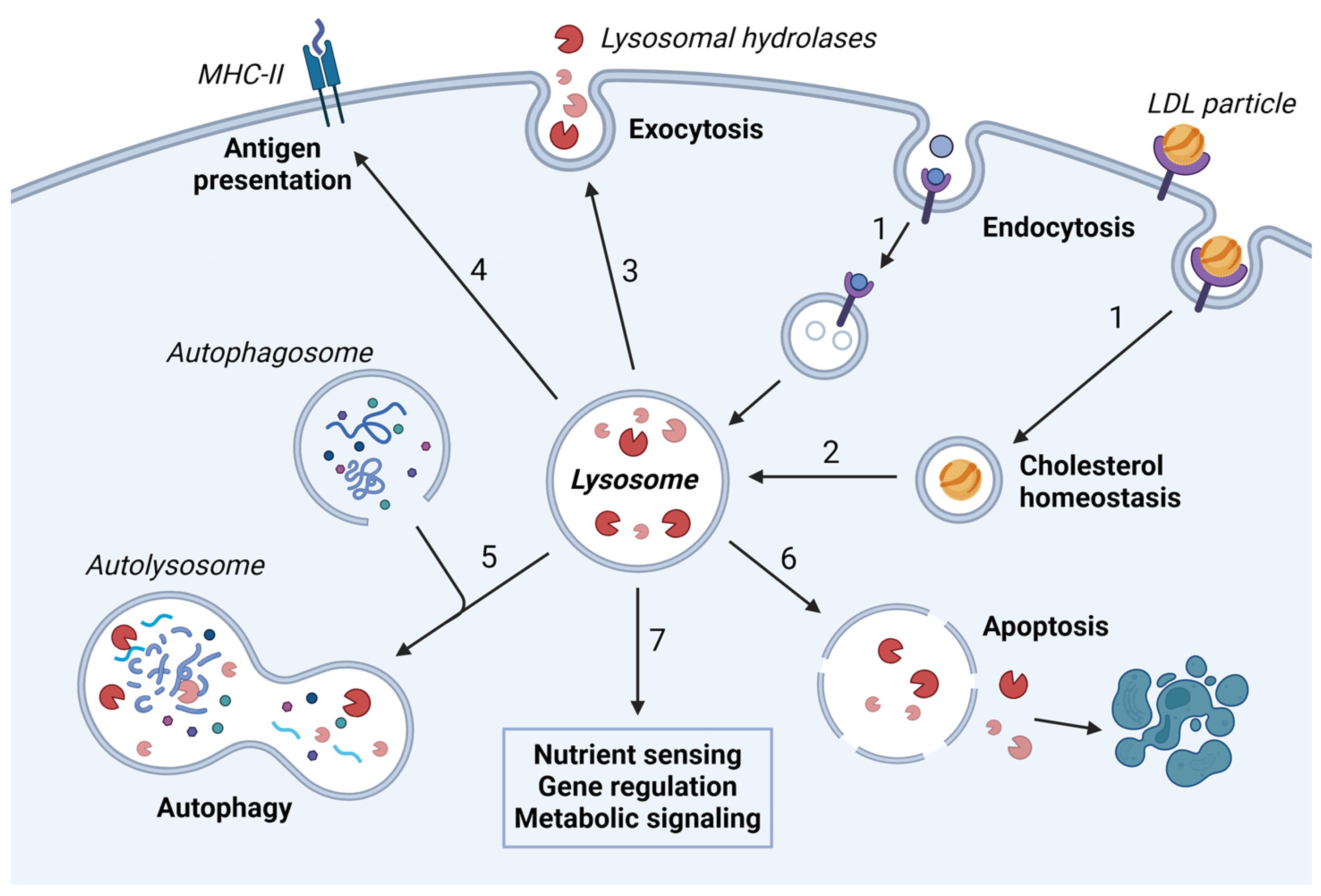
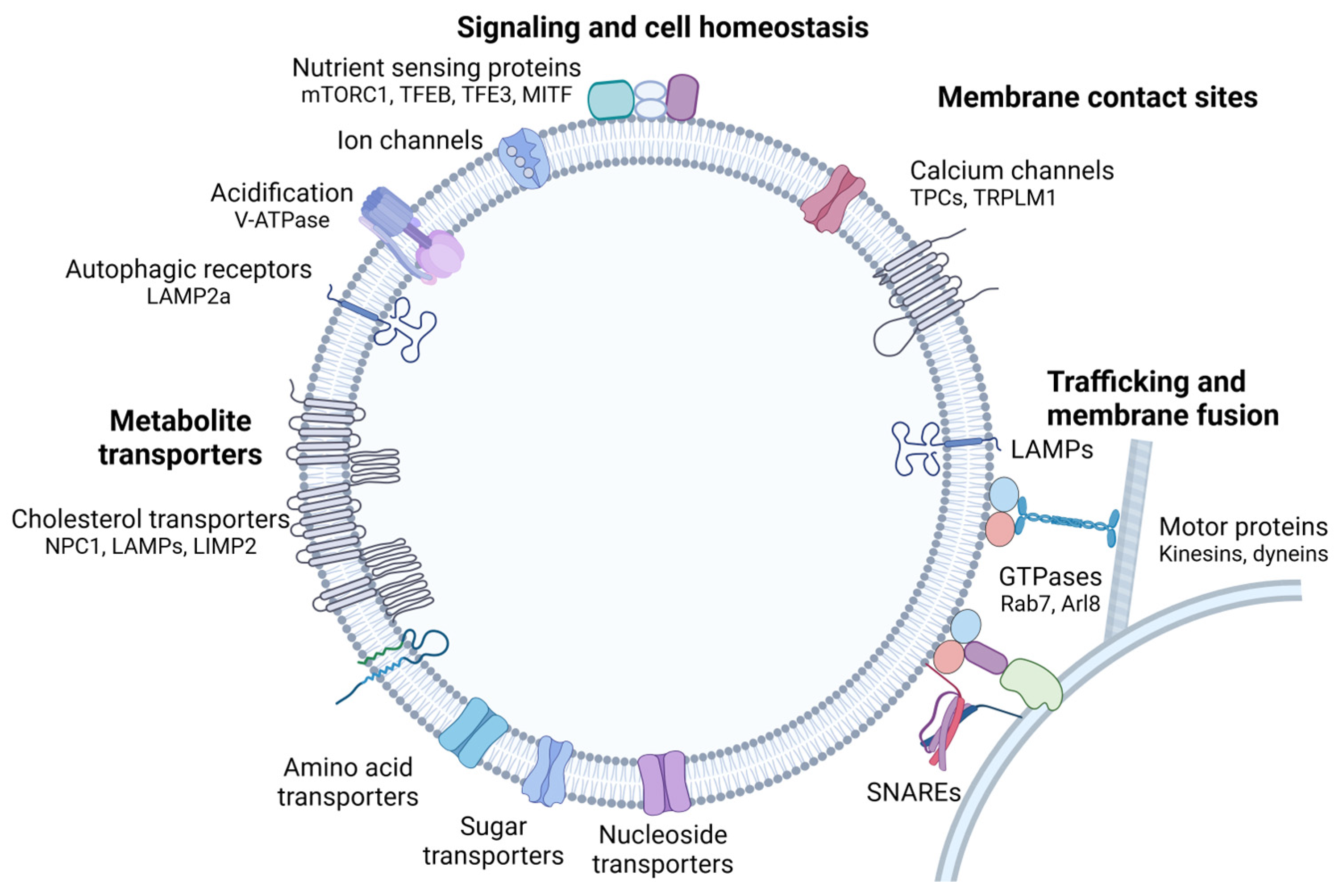
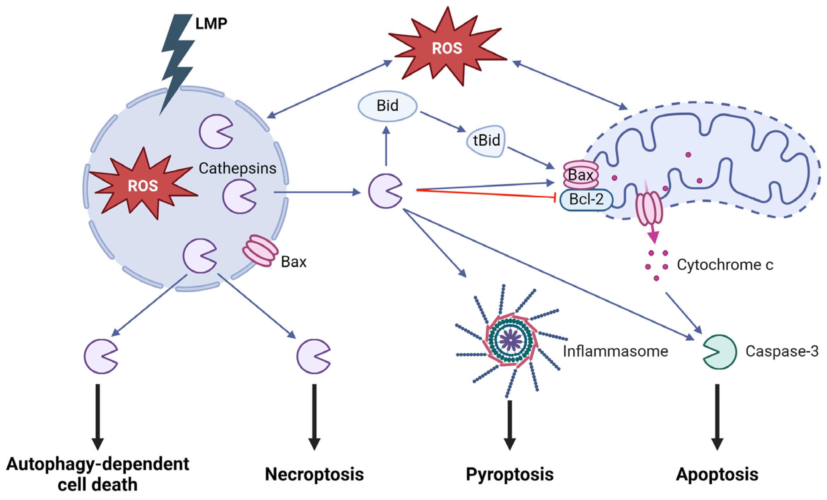
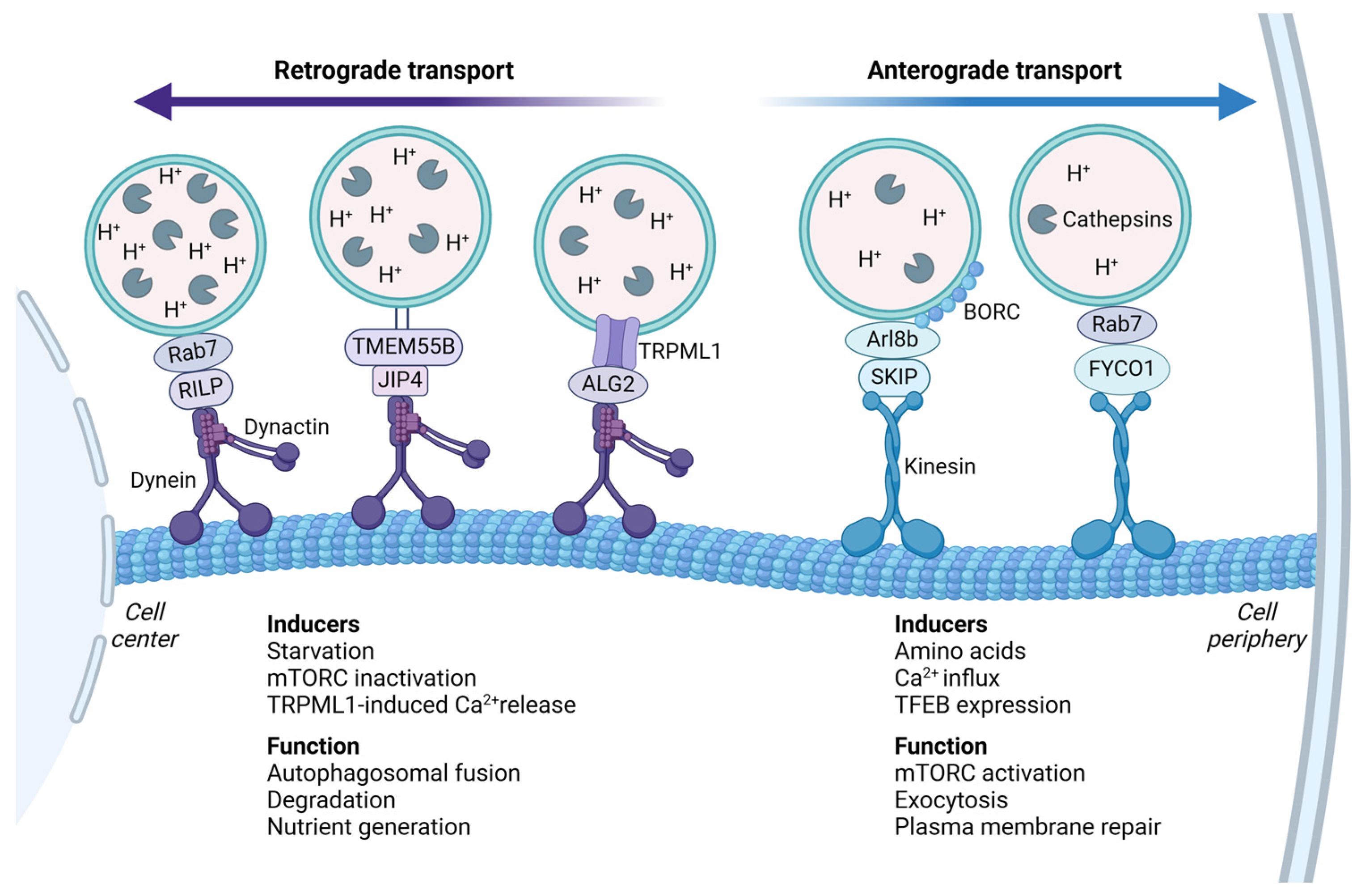
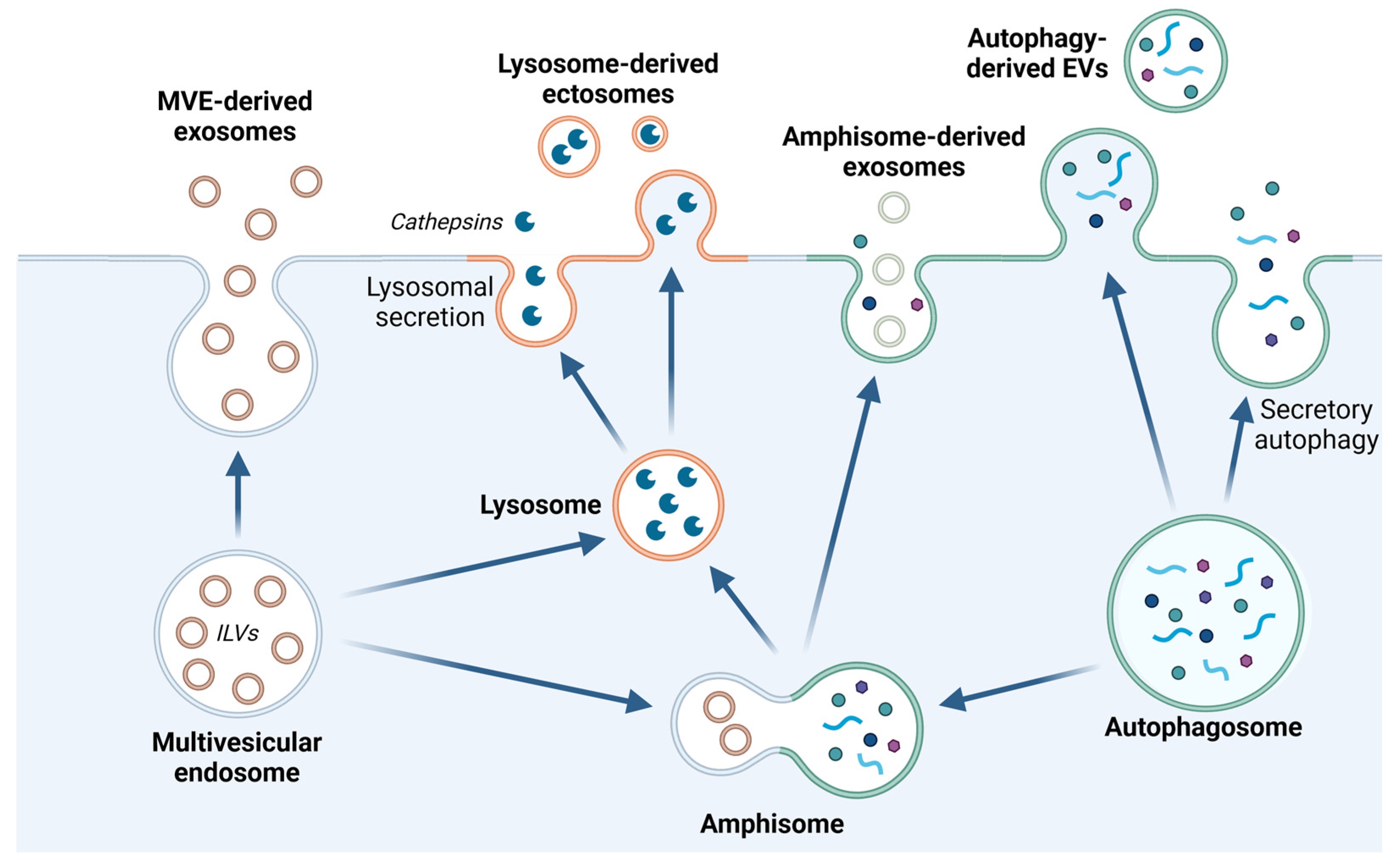
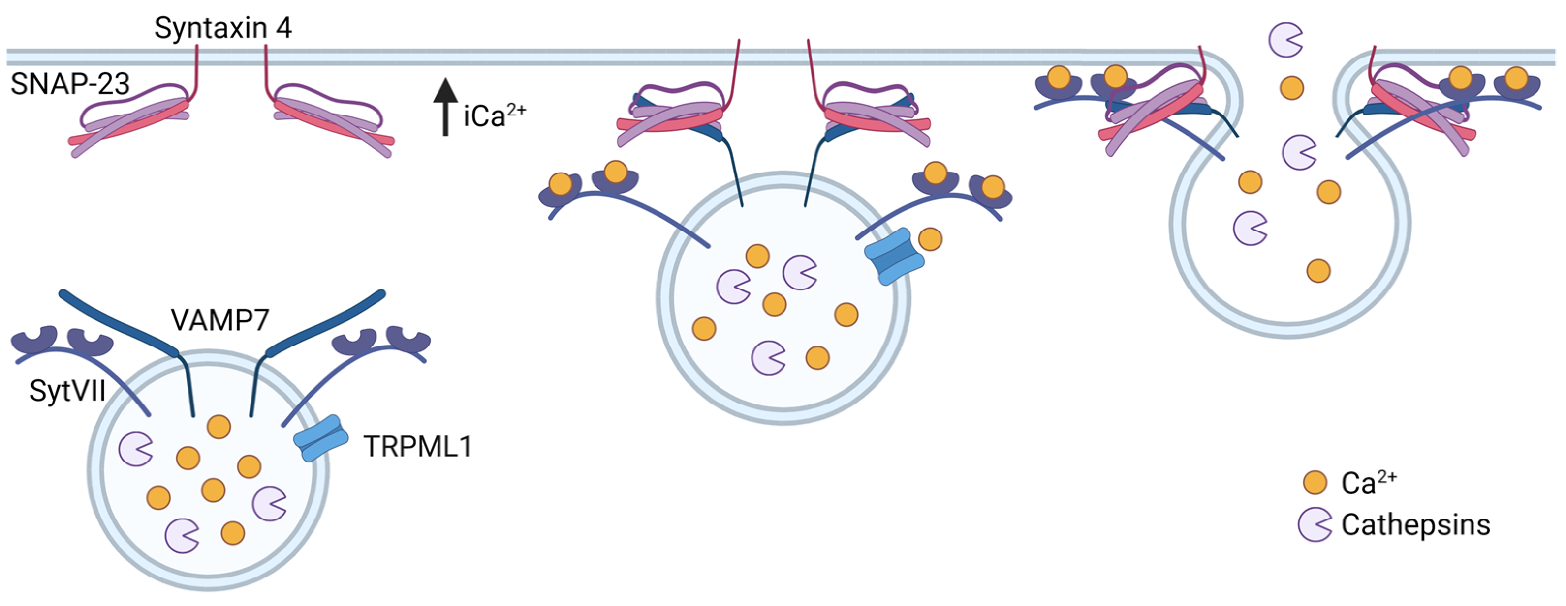
Disclaimer/Publisher’s Note: The statements, opinions and data contained in all publications are solely those of the individual author(s) and contributor(s) and not of MDPI and/or the editor(s). MDPI and/or the editor(s) disclaim responsibility for any injury to people or property resulting from any ideas, methods, instructions or products referred to in the content.

