Communication

Generation of a Transgenic Mouse Model for Investigating Mitochondria in Sperm

by
Hironmoy Sarkar
1,2,*,
Suryaprakash R. Batta
1,
Neerja Wadhwa
3,
Subeer S. Majumdar
1,4,5 and
Bhola Shankar Pradhan
1,6,*
1
Cellular Endocrinology Laboratory, National Institute of Immunology, Aruna Asaf Ali Marg, JNU Complex, New Delhi 110067, India
2
Cell Biology and Bacteriology Laboratory, Department of Microbiology, Raiganj University, Raiganj 733134, West Bengal, India
3
Embryo Biotechnology Lab, National Institute of Immunology, New Delhi 110067, India
4
National Institute of Animal Biotechnology, Miyapur, Hyderabad 500049, Telengana, India
5
Gujarat Biotechnology University, GIFT City Campus, Gandhinagar 382355, Gujarat, India
6
Lukasiewicz PORT Polish Center for Technology Development, Stablowicka 147, 54066 Wroclaw, Poland
*
Authors to whom correspondence should be addressed.
Cells 2025, 14(4), 296; https://doi.org/10.3390/cells14040296
Submission received: 31 January 2025 / Accepted: 15 February 2025 / Published: 17 February 2025
(This article belongs to the Special Issue Sperm Biology and Reproductive Health—Second Edition)

Abstract

:
Mitochondria play a crucial role in sperm development; however, the mechanisms regulating their function in sperm remain poorly understood. Developing a method to regulate the expression of a target gene within the mitochondria of sperm is a vital step in this area of research. In this study, we aimed to create a system for expressing a transgene in the mitochondria of sperm. As a proof of concept, we generated transgenic mice that express green fluorescent protein (GFP) fused with a mitochondrial localization signal (MLS) driven by the phosphoglycerate kinase 2 (PGK2) promoter, which facilitates the transgene expression in the sperm. Although the PGK2 promoter has previously shown to drive gene expression in spermatocytes and spermatids, the novelty of our approach lies in the combination of PGK2-driven MLS-GFP expression to study mitochondria in vivo. We established two founder lines of transgenic mice through pronuclear microinjection, and MLS-GFP expression was confirmed in the mitochondria of sperm cells using fluorescence microscopy and flow cytometry. Consequently, we provide a novel platform for investigating mitochondrial function in sperm, where GFP can be substituted with other genes of interest to examine their effects on mitochondria. This system specifically targets sperm mitochondria, offering an innovative approach for studying mitochondrial function in vivo.

1. Introduction

The rise in the cases of infertility has become a global concern recently, and around 7% of the male population worldwide are infertile [1,2,3,4]. The dysfunction of mitochondria contributes significantly towards male infertility [5]. Mitochondria are dynamic organelles and play a crucial role in the metabolism of spermatozoa. In the spermatozoa, approximately 50–80 mitochondria are present in the midpiece [6].They are implicated in various cellular functions such as oxidative phosphorylation and calcium homeostasis, which are important for the function of sperm [7,8,9]. The mitochondria in spermatozoa regulate important functions relating to human fertility [10,11]. The ROS-mediated damage to the spermatozoa contributes to 30% to 80% (approximately) of male infertility [12], and around 60% of idiopathic male infertility is associated with reduced sperm motility [13]. The genetic modification of mitochondria and its role in sperm development has gained significant attention in recent years. The understanding of the function of mitochondria in sperm cells may provide critical insights into the molecular basis of male infertility [6,14].
Recent reports suggested that male infertility is associated with a loss of mitochondrial proteins in spermatozoa, which induces low sperm motility, reduces OXPHOS activity, and results in male infertility [14]. The reduction in the motility of the spermatozoa in cases of asthenozoospermia is associated with reduced mitochondrial respiratory activities and reduced mitochondrial membrane potential [15,16]. The inhibition of the protein synthesis machinery of the mitochondria of the spermatozoa leads to a reduction in sperm motility [17]. Moreover, the interaction of mitochondrial fusion protein 2 (MFN2) and mitostatin-related protein 1 (MNS1), which regulate the biogenesis of sperm tails, contributes towards sperm motility [18].
Despite the advancements in understanding the role of mitochondrial dynamics in the function of the cells [19,20,21,22], the molecular mechanisms regulating this process during spermatogenesis, particularly in the development of spermatozoa, are not fully understood. Importantly, the techniques to study the roles of various genes affecting the mitochondria of the spermatozoa in vivo are limited. Therefore, there is an urgent need to develop a method to target the gene specifically to the mitochondria of the spermatozoa in vivo.
The dynamics of mitochondria in spermatozoa, including their distribution, fusion, and fission, are important for sperm function and viability. During the development of sperm, mitochondria undergo significant structural and functional changes. The role of mitochondrial fusion and fission in sperm motility is increasingly understood, as proper mitochondrial dynamics are necessary for the motility and structural integrity of the sperm tail [23]. The use of various staining dyes to label the mitochondria of the spermatozoa, such as MitoTracker, leads to the defect in the fertilizing ability of spermatozoa in vitro, highlighting the need for alternative approaches. Therefore, fluorescent proteins may be used for studying the effect of mitochondria in the development of the spermatozoa in vivo.
Previously, the PGK2 promoter has been demonstrated as a testis-specific promoter [24]. The PGK2 promoter drives expression of transgenes in spermatocytes and spermatids in vivo [25,26,27,28]. In this study, we seek to develop a novel method for studying the mitochondria within spermatozoa development by combining the GFP with a mitochondrial localization signal (MLS) along with PGK2 promoter. We hypothesize that the PGK2 promoter can drive the expression of GFP-tagged MLS in the mitochondria of the spermatozoa in vivo.
This approach not only enhances our understanding of mitochondrial behavior during spermatogenesis but also establishes a foundation for exploring the molecular basis of male infertility related to mitochondrial dysfunction. Although the role of PGK2 promoter in spermatogenesis is well established, its use for targeting the gene expression in the mitochondria of the spermatozoa is not demonstrated so far. In this study, we used the PGK2 promoter driving the expression of GFP tagged to a mitochondrial localization signal (PGK2-MLS-GFP) to express GFP specifically in the spermatozoa of transgenic mice.

2. Material and Methods

2.1. Animals

All the mice used for the present study were of the FVB/J strain. The mice were obtained from the small animal facility of the National Institute of Immunology. The mice were housed in a climate-controlled facility under standard light (14 h light–10 h dark cycle), temperature (23 °C), and humidity (50 ± 5%). The mice were housed in individually ventilated cages with (IVC) ad libitum access to acidified autoclaved water and handled by trained personnel. Mice were used as per the guidelines laid down by the CPCSEA (Committee for the Purpose of Control and Supervision of the Experiments on Animals). Protocols for experiments were approved by the Institutional Animal Ethics Committee (IAEC) of the National Institute of Immunology, India, constituted by CPCSEA (IAEC number: IAEC 249/10). Mice were euthanized by cervical dislocation as approved by the Institutional Animal Ethics Committee of the National Institute of Immunology.

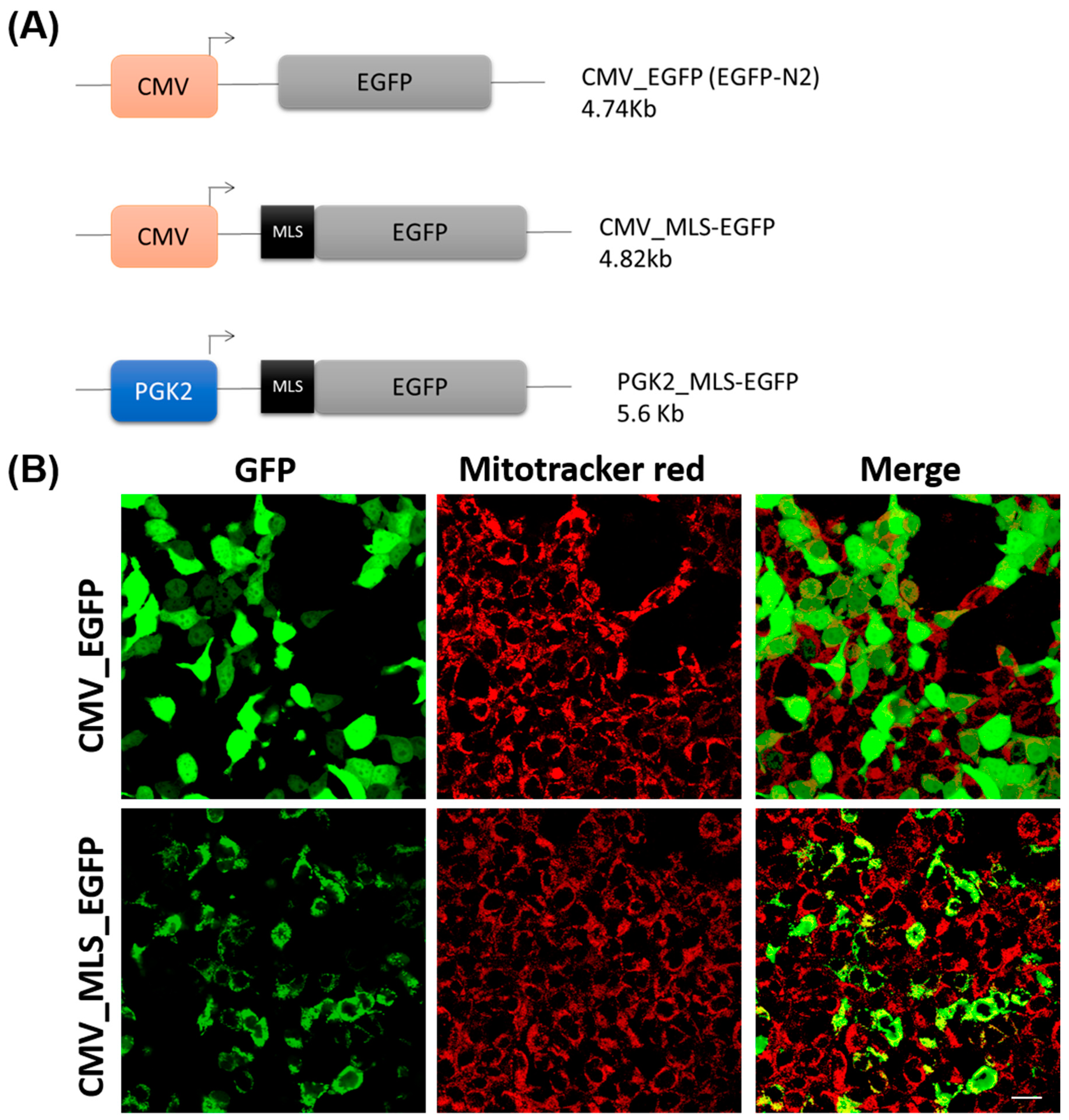
2.2. Transgene Constructs

A total of three constructs (EGFPN2, CMV_MLS-EGFP, and PGK2_MLS-EGFP) were used in this study. The schematic diagrams of the constructs were provided in Figure 1A. The constructs for mitochondrial gene targeting were generated through standard molecular cloning methods as described previously [29]. Plasmid DNA was isolated from overnight-grown culture of E. coli (dh5α) using a plasmid DNA isolation kit (Advanced Micro Devices, Bengaluru, India) following the manufacturer’s recommendation. For the generation of the CMV_MLS-EGFP construct, the mitochondrial localization signal of the COXVIII gene of murine origin (TCCGTCCTGACGCCGCTGCTGCTGCGGGGCTTGACAGGCTCGGCCCGGCGGCTCCCAGTGCCGCGCGCCAAGA) was amplified from mouse genomic DNA using primers MLSF: GCCGCCAAGCTTGTGTACTCCGTGCCATCATG and MLSR: GGCGTGGATCCCCGGCGGCAACGAATGGA. The PCR was performed using pfu DNA polymerase (Fermentas, Pittsburgh, PA, USA), following standardized thermal cycling parameters (initial denaturation at 95 °C for 2 min, denaturation at 95 °C for 30 s, annealing at 60 °C for 30 s, and extension at 68 °C for 30 s for 35 cycles, followed by final extension for 10 min at 72 °C). The amplicon (135 bp) was cloned between the two restriction enzyme sites HindIII and BamHI of the EGFPN2 vector. For the generation of the PGK2_MLS-EGFP construct, the PGK2 promoter was amplified from mouse genomic DNA using primers PGKF: GTCGT ATTAATCTAGAAATAGGATTGATGAC and PGKR: GTCGTCCTCGAGCTTGACAATATAAAGACATA. The PCR was performed using pfu DNA polymerase (Fermentas, Pittsburgh, PA, USA), following standardized thermal cycling parameters (initial denaturation at 95 °C for 2 min, denaturation at 95 °C for 30 s, annealing at 60 °C for 30 s, and extension at 68 °C for 2 min for 35 cycles, followed by final extension for 10 min at 72 °C). The amplicon (1467 bp) was cloned between the two restriction enzyme sites, AseI and XhoI, of the CMV_MLS-EGFP vector by substituting the CMV promoter with the PGK2 promoter. The sequences of the construct are provided in the Supplementary Figure S3.

2.3. Cell Culture and Transfection

The human embryonic kidney (HEK293) cells (CRL-1573, ATCC, Manassas, VA, USA) were a kind gift from Dr. A.C. Banerjea, National Institute of Immunology, New Delhi. The HEK293 cells were cultured in Dulbecco’s Modified Eagle’s Medium (DMEM) (670087, Gibco™, Life Technologies Corp., Grand Island, NY, USA) containing 10% fetal bovine serum supplemented with penicillin, streptomycin, and Fungizone at 37 °C in the presence of 5% CO2. Transfection of HEK293 cells was performed as described by us previously [29] Transient transfections with CMV_MLS-EGFP plasmid were performed with Lipofectamine 2000 (11668019, Invitrogen, Carlsbad, CA, USA), according to the manufacturer’s instructions. For the control, we used EGFPN2 (CMV_EGFP) plasmid, which lacks the mitochondrial localization signal. After 24 h, the transfected cells were stained with 20 nM of the mitochondrial-specific dye, MitoTracker Red CMXRos (M7512, Thermo Fisher Scientific, Waltham, MA, USA), for 15 min at 37 °C, followed by three washes with 1× PBS.

2.4. Pronuclear Microinjection of Transgene Cassettes and Oviductal Embryo Transfer

This method was performed as described previously [30]. Transgenic mice were generated via pronuclear microinjection [31,32,33,34]. For pronuclear microinjection, female mice were superovulated using PMSG and hCG followed by cohabitation with male mice. Oviducts were collected from donor mice (euthanized by cervical dislocation) and placed into Brinster’s Modified Oocyte Culture Media (BMOC) containing hyaluronidase (1 mg/mL). Embryos were collected and transferred to a 60 mm dish containing 100 µL drops of BMOC overlaid with mineral oil. The linearized transgene cassette (PGK2-MLS-EGFP) was prepared at a final concentration of 4 ng/µL for microinjection. DNA was microinjected into the male pronucleus of fertilized eggs using a Narishighe micromanipulator. Each manipulated embryo was then transferred into the pre-incubated BMOC-containing dish and maintained at 37 °C in a 5% CO2 incubator. Embryos were incubated until the two-cell stage. Two pseudo-pregnant female mice were generated by mating with vasectomized male mice. Two-cell stage embryos were transferred into the ampulla of the oviduct of these pseudo-pregnant recipient mice. Approximately 20 microinjected embryos were transferred to the oviductal ampulla. After the gestation period (21 ± 2 days), recipient pups were born.

2.5. Establishment of Transgenic Lines

We obtained many pups from the two pseudo-pregnant recipient mice after the embryo transfer. The pups born were screened through PCR as described by us previously [29,35]. For this, the genomic DNA was isolated from the tail biopsy of these mice (around 21 days of age) by proteinase K lysis and organic extraction with phenol-chloroform. The genotyping was performed by PCR using Taq polymerase and the following primers: MKSF: GTGTACTCCGTGCCATCATG and GFPR: TGCCGTAGGTGGCATCGCCC, following standardized thermal cycling parameters (initial denaturation at 95 °C for 2 min, denaturation at 95 °C for 30 s, annealing at 60 °C for 30 s and extension at 72 °C for 30 s for 35 cycles, followed by final extension for 10 min at 72 °C). The PCR products were analyzed by TAE agarose gel electrophoresis. The amplicon size of 258 bp indicates the transgene positive mice. As a loading control, PCR for mice gene peptidylprolyl isomerase A (Ppia) was amplified from each tail biopsy using the primer PPIAF: GTCGCTTTTCGCCGCTTGC and PPIAR: TCTGCTGTCTTTGGAACT following standardized thermal cycling parameters (initial denaturation at 95 °C for 2 min, denaturation at 95 °C for 30 s, annealing at 60 °C for 30 s, and extension at 72 °C for 30 s for 35 cycles, followed by final extension for 10 min at 72 °C). The amplicon size (120 bp) suggests good quality of genomic DNA. The founder mice were mated with either wild-type females or wild-type males to establish the separate transgenic lines, and the transgene integration in the progeny was evaluated by PCR. The litter size of the control and PGK2-MLS-GFP mice was analyzed.

2.6. Sperm Collection

The sperm cells were collected from four PGK2-MLS-GFP transgenic male mice and four wild-type male mice as described previously [30]. These mice were euthanized at around 90 days of age. The entire intact epididymis was surgically removed from these mice and transferred into a 1.5 mL microcentrifuge tube containing 1 mL of 1× PBS and ruptured thereafter for sperm cell collection. The sperm cells of the control and PGK2-MLS-GFP mice were analyzed for motility under a light microscope. For this, around 100 sperm cells were counted per field, and ten different fields were counted for each sample. A fraction of these samples was analyzed in fluorescence microscopy, and another fraction was analyzed in a flow cytometer for the detection of GFP.

2.7. Fluorescence Microscopy

The HEK293 cells transfected with CMV_EGFP plasmid (control) or with CMV_MLS-EGFP plasmid were analyzed under fluorescent microscope for GFP expression. The mitochondria of these cells were visualized by staining with MitoTracker Red CMXRos (Invitrogen, Carlsbad, CA, USA). Images were acquired from five different fields, and experiments were performed in triplicates.
The sperm cells from the PGK2-MLS-GFP transgenic male mice (n = 4) and wild-type male mice (n = 4) were analyzed for the presence of GFP under bright field and ultraviolet illumination (with FITC and BLUE/CYAN filter) using a Nikon Eclipse TE2000-S inverted microscope (Nikon Corporation, Chiyoda-ku, Tokyo, Japan) attached to a DS-5 M camera assisted by Digital Sight DS-L1 software 2.0 for capturing the images. Around 100 sperm cells were analyzed for each mouse for the presence of GFP.

2.8. Flow Cytometry Analysis

The sperm cells from the PGK2-MLS-GFP transgenic male mice (n = 4) and wild-type male mice (n = 4) were analyzed for the presence of GFP in a flow cytometer (FACS caliber, BD Biosciences, Franklin Lakes, NJ, USA). Around 10,000 sperm cells from each mouse were analyzed for the presence of GFP. Data were collected and analyzed using FlowJo software V 10.2 TreeStar, Inc., Ashland, OR, USA) to assess the percentage of GFP-positive sperm.

2.9. Statistical Analysis

All statistical analyses were performed using Graphpad Prism 5 (Graphpad Software, Lajolla, CA, USA). Data are represented as means ± SEM. Significance was determined by the Mann–Whitney test and a p value.

3. Results

3.1. Validation of MLS Sequence In Vitro

The effectiveness of the MLS sequence of the COXVIII gene of human in targeting GFP to mitochondria was demonstrated in HeLa cells before [36,37]. Since we plan to target the gene expression specifically in the mitochondria of the spermatozoa of the mice in vivo, we used the MLS sequence of the COXVIII gene of mice. However, the sperm cells are difficult to transfect in vitro. Thus, we tested the MLS sequence of COXVIII gene of mice in HEK293 cells, as these cells are comparatively easier to transfect. For this, we generated the CMV_MLS-EGFP construct, which contains the MLS sequence of the COXVIII gene of mice along with the CMV promoter, which is functional in HEK293 cells. To determine the effectiveness of MLS sequence in targeting the gene expression in mitochondria in vitro, we transfected the HEK293 cells with CMV_MLS-EGFP construct. As a control, we transfected the HEK293 cells with EGPN2 vector (which lacks the MLS sequence). We observed the tubular pattern of GFP signal in the HEK293 cells transfected with CMV_MLS-EGFP construct. Importantly, the signal for GFP in these cells was colocalized with the red signal of the mitochondria, which were stained with MitoTracker Red CMXRos (mitochondria-specific dye) suggesting that MLS was functional (Figure 1B). The control cells transfected with the EGFPN2 construct showed a cytoplasmic GFP signal that did not colocalize with the MitoTracker Red CMXRos. These results also suggest that the MLS sequence of the COXVIII gene of mice is functional in HEK293 cells.

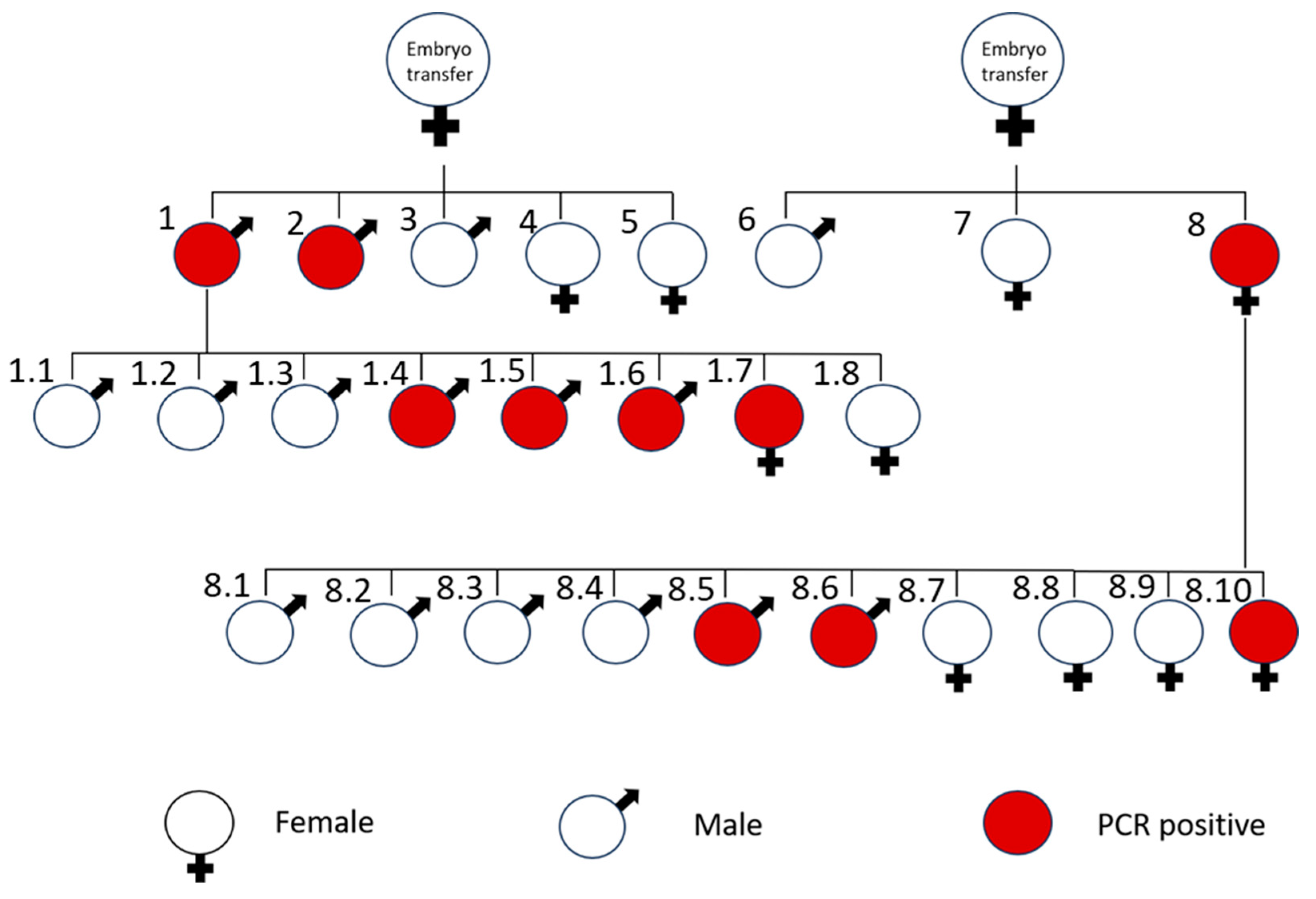
3.2. Generation of Transgenic Mice

Since the MLS sequence of the COXVIII gene of mice is effective in targeting gene expression in the mitochondria in vitro, we used this MLS sequence for testing its functionality in the spermatozoa of mice. Previously, the tissue-specific activity of the PGK2 promoter is demonstrated to drive the gene expression of the post-meiotic genes in the male germline, particularly in the spermatozoa [24]. Thus, we generated a PGK2_MLS-GFP construct by replacing the CMV promoter with the PGK2 promoter of mice in the CMV_MLS-EGFP construct. Using the classical pronuclear microinjection method, we inserted the PGK2_MLS-GFP construct into the pronucleus of the embryos and transferred these embryos into the pseudo-pregnant females. A schematic diagram of the generation of the transgenic line is presented in Figure 2. We obtained many pups from the two recipient females after the pronuclear microinjection. The pups born were screened for the integration of transgenes using the genomic DNA of tail biopsies by PCR. We observed that many mice were positive for integration of transgene (PCR product size: 258 bp) (Figure 3). As a loading control, we used the PCR for Ppia. Further, we selected two founder mice out of these for establishing the transgenic line expressing PGK2-MLS-GFP by mating them with the wild-type males or female mice. The pups born from this mating were screened for PCR, and the transgenic mice were used for further study. The litter size from the PGK2-MLS-GFP mice was compared with that of wild-type mice (control). We did not observe any significant (p ≤ 0.05) difference in the litter size of PGK2-MLS-GFP mice as compared to that of the control suggesting that PGK2-MLS-GFP mice were fertile (Supplementary Figure S2).

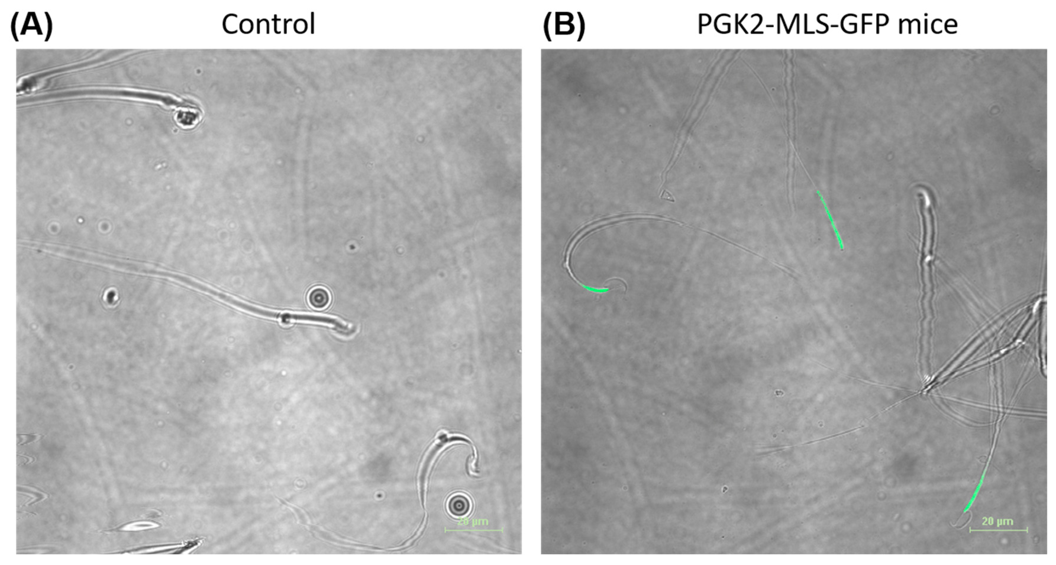
3.3. Localization of the GFP in the Mitochondria of Spermatozoa In Vivo

Since the PGK2 promoter drives expression of transgenes in spermatocytes and spermatids in vivo [25,26,27,28], we evaluated the sperm of the PGK2-MLS-GFP transgenic mice. We collected the sperm of the transgenic mice (n = 4) and control mice (n = 4). These live sperm cells (n = 100) were observed under the fluorescence microscope for the presence of GFP. We observed the GFP signal in the spermatozoa collected from transgenic mice but not from the control mice (Figure 4). The mitochondria are localized in the midpiece of the spermatozoa. Since we used the MLS sequence of the COXVIII gene of mice, we evaluated the midpiece region of the GFP-positive spermatozoa under a fluorescence microscope. We observed the GFP signal in the midpiece of the spermatozoa of the transgenic mice (Figure 4 and Figure S1). There was no signal for GFP either in the head or tail region of the spermatozoa, suggesting that GFP was expressed specifically in the midpiece (where mitochondria are localized) of the spermatozoa of PGK2-MLS-GFP transgenic mice. Moreover, the motility of sperm was analyzed, and there was no significant (p ≤ 0.05) difference between the control and transgenic mice in terms of sperm motility suggesting that GFP expression did not affect the function of the sperm cells (Supplementary Figure S2).

3.4. Analysis of GFP Expression in the Spermatozoa by Flow Cytometry (FACS)

Since we observed the GFP in the mitochondria of the spermatozoa of the PGK2-MLS-GFP transgenic mice by fluorescence microscopy, we performed the FACS analysis of sperm samples of these mice (n = 4) to validate the microscopy data. As a control, we used the sperm cells of the wild-type mice (n = 4). Around 10,000 cells per sample were analyzed for detection of GFP by flow cytometry. We observed a distinct shift in the sperm samples of PGK2-MLS-GFP transgenic mice, which suggests for the population of GFP-positive sperm (Figure 5). On the other hand, the sperm cells of the control wild-type mice did not show this shift, suggesting the absence of GFP-positive sperm. These results validate our observation from microscopic data in vivo. Moreover, these results, along with the microscopy data, also suggest that the GFP expression was specifically in the mitochondria of the spermatozoa of the PGK2-MLS-GFP transgenic mice and the method was reproducible.

4. Discussion

The present study aimed to target the expression of transgene to the mitochondria of the spermatozoa in vivo. Here, we demonstrated the targeting of GFP to the mitochondria of the spermatozoa of the transgenic mice as a proof of concept. For this, we used the construct containing the MLS sequence of the COXVIII gene and the PGK2 promoter of mice. This combination allows us to target the GFP specifically to the mitochondria of the spermatozoa. This conclusion is based on the following experimental data. First, we validated the effectiveness of the MLS sequence of the COXVIII gene in targeting GFP to the mitochondria in HEK293 cells. Secondly, we generated the PGK2-MLS-GFP transgenic mice and demonstrated the presence of GFP in the mitochondria of the spermatozoa by fluorescence microscopy and flow cytometry analysis. Our data also suggest the targeting of GFP in the midpiece of spermatozoa mitochondria did not affect its motility nor the fertility of the PGK2-MLS-GFP transgenic mice.
Previously, the MLS sequence of the COXVIII gene of both humans and mice was used for mitochondrial gene expression in HeLa cells and transgenic mice, respectively [20,36,37,38]. In this study, we used the MLS of the COXVIII gene of mice and tested its effectiveness for mitochondrial gene targeting both in HEK293 cells and in transgenic mice, which suggests the reproducibility of the MLS sequence the COXVIII gene in mitochondrial gene targeting. Moreover, the MLS sequence of the COXVIII gene of mice is functional in human cells, such as HEK293 cells, which suggests the functionality of this sequence across species.
Previous studies have utilized the constitutive promoter for GFP expression in the spermatozoa, which also led to expression of transgenes in other testicular cells such as Sertoli cells, Leydig cells, etc. [38,39,40]. This strategy will not help in studying the function of a particular gene specifically in the spermatozoa. The specificity of the PGK2 promoter ensures minimal ectopic expression, which is critical for accurate functional analyses [24]. Thus, the use of PGK2 promoter, which is active only in meiotic spermatocytes and post-meiotic spermatids [24], will address this limitation. In this study, we observed the GFP expression specifically in the spermatozoa, which is consistent with the previous studies. Further, we extended these findings by the integration of the MLS sequence along with PGK2 promoter, which enables precise targeting of GFP to the mitochondria of spermatozoa in vivo. This approach addresses a critical gap in male reproductive biology, providing insights into studying the role of mitochondria in spermatozoa development, motility, and fertilization.
Increasing evidence suggests the role of mitochondria in male infertility [5,41]. Mitochondria in the midpieces of the spermatozoa are critical for energy production and motility, and previous studies suggest a link between mitochondrial dysfunction and male infertility [14]. This approach can be used to study the effect of various mutations caused by environmental stress on mitochondrial functions, mitochondrial dynamics using advanced imaging techniques during spermatogenesis, and the role of mitochondria in assisted reproduction technologies.
In this study, we demonstrated the functional importance of mitochondrial localization sequences in directing transgenes to the mitochondria of the sperm using a germline-specific promoter. Importantly, we confirmed that GFP expression in the mitochondria of spermatozoa did not affect its functions, as we did not find any difference in the sperm motility or reduction in offspring number from the male transgenic mice. This is in agreement with the previous study, which suggests that the expression of GFP in the mitochondria of the spermatozoa does not affect its function [42].
This study may help in studies of mitochondrial health and dynamics in large populations of sperm cells [14,43]. Our study provides a platform to investigate mitochondrial biology during spermatogenesis. Mitochondria are vital for sperm function and are a major contributor to male infertility [5]. Our studies will help to study the role of mitochondria in the development of the spermatozoa.
While our study demonstrates the feasibility of targeting transgenes to the mitochondria of the spermatozoa, we did not directly assess the mitochondrial biogenesis or role of various environmental stresses on mitochondrial function. Future studies will be needed to explore the role of various genes on the function of the mitochondria of the spermatozoa and thereby their role in affecting male fertility.
In conclusion, we establish a novel in vivo model for studying the role of transgenes in the mitochondria of the spermatozoa using a germline-specific promoter such as PGK2 and mitochondrial localization sequence of the COXVIII gene. These findings may have broad implications for mitochondrial research, particularly in male infertility and sperm development.

Supplementary Materials

The following supporting information can be downloaded at: https://www.mdpi.com/article/10.3390/cells14040296/s1, Supplementary Figure S1: A representative images of the spermatozoa from the PGK2-MLS-GFP transgenic mice showing the expression of GFP in the midpiece (where the mitochondria are localized); Supplementary Figure S2: Comparison of the litter size, sperm motility between control and PGK2-MLS-GFP transgenic mice (Tg mice); Supplementary Figure S3. Full sequences of the various constructs used in this study.

Author Contributions

Experiments were conceived and designed by B.S.P., H.S. and S.S.M. Experiments were performed by H.S., S.R.B., N.W. and B.S.P. Data of the manuscript were analyzed by B.S.P. and H.S. Manuscript was written and reviewed by B.S.P. and H.S. All authors have read and agreed to the published version of the manuscript.

Funding

Department of Biotechnology, Govt. of India, for providing the financial assistance under grants BT/PR11313/AAQ/01/376/2008, BT/HRD/35/01/01/2010 (TATA Innovation Award) and BT/PR10805/AAQ/1/576/2013; Polish National Science Centre grant (2020/39/D/NZ5/02004); Department of Biotechnology, Govt. of India grant (BT/PR46488/AAQ/1/886/2022).

Institutional Review Board Statement

Mice were housed and used as per the guidelines laid down by the CPCSEA (Committee for the Purpose of Control and Supervision of the Experiments on Animals). Protocols for experiments were approved by the Institutional Animal Ethics Committee (IAEC) of National Institute of Immunology, India, constituted by CPCSEA. Approval Code: IAEC 249/10; Approval Date: 3 August 2010 (extended every four years).

Informed Consent Statement

Not applicable.

Data Availability Statement

The original contributions presented in this study are included in the article/Supplementary Material. Further inquiries can be directed to the corresponding authors.

Acknowledgments

We are thankful to all the staff of the Small Animal Facility. Thanks are due to Ram Singh, Dharamvir Singh, and Birendar Roy for the technical assistance. We are grateful to the Director, NII for valuable support.

Conflicts of Interest

The authors declare no conflicts of interest.

References

  1. Matzuk, M.M.; Lamb, D.J. The biology of infertility: Research advances and clinical challenges. Nat. Med. 2008, 14, 1197–1213. [Google Scholar] [CrossRef] [PubMed] [PubMed Central]
  2. Sharpe, R.M. Low sperm counts may be preventable. Science 2011, 333, 1380–1381. [Google Scholar] [CrossRef] [PubMed]
  3. Hentrich, A.; Wolter, M.; Szardening-Kirchner, C.; Lüers, G.H.; Bergmann, M.; Kliesch, S.; Konrad, L. Reduced numbers of Sertoli, germ, and spermatogonial stem cells in impaired spermatogenesis. Mod. Pathol. 2011, 24, 1380–1389, Erratum in Mod. Pathol. 2017, 30, 311. [Google Scholar] [CrossRef] [PubMed]
  4. Thirumavalavan, N.; Gabrielsen, J.S.; Lamb, D.J. Where are we going with gene screening for male infertility? Fertil. Steril. 2019, 111, 842–850. [Google Scholar] [CrossRef] [PubMed]
  5. Tesarik, J.; Mendoza-Tesarik, R. Mitochondria in Human Fertility and Infertility. Int. J. Mol. Sci. 2023, 24, 8950. [Google Scholar] [CrossRef] [PubMed] [PubMed Central]
  6. Freitas, M.J.; Vijayaraghavan, S.; Fardilha, M. Signaling mechanisms in mammalian sperm motility. Biol. Reprod. 2017, 96, 2–12. [Google Scholar] [CrossRef] [PubMed]
  7. Aitken, R.J.; Whiting, S.; De Iuliis, G.N.; McClymont, S.; Mitchell, L.A.; Baker, M.A. Electrophilic aldehydes generated by sperm metabolism activate mitochondrial reactive oxygen species generation and apoptosis by targeting succinate dehydrogenase. J. Biol. Chem. 2012, 287, 33048–33060. [Google Scholar] [CrossRef] [PubMed] [PubMed Central]
  8. Amaral, A.; Lourenço, B.; Marques, M.; Ramalho-Santos, J. Mitochondria functionality and sperm quality. Reproduction 2013, 146, R163–R174. [Google Scholar] [CrossRef] [PubMed]
  9. Boguenet, M.; Bouet, P.E.; Spiers, A.; Reynier, P.; May-Panloup, P. Mitochondria: Their role in spermatozoa and in male infertility. Hum. Reprod. Updat. 2021, 27, 697–719. [Google Scholar] [CrossRef] [PubMed]
  10. Spinelli, J.B.; Haigis, M.C. The multifaceted contributions of mitochondria to cellular metabolism. Nat. Cell Biol. 2018, 20, 745–754. [Google Scholar] [CrossRef] [PubMed] [PubMed Central]
  11. Fu, L.; Luo, Y.X.; Liu, Y.; Liu, H.; Li, H.Z.; Yu, Y. Potential of Mitochondrial Genome Editing for Human Fertility Health. Front. Genet. 2021, 12, 673951. [Google Scholar] [CrossRef] [PubMed] [PubMed Central]
  12. Agarwal, A.; Virk, G.; Ong, C.; du Plessis, S.S. Effect of oxidative stress on male reproduction. World J. Men’s Health 2014, 32, 1–17. [Google Scholar] [CrossRef] [PubMed] [PubMed Central]
  13. Liu, F.J.; Liu, X.; Han, J.L.; Wang, Y.W.; Jin, S.H.; Liu, X.X.; Liu, J.; Wang, W.T.; Wang, W.J. Aged men share the sperm protein PATE1 defect with young asthenozoospermia patients. Hum. Reprod. 2015, 30, 861–869. [Google Scholar] [CrossRef] [PubMed]
  14. Park, Y.J.; Pang, M.G. Mitochondrial Functionality in Male Fertility: From Spermatogenesis to Fertilization. Antioxidants 2021, 10, 98. [Google Scholar] [CrossRef] [PubMed] [PubMed Central]
  15. Ruiz-Pesini, E.; Diez, C.; Lapeña, A.C.; Pérez-Martos, A.; Montoya, J.; Alvarez, E.; Arenas, J.; López-Pérez, M.J. Correlation of sperm motility with mitochondrial enzymatic activities. Clin. Chem. 1998, 44, 1616–1620. [Google Scholar] [CrossRef] [PubMed]
  16. Piasecka, M.; Kawiak, J. Sperm mitochondria of patients with normal sperm motility and with asthenozoospermia: Morphological and functional study. Folia Histochem. Cytobiol. 2003, 41, 125–139. [Google Scholar] [PubMed]
  17. Gur, Y.; Breitbart, H. Mammalian sperm translate nuclear-encoded proteins by mitochondrial-type ribosomes. Genes Dev. 2006, 20, 411–416. [Google Scholar] [CrossRef] [PubMed] [PubMed Central]
  18. Vadnais, M.L.; Lin, A.M.; Gerton, G.L. Mitochondrial fusion protein MFN2 interacts with the mitostatin-related protein MNS1 required for mouse sperm flagellar structure and function. Cilia 2014, 3, 5. [Google Scholar] [CrossRef] [PubMed] [PubMed Central]
  19. Otera, H.; Ishihara, N.; Mihara, K. New insights into the function and regulation of mitochondrial fission. Biochim. Biophys. Acta Mol. Cell Res. 2013, 1833, 1256–1268. [Google Scholar] [CrossRef] [PubMed]
  20. Wakai, T.; Harada, Y.; Miyado, K.; Kono, T. Mitochondrial dynamics controlled by mitofusins define organelle positioning and movement during mouse oocyte maturation. Mol. Hum. Reprod. 2014, 20, 1090–1100. [Google Scholar] [CrossRef] [PubMed]
  21. Sandoval, H.; Yao, C.K.; Chen, K.; Jaiswal, M.; Donti, T.; Lin, Y.Q.; Bayat, V.; Xiong, B.; Zhang, K.; David, G.; et al. Mitochondrial fusion but not fission regulates larval growth and synaptic development through steroid hormone production. eLife 2014, 3, e03558. [Google Scholar] [CrossRef] [PubMed] [PubMed Central]
  22. Bader, G.; Enkler, L.; Araiso, Y.; Hemmerle, M.; Binko, K.; Baranowska, E.; De Craene, J.-O.; Ruer-Laventie, J.; Pieters, J.; Tribouillard-Tanvier, D.; et al. Assigning mitochondrial localization of dual localized proteins using a yeast Bi-Genomic Mitochondrial-Split-GFP. eLife 2020, 9, e56649. [Google Scholar] [CrossRef] [PubMed] [PubMed Central]
  23. Green, A.; Hossain, T.; Eckmann, D.M. Mitochondrial dynamics involves molecular and mechanical events in motility, fusion and fission. Front. Cell Dev. Biol. 2022, 10, 1010232. [Google Scholar] [CrossRef] [PubMed] [PubMed Central]
  24. Zhang, L.P.; Stroud, J.; Eddy, C.A.; Walter, C.A.; McCarrey, J.R. Multiple elements influence transcriptional regulation from the human testis-specific PGK2 promoter in transgenic mice. Biol. Reprod. 1999, 60, 1329–1337. [Google Scholar] [CrossRef] [PubMed]
  25. Kramer, J.M. Immunofluorescent localization of PGK-1 and PGK-2 isozymes within specific cells of the mouse testis. Dev. Biol. 1981, 87, 30–36. [Google Scholar] [CrossRef] [PubMed]
  26. Danshina, P.V.; Geyer, C.B.; Dai, Q.; Goulding, E.H.; Willis, W.D.; Kitto, G.B.; McCarrey, J.R.; Eddy, E.; O’Brien, D.A. Phosphoglycerate kinase 2 (PGK2) is essential for sperm function and male fertility in mice. Biol. Reprod. 2010, 82, 136–145. [Google Scholar] [CrossRef] [PubMed] [PubMed Central]
  27. McCarrey, J.R.; Berg, W.M.; Paragioudakis, S.J.; Zhang, P.L.; Dilworth, D.D.; Arnold, B.L.; Rossi, J.J. Differential transcription of Pgk genes during spermatogenesis in the mouse. Dev. Biol. 1992, 154, 160–168. [Google Scholar] [CrossRef] [PubMed]
  28. Yoshioka, H.; Geyer, C.B.; Hornecker, J.L.; Patel, K.T.; McCarrey, J.R. In vivo analysis of developmentally and evolutionarily dynamic protein-DNA interactions regulating transcription of the Pgk2 gene during mammalian spermatogenesis. Mol. Cell. Biol. 2007, 27, 7871–7885. [Google Scholar] [CrossRef] [PubMed] [PubMed Central]
  29. Pradhan, B.S.; Majumdar, S.S. An Efficient Method for Generation of Transgenic Rats Avoiding Embryo Manipulation. Mol. Ther. Nucleic Acids 2016, 5, e293. [Google Scholar] [CrossRef] [PubMed] [PubMed Central]
  30. Sarkar, R.K.; Sharma, S.S.; Mandal, K.; Wadhwa, N.; Kunj, N.; Gupta, A.; Pal, R.; Rai, U.; Majumdar, S.S. Homeobox transcription factor Meis1 is crucial to Sertoli cell mediated regulation of male fertility. Andrology 2021, 9, 689–699. [Google Scholar] [CrossRef] [PubMed]
  31. Auerbach, A.B. Production of functional transgenic mice by DNA pronuclear microinjection. Acta Biochim. Pol. 2004, 51, 9–31. [Google Scholar] [CrossRef] [PubMed]
  32. Brinster, R.L.; Chen, H.Y.; Trumbauer, M.; Senear, A.W.; Warren, R.; Palmiter, R.D. Somatic expression of herpes thymidine kinase in mice following injection of a fusion gene into eggs. Cell 1981, 27, 223–231. [Google Scholar] [CrossRef] [PubMed] [PubMed Central]
  33. Costantini, F.; Lacy, E. Introduction of a rabbit beta-globin gene into the mouse germ line. Nature 1981, 294, 92–94. [Google Scholar] [CrossRef] [PubMed]
  34. Gordon, J.W.; Scangos, G.A.; Plotkin, D.J.; Barbosa, J.A.; Ruddle, F.H. Genetic transformation of mouse embryos by microinjection of purified DNA. Proc. Natl. Acad. Sci. USA 1980, 77, 7380–7384. [Google Scholar] [CrossRef] [PubMed] [PubMed Central]
  35. Usmani, A.; Ganguli, N.; Sarkar, H.; Dhup, S.; Batta, S.R.; Vimal, M.; Ganguli, N.; Basu, S.; Nagarajan, P.; Majumdar, S.S. A non-surgical approach for male germ cell mediated gene transmission through transgenesis. Sci. Rep. 2013, 3, 3430. [Google Scholar] [CrossRef] [PubMed] [PubMed Central]
  36. Rizzuto, R.; Brini, M.; Pizzo, P.; Murgia, M.; Pozzan, T. Chimeric green fluorescent protein as a tool for visualizing subcellular organelles in living cells. Curr. Biol. 1995, 5, 635–642. [Google Scholar] [CrossRef] [PubMed]
  37. Rizzuto, R.; Brini, M.; De Giorgi, F.; Rossi, R.; Heim, R.; Tsien, R.Y.; Pozzan, T. Double labelling of subcellular structures with organelle-targeted GFP mutants in vivo. Curr. Biol. 1996, 6, 183–188. [Google Scholar] [CrossRef] [PubMed]
  38. Shitara, H.; Kaneda, H.; Sato, A.; Iwasaki, K.; Hayashi, J.; Taya, C.; Yonekawa, H. Non-invasive visualization of sperm mitochondria behavior in transgenic mice with introduced green fluorescent protein (GFP). FEBS Lett. 2001, 500, 7–11. [Google Scholar] [CrossRef] [PubMed]
  39. Barrasso, A.P.; Tong, X.; Poché, R.A. The mito::mKate2 mouse: A far-red fluorescent reporter mouse line for tracking mitochondrial dynamics in vivo. Genesis 2017, 56, e23087. [Google Scholar] [CrossRef] [PubMed] [PubMed Central]
  40. Yamaguchi, J.; Nishiyama, S.; Shimanuki, M.; Ono, T.; Sato, A.; Nakada, K.; Hayashi, J.; Yonekawa, H.; Shitara, H. Comprehensive application of an mtDsRed2-Tg mouse strain for mitochondrial imaging. Transgenic Res. 2012, 21, 439–447. [Google Scholar] [CrossRef] [PubMed]
  41. Matilainen, O.; Quirós, P.M.; Auwerx, J. Mitochondria and Epigenetics—Crosstalk in Homeostasis and Stress. Trends Cell Biol. 2017, 27, 453–463. [Google Scholar] [CrossRef] [PubMed]
  42. Wang, S.; Duan, Y.; Chen, B.; Qiu, S.; Huang, T.; Si, W. Generation of Transgenic Sperm Expressing GFP by Lentivirus Transduction of Spermatogonial Stem Cells In vivo in Cynomolgus Monkeys. Veter Sci. 2023, 10, 104. [Google Scholar] [CrossRef] [PubMed] [PubMed Central]
  43. Pradhan, B.S.; Bhattacharya, I.; Sarkar, R.; Majumdar, S.S. Pubertal down-regulation of Tetraspanin 8 in testicular Sertoli cells is crucial for male fertility. Mol. Hum. Reprod. 2020, 26, 760–772. [Google Scholar] [CrossRef] [PubMed]
Figure 1. Validation of MLS sequence in HEK293 cells. (A) A schematic diagram of the constructs used in this study. (B) HEK 293 cells were transfected with CMV_EGFP (control) and CMV_MLS-EGFP and co-stained with MitoTracker red dye. The images were taken 24 h after transfection. The left panel was expression for GFP, middle panel was for the MitoTracker of the same cells, and the right panel was the co-localization of GFP and MitoTracker red. Scale bar: 10 µm.
Cells 14 00296 g001
Figure 2. A schematic diagram of generation of PGK2-MLS-GFP transgenic mice after embryo transfer. The male and female mice were indicated in the figure. The PCR-positive progenies were marked in red. After the embryo transfer, mice No. 1, 2, and 8 were transgene positive (founder). The founder mice No. 1 and No. 8 with wild type mice, the mice No 1.4, 1.5, 1.6, 1.7, 8.5, 8.6, and 8.10 were detected to be PCR positive for transgene integration.
Cells 14 00296 g002
Figure 3. Generation of PGK2-MLS-GFP transgenic mice. Detection of transgene by PCR. PCR screening of progeny for generation of PGK2-MLS-GFP mice. (A) A band size of 258 bp represented the transgene positive mice. (B) PCR of the gene Ppia (120 bp) was used to determine the quality of the isolated gDNA from mice (loading control). Lad: 100 bp DNA ladder; Bl = blank containing only PCR master mix, P.1 to P.10 indicated the progeny numbers in the gel.
Cells 14 00296 g003
Figure 4. Detection of GFP-positive spermatozoa isolated from the epididymis of the PGK2-MLS-GFP transgenic mice by microscopy. Microscopic images of the sperm cells of wild-type mice (A) and transgenic mice (B). The GFP-positive spermatozoa was detected by microscopy in the live sperm cells of the transgenic mice. Scale bar: 20 µm.
Cells 14 00296 g004
Figure 5. Detection of GFP-positive sperm cells collected from the epididymis of the PGK2-MLS-GFP transgenic mice by flow cytometry. (A) Scatter plot of one of the representative sperm samples and the corresponding histogram of wild-type mouse No 1.7.3. (B) Scatter plot of one of the representative sperm samples and the corresponding histogram of transgenic mouse No 1.7.5.
Cells 14 00296 g005
Disclaimer/Publisher’s Note: The statements, opinions and data contained in all publications are solely those of the individual author(s) and contributor(s) and not of MDPI and/or the editor(s). MDPI and/or the editor(s) disclaim responsibility for any injury to people or property resulting from any ideas, methods, instructions or products referred to in the content.

