Review

Ion Channel Regulation in Caveolae and Its Pathological Implications

Department of Cardiology, The Fourth Affiliated Hospital, Cyrus Tang Medical Institute, Medical College, Soochow University, Suzhou 215028, China
*
Authors to whom correspondence should be addressed.
These authors contributed equally to this work.
Cells 2025, 14(9), 631; https://doi.org/10.3390/cells14090631
Submission received: 14 March 2025 / Revised: 8 April 2025 / Accepted: 8 April 2025 / Published: 24 April 2025

Abstract

:
Caveolae are distinctive, flask-shaped structures within the cell membrane that play critical roles in cellular signal transduction, ion homeostasis, and mechanosensation. These structures are composed of the caveolin protein family and are enriched in cholesterol and sphingolipids, creating a unique lipid microdomain. Caveolae contribute to the functional regulation of various ion channels through both physical interactions and involvement in complex signaling networks. Ion channels localized within caveolae are involved in critical cellular processes such as the generation and propagation of action potentials, cellular responses to mechanical forces, and regulation of metabolism. Dysregulation of caveolae function has been linked to the development of various diseases, including cardiovascular disorders, neurodegenerative diseases, metabolic syndrome, and cancer. This review summarizes the ion channel function and regulation in caveolae, and their pathological implications, offering new insights into their potential as therapeutic targets for ion channel-related diseases.

1. Introduction

1.1. Discovery of Caveolae and Characterization of Caveolin Proteins

Caveolae are unique flask-shaped invaginations of the plasma membrane, 50–100 nm in diameter, and were first observed in the mid-20th century thanks to groundbreaking electron microscopy development. In 1953, George E. Palade, during his investigation of endothelial cell ultrastructure, identified these formations as “small pits” or “plasmalemmal vesicles” that appeared to cluster dynamically at the cell surface, indicating their potential involvement in transcellular transport [1]. This groundbreaking finding established the basis for our comprehension of membrane microdomains. Two years afterward, Eichi Yamada officially coined the term caveolae, meaning “little caves within the cell,” in his comprehensive morphological study of gallbladder epithelium, highlighting their widespread occurrence in fibroblasts, adipocytes, and smooth muscle cells [2]. Initial theories viewed caveolae mainly as facilitators of endocytosis. However, subsequent investigation broadened their functional significance to encompass involvement in lipid sorting, mechanotransduction, and cellular signaling [3]. The molecular characteristics of caveolae were not clearly understood until the 1990s, when breakthroughs in protein biochemistry facilitated the identification of caveolin proteins. A pivotal study by Rothberg et al. in 1992 revealed caveolin-1 (Cav-1) as an essential structural element of caveolae. Through experiments with Rous sarcoma virus-transformed fibroblasts, they showed that Cav-1, an integral membrane protein weighing 21–24 kDa, is subject to tyrosine phosphorylation and forms high-molecular-weight oligomers that help maintain the structural integrity of caveolae [4]. Cav-1 has been demonstrated to directly interact with cholesterol through its scaffolding domain, which aids in the curvature of membranes and the formation of caveolae [5]. This finding positioned Cav-1 as a key regulator of caveolar biogenesis and a dependable marker for these microdomains. Further research identified two more mammalian caveolin isoforms. Caveolin-2 (Cav-2), co-discovered by Scherer et al. in 1996, shares about 38% sequence homology with Cav-1 and co-localizes with it in non-muscle tissues, where they form hetero-oligomers crucial for maintaining caveolar stability [6]. Cav-2 does not possess the independent capability to create caveolae and relies on Cav-1 for effective integration into the membrane. On the other hand, caveolin-3 (Cav-3), discovered by Tang et al. in 1996, is a muscle-specific isoform that shares 58% homology with Cav-1 and is essential for caveolae formation in cardiomyocytes, skeletal myocytes, and smooth muscle cells. Importantly, mice lacking Cav-3 show significant muscular dystrophy and impaired sarcolemmal structure, highlighting its critical function in muscle physiology [7]. Up to now, the caveolin protein family in mammals consists of three members: Cav-1, Cav-2, and Cav-3. Among these, only Cav-1 and Cav-3 are directly involved in the formation of caveolae at the plasma membrane [8,9]. Cav-1 is widely present across various cell types, such as endothelial cells, adipocytes, and fibroblasts [4,10,11,12]. Cav-3 expression is primarily limited to striated muscle cells and specific smooth muscle cells [13,14]. Cav-2 does not directly promote the formation of caveolae; however, it associates with the membrane and demonstrates functional activity by hetero-oligomerizing with Cav-1 [6,15]. In cells that do not naturally express Cav-1 or Cav-3, the introduction of either protein significantly enhances the formation of morphologically recognizable caveolae. Conversely, the genetic deletion of these proteins greatly impairs caveolae development [16,17]. Caveolin proteins possess a highly conserved structure characterized by four unique domains (Figure 1): an N-terminal domain, a scaffolding domain, a transmembrane domain, and a C-terminal domain [18,19,20]. They contain a highly conserved signature motif, “FEDVIAEP,” located within their hydrophobic N-terminal region, which is found in multiple species [9,21]. The Cav-1 and Cav-2 genes are situated in proximity on chromosome 7q31.1 of the human genome, with a separation of around 19 kb, while Cav-3 is found on chromosome 3p25. Examination of gene structure and sequence similarity indicates that Cav-2 could be a genomic precursor to both Cav-1 and Cav-3 [22,23]. It has been demonstrated through caveolin knockout mouse models that caveolins play a critical role in various physiological conditions, including muscular dystrophy, cardiomyopathy, obesity, pulmonary hypertension, and tumor development. Notably, the deletion of any individual caveolin gene, or even all caveolin genes, does not lead to lethality [24].

1.2. Physiological and Pathological Significance of Ion Channels and Their Association with Caveolae

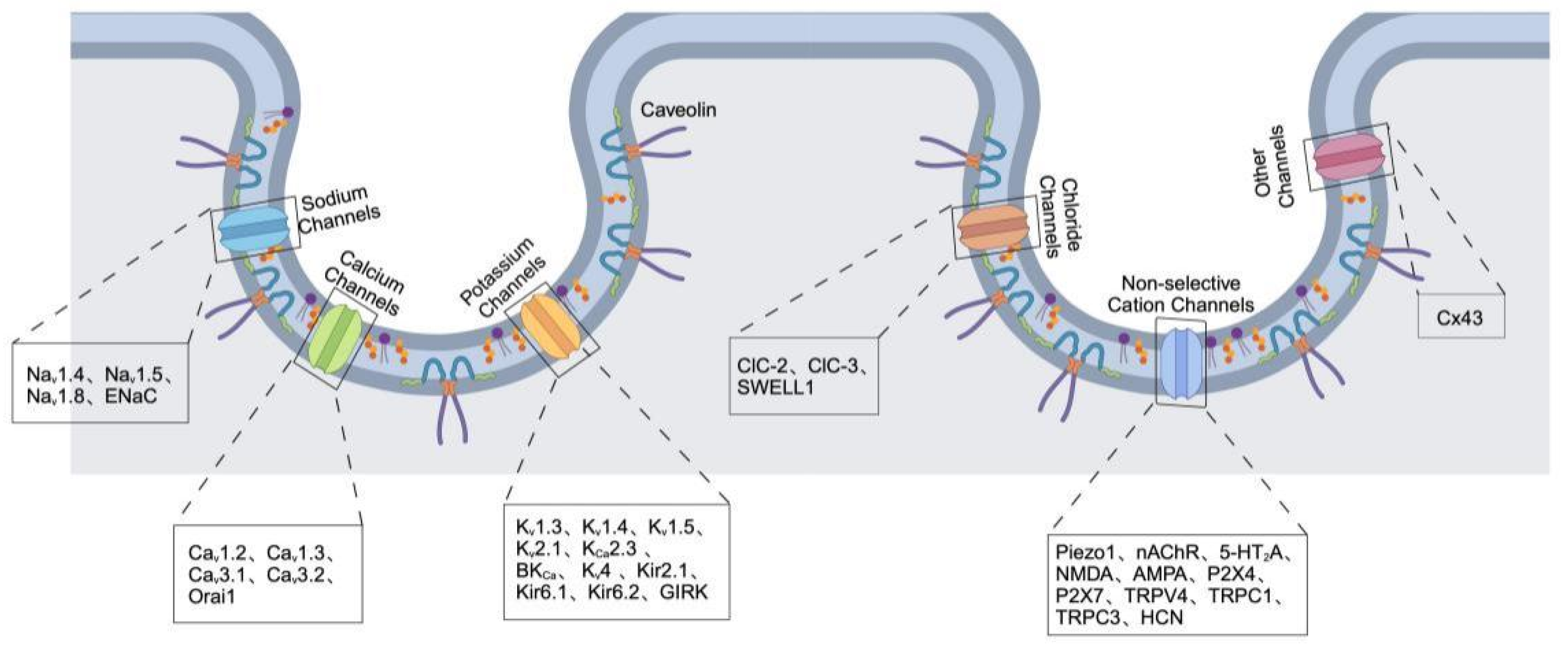
Ion channels are essential membrane proteins that regulate the movement of ions through cellular membranes, crucial for maintaining membrane potential, facilitating signal transduction, and ensuring cellular homeostasis. Abnormalities in ion channel function are associated with various diseases, such as cardiac arrhythmias, neurodegenerative disorders, and cancer [25]. For instance, voltage-gated sodium channels play a vital role in generating action potentials in excitable cells, whereas calcium-activated potassium channels (KCa) are essential for regulating vascular tone and neuronal excitability [26]. Pathogenic mutations or changes in the expression of these channels interfere with cellular function, underscoring their potential as both therapeutic targets and biomarkers for disease [27]. Caveolae are invaginations in the plasma membrane that are rich in cholesterol and contain caveolin proteins, functioning as active signaling platforms that organize ion channels and modulate their activity. Recent studies indicate that caveolae play a crucial role in the localization, trafficking, and functional regulation of ion channels (Table 1) via interactions between lipids and proteins, as well as through scaffolding proteins [28]. Cav-1 directly engages with L-type calcium channels (Cav1.2) to regulate calcium entry in cardiomyocytes, which is essential for the excitation-contraction coupling process [29]. Kv1.5 potassium channels are also localized within caveolae in vascular smooth muscle cells, with their function being influenced by the cholesterol levels present in these caveolar structures [30]. Disruption of caveolae architecture, as observed in Cav-1 knockout models, results in abnormal ion channel activity and is associated with cardiovascular diseases and muscular dystrophies [31]. These results highlight the relationship between ion channel function and the integrity of caveolae (Figure 2), offering a mechanistic basis for comprehending the development of diseases.

2. Ion Channels Localized in Caveolae

2.1. Sodium Channels

2.1.1. Voltage-Gated Sodium Channels (Nav)

Voltage-gated sodium channels (Nav) are strategically positioned within caveolar microdomains, highlighting their complex spatial organization and specialized functional roles in cellular membranes. Early biochemical studies using sucrose density gradient fractionation have effectively shown that sodium channels are concentrated in caveolin-rich membrane fractions, including areas of the plasma membrane and T-tubule membranes, emphasizing the favored localization of Nav channels within caveolae [32]. The biochemical evidence is complemented by molecular interactions that underscore the strong connection between sodium channels and caveolins. Immunoprecipitation studies have revealed a direct link between Cav-3 and the Nav1.4 isoform found in skeletal muscle, indicating a structural relationship that probably enhances specialized signaling and the regulation of channels in muscle cell membranes [33]. Imaging and biochemical studies have demonstrated the colocalization and interaction of Cav-3 with the cardiac-specific sodium channel isoform, Nav1.5. This interaction is crucial for regulating electrical excitability and rhythm in cardiomyocytes [34]. Co-immunoprecipitation studies have reinforced this idea by demonstrating the presence of molecular complexes that include Cav-1, Nav1.5, and sodium-hydrogen exchanger isoform-1 (NHE-1). This underscores the role of caveolae as active signaling platforms where various membrane proteins come together, potentially facilitating coordinated functional interactions [35,36]. Comprehensive biophysical studies utilizing surface plasmon resonance have validated direct interactions between a key segment (amino acids 85–103) of the voltage-gated sodium channel type-X (Nav1.8) α subunit and the scaffolding domain of Cav-1 (amino acids 81–100). This finding sheds light on the molecular mechanism through which Nav channels are selectively recruited and maintained within caveolar membranes [37]. The functional significance of this localization is highlighted by the observed interactions between Cav-3 and Gsα proteins in cardiac myocytes, which occur even without the conventional caveolin-binding motifs. This suggests that sodium channels associated with caveolae may engage in more complex signaling pathways than previously understood, thereby playing a crucial role in regulating cardiac function and cellular excitability [38].

2.1.2. Epithelial Sodium Channels (ENaC)

A key mechanosensitive channel found in caveolae is the epithelial sodium channel (ENaC). These channels are specifically located within caveolar regions on the apical membranes of endothelial cells, where they are closely linked to heme oxygenase-1 (HO-1) and heme oxygenase-2 (HO-2). This arrangement enhances their sensitivity to mechanical forces like stretch and shear stress, indicating that caveolae play a crucial role in the fine-tuned regulation of ENaC-driven sodium transport in reaction to mechanical changes [39]. Biochemical analyses employing detergent-resistant membranes, buoyancy density gradient fractionation, and immunolocalization have confirmed the presence of ENaC in Cav-1-enriched lipid raft compartments within mouse cortical collecting duct cells. This evidence suggests that the localization within caveolar structures could play a crucial role in the stability and functionality of ENaC [40]. Co-immunoprecipitation experiments provide strong evidence for a direct molecular interaction between Cav-1 and ENaC, underscoring the potential regulatory function of Cav-1 in influencing the surface expression and functional behavior of these mechanosensitive sodium channels [41].

2.2. Calcium Channels

2.2.1. Voltage-Gated Calcium Channels (Cav)

Calcium channels, especially the voltage-gated L-type (LTCCs), including Cav1.2 and Cav1.3, and T-type (TTCCs), including Cav3.1 and Cav3.2, are uniquely and functionally positioned within caveolar membrane microdomains. This specific localization is crucial for the accurate regulation of calcium signaling, influencing cellular excitability, contractility, and multiple signal transduction pathways. It has been shown through immunofluorescence in neonatal rat cardiomyocytes that overexpressed LTCC β2a subunits significantly colocalize with Cav-3, particularly in their phosphorylated state, highlighting caveolae as specialized sites for channel modulation and signaling interactions [42]. Electron microscopic analyses utilizing immunogold labeling provide compelling evidence for the colocalization of the Cav1.2 with Cav-3 in specific caveolar structures. Notably, distinct β-adrenergic receptor subtypes exhibit selective modulation of different LTCC populations: activation of β2-adrenergic receptors specifically increases L-type calcium current through caveolar-localized LTCCs in cardiac cells, whereas β1-adrenergic receptors primarily influence LTCCs located outside of caveolae, underscoring a complex and spatially nuanced regulatory mechanism involving caveolae in the heart [43]. Both Cav1.2 and Cav1.3 have been found in caveolar compartments closely associated with Cav-3. However, this interaction shows a decline with age, indicating a dynamic regulation of calcium channel localization throughout the lifespan [44]. Immunofluorescence studies further confirm that Cav1.2 channels are primarily situated within caveolae and other specialized membrane regions, indicating that caveolae could act as essential centers for various signaling interactions related to calcium channels [29]. Recent co-immunoprecipitation research has demonstrated direct interactions between Cav-3 and various calcium channel subtypes, notably Cav1.2 and Cav3.1, highlighting the role of caveolae in structuring specific molecular complexes. These interactions help to compartmentalize essential cardiac pacemaker proteins, including HCN4, Cav1.2, Cav1.3, Cav3.1, and the sodium-calcium exchanger NCX1, into specialized signaling hubs that are vital for maintaining rhythmic electrical activity in cardiac cells [45]. Additional findings from proximity ligation assays conducted on smooth muscle cells from mouse mesenteric arteries validated the specific positioning of the T-type calcium channel Cav3.2 within 40 nm of caveolae marked Cav-1. However, the genetic deletion of Cav-1 disrupts the formation of caveolae and hinders spontaneous transient outward currents, while not directly impacting the activity of the Cav3.2 channel itself [46]. This suggests a complex regulatory mechanism that entails the spatial organization of ion channels without changing their fundamental activity. Immunogold electron microscopy and biochemical assays conducted on ventricular myocytes have demonstrated a distinct colocalization of Cav3.2 channels with Cav-3 within caveolae. This finding is supported by co-immunoprecipitation studies that confirm direct interactions between Cav3.1/Cav3.2 channels and Cav-3. Notably, GST pull-down assays have identified the N-terminal domain of Cav-3 as crucial for its interaction with the Cav3.2 channel, highlighting a specific molecular mechanism for the recruitment of these channels into caveolae and emphasizing the role of caveolae in organizing calcium-dependent signaling pathways [47].

2.2.2. Store-Operated Calcium Channels

Caveolae serve as essential microdomains for store-operated calcium entry (SOCE) channels, notably Orai1. Fluorescence resonance energy transfer analysis, along with immunoprecipitation results, reveals direct molecular interactions between Cav-1 and Orai1, especially during SOCE activation. This interaction plays a crucial role in the dynamic regulation of calcium influx, highlighting the importance of caveolae in maintaining calcium homeostasis [48].

2.3. Potassium Channels

2.3.1. Voltage-Gated Potassium Channels (Kv)

Potassium channels represent a crucial category of voltage-gated ion channels that are strategically positioned within caveolar membrane microdomains, where they are essential for the regulation of cell excitability, metabolism, and signal transduction. Notably, Kv1-family channels, especially Kv1.3, are specifically directed to caveolae through direct interactions with Cav-1. During the differentiation of adipocytes, there is a marked increase in Kv1.3 expression, this protein is targeted to caveolae through its molecular interaction with Cav-1 [49]. This localization holds functional significance, as the integration of Kv1.3 into caveolar raft structures is essential for the proper functioning of insulin signaling pathways, highlighting the critical role of caveolar organization in metabolic regulation [26].
Kv1.5 channels exhibit a greater complexity in their association with caveolae. Biochemical analyses and plasma membrane fractionation have confirmed the co-localization of Kv1.5 with Cav-3 in isolated cardiac caveolar membranes. However, microscopy studies conducted on rat and canine atrial myocytes indicate that there is limited direct spatial overlap between Kv1.5 and Cav-3, suggesting the possibility of subtle or transient interactions occurring within specific lipid raft subdomains [50,51]. Kv1.5 are predominantly found in cholesterol-rich membrane microdomains distinct from traditional caveolae. Functional analyses indicate that the depletion of cholesterol leads to a significant increase in channel current density, highlighting the importance of the lipid microenvironment in determining Kv1.5 functionality [52]. Additionally, the Kv4 subfamily, which includes Kv4.1 and Kv4.3, has been localized within lipid raft domains of pancreatic α-cells, where they are closely associated with Cav-2 and essential signaling and SNARE proteins. This structural configuration suggests that caveolar lipid rafts serve as crucial signaling hubs that regulate hormone secretion and excitability in endocrine cells [53].
Kv1.4, Kv1.5, Kv2.1, Kv4 or Cav channels are located on membrane rafts, sometimes in a tissue-specific manner. Specifically, voltage-dependent potassium channels Kv1.5 and Kv4.3 are found in caveolae in mouse L-cells and cardiomyocytes, respectively, whereas Kv2.1 channels reside on non-caveolar membrane rafts in L-cells and neurons [28,54,55,56]. Large-conductance Ca2+-activated potassium channels (BK channels), particularly hSlo1c, exhibit functional interactions with Cav-1. Cav-1 not only facilitates intracellular trafficking of hSlo1c to plasma membrane domains but also critically modulates barrier permeability in epithelial cells through their molecular association. This relationship underscores a broader role for Cav-1 as a functional regulator beyond mere structural anchoring, intricately influencing potassium channel-mediated physiological processes [57]. Additionally, the role of Cav-1 as a modulatory scaffold for endothelial BKCa channels becomes particularly evident under chronic hypoxia conditions, where loss of Cav-1-mediated inhibition enhances BKCa channel activity, highlighting cholesterol-dependent modulation as a vital aspect of caveolae-mediated control [58].
Small-conductance calcium-activated potassium channels (KCa2.3) are also localized in caveolae. These channels are internalized through a dynamin- and Rab5-dependent process and subsequently recycled via Rab35/EPI64C pathways, which are essential for maintaining their surface expression and functional regulation [59]. The presence of KCa2.3 in caveolar domains is significant for endothelial physiology, particularly as it co-localizes with transient receptor potential channel V4 (TRPV4) in human microvascular endothelial cells, creating a caveolae-dependent signaling complex that does not include intermediate-conductance KCa3.1 channels [60]. This interaction is vital for endothelial function, as the Cav-1 scaffolding of TRPV4/KCa channels promotes vasodilation in response to shear stress. Additionally, co-immunoprecipitation studies have demonstrated that both TRPV4 and KCa2.3 channels associate with Cav-1 in freshly isolated bovine coronary artery endothelial cells, underscoring the importance of the TRPV4-KCa2.3 complex in caveolae in coronary circulation and shear stress detection [61].

2.3.2. Inwardly Rectifying Potassium Channels

Inwardly rectifying potassium channels, particularly ATP-sensitive potassium (KATP) channels, represent an essential class of ion channels prominently enriched within caveolar microdomains, reflecting their crucial roles in regulating membrane potential, cellular excitability, and metabolic responses. Accumulating studies have elegantly highlighted the functional significance of caveolae in modulating channel localization and activity. For instance, smooth muscle KATP channels, notably Kir6.1 subunits, have been localized within caveolin-containing membrane fractions, emphasizing caveolae as specialized platforms that coordinate channel distribution and activity [62,63]. Additionally, Kir6.2 channels exhibit clear colocalization with Cav-3 within cardiomyocyte caveolae, especially concentrated at the peripheral regions of isolated cardiac myocytes. This spatial arrangement underscores caveolae as critical sites for organizing KATP channel function in cardiomyocytes, potentially impacting cardiac electrical stability and myocardial metabolism [64]. Moreover, vascular Kir6.1-containing KATP channels are selectively targeted to Cav-1-rich membrane fractions within vascular smooth muscle cells under specific physiological conditions, implicating caveolae as central platforms mediating channel responsiveness to metabolic and vasoactive stimuli [65].
Caveolae play a crucial role in the dynamic regulation of Kir2 family channels. Specifically, Kir2.1 channels have been shown to interact with Cav-1, as demonstrated by co-immunoprecipitation studies revealing their direct molecular association. This interaction leads to a functional inhibition of Kir2 channel currents in HEK293 cells when Cav-1 is co-expressed, indicating that Cav-1 serves as a vital negative regulator that modulates Kir2 channel activity through caveolar compartmentalization [66]. Additionally, significant co-localization of Kir6.2 and Cav-3 has been observed in isolated cardiomyocytes, particularly in peripheral regions, underscoring the relevance of this interaction for normal cardiac electrophysiological function [64]. Moreover, G-protein gated inwardly rectifying potassium (GIRK) channels act as essential parasympathetic effectors in cardiac cells, integrating signals from multiple pathways. Following agonist stimulation, GIRK channels transiently co-localize with endothelin receptors within caveolae, but not with bradykinin receptors. This redistribution can be disrupted by methyl-β-cyclodextrin (MβCD) treatment, underscoring the necessity of caveolar integrity for effective receptor-channel coupling [67].

2.3.3. Two-Pore Domain Potassium Channels

Two-pore domain potassium (K2P) channels such as TASK-1 and TWIK-1 have been clearly demonstrated to associate with caveolae through interactions with caveolin proteins. Immunofluorescence imaging has elegantly confirmed TASK-1′s prominent colocalization with Cav-3, underscoring caveolae’s role in stabilizing and modulating this potassium channel’s function [68]. Similarly, recent biochemical analyses have identified Cav-1-mediated interactions with both TWIK-1 and TASK channel subtypes, highlighting the strategic localization and functional regulation of these two-pore domain potassium channels within specialized caveolar signaling platforms [69].

2.4. Chloride Channels

Chloride channels found in caveolar microdomains are crucial ion channels that play a significant role in regulating cellular volume, mechanical sensitivity, and epithelial barrier function through their specific membrane localization. The chloride channel ClC-2 interacts directly with Cav-1 and the small GTPase Rab5, which aids in the transport of the tight junction protein occludin to apical membranes, thus influencing epithelial integrity and permeability [70]. Additionally, caveolae membranes act as specialized platforms for mechanosensitive chloride channels (MCCs), including ClC-2, ClC-3, and SWELL1 (LRRC8A). Fluorescence resonance energy transfer imaging has confirmed the strong localization of these MCC within caveolae-rich regions, emphasizing the role of caveolae in regulating mechanosensitive chloride signaling [71]. Moreover, the activation of swelling-induced chloride currents from caveolar-localized ClC-3 and SWELL1 channels highlights their functional importance in mechanically responsive caveolar microdomains. These channels are not merely passive components; they form complex interactions with Cav-3, facilitating precise mechanotransduction and rapid cellular responses to mechanical stimuli, thereby underscoring the dynamic role of caveolae in orchestrating mechanosensitive chloride signaling essential for cellular homeostasis [72].

2.5. Non-Selective Cation Channels

2.5.1. Mechanosensitive Ion Channels and Ligand-Gated Ion Channels

Mechanosensitive ion channels are a distinct group of ion channels that are intricately located within caveolar microdomains, emphasizing the essential function of caveolae in converting mechanical stimuli into biological reactions. Notably, Piezo1 channels demonstrate significant functional modulation and a dynamic relationship with caveolae. In hypertrophic cardiomyocytes exposed to abdominal aortic constriction and angiotensin II, a reduction in the colocalization of Piezo1 with Cav-3 is associated with increased Piezo1 activity and higher channel expression. Additionally, the experimental disruption of caveolae using MβCD similarly enhances Piezo1 functionality, indicating that caveolae act as negative regulators of Piezo1 activity under normal conditions [73]. Moreover, ligand-gated ion channels, which are crucial for regulating cellular excitability and signal transduction, also show specialized localization within caveolar microdomains, indicating their functional integration into discrete signaling complexes. A key example is the nicotinic acetylcholine receptor (nAChR), which closely associates with Cav-3 at the neuromuscular junction. Cav-3 not only colocalizes with nAChRs but also plays a vital role in their clustering and stability. Cav-3 acts as a new binding partner for muscle-specific kinase (MuSK), which is essential for agrin-induced MuSK phosphorylation and subsequent Rac-1 activation, both of which are critical for the proper aggregation and stabilization of nAChR clusters on muscle membranes. The absence of Cav-3 significantly disrupts nAChR aggregation, underscoring its important structural and regulatory function within caveolae in muscle cells [74].
Additionally, serotonergic receptors, especially the 5-HT receptor subtypes, have been identified as important ligand-gated channels associated with caveolae. The 5-HT2A receptor, in particular, engages with Cav-3 when stimulated by serotonin (5-HT), promoting its movement to caveolar membrane regions. This spatial adjustment significantly affects downstream signaling pathways, altering receptor trafficking and expression. Notably, the absence of Cav-3 disrupts the distribution and functional response of 5-HT2A receptors, underscoring the role of caveolae in regulating receptor activity, trafficking, and serotonin-driven hypertrophic signaling in cardiomyocytes [75]. Furthermore, caveolae contain crucial neurotransmitter receptors, such as NMDA, AMPA, and various 5-HT subtypes. The presence of NMDA receptors in caveolae allows for interactions with key regulatory molecules like glycine, glutamate, and aspartate, enabling precise modulation of these receptors. The caveolar localization of these receptors facilitates complex downstream signaling cascades initiated by 5-HT stimulation, which may lead to the activation of adenylyl cyclase (AC), which converts adenosine triphosphate (ATP) into 3′,5′-cyclic adenosine monophosphate (cAMP). This, in turn, may activate cAMP-dependent protein kinase and subsequently AMP-activated protein kinase (AMPK), thereby modulating autophagy/mitophagy, linking caveolar positioning to essential metabolic and cellular homeostatic mechanisms [76,77]. Additionally, ATP-gated ion channels, such as purinergic P2X receptors, are also found partially within caveolae. Studies using immunofluorescence and GST pull-down assays have demonstrated that Cav-1 interacts with both P2X4 and P2X7 receptors, placing these ATP-gated channels in specific caveolar membrane compartments. This partial localization in caveolae influences ATP sensitivity and signaling specificity, indicating that caveolae-mediated compartmentalization is vital for modulating purinergic receptor function and responsiveness [78].

2.5.2. Transient Receptor Potential Channels

Transient receptor potential (TRP) channels are precisely localized within caveolar microdomains, indicating a specialized membrane structure that significantly influences their functional characteristics. Notably, TRPV4 channels are closely associated with caveolae, interacting with Cav-1 in the plasma membrane. At physiological temperature, TRPV4 shows strong colocalization with Cav-1, however, treatments that disrupt cholesterol integrity, such as MβCD or pravastatin, lead to altered membrane distribution and decreased colocalization, highlighting the reliance of caveolae on cholesterol for sustaining TRPV4 localization and signaling [79]. Freshly isolated bovine coronary endothelial cells also demonstrate targeted localization of TRPV4 channels within caveolae, alongside KCa2.3 channels, suggesting a functional interplay enabled by caveolar compartmentalization [60,61]. Interestingly, some studies indicate a more complex scenario: cholesterol depletion via MβCD paradoxically increases the membrane expression of TRPV4, Cav-1, and flotillin, yet shows limited direct colocalization, suggesting variations in caveolar association across different cell types [80]. Furthermore, advanced proximity ligation assays have confirmed direct spatial relationships among TRPV4, Cav-1, and BK channels in arterial endothelial cells, underscoring caveolae as central regulatory platforms that integrate these channels for coordinated vascular responses [81]. Additionally, canonical TRP channels like TRPC1 and TRPC3 are also enriched in caveolae, with TRPC1 showing close colocalization and physical interaction with Cav-3 in muscle tissues, further emphasizing the importance of caveolar domains in organizing TRP channel function [82]. Immunofluorescence investigations in arterial smooth muscle have shown a partial colocalization of TRPC1 with Cav-1, which supports the idea that caveolar microdomains act as structural modulators of TRPC-mediated signaling [83]. Additionally, co-immunoprecipitation analyses from cerebral artery lysates indicate direct interactions between Cav-1, TRPC3, and IP3 receptor type 1, further emphasizing the role of caveolae as essential signaling hubs for TRPC channel complexes [84].

2.5.3. Hyperpolarization-Activated Cyclic Nucleotide-Gated (HCN) Channels

Hyperpolarization-activated cyclic nucleotide-gated (HCN) channels play a crucial role in regulating cardiac pacemaker function and are specifically localized within caveolae. It has been demonstrated that direct interactions exist between HCN channels and Cav-3 in cardiac cells, indicating that caveolae are essential for maintaining channel stability, surface expression, and overall functional activity. The functional and expression levels of HCN channels are indeed modulated through interactions with Cav-3, thereby contributing to the integrity of cardiac rhythmicity and conduction systems [45,85].

2.6. Other Channels

Gap Junction Channels

Gap junction channels, primarily composed of connexins like connexin 43 (Cx43), are notably localized within caveolar microdomains, underscoring the active role of caveolae in facilitating intercellular communication rather than serving solely as structural supports. Among the 20 rodent and 21 human connexin isoforms, Cx43 is the most abundantly expressed connexin protein in the heart. As a typical integral membrane protein, Cx43 is synthesized in the rough endoplasmic reticulum, then translocates to the Golgi apparatus where it undergoes folding and oligomerization into connexons. These connexons, referred to as hemichannels (HCs), are then transported along the secretory pathway to their final destination in the plasma membrane [86,87,88,89,90,91,92]. Cardiac Cx43 creates GJCs at intercellular contacts and hemi-channels (HCs) at the peri-junctional plasma membrane and sarcolemmal caveolae/rafts compartments. These GJCs are fundamental for the direct cardiac cell-to-cell transmission of electrical and molecular signals, ensuring synchronous myocardial contraction. Electron microscopic images of adult rat heart cardiomyocytes show Cx43 structures, including the Golgi compartment where Cx43 is oligomerized into HCs, and post-Golgi HCs that are targeted to caveolae/rafts and gap junctions, forming paired HCs that establish functional GJCs between neighboring cardiomyocytes [93]. Indeed, Cx43 shows robust colocalization, co-fractionation, and co-immunoprecipitation with Cav-1. Notably, molecular interactions have identified two distinct Cav-1 domains, the scaffolding domain (residues 82–101) and the C-terminal domain (135–178), as critical sites mediating direct interactions with Cx43. These interactions suggest that caveolae create a complex spatial environment that enhances the organization, stability, and functionality of Cx43 within gap junctions [94]. The compartmentalization of gap junctions mediated by caveolae holds significant relevance across various cell types. In rat epidermal keratinocytes, Cx43 co-immunoprecipitates and colocalizes with Cav-1 and Cav-2, confirming their close spatial relationship. Importantly, this colocalization has also been observed in human epidermal keratinocytes, indicating that caveolar microdomains serve as a reliable structural framework for Cx43-mediated cell communication within the epidermal barrier [95]. Intriguingly, gap junction channels display a dynamic distribution in response to physiological stimuli, such as ATP, which prompts the rapid movement of Cx43 into caveolae, correlating with changes in gap junction communication [96]. Yeast two-hybrid studies further validate the direct interactions between caveolin and connexins, highlighting the active regulatory role of caveolae beyond mere structural support [97].
Table 1. Ion channels in caveolae: localization and caveolin interactions.
Channel TypeLocalization in Caveolae/Interaction with CaveolinsCaveolin Isoforms InvolvedReferences
Sodium Channels
Nav1.4Localized within muscle caveolae, direct interactionCav-3[32,33]
Nav1.5Caveolae-localized; forms signaling complex affecting cardiac excitabilityCav-1, Cav-3[34,35,36]
Nav1.8Stabilized within caveolae by direct interactionCav-1[37]
ENaCCaveolae-localized in endothelial cells; mechanosensitive sodium transportCav-1[39,40,41]
Calcium Channels
Cav1.2Caveolae-localized; regulate cardiac calcium signaling and pacemaker activityCav-3[29,42,43,44,45]
Cav1.3
Cav3.1, Cav3.2Localized in caveolae; regulate cardiac pacing and calcium signalingCav-1, Cav-3[45,46,47]
Orai1 Caveolae localization; modulates calcium influxCav-1[48]
Potassium Channels
Kv1.3Localized in caveolar lipid rafts; insulin signalingCav-1[49,98]
Kv1.4Tissue-specific localization in caveolar/non-caveolar raftsCav-2, Cav-3[50,51,52,53,54]
Kv1.5
Kv2.1
Kv4
KCa2.3 (SK3)Localized in caveolaeCav-1[59,60,61]
BK Channels (hSlo1c, BKCa)Localized in caveolae; regulate permeability and hypoxic responseCav-1[57,58]
Kir2.1Localized in caveolae; regulate membrane potential and metabolismCav-1, Cav-3[62,63,64,65,66].
Kir6.1
Kir6.2
GIRKLocalized in caveolaeCav-3[67]
TASK-1 and TWIK-1Colocalized with Cav-1/3Cav-1, Cav-3[68,69]
Chloride Channels
ClC-2Localized in caveolae; regulate cell volume and mechanosensitivityCav-1, Cav-3[70,71,72]
ClC-3
SWELL1
Non-selective Cation Channels
Piezo1Colocalized in cardiomyocytes with Cav-3Cav-3[73]
nAChR, 5-HT2A, NMDA, AMPA, P2X4, P2X7Localized in caveolae; modulate neurotransmitter responses and excitabilityCav-1, Cav-3[75,76,77,78,99]
TRPV4, TRPC1, TRPC3Localized in caveolae; regulate thermal and mechanical responsesCav-1, Cav-3[60,61,79,80,81,82,83,84]
HCNCaveolae-localized; regulate cardiac pacing and rhythmicityCav-3[45,85]
Other Channels
Cx43Caveolae-localized; regulate intercellular communication and stabilityCav-1, Cav-2[94,95,96,97]

3. Regulation of Ion Channels by Caveolae

3.1. Direct Interaction Between Caveolin Scaffolding Domains and Ion Channels

Caveolins, as the structural proteins of caveolae, play a crucial role in modulating ion channel function by directly interacting with specific channel domains. The caveolin scaffolding domain (CSD) acts as a primary mediator in these interactions, recognizing and binding to ion channel proteins through a conserved caveolin-binding motif (CBM), characterized by an arrangement of aromatic amino acids in the sequence QxQxxxxQ (Table 2), where Q represents tyrosine (Y), tryptophan (W), or phenylalanine (F) [100]. These interactions impact channel localization, function, and regulatory mechanisms, influencing physiological and pathological processes. In Kir channels, both the scaffolding and membrane domains of Cav-3 are necessary for proper channel localization within caveolar microdomains, since disruption of either the Kir2.x CBM sequence or the Cav-3 domain alters channel distribution and function [101,102]. Similarly, KATP channels, specifically the Kir6.2/SUR2A complex, are negatively regulated by Cav-3, with evidence showing that a Cav-3-derived scaffolding domain peptide significantly inhibits channel function [103]. TRP channels are another class of ion channels regulated by caveolin scaffolding domains. The primary Cav-1 binding region in TRPC5 has been mapped to residues 295–322, highlighting a direct molecular interaction [104]. Furthermore, Cav-1 directly associates with TRPC1, particularly its C-terminal domain, playing an essential role in store-operated calcium entry in endothelial cells [105]. Modulation of this interaction, either by increasing Cav-1 scaffolding domain expression or inhibiting calpain activity, has been shown to significantly alleviate lipopolysaccharide -induced cardiac dysfunction and myocyte apoptosis/autophagy [106].
The large-conductance, voltage- and calcium-activated potassium (BK, MaxiK) channel also interacts with Cav-1, with its α-subunit (Slo1) containing a specific sequence (YNMLCFGIY) that facilitates this interaction [107]. This binding appears to serve dual functions: (i) maintaining BK channels in intracellular compartments, thereby limiting surface expression, and (ii) anchoring plasma membrane-resident channels to Cav-1-enriched domains. Importantly, Cav-1 exerts an inhibitory effect on BK channel activity through its scaffolding domain (residues 82–101), a regulation that may be relieved under conditions such as chronic hypoxia, likely via cholesterol-dependent mechanisms [58,108]. In addition, Cx43 has been shown to interact with two distinct regions of Cav-1, the scaffolding domain and the C-terminal domain, suggesting that Cav-1 plays a complex role in regulating intercellular communication through direct channel binding [94].

3.2. Lipid-Dependent Modulation of Ion Channel Function

Caveolae possess a unique lipid composition, enriched with cholesterol, sphingolipids (sphingomyelin and glycosphingolipids), and phosphatidylinositol, which are essential for their structural integrity and functional properties [109]. These lipid components play a crucial role in the regulation of ion channels, influencing their localization, stability, and activity within the membrane microdomains.
Cholesterol is a key regulator of ion channel function, exerting both positive and negative effects on different channels through multiple mechanisms. It can directly interact with specific amino acid residues in ion channels or regulate their recruitment into specialized membrane domains such as lipid rafts and caveolae [110]. For example, TRPV4 is localized within cholesterol-rich caveolar domains, and its activity is strongly influenced by its surrounding membrane lipids, including cholesterol and phosphatidylinositol. The depletion of cholesterol has been shown to modulate TRPV4 activation, enhancing calcium influx in a dose-dependent manner [111]. Similarly, cholesterol depletion has profound effects on voltage-gated potassium channels. The interaction of KV7.1 and Cav-1 is significantly disrupted upon cholesterol depletion, leading to increased membrane localization of Kv7.4, mimicking the effect of ciliobrevin D treatment [112]. Kv1.5, although not localized to caveolae, resides in cholesterol-rich lipid rafts, and its redistribution following cholesterol depletion increases its current-carrying capacity [52]. Kv2.1 activation at more negative potentials is induced by sphingomyelinase D treatment, highlighting the impact of lipid modifications on channel gating [113]. Cholesterol plays a crucial role in modulating calcium channels in a highly channel-specific manner. For example, cholesterol depletion enhances CaV2.1channel current [114]. In cardiac myocytes, cholesterol depletion affects Orai1 channels by promoting their internalization, leading to reduced calcium influx and altered diffusion of the channel within the cell membrane. Notably, Cav-1 overexpression restores Orai1 localization and function, highlighting the role of cholesterol-rich domains in maintaining calcium channel activity [48].
Beyond cholesterol, polyunsaturated fatty acids (PUFAs), another key component of caveolae, significantly alter membrane microdomain composition and ion channel activity. In cardiac cells, PUFAs modulate sodium channel gating, reduce sodium current, and shorten the action potential duration [115]. They also directly regulate L-type calcium current, reducing calcium transients and calcium-activated membrane currents [116]. Likewise, phosphatidylinositol 4,5-bisphosphate (PIP2), a negatively charged phospholipid enriched in caveolae, plays a crucial role in ion channel modulation, particularly affecting Na/Ca exchangers and KATP channels, both of which are present in cardiac caveolae. Generally, increased PIP2 levels activate Kir channels, whereas depletion inhibits their activity, demonstrating the critical role of membrane lipids in potassium channel regulation [117]. The functional importance of lipid-caveolae interactions is further illustrated in vascular regulation.
Table 2. Interaction of caveolin proteins/caveolae with lipids and proteins in ion channel regulation.
Caveolin/Caveolae ComponentInteraction PartnerKey Interaction Domain or MechanismEffect on Ion Channel Localization, Trafficking, or FunctionReference
Caveolin-3Kir2.x (Kir channels)Caveolin scaffolding domain (CSD); Caveolin-binding motif (CBM)Regulates proper localization in caveolae; disruption alters channel distribution and functionand function[100,101,102]
Caveolin-3Kir6.2/SUR2A (KATP channels)Caveolin scaffolding domain peptideNegative regulation; inhibits channel function [103]
Caveolin-1TRPC5Amino acids 295–322Direct binding modulates channel localization and function[104]
Caveolin-1TRPC1C-terminal domainRegulates store-operated calcium entry; affects cardiac cell survival and function[105,106]
Caveolin-1BK (MaxiK) channel (Slo1 <-subunit)Slo1 sequence: YNMLCFGIY; Caveolin scaffolding domain residues 82–101Dual function: Limits surface expression; anchors channels in caveolin-rich domains; negatively regulates channel activity[58,107,108]
Caveolin-1Connexin 43 (Cx43)Scaffolding and C-terminal domainsComplex regulation of intercellular communication[94]
Cholesterol (lipid)TRPV4Cholesterol-rich caveolar domainsCholesterol depletion enhances TRPV4-mediated Ca2+ influx[110,111]
CholesterolKV7.1/KV7.4Cholesterol-dependent caveolin interactionsCholesterol depletion disrupts interaction, alters channel localization[112]
CholesterolKV1.5Cholesterol-rich lipid raft localizationCholesterol depletion increases channel current capacity[52]
Cholesterol/SphingolipidsKV2.1Membrane lipid modification by sphingomyelinase DInduces activation at more negative potentials[113]
CholesterolCaV2.1Cholesterol depletionEnhances channel current[114]
CholesterolOrai1Cholesterol-rich domainsDepletion causes internalization and reduced Ca2+ influx; caveolin-1 reverses effects[48]
PUFAsCardiac Na+ and L-type Ca2+ channelsLipid microdomain modulationReduces currents; shortens action potential duration[115,116]
PIP2Kir channels; Na+/Ca2+ exchangers; KATP channelsMembrane lipid modulationIncreased PIP2 activates Kir channels; depletion inhibits activity[117]

3.3. Compartmentalization of Signaling Cascades

Caveolae serve as specialized membrane microdomains that compartmentalize key signaling molecules, facilitating efficient and localized signal transduction (Figure 3). These domains are enriched with G protein-coupled receptors (GPCRs), heterotrimeric G proteins, steroid hormone receptors, receptor and non-receptor tyrosine kinases, protein kinase A (PKA) and C (PKC), phosphoinositide 3-kinase (PI3K)/Akt, mitogen-activated protein kinases (MAPK), and nitric oxide synthases (NOS), all of which have been shown to interact directly with caveolins [118]. This spatial organization enables precise regulation of ion channel function through localized cAMP/PKA signaling, nitric oxide (NO)-mediated modulation, and kinase-dependent phosphorylation events.
Caveolae play a critical role in modulating ion channel function through NOS-related signaling cascades. In cardiomyocytes, Cav-3 acts as a negative regulator of late sodium current via nNOS-dependent S-nitrosylation of SCN5A, underscoring the role of caveolae in regulating cardiac excitability [119]. Similarly, TRPC5 activation upon PLC-mediated ATP stimulation triggers Ca2+ influx, leading to endothelial NO synthase (eNOS) activation and NO production, which in turn induces further TRPC5 activation via cysteine S-nitrosylation. Mutations in the Cav-1 binding domain of TRPC5 disrupt this interaction, impairing both Ca2+ influx and NO production, suggesting that Cav-1 scaffolds TRPC5 and eNOS into a functional signaling complex [104].
Caveolae also serve as key regulators of cAMP/PKA-dependent ion channel modulation by compartmentalizing signaling components. Disrupting caveolae with MβCD reduces whole-cell KATP currents sensitive to PKA, indicating that caveolar integrity is crucial for adenylyl cyclase (AC)-mediated channel regulation [120]. In ventricular myocytes, α1-adrenergic receptor (α1-AR) activation modulates transient outward K+ current through a PKA-dependent pathway, but this effect remains spatially restricted within caveolae. Components of this pathway, including Kv4.2/Kv4.3 channels, PKA, and AKAP100, are assembled in caveolae through interactions with Cav-3, forming a functional microdomain distinct from α1-AR, Gαs, and adenylyl cyclase complexes [54,121]. L-type calcium channels are also targeted to caveolae, where CaMKII-mediated phosphorylation promotes cardiac hypertrophy. Disrupting caveolar integrity with MβCD significantly attenuates this effect, likely due to the loss of PKA-mediated channel phosphorylation and microdomain-specific signaling [42,45].
β-adrenergic receptor (β-AR) signaling is another pathway that is compartmentalized within caveolae. β2-ARs are primarily localized to caveolae in the basal state but translocate out of caveolae upon activation, leading to cAMP production by adenylyl cyclase V/VI, which is also highly concentrated within caveolae [122]. Cholesterol depletion disrupts β2-AR signaling, suggesting that caveolae act as negative regulators of cAMP accumulation, restricting its diffusion and ensuring localized modulation of ion channels. In ventricular myocytes, β2-AR activation leads to localized cAMP production near LTCCs, enhancing Ca2+ influx via PKA-dependent phosphorylation without broadly affecting other cAMP-dependent processes [43,44]. This compartmentalization is essential for precise regulation of cardiac pacemaking, as β2-ARs in caveolae localize near HCN4, CaV1.2, and CaV1.3 channels, ensuring their coordinated regulation [44].
Caveolae also mediate Angiotensin II (Ang II)-dependent inhibition of KATP channels in vascular smooth muscle cells. Upon Ang II stimulation, PKCη is selectively recruited to caveolae, where it phosphorylates and inhibits KATP channels, highlighting the role of caveolae in spatially organizing PKC isoforms for targeted channel modulation [123]. Additionally, caveolae coordinate Ca2+-dependent signaling cascades by scaffolding key proteins such as TRPV4, BKCa, and RyRs, which regulate vascular tone and excitation-contraction coupling. In aged VSMCs, the loss of caveolar CaV3.2-RyR coupling alters Ca2+ signaling, suggesting that caveolae spatially control local Ca2+ release and vasodilation [124]. The activation of PKCε has been shown to mediate the internalization of vascular KATP channels (Kir6.1/SUR2B) via Cav-1, leading to channel inhibition. This process is disrupted by cholesterol depletion, dynamin mutations (K44E), or Cav-1 knockdown, further emphasizing the role of caveolae as essential platforms for channel trafficking [125].

3.4. Mechanosensitive Regulation of Ion Channels by Caveolae

Caveolae serve as mechanically deformable membrane invaginations, providing structural resilience to cells experiencing mechanical stress. These cholesterol-rich microdomains play a crucial role in mechanotransduction, buffering mechanical forces and modulating ion channel activity in response to changes in membrane tension. The loss or disruption of caveolae has been associated with increased mechanical sensitivity, dysregulated ion channel activity, and pathological remodeling in cardiovascular and muscular diseases.
Cav-3, the predominant scaffolding protein in muscle caveolae, plays a critical role in maintaining membrane stability and regulating mechanosensitive ion channels. Mutations in Cav-3 lead to limb-girdle muscular dystrophy, resulting in cytoskeletal defects that cause excessive activation of mechanosensitive cation channels and increased intracellular Ca2+ levels. While Cav-3 normally protects the sarcolemma from mechanical stress, its upregulation in dystrophic conditions may act as a compensatory mechanism to counteract mechanical instability [126]. In the cardiovascular system, caveolae regulate mechanotransduction in the sinoatrial node, integrating mechanical and biochemical signaling factors that influence cardiac automaticity. The presence of eNOS, NOX2-derived reactive oxygen species (ROS), and Ca2+ dynamics within caveolae is believed to suppress excessive mechanotransduction. However, the chronic loss of caveolae under sustained shear stress leads to uncontrolled activation of these signaling pathways, creating aberrant feedback loops associated with cardiac pathologies [127].
Caveolae function as a reserve to absorb mechanical forces and limit excessive activation of mechanosensitive channels. Disrupting caveolae, such as through cholesterol depletion, diminishes this reserve capacity, leading to accelerated cell swelling under hypotonic conditions and enhanced activation of swelling-activated chloride current [128]. Cav-3 has also been shown to influence SWELL1-mediated Cl- currents, where its expression enhances SWELL1 activity, conferring resistance to swelling-induced membrane damage [71]. In cardiac myocytes, Cav-3 downregulation increases sensitivity, which may contribute to atrial fibrillation in hypertensive patients [72]. These findings highlight the importance of caveolae in maintaining cellular homeostasis under mechanical stress by modulating ion channel responsiveness.
Cav-1 serves as a scaffold for P2Y1 and P2Y2 receptors, stabilizing them in cholesterol-rich microdomains to regulate downstream signaling cascades involved in mucosal secretion, motility, and pain transmission. In alveolar cells, ATP release in response to mechanical expansion activates P2Y2 receptor-dependent Ca2+ signaling, which in turn regulates surfactant secretion, emphasizing the role of caveolae in lung mechanotransduction [129]. Piezo1, a key mechanosensitive channel implicated in cardiac hypertrophy and fibrosis, is similarly regulated by Cav-3. Under conditions of pressure overload or angiotensin II stimulation, Piezo1 relocates out of caveolae, leading to enhanced channel function and increased expression [73]. This suggests that caveolae act as regulatory compartments, restricting Piezo1 activity under normal conditions to prevent excessive mechanotransduction that contributes to pathological cardiac remodeling.
Caveolae also contribute to endothelial mechanosensitivity by regulating ENaCs. These channels reside in Cav-1-enriched microdomains, where their function is influenced by heme oxygenase activity. Under shear stress, HO-1 upregulation leads to increased carbon monoxide production, which enhances ENaC activity and promotes Na+ influx. Persistent ENaC activation is linked to endothelial dysfunction and vascular diseases, establishing a connection between caveolae disruption and pathological states such as atherosclerosis [39].

3.5. Caveolae-Mediated Regulation of Ion Channels by Other Channels and Proteins

Caveolae, specialized lipid microdomains in the plasma membrane, serve as platforms for the regulation of various ion channels. These lipid rafts not only facilitate channel trafficking and localization but also regulate their functional activity through interactions with other ion channels and membrane-associated proteins.
Caveolae facilitate cross-talk between different ion channels, influencing their activity and functional coordination. A notable example is the interaction between KCa3.1 and BKCa channels, where KCa3.1 activation suppresses BKCa channel activity. This cross-regulation is cholesterol-dependent and cytoskeleton-regulated, as cholesterol depletion weakens the interaction, whereas cholesterol enrichment enhances it, demonstrating that caveolae and lipid raft integrity are essential for inter-channel modulation [130]. In vascular smooth muscle cells, caveolae also facilitate the coupling between BKCa and LTCC, enabling coordinated regulation of vascular tone and excitation-contraction coupling. Cav-1 directly interacts with Cav1.2 to regulate calcium signaling, forming a molecular complex that governs the spatial and temporal calcium dynamics necessary for smooth muscle excitability and contraction [131].
In endothelial cells, caveolae are integral to the regulation of ion channels that control vascular relaxation and mechanosensation. TRPV4 and KCa2.3 channels, which are critical for endothelial-dependent vasodilation, co-localize within caveolae, forming a functional complex that contributes to coronary circulation regulation [61]. Additionally, ENaCs are mechanosensitive and reside in caveolae, where they modulate vascular function in response to shear stress. The upregulation of heme oxygenase-1 leads to increased carbon monoxide production, which enhances ENaC activity and sodium influx, potentially contributing to endothelial dysfunction and vascular disease [39].
Beyond direct ion channel interactions, caveolae also regulate channel trafficking and endocytosis through associated proteins. In cardiomyocytes, EHD2 interacts with caveolae to stabilize the localization of KATP channels, thereby influencing their trafficking and turnover. This regulatory mechanism highlights the broader role of caveolae in controlling ion channel distribution and function through protein interactions [64].

4. Caveolae and Diseases

4.1. Cardiovascular Diseases

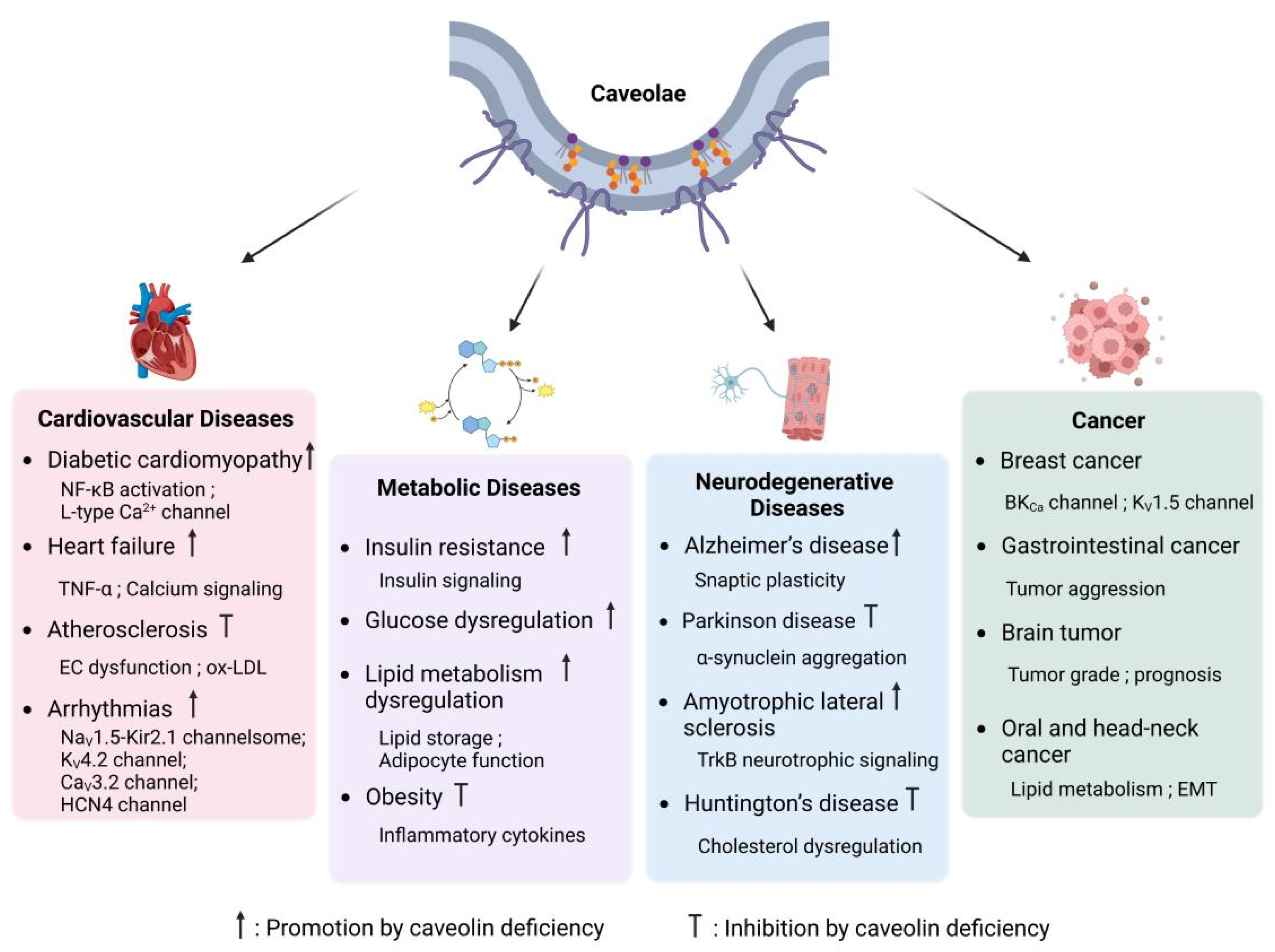
Caveolae are essential components in cardiovascular physiology, playing a pivotal role in regulating endothelial function, lipid metabolism, ion channel activity, and myocardial signaling. Dysfunction of caveolae is implicated in the pathogenesis of a wide range of cardiovascular diseases, such as atherosclerosis, diabetic cardiomyopathy, heart failure, and arrhythmias (Figure 4). By modulating mechanotransduction, inflammatory pathways, and the compartmentalization of ion channels, caveolae significantly influence disease progression and emerge as potential therapeutic targets for cardiovascular disorders.

4.1.1. Atherosclerosis

Caveolae play a pivotal role in the initiation and progression of atherosclerosis, primarily through their involvement in lipid metabolism and endothelial dysfunction. It has been demonstrated that caveolae mediate the endocytosis and transcytosis of oxidized low-density lipoprotein (ox-LDL), promoting lipid accumulation within vascular cells [132]. Inhibition of this pathway, such as through Cav-1 silencing, significantly reduces ox-LDL uptake and foam cell formation, underscoring the contribution of caveolae to lipid-driven atherogenesis [133,134]. Beyond lipid transport, caveolae are critical regulators of vascular inflammation and remodeling. They participate in the activation of NF-κB signaling, a key pathway driving endothelial dysfunction and vascular inflammation [135,136]. Furthermore, caveolae modulate vascular smooth muscle cell (VSMC) behavior, where their dysfunction enhances VSMC proliferation and migration—processes closely linked to plaque instability and restenosis [135,137,138,139].

4.1.2. Diabetic Cardiomyopathy

The role of caveolae in diabetic cardiomyopathy (DCM) is closely tied to their regulation of inflammatory signaling and myocardial remodeling. In diabetic mouse models, Cav-1 deficiency exacerbates cardiac dysfunction, characterized by increased hypertrophy, fibrosis, and inflammation [140]. This pathological progression is associated with enhanced NF-κB activation, which upregulates inflammatory and hypertrophic genes, further driving disease severity. Conversely, Cav-1 overexpression has been shown to mitigate these effects, highlighting its protective role in DCM. In cardiomyocytes, caveolae play a critical role in maintaining calcium homeostasis and excitation-contraction coupling. The disruption of caveolae, particularly through Cav-3 depletion, impairs T-tubule organization and L-type Ca2+ currents, resulting in calcium mishandling and exacerbating diabetic cardiomyopathy [141,142,143]. Furthermore, caveolae are implicated in the development of left ventricular hypertrophy and pulmonary hypertension. Their loss promotes excessive MAPK signaling and endothelial dysfunction, which collectively worsens cardiac remodeling [144].

4.1.3. Heart Failure

Caveolae play a critical role in maintaining myocardial integrity, and their dysfunction is closely related to the progression of heart failure. In heart failure models, Cav-3 expression is significantly reduced, which correlates with deteriorating left ventricular function and T-tubule disorganization. In patients with heart failure, reduced levels of Cav-3 are associated with impaired calcium signaling and contractile dysfunction, underscoring its critical role in maintaining myocardial function. Moreover, overexpression of tumor necrosis factor-α (TNF-α) has been shown to contribute to Cav-3 depletion in failing hearts, suggesting an inflammation-mediated mechanism underlying caveolar loss. Given its essential functions in ion channel compartmentalization and T-tubule stabilization, Cav-3 deficiency leads to electrical instability and contractile impairment, further contributing to heart failure pathogenesis [145].

4.1.4. Arrhythmias

Caveolae are essential for maintaining cardiac excitability, and their dysfunction is associated with the occurence of arrhythmias. Specifically, mutations or dysregulation of Cav-3, a key structural protein of caveolae, can significantly impair ion channel function, thereby inducing proarrhythmic alterations. For instance, Cav-3 mutations have been shown to diminish inward rectifier potassium currents [102,146], while enhancing late sodium currents, predisposing the heart to ventricular arrhythmias. Additionally, overexpression of Cav-3 can disrupt diastolic Ca2+ transients, leading to aberrant L-type Ca2+ channel activity and further exacerbating the propensity for ventricular arrhythmias [47,147,148]. Furthermore, Cav-3 deficiency disrupts HCN4 channel function, impairing pacemaker activity and contributing to conduction disorders [149,150]. Given the role of caveolae in ion channel compartmentalization, their disruption leads to electrical instability, increasing susceptibility to atrial and ventricular arrhythmias.

4.2. Metabolic Diseases

Caveolae play a pivotal role in the regulation of metabolic processes, serving as critical platforms for insulin signaling, lipid metabolism, and the modulation of inflammatory pathways. Dysfunction of caveolae has been strongly associated with the pathogenesis of metabolic disorders, including insulin resistance, obesity, and type 2 diabetes. These conditions are characterized by disrupted glucose homeostasis, aberrant lipid metabolism, and an elevated risk of cardiovascular complications. By orchestrating interactions between membrane-associated proteins and key signaling molecules, caveolae finely tune hormonal responses and energy balance, underscoring their essential role in preserving metabolic stability and overall physiological homeostasis.

4.2.1. Insulin Resistance and Glucose Dysregulation

Caveolae play a critical role in the proper functioning of the insulin receptor (IR) and the facilitation of insulin-mediated glucose uptake. Specifically, the IR forms a dynamic complex with Cav-1 and ganglioside GM3 within caveolae, a structural arrangement essential for optimal receptor activity. Disruption of this tripartite interaction compromises IR mobility and stability, leading to impaired insulin signaling and subsequent metabolic dysregulation [151]. In insulin-resistant adipocytes, elevated levels of ganglioside GM3 disrupt the IR-Cav-1 complex, leading to reduced insulin sensitivity and contributing to adipose tissue insulin resistance, a hallmark of metabolic syndrome. Similarly, in the heart, insulin signaling is critically dependent on caveolae. Under high-fat diet conditions, Cav-3 dissociates from the insulin receptor β-subunit, thereby impairing insulin-mediated cardioprotective effects. This pathological dissociation is associated with tyrosine nitration at Tyr73 in Cav-3, a modification that disrupts IR complex formation and inhibits downstream insulin signaling pathways [152]. Restoration of Cav-3 expression or inhibition of its nitration rescues insulin signaling and provides protection against diabetes-induced ischemic heart failure, underscoring the importance of maintaining caveolae integrity for preserving insulin sensitivity in cardiac tissues. Additionally, genetic variations in Cav-1 have been implicated in systemic insulin resistance. Epidemiological studies in hypertensive populations have demonstrated that Cav-1 single nucleotide polymorphisms (SNPs) are associated with elevated fasting insulin levels, increased homeostasis model assessment of insulin resistance scores, and impaired glucose disposal during insulin clamp studies [153]. In Cav-1 knockout mice, metabolic dysfunctions are intensified, indicating that genetic variations in Cav-1 may act as biomarkers for the early identification and management of insulin resistance and metabolic disorders. In skeletal muscle, the predominant isoform is Cav-3, and its deficiency causes considerable impairment in glucose uptake, which in turn leads to diminished insulin-stimulated signaling, glucose intolerance, and dyslipidemia [154]. These results emphasize the importance of caveolae in preserving insulin sensitivity and regulating glucose metabolism in muscle tissue, underscoring Cav-3 as a crucial element in muscle insulin resistance and overall metabolic health.

4.2.2. Obesity and Lipid Metabolism Dysregulation

Caveolae are essential for lipid storage and adipocyte function, with Cav-1 and Cav-3 being vital for maintaining adipose tissue balance. In cases of obesity, there is a marked increase in Cav-1 expression, which is associated with elevated levels of pro-inflammatory cytokines like TNF-α and the activation of NF-κB [155]. Inflammatory pathways contribute to the dysfunction of adipose tissue and the development of metabolic syndrome, reinforcing the importance of caveolae in regulating adipose inflammation and lipid metabolism. In contrast, the absence of Cav-1 offers protection against obesity caused by diet. Investigation involving Cav-1 knockout mice demonstrates a lean phenotype, resistance to obesity from high-fat diets, and notable decreases in adipose tissue mass [31]. These mice exhibit severe lipid metabolism defects, including reduced triglyceride storage, elevated circulating free fatty acids, and impaired lipid droplet formation. Mechanistically, Cav-1 loss disrupts adipocyte caveolae, impairing lipid uptake and storage, which may contribute to altered systemic lipid homeostasis.
In pediatric populations, Cav-1 gene polymorphisms have been associated with metabolic syndrome in obese children [156]. This suggests that caveolae dysfunction may contribute to early-onset obesity and metabolic disturbances, reinforcing their role as potential therapeutic targets for metabolic disease prevention.

4.3. Neurodegenerative Diseases

Caveolae, as cholesterol-rich membrane microdomains, play a crucial role in neuronal signaling, synaptic stability, and neuroprotection. The structural protein Cav-1 regulates various signaling pathways involved in synaptic plasticity, neurotransmitter receptor function, and neuronal survival. Dysregulation of caveolae is implicated in the progression of multiple neurodegenerative diseases, including Alzheimer’s disease (AD), Parkinson disease (PD), amyotrophic lateral sclerosis (ALS), and Huntington’s disease (HD).

4.3.1. Alzheimer’s Disease and Cognitive Decline

Cav-1 plays a crucial role in regulating synaptic plasticity and signaling for neuronal survival. It organizes glutamate receptors, neurotrophic factor receptors, and pre-survival kinases, which enhances cAMP production and promotes dendritic development [157]. Elevated levels of Cav-1 in neurons boost synaptic signaling and plasticity, resulting in improved memory and cognitive abilities in Alzheimer’s disease models. In contrast, the absence of Cav-1 results in accelerated neuronal aging, heightened accumulation of Aβ and phosphorylated Tau, and a decrease in hippocampal synapses, resembling the pathology associated with Alzheimer’s disease [158].
In aging brains, Cav-1 deficiency disrupts membrane lipid raft organization, impairing the localization of PSD-95, NMDA receptor (NR2A/NR2B), and BDNF receptors (TrkBR), which are critical for synaptic function [158]. Targeted re-expression of Cav-1 in Cav-1 null neurons restores synaptic integrity and reduces Aβ accumulation, supporting its role as a potential neuroprotective target in AD.
Cognitive decline in aging is linked to MLR disorganization, synaptic loss, and impaired long-term potentiation [159]. Cav-1 overexpression in aged mice improves neuroplasticity and enhances learning and memory associated with the hippocampus, whereas its deficiency leads to accelerated cognitive decline. These results indicate that Cav-1 plays a vital role in maintaining cognitive resilience and may serve as a promising therapeutic target for neurodegenerative conditions related to aging.

4.3.2. Parkinson Disease and α-Synuclein Aggregation

Parkinson disease (PD) is characterized by the progressive accumulation and intercellular transmission of misfolded α-synuclein, a hallmark of its pathogenesis. Cav-1 expression increases with age and is upregulated in α-synuclein-overexpressing PD models [160]. Cav-1 overexpression promotes neuronal α-synuclein uptake and the formation of Lewy body-like inclusions, underscoring its significance in the propagation of α-synuclein and the advancement of the disease.
Mechanistically, Cav-1 phosphorylation at Y14 is essential for α-synuclein uptake and aggregation, suggesting a direct involvement of caveolae in PD pathology. These findings provide new insights into the molecular mechanisms of α-synuclein intercellular transmission, implicating Cav-1 as a potential modulator of PD progression.

4.3.3. Amyotrophic Lateral Sclerosis (ALS) and Neuroprotection

In ALS, Cav-1 exerts neuroprotective effects by preserving neuromuscular function. Overexpression of Cav-1 in ALS mouse models delays disease progression, improves motor function, and enhances survival [161]. Cav-1 upregulation maintains muscle innervation, sustains TrkB neurotrophic signaling, and protects mitochondrial function, demonstrating its essential role in preventing ALS-related neuronal degeneration. These findings indicate that Cav-1 gene therapy may offer a novel therapeutic strategy for ALS, by maintaining neurotrophic signaling and stabilizing synaptic function.

4.3.4. Huntington’s Disease and Cholesterol Dysregulation

Huntington’s disease (HD) is characterized by abnormal cholesterol metabolism and vesicular trafficking deficits. Mutant huntingtin directly interacts with Cav-1, leading to intracellular cholesterol accumulation and impaired caveolae-dependent endocytosis [162]. This interaction disrupts lipid homeostasis and neuronal function, contributing to HD progression. In HD models, Cav-1 loss or reduction rescues neuronal cholesterol abnormalities and significantly delays motor dysfunction and intracellular inclusion formation [163]. These findings suggest that targeting Cav-1-mediated cholesterol regulation could provide a therapeutic avenue for HD.

4.4. Cancer

Caveolae have a complex role in cancer progression, affecting tumor growth, invasion, metastasis, and treatment resistance. involvement varies depending on the tumor type, stage, and microenvironment, functioning as either tumor suppressors or promoters. In certain cancers, reduced caveolae levels are associated with heightened proliferation and angiogenesis, while in others, increased levels contribute to improved survival and metastatic capabilities [164].
In breast cancer, caveolae have been implicated in tumor progression and metastatic potential. Recent findings indicate that Cav-1 acts as a critical regulator of late-stage breast cancer metastasis, with its phosphorylation state playing a pivotal role. Specifically, potassium channel activation-dependent Cav-1 dephosphorylation was observed in highly metastatic breast cancer cells, suggesting that targeting this pathway could be a novel therapeutic approach to inhibit cancer cell migration and enhance contact inhibition [165]. Moreover, siRNA-mediated silencing of Cav-1 results in the activation of BKCa channels and enhances their surface expression, thereby driving breast cancer cell proliferation and invasiveness. In contrast, upregulation of Cav-1 suppresses BKCa channel activity and reduces their surface localization, ultimately inhibiting cancer progression [166]. Furthermore, in MCF-7 breast cancer cells, the co-expression of Cav-1 and Kv1.5 significantly enhances cell survival. In contrast, overexpression of Kv1.5 alone does not markedly influence cell viability, highlighting a potential regulatory role of Cav-1 in ion channel-mediated mechanisms that promote cancer cell survival [167].
In brain tumors, their expression has been associated with tumor grade and prognosis. In glioblastomas, they contribute to tumor progression, therapy resistance, and blood-tumor barrier regulation, making them a factor in treatment challenges [168]. Their presence in lower-grade astrocytomas and oligodendrogliomas is inconsistent, with some indications pointing to a possible tumor-suppressive role in the initial stages. Conversely, expression levels rise in high-grade tumors, which is associated with more aggressive characteristics. In meningiomas, elevated expression levels have been associated with a higher risk of recurrence and poorer survival rates.
In gastrointestinal cancers, including esophageal and pancreatic cancer, as well as hepatocellular carcinoma, elevated expression levels correlate with poor prognosis and increased tumor aggression. Conversely, in colorectal cancer, stromal expression seems to play a protective role, possibly affecting interactions within the tumor microenvironment [169]. This variability underscores the intricate nature of their functions across various tumor types. Their involvement in soft-tissue sarcomas and mesenchymal tumors suggests a role in cellular homeostasis, as they are highly expressed in benign mesenchymal tumors such as fibromas and lipomas [170]. However, in aggressive sarcomas, their role is more complex, with studies indicating both tumor-suppressive and tumor-promoting effects depending on the cellular and molecular context. In oral and head-neck cancers, caveolae-related mechanisms are implicated in lipid metabolism and epithelial-mesenchymal transition (EMT), particularly in early tumor development [171]. Their regulation of cell adhesion and migration further supports their role in metastasis and invasion. A significant aspect of their function in cancer is their response to hypoxia and therapy resistance. Under hypoxic conditions, they regulate membrane protein internalization and influence cellular adaptation to stress, independent of hypoxia-inducible factor signaling [172]. This contributes to tumor cell survival, drug resistance, and metastatic potential. Additionally, their involvement in the unfolded protein response and endoplasmic reticulum stress pathways has been linked to tumor cell survival under adverse conditions, further supporting their role in therapy resistance [173].

5. Conclusions and Future Perspectives

Caveolae serve as versatile regulatory platforms that influence ion channel activity through multiple mechanisms. Caveolins offer a structural framework to engage directly with ion channels, affecting their gating and transport. Additionally, caveolae establish a distinct lipid microenvironment that adjusts channel function in reaction to mechanical and biochemical signals. This multi-tiered regulation allows for accurate management of electrical signaling, mechanotransduction, and metabolic equilibrium. Future research should aim to delineate the intricate molecular structure and dynamic formation of caveolae-associated complexes through cutting-edge imaging and proteomic techniques, while also examining how these processes differ among various tissues and disease conditions. These findings are anticipated to reveal new therapeutic targets for reinstating ion channel functionality in a range of disorders.

Author Contributions

J.H., L.M., X.L. and Y.D. wrote the manuscript. J.H., L.M., X.L., Y.D. and H.Y. critically reviewed and revised the manuscript. All authors have read and agreed to the published version of the manuscript.

Funding

This work was supported by the National Natural Science Foundation of China (82270268); the Jiangsu Provincial Natural Science Foundation (BK20221358); and the High Level Personnel Project of Jiangsu Province (JSSCRC2022472 and JSSCTD202353).

Institutional Review Board Statement

Not applicable.

Informed Consent Statement

Not applicable.

Data Availability Statement

No new data were created or analyzed in this study.

Conflicts of Interest

The authors declare no conflicts of interest.

References

  1. Palade, G.E. An Electron Microscope Study of the Mitochondrial Structure. J. Histochem. Cytochem. 1953, 1, 188–211. [Google Scholar] [CrossRef] [PubMed]
  2. Yamada, E. The fine structure of the gall bladder epithelium of the mouse. J. Biophys. Biochem. Cytol. 1955, 1, 445–458. [Google Scholar] [CrossRef] [PubMed]
  3. Parton, R.G.; del Pozo, M.A. Caveolae as plasma membrane sensors, protectors and organizers. Nat. Rev. Mol. Cell Biol. 2013, 14, 98–112. [Google Scholar] [CrossRef] [PubMed]
  4. Rothberg, K.G.; Heuser, J.E.; Donzell, W.C.; Ying, Y.S.; Glenney, J.R.; Anderson, R.G. Caveolin, a protein component of caveolae membrane coats. Cell 1992, 68, 673–682. [Google Scholar] [CrossRef]
  5. Murata, M.; Peranen, J.; Schreiner, R.; Wieland, F.; Kurzchalia, T.V.; Simons, K. VIP21/caveolin is a cholesterol-binding protein. Proc. Natl. Acad. Sci. USA 1995, 92, 10339–10343. [Google Scholar] [CrossRef]
  6. Scherer, P.E.; Okamoto, T.; Chun, M.; Nishimoto, I.; Lodish, H.F.; Lisanti, M.P. Identification, sequence, and expression of caveolin-2 defines a caveolin gene family. Proc. Natl. Acad. Sci. USA 1996, 93, 131–135. [Google Scholar] [CrossRef]
  7. Tang, Z.; Scherer, P.E.; Okamoto, T.; Song, K.; Chu, C.; Kohtz, D.S.; Nishimoto, I.; Lodish, H.F.; Lisanti, M.P. Molecular cloning of caveolin-3, a novel member of the caveolin gene family expressed predominantly in muscle. J. Biol. Chem. 1996, 271, 2255–2261. [Google Scholar] [CrossRef]
  8. Cohen, A.W.; Hnasko, R.; Schubert, W.; Lisanti, M.P. Role of caveolae and caveolins in health and disease. Physiol. Rev. 2004, 84, 1341–1379. [Google Scholar] [CrossRef]
  9. Williams, T.M.; Lisanti, M.P. The caveolin proteins. Genome Biol. 2004, 5, 214. [Google Scholar] [CrossRef]
  10. Drab, M.; Verkade, P.; Elger, M.; Kasper, M.; Lohn, M.; Lauterbach, B.; Menne, J.; Lindschau, C.; Mende, F.; Luft, F.C.; et al. Loss of caveolae, vascular dysfunction, and pulmonary defects in caveolin-1 gene-disrupted mice. Science 2001, 293, 2449–2452. [Google Scholar] [CrossRef]
  11. Razani, B.; Engelman, J.A.; Wang, X.B.; Schubert, W.; Zhang, X.L.; Marks, C.B.; Macaluso, F.; Russell, R.G.; Li, M.; Pestell, R.G.; et al. Caveolin-1 null mice are viable but show evidence of hyperproliferative and vascular abnormalities. J. Biol. Chem. 2001, 276, 38121–38138. [Google Scholar] [CrossRef] [PubMed]
  12. Razani, B.; Lisanti, M.P. Two distinct caveolin-1 domains mediate the functional interaction of caveolin-1 with protein kinase A. Am. J. Physiol. Cell Physiol. 2001, 281, C1241–C1250. [Google Scholar] [CrossRef] [PubMed]
  13. Gazzerro, E.; Sotgia, F.; Bruno, C.; Lisanti, M.P.; Minetti, C. Caveolinopathies: From the biology of caveolin-3 to human diseases. Eur. J. Hum. Genet. 2010, 18, 137–145. [Google Scholar] [CrossRef] [PubMed]
  14. Way, M.; Parton, R.G. M-caveolin, a muscle-specific caveolin-related protein. FEBS Lett. 1995, 376, 108–112. [Google Scholar] [CrossRef]
  15. Scherer, P.E.; Lewis, R.Y.; Volonte, D.; Engelman, J.A.; Galbiati, F.; Couet, J.; Kohtz, D.S.; van Donselaar, E.; Peters, P.; Lisanti, M.P. Cell-type and tissue-specific expression of caveolin-2. Caveolins 1 and 2 co-localize and form a stable hetero-oligomeric complex in vivo. J. Biol. Chem. 1997, 272, 29337–29346. [Google Scholar] [CrossRef]
  16. Jung, W.; Sierecki, E.; Bastiani, M.; O’Carroll, A.; Alexandrov, K.; Rae, J.; Johnston, W.; Hunter, D.J.B.; Ferguson, C.; Gambin, Y.; et al. Cell-free formation and interactome analysis of caveolae. J. Cell Biol. 2018, 217, 2141–2165. [Google Scholar] [CrossRef]
  17. Parton, R.G.; Hanzal-Bayer, M.; Hancock, J.F. Biogenesis of caveolae: A structural model for caveolin-induced domain formation. J. Cell Sci. 2006, 119, 787–796. [Google Scholar] [CrossRef]
  18. Fiala, G.J.; Minguet, S. Caveolin-1: The Unnoticed Player in TCR and BCR Signaling. Adv. Immunol. 2018, 137, 83–133. [Google Scholar] [CrossRef]
  19. Parton, R.G. Caveolae: Structure, Function, and Relationship to Disease. Annu. Rev. Cell Dev. Biol. 2018, 34, 111–136. [Google Scholar] [CrossRef]
  20. Root, K.T.; Plucinsky, S.M.; Glover, K.J. Recent progress in the topology, structure, and oligomerization of caveolin: A building block of caveolae. Curr. Top. Membr. 2015, 75, 305–336. [Google Scholar] [CrossRef]
  21. Head, B.P.; Insel, P.A. Do caveolins regulate cells by actions outside of caveolae? Trends Cell Biol. 2007, 17, 51–57. [Google Scholar] [CrossRef] [PubMed]
  22. Engelman, J.A.; Zhang, X.L.; Lisanti, M.P. Genes encoding human caveolin-1 and -2 are co-localized to the D7S522 locus (7q31.1), a known fragile site (FRA7G) that is frequently deleted in human cancers. FEBS Lett. 1998, 436, 403–410. [Google Scholar] [CrossRef] [PubMed]
  23. Engelman, J.A.; Zhang, X.L.; Lisanti, M.P. Sequence and detailed organization of the human caveolin-1 and -2 genes located near the D7S522 locus (7q31.1). Methylation of a CpG island in the 5′ promoter region of the caveolin-1 gene in human breast cancer cell lines. FEBS Lett. 1999, 448, 221–230. [Google Scholar] [CrossRef] [PubMed]
  24. Schilling, J.M.; Head, B.P.; Patel, H.H. Caveolins as Regulators of Stress Adaptation. Mol. Pharmacol. 2018, 93, 277–285. [Google Scholar] [CrossRef]
  25. Hille, B. Ion Channels of Excitable Membranes, 3rd ed.; Sinauer: Sunderland, MA, USA, 2001; p. xviii. 814p. [Google Scholar]
  26. Ashcroft, F.M. Ion Channels and Disease: Channelopathies; Academic Press: San Diego, CA, USA, 2000; p. xxi. 481p. [Google Scholar]
  27. Wulff, H.; Castle, N.A.; Pardo, L.A. Voltage-gated potassium channels as therapeutic targets. Nat. Rev. Drug Discov. 2009, 8, 982–1001. [Google Scholar] [CrossRef]
  28. Patel, H.H.; Murray, F.; Insel, P.A. Caveolae as organizers of pharmacologically relevant signal transduction molecules. Annu. Rev. Pharmacol. Toxicol. 2008, 48, 359–391. [Google Scholar] [CrossRef]
  29. Balijepalli, R.C.; Foell, J.D.; Hall, D.D.; Hell, J.W.; Kamp, T.J. Localization of cardiac L-type Ca2+ channels to a caveolar macromolecular signaling complex is required for beta(2)-adrenergic regulation. Proc. Natl. Acad. Sci. USA 2006, 103, 7500–7505. [Google Scholar] [CrossRef]
  30. Martens, J.R.; Navarro-Polanco, R.; Coppock, E.A.; Nishiyama, A.; Parshley, L.; Grobaski, T.D.; Tamkun, M.M. Differential targeting of Shaker-like potassium channels to lipid rafts. J. Biol. Chem. 2000, 275, 7443–7446. [Google Scholar] [CrossRef]
  31. Razani, B.; Combs, T.P.; Wang, X.B.; Frank, P.G.; Park, D.S.; Russell, R.G.; Li, M.; Tang, B.; Jelicks, L.A.; Scherer, P.E.; et al. Caveolin-1-deficient mice are lean, resistant to diet-induced obesity, and show hypertriglyceridemia with adipocyte abnormalities. J. Biol. Chem. 2002, 277, 8635–8647. [Google Scholar] [CrossRef]
  32. Yarbrough, T.L.; Lu, T.; Lee, H.C.; Shibata, E.F. Localization of cardiac sodium channels in caveolin-rich membrane domains: Regulation of sodium current amplitude. Circ. Res. 2002, 90, 443–449. [Google Scholar] [CrossRef]
  33. Murphy, R.M.; Mollica, J.P.; Lamb, G.D. Plasma membrane removal in rat skeletal muscle fibers reveals caveolin-3 hot-spots at the necks of transverse tubules. Exp. Cell Res. 2009, 315, 1015–1028. [Google Scholar] [CrossRef] [PubMed]
  34. Vatta, M.; Ackerman, M.J.; Ye, B.; Makielski, J.C.; Ughanze, E.E.; Taylor, E.W.; Tester, D.J.; Balijepalli, R.C.; Foell, J.D.; Li, Z.; et al. Mutant caveolin-3 induces persistent late sodium current and is associated with long-QT syndrome. Circulation 2006, 114, 2104–2112. [Google Scholar] [CrossRef] [PubMed]
  35. Brisson, L.; Driffort, V.; Benoist, L.; Poet, M.; Counillon, L.; Antelmi, E.; Rubino, R.; Besson, P.; Labbal, F.; Chevalier, S.; et al. NaV1.5 Na(+) channels allosterically regulate the NHE-1 exchanger and promote the activity of breast cancer cell invadopodia. J. Cell Sci. 2013, 126, 4835–4842. [Google Scholar] [CrossRef] [PubMed]
  36. Brisson, L.; Gillet, L.; Calaghan, S.; Besson, P.; Le Guennec, J.Y.; Roger, S.; Gore, J. Na(V)1.5 enhances breast cancer cell invasiveness by increasing NHE1-dependent H(+) efflux in caveolae. Oncogene 2011, 30, 2070–2076. [Google Scholar] [CrossRef]
  37. Ohman, E.; Nilsson, A.; Madeira, A.; Sjogren, B.; Andren, P.E.; Svenningsson, P. Use of surface plasmon resonance coupled with mass spectrometry reveals an interaction between the voltage-gated sodium channel type X alpha-subunit and caveolin-1. J. Proteome Res. 2008, 7, 5333–5338. [Google Scholar] [CrossRef]
  38. Palygin, O.A.; Pettus, J.M.; Shibata, E.F. Regulation of caveolar cardiac sodium current by a single Gsalpha histidine residue. Am. J. Physiol. Heart Circ. Physiol. 2008, 294, H1693–H1699. [Google Scholar] [CrossRef]
  39. Guo, D.; Liang, S.; Wang, S.; Tang, C.; Yao, B.; Wan, W.; Zhang, H.; Jiang, H.; Ahmed, A.; Zhang, Z.; et al. Role of epithelial Na+ channels in endothelial function. J. Cell Sci. 2016, 129, 290–297. [Google Scholar] [CrossRef]
  40. Hill, W.G.; Butterworth, M.B.; Wang, H.; Edinger, R.S.; Lebowitz, J.; Peters, K.W.; Frizzell, R.A.; Johnson, J.P. The epithelial sodium channel (ENaC) traffics to apical membrane in lipid rafts in mouse cortical collecting duct cells. J. Biol. Chem. 2007, 282, 37402–37411. [Google Scholar] [CrossRef]
  41. Lee, I.H.; Campbell, C.R.; Song, S.H.; Day, M.L.; Kumar, S.; Cook, D.I.; Dinudom, A. The activity of the epithelial sodium channels is regulated by caveolin-1 via a Nedd4-2-dependent mechanism. J. Biol. Chem. 2009, 284, 12663–12669. [Google Scholar] [CrossRef]
  42. Tonegawa, K.; Otsuka, W.; Kumagai, S.; Matsunami, S.; Hayamizu, N.; Tanaka, S.; Moriwaki, K.; Obana, M.; Maeda, M.; Asahi, M.; et al. Caveolae-specific activation loop between CaMKII and L-type Ca2+ channel aggravates cardiac hypertrophy in alpha(1)-adrenergic stimulation. Am. J. Physiol. Heart Circ. Physiol. 2017, 312, H501–H514. [Google Scholar] [CrossRef]
  43. Best, J.M.; Kamp, T.J. Different subcellular populations of L-type Ca2+ channels exhibit unique regulation and functional roles in cardiomyocytes. J. Mol. Cell Cardiol. 2012, 52, 376–387. [Google Scholar] [CrossRef] [PubMed]
  44. Choi, S.; Vivas, O.; Baudot, M.; Moreno, C.M. Aging Alters the Formation and Functionality of Signaling Microdomains Between L-type Calcium Channels and beta2-Adrenergic Receptors in Cardiac Pacemaker Cells. Front. Physiol. 2022, 13, 805909. [Google Scholar] [CrossRef] [PubMed]
  45. Lang, D.; Ni, H.; Medvedev, R.Y.; Liu, F.; Alvarez-Baron, C.P.; Tyan, L.; Turner, D.G.P.; Warden, A.; Morotti, S.; Schrauth, T.A.; et al. Caveolar Compartmentalization of Pacemaker Signaling is Required for Stable Rhythmicity of Sinus Nodal Cells and is Disrupted in Heart Failure. bioRxiv 2024. [Google Scholar] [CrossRef]
  46. Hashad, A.M.; Harraz, O.F.; Brett, S.E.; Romero, M.; Kassmann, M.; Puglisi, J.L.; Wilson, S.M.; Gollasch, M.; Welsh, D.G. Caveolae Link Ca(V)3.2 Channels to BK(Ca)-Mediated Feedback in Vascular Smooth Muscle. Arterioscler. Thromb. Vasc. Biol. 2018, 38, 2371–2381. [Google Scholar] [CrossRef]
  47. Markandeya, Y.S.; Fahey, J.M.; Pluteanu, F.; Cribbs, L.L.; Balijepalli, R.C. Caveolin-3 regulates protein kinase A modulation of the Ca(V)3.2 (alpha1H) T-type Ca2+ channels. J. Biol. Chem. 2011, 286, 2433–2444. [Google Scholar] [CrossRef]
  48. Bohorquez-Hernandez, A.; Gratton, E.; Pacheco, J.; Asanov, A.; Vaca, L. Cholesterol modulates the cellular localization of Orai1 channels and its disposition among membrane domains. Biochim. Biophys. Acta Mol. Cell Biol. Lipids 2017, 1862, 1481–1490. [Google Scholar] [CrossRef]
  49. Perez-Verdaguer, M.; Capera, J.; Ortego-Dominguez, M.; Bielanska, J.; Comes, N.; Montoro, R.J.; Camps, M.; Felipe, A. Caveolar targeting links Kv1.3 with the insulin-dependent adipocyte physiology. Cell Mol. Life Sci. 2018, 75, 4059–4075. [Google Scholar] [CrossRef]
  50. Eldstrom, J.; Van Wagoner, D.R.; Moore, E.D.; Fedida, D. Localization of Kv1.5 channels in rat and canine myocyte sarcolemma. FEBS Lett. 2006, 580, 6039–6046. [Google Scholar] [CrossRef]
  51. Shibata, E.F.; Brown, T.L.; Washburn, Z.W.; Bai, J.; Revak, T.J.; Butters, C.A. Autonomic regulation of voltage-gated cardiac ion channels. J. Cardiovasc. Electrophysiol. 2006, 17 (Suppl. S1), S34–S42. [Google Scholar] [CrossRef]
  52. Abi-Char, J.; Maguy, A.; Coulombe, A.; Balse, E.; Ratajczak, P.; Samuel, J.L.; Nattel, S.; Hatem, S.N. Membrane cholesterol modulates Kv1.5 potassium channel distribution and function in rat cardiomyocytes. J. Physiol. 2007, 582, 1205–1217. [Google Scholar] [CrossRef]
  53. Xia, F.; Leung, Y.M.; Gaisano, G.; Gao, X.; Chen, Y.; Fox, J.E.; Bhattacharjee, A.; Wheeler, M.B.; Gaisano, H.Y.; Tsushima, R.G. Targeting of voltage-gated K+ and Ca2+ channels and soluble N-ethylmaleimide-sensitive factor attachment protein receptor proteins to cholesterol-rich lipid rafts in pancreatic alpha-cells: Effects on glucagon stimulus-secretion coupling. Endocrinology 2007, 148, 2157–2167. [Google Scholar] [CrossRef] [PubMed]
  54. Gallego, M.; Alday, A.; Alonso, H.; Casis, O. Adrenergic regulation of cardiac ionic channels: Role of membrane microdomains in the regulation of kv4 channels. Biochim. Biophys. Acta 2014, 1838, 692–699. [Google Scholar] [CrossRef] [PubMed]
  55. Maguy, A.; Hebert, T.E.; Nattel, S. Involvement of lipid rafts and caveolae in cardiac ion channel function. Cardiovasc. Res. 2006, 69, 798–807. [Google Scholar] [CrossRef] [PubMed]
  56. Martens, J.R.; Sakamoto, N.; Sullivan, S.A.; Grobaski, T.D.; Tamkun, M.M. Isoform-specific localization of voltage-gated K+ channels to distinct lipid raft populations. Targeting of Kv1.5 to caveolae. J. Biol. Chem. 2001, 276, 8409–8414. [Google Scholar] [CrossRef]
  57. Song, Y.; Wang, P.; Ma, J.; Xue, Y. C-terminus of human BKca channel alpha subunit enhances the permeability of the brain endothelial cells by interacting with caveolin-1 and triggering caveolin-1 intracellular trafficking. Neuromolecular Med. 2014, 16, 499–509. [Google Scholar] [CrossRef]
  58. Riddle, M.A.; Hughes, J.M.; Walker, B.R. Role of caveolin-1 in endothelial BKCa channel regulation of vasoreactivity. Am. J. Physiol. Cell Physiol. 2011, 301, C1404–C1414. [Google Scholar] [CrossRef]
  59. Gao, Y.; Bertuccio, C.A.; Balut, C.M.; Watkins, S.C.; Devor, D.C. Dynamin- and Rab5-dependent endocytosis of a Ca2+-activated K+ channel, KCa2.3. PLoS ONE 2012, 7, e44150. [Google Scholar] [CrossRef]
  60. Goedicke-Fritz, S.; Kaistha, A.; Kacik, M.; Markert, S.; Hofmeister, A.; Busch, C.; Banfer, S.; Jacob, R.; Grgic, I.; Hoyer, J. Evidence for functional and dynamic microcompartmentation of Cav-1/TRPV4/K(Ca) in caveolae of endothelial cells. Eur. J. Cell Biol. 2015, 94, 391–400. [Google Scholar] [CrossRef]
  61. Lu, T.; Wang, X.L.; Chai, Q.; Sun, X.; Sieck, G.C.; Katusic, Z.S.; Lee, H.C. Role of the endothelial caveolae microdomain in shear stress-mediated coronary vasorelaxation. J. Biol. Chem. 2017, 292, 19013–19023. [Google Scholar] [CrossRef]
  62. Garg, V.; Jiao, J.; Hu, K. Regulation of ATP-sensitive K+ channels by caveolin-enriched microdomains in cardiac myocytes. Cardiovasc. Res. 2009, 82, 51–58. [Google Scholar] [CrossRef]
  63. Puddu, A.; Salani, B.; Cordera, R.; Viviani, G.L.; Maggi, D. Caveolin-1 is essential for glimepiride-induced insulin secretion in the pancreatic betaTC-6 cell line. Biochem. Biophys. Res. Commun. 2008, 375, 235–237. [Google Scholar] [CrossRef] [PubMed]
  64. Yang, H.Q.; Jana, K.; Rindler, M.J.; Coetzee, W.A. The trafficking protein, EHD2, positively regulates cardiac sarcolemmal K(ATP) channel surface expression: Role in cardioprotection. FASEB J. 2018, 32, 1613–1625. [Google Scholar] [CrossRef] [PubMed]
  65. Davies, L.M.; Purves, G.I.; Barrett-Jolley, R.; Dart, C. Interaction with caveolin-1 modulates vascular ATP-sensitive potassium (KATP) channel activity. J. Physiol. 2010, 588, 3255–3266. [Google Scholar] [CrossRef] [PubMed]
  66. Han, H.; Rosenhouse-Dantsker, A.; Gnanasambandam, R.; Epshtein, Y.; Chen, Z.; Sachs, F.; Minshall, R.D.; Levitan, I. Silencing of Kir2 channels by caveolin-1: Cross-talk with cholesterol. J. Physiol. 2014, 592, 4025–4038. [Google Scholar] [CrossRef]
  67. Cui, S.; Ho, W.K.; Kim, S.T.; Cho, H. Agonist-induced localization of Gq-coupled receptors and G protein-gated inwardly rectifying K+ (GIRK) channels to caveolae determines receptor specificity of phosphatidylinositol 4,5-bisphosphate signaling. J. Biol. Chem. 2010, 285, 41732–41739. [Google Scholar] [CrossRef]
  68. Kang, C.; Hernandez, V.A.; Hu, K. Functional interaction of the two-pore domain potassium channel TASK-1 and caveolin-3. Biochim. Biophys. Acta Mol. Cell Res. 2017, 1864, 1537–1544. [Google Scholar] [CrossRef]
  69. Zuniga, R.; Mancilla, D.; Rojas, T.; Vergara, F.; Gonzalez, W.; Catalan, M.A.; Zuniga, L. A Direct Interaction between Cyclodextrins and TASK Channels Decreases the Leak Current in Cerebellar Granule Neurons. Biology 2022, 11, 1097. [Google Scholar] [CrossRef]
  70. Nighot, P.K.; Blikslager, A.T. Chloride channel ClC-2 modulates tight junction barrier function via intracellular trafficking of occludin. Am. J. Physiol. Cell Physiol. 2012, 302, C178–C187. [Google Scholar] [CrossRef]
  71. Turner, D.G.P.; Tyan, L.; DeGuire, F.C.; Medvedev, R.Y.; Stroebel, S.J.; Lang, D.; Glukhov, A.V. Caveolin-3 prevents swelling-induced membrane damage via regulation of I(Cl,swell) activity. Biophys. J. 2022, 121, 1643–1659. [Google Scholar] [CrossRef]
  72. Egorov, Y.V.; Lang, D.; Tyan, L.; Turner, D.; Lim, E.; Piro, Z.D.; Hernandez, J.J.; Lodin, R.; Wang, R.; Schmuck, E.G.; et al. Caveolae-Mediated Activation of Mechanosensitive Chloride Channels in Pulmonary Veins Triggers Atrial Arrhythmogenesis. J. Am. Heart Assoc. 2019, 8, e012748. [Google Scholar] [CrossRef]
  73. Li, J.; Li, J.; Wu, F.; Yu, Z.; Yang, L. Disruption of the interaction between caveolae and Piezo1 promotes pressure overload-induced cardiac remodeling. Biochem. Biophys. Res. Commun. 2024, 735, 150456. [Google Scholar] [CrossRef] [PubMed]
  74. Hezel, M.; de Groat, W.C.; Galbiati, F. Caveolin-3 promotes nicotinic acetylcholine receptor clustering and regulates neuromuscular junction activity. Mol. Biol. Cell 2010, 21, 302–310. [Google Scholar] [CrossRef] [PubMed]
  75. Mialet-Perez, J.; D’Angelo, R.; Villeneuve, C.; Ordener, C.; Negre-Salvayre, A.; Parini, A.; Vindis, C. Serotonin 5-HT2A receptor-mediated hypertrophy is negatively regulated by caveolin-3 in cardiomyoblasts and neonatal cardiomyocytes. J. Mol. Cell Cardiol. 2012, 52, 502–510. [Google Scholar] [CrossRef]
  76. Nakashima, M.; Suga, N.; Fukumoto, A.; Yoshikawa, S.; Matsuda, S. Caveolae with serotonin and NMDA receptors as promising targets for the treatment of Alzheimer’s disease. Int. J. Physiol. Pathophysiol. Pharmacol. 2024, 16, 96–110. [Google Scholar] [CrossRef]
  77. Nakashima, M.; Suga, N.; Yoshikawa, S.; Matsuda, S. Caveolae with GLP-1 and NMDA Receptors as Crossfire Points for the Innovative Treatment of Cognitive Dysfunction Associated with Neurodegenerative Diseases. Molecules 2024, 29, 3922. [Google Scholar] [CrossRef]
  78. Barth, K.; Weinhold, K.; Guenther, A.; Linge, A.; Gereke, M.; Kasper, M. Characterization of the molecular interaction between caveolin-1 and the P2X receptors 4 and 7 in E10 mouse lung alveolar epithelial cells. Int. J. Biochem. Cell Biol. 2008, 40, 2230–2239. [Google Scholar] [CrossRef]
  79. Das, R.; Goswami, C. TRPV4 expresses in bone cell lineages and TRPV4-R616Q mutant causing Brachyolmia in human reveals “loss-of-interaction” with cholesterol. Biochem. Biophys. Res. Commun. 2019, 517, 566–574. [Google Scholar] [CrossRef]
  80. Lakk, M.; Hoffmann, G.F.; Gorusupudi, A.; Enyong, E.; Lin, A.; Bernstein, P.S.; Toft-Bertelsen, T.; MacAulay, N.; Elliott, M.H.; Krizaj, D. Membrane cholesterol regulates TRPV4 function, cytoskeletal expression, and the cellular response to tension. J. Lipid Res. 2021, 62, 100145. [Google Scholar] [CrossRef]
  81. Naik, J.S.; Walker, B.R. Endothelial-dependent dilation following chronic hypoxia involves TRPV4-mediated activation of endothelial BK channels. Pflug. Arch. 2018, 470, 633–648. [Google Scholar] [CrossRef]
  82. Gervasio, O.L.; Whitehead, N.P.; Yeung, E.W.; Phillips, W.D.; Allen, D.G. TRPC1 binds to caveolin-3 and is regulated by Src kinase—Role in Duchenne muscular dystrophy. J. Cell Sci. 2008, 121, 2246–2255. [Google Scholar] [CrossRef]
  83. Bergdahl, A.; Gomez, M.F.; Dreja, K.; Xu, S.Z.; Adner, M.; Beech, D.J.; Broman, J.; Hellstrand, P.; Sward, K. Cholesterol depletion impairs vascular reactivity to endothelin-1 by reducing store-operated Ca2+ entry dependent on TRPC1. Circ. Res. 2003, 93, 839–847. [Google Scholar] [CrossRef] [PubMed]
  84. Adebiyi, A.; Narayanan, D.; Jaggar, J.H. Caveolin-1 assembles type 1 inositol 1,4,5-trisphosphate receptors and canonical transient receptor potential 3 channels into a functional signaling complex in arterial smooth muscle cells. J. Biol. Chem. 2011, 286, 4341–4348. [Google Scholar] [CrossRef] [PubMed]
  85. Dong, X.; Song, Q.; Zhu, J.; Zhao, J.; Liu, Q.; Zhang, T.; Long, Z.; Li, J.; Wu, C.; Wang, Q.; et al. Interaction of Caveolin-3 and HCN is involved in the pathogenesis of diabetic cystopathy. Sci. Rep. 2016, 6, 24844. [Google Scholar] [CrossRef]
  86. Das Sarma, J.; Kaplan, B.E.; Willemsen, D.; Koval, M. Identification of rab20 as a potential regulator of connexin 43 trafficking. Cell Commun. Adhes. 2008, 15, 65–74. [Google Scholar] [CrossRef]
  87. Epifantseva, I.; Shaw, R.M. Intracellular trafficking pathways of Cx43 gap junction channels. Biochim. Biophys. Acta Biomembr. 2018, 1860, 40–47. [Google Scholar] [CrossRef]
  88. Evans, W.H. Cell communication across gap junctions: A historical perspective and current developments. Biochem. Soc. Trans. 2015, 43, 450–459. [Google Scholar] [CrossRef]
  89. Salameh, A. Life cycle of connexins: Regulation of connexin synthesis and degradation. Adv. Cardiol. 2006, 42, 57–70. [Google Scholar] [CrossRef]
  90. Smyth, J.W.; Hong, T.T.; Gao, D.; Vogan, J.M.; Jensen, B.C.; Fong, T.S.; Simpson, P.C.; Stainier, D.Y.; Chi, N.C.; Shaw, R.M. Limited forward trafficking of connexin 43 reduces cell-cell coupling in stressed human and mouse myocardium. J. Clin. Investig. 2010, 120, 266–279. [Google Scholar] [CrossRef]
  91. Solan, J.L.; Lampe, P.D. Spatio-temporal regulation of connexin43 phosphorylation and gap junction dynamics. Biochim. Biophys. Acta Biomembr. 2018, 1860, 83–90. [Google Scholar] [CrossRef]
  92. Su, V.; Lau, A.F. Connexins: Mechanisms regulating protein levels and intercellular communication. FEBS Lett. 2014, 588, 1212–1220. [Google Scholar] [CrossRef]
  93. Andelova, K.; Egan Benova, T.; Szeiffova Bacova, B.; Sykora, M.; Prado, N.J.; Diez, E.R.; Hlivak, P.; Tribulova, N. Cardiac Connexin-43 Hemichannels and Pannexin1 Channels: Provocative Antiarrhythmic Targets. Int. J. Mol. Sci. 2020, 22, 260. [Google Scholar] [CrossRef] [PubMed]
  94. Schubert, A.L.; Schubert, W.; Spray, D.C.; Lisanti, M.P. Connexin family members target to lipid raft domains and interact with caveolin-1. Biochemistry 2002, 41, 5754–5764. [Google Scholar] [CrossRef] [PubMed]
  95. Langlois, S.; Cowan, K.N.; Shao, Q.; Cowan, B.J.; Laird, D.W. Caveolin-1 and -2 interact with connexin43 and regulate gap junctional intercellular communication in keratinocytes. Mol. Biol. Cell 2008, 19, 912–928. [Google Scholar] [CrossRef]
  96. Ampey, B.C.; Morschauser, T.J.; Ramadoss, J.; Magness, R.R. Domain-Specific Partitioning of Uterine Artery Endothelial Connexin43 and Caveolin-1. Hypertension 2016, 68, 982–988. [Google Scholar] [CrossRef] [PubMed]
  97. Mattera, M.D. Changing practice. RN 1998, 61, 7. [Google Scholar]
  98. Perez-Verdaguer, M.; Capera, J.; Martinez-Marmol, R.; Camps, M.; Comes, N.; Tamkun, M.M.; Felipe, A. Caveolin interaction governs Kv1.3 lipid raft targeting. Sci. Rep. 2016, 6, 22453. [Google Scholar] [CrossRef]
  99. Lucas, K.C.; Hart, D.A.; Becker, R.W. Porcine proximal tubular cells (LLC-PK1) are able to tolerate high levels of lithium chloride in vitro: Assessment of the influence of 1-20 mM LiCl on cell death and alterations in cell biology and biochemistry. Cell Biol. Int. 2010, 34, 225–233. [Google Scholar] [CrossRef]
  100. Couet, J.; Li, S.; Okamoto, T.; Ikezu, T.; Lisanti, M.P. Identification of peptide and protein ligands for the caveolin-scaffolding domain. Implications for the interaction of caveolin with caveolae-associated proteins. J. Biol. Chem. 1997, 272, 6525–6533. [Google Scholar] [CrossRef]
  101. Vaidyanathan, R.; Reilly, L.; Eckhardt, L.L. Caveolin-3 Microdomain: Arrhythmia Implications for Potassium Inward Rectifier and Cardiac Sodium Channel. Front. Physiol. 2018, 9, 1548. [Google Scholar] [CrossRef]
  102. Vaidyanathan, R.; Van Ert, H.; Haq, K.T.; Morotti, S.; Esch, S.; McCune, E.C.; Grandi, E.; Eckhardt, L.L. Inward Rectifier Potassium Channels (Kir2.x) and Caveolin-3 Domain-Specific Interaction: Implications for Purkinje Cell-Dependent Ventricular Arrhythmias. Circ. Arrhythm. Electrophysiol. 2018, 11, e005800. [Google Scholar] [CrossRef]
  103. Garg, V.; Sun, W.; Hu, K. Caveolin-3 negatively regulates recombinant cardiac K(ATP) channels. Biochem. Biophys. Res. Commun. 2009, 385, 472–477. [Google Scholar] [CrossRef] [PubMed]
  104. Sakaguchi, R.; Takahashi, N.; Yoshida, T.; Ogawa, N.; Ueda, Y.; Hamano, S.; Yamaguchi, K.; Sawamura, S.; Yamamoto, S.; Hara, Y.; et al. Dynamic remodeling of TRPC5 channel-caveolin-1-eNOS protein assembly potentiates the positive feedback interaction between Ca2+ and NO signals. J. Biol. Chem. 2024, 300, 107705. [Google Scholar] [CrossRef] [PubMed]
  105. Kwiatek, A.M.; Minshall, R.D.; Cool, D.R.; Skidgel, R.A.; Malik, A.B.; Tiruppathi, C. Caveolin-1 regulates store-operated Ca2+ influx by binding of its scaffolding domain to transient receptor potential channel-1 in endothelial cells. Mol. Pharmacol. 2006, 70, 1174–1183. [Google Scholar] [CrossRef] [PubMed]
  106. Tian, W.; Liu, S.Y.; Zhang, M.; Meng, J.R.; Tang, N.; Feng, Y.D.; Sun, Y.; Gao, Y.Y.; Zhou, L.; Cao, W.; et al. TRPC1 contributes to endotoxemia-induced myocardial dysfunction via mediating myocardial apoptosis and autophagy. Pharmacol. Res. 2022, 181, 106262. [Google Scholar] [CrossRef]
  107. Alioua, A.; Lu, R.; Kumar, Y.; Eghbali, M.; Kundu, P.; Toro, L.; Stefani, E. Slo1 caveolin-binding motif, a mechanism of caveolin-1-Slo1 interaction regulating Slo1 surface expression. J. Biol. Chem. 2008, 283, 4808–4817. [Google Scholar] [CrossRef]
  108. Wang, X.L.; Ye, D.; Peterson, T.E.; Cao, S.; Shah, V.H.; Katusic, Z.S.; Sieck, G.C.; Lee, H.C. Caveolae targeting and regulation of large conductance Ca2+-activated K+ channels in vascular endothelial cells. J. Biol. Chem. 2005, 280, 11656–11664. [Google Scholar] [CrossRef]
  109. Liu, J.; Oh, P.; Horner, T.; Rogers, R.A.; Schnitzer, J.E. Organized endothelial cell surface signal transduction in caveolae distinct from glycosylphosphatidylinositol-anchored protein microdomains. J. Biol. Chem. 1997, 272, 7211–7222. [Google Scholar] [CrossRef]
  110. Morales-Lazaro, S.L.; Rosenbaum, T. Multiple Mechanisms of Regulation of Transient Receptor Potential Ion Channels by Cholesterol. Curr. Top. Membr. 2017, 80, 139–161. [Google Scholar] [CrossRef]
  111. Kuwashima, Y.; Yanagawa, M.; Maekawa, M.; Abe, M.; Sako, Y.; Arita, M. TRPV4-dependent Ca2+ influx determines cholesterol dynamics at the plasma membrane. Biophys. J. 2024, 123, 867–884. [Google Scholar] [CrossRef]
  112. van der Horst, J.; Rognant, S.; Abbott, G.W.; Ozhathil, L.C.; Hagglund, P.; Barrese, V.; Chuang, C.Y.; Jespersen, T.; Davies, M.J.; Greenwood, I.A.; et al. Dynein regulates Kv7.4 channel trafficking from the cell membrane. J. Gen. Physiol. 2021, 153, e202012760. [Google Scholar] [CrossRef]
  113. Ramu, Y.; Xu, Y.; Lu, Z. Enzymatic activation of voltage-gated potassium channels. Nature 2006, 442, 696–699. [Google Scholar] [CrossRef] [PubMed]
  114. Pani, B.; Singh, B.B. Lipid rafts/caveolae as microdomains of calcium signaling. Cell Calcium 2009, 45, 625–633. [Google Scholar] [CrossRef] [PubMed]
  115. Seo, J.; Barhoumi, R.; Johnson, A.E.; Lupton, J.R.; Chapkin, R.S. Docosahexaenoic acid selectively inhibits plasma membrane targeting of lipidated proteins. FASEB J. 2006, 20, 770–772. [Google Scholar] [CrossRef] [PubMed]
  116. Xiao, Y.F.; Gomez, A.M.; Morgan, J.P.; Lederer, W.J.; Leaf, A. Suppression of voltage-gated L-type Ca2+ currents by polyunsaturated fatty acids in adult and neonatal rat ventricular myocytes. Proc. Natl. Acad. Sci. USA 1997, 94, 4182–4187. [Google Scholar] [CrossRef]
  117. Suh, B.C.; Hille, B. Regulation of ion channels by phosphatidylinositol 4,5-bisphosphate. Curr. Opin. Neurobiol. 2005, 15, 370–378. [Google Scholar] [CrossRef]
  118. Balijepalli, R.C.; Kamp, T.J. Caveolae, ion channels and cardiac arrhythmias. Prog. Biophys. Mol. Biol. 2008, 98, 149–160. [Google Scholar] [CrossRef]
  119. Cheng, J.; Valdivia, C.R.; Vaidyanathan, R.; Balijepalli, R.C.; Ackerman, M.J.; Makielski, J.C. Caveolin-3 suppresses late sodium current by inhibiting nNOS-dependent S-nitrosylation of SCN5A. J. Mol. Cell Cardiol. 2013, 61, 102–110. [Google Scholar] [CrossRef]
  120. Sampson, L.J.; Hayabuchi, Y.; Standen, N.B.; Dart, C. Caveolae localize protein kinase A signaling to arterial ATP-sensitive potassium channels. Circ. Res. 2004, 95, 1012–1018. [Google Scholar] [CrossRef]
  121. Alday, A.; Urrutia, J.; Gallego, M.; Casis, O. alpha1-adrenoceptors regulate only the caveolae-located subpopulation of cardiac K(V)4 channels. Channels 2010, 4, 168–178. [Google Scholar] [CrossRef]
  122. Rybin, V.O.; Xu, X.; Lisanti, M.P.; Steinberg, S.F. Differential targeting of beta -adrenergic receptor subtypes and adenylyl cyclase to cardiomyocyte caveolae. A mechanism to functionally regulate the cAMP signaling pathway. J. Biol. Chem. 2000, 275, 41447–41457. [Google Scholar] [CrossRef]
  123. Sampson, L.J.; Davies, L.M.; Barrett-Jolley, R.; Standen, N.B.; Dart, C. Angiotensin II-activated protein kinase C targets caveolae to inhibit aortic ATP-sensitive potassium channels. Cardiovasc. Res. 2007, 76, 61–70. [Google Scholar] [CrossRef] [PubMed]
  124. Kassmann, M.; Fan, G.; Gollasch, M. Arterial elementary calcium signaling in aging. Aging 2020, 12, 24476–24478. [Google Scholar] [CrossRef] [PubMed]
  125. Jiao, J.; Garg, V.; Yang, B.; Elton, T.S.; Hu, K. Protein kinase C-epsilon induces caveolin-dependent internalization of vascular adenosine 5′-triphosphate-sensitive K+ channels. Hypertension 2008, 52, 499–506. [Google Scholar] [CrossRef] [PubMed]
  126. Huang, H.; Bae, C.; Sachs, F.; Suchyna, T.M. Caveolae regulation of mechanosensitive channel function in myotubes. PLoS ONE 2013, 8, e72894. [Google Scholar] [CrossRef]
  127. Turner, D.; Kang, C.; Mesirca, P.; Hong, J.; Mangoni, M.E.; Glukhov, A.V.; Sah, R. Electrophysiological and Molecular Mechanisms of Sinoatrial Node Mechanosensitivity. Front. Cardiovasc. Med. 2021, 8, 662410. [Google Scholar] [CrossRef]
  128. Kozera, L.; White, E.; Calaghan, S. Caveolae act as membrane reserves which limit mechanosensitive I(Cl,swell) channel activation during swelling in the rat ventricular myocyte. PLoS ONE 2009, 4, e8312. [Google Scholar] [CrossRef]
  129. Kong, Q.; Quan, Y.; Tian, G.; Zhou, J.; Liu, X. Purinergic P2 Receptors: Novel Mediators of Mechanotransduction. Front. Pharmacol. 2021, 12, 671809. [Google Scholar] [CrossRef]
  130. Romanenko, V.G.; Roser, K.S.; Melvin, J.E.; Begenisich, T. The role of cell cholesterol and the cytoskeleton in the interaction between IK1 and maxi-K channels. Am. J. Physiol. Cell Physiol. 2009, 296, C878–C888. [Google Scholar] [CrossRef]
  131. Suzuki, Y.; Yamamura, H.; Ohya, S.; Imaizumi, Y. Caveolin-1 facilitates the direct coupling between large conductance Ca2+-activated K+ (BKCa) and Cav1.2 Ca2+ channels and their clustering to regulate membrane excitability in vascular myocytes. J. Biol. Chem. 2013, 288, 36750–36761. [Google Scholar] [CrossRef]
  132. Tian, J.; Popal, M.S.; Huang, R.; Zhang, M.; Zhao, X.; Zhang, M.; Song, X. Caveolin as a Novel Potential Therapeutic Target in Cardiac and Vascular Diseases: A Mini Review. Aging Dis. 2020, 11, 378–389. [Google Scholar] [CrossRef]
  133. Gerbod-Giannone, M.C.; Dallet, L.; Naudin, G.; Sahin, A.; Decossas, M.; Poussard, S.; Lambert, O. Involvement of caveolin-1 and CD36 in native LDL endocytosis by endothelial cells. Biochim. Biophys. Acta Gen. Subj. 2019, 1863, 830–838. [Google Scholar] [CrossRef] [PubMed]
  134. Pavlides, S.; Gutierrez-Pajares, J.L.; Iturrieta, J.; Lisanti, M.P.; Frank, P.G. Endothelial caveolin-1 plays a major role in the development of atherosclerosis. Cell Tissue Res. 2014, 356, 147–157. [Google Scholar] [CrossRef] [PubMed]
  135. Bernatchez, P.; Sharma, A.; Bauer, P.M.; Marin, E.; Sessa, W.C. A noninhibitory mutant of the caveolin-1 scaffolding domain enhances eNOS-derived NO synthesis and vasodilation in mice. J. Clin. Investig. 2011, 121, 3747–3755. [Google Scholar] [CrossRef] [PubMed]
  136. Sun, S.W.; Zu, X.Y.; Tuo, Q.H.; Chen, L.X.; Lei, X.Y.; Li, K.; Tang, C.K.; Liao, D.F. Caveolae and caveolin-1 mediate endocytosis and transcytosis of oxidized low density lipoprotein in endothelial cells. Acta Pharmacol. Sin. 2010, 31, 1336–1342. [Google Scholar] [CrossRef]
  137. Forrester, S.J.; Elliott, K.J.; Kawai, T.; Obama, T.; Boyer, M.J.; Preston, K.J.; Yan, Z.; Eguchi, S.; Rizzo, V. Caveolin-1 Deletion Prevents Hypertensive Vascular Remodeling Induced by Angiotensin II. Hypertension 2017, 69, 79–86. [Google Scholar] [CrossRef]
  138. Wang, D.X.; Pan, Y.Q.; Liu, B.; Dai, L. Cav-1 promotes atherosclerosis by activating JNK-associated signaling. Biochem. Biophys. Res. Commun. 2018, 503, 513–520. [Google Scholar] [CrossRef]
  139. Zhou, L.J.; Chen, X.Y.; Liu, S.P.; Zhang, L.L.; Xu, Y.N.; Mu, P.W.; Geng, D.F.; Tan, Z. Downregulation of Cavin-1 Expression via Increasing Caveolin-1 Degradation Prompts the Proliferation and Migration of Vascular Smooth Muscle Cells in Balloon Injury-Induced Neointimal Hyperplasia. J. Am. Heart Assoc. 2017, 6, e005754. [Google Scholar] [CrossRef]
  140. Gong, W.; Jiao, Q.; Yuan, J.; Luo, H.; Liu, Y.; Zhang, Y.; Chen, Z.; Xu, X.; Bai, L.; Zhang, X. Cardioprotective and anti-inflammatory effects of Caveolin 1 in experimental diabetic cardiomyopathy. Clin. Sci. 2023, 137, 511–525. [Google Scholar] [CrossRef]
  141. Bryant, S.M.; Kong, C.H.T.; Cannell, M.B.; Orchard, C.H.; James, A.F. Loss of caveolin-3-dependent regulation of I(Ca) in rat ventricular myocytes in heart failure. Am. J. Physiol. Heart Circ. Physiol. 2018, 314, H521–H529. [Google Scholar] [CrossRef]
  142. Bryant, S.M.; Kong, C.H.T.; Watson, J.J.; Gadeberg, H.C.; James, A.F.; Cannell, M.B.; Orchard, C.H. Caveolin 3-dependent loss of t-tubular I(Ca) during hypertrophy and heart failure in mice. Exp. Physiol. 2018, 103, 652–665. [Google Scholar] [CrossRef]
  143. Bryant, S.M.; Kong, C.H.T.; Watson, J.J.; Gadeberg, H.C.; Roth, D.M.; Patel, H.H.; Cannell, M.B.; James, A.F.; Orchard, C.H. Caveolin-3 KO disrupts t-tubule structure and decreases t-tubular I(Ca) density in mouse ventricular myocytes. Am. J. Physiol. Heart Circ. Physiol. 2018, 315, H1101–H1111. [Google Scholar] [CrossRef] [PubMed]
  144. Woodman, S.E.; Park, D.S.; Cohen, A.W.; Cheung, M.W.; Chandra, M.; Shirani, J.; Tang, B.; Jelicks, L.A.; Kitsis, R.N.; Christ, G.J.; et al. Caveolin-3 knock-out mice develop a progressive cardiomyopathy and show hyperactivation of the p42/44 MAPK cascade. J. Biol. Chem. 2002, 277, 38988–38997. [Google Scholar] [CrossRef] [PubMed]
  145. Feiner, E.C.; Chung, P.; Jasmin, J.F.; Zhang, J.; Whitaker-Menezes, D.; Myers, V.; Song, J.; Feldman, E.W.; Funakoshi, H.; Degeorge, B.R., Jr.; et al. Left ventricular dysfunction in murine models of heart failure and in failing human heart is associated with a selective decrease in the expression of caveolin-3. J. Card. Fail. 2011, 17, 253–263. [Google Scholar] [CrossRef]
  146. Miake, J.; Marban, E.; Nuss, H.B. Functional role of inward rectifier current in heart probed by Kir2.1 overexpression and dominant-negative suppression. J. Clin. Investig. 2003, 111, 1529–1536. [Google Scholar] [CrossRef] [PubMed]
  147. Chemin, J.; Taiakina, V.; Monteil, A.; Piazza, M.; Guan, W.; Stephens, R.F.; Kitmitto, A.; Pang, Z.P.; Dolphin, A.C.; Perez-Reyes, E.; et al. Calmodulin regulates Ca(v)3 T-type channels at their gating brake. J. Biol. Chem. 2017, 292, 20010–20031. [Google Scholar] [CrossRef]
  148. Markandeya, Y.S.; Phelan, L.J.; Woon, M.T.; Keefe, A.M.; Reynolds, C.R.; August, B.K.; Hacker, T.A.; Roth, D.M.; Patel, H.H.; Balijepalli, R.C. Caveolin-3 Overexpression Attenuates Cardiac Hypertrophy via Inhibition of T-type Ca2+ Current Modulated by Protein Kinase Calpha in Cardiomyocytes. J. Biol. Chem. 2015, 290, 22085–22100. [Google Scholar] [CrossRef]
  149. Barbuti, A.; Terragni, B.; Brioschi, C.; DiFrancesco, D. Localization of f-channels to caveolae mediates specific beta2-adrenergic receptor modulation of rate in sinoatrial myocytes. J. Mol. Cell Cardiol. 2007, 42, 71–78. [Google Scholar] [CrossRef]
  150. Kuwabara, Y.; Kuwahara, K.; Takano, M.; Kinoshita, H.; Arai, Y.; Yasuno, S.; Nakagawa, Y.; Igata, S.; Usami, S.; Minami, T.; et al. Increased expression of HCN channels in the ventricular myocardium contributes to enhanced arrhythmicity in mouse failing hearts. J. Am. Heart Assoc. 2013, 2, e000150. [Google Scholar] [CrossRef]
  151. Kabayama, K.; Sato, T.; Saito, K.; Loberto, N.; Prinetti, A.; Sonnino, S.; Kinjo, M.; Igarashi, Y.; Inokuchi, J. Dissociation of the insulin receptor and caveolin-1 complex by ganglioside GM3 in the state of insulin resistance. Proc. Natl. Acad. Sci. USA 2007, 104, 13678–13683. [Google Scholar] [CrossRef]
  152. Meng, Z.; Zhang, Z.; Zhao, J.; Liu, C.; Yao, P.; Zhang, L.; Xie, D.; Lau, W.B.; Tsukuda, J.; Christopher, T.A.; et al. Nitrative Modification of Caveolin-3: A Novel Mechanism of Cardiac Insulin Resistance and a Potential Therapeutic Target Against Ischemic Heart Failure in Prediabetic Animals. Circulation 2023, 147, 1162–1179. [Google Scholar] [CrossRef]
  153. Pojoga, L.H.; Underwood, P.C.; Goodarzi, M.O.; Williams, J.S.; Adler, G.K.; Jeunemaitre, X.; Hopkins, P.N.; Raby, B.A.; Lasky-Su, J.; Sun, B.; et al. Variants of the caveolin-1 gene: A translational investigation linking insulin resistance and hypertension. J. Clin. Endocrinol. Metab. 2011, 96, E1288–E1292. [Google Scholar] [CrossRef] [PubMed]
  154. Oshikawa, J.; Otsu, K.; Toya, Y.; Tsunematsu, T.; Hankins, R.; Kawabe, J.; Minamisawa, S.; Umemura, S.; Hagiwara, Y.; Ishikawa, Y. Insulin resistance in skeletal muscles of caveolin-3-null mice. Proc. Natl. Acad. Sci. USA 2004, 101, 12670–12675. [Google Scholar] [CrossRef] [PubMed]
  155. Al Madhoun, A.; Kochumon, S.; Haddad, D.; Thomas, R.; Nizam, R.; Miranda, L.; Sindhu, S.; Bitar, M.S.; Ahmad, R.; Al-Mulla, F. Adipose Tissue Caveolin-1 Upregulation in Obesity Involves TNF-alpha/NF-kappaB Mediated Signaling. Cells 2023, 12, 1019. [Google Scholar] [CrossRef] [PubMed]
  156. Nizam, R.; Al-Ozairi, E.; Goodson, J.M.; Melhem, M.; Davidsson, L.; Alkhandari, H.; Al Madhoun, A.; Shamsah, S.; Qaddoumi, M.; Alghanim, G.; et al. Caveolin-1 Variant Is Associated With the Metabolic Syndrome in Kuwaiti Children. Front. Genet. 2018, 9, 689. [Google Scholar] [CrossRef]
  157. Head, B.P.; Hu, Y.; Finley, J.C.; Saldana, M.D.; Bonds, J.A.; Miyanohara, A.; Niesman, I.R.; Ali, S.S.; Murray, F.; Insel, P.A.; et al. Neuron-targeted caveolin-1 protein enhances signaling and promotes arborization of primary neurons. J. Biol. Chem. 2011, 286, 33310–33321. [Google Scholar] [CrossRef]
  158. Head, B.P.; Peart, J.N.; Panneerselvam, M.; Yokoyama, T.; Pearn, M.L.; Niesman, I.R.; Bonds, J.A.; Schilling, J.M.; Miyanohara, A.; Headrick, J.; et al. Loss of caveolin-1 accelerates neurodegeneration and aging. PLoS ONE 2010, 5, e15697. [Google Scholar] [CrossRef]
  159. Tang, W.; Li, Y.; Li, Y.; Wang, Q. Caveolin-1, a novel player in cognitive decline. Neurosci. Biobehav. Rev. 2021, 129, 95–106. [Google Scholar] [CrossRef]
  160. Ha, T.Y.; Choi, Y.R.; Noh, H.R.; Cha, S.H.; Kim, J.B.; Park, S.M. Age-related increase in caveolin-1 expression facilitates cell-to-cell transmission of alpha-synuclein in neurons. Mol. Brain 2021, 14, 122. [Google Scholar] [CrossRef]
  161. Sawada, A.; Wang, S.; Jian, M.; Leem, J.; Wackerbarth, J.; Egawa, J.; Schilling, J.M.; Platoshyn, O.; Zemljic-Harpf, A.; Roth, D.M.; et al. Neuron-targeted caveolin-1 improves neuromuscular function and extends survival in SOD1(G93A) mice. FASEB J. 2019, 33, 7545–7554. [Google Scholar] [CrossRef]
  162. Trushina, E.; Canaria, C.A.; Lee, D.Y.; McMurray, C.T. Loss of caveolin-1 expression in knock-in mouse model of Huntington’s disease suppresses pathophysiology in vivo. Hum. Mol. Genet. 2014, 23, 129–144. [Google Scholar] [CrossRef]
  163. Trushina, E.; Singh, R.D.; Dyer, R.B.; Cao, S.; Shah, V.H.; Parton, R.G.; Pagano, R.E.; McMurray, C.T. Mutant huntingtin inhibits clathrin-independent endocytosis and causes accumulation of cholesterol in vitro and in vivo. Hum. Mol. Genet. 2006, 15, 3578–3591. [Google Scholar] [CrossRef] [PubMed]
  164. Ketteler, J.; Klein, D. Caveolin-1, cancer and therapy resistance. Int. J. Cancer 2018, 143, 2092–2104. [Google Scholar] [CrossRef] [PubMed]
  165. Jiang, Y.; Senyuk, V.; Ma, K.; Chen, H.; Qin, X.; Li, S.; Liu, Y.; Gentile, S.; Minshall, R.D. Pharmacological Activation of Potassium Channel Kv11.1 with NS1643 Attenuates Triple Negative Breast Cancer Cell Migration by Promoting the Dephosphorylation of Caveolin-1. Cells 2022, 11, 2461. [Google Scholar] [CrossRef] [PubMed]
  166. Du, C.; Chen, L.; Zhang, H.; Wang, Z.; Liu, W.; Xie, X.; Xie, M. Caveolin-1 limits the contribution of BKCa channel to MCF-7 breast cancer cell proliferation and invasion. Int. J. Mol. Sci. 2014, 15, 20706–20722. [Google Scholar] [CrossRef]
  167. Qu, C.; Sun, J.; Liu, Y.; Wang, X.; Wang, L.; Han, C.; Chen, Q.; Guan, T.; Li, H.; Zhang, Y.; et al. Caveolin-1 facilitated KCNA5 expression, promoting breast cancer viability. Oncol. Lett. 2018, 16, 4829–4838. [Google Scholar] [CrossRef]
  168. Eser Ocak, P.; Ocak, U.; Tang, J.; Zhang, J.H. The role of caveolin-1 in tumors of the brain—Functional and clinical implications. Cell Oncol. 2019, 42, 423–447. [Google Scholar] [CrossRef]
  169. Kamposioras, K.; Dinas, P.C.; Barriuoso, J.; Trachana, V.; Dimas, K. Caveolin-1 protein expression as a prognostic biomarker of gastrointestinal tumours: A systematic review and meta-analysis. Eur. J. Clin. Investig. 2023, 53, e14065. [Google Scholar] [CrossRef]
  170. Wiechen, K.; Sers, C.; Agoulnik, A.; Arlt, K.; Dietel, M.; Schlag, P.M.; Schneider, U. Down-regulation of caveolin-1, a candidate tumor suppressor gene, in sarcomas. Am. J. Pathol. 2001, 158, 833–839. [Google Scholar] [CrossRef]
  171. Chen, X.J.; Bai, Y.T.; Xie, J.R.; Zhou, G. Lipid droplets’ functional protein caveolin-2 is associated with lipid metabolism-related molecule FABP5 and EMT marker E-cadherin in oral epithelial dysplasia. J. Clin. Pathol. 2024, 77, 330–337. [Google Scholar] [CrossRef]
  172. Bourseau-Guilmain, E.; Menard, J.A.; Lindqvist, E.; Indira Chandran, V.; Christianson, H.C.; Cerezo Magana, M.; Lidfeldt, J.; Marko-Varga, G.; Welinder, C.; Belting, M. Hypoxia regulates global membrane protein endocytosis through caveolin-1 in cancer cells. Nat. Commun. 2016, 7, 11371. [Google Scholar] [CrossRef]
  173. Diaz, M.I.; Diaz, P.; Bennett, J.C.; Urra, H.; Ortiz, R.; Orellana, P.C.; Hetz, C.; Quest, A.F.G. Caveolin-1 suppresses tumor formation through the inhibition of the unfolded protein response. Cell Death Dis. 2020, 11, 648. [Google Scholar] [CrossRef]
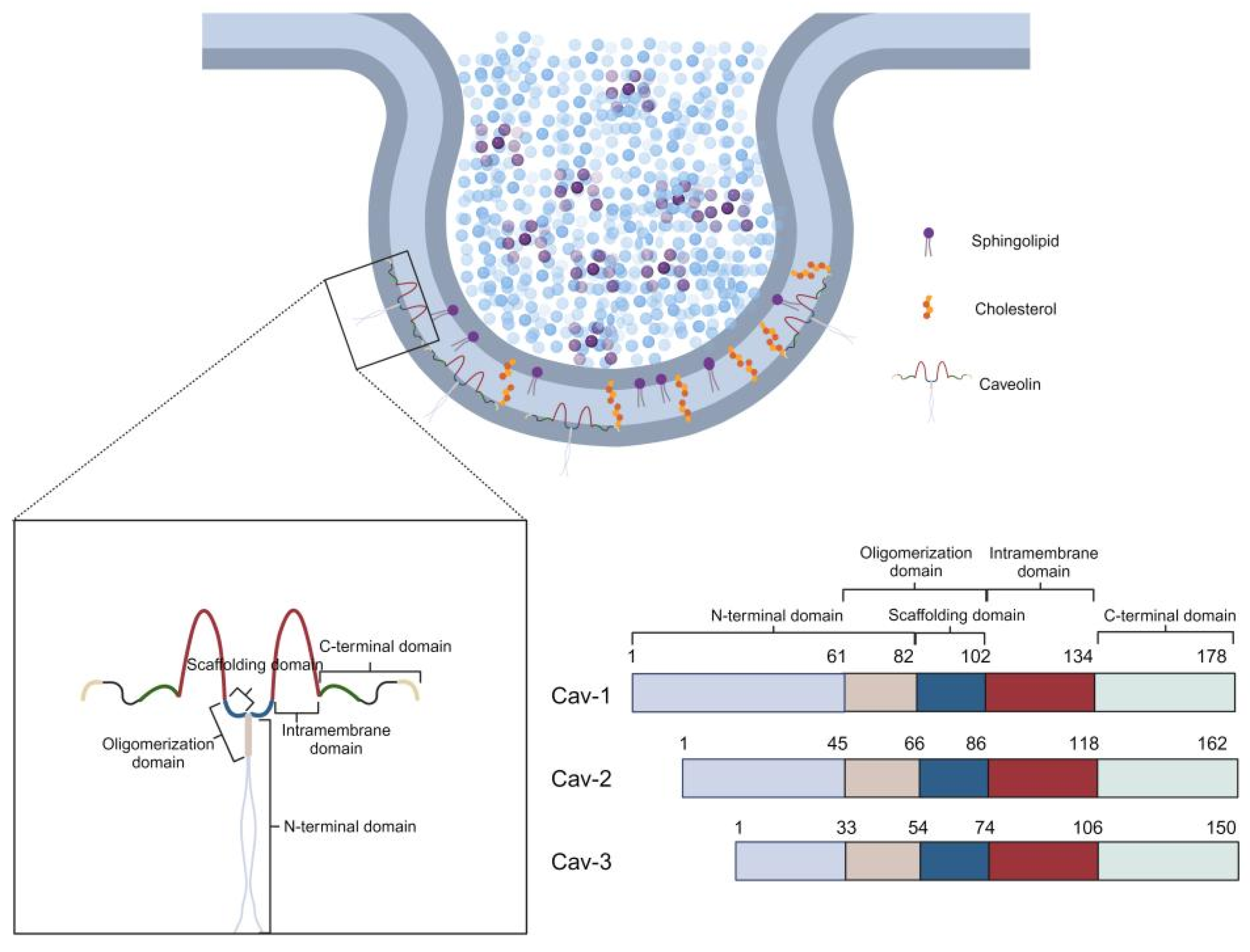
Figure 1. Structure of caveolae and the three caveolin proteins (Cav-1, Cav-2, Cav-3). Caveolin proteins contain distinct domains: an N-terminal domain, a scaffolding domain (SD), an intramembrane domain, and a C-terminal domain.
Cells 14 00631 g001
Figure 2. Structure of caveolae and distribution of ion channels. The flask-shaped caveolae structures in the plasma membrane are associated with various ion channels, including sodium channels (Nav1.4, Nav1.5, Nav1.8, ENaC), calcium channels (Cav1.2, Cav1.3, Cav3.1, Cav3.2, Orai1), potassium channels (Kv1.3, Kv1.4, Kv1.5, Kv2.1, KCa2.3, BKCa, Kv4, Kir2.1, Kir6.1, Kir6.2, GIRK), chloride channels (CIC-2, CIC-3, SWELL1), non-selective cation channels (Piezo1, nAChR, 5-HT2A, NMDA, AMPA, P2X4, P2X7, TRPV4, TRPC1, TRPC3, HCN) and other channels (Cx43).
Cells 14 00631 g002
Figure 3. Regulation of ion channels by caveolae and caveolins via signaling pathways. Key signaling components enriched in caveolae include G protein-coupled receptors (GPCRs), heterotrimeric G proteins, steroid hormone receptors, receptor and non-receptor tyrosine kinases, protein kinase A (PKA), protein kinase C (PKC), phosphoinositide 3-kinase (PI3K)/Akt, mitogen-activated protein kinases (MAPK), nitric oxide synthases (NOS) and others.
Cells 14 00631 g003
Figure 4. Caveolae-associated diseases. Caveolae are associated with various diseases, including cardiovascular diseases, metabolic disorders, neurodegenerative diseases and cancer, and play a regulatory role in their pathophysiology.
Cells 14 00631 g004
Disclaimer/Publisher’s Note: The statements, opinions and data contained in all publications are solely those of the individual author(s) and contributor(s) and not of MDPI and/or the editor(s). MDPI and/or the editor(s) disclaim responsibility for any injury to people or property resulting from any ideas, methods, instructions or products referred to in the content.