Article

A Novel Kelch-Like-1 Is Involved in Antioxidant Response by Regulating Antioxidant Enzyme System in Penaeus vannamei

Guangzhou Key Laboratory of Subtropical Biodiversity and Biomonitoring, Guangdong Provincial Key Laboratory for Healthy and Safe Aquaculture, Key Laboratory of Ecology and Environmental Science in Guangdong Higher Education, College of Life Science, South China Normal University, Guangzhou 510631, China
*
Author to whom correspondence should be addressed.
Genes 2020, 11(9), 1077; https://doi.org/10.3390/genes11091077
Submission received: 29 June 2020 / Revised: 3 September 2020 / Accepted: 10 September 2020 / Published: 15 September 2020

Abstract

:
Heavy metals are typical cumulative pollutants that can enter and poison the human body through the food chain. However, the molecular mechanism of heavy metal-induced oxidative stress is unclear. In this study, we characterize PvKelch-like-1 from P. vannamei and explore its antioxidant roles in immune regulation of crustaceans. PvKelch-like-1 full length contains 2107 nucleotides, consists of a 5′ untranslated region (UTR) of 79 bp, a 3′ UTR of 180 bp, and a ORF of 1848 encoded 615 amino acids, which contain a BTB, BACK and Kelch motif, putative molecular mass and isoelectric point were 69 KDa and 6.54. PvKelch-like-1 mRNA was ubiquitously expressed in all detected tissue of P. vannamei, and mRNA expression levels were significantly up-regulated from 6 to 24 h after cadmium stress and reached the highest level (3.2-fold) at 12 h in the hepatopancreas. Subcellular localization analysis revealed that PvKelch-like-1 was localized in the nucleus. Silencing PvKelch-like-1 significantly increased reactive oxygen species (ROS) (1.61-fold) production and DNA damage (1.32-fold) in the shrimp hemolymph, and significantly decreased total hemocyte counts (THC) (0.64-fold) at 6 h in hemolymph. Additionally, the antioxidant genes PvCAT (0.43-fold), PvMnSOD (0.72-fold), PvGST (0.31-fold) and PvGPx (0.59-fold) at 6 h were decreased significantly in PvKelch-like-1 silenced shrimp after cadmium stress. Overexpression of PvKelch-like-1 has the opposite results in enzyme activity. The SOD (2.44-fold) and CAT (2.19-fold) activities were significantly increased after overexpressing PvKelch-like-1. These results suggest that PvKelch-like-1 plays a vital role in shrimp innate immune defense by positively regulating the expression of antioxidant enzyme genes to respond to cadmium stress.

1. Introduction

In recent years, heavy metal pollution has become increasingly serious in China, and has become an important pollution factor for aquatic water environments, which is causing serious harm to farmed animals. Cadmium (Cd) may endanger the growth and development of aquatic organisms through toxic effects [1,2]. Accumulating evidence suggests that cadmium can damage the immune system of shrimp, trigger inhibition of physiology of shrimp and cause huge economic losses in the shrimp farming industry [3,4,5]. Recent studies on heavy metal pollutants have focused mainly on the effects of the acute toxicity and antioxidant system enzymes of organisms. However, there are few studies on the mechanism of detoxification of organisms.
Over the past thirty years, marine invertebrates subjected to various harmful stimuli induced oxidative stress have been increasingly studied. Recent studies have shown that reactive oxygen species (ROS) as a secondary messenger is crucial in the response to environmental stresses, growth and development [6,7]. However, excessive ROS can cause imbalance in the oxidation system and the antioxidant system, leading to oxidation damage [8]. Our initial findings are that PvDJ-1 is of critical importance in the antioxidant function of shrimp [9]. Meanwhile, DJ-1 as transcription factors has been confirmed in the involvement of the Kelch-like ECH-associated protein 1 (Keap1)-Nrf2 antioxidant signaling pathway in mammals or fruit flies [10,11,12].
Keap1 belongs to the BBK protein family because it contains BTB, BACK and Kelch domains. The BTB domain is a conserved domain comprising approximately 115 hydrophobic amino acids, which play a functional role in mediating protein–protein interactions and is capable of protein self-association or interacting with other non-BTB proteins [13,14]. Kelch domains (Kelch-repeat motifs) consist of 3–7 Kelch, one of which contains 44–56 amino acids and it was found for the first time in Drosophila melanogaster [15]. Various proteins containing Kelch domain have been found in vertebrates, which belong to the Kelch-repeat superfamily protein [16]. The Kelch domain generally exists at the C-terminal of the protein, and conserved BTB or other domains exist at the N-terminal. At present, research on the BBK protein family focuses on the following aspects: cell morphology [17], protein ubiquitination degradation [18], and signal transduction [19,20]. It has been found in both vertebrates and fruit flies that the Keap1-Nrf2 signaling pathway plays an important role in the antioxidant function [21,22,23]. However, there is no report on this signaling pathway in shrimp.
Kelch-like ECH-associated protein 1 (Keap1) is an important model in the study of BBK family proteins. Therefore, this paper studied the antioxidative function of PvKelch-like-1 under cadmium stress, and combined with the mRNA regulation relationship between PvDJ-1 and PvKelch-like-1, it has enriched the research on the antioxidant system of P. vannamei and provided a theoretical basis for solving the oxidative stress of shrimp in response to environmental stress.

2. Materials and Methods

2.1. Animal

P. vannamei (2–3 g in weight) were used in this study. The shrimp were maintained in 120 × 120 × 80 cm aquariums of seawater (5 ‰ salinity, pH 7.4 and 24 to 25 °C) and acclimatized for 3 days prior to the experiments.

2.2. CDNA Synthesis and RT-qPCR

Total RNA was extracted using Trizol (Invitrogen, Carlsbad, CA, USA) according to manufacturer’s instructions. RNA concentration was determined using NanoDrop 2000 (Montchanin, DE, USA), and RNA integrity was verified by 1% agarose gels. cDNA was synthesized with a PrimeScript TM RT-PCR Kit (TaKaRa, Shiga, Japan) following the manufacturer’s instructions. The mRNA expression level was determined by quantitative real-time PCR (qRT-PCR) using the ABI 7500 system. The PCR products were further verified by sequencing, all primers are shown in Table S1. Three independent qPCR experiments were performed. Each experiment was performed in triplicate.

2.3. Identification PvKelch-Like-1

Based on the data of the P. vannamei genome, we cloned the full-length of PvKelch-like-1 with rapid amplification with SMARTer RACE 5′/3′ Kit (TAKARA, Japan), following manufacturer’s instructions and used primer (Table S1). We used BLAST program of NCBI (http://www.ncbi.nlm.nih.gov/blast) to analyze the sequence. Multiple sequence alignment was performed using the Bio-Edit (version 7.0.9.0).
A phylogenic tree was constructed using the MEGA 7.0 program with neighbor-joining methods at 1000 bootstrap replication.

2.4. Polyclonal Antibody Preparation

The ORF of PvKelch-like-1 was amplified by PCR, digested by a combination of restriction enzymes EcoRV and HindIII, and then used to construct the recombinant vector and transformed into Escherichia coli (E. coli) BL21 (DE3) (Table S1). After induction for 6 h with 1.0% isopropyl β-D-thiogalactopy ranoside (IPTG), we used His-Bind Resin (Novagen) to purify recombinant protein, according to the manufacturer’s instructions. Recombinant protein concentration was determined by the BCA method, correctness was reanalyzed by SDS-PAGE. Healthy Kunming mice (qualified number: Guangdong verification word 2007A064) were intraperitoneally injected with 100 ug recombinant protein emulsified with Freund’s complete adjuvant (Sigma) initially. Then, they were injected three times with emulsified Freund’s incomplete adjuvant (Sigma) at intervals of one week. Antisera were harvested seven days after the last injection from ear veins and stored at −80 °C. Each experiment was performed in triplicate.

2.5. Tissue-Specific Expression and Immunofluorescence

The healthy shrimp’s hepatopancreas, heart, stomach, eyestalk, gill, intestine, hemolymph, foot and muscle were collected. The real-time PCR was performed to analyze the expression pattern. All primers are shown in Table S1.
The muscle tissue of the normal shrimp was prepared in 4% paraformaldehyde, and then the green fluorescent protein was labeled with the obtained antibody. After incubation, the nucleus was stained with DAPI, and the distribution of PvKelch-like-1 protein was observed under a fluorescence microscope. Each experiment was performed in triplicate.

2.6. Cadmium Challenge

Sixty healthy P. vannamei individuals were randomly separated into two groups. One group was the challenge with 4.25 µM/L CdCl2 (Kermel, Tianjin, China), the other group as controls. Each group was performed in triplicate. The hepatopancreases were collected at 0, 1.5, 3, 6, 12 and 24 h post challenge, and stored at −80 °C for RNA extraction.

2.7. Overexpression of PvKelch-Like-1 in S2 Cell

Drosophila S2 cells (Invitrogen, USA) were cultured in Schneider’s Drosophila media (Gibco, Gaithersburg, MD, USA) containing 10% fetal bovine serum (FBS, Gibco, USA) at 28 °C. The recombination plasmid pAc5.1-PvKelch-like-1-V5/HisB or pAc5.1-V5/HisB was mixed with FuGENE® HD Transfection Reagent (Promega, San Luis Obispo, CA, USA), and then transfected in S2 cells according to the manufacturer’s protocol. After incubation for 48 h, cells were collected. The enzyme activity was detected according to the Total Superoxide Dismutase Assay Kit with WST-8 (Beyotime, Shanghai, China) and Catalase Assay Kit (Beyotime, China) according to the manufacturer’s protocol. Each experiment was performed in triplicate.

2.8. Silenced PvKelch-Like-1 In Vivo

Double RNA was obtained in vitro using the T7 RiboMAX TM Express RNA Production System (Promega, USA) according to the manufacturer’s protocol. Then, the double RNA concentration was detected with application of NanoDrop 2000 and confirmed by 1.2% agarose gel. Finally, the dsRNA were stored at −80 °C.
Sixty healthy shrimp were randomly separated into 2 groups. Then, equal amounts of double RNA was injected intramuscularly with 10 ug (1 μg/μL solution) (dsPvKelch-like-1 and dsGFP as a control). Each group was performed in triplicate. The hepatopancreases were collected 0, 1, 2, 3, and 4 d post injection, and stored at −80 °C for RNA extraction. RT-qPCR was performed to analyze the interference efficiency.
The hepatopancreases were collected 0, 1.5, 3 and 6 h after the cadmium challenge. Each experiment was performed in triplicate. Hepatopancreas was used to detect the expression of antioxidant enzyme genes.

2.9. Comet Assays and Total Hemocyte Counts (THC)

The hemolymph samples were mixed into low melting agarose after being diluted to 105 cells mL−1, and were applied to the slides pre-plated with normal melting agarose, placed in the lysing buffer for 2 h at 4 °C, and then the slides experienced electrophoresis at 20 V and then 200 mA for 25 min of ice-cold, cold neutralization buffer twice for 10 min then dehydrated in ethanol for 15 min and stored in the dark. Then, we used SYRB Green I (1:10,000) staining, and the cells were observed using fluorescence microscopy (Leica, Frankfurt, Germany). The CASP image analysis software measures the comet tail moment (OTM).
The total hemocyte counts (THC) used a hemocytometer and a light microscope (Olympus, Beijing, China) to measure, according to the manufacturer’s protocol.

2.10. ROS Accumulation

The hemolymph ROS accumulation was detected with a DCFH-DA kit (Beyotime, China), according to the manufacturer’s instructions. Briefly, the hemolymph cells were incubated at 37 °C for 20 min with 10 μM DCFH-DA. After PBS elution twice, the cells were detected with a FACS AriaIII flow cytometer system (BD Biosciences, New York, NY, USA). Each experiment was performed in triplicate.

2.11. Western Blotting

The tissue was lysed in RIPA buffer (Beyotime, China), containing 1 mm PMSF for 30 min at 4 °C, and centrifuged at 12,000× g for 15 min at 4 °C. The tissue lysate was mixed with 5× SDS-PAGE buffer (Beyotime) and boiled for 10 min. Then, 30 μg of each sample was subjected to 10% SDS-PAGE and Western blotting, as previously reported [24]. The primary antibody diluted 1:1000 and secondary antibody diluted 1:2000 in 0.5% BSA-TBST. Following a further four 10 min washes in TBST, the membranes were detected with 5-bromo-4-chloro-3-indolyl-phosphate (BCIP)/nitroblue tetrazolium (NBT) substrate (Sangon Biotech, Shanghai, China). Each experiment was performed in triplicate.

2.12. Statistical Analysis

All data were presented as means ± SD. All data were performed using SPSS 20.0. Significant differences between means were analyzed with a one-way ANOVA analysis, p Values < 0.05 were considered statistically significant.

3. Results

3.1. Characterization of PvKelch-Like-1

The PvKelch-like-1 full length sequence consisted of 2107 nucleotides and contained an 1848 bp ORF that encoded a putative protein with 615 amino acids, a 5′ untranslated region (UTR) of 79 bp, and a 3′ UTR of 180 bp. The molecular mass and isoelectric point (pI) of this protein were predicted at 69 KDa and 6.54. It is predicted by the SMART program (http://smart.embl-heidelberg.de/, use default parameters) that the PvKelch-like-1 protein contains three domains: BTB at positions 76–173, BACK at positions 178–279, and Kelch at positions 320–606 (Figure S1A). The multiple sequence alignment revealed that the PvKelch-like-1 three domains BTB, BACK and Kelch are conservative. PvKelch-like-1 amino acid sequence is most similar to that of Cryptotermes secundus (accession no. XP_023726037.1) with 64.37% and Zootermopsis nevadensis (accession no. XP_021934020.1) with 64.02%, PvKelch-like-1 shares 36.08–64.37% similarity with other known sequences in this family (Figure S1B). Moreover, a phylogenic tree showed that PvKelch-like-1 has separated branches including vertebrates and invertebrates and was close to the arthropod (Figure S1C).

3.2. Preparation of Polyclonal Antibody

The recombinant PvKelch-like-1 protein has a significant band of approximately 88 KDa target protein (Figure 1A), and conformed to the expected results. Western blotting recognized a constituent of recombinant protein with an apparent molecular mass of 88 kDa, corresponding to that predicted from the PvKelch-like-1 (Figure 1B).

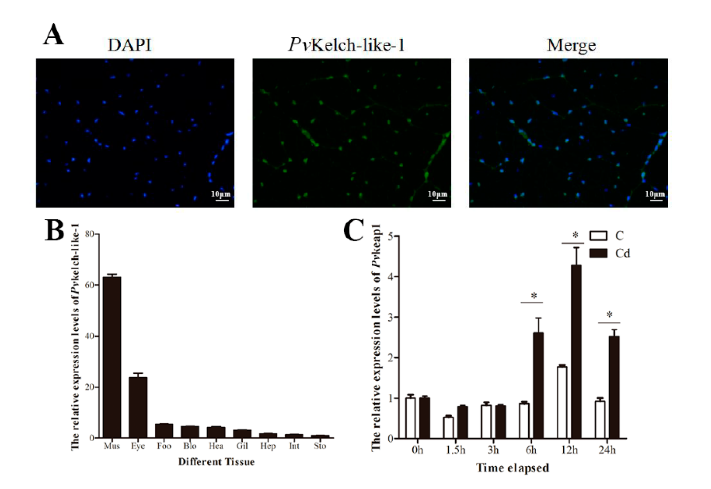
3.3. Subcellular Localization and Tissue-Specific Expression

The subcellular localization of PvKelch-like-1 was determined by a immunofluorescence assay. The results show that the PvKelch-like-1 localization nucleus of muscle cells use the polyclonal antibody of P. vannamei (Figure 2A). PvKelch-like-1 was detected in all tissue, with the highest expression in muscle (Figure 2B).

3.4. Expression Pattern of PvKelch-Like-1 after Cadmium Challenge

Expression of PvKelch-like-1 was analyzed in hepatopancreas samples after cadmium challenge of P. vannamei. The expression of PvKelch-like-1 rose gradually for the first 6 h, showing a 2.3-fold increase relative to the start of the experiment, at 12 h it reached maximum (3.2-fold) and then subsided slightly at 24 h after cadmium challenge (Figure 2C). In contrast, no significant change of PvKelch-like-1 expression was observed in the control shrimp.

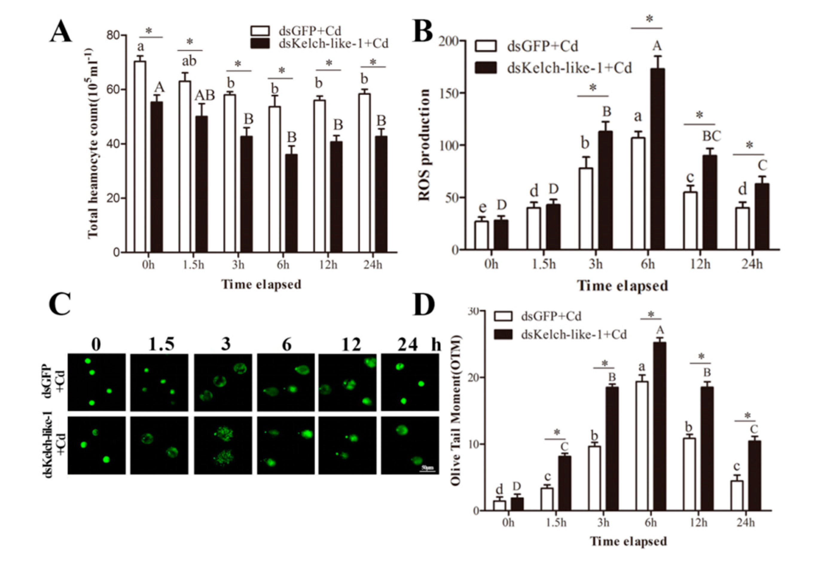
3.5. PvKelch-Like-1 Participates Injury Response under Cadmium Stress

The PvKelch-like-1 was knocked down (0.37-fold, 0.42-fold and 0.47-fold compared with GFP-RNAi) at 2, 3 and 4 d after PvKelch-like-1 group in P. vannamei, respectively (Figure S2A). In contrast, the dsGFP group had no significant effects on PvKelch-like-1 expression. As shown in Figure 3A, the THC in PvKelch-like-1-silenced shrimp was significantly lower than in the control group at 0 h post challenge. The THC of both groups significantly decreased after cadmium challenge and reached its minimum at 6 h. As shown in Figure 3B, ROS production increased at first and then decreased after cadmium challenge in both PvKelch-like-1 silenced and control groups, and reached the maximum at 6 h. During the 3 h to 24 h after cadmium challenge, the ROS levels in the PvKelch-like-1 silenced group were significantly higher than the control group. Moreover, this decrease was more prominent in the PvKelch-like-1 silenced group than in the control group. As shown in Figure 3C,D, the OTM value describing hemolymph DNA damage for both groups was significantly increased compared to 0 h under cadmium challenge.

3.6. PvKelch-Like-1 Responds to Cadmium Stress by Regulating Antioxidant Enzyme System

The expression of the antioxidant enzyme gene was detected on the second day of PvKelch-like-1 silenced shrimp, and the results showed that the expression of PvCAT, PvMnSOD, PvGPx and PvGST in the PvKelch-like-1 silenced group were significantly decreased compared to the dsGFP silenced group (Figure S2B).
The expression of antioxidant enzymes in both groups showed significant differences under cadmium stress. The expression of PvCAT and PvGST reached the maximum at 3 h after cadmium challenge and then decreased, and the expression of PvMnSOD and PvGPx reached the maximum at 6 h after cadmium challenge and then decreased. Antioxidant enzymes gene PvCAT, PvMnSOD, PvGPx and PvGST expression were significantly decreased in the PvKelch-like-1 silenced group than in the control group at 0, 1.5, 3, 6, 12 and 24 h post challenge, respectively (Figure 4).

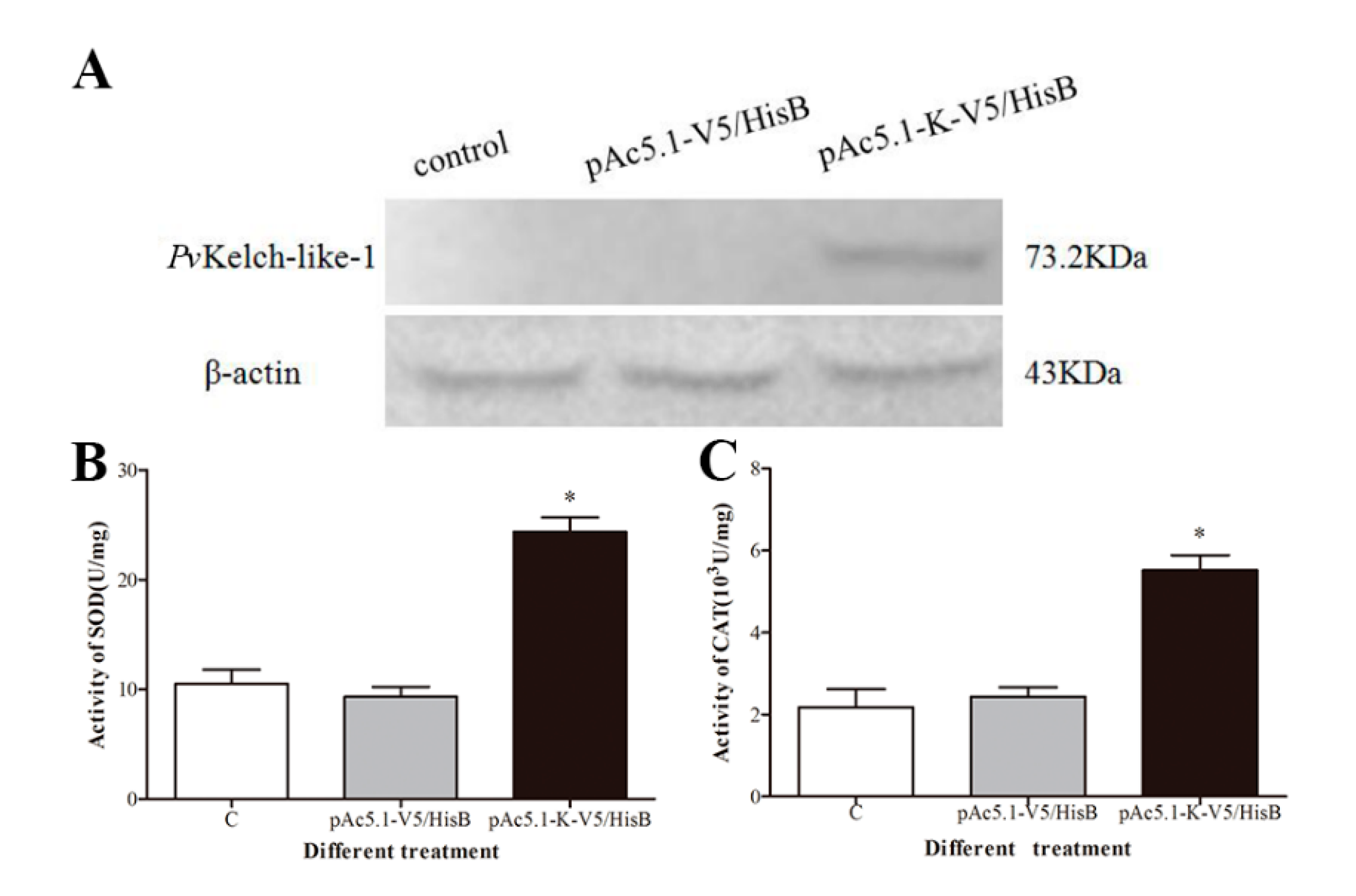
3.7. Effect of Enzyme Activity after Overexpression of PvKelch-Like-1 in S2 Cell

PvKelch-like-1 recombinant eukaryotic protein with a molecular weight of 73.2 kDa was successfully expressed in S2 cells by Western blot (Figure 5A). The SOD and CAT activities were significantly increased compared to the transfected, with empty plasmids group after overexpressing PvKelch-like-1 in S2 cells (Figure 5B,C).

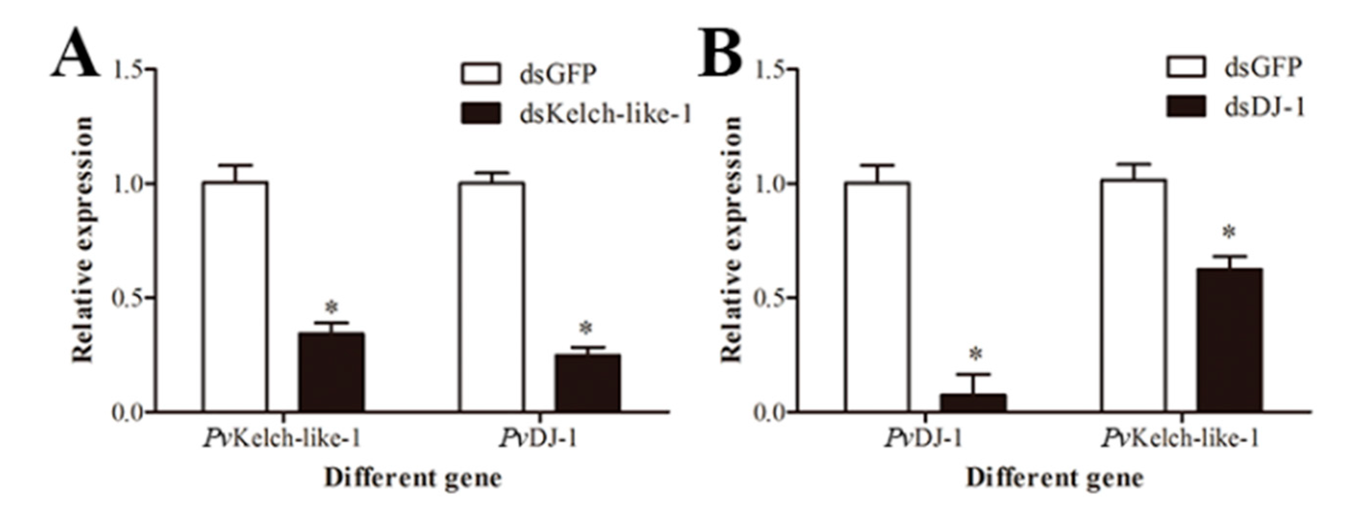
3.8. Relationship between PvKelch-Like-1 and PvDJ-1

In order to verify whether there is a regulatory relationship between PvKelch-like-1 and PvDJ-1, the expression of PvDJ-1 was detected in PvKelch-like-1-silenced shrimp, and the expression of PvKelch-like-1 was detected in PvDJ-1-silenced shrimp. The results show that PvDJ-1 significantly decreased after silenced PvKelch-like-1 (Figure 6A), and the expression of PvKelch-like-1 significantly decreased after silenced PvDJ-1(Figure 6B).

4. Discussion

The dynamic balance of oxidative stress is essential for the physiological activities of living organisms [25]. Studies have shown that when the external environmental factors change, the oxidative stress reaction of shrimp causes excessive ROS to damage the innate immune system of shrimp, including the antioxidant enzyme system [26,27,28]. At present, there is little basic research on the antioxidation system of shrimp. PvDJ-1, in the previous work, has confirmed that it has significant antioxidant effects similar to vertebrates [9,29,30]. In this paper, cloning and functional studies reveal that PvKelch-like-1 in the PvDJ-1 transcriptome occupies a position in the shrimp antioxidant system.
Sequence analysis revealed that PvKelch-like-1 belongs to the BBK family with poor nucleotide conservation. It is speculated that there are functional differences between aquatic organism BBK family genes and vertebrates. Tissue distribution analyses in P. vannamei showed that PvKelch-like-1 had the highest expression in the muscle, which was supposed to be related to its function in the Kelch domain. Kelch was found in Drosophila interacted with actin and had an important role in the growth and development [31,32].
Keap1, a cytoplasmic protein, is one of the most widely studied antioxidant-related genes in the BBK family [33,34] and itinteractes with the nuclear transcription factor Nrf2 to maintain redox homeostasis [35,36]. Immunofluorescence verified that the PvKelch-like-1 protein was localized in the nucleus of the shrimp cells, which was different from the cytoplasmic localization result of Keap1. BBK family proteins play an important role by localization in a different location [37,38]. KLHL31 protein is localized in both the nucleus and the cytoplasm to inhibit the transcriptional activities of TRE and SRE [39]. KLHL7 protein is expressed in the nuclei of neurons, and its antibodies can be used as paraneoplastic markers [40]. According to the localization results of PvKelch-like-1 protein, it is speculated that PvKelch-like-1 may be involved in nuclear transcriptional regulation.
It has been reported that cadmium exposure reduces the THC, induces DNA damage and ROS production of hemolymph in shrimp [41,42,43]. Our results found that the THC was significantly decreased, the DNA damage was significantly increased, and the ROS level was significantly increased in the PvKelch-like-1 silenced group, indicating that the ability to regulate oxidative stress of the hemolymph in shrimp under cadmium stress was impaired after PvKelch-like-1 was silenced. The hepatopancreas plays important roles in several metabolic processes and the immunoregulatory process in shrimp [44,45]. It was found that the hepatopancreas of shrimp developed lesions under heavy metal stress, and expression of antioxidant enzymes was induced [46,47,48]. Whether PvKelch-like-1 affect the expression of antioxidant enzyme gene in P. vannamei. In this study, expression of antioxidant enzymes gene significantly decreased in the PvKelch-like-1 silenced group compared with GFP silenced group after cadmium stress. Knockout Keap1 revealed a significant increase in the antioxidant enzyme gene HO-1 and Nrf2 [49,50]. Studies have shown that overexpression of the antioxidant-related gene Keap1 in vertebrate cells significantly reduces the expression of antioxidant enzymes, which is different from our results [51,52]. Our results found that the activity of antioxidant enzymes SOD and CAT was significantly increased after overexpressed PvKelch-like-1 in Drosophila S2 cells. All results reveal that PvKelch-like-1 positively regulates the expression of antioxidant enzyme genes under response to cadmium stress.
Recently published data suggest that DJ-1 is necessary for the Nrf2/Keap1 axis and antioxidant stress [10]. Preliminary investigation on the regulatory relationship between the antioxidant genes PvKelch-like-1 and PvDJ-1 was performed by detecting mRNA expression. The results show that there is a bidirectional regulation relationship between PvKelch-like-1 and PvDJ-1, but the specific regulatory mechanism needs further research.
In conclusion, we report the molecular characterization and functional investigation of PvKelch-like-1. Overexpression and silencing indicate that PvKelch-like-1 has a positive regulatory effect on the expression of antioxidant enzyme gene. Furthermore, the mRNA regulation relationship between PvKelch-like-1 and PvDJ-1 is confirmed. These results reveal the function of the BBK family gene PvKelch-like-1 and provide a basis for the antioxidant system of P. vannamei.

Supplementary Materials

The following are available online at https://www.mdpi.com/2073-4425/11/9/1077/s1, Figure S1 Characterization of PvKelch-like-1. (A) Nucleotide and deduced amino acid sequences of PvKelch-like-1. The translation initiation (ATG) and stop codons (TGA) are boxed. Three conserved sequences of PvKelch-like-1 protein domains are shaded in different gray (76-173 BTB; 178-279 BACK; 320-606 Kelch); (B) multiple sequence alignment of Kelch-like-1 protein between P. vannamei and other species. Mus musculus (BAA34639.1), Drosophila melanogaster (ABD14408.1), Homo sapiens (NP_987096.1), Danio rerio (NP_878284.2), Latimeria chalumnae (XP_005994374.1), Limulus Polyphemus (XP_013773778.1), Wasmannia auropunctata (XP_011697788.1). Similar amino acid residues (>50%) are labeled in gray while uniform amino acid residues are labeled in black; (C) phylogenetic tree of the PvKelch-like-1 was constructed with NJ method in MEGA 5.1 and a bootstrap analysis was performed using 1000 replicates to test the relative support for particular clades; Figure S2 Effect of silencing PvKelch-like-1 on antioxidant gene expression. (A) PvKelch-like-1 silenced efficiency detection in vivo; (B) the mRNA expression of PvCAT, PvMnSOD, PvGPx and PvGST in the hepatopancreas after PvKelch-like-1 knockdown.

Author Contributions

Software and writing—original draft preparation, X.-L.Q.; validation and writing—review and editing, Q.-J.L.; project administration and funding acquisition, Y.L. and W.-N.W. All authors have read and agreed to the published version of the manuscript.

Funding

This work was supported by the National Science Foundation for Young Scientists of China (grant no.31500325), the National Nature Science Foundation of China (grant no.31971417 and 31670423) and Guangdong Province Natural Science Foundation of China (grant no. 2019A1515011442).

Conflicts of Interest

The authors declare no conflict of interest.

References

  1. Krumschnabel, G.; Ebner, H.L.; Hess, M.W.; Villunger, A. Apoptosis and necroptosis are induced in rainbow trout cell lines exposed to cadmium. Aquat. Toxicol. 2010, 99, 73–85. [Google Scholar] [CrossRef] [PubMed]
  2. Tan, X.Y.; Zhi, L.; Zhang, G.Y.; Liu, X.J.; Min, J. Effect of dietary cadmium level on the growth, body composition and several hepatic enzymatic activities of juvenile yellow catfish, Pelteobagrus fulvidraco. Aquac. Res. 2010, 41, 1022–1029. [Google Scholar] [CrossRef]
  3. Wu, J.P.; Chen, H.C.; Huang, D.J. Histopathological and biochemical evidence of hepatopancreatic toxicity caused by cadmium and zinc in the white shrimp, Litopenaeus vannamei. Chemosphere 2008, 73, 1019–1026. [Google Scholar] [CrossRef] [PubMed]
  4. Wu, J.P.; Chen, H.C.; Huang, D.J. Histopathological Alterations in Gills of White Shrimp, Litopenaeus vannamei (Boone) After Acute Exposure to Cadmium and Zinc. Bull. Environ. Contam. Toxicol. 2009, 82, 90–95. [Google Scholar] [CrossRef] [PubMed]
  5. Boudet, L.C.; Polizzi, P.; Romero, M.B.; Robles, A.; Marcovecchio, J.E.; Gerpe, M.S. Histopathological and biochemical evidence of hepatopancreatic toxicity caused by cadmium in white shrimp, Palaemonetes argentinus. Ecotoxicol. Environ. Saf. 2015, 113, 231–240. [Google Scholar] [CrossRef] [PubMed]
  6. Li, L.; Tan, J.; Miao, Y.; Lei, P.; Zhang, Q. ROS and Autophagy: Interactions and Molecular Regulatory Mechanisms. Cell. Mol. Neurobiol. 2015, 35, 615–621. [Google Scholar] [CrossRef]
  7. Russell, E.G.; Cotter, T.G. New Insight into the Role of Reactive Oxygen Species (ROS) in Cellular Signal-Transduction Processes. Int. Rev. Cell Mol. Biol. 2015, 319, 221–254. [Google Scholar] [PubMed]
  8. Xu, Z.; Regenstein, J.M.; Xie, D.; Lu, W.; Ren, X.; Yuan, J.; Mao, L. The oxidative stress and antioxidant responses of Litopenaeus vannamei to low temperature and air exposure. Fish Shellfish. Immunol. 2018, 72, 564–571. [Google Scholar] [CrossRef]
  9. Huang, M.Z.; Liu, Y.; Xie, C.Y.; Wang, W.N. LvDJ-1 plays an important role in resistance against Vibrio alginolyticus in Litopenaeus vannamei. Fish Shellfish. Immunol. 2015, 44, 180–186. [Google Scholar] [CrossRef]
  10. Gan, L.; Johnson, D.A.; Johnson, J.A. Keap1-Nrf2 activation in the presence and absence of DJ-1. Eur. J. Neurosci. 2010, 31, 967–977. [Google Scholar] [CrossRef] [Green Version]
  11. Yang, J.; Kim, M.J.; Yoon, W.; Kim, E.Y.; Kim, H.; Lee, Y.; Min, B.; Kang, K.S.; Son, J.H.; Park, H.T.; et al. Isocitrate protects DJ-1 null dopaminergic cells from oxidative stress through NADP(+)—Dependent isocitrate dehydrogenase (IDH). PLoS Genet. 2017, 13, e1006975. [Google Scholar] [CrossRef] [PubMed]
  12. Zhang, X.L.; Yuan, Y.H.; Shao, Q.H.; Wang, Z.Z.; Zhu, C.G.; Shi, J.G.; Ma, K.L.; Yan, X.; Chen, N.H. DJ-1 regulating PI3K-Nrf2 signaling plays a significant role in bibenzyl compound 20C-mediated neuroprotection against rotenone-induced oxidative insult. Toxicol. Lett. 2017, 271, 74–83. [Google Scholar] [CrossRef] [PubMed]
  13. Albagli, O.; Dhordain, P.; Deweindt, C.; Lecocq, G.; Leprince, D. The BTB/POZ domain: A new protein-protein interaction motif common to DNA- and actin-binding proteins. Cell Growth Differ. Mol. Biol. J. Am. Assoc. Cancer Res. 1995, 6, 1193–1198. [Google Scholar]
  14. Chaharbakhshi, E.; Jemc, J.C. Broad-complex, tramtrack, and bric-a-brac (BTB) proteins: Critical regulators of development. Genesis 2016, 54, 505–518. [Google Scholar] [CrossRef]
  15. Xue, F.; Cooley, L. kelch encodes a component of intercellular bridges in Drosophila egg chambers. Cell 1993, 72, 681–693. [Google Scholar] [CrossRef]
  16. Adams, J.; Kelso, R.; Cooley, L. The kelch repeat superfamily of proteins: Propellers of cell function. Trends Cell Biol. 2000, 10, 17–24. [Google Scholar] [CrossRef]
  17. Seng, S.; Avraham, H.K.; Jiang, S.; Venkatesh, S.; Avraham, S. KLHL1/MRP2 Mediates Neurite Outgrowth in a Glycogen Synthase Kinase 3β-Dependent Manner. Mol. Cell. Biol. 2006, 26, 8371–8384. [Google Scholar] [CrossRef] [PubMed] [Green Version]
  18. Sumara, I.; Quadroni, M.; Frei, C.; Olma, M.H.; Sumara, G.; Ricci, R.; Peter, M. A Cul3-based E3 ligase removes Aurora B from mitotic chromosomes, regulating mitotic progression and completion of cytokinesis in human cells. Dev. Cell 2007, 2, 887–900. [Google Scholar] [CrossRef] [Green Version]
  19. Wang, Q.; Burles, K.; Couturier, B.; Randall, C.M.; Shisler, J.; Barry, M. Ectromelia virus encodes a BTB/kelch protein, EVM150, that inhibits NF-kappaB signaling. J. Virol. 2014, 88, 4853–4865. [Google Scholar] [CrossRef] [PubMed] [Green Version]
  20. Itoh, K.; Wakabayashi, N.; Katoh, Y.; Ishii, T.; Igarashi, K.; Engel, J.D.; Yamamoto, M. Keap1 represses nuclear activation of antioxidant responsive elements by Nrf2 through binding to the amino-terminal Neh2 domain. Genes Dev. 1999, 13, 76–86. [Google Scholar] [CrossRef] [PubMed] [Green Version]
  21. Zhang, D.D.; Lo, S.C.; Cross, J.V.; Templeton, D.J.; Hannink, M. Keap1 is a redox-regulated substrate adaptor protein for a Cul3-dependent ubiquitin ligase complex. Mol. Cell. Biol. 2004, 24, 10941–10953. [Google Scholar] [CrossRef] [PubMed] [Green Version]
  22. Dinkova-Kostova, A.T.; Holtzclaw, W.D.; Kensler, T.W. The role of Keap1 in cellular protective responses. Chem. Res. Toxicol. 2005, 18, 1779–1791. [Google Scholar] [CrossRef] [PubMed]
  23. Velichkova, M.; Hasson, T. Keap1 regulates the oxidation-sensitive shuttling of Nrf2 into and out of the nucleus via a Crm1-dependent nuclear export mechanism. Mol. Cell. Biol. 2005, 25, 4501–4513. [Google Scholar] [CrossRef] [PubMed] [Green Version]
  24. Liang, Q.; Ou, M.; Li, Z.; Ren, Y.; Wei, W.; Qiao, X.; Hu, R.; Wu, X.; Liu, Y.; Wang, W. Functional analysis target of rapamycin (TOR) on the Penaeus vannamei in response to acute low temperature stress. Fish Shellfish. Immunol. 2019, 96, 53–61. [Google Scholar] [CrossRef] [PubMed]
  25. Camhi, S.L.; Lee, P.; Choi, A.M. The oxidative stress response. New Horiz. 1995, 3, 170–182. [Google Scholar] [PubMed]
  26. Lushchak, V.I. Environmentally induced oxidative stress in aquatic animals. Aquat. Toxicol. 2011, 101, 13–30. [Google Scholar] [CrossRef] [PubMed]
  27. Zenteno-Savín, T.; Saldierna, R.; Ahuejote-Sandoval, M. Superoxide radical production in response to environmental hypoxia in cultured shrimp. Comp. Biochem. Physiol. Part C Toxicol. Pharmacol. 2006, 142, 301–308. [Google Scholar] [CrossRef]
  28. Guo, H.; Li, K.; Wang, W.; Wang, C.; Shen, Y. Effects of copper on hemocyte apoptosis, ROS production, and gene expression in white shrimp Litopenaeus vannamei. Biol. Trace Elem. Res. 2017, 179, 318–326. [Google Scholar] [CrossRef]
  29. Mullett, S.J.; Hinkle, D.A. DJ-1 knock-down in astrocytes impairs astrocyte-mediated neuroprotection against rotenone. Neurobiol. Dis. 2009, 33, 28–36. [Google Scholar] [CrossRef] [Green Version]
  30. Wang, H.; Li, Y.Y.; Qiu, L.Y.; Yan, Y.F.; Liao, Z.P.; Chen, H.P. Involvement of DJ-1 in ischemic preconditioninginduced delayed cardioprotection in vivo. Mol. Med. Rep. 2017, 15, 995–1001. [Google Scholar] [CrossRef]
  31. Hudson, A.M.; Mannix, K.M.; Cooley, L. Actin Cytoskeletal Organization in Drosophila Germline Ring Canals Depends on Kelch Function in a Cullin-RING E3 Ligase. Genetics 2015, 201, 1117–1131. [Google Scholar] [CrossRef] [PubMed] [Green Version]
  32. Robinson, D.N.; Cooley, L. Drosophila kelch is an oligomeric ring canal actin organizer. J. Cell Biol. 1997, 138, 799–810. [Google Scholar] [CrossRef] [PubMed] [Green Version]
  33. Sihvola, V.; Levonen, A.L. Keap1 as the redox sensor of the antioxidant response. Arch. Biochem. Biophys. 2017, 617, 94–100. [Google Scholar] [CrossRef]
  34. Dinkova-Kostova, A.T.; Kostov, R.V.; Canning, P. Keap1, the cysteine-based mammalian intracellular sensor for electrophiles and oxidants. Arch. Biochem. Biophys. 2017, 617, 84–93. [Google Scholar] [CrossRef] [Green Version]
  35. Itoh, K.; Tong, K.I.; Yamamoto, M. Molecular mechanism activating Nrf2-Keap1 pathway in regulation of adaptive response to electrophiles. Free Radic. Biol. Med. 2004, 36, 1208–1213. [Google Scholar] [CrossRef] [PubMed]
  36. Kundu, J.K.; Surh, Y.J. Nrf2-Keap1 signaling as a potential target for chemoprevention of inflammation-associated carcinogenesis. Pharm. Res. 2010, 27, 999–1013. [Google Scholar] [CrossRef]
  37. Hu, J.; Yuan, W.; Tang, M.; Wang, Y.; Fan, X.; Mo, X.; Li, Y.; Ying, Z.; Wan, Y.; Ocorr, K.; et al. KBTBD7, a novel human BTB-kelch protein, activates transcriptional activities of SRE and AP-1. BMB Rep. 2010, 43, 17–22. [Google Scholar] [CrossRef] [Green Version]
  38. Luhrig, S.; Kolb, S.; Mellies, N.; Nolte, J. The novel BTB-kelch protein, KBTBD8, is located in the Golgi apparatus and translocates to the spindle apparatus during mitosis. Cell Div. 2013, 8, 3. [Google Scholar] [CrossRef] [Green Version]
  39. Yu, W.; Li, Y.; Zhou, X.; Deng, Y.; Wang, Z.; Yuan, W.; Li, D.; Zhu, C.; Zhao, X.; Mo, X.; et al. A novel human BTB-kelch protein KLHL31, strongly expressed in muscle and heart, inhibits transcriptional activities of TRE and SRE. Mol. Cells Springer Sci. Bus. Media BV 2008, 26, 443–453. [Google Scholar]
  40. Bredholt, G.; Storstein, A.; Haugen, M.; Krossnes, B.K.; Husebye, E.; Knappskog, P.; Vedeler, C.A. Detection of Autoantibodies to the BTB-kelch Protein KLHL7 in Cancer Sera. Scand. J. Immunol. 2006, 64, 325–335. [Google Scholar] [CrossRef]
  41. Bautista-Covarrubias, J.C.; Velarde-Montes, G.J.; Voltolina, D.; García-de la Parra, L.M.; Soto-Jiménez, M.F.; Frías-Espericueta, M.G. Humoral and haemocytic responses of Litopenaeus vannamei to Cd exposure. Sci. World J. 2014, 2014, 903452. [Google Scholar] [CrossRef] [PubMed] [Green Version]
  42. Chang, M.; Wang, W.N.; Wang, A.L.; Tian, T.T.; Wang, P.; Zheng, Y.; Liu, Y. Effects of cadmium on respiratory burst, intracellular Ca2+ and DNA damage in the white shrimp Litopenaeus vannamei. Comp. Biochem. Physiol. C-Toxicol. Pharmacol. 2009, 149, 581–586. [Google Scholar] [CrossRef] [PubMed]
  43. Xian, J.A.; Wang, A.L.; Miao, Y.T.; Li, B. Flow Cytometric Analysis of In Vitro Cytotoxicity of Cadmium in Haemocytes from the Tiger Shrimp, Penaeus monodon. Bull. Environ. Contam. Toxicol. 2013, 90, 46–50. [Google Scholar] [CrossRef] [PubMed]
  44. Aguirre-Guzman, G.; Sanchez-Martinez, J.G.; Campa-Cordova, A.I.; Luna-Gonzalez, A.; Ascencio, F. Penaeid shrimp immune system. Thai J. Vet. Med. 2009, 39, 205–215. [Google Scholar]
  45. Alday-Sanz, V.; Roque, A.; Turnbull, J. Clearing mechanisms of Vibrio vulnificus biotype I in the black tiger shrimp Penaeus monodon. Dis. Aquat. Org. 2002, 48, 91–99. [Google Scholar] [CrossRef] [PubMed] [Green Version]
  46. Keating, J.; Delaney, M.; Meehan-Meola, D.; Warren, W.; Alcivar, A.; Alcivar-Warren, A. Histological findings, cadmium bioaccumulation, and isolation of expressed sequence tags (ESTs) in cadmium-exposed, specific pathogen-free shrimp, Litopenaeus vannamei postlarvae. J. Shellfish. Res. 2007, 26, 1225–1237. [Google Scholar] [CrossRef]
  47. Yu, Y.Y.; Chen, S.J.; Chen, M.; Tian, L.X.; Niu, J.; Liu, Y.J.; Xu, D.H. Effect of cadmium-polluted diet on growth, salinity stress, hepatotoxicity of juvenile Pacific white shrimp (Litopenaeus vannamei): Protective effect of Zn(II)-curcumin. Ecotoxicol. Environ. Saf. 2016, 125, 176–183. [Google Scholar] [CrossRef]
  48. Qian, Z.; Liu, T.; Liu, Q.; He, S.; Liu, Y.; Hou, F.; Wang, X.; Mi, X.; Cai, C.; Liu, X. p53 is involved in shrimp survival via its regulation roles on MnSOD and GPx in response to acute environmental stresses. Comp. Biochem. Physiol. Part C Toxicol. Pharmacol. 2014, 159, 38–51. [Google Scholar] [CrossRef]
  49. Gonzalez-Rodriguez, A.; Reibert, B.; Amann, T.; Constien, R.; Rondinone, C.M.; Valverde, A.M. In vivo siRNA delivery of Keap1 modulates death and survival signaling pathways and attenuates concanavalin-A-induced acute liver injury in mice. Dis. Models Mech. 2014, 7, 1093–1100. [Google Scholar] [CrossRef] [Green Version]
  50. Kim, J.Y.; Lee, H.; Lee, E.J.; Kim, M.; Kim, T.G.; Kim, H.P.; Oh, S.H. Keap1 knockdown in melanocytes induces cell proliferation and survival via HO-1-associated beta-catenin signaling. J. Dermatol. Sci. 2017, 88, 85–95. [Google Scholar] [CrossRef] [Green Version]
  51. Kanzaki, H.; Shinohara, F.; Kajiya, M.; Kodama, T. The Keap1/Nrf2 protein axis plays a role in osteoclast differentiation by regulating intracellular reactive oxygen species signaling. J. Biol. Chem. 2013, 288, 23009–23020. [Google Scholar] [CrossRef] [PubMed] [Green Version]
  52. Weng, X.; Yan, Y.Y.; Tong, Y.H.; Fan, Y.; Zeng, J.M.; Wang, L.L.; Lin, N.M. Overexpression of Keap1 inhibits the cell proliferation and metastasis and overcomes the drug resistance in human lung cancer A549 cells. Chin. J. Oncol. 2016, 38, 404–410. [Google Scholar]
Figure 1. Preparation of polyclonal antibody. (A) Recombinant expression of PvKelch-like-1. Lane M, protein marker; Lane 1, control group with no isopropyl β-D-thiogalactopy ranoside (IPTG); Lane 2, control group inducted for 2 h with 1.0 mM IPTG; Lane 3, the sample of supernatant; Lane 4, dissolution of precipitation; Lane 5, purified protein; (B) Western blot analysis of the polyclonal antibody. Lane M, protein marker; Lane 1, PvKelch-like-1 recombinant protein incubated with experimental group serum; Lane 2, PvKelch-like-1 recombinant protein incubated with control group serum.
Genes 11 01077 g001
Figure 2. Subcellular localization and expression pattern of PvKelch-like-1. (A) Immunofluorescence analysis. Blue fluorescence is DAPI-stained nucleus of muscle cells, green fluorescence is the distribution of PvKelch-like-1 protein in cells. Pictures were taken at a magnification of 400 × fluorescence microscope; (B) the differential mRNA expression of PvKelch-like-1 in muscle (Mus), eyetalk (Eye), foot (Foo), hemolymph (Hem), hepatopancreas (Hep), gill (Gil), stomach (Sto), heart (Hea), intestine (Int). Vertical bars are presented as mean ± SD (n = 3); (C) expression of PvKelch-like-1 in the hepatopancreas of shrimp after cadmium challenge. Vertical bars represent the mean ± SD (n = 3). Different letters indicate statistically significant differences (p < 0.05) relative to normal shrimp (0 h). Asterisks indicate statistically significant differences (p < 0.05) between the two treatment groups.
Genes 11 01077 g002
Figure 3. Effect of knockout PvKelch-like-1 in hemolymph. (A) Total hemocyte count; (B) endogenous reactive oxygen species (ROS) in the hemocytes; (C) images of DNA damage from the assayed hemocytes; (D) the change in DNA comet tail moment (OTM) value of hemocytes. Vertical bars represent the mean ± SD (n = 3). Different letters indicate statistically significant differences (p < 0.05) between Cd challenged and unchallenged shrimp (0 h). Asterisks indicate statistically significant differences (p < 0.05) between the two treatment groups.
Genes 11 01077 g003
Figure 4. The mRNA expression of (A) PvCAT (Catalase), (B) PvMnSOD (Superoxide dismutase), (C) PvGPx (Glutathione peroxidase) and (D) PvGST (Glutathione S-transferase) in PvKelch-like-1-silenced shrimp after cadmium challenge. Vertical bars represent the mean ± SD (n = 3). Different letters indicate statistically significant differences (p < 0.05) between Cd challenged and unchallenged shrimp (0 h). Asterisks indicate statistically significant differences (p < 0.05) between the two treatment groups.
Genes 11 01077 g004
Figure 5. Effects of overexpression of PvKelch-like-1 on antioxidase activity. (A) Western blot detected PvKelch-like-1; (B) activity detection of SOD after overexpression of PvKelch-like-1; (C) activity detection of CAT after overexpression of PvKelch-like-1. Vertical bars represent the mean ± SD (n = 3). Asterisks indicate statistically significant differences (p < 0.05) between the three treatment groups.
Genes 11 01077 g005
Figure 6. Regulatory relationship of PvKelch-like-1 and PvDJ-1. (A) Expression of PvDJ-1 in PvKelch-like-1-silenced shrimp; (B) expression of PvKelch-like-1 in PvDJ-1-silenced shrimp. Vertical bars represent the mean ± SD (n = 3). Asterisks indicate statistically significant differences (p < 0.05) between the two treatment groups.
Genes 11 01077 g006

