Article

Gene Expression and Mutational Profile in BAP-1 Inactivated Melanocytic Lesions of Progressive Malignancy from a Patient with Multiple Lesions

1
Department of Laboratory Medicine and Pathology, University of Minnesota, Minneapolis, MN 55455, USA
2
Masonic Cancer Center, University of Minnesota, Minneapolis, MN 55455, USA
3
Minnesota Supercomputing Institute, University of Minnesota, Minneapolis, MN 55455, USA
4
Department of Medicine, University of Minnesota, Minneapolis, MN 55455, USA
*
Author to whom correspondence should be addressed.
Genes 2022, 13(1), 10; https://doi.org/10.3390/genes13010010
Submission received: 26 November 2021 / Revised: 14 December 2021 / Accepted: 17 December 2021 / Published: 22 December 2021
(This article belongs to the Special Issue Genetics and Genomics of Melanoma)

Abstract

:
BAP-1 (BRCA1-associated protein 1) inactivated melanocytic lesions are a group of familial or sporadic lesions with unique histology and molecular features. They are of great clinical interest, at least in part due to the potential for malignant transformation and association with a familial cancer predisposition syndrome. Here, we describe a patient with multiple spatially and temporally distinct melanocytic lesions with loss of BAP1 expression by immunohistochemistry. RNA sequencing was performed on three independent lesions spanning the morphologic spectrum: a benign nevus, an atypical tumor, and a melanoma arising from a pre-existing BAP1-inactivated nevus. The three lesions demonstrated largely distinct gene expression and mutational profiles. Gene expression analysis revealed that genes involved in receptor protein kinase pathways were progressively upregulated from nevus to melanoma. Moreover, a clear enrichment of genes regulated in response to UV radiation was found in the melanoma from this patient, as well as upregulation of MAPK pathway-related genes and several transcription factors related to melanomagenesis.

1. Introduction

BAP1 (BRCA1-associated protein 1) gene, located on chromosome 3p21, is a tumor suppressor gene that encodes a deubiquitination enzyme regulating several key cellular pathways [1]. Inherited germline inactivating mutations in BAP1 have recently been found associated with a cancer predisposition syndrome, initially described in two unrelated families by Wiesner et al. [2]. It is characterized by the occurrence of multiple epithelioid melanocytic neoplasms resembling Spitz nevi and increased susceptibility for developing several malignancies in the affected individuals, including uveal melanoma, cutaneous melanoma, renal cell carcinoma, mesothelioma, and other tumors [3,4]. A variety of mutations in both coding and noncoding regions throughout the BAP1 gene has been identified in the melanocytic lesions and can impair its protein function [5]. Interestingly these lesions harbor a BRAF V600E mutation, and characterization of this feature has helped distinguish them from Spitz lesions, which do not have this mutation in their genetic background [2,3,6]. Furthermore, in lesions with combined morphologies, BRAF V600E was found in all melanocytes, whereas BAP1 mutation was restricted to the epithelioid cells, suggesting that the BAP1-inactivated melanocytic tumors might arise from common acquired nevi [2,7].
Besides presenting as a part of the familial cancer syndrome, BAP1-inactivated melanocytic tumors, including benign-appearing nevi, atypical tumors, and melanomas can also occur in a sporadic fashion [8,9]. For both the familial and sporadic lesions, prior studies found that only a minority of the BAP1-inactivated melanocytic lesions progress to melanoma, suggesting a relatively low malignant potential [3,7,10]. For an isolated lesion, a conservative complete excision with close clinical follow-up is the standard of care. When multiple occurrences of such melanocytic lesions are present in the same individual, genetic counseling and testing for germline BAP1 mutation are recommended. However, a considerable number of patients who presented with multiple cutaneous BAP1-inactivated melanocytic lesions had no prior history of BAP1-associated malignancies [5], and long-term follow-up is usually recommended.
While many cases of BAP1-inactivated melanocytic tumors have been reported to date in the literature, data on the gene expression profile of these lesions is limited, and little we know about the differential expression of genes of progressively malignant lesions in this category of melanocytic neoplasms. To explore the expression profiles of a full spectrum of BAP1-inactivated lesions, here we performed RNA sequencing on three lesions from a young patient with a nevus, an atypical tumor, and a melanoma with BAP1 loss. We also compared the gene expression profiles among the lesions to explore potential markers of tumor progression.

2. Materials and Methods

One melanocytic nevus, one atypical tumor, and a melanoma, all with loss of BAP1 expression, from the same patient were selected for subsequent RNA extraction and sequencing studies. Histologic diagnoses of these lesions were confirmed by an expert second opinion.

2.1. RNA Sequencing

Unstained slides prepared from the archival paraffin-embedded tissue of the three specimens were macro-dissected to obtain RNA from lesional tissue. Total RNA was extracted and purified using an RNeasy FFPE Kit (Qiagen, Germantown, MD, USA). RNA samples were then quantified and analyzed for quality (Agilent RNA 6000 Nano Kit, Agilent, Santa Clara, CA, US). Library preparation and targeted gene enrichment were performed with the TruSight RNA Pan-Cancer Panel Kit according to the manufacturer’s protocol (Illumina, San Diego, CA, USA). Libraries were sequenced on the Illumina NextSeq 550 System. FASTQ file analysis was performed using the Illumina BaseSpace RNA-Seq Alignment Application 2.0.2. Gene-level counts [11] were created using this pipeline developed and supported by Illumina.
Annovar [12] (15 April 2018 version) was used to annotate the variant call files with clinical genomic information, including gnomAD minor allele fractions, COSMIC cancer listings, and NCBI ClinVar clinical significance. Variants that meet the following criteria were kept: (1) alternate allele depth (AD) > 5 and VAF ≥ 5% or (2) AD = 4 or 5 and VAF ≥ 15%. Plus, we excluded small indels in repetitive sequence with VAF < 10%. Annotated and quality-filtered variant calls were reviewed by a board-certified molecular pathologist for potential or known clinical significance using established professional guidelines [13].

2.2. RNA Expression and Pathway Analysis

Gene-level counts from the BaseSpace RNA-Seq Alignment Application were analyzed using custom R scripts and open-source R packages. Any genes that did not have at least 1 cpm (count per million) were removed. A 1.5-fold cutoff of log2 cpm values was used to evaluate gene expression differences between any of the two samples. Several approaches were used for pathway analysis based on all the differentially expressed genes: GO term enrichment analysis, ToppGene Suite [14] and EnrichR [15,16,17] for functional enrichment analysis, and Gene Set Enrichment Analysis (GSEA) [18].

3. Results

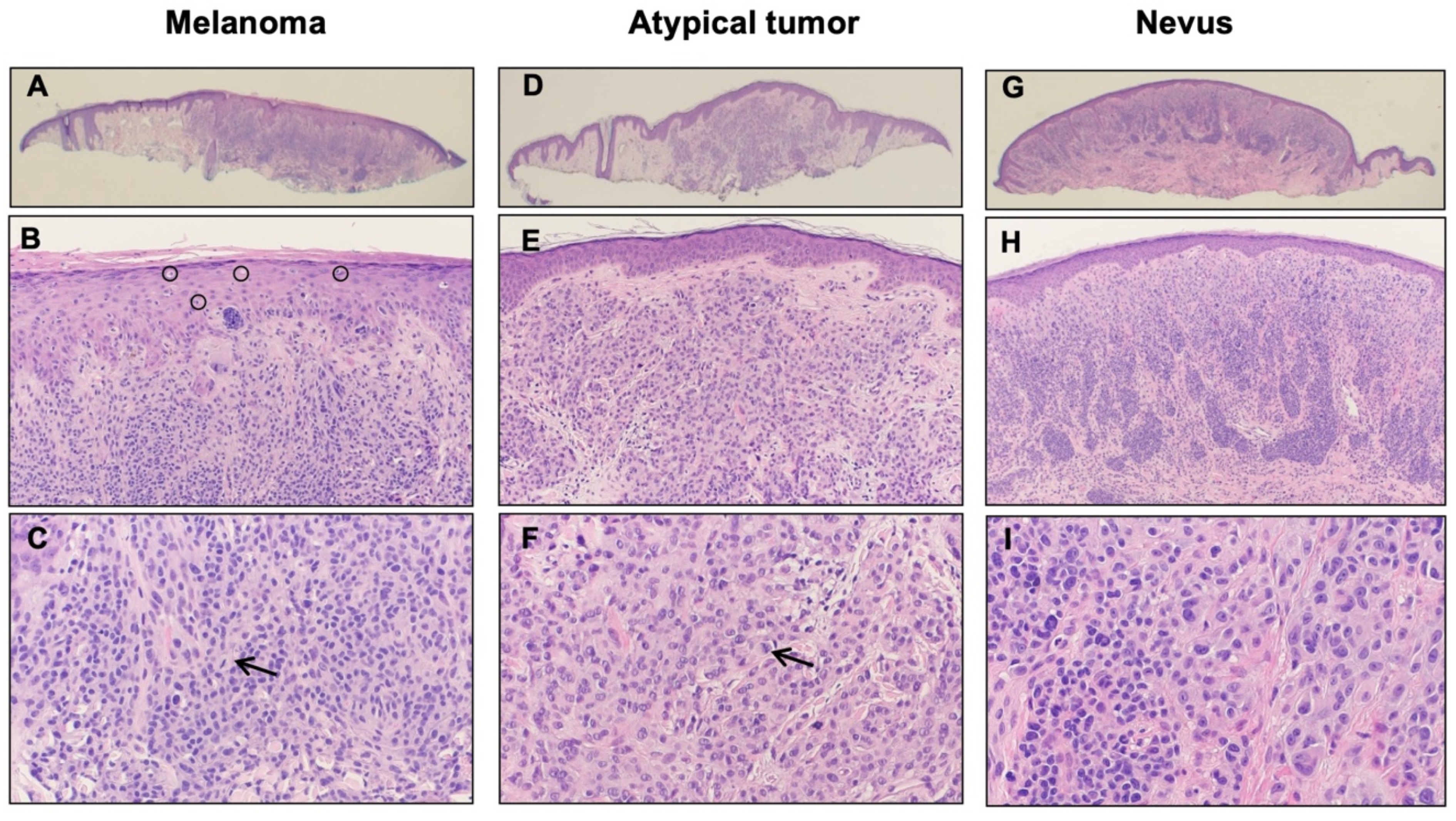
3.1. Case Presentation

A 31-year-old man presented with a history of multiple atypical melanocytic lesions, biopsied at an outside institution. He first presented subsequently at our hospital with an ear lesion in the right superior helix with the clinical appearance of a papule, which was tender. On histology, there was a severely atypical compound melanocytic proliferation with abundant pagetoid scatter of single melanocytes across the breath of the epidermis and multiple dermal mitoses (mitotic index of 5 per mm2) (Figure 1A–C). The ki67 stain demonstrated a modest increase in the labeling of dermal melanocytes, while p16 expression was lost in the atypical melanocytes, which suggested CDKN2A genomic loss. BAP1 immunohistochemistry demonstrated a loss in part of the lesion. Altogether a diagnosis was rendered of melanoma developing in the context of a melanocytic nevus with associated BAP1 genomic loss (melanoma ex-“BAPoma”). The Breslow thickness was 0.75 mm with no ulceration, no vascular invasion, and no regression, placing the pathologic staging as a pT1a lesion.
Another lesion was detected one year later in the right posterior ear, presenting clinically as a 2 mm pink pearly papule. Histologically the lesion presented as a mostly intradermal proliferation of large, oval melanocytes with abundant pale cytoplasm and large but monomorphous vesicular nuclei and small nucleoli, sparse mitoses, and with loss of BAP1 expression (Figure 1D–F). A final diagnosis of atypical Spitz nevus with BAP1 loss was rendered.
The following year, other melanocytic lesions were removed, including a lesion in the posterior neck with a clinical appearance of a dome-shaped lesion. On histology, the lesion presented as a dermal melanocytic proliferation composed of a regular melanocytic nevus in conjunction with more ovoid melanocytes with BAP1 loss (Figure 1G–I). Given the histologic and immunohistochemical features, this lesion was classified as a BAP-1 inactivated nevus (“BAPoma”).

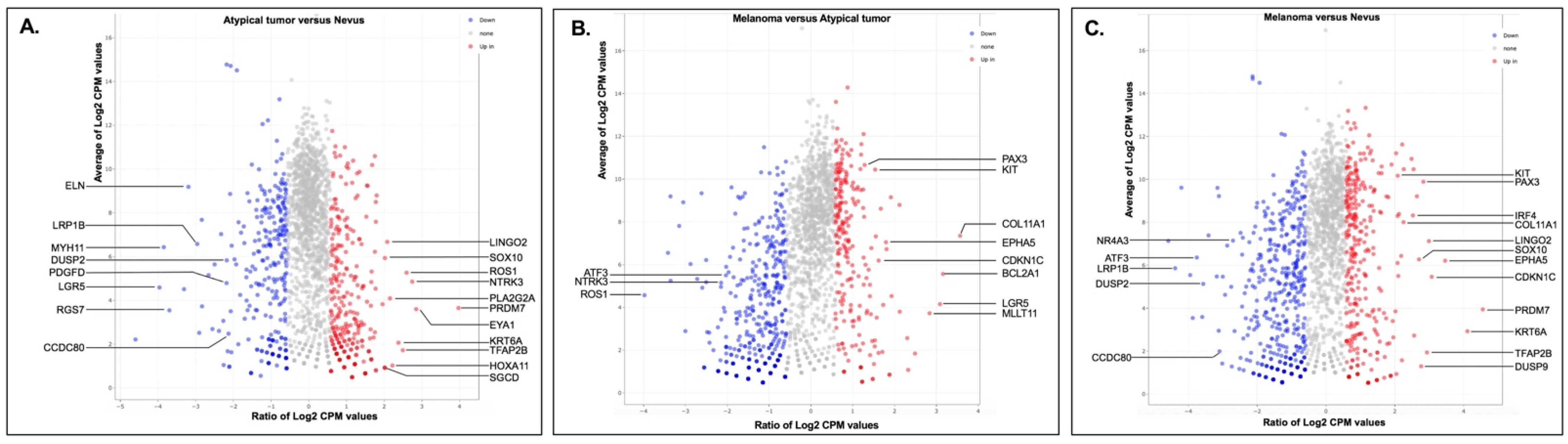
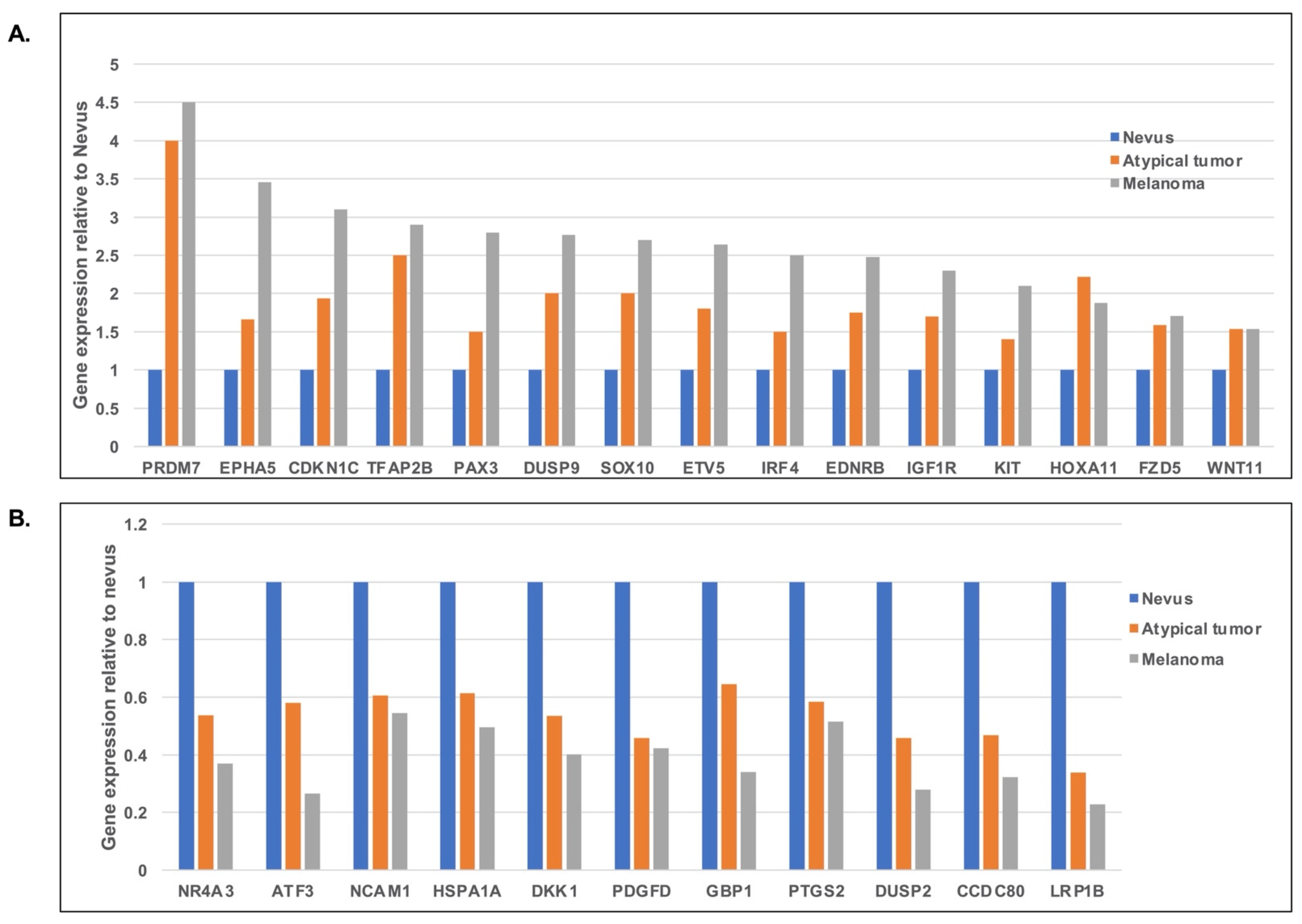
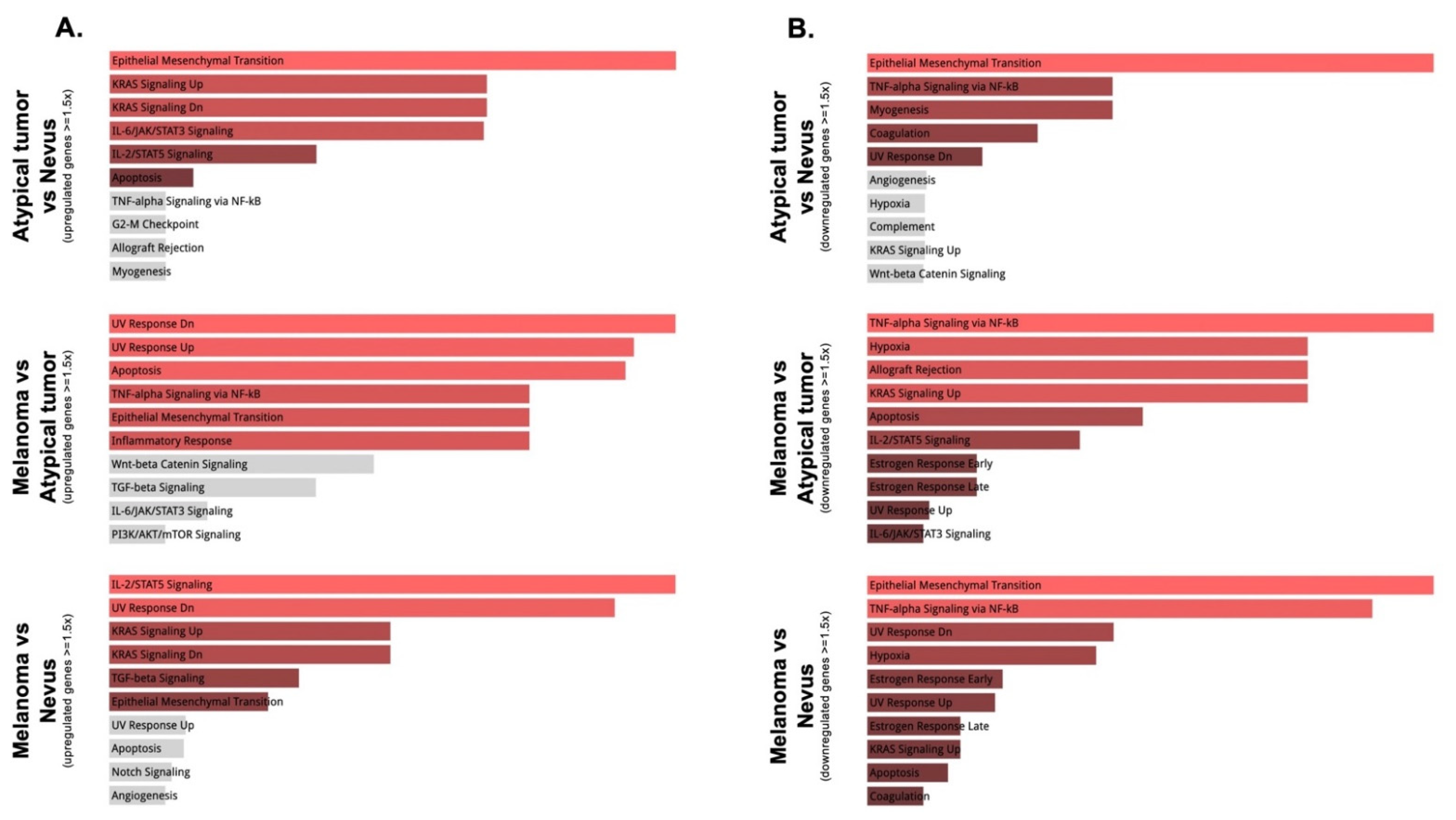
3.2. Gene Expression and Mutation Analyses

We performed a pairwise exploration of the relative differences in gene expression levels between these lesions (Figure 2A–C). A total of 40 upregulated and 53 downregulated genes were identified in the atypical tumor compared to the BAP1-inactivated nevus, 26 upregulated and 106 downregulated genes in the melanoma compared to the atypical tumor, and finally, 51 upregulated and 158 downregulated genes in the melanoma compared to the BAP1-inactivated nevus. Among the genes upregulated in the atypical tumor compared to the nevus were: SOX10, the receptor tyrosine kinases ROS1 and NTRK3, PLA2G2A, and HOXA11, while DUSP2, PDGFD, and ELN were downregulated (Figure 2A). PAX3, a transcription factor involved in melanocyte development, the receptor tyrosine kinase KIT, CDKN1C, and EPHA5 were upregulated in the melanoma compared to the atypical nevus, while NTRK3 and ROS1 were downregulated in the same comparison (Figure 2B). Finally, Figure 2C illustrates a volcano plot of the comparison of the melanoma versus the nevus, with notable upregulation of KIT, PAX3, SOX10, CDKN1C, and DUSP9, and downregulation of ATF3 and DUSP2.
By analyzing and comparing the relative expression levels of the top 50 most variable genes among the three lesions, we identified a group of differentially expressed genes (Supplemental Figure S1). We then wanted to explore which genes are progressively upregulated or downregulated from a benign BAP1-inactivated nevus to a BAP1-inactivated melanoma. Figure 3 illustrates this concept. Overall, genes that are progressively upregulated from nevus to atypical tumor to melanoma include SOX10, a transcription factor important in melanocytic differentiation, the protein tyrosine kinases IGF1R, KIT (the latter mutated in acral melanoma, mucosal melanoma, and melanoma of chronically sun-damaged skin) and EPHA5, the transcription factor PAX3 (involved in melanocyte development), the Dual Specificity Phosphatase DUSP9, WNT11 and IRF4/MUM1 (a gene regulated by MITF in melanocytic cells). In contrast, genes that are progressively downregulated from nevus to atypical tumor to melanoma include NCAM1 (implicated in cell-cell adhesion and reportedly downregulated in several human cancer, suggesting a tumor repressor role), HSPA1A, DKK1 (a secreted inhibitor of the β-catenin dependent Wnt signaling pathway and involved in induction of cancer evasion of immune surveillance), the growth factor PDGFD, the protein phosphatase DUSP2 (involved in the negative regulation of members of the mitogen-activated protein kinase (MAPK) superfamily), and LRP1B (frequently mutated in melanoma) [19].
In order to understand the collective functions and the cellular molecular pathways related to the differentially expressed genes in our samples, we performed gene set enrichment analysis. Using the Hallmark gene sets within the EnrichR software analysis, when comparing the atypical tumor with the nevus, there was a clear modulation of the epithelial-mesenchymal transition pathway, as well as upregulation of the KRAS signaling pathway and interleukin/STAT signaling, and downregulation of TNF-α signaling. When comparing the melanoma with both the nevus and the atypical tumor, it was interesting to observe regulation of the UV response, particularly important in melanomagenesis. Specifically, genes upregulated or downregulated in response to UV radiation involve CDKN1C, CDK2, COL11A1, KIT, and IGF1R, the majority of which are identified as differentially upregulated in melanoma versus atypical tumor or nevus (Figure 2B,C and Figure 3A). In this comparison, there was also the regulation of TNF-α signaling and epithelial-mesenchymal transition (Figure 4A,B).
GO term enrichment analysis was also performed and showed that expression profiles involving the extracellular region and component of the membrane were enriched progressively from nevus to atypical tumor to melanoma (Supplemental Figure S2). Particularly, pathways related to protein kinase activity in general as well MAPK activity and protein tyrosine kinase activity were significantly upregulated in both the atypical tumor and melanoma compared to the nevus (Supplemental Table S1).
Genes harboring pathogenic or likely pathogenic mutations were heterogeneous across the three lesions (Table 1). NIPBL, AKAP9, and EP400 were mutated in both the nevus and atypical tumor, although the specific mutations were different between the two lesions. In addition, mutations in PDGFRB, HIF1A, and MAP2K1 genes were identified in the nevus, and mutations in HDAC2, PRK2, ARID2, BRCA2, NOTCH3, NOTCH2, BRAF, CSF1R, and PTEN, among others, were found in the atypical nevus. To be noted, BRAF V600E was identified in both atypical tumors and melanoma. The mutation in BAP1 was captured in the melanoma, as well as mutations in KMT2A (a known cancer driver mutation in melanoma) and TIAM1 (a gene involved in the RAC1 signaling pathway affecting cell shape and migration).

4. Discussion

BAP1-inactivated melanocytic lesions exist in a spectrum that goes from benign melanocytic nevi, usually combined (“BAPomas”), to intermediate lesions with atypical features (akin to the atypical Spitz tumor) and ending with malignant melanoma. While initially categorized within the Spitz family of melanocytic lesions, genetic analysis has revealed a molecular profile, including the presence of BRAF mutation, that is not compatible with this classification. Thus, in the recent WHO classification, these lesions have been classified separately as combined melanocytic lesions with a distinct morphologic and molecular signature (BAP1 inactivation and BRAF V600E mutation). From their first description in the seminal paper by Wiesner et al. [2], there has been a lot of interest in these lesions as a paradigm for newly recognized melanocytic lesions with distinct histologic morphology and a well-defined molecular signature. The last WHO classification of cutaneous tumors classifies BAP1-inactivated lesions in a separate category, recognizing its unicity. However, besides the pathognomonic mutation, little is known regarding the gene expression profile and other gene mutations occurring in these lesions. To our knowledge, this is the first study to compare gene expression levels in a series of BAP1-inactivated melanocytic lesions of progressive malignancy, from nevus to atypical tumor to melanoma in a single patient.
The same WHO classification released in 2018 put an accent on the role of UV-related damage in melanocytic tumorigenesis by dividing the most common melanocytic lesions in those related to low cumulative solar damage (CSD) and high CSD. BAP1 mutation may occur as the driver genetic event in low CSD melanoma, but there is not a specific relation with sun—damage reported for the new category of BAP1-inactivated lesions. In our study, we found a clear enrichment of genes regulated in response to UV radiation, based on the Molecular Signature Database (MSigDB), suggesting that sun damage is a relevant step in the multistep process of tumor progression in these lesions.
Our study possesses potential clinical significance by suggesting multiple therapeutic targets in patients with BAP1-inactivated melanoma, the treatment for whom is otherwise limited to conventional therapies. These targets may involve genes in protein kinase pathways or governing transcriptional regulation, as discussed below. Based on our analysis, activation of protein kinase pathways, especially the MAPK pathway, is largely observed in the atypical tumor and melanoma arising from BAP1-inactivated nevus compared to nevus, consistent with current knowledge of melanocytic progression. MAPK activation represents a common aberrant signaling pathway in cutaneous melanoma associated with a wide variety of somatic genetic alterations. This knowledge has resulted in effective targeted therapies targeting this pathway; the success of BRAF inhibitors alone or in combination with MEK inhibitors [20] is a testament to the importance of molecular and mechanistic studies to understand the pathogenesis and discover relevant targets for cancer therapy. Thus, our findings support the use of similar therapeutic regimens in our patient as adjuvant treatment. To further support this, we found downregulation of the protein phosphatase DUSP2, involved in the negative regulation of members of the MAPK superfamily, and upregulation of the KRAS signaling pathway. In addition, genes encoding protein tyrosine kinases, KIT and EPHA5, relevant receptors upstream of the MAPK pathway, are particularly highly expressed in the melanoma compared to BAP1-inactivated nevus. Interestingly, a mutation in the MAP2K1 gene was identified in the BAPoma from our patient; similar mutations in this gene have been reported in a large spectrum of melanocytic lesions, including BAPomas [21].
IGF1R (insulin-like growth factor I receptor), another receptor tyrosine kinases found to be upregulated in our sample, has been found to have anti-apoptotic properties and to be overexpressed in multiple cancers including melanoma [22], where may represent a potential therapeutic target.
Among the protein kinase activation pathways, the expression levels of ROS1 and NTRK3 are only upregulated in the atypical tumor compared to the nevus and melanoma. Interestingly, various kinase fusions, including ROS1 and NTRK3, were found in Spitz melanocytic lesions [23]. These various fusion events are mostly mutually exclusive and are believed to be an early event in tumorigenesis [24], but do not necessarily confer malignant potential and are actually present often in benign or intermediate lesions, but rarely in melanoma.
Another interesting group of upregulated genes in the atypical tumor and the melanoma is related to sequence-specific DNA binding and regulatory region nucleic acid binding, specifically transcriptional regulation. This gene category includes HOXA11, SOX10, ETV5, IRF4, PAX3, PRDM7, TFAP2B, and WNT11. Among those, the transcription factor SOX10 has been shown to be involved in melanomagenesis in animal models [25,26] and, more recently, in the regulation of melanoma cell invasion, through the regulation of melanoma inhibitory activity (MIA) expression [27]; thus SOX10, besides its role as a marker of melanocytic differentiation in diagnostic pathology, represents a potential therapeutic target for melanoma. SOX10 was also found to regulate immunogenicity in melanoma through IRF4 (interferon regulatory factor 4)/MUM1 [28], another transcription factor discovered in our dataset. IRF4 is a gene regulated by MITF in melanocytic cells, and it was also found to have a functional variant associated with increased Breslow thickness, conferring a worse survival in melanoma [29]. WNT11 was shown to play an important role in neural crest migration and appears to have a role in the aberrant activation of Wnt signaling in melanoma. Finally, PAX3 (paired box gene 3 transcription factor) was shown, in conjunction with the transcription factor ETS1, to promote melanoma cells proliferation and metastasis by increasing the expression of MET, the HGF receptor [30]. Altogether, these findings highlight the role of several transcription factors as critical players in melanoma. Given their role as focal and convergence points of several signaling pathways and their role in tumor progression and resistance to therapy [31], these transcription factors are potential targets to explore for the development of future therapy for melanoma, including those arising in the background of BAP1 inactivation.
There are some limitations in our study that we need to acknowledge. First, only three samples from the same patient were available at the time of the experiment; thus, these data represent a single set of lesions, and future studies on multiple samples from several patients will be needed to generalize any of the biomarkers identified. Second, only a panel of 1400 cancer-related genes were analyzed, and the source material was formalin-fixed and paraffin-embedded, a challenging source that is prone to RNA fragmentation impacting the number of genes that we were able to capture with our assay. Future experiments on a larger panel of genes or the entire transcriptome may reveal additional significant markers of tumor progression in this type of lesion.

5. Conclusions

In conclusion, our study suggests that a BAP1-inactivated nevus, an atypical tumor, and melanoma with the same background from the same patient possess distinct gene expression profiles, with notable upregulation of genes involved in protein kinase pathways in the progression from nevus to melanoma. Moreover, we found a clear enrichment of genes regulated in response to UV radiation in the melanoma from this patient, as well as upregulation of MAPK pathway-related genes and several transcription factors related to melanomagenesis. Current treatments for patients with BAP1-inactivated melanoma are limited to conventional therapies used in other more common melanomas, which may not address the molecular mechanisms of these lesions entirely. Thus, our study may suggest potential novel targets for personalized therapy in these patients.

Supplementary Materials

The following are available online at https://www.mdpi.com/article/10.3390/genes13010010/s1, Figure S1: Heatmap showing the z-scored expression levels of the genes that are differentially expressed among the three lesions, Figure S2: Gene enrichment analyses between (A) Atypical tumor versus Nevus; (B) Melanoma versus Atypical tumor; (C) Melanoma versus Nevus, Table S1: Selected pathways that are upregulated and the involved genes in these pathways between either of the two BAP1-inactivated lesions.

Author Contributions

Conceptualization, A.G. and Y.Z.; Methodology, A.G., K.Y.S. and A.C.N.; Formal Analysis, A.G., Y.Z., A.C.N., Y.H., S.A.M., K.Y.S. and E.D.-M.; Resources, A.G.; Writing—Original Draft Preparation, A.G. and Y.Z.; Writing—Review and Editing, A.G., Y.Z., A.C.N., Y.H., S.A.M., K.Y.S. and E.D.-M.; Visualization, A.G., Y.Z. and S.A.M.; Supervision, A.G.; Project Administration, A.G.; Funding Acquisition, A.G. All authors have read and agreed to the published version of the manuscript.

Funding

This research was funded by start-up funds from the Department of Laboratory Medicine and Pathology/Masonic Cancer Center, University of Minnesota.

Institutional Review Board Statement

The study was conducted according to the guidelines of the Declaration of Helsinki and approved by the Institutional Review Board of the University of Minnesota (protocol code STUDY00002958 and date of approval 3 December 2018).

Informed Consent Statement

Written informed consent has been obtained from the patient to publish this paper.

Data Availability Statement

The data presented in this study are available in the supplemental material and also upon request from the corresponding author.

Acknowledgments

We would like to thank Colleen Forster for histologic preparations and the staff at the University of Minnesota Genomic Center.

Conflicts of Interest

The authors declare no conflict of interest.

References

  1. Carbone, M.; Yang, H.; Pass, H.I.; Krausz, T.; Testa, J.R.; Gaudino, G. BAP1 and cancer. Nat. Rev. Cancer 2013, 13, 153–159. [Google Scholar] [CrossRef]
  2. Wiesner, T.; Obenauf, A.; Murali, R.; Fried, I.; Griewank, K.; Ulz, P.; Windpassinger, C.; Wackernagel, W.; Loy, S.; Wolf, I.; et al. Germline mutations in BAP1 predispose to melanocytic tumors. Nat. Genet. 2011, 43, 1018–1021. [Google Scholar] [CrossRef] [Green Version]
  3. Wiesner, T.; Fried, I.; Ulz, P.; Stacher, E.; Popper, H.; Murali, R.; Kutzner, H.; Lax, S.; Smolle-Jüttner, F.; Geigl, J.B.; et al. Toward an Improved Definition of the Tumor Spectrum Associated With BAP1 Germline Mutations. J. Clin. Oncol. 2012, 30, e337–e340. [Google Scholar] [CrossRef] [PubMed]
  4. Walpole, S.; Pritchard, A.L.; Cebulla, C.M.; Pilarski, R.; Stautberg, M.; Davidorf, F.H.; De La Fouchardière, A.; Cabaret, O.; Golmard, L.; Stoppa-Lyonnet, D.; et al. Comprehensive Study of the Clinical Phenotype of Germline BAP1 Variant-Carrying Families Worldwide. J. Natl. Cancer Inst. 2018, 110, 1328–1341. [Google Scholar] [CrossRef]
  5. Haugh, A.M.; Njauw, C.N.; Bubley, J.A.; Verzì, A.E.; Zhang, B.; Kudalkar, E.; Van den Boom, T.; Walton, K.; Swick, B.L.; Kumar, R.; et al. Genotypic and Phenotypic Features of BAP1 Cancer Syndrome: A Report of 8 New Families and Review of Cases in the Literature. JAMA Dermatol. 2017, 153, 999–1006. [Google Scholar] [CrossRef] [PubMed]
  6. Yeh, I.; Mully, T.W.; Wiesner, T.; Vemula, S.S.; Mirza, S.A.; Sparatta, A.J.; McCalmont, T.H.; Bastian, B.; LeBoit, P.E. Ambiguous Melanocytic Tumors With Loss of 3p21. Am. J. Surg. Pathol. 2014, 38, 1088–1095. [Google Scholar] [CrossRef] [PubMed] [Green Version]
  7. Busam, K.J.; Sung, J.; Wiesner, T.; Von Deimling, A.; Jungbluth, A. Combined BRAF(V600E)-positive melanocytic lesions with large epithelioid cells lacking BAP1 expression and conventional nevomelanocytes. Am. J. Surg. Pathol. 2013, 37, 193–199. [Google Scholar] [CrossRef]
  8. Wiesner, T.; Murali, R.; Fried, I.; Cerroni, L.; Busam, K.; Kutzner, H.; Bastian, B.C. A Distinct Subset of Atypical Spitz Tumors is Characterized by BRAF Mutation and Loss of BAP1 Expression. Am. J. Surg. Pathol. 2012, 36, 818–830. [Google Scholar] [CrossRef] [Green Version]
  9. Aung, P.P.; Nagarajan, P.; Tetzlaff, M.T.; Curry, J.L.; Tang, G.; Abdullaev, Z.; Pack, S.D.; Ivan, D.; Prieto, V.G.; Torres-Cabala, C.A. Melanoma With Loss of BAP1 Expression in Patients With No Family History of BAP1-Associated Cancer Susceptibility Syndrome: A Case Series. Am. J. Dermatopathol. 2019, 41, 167–179. [Google Scholar] [CrossRef]
  10. Busam, K.J.; Wanna, M.; Wiesner, T. Multiple epithelioid Spitz nevi or tumors with loss of BAP1 expression: A clue to a hereditary tumor syndrome. JAMA Dermatol. 2013, 149, 335–339. [Google Scholar] [CrossRef]
  11. Patro, R.; Duggal, G.; Love, M.I.; Irizarry, R.A.; Kingsford, C. Salmon provides fast and bias-aware quantification of transcript expression. Nat. Methods 2017, 14, 417–419. [Google Scholar] [CrossRef] [Green Version]
  12. Wang, K.A.; Li, M.; Hakonarson, H. ANNOVAR: Functional annotation of genetic variants from high-throughput sequencing data. Nucleic Acids Res. 2010, 38, e164. [Google Scholar] [CrossRef] [PubMed]
  13. Li, M.M.; Datto, M.; Duncavage, E.J.; Kulkarni, S.; Lindeman, N.I.; Roy, S.; Tsimberidou, A.M.; Vnencak-Jones, C.L.; Wolff, D.J.; Younes, A.; et al. Standards and Guidelines for the Interpretation and Reporting of Sequence Variants in Cancer: A Joint Consensus Recommendation of the Association for Molecular Pathology, American Society of Clinical Oncology, and College of American Pathologists. J. Mol. Diagn. 2017, 19, 4–23. [Google Scholar] [CrossRef] [PubMed] [Green Version]
  14. Chen, J.; Bardes, E.E.; Aronow, B.J.; Jegga, A.G. ToppGene Suite for gene list enrichment analysis and candidate gene prioritization. Nucleic Acids Res. 2009, 37, W305–W311. [Google Scholar] [CrossRef]
  15. Chen, E.Y.; Tan, C.M.; Kou, Y.; Duan, Q.; Wang, Z.; Meirelles, G.V.; Clark, N.R.; Ma’Ayan, A. Enrichr: Interactive and collaborative HTML5 gene list enrichment analysis tool. BMC Bioinform. 2013, 14, 128. [Google Scholar] [CrossRef] [Green Version]
  16. Kuleshov, M.V.; Jones, M.R.; Rouillard, A.D.; Fernandez, N.F.; Duan, Q.; Wang, Z.; Koplev, S.; Jenkins, S.L.; Jagodnik, K.M.; Lachmann, A.; et al. Enrichr: A comprehensive gene set enrichment analysis web server 2016 update. Nucleic Acids Res. 2016, 44, W90–W97. [Google Scholar] [CrossRef] [PubMed] [Green Version]
  17. Xie, Z.; Bailey, A.; Kuleshov, M.V.; Clarke, D.J.B.; Evangelista, J.E.; Jenkins, S.L.; Lachmann, A.; Wojciechowicz, M.L.; Kropiwnicki, E.; Jagodnik, K.M.; et al. Gene Set Knowledge Discovery with Enrichr. Curr. Protoc. 2021, 1, e90. [Google Scholar] [CrossRef]
  18. Subramanian, A.; Tamayo, P.; Mootha, V.K.; Mukherjee, S.; Ebert, B.L.; Gillette, M.A.; Paulovich, A.; Pomeroy, S.L.; Golub, T.R.; Lander, E.S.; et al. Gene set enrichment analysis: A knowledge-based approach for interpreting genome-wide expression profiles. Proc. Natl. Acad. Sci. USA 2005, 102, 15545–15550. [Google Scholar] [CrossRef] [Green Version]
  19. Brown, L.C.; Tucker, M.D.; Sedhom, R.; Schwartz, E.B.; Zhu, J.; Kao, C.; Labriola, M.K.; Gupta, R.T.; Marin, D.; Wu, Y.; et al. LRP1B mutations are associated with favorable outcomes to immune checkpoint inhibitors across multiple cancer types. J. Immunother. Cancer 2021, 9, e001792. [Google Scholar] [CrossRef]
  20. Jenkins, R.W.; Fisher, D.E. Treatment of Advanced Melanoma in 2020 and Beyond. J. Investig. Dermatol. 2020, 141, 23–31. [Google Scholar] [CrossRef]
  21. Sunshine, J.C.; Kim, D.; Zhang, B.; Compres, E.V.; Khan, A.U.; Busam, K.J.; Gerami, P. Melanocytic Neoplasms With MAP2K1 in Frame Deletions and Spitz Morphology. Am. J. Dermatopathol. 2020, 42, 923–931. [Google Scholar] [CrossRef]
  22. Capoluongo, E. Insulin-Like Growth Factor System and Sporadic Malignant Melanoma. Am. J. Pathol. 2011, 178, 26–31. [Google Scholar] [CrossRef] [PubMed]
  23. Quan, V.L.; Panah, E.; Zhang, B.; Shi, K.; Mohan, L.S.; Gerami, P. The role of gene fusions in melanocytic neoplasms. J. Cutan. Pathol. 2019, 46, 878–887. [Google Scholar] [CrossRef] [Green Version]
  24. Tetzlaff, M.T.; Reuben, A.; Billings, S.D.; Prieto, V.G.; Curry, J.L. Toward a Molecular-Genetic Classification of Spitzoid Neoplasms. Clin. Lab. Med. 2017, 37, 431–448. [Google Scholar] [CrossRef]
  25. Shakhova, O.; Zingg, D.; Schaefer, S.M.; Hari, L.; Civenni, G.; Blunschi, J.; Claudinot, S.; Okoniewski, M.; Beermann, F.; Mihic-Probst, D.; et al. Sox10 promotes the formation and maintenance of giant congenital naevi and melanoma. Nature 2012, 14, 882–890. [Google Scholar] [CrossRef] [PubMed]
  26. Cronin, J.C.; Watkins-Chow, D.; Incao, A.; Hasskamp, J.H.; Schönewolf, N.; Aoude, L.; Hayward, N.; Bastian, B.; Dummer, R.; Loftus, S.K.; et al. SOX10 Ablation Arrests Cell Cycle, Induces Senescence, and Suppresses Melanomagenesis. Cancer Res. 2013, 73, 5709–5718. [Google Scholar] [CrossRef] [PubMed] [Green Version]
  27. Graf, S.A.; Busch, C.; Bosserhoff, A.-K.; Besch, R.; Berking, C. SOX10 Promotes Melanoma Cell Invasion by Regulating Melanoma Inhibitory Activity. J. Investig. Dermatol. 2014, 134, 2212–2220. [Google Scholar] [CrossRef] [Green Version]
  28. Yokoyama, S.; Takahashi, A.; Kikuchi, R.; Nishibu, S.; Lo, J.A.; Hejna, M.; Moon, W.M.; Kato, S.; Zhou, Y.; Hodi, F.S.; et al. SOX10 regulates melanoma immunogenicity through an IRF4-IRF1 axis. Cancer Res. 2021. [Google Scholar] [CrossRef]
  29. Gibbs, D.C.; Ward, S.V.; Orlow, I.; Cadby, G.; Kanetsky, P.A.; Luo, L.; Busam, K.J.; Kricker, A.; Armstrong, B.K.; Cust, A.E.; et al. Functional melanoma-risk variant IRF 4 rs12203592 associated with Breslow thickness: A pooled international study of primary melanomas. Br. J. Dermatol. 2017, 177, e180–e182. [Google Scholar] [CrossRef]
  30. Kubic, J.D.; Little, E.C.; Lui, J.W.; Iizuka, T.; Lang, D. PAX3 and ETS1 synergistically activate MET expression in melanoma cells. Oncogene 2014, 34, 4964–4974. [Google Scholar] [CrossRef] [Green Version]
  31. Cohen-Solal, K.A.; Kaufman, H.L.; Lasfar, A. Transcription factors as critical players in melanoma invasiveness, drug resistance, and opportunities for therapeutic drug development. Pigment. Cell Melanoma Res. 2017, 31, 241–252. [Google Scholar] [CrossRef] [PubMed] [Green Version]
Figure 1. Histopathology of three representative lesions. (AC) Melanoma; an atypical compound melanocytic proliferation with a nevoid appearance at low power (A), with abundant pagetoid scatter of single melanocytes within the epidermis ((B), circles) and multiple dermal mitoses ((C), arrow); (DF) atypical tumor; a predominantly dermal melanocytic proliferation of atypical yet monomorphous large oval melanocytes with occasional mitoses ((F), arrow); (GI) nevus; a dermal proliferation of small regular melanocytes in conjunction with large ovoid melanocytes (I). Circles (B): pagetoid spread of the tumor cells. Arrows (C,F): mitoses.
Genes 13 00010 g001
Figure 2. The MA plots showing upregulation and downregulation of the genes between each of the two BAP1-inactivated melanocytic lesions. (A) Atypical tumor versus Nevus; (B) Melanoma versus Atypical tumor; (C) Melanoma versus Nevus.
Genes 13 00010 g002
Figure 3. (A) Relative expression levels of the genes showing the upregulated trend of expression from nevus, atypical tumor to melanoma. (B) Relative expression levels of the genes showing the downregulated trend of expression from nevus, atypical tumor to melanoma.
Genes 13 00010 g003
Figure 4. Gene enrichment analysis using the EnrichR platform. Upregulated (A) and downregulated (B) pathways (in the Hallmark category) in the following comparisons: Atypical tumor versus Nevus; Melanoma versus Atypical tumor; and Melanoma versus Nevus. Statistical significantly enriched terms are displayed in red, while gray bars are not statistically significant.
Genes 13 00010 g004
Table 1. List of genes with identified pathogenic or likely pathogenic mutations in the BAP-1 inactivated nevus, atypical tumor, and melanoma.
LesionGeneMutationType
NevusNIPBLp.Q338Xstopgain
AKAP9p.Q2911Xstopgain
EP400p.E2200Xstopgain
CREBBPp.Q1075Xstopgain
COL1A1p.P817fsframeshift deletion
ZNF687p.Q474Xstopgain
PDGFRBp.Q412Xstopgain
ASPHp.R659Xstopgain
NT5C2p.Q173Xstopgain
NINp.Q1291Xstopgain
HIF1Ap.Q379Xstopgain
CHD2p.Q366Xstopgain
USP7p.Q822Xstopgain
MYO18Ap.Q830Xstopgain
KPNB1p.Q111Xstopgain
RPS6KA3p.R383Wnonsynonymous SNV
TBL1XR1p.S332Fnonsynonymous SNV
MAP2K1p.A172Vnonsynonymous SNV
Atypical tumorNIPBLp.Q1567Xstopgain
AKAP9p.Q2480Xstopgain
EP400p.Q148Xstopgain
CADp.Q30Xstopgain
PPP1CBp.Q293Xstopgain
BIRC6p.Q1072Xstopgain
COL6A3p.Q2366Xstopgain
TFGp.Q266Xstopgain
EIF4A2p.Q209Xstopgain
AFF4p.Q537Xstopgain
ARHGAP26p.R120Xstopgain
MAML1p.Q683Xstopgain
DSTp.Q1890Xstopgain
HDAC2p.Q128Xstopgain
PTK2p.Q734Xstopgain
SYKp.Q239Xstopgain
NUMA1p.Q832Xstopgain
PICALMp.Q256Xstopgain
EEDp.Q302Xstopgain
SIK3p.Q675Xstopgain
KDM5Ap.R266Xstopgain
PRICKLE1p.Q348Xstopgain
ARID2p.Q720Xstopgain
NUP107p.Q236Xstopgain
BRCA2p.Q2943Xstopgain
TCF12p.Q605Xstopgain
TOP2Ap.Q517Xstopgain
ITGB3p.Q616Xstopgain
SMARCA4p.R978Xstopgain
NOTCH3p.Q646Xstopgain
CSNK2A1p.Q71Xstopgain
NCOA3p.Q478Xstopgain
ETS2p.Q234Xstopgain
EP300p.Q523Xstopgain
OFD1p.Q39Xstopgain
ARp.Q825Xstopgain
ZMYM3p.Q763Xstopgain
BMPR1Ap.R244Xstopgain
NOTCH2p.Q1814Xstopgain
TPRp.Q2344Xstopgain
BRAFp.V600Enonsynonymous SNV
CSF1Rp.A781Vnonsynonymous SNV
PTENp.T277Inonsynonymous SNV
MelanomaLRPPRCp.G1050fsframeshift insertion
EXT2p.R215Xstopgain
SPENp.Q3373Xstopgain
BAP1p.Y223Xstopgain
AHRp.Q705Xstopgain
KMT2Ap.Q1207Xstopgain
CDH1p.Q388Xstopgain
RABEP1p.Q225Xstopgain
CLTCp.Q1358Xstopgain
SUGP2p.R831Xstopgain
TIAM1p.Q714Xstopgain
BRAFp.V600Enonsynonymous SNV
Publisher’s Note: MDPI stays neutral with regard to jurisdictional claims in published maps and institutional affiliations.

