Hypothesis

The Theory of Carcino-Evo-Devo and Its Non-Trivial Predictions

1
Vavilov Institute of General Genetics, Russian Academy of Sciences, 3 Gubkina Street, 117971 Moscow, Russia
2
Peter the Great St. Petersburg Polytechnic University, 29 Polytechnicheskaya Street, 195251 St. Petersburg, Russia
3
Biomedical Center, 8 Viborgskaya Street, 194044 St. Petersburg, Russia
Genes 2022, 13(12), 2347; https://doi.org/10.3390/genes13122347
Submission received: 8 October 2022 / Revised: 4 December 2022 / Accepted: 8 December 2022 / Published: 12 December 2022
(This article belongs to the Special Issue Carcinogenesis as an Evolutionary Process)

Abstract

:
To explain the sources of additional cell masses in the evolution of multicellular organisms, the theory of carcino-evo-devo, or evolution by tumor neofunctionalization, has been developed. The important demand for a new theory in experimental science is the capability to formulate non-trivial predictions which can be experimentally confirmed. Several non-trivial predictions were formulated using carcino-evo-devo theory, four of which are discussed in the present paper: (1) The number of cellular oncogenes should correspond to the number of cell types in the organism. The evolution of oncogenes, tumor suppressor and differentiation gene classes should proceed concurrently. (2) Evolutionarily new and evolving genes should be specifically expressed in tumors (TSEEN genes). (3) Human orthologs of fish TSEEN genes should acquire progressive functions connected with new cell types, tissues and organs. (4) Selection of tumors for new functions in the organism is possible. Evolutionarily novel organs should recapitulate tumor features in their development. As shown in this paper, these predictions have been confirmed by the laboratory of the author. Thus, we have shown that carcino-evo-devo theory has predictive power, fulfilling a fundamental requirement for a new theory.

1. Introduction

Additional cellular masses with high biosynthetic and morphogenetic potential are necessary for the evolution of multicellular organisms, especially in the line Deuterostomia—Chordata—Vertebrata. The origin of such additional cellular masses is unclear [1].
In multicellular organisms, cell division is regulated by functional feedbacks. Formation of additional cell masses means escape from the regulatory control. Unregulated cell division is one of the features of a tumor.
The hypothesis of the evolutionary role of hereditary tumors as the source of additional cell masses in evolution was first formulated in the author’s 1979 paper [2]. Since then, the concept of the positive evolutionary role of tumors has been developed in a series of publications [3,4,5,6,7,8]. In the book Evolution by Tumor Neofunctionalization [9], with its twelve chapters and over one thousand references, this concept started to gain the shape of the theory, which was further developed in subsequent publications [10,11,12,13,14]. In a 2019 paper, the author called this new theory the “carcino-evo-devo” theory to stress the role of heritable tumors in the evolution of development [11].
The important demand for a new theory in experimental science is the capability to formulate non-trivial predictions, which can be experimentally confirmed. The formulation of predictions is largely determined by the main hypothesis of a theory. The main hypothesis of carcino-evo-devo theory is the hypothesis of evolution by tumor neofunctionalization.
The hypothesis of evolution by tumor neofunctionalization in [9] was defined as follows:
“Tumors are the source of extra cell masses, which may be used in the evolution of multicellular organisms for the expression of evolutionarily new genes, for the origin of new differentiated cell types with novel functions and for building new structures, which constitute evolutionary innovations and morphological novelties”.
Hereditary tumors may play an evolutionary role by providing conditions (space and resources) for the expression of genes newly evolving in the DNA of germ cells. As a result of expression of novel genes, tumor cells may acquire new functions and differentiate in new directions, which may lead to the origin of new cell types, tissues and organs. New cell type is inherited in progeny generations due to genetic and epigenetic mechanisms similar to those for pre-existing cell types.
Tumors at the early stages of progression, benign tumors, pseudoneoplasms and tumor-like processes, which provide evolving multicellular organisms with extra cell masses functionally unnecessary to the organism, are considered as potentially evolutionarily meaningful. Malignant tumors at the late stages of progression, however, are not” [9].
The main hypothesis of carcino-evo-devo theory helped to formulate several non-trivial predictions.

2. Non-Trivial Predictions of the Carcino-Evo-Devo Theory

Several non-trivial predictions were formulated, four of which will be discussed in the present paper:
(1)
The number of cellular oncogenes should correspond to the number of cell types in the organism. Evolution of oncogenes, tumor suppressor and differentiation gene classes should proceed concurrently.
(2)
Evolutionarily new and evolving genes should be specifically expressed in tumors (TSEEN genes).
(2’)
The whole classes of genes with tumor-specific expression may be evolutionarily novel:
  • CT antigen genes;
  • HERVs;
  • ncRNA genes;
  • pan-cancer genes.
(3)
Human orthologs of fish TSEEN genes should acquire progressive functions connected with new cell types, tissues and organs.
(4)
Selection of tumors for new functions in the organism is possible. Evolutionarily novel organs should recapitulate tumor features in their development.

3. Confirmation of Non-Trivial Predictions

3.1. The Number of Cellular Oncogenes Should Correspond to the Number of Cell Types in the Organism

Originally, this prediction was formulated as follows:
“The evolutionary role of cellular oncogenes, or proto-oncogenes might consist in sustaining a definite genetically determined level of autonomous proliferative processes in evolving populations of multicellular organisms and in promoting the expression of evolutionarily new genes in anaplastic cells of extra cell masses. After the origin of a new cell type, the corresponding oncogene should have turned into a cell type-specific regulator of cell division. If such scenario is true, then the number of different proto-oncogenes should be about 200—in accordance with the number of cell types in the multicellular organism”.
[4]
When this prediction was first formulated, only about a dozen oncogenes and two hundred cell types were known.
In 1996, the author wrote on the same prediction:
“The evolutionary role of cellular oncogenes may consist in sustaining the definite level of autonomous proliferative processes in the evolving populations of organisms and in promoting the expression of evolutionarily new genes. After the origin of a new cell type, the corresponding oncogene should have turned into a cell type-specific regulator of cell division and gene expression. If true, the number of cellular oncogenes should correspond to the number of cell types in higher animals (10-fold higher than the 20 or so limit predicted a few years ago and now already about 70)”.
[6]
Determination of the correct number of cellular oncogenes took thirty years of work by thousands of scientists in hundreds of labs, and the work is being continued. In 2019, we performed a comparative analysis of the discovered numbers of oncogenes and cell types [15].
The published estimates of the number of cell types in humans produced numbers ranging from 240 [16,17,18] to 411 [19] cell types.
At the same time, the TAG database contained 246 oncogenes and the COSMIC database had 312 cancer genes, which approximately corresponded to the number of cell types. The number of oncogenes in the other multicellular organisms for which such information was available also corresponded to the number of cell types in these organisms [15]. Thus, the prediction about correspondence of the number of cellular oncogenes to the number of cell types is confirmed.

Evolution of Oncogenes, Tumor Suppressor and Differentiation Gene Classes Should Proceed Concurrently

The author further hypothesized that at least three different classes of genes are necessary for the origin and evolutionary enhancement of functional molecular feedback loops in a new cell type during evolution—oncogenes, tumor suppressor genes (TSG) and evolutionarily novel genes, which determine new functions (Figure 1).
Such a relationship predicted concurrent evolution of these gene classes. In order to prove this prediction, we performed a special study of phylogenetic distribution of orthologs of human oncogenes, tumor suppressor genes and differentiation genes. We have shown that gene age distribution curves of oncogenes, tumor suppressor genes and differentiation genes overlap. They form a cluster with perfect (100%) bootstrap reliability which confirms the coevolution of these gene classes. Moreover, we found intersections between these classes, i.e., some genes which belong to several classes (onco × diff, TSG × diff). We found that TGFβ gene belongs to all three classes, i.e., it is the triple function gene. This suggests functional co-evolution on a single gene level [15]. Thus, the prediction about concurrent evolution of oncogenes, tumor suppressor and differentiation gene classes is confirmed.

3.2. Evolutionarily New and Evolving Genes Should Be Specifically Expressed in Tumors (TSEEN Genes)

The important non-trivial prediction of the main hypothesis that evolutionarily new genes should be specifically expressed in tumors is contained in the formulation of the hypothesis. This prediction has been addressed in many of our papers [10,15,20,21,22,23,24,25,26,27,28,29,30,31,32,33,34,35,36,37].
Before the era of genomics came, it was not possible to prove this prediction because the sensitivity of methods was low (see e.g., [20]).
In our genomic studies that followed, the age of the gene was defined by the most recent common ancestor on the human evolutionary timeline which contained genes with similar sequences, i.e., with a significant BLAST score (or HMMER E-value) [38].
The age of the gene class was described by distribution of ages of genes belonging to this gene class. For convenience, the age of the gene class was measured numerically in million years (Ma) at the median of distribution, i.e., at the time point on the human evolutionary timeline which corresponds to the origin of 50% of orthologs of the functional gene class [15].
Tumor specificity of gene expression was studied using subtractions of all known normal cDNA libraries from all tumor DNA libraries [21,24]. We called this approach “the global subtraction”. It stemmed from our earlier saturation hybridization experiments in which we used combined RNA preparations from all experimentally available tissues of rat [20]. With the development of in silico databases, for global subtractions we used databases such as GTEx and TCGA. Genes obtained as a result of such subtractions may be called the “pan-cancer genes”.
Tumor specificity of gene expression was experimentally confirmed using PCR on normal and tumor cDNA panels (reviewed in [9,10]).
We described several evolutionarily new and young human genes with tumor-specific or tumor-predominant expression, and the whole classes of such genes. The author called them TSEEN genes—tumor specifically expressed, evolutionarily novel genes (reviewed in [9,10]).

3.2.1. Single TSEEN Genes

PBOV1 gene. In our 2013 paper [31], we described the de novo origin of the human TSEEN protein coding gene, PBOV1. It was among the first de novo originated human genes described in the literature (see discussion in [39]).
We noticed this gene among human genes without orthologs in the mouse and dog genomes in the paper of Clamp and co-authors [40] and we studied its evolutionary novelty more carefully. We found that PBOV1 is a single-exon gene, and its ORF encodes a 135-aa protein; the ORF originated in humans through a series of frame-shift and stop codon mutations; 80% of the protein sequence is unique to humans; the protein existence is confirmed by Western blot and MS/MS identifications; the protein lacks any annotated or predicted domains; over 60% of the protein is predicted to be disordered; a protein-coding sequence is not conserved, Ka/Ks ~ 1.0, indicating that the amino acid sequence evolved neutrally; and Gene Ontology (GO) shows few functions. These findings strongly suggested a very recent de novo evolutionary origin for the PBOV1 gene [31].
The PBOV1 gene was originally discovered by An and co-authors, who have shown its overexpression in prostate, breast and bladder cancer [41]. In our paper, we have shown that the PBOV1 gene is expressed in a much broader range of tumors [31]. High levels of PBOV1 expression are connected with a favorable clinical outcome for breast cancer and glioma [31]. We hypothesized that PBOV1 protein acts as an immunological tumor suppressor [31].
Similar results have been obtained by other authors using an ovarian cancer model [42].
However, PBOV1 promotes prostate cancer [43] and can be a biomarker for more advanced prostate cancer [44]. High PBOV1 expression level is also a marker of poor prognosis for patients with hepatocellular carcinoma (HCC) and therapeutic target for HCC [45,46]. Its function in HCC may be connected with its role in epithelial-to-mesenchymal transition [45,47]. PBOV1 rs6927706 polymorphism is linked to the development of breast cancer [48].
Therefore, depending on context, PBOV1 can serve either as a tumor suppressor or an oncogene. Similar situations will be discussed below for other genes.
PBOV1 may also have other functions [49,50].
ELFN1-AS1 gene. The other TSEEN gene—ELFN1-AS1 was described by us as a novel primate gene expressed predominantly in tumors [32]. It has an extensive literature now as an lncRNA oncogene in many tumors, with appropriate references to our original paper as the first description of this gene [51,52,53,54]. In a recent review, it is presented as a human-specific de novo originated gene [55].
The TSEEN nature of PBOV1 or ELFN1-AS1 genes was studied with two different approaches. In the case of PBOV1, we first studied in silico the evolutionary novelty of the gene. After it was established, we studied experimentally the tumor specificity of its expression using cDNA panels from normal and tumor tissues, and we found the broad range of tumors in which PBOV1 was specifically expressed [31].
ELFN1-AS1 was found among EST sequences obtained by global subtraction of all known normal cDNA libraries from all tumor cDNA libraries (not the pairwise comparison of each normal tissue and corresponding tumor, as it is usually done) [21,24]. After the tumor specificity of the expression of the gene was confirmed experimentally [22], we studied its evolutionary age and discovered that it was novel for primates [32].
The de novo origin is one of fundamental modes of gene origin (see [9] for discussion). It is difficult to prove de novo origin, however. Several papers have double-checked the de novo genes described by other authors [39,55,56]. Two papers [39,55] studied the de novo origin of PBOV1 and ELFN1-AS1 genes again. Both genes have successfully passed the scrutiny. This confirms the correctness of our approach to TSEEN genes discovery.
In [55], though, there is serious inaccuracy in citation: the author did not correctly cite the paper in which de novo origin of the PBOV1 gene was described. An and co-authors in their paper [41] did not describe the de novo origin of the PBOV1 gene. Other examples of individual TSEEN genes/sequences described in the lab of the author are presented in [10].

3.2.2. TSEEN Gene Classes

CT genes. We have also described the evolutionary novelty of a whole class of genes—cancer/testis antigen (CT) genes [30]. The evolutionary novelty of CT genes was later confirmed by other authors with appropriate reference to our priority [57].
The reasoning for prediction of the evolutionary novelty of CT genes was as follows. The author asked the question—“Why cancer/testis?”, why such unusual similarity of expression patterns in testis and different cancers? This question had never been appropriately addressed. The answer was hinted at by the fact that evolutionarily novel genes originate in the germ cells, and the germ cells are in the testis. Many genes originate through retroposition mechanism, which is why their expression in the testis is necessary. According to our hypothesis, evolutionarily novel genes should be expressed in tumors. Hence, CT genes should be young or novel genes, exactly what we found in [30]. The same line of reasoning explains the broader phenomenon of cancer/testis/brain genes, i.e., through the suggestion that an evolutionarily novel organ, the human brain, originated from a tumor-like structure (see more detailed discussion in the author’s book [9]).
In our recent paper, we confirmed the evolutionary novelty of CT genes using a different methodology [15].

Evolutionarily Novel HERVs Expressed Predominantly in Tumors

Different families of HERVs infected human ancestors and integrated their genome during different phylogenetic periods [58,59]. The author suggested that expression of HERVs in normal and tumor tissues should depend on their evolutionary novelty. The existing data on the expression of HERVs and other retrotransposons in tumors supported overexpression of many HERVs in tumors [10]. However, the data also suggested that HERV-derived RNAs were more widely expressed in normal tissues than originally anticipated. The author further suggested that the evolutionarily youngest HERVs should be expressed more specifically in tumors. There are very young HERV families, e.g., HERV-K HML-2, with estimated time of infection less than 1 Ma [60]. HERV-K HML-2 proviruses located on the X chromosome were chosen because the burst of X-linked gene originations happened after the split of human and chimp [61].
According to our prediction, HERV-K HML-2 located on X chromosome (HERV-K HML-2 (X)) should have higher expression in tumor tissues. The expression level of 12 HERV-K HML-2 (X) sequences were analyzed in normal tissues (lung, colon and lymphocytes) and in tumors (small cell carcinoma, colon cancer and acute myeloid leukemia). We found that the expression level of HERV-K HML-2 sequences located on the X-chromosome was dramatically higher in tumors as compared with normal tissues [62]. Therefore, a new family of TSEEN genes –HERV-K HML-2 (X)—was described as predicted.
ncRNA genes/sequences. About half of the tumor-specific sequences produced by global subtractions [21,24] lack long reading frames and may be referred to non-coding RNAs [10]. Among them, the ELFN1-AS1 gene was discovered as a candidate microRNAgene [32], but later was referred to lncRNA genes [63,64]. We also described a new long non-coding RNA gene—OTP-AS1—which belongs to cancer/testis genes and is evolutionarily novel for eutherians, with almost 100% tumor specificity of expression in many tumors [22,36,65]. A class of ncRNA genes obtained by global subtractions is the youngest class of human genes [15]. Noncoding RNA genes may represent proto-genes evolving to new organismal functions. Proto-genes are defined as gene precursors, which have not acquired functions yet [66].

The Evolutionary Novelty of Tumor-Specifically Expressed Sequences Obtained by Global Subtraction (Pan-Cancer Genes)

In [15], the evolutionary ages of different gene classes have been compared.The protein-coding sequences have been studied by Protein Historian tool, and the non-coding sequencesby BLAST algorithm and the original Python script. The curves of phylogenetic distribution for orthologs of the different gene classes have been generated. They formed three clusters. Cluster I contained the oldest gene classes—housekeeping genes, oncogenes, differentiation genes and tumor suppressor genes. Cluster II contained homeobox genes, apoptosis genes, autosomal CT genes and protein coding genes from [21,24]. Cluster III was formed by the youngest gene classes—CT(X) and noncoding genes from [21,24]. Each studied gene class contained human-specific genes, even the class of house-keeping genes, which was a surprise. But the largest proportion of genes evolutionarily novel for humans was in globally subtracted noncoding RNA genes, located on the X chromosome (25%). Therefore, the results showed that noncoding tumor-specific sequences obtained by global subtraction form the youngest gene class in humans [15].
The global subtraction of normal and tumor cDNA libraries produced not only genes evolutionarily novel or young for humans. Using this approach, we described a new long non-coding RNA gene—OTP-AS1– novel to eutherians [36]. We also described the high tumor specificity of expression of human Brachyury homolog [26,67], orthologs of which originated in fishes. The product of this gene turned out to be a promising antigen for tumor vaccines. The respective US patent has been obtained [68]. About thirty clinical trials of tumor vaccines based on Brachyury are currently under way (ClinicalTrials.gov). Brachyury homolog participates in animal organs’ formation and may belong to the carcino-evo-devo genes discussed below.

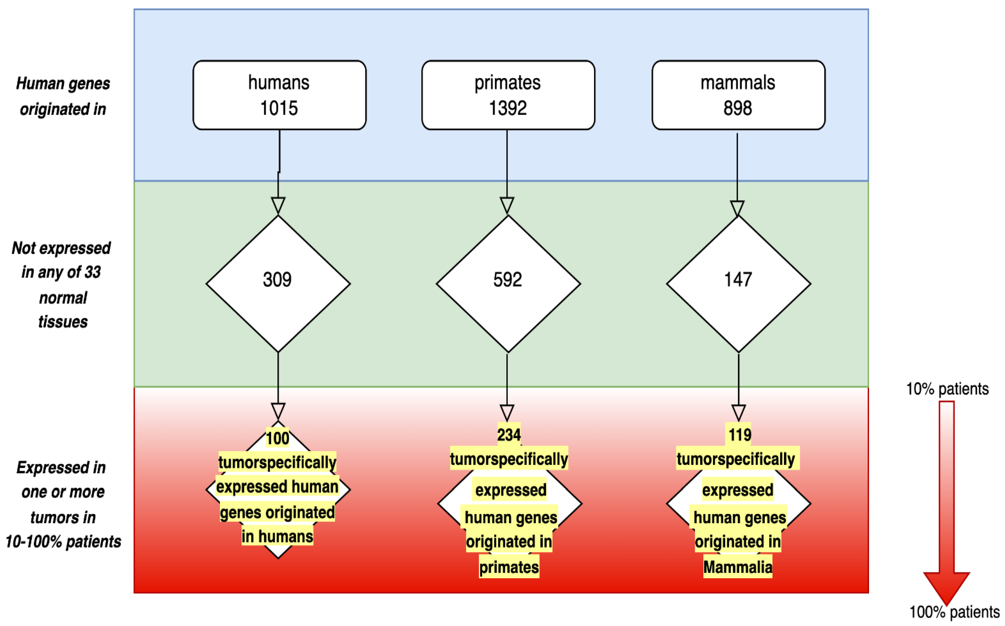
3.2.3. Human TSEEN Protein-Coding Genes Database

In 2021, we registered a TSEEN genes database [69]. This database was obtained in silico using the following databases and tools: GTEx database (RNASeq data of 53 normal tissues from 100 patients who died as a result of an accident and had no serious pathologies during their life); TCGA database (Data Release 28.0, 2 February 2021; RNASeq data of total RNA samples which were isolated from 20,000 biosamples covering 33 different tumors); GENEVESTIGATOR (curated database and expression data analysis tool, which includes RNASeq data of total RNA samples from eighttypes of stem cells).
The database contains 100 genes originated in humans, 234 genes originated in primates and 119 genes originated in mammals (Figure 2).
As shown in the lower part of Figure 2, human TSEEN genes are expressed in a broad range of tumors and in a considerable proportion of patients.

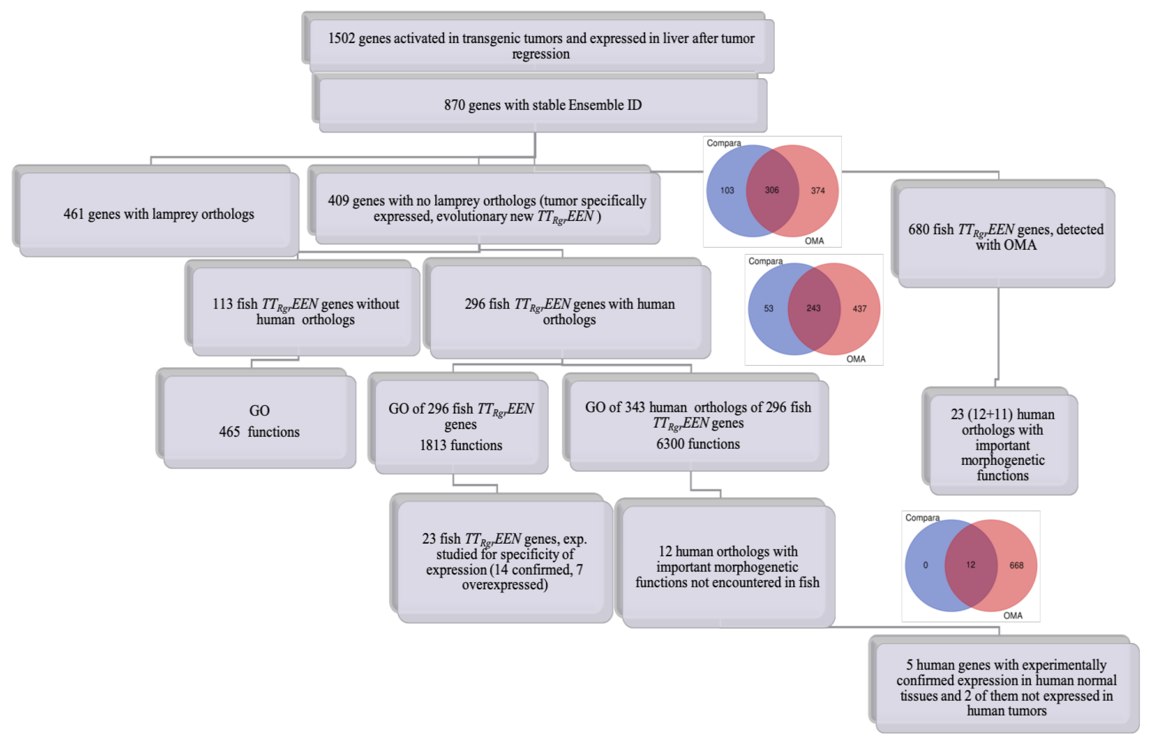
3.3. Human Orthologs of Fish TSEEN Genes Should Acquire Progressive Functions Connected with New Cell Types, Tissues and Organs

We studied this prediction using a transgenic fish inducible-tumor model. The transgenic inducible fish tumor after regression can be considered as an approximation to an organ evolving from a tumor. In so far as many evolutionarily novel genes have no functions, we studied new gene functions by comparing fish novel genes with their human orthologs using the Gene Ontology (GO) approach. We found that orthologs of many human genes, which are involved in the development of lung, mammary gland, placenta, ventricular septum, etc., originated in fish and were expressed in fish tumors and tumors after regression [35]. These data may be regarded as direct confirmation of the main hypothesis.
Figure 3 demonstrates the sequential steps of our analysis:
From 1502 genes expressed in fish tumors and tumors after regression, using Ensemble Compara we detected 409 genes with no lamprey orthologs, i.e., tumor and tumor after regression expressed evolutionary novel (TTRgrEEN) fish genes. From 343 human orthologs of fish TTRgrEEN genes, using GO, we selected 12 genes with important developmental and morphogenetic functions not encountered in fish. An additional 11 human orthologs of fish TTRgrEEN genes with functions that do not exist in fish were obtained by OMA ortholog search algorithm and GO (Figure 3 and Table 1).
The progressive functions of human orthologs presented in Table 1 do not exist in fish and will never be discovered there.
Highly represented among fish, TTRgrEEN genes are protein kinase, DNA binding and transcriptional functions, the most common domains encoded by cancer genes [35]. Other authors have also demonstrated oncogene properties in some evolutionarily novel genes [57]. The progressive functions of human orthologs have been added to fish proto-genes in the course of evolution and involved more organismic than molecular functions [35]. The novel progressive functions in human orthologs balanced the original oncogenic potential of fish proto-genes.
Some human orthologs of fish genes from Table 1 are involved in the development of several progressive features in humans. On the other hand, the development of some human progressive traits involves several human genes from this table [35]. The gene network that participates in mammalian adipose organ development will be described below.
The author suggests calling genes that originated from ancestral TSEEN genes and acquired progressive functions the “carcino-evo-devo” genes. This term was originally used as a synonym of TSEEN genes in [9]. In the present meaning, it was first used in [35] to stress their role in evo-devo and connection to carcinoembryonic antigen genes [70].

Conclusion on Predictions 2 and 3: TSEEN Genes—A New Biological Phenomenon and the Superclass of Novel and Evolving Genes Expressed in Tumors

The author has suggested considering the expression of evolutionarily novel and evolving genes in tumors as a new biological phenomenon, which is a part of a greater biological phenomenon defined by the main hypothesis, i.e., evolution by tumor neofunctionalization [9,10].
TSEEN genes as a new class of genes were reviewed for the first time in [9,10]. This new class of TSEEN genes has peculiarities when compared to other gene classes. First, it is not a functional gene class like the classes of oncogenes or differentiation genes. Evolutionarily novel genes often do not have functions yet [9]. The generic features of TSEEN genes are their tumor specificity of expression, and evolutionary novelty and continuing evolution towards new function acquisition.
The dual specificity of TSEEN genes determines two complementary ways for their study, as discussed above. Tumor specificity of expression can first be studied, followed by estimation of the evolutionary age; and vice-versa, evolutionarily novel genes can first be found, followed by determination of the tumor specificity of their expression [10,69].
Evolutionary novelty of a gene class is the relative characteristic. Even the evolutionarily oldest gene classes such as housekeeping genes contain genes originated in humans, but their proportion is extremely low [15]. Various classes of human TSEEN genes, for example, CT antigen genes, contain older genes, and not only human and primate specific genes. However, TSEEN gene classes contain the highest proportions of evolutionarily new and young genes. The median of gene age distribution for TSEEN gene classes demonstrates that in humans they are the youngest gene classes [15].
The main hypothesis does not specify the strict borders of the TSEEN gene class. As shown earlier by us and other authors, genes originate somewhat earlier than corresponding morphological structures appear [15,35,71]. Our data on fish TSEEN genes suggest that their human orthologs were important for the origin of progressive mammalian traits [35]. Higher prevalence of TSEEN genes in primates (Figure 2) suggests their role in human evolution. Human tumor-specifically expressed genes that originated in mammals have not acquired functions in normal tissues yet, which is why they were considered as evolving genes and were placed in Figure 2. Genes represented in Figure 2 demonstrate the gradient of evolutionary novelty that reflects an evolutionary continuum of evolving genes. Expression of proto-genes, young and novel genes in hereditary tumors may represent the early stages in the origin of novel gene functions. This view corresponds well to the concept of proto-gene [66] and to several models of gene origin described in [72]. TSEEN genes may represent a reservoir of proto-genes pervasively expressed in tumors. It would be most interesting to study the process of new progressive function acquisition by TSEEN genes, which have not acquired functions yet. The algorithm for such studies is suggested in [14].
Various TSEEN genes can acquire different functions in evolution, as shown in [35]. TSEEN gene classes of various phyla of organisms are different and may evolve in different directions. As shown in Figure 3, a considerable proportion of fish TTRgrEEN genes do not have orthologs in humans.
The author suggests considering TSEEN genes as a new superclass of novel and evolving genes with tumor-specific expression, with several classes and families of TSEEN genes, which includes TSEEN genes of various phyla of organisms. The author has called TSEEN genes’ descendants, which acquired progressive developmental and morphogenetic functions during evolution, the carcino-evo-devo genes. The discovery of carcino-evo-devo genes in [35] may be considered as the direct confirmation of the main hypothesis.
The expression of TSEEN genes in a wide range of tumors and in a considerable proportion of patients is their important characteristic that makes them perspective targets for the development of universal tumor test systems, prophylactic and therapeutic tumor vaccines, and conventional therapeutic agents.
Other authors have recognized the connection of evolutionarily novel genes with tumors. In their reviews, they discuss the TSEEN genes described by us and others [39,55,72].

3.4. Selection of Tumors for New Functions in the Organism Is Possible. Evolutionarily Novel Organs Should Recapitulate Tumor Features in Their Development

3.4.1. Selection of Tumors for New Functions

The main hypothesis predicts that hereditary tumors may be selected for new organismal functions. A special paper was devoted to confirmation of this prediction [73]. We have shown that so called “hoods” on the heads of certain varieties of goldfish are benign tumors. These tumors were selected for hundreds of years by Chinese breeders. As a result of this selection, a new organ—the hood—originated. The symmetrical shape of the hood, its benign properties and its appearance at certain stages of development are features of the normal organ. The capability for unlimited growth and histological peculiarities are tumor features. The author suggested calling such organs of dual nature the “tumor-like” organs [9].
To our knowledge, the “hood” of goldfish was the first example of artificial selection of tumors described in the literature. This was discussed in the book [9] and in a recent paper devoted to tumor-like organs [12].
Symbiovilli in voles originated by natural selection of the early stages of papillomatosis [74], and macromelanophores in swordtails by sexual selection [75]. We may conclude that the nontrivial prediction about the possibility of selection of hereditary tumors for new organismal functions works. Positive selection of many tumor-related genes in the primate lineage discussed in the book [9] supports this view.

3.4.2. Evolutionarily Novel Organs That Recapitulate Tumor Features in Their Development: Mammalian Tumor-like Organs

The author addressed this topic in the book [9] and in two recently published papers devoted solely to this question [12,13]. In [12], the author was looking for tumor features of four mammalian evolutionarily novel organs: placenta, mammary gland, prostate and infantile human brain. In [13], the tumor-like features of mammalian adipose were described for the first time in the literature, a discovery that can change the medicine of obesity and cancer.
Placenta was the first organ the tumor-like features of which were recognized because it has dozens of such features (reviewed in [9,12]). The role of ancient retrovirus infection and syncytin gene domestication was also early recognized [76,77,78]. A special symposium was held on this particular organ and its resemblance to tumors [79]. A term “tumor-like organ” is used for placenta [9,80,81].
The mammary gland and prostate are characterized by the highest incidences of cancer. It was shown that the high incidence of cancer in these organs is connected with their evolutionary novelty [82]. Like placenta, they have a regulated invasion stage in their organogenesis. Both glands have many other similarities with tumors (reviewed in [12]) that indicate the tumor-like nature of mammary gland and prostate.
Human brain recapitulates many features of tumors and demonstrates more of its tumor-like nature during childhood and infancy. Brain tumors are the most common solid tumors and the leading cause of cancer death in children (reviewed in [9,12]).
The tumor-like features of mammalian adipose [13] will be discussed below.
One more evolutionary novel organ (or tissue) with tumor features can be added to the list of organs discussed in [12,13]: the ependymal region of the adult human spinal cord, which differs from other species and shows ependymoma-like features [83].
The features of tumor-like organs position them close to the so-called “atypical tumor organs”. The concept of atypical tumor organs is already accepted among oncologists. It reflects the complex structure of solid tumors, which consist of parenchyma with a hierarchy of cell types and stroma with connective tissue, blood vessels and accessory cells (see [84] for review). According to the main hypothesis, normal tumor-like organs could evolve from hereditary atypical tumor organs. The relationship between tumor-like organs and atypical tumor organs represents an essential part of the carcino-evo-devo relationship, i.e., coevolution of normal and neoplastic development [12]. The term “carcino-evo-devo” gave a name to the theory of the evolutionary role of hereditary tumors.
The series of our two papers devoted to mammalian tumor-like organs answered the important and frequent question: how often in evolution could tumor neofunctionalization happen? The answer is that in mammalian evolution it happened often.

3.4.3. Tumor Features of Mammalian Adipose. Obesity as a Tumor-like Process

An unexpected discovery was the finding of many tumor features in mammalian adipose [13]. It was stimulated by two circumstances: the concept that several mammalian adipose tissues constitute the mammalian adipose organ, and the evolutionary novelty of this organ. Adipose is currently recognized as a metabolic and endocrine organ operating “as a structured whole” [85,86]. Although the storage of energy in lipids is evolutionarily conserved, and lipid-storing cells and proteins are ancient [87,88], the adipose organ is evolutionarily novel to mammals [89,90]. Brown adipose tissue (BAT) has not been described in fishes, amphibians, reptiles or birds, and is present only in higher mammals [91,92,93].
After understanding these things, the author started looking for tumor-like features of mammalian adipose and found many such features. Similarities of mammalian adipose with tumors include the following: the capability to unlimited expansion; lipomas as the most frequent soft tissue tumors; reversible plasticity; induction of angiogenesis; chronic inflammation; remodeling and disfunction; systemic influence on the organism; hormone production; production of miRNAs that influence other tissues; immunosuppression; DNA damage and resistance to apoptosis; infiltration of other organs and tissues; and similar drugs that may be used for treatment of obesity and cancer [13].The most luminous similarities are the capability to unlimited expansion, due to hyperplasia and hypertrophy, and infiltration of other organs and tissues. Many of the common features of tumors and the adipose organ are in the list of so-called “hallmarks of cancer” [94,95], and many of them are connected with obesity. Thus, mammalian adipose is a tumor-like organ, and obesity is a tumor-like process, a discovery that may change the landscape of public health and medicine. It means that we can probably use the approaches developed in oncology to fight obesity, and the approaches used against obesity to fight cancer.
The tumor-like nature of mammalian adipose suggests its origin from some ancestral mesenchymal tumors. The author has used our data obtained with the transgenic fish hepatoma model in [35] to explore this possibility [13]. It was found that five genes (LEP, SPRY1, PPARG, ID2 and NOTCH1) among the 23 human orthologs of fish TTRgrEEN genes with progressive functions not encountered in fish described in [35] also participate in mammalian adipose organ development. Progressive functions connected with the adipose organ were not mentioned in [35] because we were not interested in mammalian adipose at that time and did not look for adipose-related functions. One more gene involved in mammalian adipose development—the CIDEA gene—was found in [13] among 343 human orthologs of the fish TTRgrEEN genes described in [35]. In Table 2, the functions of genes related to the mammalian adipose organ are presented:
These are progressive functions connected mainly with white adipose beiging, BAT and thermoregulation, and not encountered in fish.
Besides their function in adipose development, these genes also participate in tumor development in humans. Depending on the context, they can play tumor-promoting or tumor-suppressing roles (reviewed in [13]).
These genes also interact with each other and form a gene network. In the author’s presentation at the American Association for Cancer Research Annual Meeting 2022 (AACR 2022) [37], the following diagram describing interactions of LEP, SPRY1, PPARG, ID2, CIDEA and NOTCH1 and their possible roles in tumorigenesis was presented (Figure 4):
In this gene network, LEP and PPARG play particular roles. LEP, which encodes leptin, became the central regulator of energy metabolism in mammals. PPARG is a major regulator of adipocyte differentiation and function (reviewed in [13]).
After the conference, one more member of this gene network was found among the 343 human orthologs of fish TTRgrEEN genes—the ZAG or AZGP1 gene. ZAG plays a key role in lipid metabolism in adipocytes and participates in white adipose tissue browning. It affects glucose metabolism and is linked to insulin resistance, and it has a role in reducing obesity and improving insulin sensitivity. ZAG inhibits LEP. ZAG can both stimulate and inhibit expression of PPARG. On the other hand, the increased expression of ZAG is influenced by PPARG [96,97,98,99].
Like other members of the adipose gene network, the ZAG gene is also involved in the development of many different types of tumors [100]. It functions mostly as a tumor suppressor gene [100,101,102,103,104], but sometimes also as an oncogene [105]. ZAG has a pro-carcinogenic effect on breast cancer cells and an anti-carcinogenic effect on nonmalignant cells [106].
The upgraded adipose gene network, which includes the ZAG gene and its interactions, is presented at Figure 5:
In the upgraded adipose gene network, the ZAG gene as the major lipid mobilizing factor may play the important role comparable to that of LEP and PPRG.
The work on the adipose gene network will be continued and will certainly bring to light new gene members and new interactions. However, important conclusions can be made right now.
We can summarize that orthologs of LEP, NOTCH1, SPRY1, PPARG, ID2, CIDEA and ZAG genes originated in fishes and were expressed in fish tumors and tumors after regression. In mammals, these genes acquired progressive functions connected with the adipose organ, form a gene network with mutual influences, and participate in tumor processes. These data suggest the tumor-like nature of mammalian adipose and support the possibility of its origin from ancestral hereditary tumors.
The most important circumstance is that all genes in the above diagram play the dual role in tumor development. Depending on the context, they play roles of either oncogenes or tumor suppressor genes. Similar duality was described for other tumor-related genes. TGFβ acts as tumor suppressor in normal cells and as oncogene in malignant cells [107,108]. Mouse models of BRCA1 and BRCA2 deficiency demonstrated a similar paradox: BRCA-deficient tumor cells proliferate rapidly, but developing BRCA-deficient embryos suffer from proliferation defect [109]. High and low levels of TNF have opposing effects on tumor growth [110]. Tumor suppressor p53 is characterized by antagonistic bifunctionality, producing both positive and negative signals on cell migration, metabolism, differentiation and survival [111]. In a recent publication, a paradoxical promotion of liver carcinogenesis by constitutive activation of p53 was demonstrated [112].
All seven members of the adipose gene network have similar antagonistic tumor duality. This may be connected with several circumstances: (1) the ancestral fish TSEEN genes duality; (2) the coevolution of oncogenes, tumor suppressor genes and differentiation genes; and (3) the addition in the course of evolution of domains with progressive functions to fish cancer genes. Indeed, in our previous article, we demonstrated that many genes belong to two functional classes, and TGFβ even has triple specificity [15]. The new progressive functions could neutralize the original tumorigenic potential of fish proto-genes and create a basis for governing homeostasis.
The phenomenon of dual functionality of tumor-related genes may have fundamental importance. The key to control over cancer and obesity may consist in understanding the antagonistic bifunctionality of tumor- and obesity-related genes and developing technologies to regulate the networks of genes with multiple antagonistic functionalities.

4. Conclusions: Towards a Comprehensive Theory of Evolutionary Oncology

Throughout the work on development of carcino-evo-devo theory, a considerable amount of effort by the author’s lab has been devoted to confirmation of non-trivial predictions, which were formulated with the help of the main hypothesis. Several predictions have been confirmed, as shown in this paper. Thus, we have shown that carcino-evo-devo theory has predictive power in several related areas of biology, fulfilling a fundamental requirement for a new theory.
Other monographs devoted to problems of evolutionary oncology [113,114,115] have explored other directions and do not contradict our theory.
The field of evolutionary oncology is actively developing. The author hopes that at some point we will have a comprehensive theory of evolutionary oncology.

Funding

This research was funded by the Ministry of Science and Higher Education of the Russian Federation under the strategic academic leadership program ‘Priority 2030′ (Agreement 075-15-2021-1333 dated 30 September 2021).

Institutional Review Board Statement

Not applicable.

Informed Consent Statement

Not applicable.

Data Availability Statement

Not applicable.

Acknowledgments

The author wishes to thank anonymous reviewers for their helpful comments.

Conflicts of Interest

The author declares no conflict of interest.

References

  1. Isaeva, V.V. Heterochronies, heterotopies, and cell resources of development in ontogenetic and evolutionary transformations. Paleontol. J. 2015, 49, 1530–1537. [Google Scholar] [CrossRef]
  2. Kozlov, A.P. Evolution of Living Organisms as a Multilevel Process. J. Theor. Biol. 1979, 81, 1–17. [Google Scholar] [CrossRef] [PubMed]
  3. Kozlov, A.P. The principles of multilevel development of organisms. In Problems of Analysis of Biological Systems; Maximov, V.N., Ed.; Moscow University Press: Moscow, Russia, 1983; pp. 48–62. [Google Scholar]
  4. Kozlov, A.P. Gene competition and the possible evolutionary role of tumors and cellular oncogenes. In Theoretical and Mathematical Aspects of Morphogenesis; Presnov, E.V., Maresin, V.M., Zotin, A.I., Eds.; Nauka: Moscow, Russia, 1987; pp. 136–140. [Google Scholar]
  5. Kozlov, A.P. Conservation principles in the system of molecular-biological laws. Trans. Leningr. Soc. Nat. Sci. 1988, 87, 4–21. [Google Scholar]
  6. Kozlov, A.P. Gene competition and the possible evolutionary role of tumors. Med. Hypotheses 1996, 46, 81–84. [Google Scholar] [CrossRef] [PubMed]
  7. Kozlov, A.P. Tumors and evolution. Vopr. Oncol. 2008, 54, 695–705. [Google Scholar]
  8. Kozlov, A.P. The Possible Evolutionary Role of Tumors in the Origin of New Cell Types. Med. Hypotheses 2010, 74, 177–185. [Google Scholar] [CrossRef]
  9. Kozlov, A.P. Evolution by Tumor Neofunctionalization. The Role of Tumors in the Origin of New Cell Types, Tissues and Organs; Elsevier; Academic Press: Amsterdam, The Netherlands; Boston, MA, USA; Heidelberg, Germany; London, UK; New York, NY, USA; Oxford, UK; Paris, France; San Diego, CA, USA; San Francisco, CA, USA; Singapore; Sydney, Australia; Tokyo, Japan, 2014; p. 248. [Google Scholar]
  10. Kozlov, A.P. Expression of evolutionarily novel genes in tumors. Infect. Agents Cancer 2016, 11, 34. [Google Scholar] [CrossRef] [Green Version]
  11. Kozlov, A.P. The role of heritable tumors in evolution of development: A new theory of carcino-evo-devo. Acta Nat. 2019, 11, 65–72. [Google Scholar] [CrossRef]
  12. Kozlov, A.P. Mammalian tumor-like organs.1. The role of tumor-like normal organs and atypical tumor organs in the evolution of development (carcino-evo-devo). Infect. Agents Cancer 2022, 17, 2. [Google Scholar] [CrossRef]
  13. Kozlov, A.P. Mammalian tumor-like organs. 2. Mammalian adipose has many tumor features and obesity is a tumor-like process. Infect. Agents Cancer 2022, 17, 15. [Google Scholar] [CrossRef]
  14. Kozlov, A.P. Biological computation and compatibility search in the possibility space as the mechanism of complexity increase during progressive evolution. Evol. Bioinform. 2022, 18, 1–5. [Google Scholar] [CrossRef]
  15. Makashov, A.; Malov, S.V.; Kozlov, A.P. Oncogenes, tumor suppressor and differentiation genes represent the oldest human gene classes and evolve concurrently. Sci. Rep. 2019, 9, 16410. [Google Scholar] [CrossRef]
  16. Valentine, J.W. Late Precambrian bilaterians: Grades and clades. Proc. Natl. Acad. Sci. USA 1994, 91, 6751–6757. [Google Scholar] [CrossRef] [Green Version]
  17. Bell, G.; Mooers, A.O. Size and complexity among multicellular organisms. Biol. J. Linn. Soc. 1997, 60, 345–363. [Google Scholar] [CrossRef]
  18. Chen, L.; Stephen, J.B.; Jaime, M.T.C.; Atahualpa, C.M.; Araxi, O.U. Correcting for Differential Transcript Coverage Reveals a Strong Relationship between Alternative Splicing and Organism Complexity. Mol. Biol. Evol. 2014, 31, 1402–1413. [Google Scholar] [CrossRef]
  19. Vickaryous, M.K.; Hall, B.K. Human cell type diversity, evolution, development, and classification with special reference to cells derived from neural crest. Biol. Rev. Camb. Philos. Soc. 2006, 81, 425–455. [Google Scholar] [CrossRef]
  20. Evtushenko, V.I.; Hanson, K.P.; Barabitskaya, O.V.; Emelyanov, A.V.; Reshetnikov, V.L.; Kozlov, A.P. Determination of the upper limit of rat genome expression. Mol. Biol. 1989, 23, 663–675. [Google Scholar]
  21. Baranova, A.V.; Lobashev, A.V.; Ivanov, D.V.; Krukovskaya, L.L.; Yankovsky, N.K.; Kozlov, A.P. In silico screening for tumor-specific expressed sequences in human genome. FEBS Lett. 2001, 508, 143–148. [Google Scholar] [CrossRef] [Green Version]
  22. Krukovskaya, L.L.; Baranova, A.; Tyezelova, T.; Polev, D.; Kozlov, A.P. Experimental study of human expressed sequences newly identified in silico as tumor specific. Tumor Biol. 2005, 26, 17–24. [Google Scholar] [CrossRef]
  23. Kozlov, A.P.; Galachyants, Y.P.; Dukhovlinov, I.V.; Samusik, N.A.; Baranova, A.V.; Polev, D.E.; Krukovskaya, L.L. Evolutionarily new sequences expressed in tumors. Infect. Agents Cancer 2006, 1, 8. [Google Scholar] [CrossRef] [Green Version]
  24. Galachyants, Y.; Kozlov, A.P. CDD as a tool for discovery of specifically-expressed transcripts. Russ. J. AIDS Cancer Public Health 2009, 13, 60–61. [Google Scholar]
  25. Polev, D.E.; Nosova, J.K.; Krukovskaya, L.L.; Kozlov, A.P. Expression of transcripts corresponding to cluster Hs.633957 in human healthy and tumor tissues. Mol. Biol. 2009, 43, 88–92. [Google Scholar] [CrossRef]
  26. Krukovskaia, L.L.; Polev, D.E.; Nosova, I.K.; Baranova, A.V.; Koliubaeva, S.N.; Kozlov, A.P. Investigation of transcription factor Brachyury (T) expression in human normal and tumor tissues. Vopr. Oncol. 2008, 54, 739–743. [Google Scholar]
  27. Krukovskaya, L.L.; Samusik, N.; Shilov, E.S.; Polev, D.; Kozlov, A.P. Tumor-specific expression of PBOV1, a new gene in evolution. Vopr. Onkol. 2010, 56, 327–332. [Google Scholar]
  28. Polev, D.E.; Krukovskaya, L.L.; Kozlov, A.P. Expression of the locus Hs.633957 in human digestive system and tumors. Vopr. Oncol. 2011, 57, 48–49. [Google Scholar]
  29. Samusik, N.A.; Galachyants, Y.P.; Kozlov, A.P. Analysis of evolutionary novelty of tumor-specifically expressed sequences. Russ. J. Genet. Appl. Res. 2011, 1, 138–148. [Google Scholar] [CrossRef]
  30. Dobrynin, P.; Matyunina, E.; Malov, S.V.; Kozlov, A.P. The novelty of human cancer/testis antigen encoding genes in evolution. Int. J. Genom. 2013, 2013, e105108. [Google Scholar] [CrossRef]
  31. Samusik, N.; Krukovskaya, L.; Meln, I.; Shilov, E.; Kozlov, A.P. PBOV1 is a human de novo gene with tumor-specific expression that is associated with a positive clinical outcome of cancer. PLoS ONE 2013, 8, e56162. [Google Scholar] [CrossRef]
  32. Polev, D.E.; Karnaukhova, J.K.; Krukovskaya, L.L.; Kozlov, A.P. ELFN1-AS1, a novel primate gene with possible microRNA function expressed predominantly in tumors. BioMed Res. Int. 2014, 2014, e398097. [Google Scholar] [CrossRef] [Green Version]
  33. Matyunina, E.; Emelyanov, A.; Kozlov, A. Evolutionarily novel genes expressed in fish tumors determine progressive evolutionary characters. In Proceedings of the 106th Annual Meeting of the American Association for Cancer Research, Philadelphia, PA, USA, 18–22 April 2015; AACR: Philadelphia, PA, USA, 2015. Abstract Number 1927. [Google Scholar]
  34. Krukovskaya, L.L.; Polev, D.E.; Kurbatova, T.V.; Karnaukhova, Y.K.; Kozlov, A.P. The study of the tumor specificity of expression of some evolutionarily novel genes. Vopr. Oncol. 2016, 62, 495–500. [Google Scholar]
  35. Matyunina, E.A.; Emelyanov, A.V.; Kurbatova, T.V.; Makashov, A.A.; Mizgirev, I.V.; Kozlov, A.P. Evolutionary novel genes are expressed in transgenic fish tumors and their orthologs are involved in development of progressive traits in humans. Infect. Agents Cancer 2019, 14, 46. [Google Scholar] [CrossRef] [Green Version]
  36. Karnaukhova, I.K.; Polev, D.E.; Krukovskaya, L.L.; Makashov, A.A.; Masharsky, A.E.; Nazarenko, O.V.; Poverennaya, I.V.; Makeev, V.J.; Kozlov, A.P. A new cancer/testis long noncoding RNA, the OTP-AS1 RNA. Sci. Rep. 2022. [Google Scholar] [CrossRef]
  37. Kozlov, A.P. Mammalian adipose has many tumor features and obesity is the tumor-like process. In Proceedings of the Annual Meeting of the American Association for Cancer Research 2022, New Orleans, LA, USA, 8–13 April 2022; AACR: New Orleans, LA, USA, 2022. Abstract Number 3371/6077. [Google Scholar]
  38. Koonin, E.V. Orthologs, paralogs, and evolutionary genomics. Annu. Rev. Genet. 2005, 39, 309–338. [Google Scholar] [CrossRef] [Green Version]
  39. McLysaght, A.; Hurst, L.D. Open questions in the study of de novo genes: What, how and why? Nat. Rev. Genet. 2016, 17, 567. [Google Scholar] [CrossRef]
  40. Clamp, M.; Fry, B.; Kamal, M.; Xie, X.; Cuff, J.; Linn, M.F.; Kellis, M.; Lindblad-Toh, K.; Lander, E.S. Distinguishing protein-coding and noncoding genes in the human genome. Proc. Natl. Acad. Sci. USA 2007, 104, 19428–19433. [Google Scholar] [CrossRef] [Green Version]
  41. An, G.; Ng, A.Y.; Meka, C.S.R.; Luo, G.; Bright, S.P.; Cazares, L.; Wright, G.L., Jr.; Veltri, R.W. Cloning and characterization UROC28, a novel gene overexpressed in prostate, breast and bladder cancer. Cancer Res. 2000, 60, 7014–7020. [Google Scholar]
  42. Wang, L.; Niu, C.H.; Wu, S.; Wu, H.M.; Ouyang, F.; He, M.; He, S.Y. PBOV1 correlates with progression of ovarian cancer and inhibits proliferation of ovarian cancer calls. Oncol. Rep. 2016, 35, 488–496. [Google Scholar] [CrossRef] [Green Version]
  43. Pan, T.; Wu, R.; Liu, B.; Wen, H.; Tu, Z.; Guo, J.; Yang, J.; Shen, G. PBOV1 promotes prostate cancer proliferation by promoting G1/S transition. Onco. Targets Ther. 2016, 9, 787–795. [Google Scholar] [CrossRef] [Green Version]
  44. Carleton, N.M.; Zhu, G.; Gorbunov, M.; Miller, M.C.; Pienta, K.J.; Resar, L.M.S.; Veltri, R.W. PBOV1 as a potential biomarker for more advanced prostate cancer based on protein and digital histomorphometric analysis. Prostate 2018, 78, 547–559. [Google Scholar] [CrossRef]
  45. Guo, Y.; Wu, Z.; Shen, S.; Guo, R.; Wang, J.; Wang, W.; Zhao, K.; Kuang, M.; Shuai, X. Nanomedicines reveal how PBOV1 promotes hepatocellular carcinoma for effective gene therapy. Nat. Commun. 2018, 9, 3430. [Google Scholar] [CrossRef]
  46. Xue, C.; Zhong, Z.; Ye, S.; Wang, Y.; Ye, O. Association between the overexpression of PBOV1 and the prognosis of patients with hepatocellular carcinoma. Oncol. Lett. 2018, 16, 3401–3407. [Google Scholar] [CrossRef] [PubMed] [Green Version]
  47. Kong, Q.; Han, J.; Deng, H.; Wu, F.; Guo, S.; Ye, Z. miR-431-5p alters the epithelial-to-mesenchimal transition markers targeting UROC28 in hepatoma cells. Onco. Targets Ther. 2018, 11, 6489–6503. [Google Scholar] [CrossRef] [PubMed] [Green Version]
  48. Loizidou, M.A.; Cariolou, M.A.; Neuhausen, S.L.; Newbold, R.F.; Bashiardes, E.; Marcou, Y.; Michael, T.; Daniel, M.; Kakouri, E.; Papadopoulos, P.; et al. Genetic variation in genes interacting with BRCA1/2 and risk of breast cancer in the Cypriot population. Breast Cancer Res. Treat. 2010, 121, 147–156. [Google Scholar] [CrossRef] [PubMed]
  49. Zhang, S.Y.; Gao, F.; Peng, C.G.; Zheng, C.J.; Wu, M.F. Has-miR-203 inhibits fracture healing via targeting PBOV1. Eur. Rev. Med. Pharmacol. Sci. 2018, 22, 5797–5803. [Google Scholar] [PubMed]
  50. Yang, C.A.; Li, J.P.; Yen, J.C.; Lai, I.L.; Ho, Y.C.; Chen, Y.V.; Lan, J.L.; Chang, J.G. lncRNA NTT/PBOV1 axis promotes monocyte differentiation and is elevated in rheumatoid arthritis. Int. J. Mol. Sci. 2018, 19, 2806. [Google Scholar] [CrossRef] [Green Version]
  51. Jie, Y.; Ye, L.; Chen, H.; Yu, X.; Cai, L.; He, W.; Fu, Y. ELFN1-AS1 accelerates cell proliferation, invasion and migration via regulating miR-497-3p/CLDN4 axis in ovarian cancer. Bioengineered 2020, 11, 872–882. [Google Scholar] [CrossRef]
  52. Du, Y.; Hou, Y.; Shi, Y.; Liu, J.; Li, T. Long non-coding RNA ELFN1-AS1 promoted colon cancer cell growth and migration via the miR-191-5p/special AT-rich sequence-binding protein axis. Front. Oncol. Sec. Cancer Genet. 2021, 10, 588360. [Google Scholar] [CrossRef]
  53. Ma, G.; Li, G.; Gou, A.; Xiao, Z.; Xu, Y.; Song, S.; Guo, K.; Liu, Z. Long non-coding RNA ELFN1-AS1 in the pathogenesis of pancreatic cancer. Ann. Transl. Med. 2021, 9, 10. [Google Scholar] [CrossRef]
  54. Li, Y.; Gan, Y.; Liu, J.; Li, J.; Zhou, Z.; Tian, R.; Sun, R.; Liu, J.; Xiao, Q.; Li, Y.; et al. Downregulation of MEIS1 mediated by ELFN1-AS1/EZH2/DNMT3a axis promotes tumorigenesis and oxaliplatin resistance in colorectal cancer. Signal Transduct. Target. Ther. 2022, 7, 87. [Google Scholar] [CrossRef]
  55. Weisman, C.M. The origins and functions of de novo genes: Against all odds? J. Mol. Evol. 2022, 90, 244–257. [Google Scholar] [CrossRef]
  56. Casola, C. From de novo to “de nono”: The majority of novel protein-coding genes identified with phylostratigraphy are old genes or recent duplicates. Genome Biol. Res. 2018, 10, 2906–2918. [Google Scholar] [CrossRef]
  57. Zhang, Y.E.; Long, M. New genes contribute to genetic and phenotypic novelties in human evolution. Curr. Opin. Genet. Dev. 2014, 29, 90–96. [Google Scholar] [CrossRef] [Green Version]
  58. Sverdlov, E.D. Retroviruses and primate evolution. Bioessays 2000, 22, 161–171. [Google Scholar] [CrossRef]
  59. Khodosevich, K.; Lebedev, Y.; Sverdlov, E. Endogenous retroviruses and human evolution. Comp. Funct. Genom. 2002, 3, 494–498. [Google Scholar] [CrossRef] [Green Version]
  60. Brinzevich, D.; Young, G.R.; Sebra, R.; Ayllon, J.; Maio, S.M.; Deikus, G.; Chen, B.K.; Fernandez-Sesma, A.; Simon, V.; Mulder, L.C. HIV-1 interacts with human endogenous retrovirus K (HML-2) envelopes derived from human primary lymphocytes. J. Virol. 2014, 88, 6213–6223. [Google Scholar] [CrossRef] [Green Version]
  61. Zhang, Y.E.; Vibranovsky, M.D.; Landback, P.; Marais, G.A.B.; Long, M. Chromosomal redistribution of male-biased genes in mammalian evolution with two bursts of gene gain on X chromosome. PLoS Biol. 2010, 8, e1000494. [Google Scholar] [CrossRef]
  62. Makashov, A.A.; Malov, S.V.; Kozlov, A.P. Expression of HERV-K HML-2 in Tumor Tissues. Symposium “Evolutionary Oncology and Evolutionary Virology” within the Framework of the VII St. Petersburg International Oncology Forum “White Nights 2021”; Russian Federation: St. Petersburg, Russia, 2021. [Google Scholar]
  63. Kim, T.; Jeon, Y.J.; Cui, R.; Lee, J.H.; Peng, Y.; Kim, S.H.; Tili, E.; Alder, H.; Croce, C.M. Role of MYC-regulated long noncoding RNAs in cell cycle regulation and tumorigenesis. J. Natl. Cancer Inst. 2015, 107, dju505. [Google Scholar] [CrossRef]
  64. Wu, S.M.; Liu, H.; Huang, P.J.; Chang, I.Y.; Lee, C.C.; Yang, C.Y.; Tsai, W.S.; Tan, B.C. circlncRNAnet: An integrated web-based resource for mapping functional networks of long or circular forms of noncoding RNAs. GigaScience 2018, 7, gix 118. [Google Scholar] [CrossRef] [Green Version]
  65. Karnaukhova, Y.K.; Polev, D.E.; Krukovskaya, L.L.; Kozlov, A.P. Study of the expression of gene Orthopedia homeobox in various tumor and normal human tissues. Vopr. Oncol. 2017, 63, 128–134. [Google Scholar]
  66. Carvunis, A.R.; Rolland, T.; Wapinski, I.; Calderwood, M.A.; Yildirim, M.A.; Simonis, M.; Charloteaux, B.; Hidalgo, C.A.; Barbette, J.; Santhanam, B.; et al. Proto-genes and de novo gene birth. Nature 2012, 487, 370–374. [Google Scholar] [CrossRef]
  67. Palena, C.; Polev, D.E.; Tsang, K.Y.; Fernando, R.I.; Litzinger, M.; Krukovskaya, L.L.; Baranova, A.V.; Kozlov, A.P.; Schlom, J. The human T-box mesodermal transcription factor Brachyury is a candidate target for T-cell mediated cancer immunotherapy. Clin. Cancer Res. 2007, 13, 2471–2478. [Google Scholar] [CrossRef] [PubMed] [Green Version]
  68. Schlom, J.; Palena, C.M.; Kozlov, A.P.; Tsang, K.Y. Brachyury Polypeptides and Methods for Use. U.S. Patent No. 8,188,214 B2; Application Granted, 29 May 2012. Available online: https://patents.google.com/patent/US8188214B2/en (accessed on 7 October 2022).
  69. Kozlov, A.P.; Matyunina, E.A.; Makashov, A.A. The Biomedical Center TSEEN Genes Database. Certificate about Federal Registration of the Database №2021621840. 2021. Available online: https://tseendb.org/#/ (accessed on 7 October 2022).
  70. Gold, P.; Freedman, S.O. Demonstration of tumor-specific antigens in human colonic carcinomata by immunological tolerance and absorption techniques. J. Exp. Med. 1965, 121, 439–462. [Google Scholar] [CrossRef] [PubMed] [Green Version]
  71. Domazet-Loso, T.; Brajkovic, J.; Tautz, D. A phylostratigraphy approach to uncover the genomic history of major adaptations in metazoan lineages. Trends Genet. 2007, 23, 533–539. [Google Scholar] [CrossRef] [PubMed]
  72. Van Oss, S.B.; Carvunis, A.R. De novo gene birth. PLoS Genet. 2019, 15, e1008160. [Google Scholar] [CrossRef] [PubMed] [Green Version]
  73. Kozlov, A.P.; Zabezhinski, M.A.; Popovich, I.G.; Polev, D.E.; Shilov, E.S.; Murashev, B.V. Hyperplastic skin growth on the head of goldfish-comparative oncology aspects. Vopr. Oncol. 2012, 58, 387–393. [Google Scholar]
  74. Vorontsov, N.N. Macromutations and evolution: Fixation of Goldschmidt’s macromutations as species and genus characters. Papillomatosis and appearance of macrovilli in the rodent stomach. Russ. J. Genet. 2003, 39, 422–426. [Google Scholar] [CrossRef]
  75. Fernandez, A.A.; Morris, M.R. Mate choice for more melanin as a mechanism to maintain functional oncogene. Proc. Natl. Acad. Sci. USA 2008, 105, 13503–13507. [Google Scholar] [CrossRef] [Green Version]
  76. Harris, J.R. The evolution of placental mammals. FEBS Lett. 1991, 295, 3–4. [Google Scholar] [CrossRef] [Green Version]
  77. Blond, J.L.; Beseme, F.; Duret, L. Molecular characterization and placental expression of HERV-W, a new human endogenous retrovirus family. J. Virol. 1999, 73, 1175–1185. [Google Scholar] [CrossRef] [Green Version]
  78. Mi, S.; Lee, X.; Li, X.; Veldman, G.M.; Finnerty, H.; Racie, L.; La Vallie, E.; Tang, X.Y.; Edouard, P.; Howes, S.; et al. Syncytin is a captive retroviral envelope protein involved in human placental morphogenesis. Nature 2000, 403, 785–789. [Google Scholar] [CrossRef]
  79. Kurlak, L.O.; Knofler, M.; Mistry, H.D. Lumps & Bumps: Common features between placental development and cancer growth. Placenta 2017, 56, 2–4. [Google Scholar]
  80. Lala, P.K.; Lee, B.P.; Xu, G.; Chakraborty, C. Human placental trophoblast as an in vitro model for tumor progression. Can. J. Physiol. Pharmacol. 2002, 80, 142–149. [Google Scholar] [CrossRef]
  81. Lala, P.K.; Nandi, P.; Hadi, A.; Halari, C. A crossroad between placental and tumor biology: What have we learnt? Placenta 2021, 116, 12–30. [Google Scholar] [CrossRef]
  82. Davies, J.A. Inverse correlation between an organ’s cancer rate and its evolutionary antiquity. Organogenesis 2004, 1, 60–63. [Google Scholar] [CrossRef] [Green Version]
  83. Garcia-Ovejero, D.; Arevalo-Martin, A.; Paniaga-Torija, B.; Florensa-Vila, J.; Ferrer, I.; Grassner, L.; Molina-Holgado, E. The ependymal region of the adult human spinal cord differs from other species and shows ependymoma-like features. Brain 2015, 138, 1583–1597. [Google Scholar] [CrossRef] [Green Version]
  84. Egeblad, M.; Nakasone, E.S.; Werb, Z. Tumors as organs: Complex tissues that interface with the entire organism. Dev. Cell 2010, 18, 884–901. [Google Scholar] [CrossRef] [Green Version]
  85. Cinti, S. The Adipose Organ; Editrice Kurtis: Milan, Italy, 2001; plates 94. [Google Scholar]
  86. Frayn, K.N.; Karpe, F.; Fielding, B.A.; Macdonald, I.A.; Coppack, S.W. Integrative physiology of human adipose tissue. Int. J. Obes. 2003, 27, 875–888. [Google Scholar] [CrossRef] [Green Version]
  87. Kadereit, B.; Kumar, P.; Wang, W.J.; Miranda, D.; Snapp, E.; Severina, N.; Torregroza, I.; Evans, T.; Silver, D. Evolutionarily conserved gene family important for fat storage. Proc. Natl. Acad. Sci. USA 2008, 105, 94–99. [Google Scholar] [CrossRef] [Green Version]
  88. Zwick, R.K.; Guerrero-Juarez, C.F.; Horsley, V.; Plikus, M.V. Anatomical, physiological and functional diversity of adipose tissue. Cell Metab. 2018, 27, 63–83. [Google Scholar] [CrossRef] [Green Version]
  89. Cinti, S. The adipose organ. In Adipose Tissue and Adipokines in Health and Disease, Nutrition and Health; Fantuzzi, G., Mazzone, T., Eds.; Humana Press Inc.: Totowa, NJ, USA, 2007; pp. 3–19. [Google Scholar]
  90. Pond, C.M. The Evolution of Mammalian Adipose Tissue. In Adipose Tissue Biology; Symonds, M., Ed.; Springer: New York, NY, USA, 2011; pp. 227–269. [Google Scholar]
  91. Cannon, B.; Nedergaard, J. Brown adipose tissue: Function and physiological significance. Physiol. Rev. 2004, 84, 277–359. [Google Scholar] [CrossRef]
  92. Gesta, S.; Tseng, Y.H.; Kahn, C.R. Developmental origin of fat: Tracking obesity to its source. Cell 2007, 131, 242–256. [Google Scholar] [CrossRef] [PubMed] [Green Version]
  93. Mezentseva, N.V.; Kumaratilake, J.S.; Newman, S.A. The brown adipocyte differentiation pathway in birds: An evolutionary road not taken. BMC Biol. 2008, 6, 17. [Google Scholar] [CrossRef] [PubMed]
  94. Foulds, L. The experimental study of tumor progression: A review. Cancer Res. 1954, 14, 327–339. [Google Scholar] [PubMed]
  95. Hanahan, D.; Weinberg, R.A. Hallmarks of cancer: The next generation. Cell 2011, 144, 646–674. [Google Scholar] [CrossRef] [PubMed] [Green Version]
  96. Zhu, H.J.; Ding, H.H.; Deng, J.J.; Pan, H.; Wang, L.; Li, N.; Wang, X.; Shi, Y.; Gong, F. Inhibition of preadipocyte differentiation and adipogenesis by zinc-a2-glycoprotein treatment in 3T3-L1 cells. J. Diabet. Investig. 2013, 4, 252–260. [Google Scholar] [CrossRef] [PubMed] [Green Version]
  97. Wei, X.; Liu, X.; Tan, C.; Wang, H.; Peng, X.; Deng, F.; Chen, L. Expression and function of zinc-a2-glycoprotein. Neurosci. Bull. 2019, 35, 540–550. [Google Scholar] [CrossRef] [PubMed]
  98. Banaszak, M.; Gorna, I.; Przyslawski, J. Zink and the innovative zink-a2-plycoprotein adipokine play an important role in lipid metabolism: A critical review. Nutrients 2021, 13, 2023. [Google Scholar] [CrossRef]
  99. Tang, Y.; Zhang, W.; Sheng, T.; He, X.; Xiong, X. Overview of molecular mechanisms contributing to the formation of cancer-associated adipocytes. Mol. Med. Rep. 2021, 24, 768. [Google Scholar] [CrossRef]
  100. Hassan, M.I.; Waheed, A.; Yadav, S.; Singh, T.P.; Ahmad, F. Zinc-a2-glycoprotein: A multidisciplinary protein. Mol. Cancer Res. 2008, 6, 892–906. [Google Scholar] [CrossRef] [Green Version]
  101. Kong, B.; Michalsky, C.W.; Hong, X.; Valkovskaya, N.; Rieder, S.; Abiatari, I.; Streit, S.; Erkan, M.; Esposito, I.; Friess, H.; et al. AZP1 is a tumor suppressor in pancreatic cancer inducing mesenchymal-to-epithelial transdifferentiation by inhibiting TGF-beta-mediated ERK signaling. Oncogene 2010, 29, 5146–5158. [Google Scholar] [CrossRef] [Green Version]
  102. Huang, C.; Zhao, J.; Lv, L.; Chen, Y.; Li, Y.; Jiang, S.; Wang, W.; Pan, K.; Zheng, Y.; Zhao, B.; et al. Decreased expression of AZGP1 is associated with poor prognosis in primary gastric cancer. PLoS ONE 2013, 8, e69155. [Google Scholar] [CrossRef] [Green Version]
  103. Tian, H.; Ge, C.; Zhao, F.; Zhu, M.; Zhang, L.; Huo, Q.; Li, H.; Chen, T.; Xie, H.; Cui, Y.; et al. Down regulation of AZGP1 by Ikaros and histone deacetylase promotes tumor progression through the PTEN/Akt and CD44s pathways in hepatocellular carcinoma. Carcinogenesis 2017, 38, 207–217. [Google Scholar]
  104. Liu, J.; Han, H.; Fan, Z.; El Beaino, M.; Fang, Z.; Li, S.; Ji, J. AZGP1 inhibits soft tissue sarcoma cells invasion and migration. BMC Cancer 2018, 18, 89. [Google Scholar] [CrossRef]
  105. Ji, M.; Li, W.; He, G.; Zhu, D.; Lv, S.; Tang, W.; Jian, M.; Zheng, P.; Yang, L.; Qi, Z.; et al. Zinc-a2-glycoprotein 1 promotes EMT in colorectal cancer by filamin A mediated focal adhesion pathway. J. Cancer 2019, 10, 5557–5566. [Google Scholar] [CrossRef]
  106. Delort, L.; Perrier, S.; Dubois, V.; Billard, H.; Mracek, T.; Bing, C.; Vasson, M.P.; Caldefie-Chézet, F. Zinc-a2-glycoprotein: A proliferative factor for breast cancer? In vitro study and molecular mechanisms. Oncol. Rep. 2013, 29, 2025–2029. [Google Scholar] [CrossRef] [Green Version]
  107. Massague, J. TGFb in cancer. Cell 2008, 134, 215–230. [Google Scholar] [CrossRef] [Green Version]
  108. Tian, M.; Neil, J.R.; Schiemann, W.P. Transforming growth factor-β and the hallmarks of cancer. Cell Signal. 2011, 23, 951–962. [Google Scholar] [CrossRef] [Green Version]
  109. Evers, B.; Jonkers, J. Mouse models of BRCA1 and BRCA2 deficiency: Past lessons, current understanding and future prospects. Oncogene 2006, 25, 5885–5897. [Google Scholar] [CrossRef] [Green Version]
  110. Montfort, A.; Colacios, C.; Levade, T.; Andrieu-Abadie, N.; Meyer, N.; Ségui, B. The TNF paradox in cancer progression and immunotherapy. Front. Immunol. 2019, 10, 1818. [Google Scholar] [CrossRef] [Green Version]
  111. Aylon, Y.; Oren, M. The paradox of p53: What, how and why? Cold Spring Harb. Perspect. Med. 2016, 6, a026328. [Google Scholar] [CrossRef] [Green Version]
  112. Makino, Y.; Hikita, H.; Fukumoto, K.; Sung, J.; Sakano, Y.; Murai, K.; Sakane, S.; Kodama, T.; Sakamori, R.; Kondo, J.; et al. Constitutive activation of the tumor suppressor p53 in hepatocytes paradoxically promotes non-cell autonomous liver carcinogenesis. Cancer Res. 2022, 82, 2860–2873. [Google Scholar] [CrossRef] [PubMed]
  113. Graham, J. Cancer Selection. The New Theory of Evolution; Aculeus Press Inc.: Lexington, VA, USA, 1992; p. 226. [Google Scholar]
  114. Greaves, M. Cancer. In The Evolutionary Legacy; Oxford University Press: Oxford, USA, 2000; p. 290. [Google Scholar]
  115. Aktipis, A. The Cheating Cell. How Evolution Helps Us Understand and Treat Cancer; Princeton University Press: Princeton, NJ, USA, 2020; p. 238. [Google Scholar]
Figure 1. Three different classes of genes are necessary for the origin and evolutionary enhancement of functional molecular feedback loops in a new cell type during evolution—oncogenes (Onc), tumor suppressor genes (TSG) and evolutionarily novel genes, which determine new functions (ENG). Reprinted with permission from Ref. [9].Copyright 2014 Elsevier Inc.
Genes 13 02347 g001
Figure 2. Human TSEEN protein-coding genes database.
Genes 13 02347 g002
Figure 3. Flow diagram for the study of selected groups of zebrafish genes and their human orthologs. Reprinted with permission from Ref. [35]. Copyright 2019 of the authors.
Genes 13 02347 g003
Figure 4. Mammalian adipose gene network originated from fish TTRgrEEN genes, which participates in adipose organ development and tumor formation.
Genes 13 02347 g004
Figure 5. Upgraded version of adipose gene network, which includes ZAG gene.
Genes 13 02347 g005
Table 1. Selected human orthologs of fish TTRgrEEN genes with functions that do not exist in fish. Adapted with permission from Ref. [35]. Copyright 2019 of the authors.
Name of Gene
(Fish Gene/Human Gene)
GO DomainSelected GO Progressive Functions Not Encountered in Fish ([Fish Gene]/[Human Gene])
Molecular Function (Fish Gene/Human Gene)Cellular Component (Fish Gene/Human Gene)Biological Process (Fish Gene/Human Gene)
Fish tgfbr2b/
Human TGFBR2
10/184/129/84[NO]/[bronchus development, bronchus morphogenesis, embryo implantation, in utero embryonic development, lung development, lung lobe morphogenesis, lung morphogenesis, mammary gland morphogenesis, ventricular septum development]
Fish lepa/
Human LEP
2/43/315/106[NO]/[placenta development
Fish sema7a/
Human SEMA7A
1/30/41/16[NO]/[olfactory lobe development
Fish klf1/Human KLF13/71/23/6[NO]/[maternal process involved in female pregnancy
Fish ephb3a/
Human EPHB3
7/93/85/25[NO]/corpus callosum development
Fish dazap1/Human DAZAP12/60/60/6[NO]/maternal placenta development
Fish spry1/
Human SPRY1
0/11/66/16[NO]/bud elongation involved in lung branching
Fish lmx1bb/
Human LMX1B
3/71/113/9[NO]/in utero embryonic development
Fish nr2e1/
Human NR2E1
5/91/22/41[NO]/cerebral cortex development, cerebral cortex neuron differentiation, dentate gyrus development, layer formation in cerebral cortex
Fish sobpa/
Human SOBP
0/20/10/5[NO]/cochlea development
Fish ccdc40/Human CCDC400/04/511/14[NO]/lung development
Fish fosl1a/
Human FOSL1
0/70/60/29[NO]/placenta blood vessel development
Fish atxn1l/Human ATXN1L1/31/50/10[NO]/lung alveolus development
Fish id2a/Human ID21/32/48/56[NO]/epithelial cell differentiation involved in mammary gland alveolus development, mammary gland epithelial cell proliferation, mammary gland alveolus development, ventricular septum development
Fish ccr11.1/Human CX3CR13/42/74/17[NO]/cerebral cortex cell migration
Fish cntnap2a/Human CNTNAP20/22/151/8[NO]/cerebral cortex development
Fish mycn/Human MYCN2/71/31/20[NO]/lung development
Fish neflb/Human NEFL1/102/101/29[NO]/cerebral cortex development
Fish notch1b/Human NOTCH13/151/2015/162[NO]/lung development
Fish reck/Human RECK0/50/47/8[NO]/embryo implantation
Fish srd5a1/Human SRD5A12/72/114/40[NO]/cerebral cortex development
Fish wnt7bb/Human WNT7B2/33/94/42[NO]/trachea cartilage morphogenesis, lobar bronchus development, lung epithelium development, lung development, lung morphogenesis, chorio-allantoic fusion, embryonic placenta morphogenesis, mammary gland epithelium development
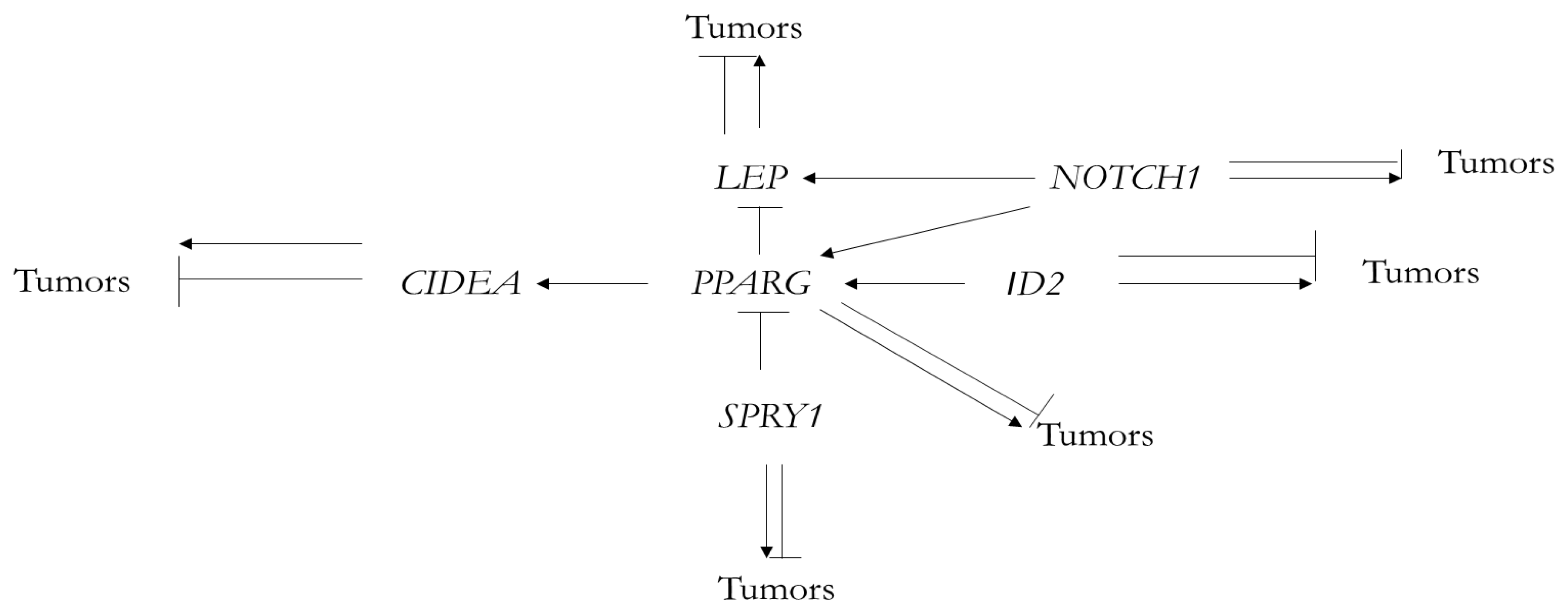
Fish pparg/Human PPARG7/302/86/81[NO]/placenta development
Table 2. Functions of human orthologs of fish TTRgrEEN genes involved in human adipose development [13].
Name of GeneProgressive Functions Connected with Beiging, BAT and Thermoregulation, Not Encountered in Fish
LEPRegulation of energy metabolism in mammals
Regulation of beige/brown fat cell differentiation
Lipostatic function and thermoregulation
NOTCH1Regulation of adipose browning, energy metabolism and thermogenesis
SPRY1Initiation and regulation of adipogenesis
Maintaining proliferation and differentiation of adipose stem cells (ASCs)
PPARGDifferentiation of adipocytes
Activation of thermogenic gene expression in brown adipocytes
The role in lipodystrophy, obesity and diabetes
ID2Stimulation of adipocyte differentiation and adipogenesis
The role in obesity
CIDEAAssociation with lipid droplets
Regulation of lipid metabolism
Regulation of adipocyte beiging
Publisher’s Note: MDPI stays neutral with regard to jurisdictional claims in published maps and institutional affiliations.

