Article

Rare Variant Analysis and Molecular Dynamics Simulation in Alzheimer’s Disease Identifies Exonic Variants in FLG

by
Weixue Xiong
1,†,
Jiahui Cai
1,†,
Ruijia Li
2,
Canhong Wen
2,*,
Haizhu Tan
1,* and
on behalf of the Alzheimer’s Disease Neuroimaging Initiative (ADNI) Database
1
Department of Preventive Medicine, Shantou University Medical College, Shantou 515000, China
2
Department of Statistics and Finance, School of Management, University of Science and Technology of China, Hefei 230000, China
*
Authors to whom correspondence should be addressed.
These authors contributed equally to this work.
The ADNI was launched by the National Institute on Aging (NIA), the National Institute of Biomedical Imaging and Bioengineering (NIBIB), the Food and Drug Administration (FDA), private pharmaceutical companies and non-profit organizations. A complete listing of ADNI investigators can be found in the acknowledgments part.
Genes 2022, 13(5), 838; https://doi.org/10.3390/genes13050838
Submission received: 22 April 2022 / Revised: 3 May 2022 / Accepted: 4 May 2022 / Published: 7 May 2022
(This article belongs to the Special Issue Bioinformatics and Genetics of Human Diseases)

Abstract

:
Background: Although an increasing number of common variants contributing to Alzheimer’s disease (AD) are uncovered by genome-wide association studies, they can only explain less than half of the heritability of AD. Rare variant association studies (RVAS) has become an increasingly important area to explain the risk or trait variability of AD. Method: To investigate the potential rare variants that cause AD, we screened 70,209 rare variants from two cohorts of a 175 AD cohort and a 214 cognitively normal cohort from the Alzheimer’s Disease Neuroimaging Initiative database. MIRARE, a novel RVAS method, was performed on 232 non-synonymous variants selected by ANNOVAR annotation. Molecular docking and molecular dynamics (MD) simulation were adopted to verify the interaction between the chosen functional variants and BACE1. Results: MIRAGE analysis revealed significant associations between AD and six potential pathogenic genes, including PREX2, FLG, DHX16, NID2, ZnF585B and ZnF875. Only interactions between FLG (including wild type and rs3120654(SER742TYR)) and BACE1 were verified by molecular docking and MD simulation. The interaction of FLG(SER742TYR) with BACE1 was greater than that of wildtype FLG with BACE1. Conclusions: According to the literature search, bio-informatics analysis, and molecular docking and MD simulation, we find non-synonymous rare variants in six genes, especially FLG(rs3120654), that may play key roles in AD.

1. Introduction

Alzheimer’s disease (AD) is a devastating illness that always causes progressive neurodegenerative and cognitive decline [1]. According to the report from WHO (key facts of dementia. Available online: https://www.who.int/news-room/fact-sheets/detail/dementia (accessed on 2 September 2021)) [2], there were approximately 35 million AD patients worldwide in 2021. The heritability of AD is high (79%) and more than 50% of the phenotypic variance can be explained by genetic variations [3,4]. An increasing number of common variants (minor allele frequency (MAF) > 0.05) that cause AD has been uncovered by large-scale genome-wide association studies. For example, RBFOX1 with a higher amyloid-β burden has been linked to early AD [5]. Apolipoprotein E-4 (APOE4), an important possible AD therapeutic target, is highly expressed in more than half of AD patients [6]. However, these common variants explain less than half of the heritability to date [7,8]. For example, the total proportion of heritability of APOE4 is estimated to be 25% [9,10]. With the development of base-by-base resolution of whole-genome sequencing, low-frequency variants (0.01 < MAF < 0.05) and rare variants (0.005 < MAF < 0.01), previously difficult to detect by microarrays, are becoming attractive alternative biomarks to AD [11,12,13]. The reason might be that these two types of variants can explain additional disease or trait variability because the growth of the effect sizes of associated variants along with the drop of allele frequencies can exceed the limits imposed by natural selection on more common variants [11]. For example, rare variants in TREM2 have been reported to be associated with a significant increase in the risk of AD [14], and rare variants in three genes (APP, PSEN1 & PSEN2) can explain 5–10% of the occurrence of early onset AD [15,16]. Therefore, the identification of rare variants associated with AD is becoming increasingly important.
Due to the importance of rare variants in sequencing studies, rare variant association studies (RVAS) have become an increasingly important area to explain additional disease risk or trait variability [17,18]. Current approaches for RVAS in complex traits involve burden tests, sequence kernel association tests (SKAT), variable threshold (VT), MIxture model based Rare variant Analysis on GEnes (MIRAGE), etc. In order to determine causative gene variants, MIRAGE, a novel method for determining causative gene variants, was applied in our study because it is an effective method to analyze rare variant data from case–control studies. MIRAGE uses an empirical Bayesian approach to estimate these prior probabilities by combining information across genes [19]. It not only better accounts for the heterogeneity of variant effects of genes, but also selects putative causal variants by fully using external information [19].
After selecting the significant variants for AD by using the RVAS method, it is very important to verify their relevant functions and mechanisms for AD. Over 80% of gene substrates (proteins) rarely act alone, but in complexes [20]. Protein–protein interactions (PPIs) are a useful way to predict the functionality and mechanism of a potential protein by acting with a known protein to disease [21] because proteins involved in the same cellular processes always interact with each other [21]. In an abundance of studies, amyloid β (Aβ) accumulation occurs before clinical symptoms in the early progression of AD [22,23]. Aβ is generated by β-site APP-cleaving enzyme 1 (BACE1) by catalyzing the amyloid precursor protein (APP), and inhibition of BACE1 activity can prevent AD development [22,23,24,25]. However, natural and synthetic BACE1 inhibitors have consistently failed in the treatment of AD for reasons such as liver toxicity and lack of improvement in cognitive decline [26]. Further exploration of the pathology of BACE1 in AD is needed. Hence, BACE1 is chosen as the interaction party by several while investigating PPIs between BACE1 and the potential proteins of AD by using molecular docking, molecular dynamics (MD) simulation, etc.
The aim of our study is to explore the genetic architecture of AD. Genetic data in our study are obtained from the Alzheimer’s Disease Neuroimaging Initiative (ADNI) database (http://adni.loni.usc.edu, accessed on 9 September 2020) [27]. To efficiently boost the power of the RVAS and genomic evaluation accuracy, whole-genotype imputation on those preprocessed genomes was first implemented on the Michigan imputation server (https://imputationserver.sph.umich.edu, accessed on 25 November 2020). Then, imputed variants were functionally annotated and classified as downstream, exonic, intergenic, intronic, ncRNA-exonic, ncRNA-intronic, ncRNA splicing, splicing, and upstream by using ANNOVAR (modified on 7 October 2020) [28], using several databases, including RefGene, exac03, avsnp147, dbnsfp30a, and ljb26_all. The MIRAGE method was subsequently adopted to perform RVAS analysis [19]. After that, several bio-informatics tools further provided a comprehensive demonstration of the significant selected variants. In addition, homology modeling, molecular docking, and MD simulation were performed to find the PPIs between the selected genes and BACE1 [29], and the effects of mutations on them.

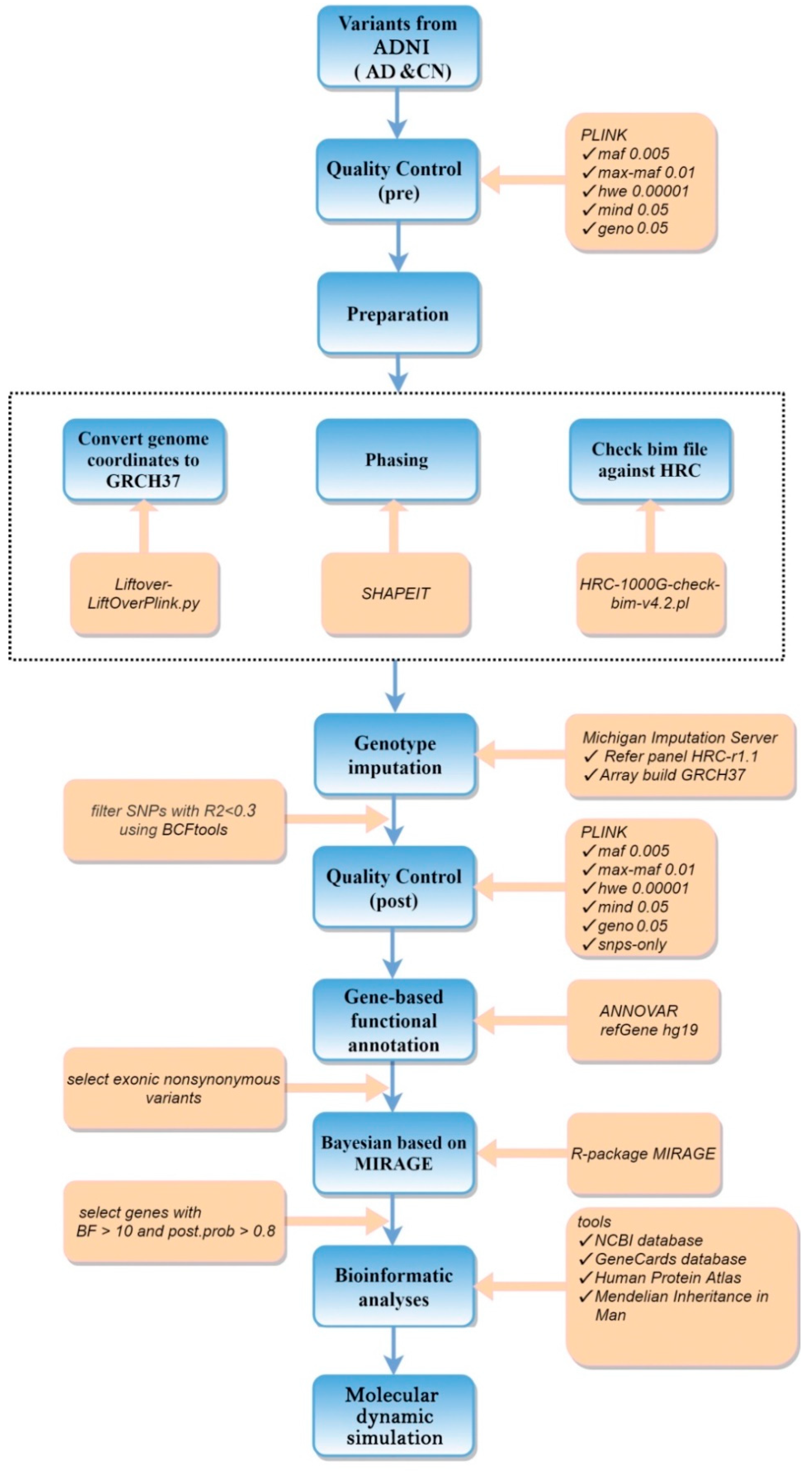
2. Materials and Methods

Detailed RVAS workflow is provided in Figure 1.

2.1. Participants

Data in this study was downloaded from the ADNI-1 database, including 600,470 genetic data, several demographic and clinical data (such as age, gender, and race). Overall, 175 AD and 214 cognitively normal (CN) subjects, aged from 56.4 to 90.9 years, were enrolled. Written informed consent was obtained from all participants. The full list of inclusion and exclusion criteria can be accessed online at https://adni.loni.usc.edu/wp-content/uploads/2008/07/adni2-procedures-manual.pdf, accessed on 3 May 2022.

2.2. Statistical Analysis

2.2.1. Demographic Comparisons

The two-tailed student’s t-test for two cohort comparisons in age was performed. The chi-squared test was adopted to determine the differences in gender and race distribution between two groups.

2.2.2. Data Preprocessing

A series of steps, including quality control (QC) before genotype imputation like filtering for single nucleotide polymorphism (SNPs) with call rate (missing > 5%), MAF > 1% and MAF < 0.5%), and the Hardy–Weinberg equilibrium (p-value < 1 × 10⁻⁵), LiftOver for converting the genome coordinates into hg19, SHAPEIT2 for phasing and whole-genotype imputation on the Michigan imputation server (https://imputationserver.sph.umich.edu), were carried out. To filter imputed data with high degrees of uncertainty, an R-square of 0.3 was set for inclusion after imputation. Then, post-imputation QC and gene-based annotation were implemented. In this study, non-synonymous exonic variants occurring in 330 subjects were collected from the annotated variants because non-synonymous mutations were demonstrated to have a much greater effect on an individual than synonymous mutations [30]. Non-synonymous variants were subsequently predicted to be at risk or non-risk variants by considering three functionally damaging effect scores, such as polyPhen2 HDIV scores [31], Sorting Intolerant From Tolerant (SIFT) scores [32] and Combined Annotation-Dependent Depletion (CADD) scores [33] together.

2.2.3. RVAS Analysis

MIRAGE was conducted to do RVAS analysis on whole exome sequencing data of 330 subjects. Variant groups are defined depending on their annotations. Those variants with 64 PolyPhen scores lower than 0.1, a CADD score 10 or SIFT score < 0.05 were labeled as group1; others were labeled group 2. The number of the case and control groups was equal to the total number of alleles in cases and controls at a genomic locus. γ was set to 2. With the estimated parameters, the Bayes factor (BF) and posterior probabilities of each gene were computed to assess its association with the disease by comparing the likelihood of the full model (group 1) to the likelihood of the null model (group 2). In this study, a threshold of BF > 3 was set to select rare potential risk variants associated with AD.

2.3. Bioinformatic Analyses

Several in silico prediction tools were used to verify the underlying biological interpretations of the identified SNPs based on bio-informatics databases and resources, including NCBI (https://www.ncbi.nlm.nih.gov/gene/, accessed on 3 May 2022), GeneCards (https://www.genecards.org/, accessed on 3 May 2022), UCSC Genome Browser on Human (https://genome.ucsc.edu/, accessed on 3 May 2022), and Online Mendelian Inheritance in Man (https://omim.org/entry/, accessed on 3 May 2022). Online tools for the effect of SNPs on protein structure and function were also used, such as: polyPhen2 (http://genetics.bwh.harvard.edu/pph2/index.shtml, accessed on 3 May 2022), SIFT (http://provean.jcvi.org/index.php, accessed on 3 May 2022), and CADD (https://cadd.gs.washington.edu/snv, accessed on 3 May 2022).

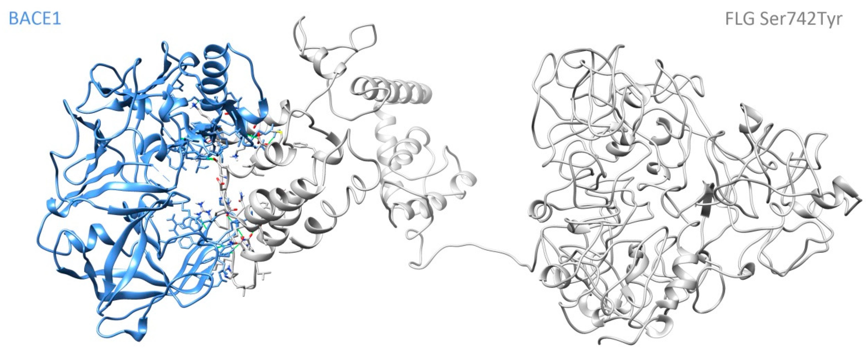
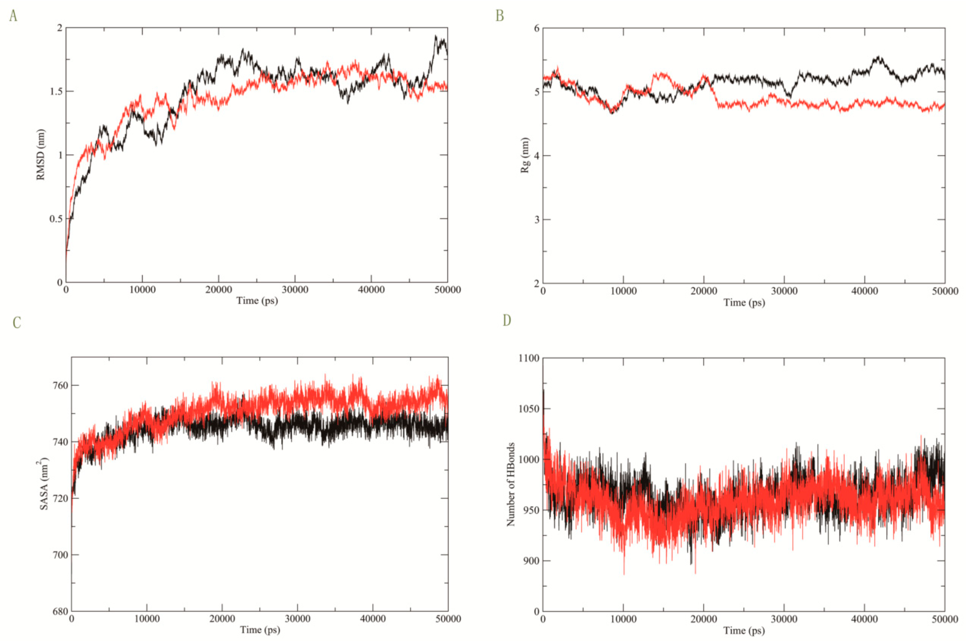
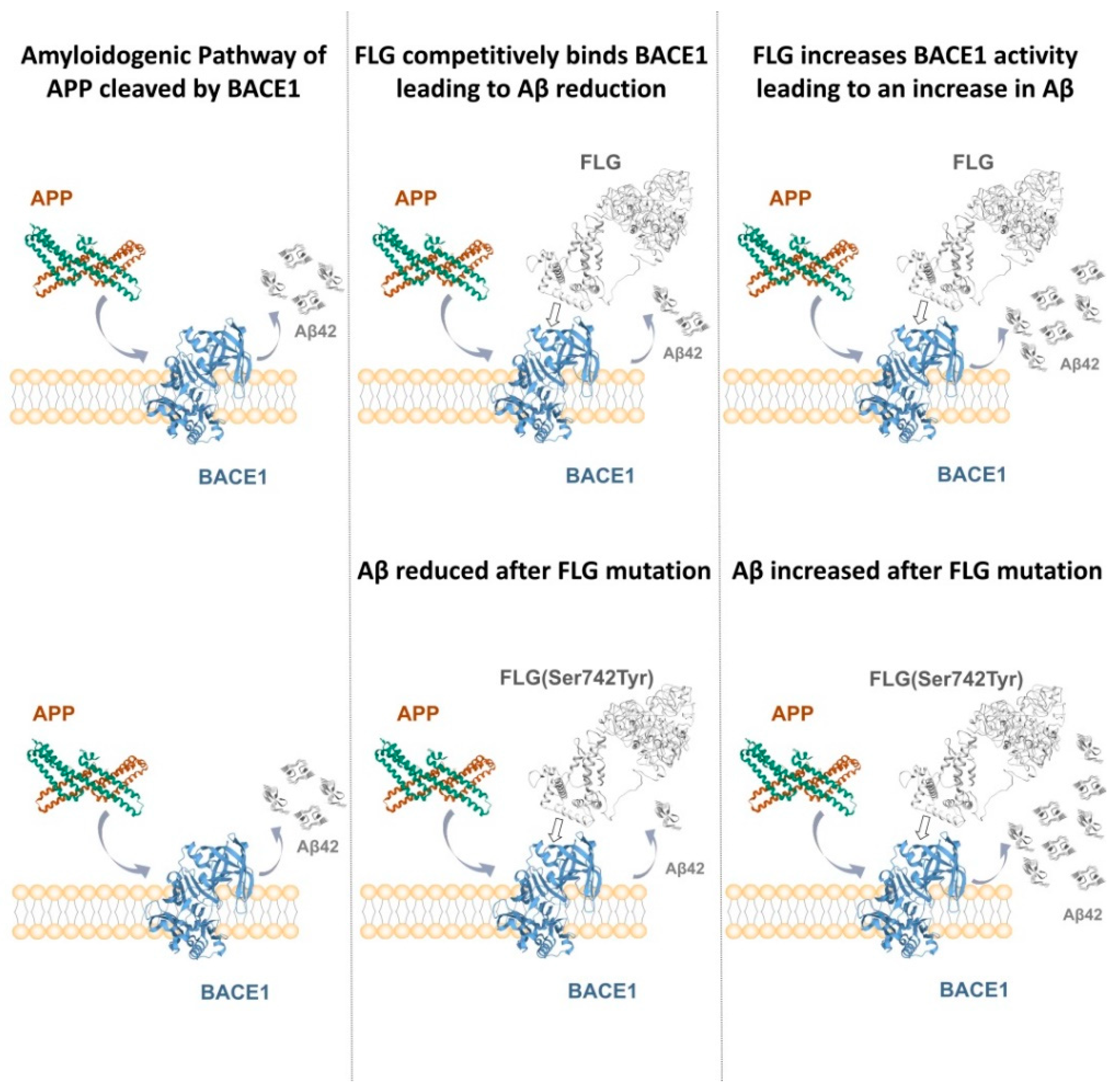
2.4. Molecular Dynamics Simulation

To demonstrate whether the proteins transcribed by potential pathogenic genes were involved in the pathogenic mechanism of AD or not, MD simulations were used to verify the stability of the complex docked with BACE1, which is involved in the formation of Aβ. All of the proteins that were finally chosen by MIRAGE were modeled on the trRosseta server, and the point amino acid was mutated by Chimera 1.14 [34,35,36]. The quality of protein structure was evaluated by PROCHECK [37]. Each selected protein was separately docked with BACE1 by the High Ambiguity Driven protein-protein Docking (HADDOCK) [38]. Finally, MD simulation was performed to test whether the above genes were involved in the formation of Aβ or not, and the influence of non-synonymous mutations on them.

3. Results

3.1. Demographic Comparisons

Table 1 describes the results of the difference tests between the AD and CN groups. From Table 1, we find that there were no significant differences in age (p-value = 0.907), sex (p-value = 0.815) and race (p-value = 0.357) between the two groups.

3.2. RVAS Analysis

After whole-genotype imputation and QC steps, a total of 330 participants with 70,209 variants passing were reserved.
The predicted model using a data set that includes 232 non-synonymous variants in 150 ADs and 180 NCs was built by MIRAGE. Table 2 shows that two non-synonymous variants (FLG (BF = 4040.782) and ZNF585B (BF = 87.062)) are likely to be pathogenic when considering BF and post-probability together.

3.3. Molecular Dynamics Simulation

The interaction strength of the protein complex that binds is denoted by the overall HADDOCK score, which is composed of the weighted average of the Van der Waal’s interactions, electrostatic energy, desolvation energy, and the buried surface area (BSA). The lower the HADDOCK score is, the stronger the interaction will be. As Table 3 shows, the interaction between BACE1- mutant FLG (rs3120654 (Ser742Tyr)) is stronger than the interaction between BACE1-FLG because the HADDOCK score of BACE1-FLG (Ser742Tyr) is lower than that of BACE1-FLG. Figure 2 shows the 3D protein structure of the complex BACE1-FLG (Ser742Tyr). There was no interaction between ZNF585B and BACE1.
The MD simulation results are shown in Figure 3 below. The root-mean-square deviation (RMSD) plot depicts that there are no erratic fluctuations in the molecular systems and all complexes are stable (see Figure 3A). The result of the radius of gyration (Rg) in Figure 3B elucidates the volumetric and compactness variations induced in the complex. Figure 3C shows the results of solvent-accessible surface area (SASA) for the protein structures with dimensional discrepancy. From Figure 3D, we can also find that the hydrogen bonds reflect the protein’s rigidity and its ability to interact with its partners (Color scheme: black: BACE1-FLG, red: BACE1-FLG (Ser742Tyr) mutant).

4. Discussion

The results of MIRAGE showed that PREX2, FLG, DHX16, NID2, ZnF585B, and ZnF875 have pathogenic risk rare variants for AD. PREX2 is associated with brain arteriovenous malformations that can induce vascular amyloid β deposition, which is a significant risk factor for AD [39,40,41]. Elena Galea et al. also suggest that PREX2 is one of the top genes detected by principal component analysis when they define AD astrocytes in multi-transcriptomic analysis [42]. The RNA helicase Dhx16 induces transcription alterations and DNA methylation changes that are involved in memory-related neurological and neuropsychiatric diseases [43]. In single variant analysis, candidate genes including DHX16 were identified as novel candidates for early onset AD in the study by Victoria Fernandez et al. [44]. Nidogen 2 (NID2) has been discovered to cause neurological disease [45]. NID2 is a pathogenic gene for neurological disease because the severity of cerebral amyloid angiopathy is least in the striatum, where NID2 is reduced [46]. ZnF875 (also known as HKR1) is associated with aging [47]. Miren Altuna’s studies find that the methylation level of HKR1 is significantly correlated with the burden of phosphorylated tau deposits [48]. The relationship between ZNF585B or ZnF875 and AD is still unknown.
Since BF greater than 10 is considered strong evidence, while a posterior probability greater than 0.8 indicates a potential efficacy signal. FLG (BF = 4040.782, posterior probability = 0.996) and ZNF585B (BF = 87.062, posterior probability = 0.842) are considered as the most likely candidate genes for rare variants [49,50]. ZNF585B is a P53 inhibitor, and P53 is up-regulated in AD [51,52]. ZNF585B is up-regulated in the olfactory bulb neural stem (OBNS) as a modulated gene, whereas OBNS enriches the ErbB signaling pathway [53]. Lack of ErbB signaling in humans has been implicated in the development of neurodegenerative diseases, such as AD [53].
In our study, filaggrin (FLG) was the only one of these genes with rare variants involved in the BACE1 pathway by molecular docking and MD simulation. The protein encoded by FLG is an intermediate filament-associated protein that aggregates keratin intermediate filaments in mammalian epidermis [54]. Shafiq M et al. suggested that significantly higher levels of FLG are observed in rapidly progressive Alzheimer’s disease high density factions (HDFs) compared with sporadic Creutzfeldt-Jakob disease-specific HDFs [55]. Our bioinformatics analysis identified that three Reactome pathways for the FLG gene are related to developmental biology and include nervous system development (https://reactome.org/PathwayBrowser/#/R-HSA-1266738, accessed on 3 May 2022), formation of the cornified envelope, which is involved in axonal regeneration [56], and keratinization (Keratin 9 has been identified as an important biomarker for AD [57]).
The validation of interactions, using 50 ns (50,000 ps) MD simulations, between FLG and BACE1 showed stable binding between BACE1 and FLG. This suggests that FLG may affect the formation of Aβ by affecting the activity of BACE1, and thus participate in the pathogenesis of AD. If FLG was competitively bound to BACE1 or the interaction could lead to a decrease in BACE1 activity, then the competitive binding of APP to BACE1 would be decreased, leading to a reduction in Aβ42 produced by hydrolysis, which can alleviate the symptoms of AD. If the interaction between FLG and BACE1 could increase the activity of BACE1, the activity of BACE1 would be increased, leading to an increase in Aβ42 produced by hydrolysis, promoting the development of AD symptoms. Additionally, mutant FLG (Ser742Tyr) can affect the interaction with BACE1. These results show that the interaction between FLG and BACE1 is enhanced by the FLG (Ser742Tyr) mutation, which further supports the effect of the rare variants we found related to AD. The theoretical pathway figure is as shown in Figure 4 below.
Thus far, a large amount of literature, the results of bioinformatics analysis and the results of MD simulation support the existence of pathogenic rare variants in these selected genes. Only BACE1 was introduced to interact with each significant gene in our docking and MD experiment in this study. Hence, in the future, many known proteins related with AD will be introduced to do the validation, and experiments in vitro and vivo will be performed to assess the computational work. In addition, the sample size in our study is not large enough for our RVAS research.
In conclusion, we identified six genes with rare variants affecting AD, especially FLG. We hope our study may aid in furthering study of exploring the underlying pathology mechanism of AD.

Author Contributions

H.T. and C.W. conceived and oversaw the project. W.X., J.C. and R.L. contributed to the data analysis and MD simulation and performed the bioinformatics analysis. All authors made critical contributions to the manuscript. All authors have read and agreed to the published version of the manuscript.

Funding

Our research was supported by the National Key Research and Development Program of China (Reference: 2018YFC1315400); the Science and Technology Planning Project of Guangdong Province (Reference: 2017A010101030); the Third Medical Technology Projects of Shantou in 2018 (Reference: 41368043); the Natural Science Foundation of China (References: 11771462, 71991474, 72171216), the National Key Research, the Key Research and Development Program of Guangdong, China (Reference: 2019B020228001), the Science and Technology Program of Guangzhou, China (Grant No. 202002030129), and the Natural Science Foundation of Anhui (Reference: BJ2040170017).

Institutional Review Board Statement

Publicly available databases were analyzed in this study. The ADNI database was conducted in accordance with the Declaration of Helsinki, and approved by the ethics committees of the National Institute on Aging, the National Institute of Biomedical Imaging and Bioengineering, and the local ethics committee of each participating site (https://adni.loni.usc.edu/methods/documents/, accessed on 3 May 2022).

Informed Consent Statement

Informed consent was obtained from all subjects involved in the study. Written informed consent has been obtained from the patient(s) to publish this paper.

Data Availability Statement

Data in our study are obtained from the Alzheimer’s Disease Neuroimaging Initiative (ADNI) database (http://adni.loni.usc.edu, accessed on 3 May 2022), an open source database.

Acknowledgments

We gratefully thank Geng Dong for his support in computational biology and bioinformatics technology. The authors would like to thank blogger Wenyan Chen and Mo Xv for their help. Thanks to investigators within ADNI such as Michael W. Weiner who contributed to the design and implementation of ADNI and provided data, but did not participate in the analysis or writing of this report. A complete listing of ADNI investigators can be found at: https://adni.loni.usc.edu/wp-content/uploads/how_to_apply/ADNI_Acknowledgement_List.pdf, accessed on 3 May 2022.

Conflicts of Interest

The authors declare no conflict of interest.

References

  1. Aggleton, J.P.; Pralus, A.; Nelson, A.J.; Hornberger, M. Thalamic pathology and memory loss in early Alzheimer’s disease: Moving the focus from the medial temporal lobe to Papez circuit. Brain 2016, 139, 1877–1890. [Google Scholar] [CrossRef] [PubMed]
  2. Key Facts of Dementia. Available online: https://www.who.int/news-room/fact-sheets/detail/dementia (accessed on 2 September 2021).
  3. Gatz, M.; Reynolds, C.A.; Fratiglioni, L.; Johansson, B.; Mortimer, J.A.; Berg, S.; Fiske, A.; Pedersen, N.L. Role of genes and environments for explaining Alzheimer disease. Arch. Gen. Psychiatry 2006, 63, 168–174. [Google Scholar] [CrossRef] [PubMed]
  4. Ridge, P.G.; Hoyt, K.B.; Boehme, K.; Mukherjee, S.; Crane, P.K.; Haines, J.L.; Mayeux, R.; Farrer, L.A.; Pericak-Vance, M.A.; Schellenberg, G.D. Assessment of the genetic variance of late-onset Alzheimer’s disease. Neurobiol. Aging 2016, 41, 200.e213–200.e220. [Google Scholar] [CrossRef] [PubMed] [Green Version]
  5. Raghavan, N.S.; Dumitrescu, L.; Mormino, E.; Mahoney, E.R.; Lee, A.J.; Gao, Y.; Bilgel, M.; Goldstein, D.; Harrison, T.; Engelman, C.D. Association between common variants in RBFOX1, an RNA-binding protein, and brain amyloidosis in early and preclinical Alzheimer disease. JAMA Neurol. 2020, 77, 1288–1298. [Google Scholar] [CrossRef]
  6. Michaelson, D.M. APOE ε4: The most prevalent yet understudied risk factor for Alzheimer’s disease. Alzheimer’s Dement. 2014, 10, 861–868. [Google Scholar] [CrossRef]
  7. Zuk, O.; Schaffner, S.F.; Samocha, K.; Do, R.; Hechter, E.; Kathiresan, S.; Daly, M.J.; Neale, B.M.; Sunyaev, S.R.; Lander, E.S. Searching for missing heritability: Designing rare variant association studies. Proc. Natl. Acad. Sci. USA 2014, 111, E455–E464. [Google Scholar] [CrossRef] [Green Version]
  8. Cruchaga, C.; Chakraverty, S.; Mayo, K.; Vallania, F.L.; Mitra, R.D.; Faber, K.; Williamson, J.; Bird, T.; Diaz-Arrastia, R.; Foroud, T.M. Rare variants in APP, PSEN1 and PSEN2 increase risk for AD in late-onset Alzheimer’s disease families. PLoS ONE 2012, 7, e31039. [Google Scholar] [CrossRef]
  9. So, H.C.; Gui, A.H.; Cherny, S.S.; Sham, P.C. Evaluating the heritability explained by known susceptibility variants: A survey of ten complex diseases. Genet. Epidemiol. 2011, 35, 310–317. [Google Scholar] [CrossRef]
  10. Bagyinszky, E.; Giau, V.V.; An, S.A. Transcriptomics in Alzheimer’s Disease: Aspects and Challenges. Int. J. Mol. Sci. 2020, 21, 3517. [Google Scholar] [CrossRef]
  11. Cirulli, E.T.; White, S.; Read, R.W.; Elhanan, G.; Metcalf, W.J.; Tanudjaja, F.; Fath, D.M.; Sandoval, E.; Isaksson, M.; Schlauch, K.A. Genome-wide rare variant analysis for thousands of phenotypes in over 70,000 exomes from two cohorts. Nat. Commun. 2020, 11, 542. [Google Scholar] [CrossRef]
  12. Huffman, J.E.; De Vries, P.S.; Morrison, A.C.; Sabater-Lleal, M.; Kacprowski, T.; Auer, P.L.; Brody, J.A.; Chasman, D.I.; Chen, M.-H.; Guo, X. Rare and low-frequency variants and their association with plasma levels of fibrinogen, FVII, FVIII, and vWF. Blood 2015, 126, e19–e29. [Google Scholar] [CrossRef] [PubMed] [Green Version]
  13. Manolio, T.A.; Collins, F.S.; Cox, N.J.; Goldstein, D.B.; Hindorff, L.A.; Hunter, D.J.; McCarthy, M.I.; Ramos, E.M.; Cardon, L.R.; Chakravarti, A. Finding the missing heritability of complex diseases. Nature 2009, 461, 747–753. [Google Scholar] [CrossRef] [PubMed] [Green Version]
  14. Guerreiro, R.; Wojtas, A.; Bras, J.; Carrasquillo, M.; Rogaeva, E.; Majounie, E.; Cruchaga, C.; Sassi, C.; Kauwe, J.S.; Younkin, S. TREM2 variants in Alzheimer’s disease. N. Engl. J. Med. 2013, 368, 117–127. [Google Scholar] [CrossRef] [Green Version]
  15. Van Cauwenberghe, C.; Van Broeckhoven, C.; Sleegers, K. The genetic landscape of Alzheimer disease: Clinical implications and perspectives. Genet. Med. 2016, 18, 421–430. [Google Scholar] [CrossRef] [Green Version]
  16. Hollingworth, P.; Harold, D.; Sims, R.; Gerrish, A.; Lambert, J.-C.; Carrasquillo, M.M.; Abraham, R.; Hamshere, M.L.; Pahwa, J.S.; Moskvina, V. Common variants at ABCA7, MS4A6A/MS4A4E, EPHA1, CD33 and CD2AP are associated with Alzheimer’s disease. Nat. Genet. 2011, 43, 429–435. [Google Scholar] [CrossRef] [PubMed] [Green Version]
  17. Liu, L.; Wang, P.; Meng, J.; Chen, L.; Zhu, W.; Ma, W. A permutation method for detecting trend correlations in rare variant association studies. Genet. Res. 2019, 101, E13. [Google Scholar] [CrossRef] [Green Version]
  18. Lee, S.; Abecasis, G.R.; Boehnke, M.; Lin, X. Rare-variant association analysis: Study designs and statistical tests. Am. J. Hum. Genet. 2014, 95, 5–23. [Google Scholar] [CrossRef] [Green Version]
  19. Han, S.; Knoblauch, N.; Wang, G.; Zhao, S.; Liu, Y.; Xie, Y.; Sheng, W.; Nguyen, H.T.; He, X. A Bayesian method for rare variant analysis using functional annotations and its application to Autism. bioRxiv 2019, 828061. [Google Scholar]
  20. Hu, G.; Di Paola, L.; Liang, Z.; Giuliani, A. Comparative study of elastic network model and protein contact network for protein complexes: The hemoglobin case. BioMed Res. Int. 2017, 2017, 2483264. [Google Scholar] [CrossRef]
  21. Von Mering, C.; Krause, R.; Snel, B.; Cornell, M.; Oliver, S.G.; Fields, S.; Bork, P. Comparative assessment of large-scale data sets of protein–protein interactions. Nature 2002, 417, 399–403. [Google Scholar] [CrossRef]
  22. Koelsch, G. BACE1 function and inhibition: Implications of intervention in the amyloid pathway of Alzheimer’s disease pathology. Molecules 2017, 22, 1723. [Google Scholar] [CrossRef] [PubMed] [Green Version]
  23. Yan, R.; Vassar, R. Targeting the β secretase BACE1 for Alzheimer’s disease therapy. Lancet Neurol. 2014, 13, 319–329. [Google Scholar] [CrossRef] [Green Version]
  24. Franzmeier, N.; Dewenter, A.; Frontzkowski, L.; Dichgans, M.; Rubinski, A.; Neitzel, J.; Smith, R.; Strandberg, O.; Ossenkoppele, R.; Buerger, K. Patient-centered connectivity-based prediction of tau pathology spread in Alzheimer’s disease. Sci. Adv. 2020, 6, eabd1327. [Google Scholar] [CrossRef] [PubMed]
  25. Swaminathan, G.; Zhu, W.; Plowey, E.D. BECN1/Beclin 1 sorts cell-surface APP/amyloid β precursor protein for lysosomal degradation. Autophagy 2016, 12, 2404–2419. [Google Scholar] [CrossRef] [PubMed] [Green Version]
  26. Chakravarthy, M.; Veedu, R.N. BACE1 inhibition using 2′-OMePS steric blocking antisense oligonucleotides. Genes 2019, 10, 705. [Google Scholar] [CrossRef] [Green Version]
  27. Spooner, A.; Chen, E.; Sowmya, A.; Sachdev, P.; Kochan, N.A.; Trollor, J.; Brodaty, H. A comparison of machine learning methods for survival analysis of high-dimensional clinical data for dementia prediction. Sci. Rep. 2020, 10, 1–10. [Google Scholar] [CrossRef]
  28. Wang, K.; Li, M.; Hakonarson, H. ANNOVAR: Functional annotation of genetic variants from high-throughput sequencing data. Nucleic Acids Res. 2010, 38, e164. [Google Scholar] [CrossRef]
  29. Ghosh, A.K.; Osswald, H.L. BACE1 (β-secretase) inhibitors for the treatment of Alzheimer’s disease. Chem. Soc. Rev. 2014, 43, 6765–6813. [Google Scholar] [CrossRef] [Green Version]
  30. Agashe, D.; Sane, M.; Phalnikar, K.; Diwan, G.D.; Habibullah, A.; Martinez-Gomez, N.C.; Sahasrabuddhe, V.; Polachek, W.; Wang, J.; Chubiz, L.M. Large-effect beneficial synonymous mutations mediate rapid and parallel adaptation in a bacterium. Mol. Biol. Evol. 2016, 33, 1542–1553. [Google Scholar] [CrossRef] [Green Version]
  31. Adzhubei, I.A.; Schmidt, S.; Peshkin, L.; Ramensky, V.E.; Gerasimova, A.; Bork, P.; Kondrashov, A.S.; Sunyaev, S.R. A method and server for predicting damaging missense mutations. Nat. Methods 2010, 7, 248–249. [Google Scholar] [CrossRef] [Green Version]
  32. Ng, P.C.; Henikoff, S. SIFT: Predicting amino acid changes that affect protein function. Nucleic Acids Res. 2003, 31, 3812–3814. [Google Scholar] [CrossRef] [PubMed] [Green Version]
  33. Kircher, M.; Witten, D.M.; Jain, P.; O’Roak, B.J.; Cooper, G.M.; Shendure, J. A general framework for estimating the relative pathogenicity of human genetic variants. Nat. Genet. 2014, 46, 310–315. [Google Scholar] [CrossRef] [PubMed] [Green Version]
  34. Yang, J.; Yan, R.; Roy, A.; Xu, D.; Poisson, J.; Zhang, Y. The I-TASSER Suite: Protein structure and function prediction. Nat. Methods 2015, 12, 7–8. [Google Scholar] [CrossRef] [PubMed] [Green Version]
  35. Pettersen, E.F.; Goddard, T.D.; Huang, C.C.; Couch, G.S.; Greenblatt, D.M.; Meng, E.C.; Ferrin, T.E. UCSF Chimera—A visualization system for exploratory research and analysis. J. Comput. Chem. 2004, 25, 1605–1612. [Google Scholar] [CrossRef] [Green Version]
  36. Bassot, C.; Elofsson, A. Accurate contact-based modelling of repeat proteins predicts the structure of new repeats protein families. PLoS Comput. Biol. 2021, 17, e1008798. [Google Scholar] [CrossRef]
  37. Laskowski, R.A.; MacArthur, M.W.; Moss, D.S.; Thornton, J.M. PROCHECK: A program to check the stereochemical quality of protein structures. J. Appl. Crystallogr. 1993, 26, 283–291. [Google Scholar] [CrossRef]
  38. Dominguez, C.; Boelens, R.; Bonvin, A.M. HADDOCK: A protein—Protein docking approach based on biochemical or biophysical information. J. Am. Chem. Soc. 2003, 125, 1731–1737. [Google Scholar] [CrossRef] [Green Version]
  39. Wang, K.; Zhao, S.; Liu, B.; Zhang, Q.; Li, Y.; Liu, J.; Shen, Y.; Ding, X.; Lin, J.; Wu, Y. Perturbations of BMP/TGF-β and VEGF/VEGFR signalling pathways in non-syndromic sporadic brain arteriovenous malformations (BAVM). J. Med. Genet. 2018, 55, 675–684. [Google Scholar] [CrossRef]
  40. Hart, M.; Merz, P.; Bennett-Gray, J.; Menezes, A.; Goeken, J.; Schelper, R.; Wisniewski, H. β-amyloid protein of Alzheimer’s disease is found in cerebral and spinal cord vascular malformations. Am. J. Pathol. 1988, 132, 167. [Google Scholar]
  41. Tsonou, E. Exploring the Importance of PREX Proteins in Glucose Homeostasis and Insulin Signalling. Ph.D. Thesis, University of Cambridge, Cambridge, UK, 2020. [Google Scholar]
  42. Galea, E.; Weinstock, L.D.; Larramona-Arcas, R.; Pybus, A.F.; Giménez-Llort, L.; Escartin, C.; Wood, L.B. Multi-transcriptomic analysis points to early organelle dysfunction in human astrocytes in Alzheimer’s disease. Neurobiol. Dis. 2022, 166, 105655. [Google Scholar] [CrossRef]
  43. Duke, C.G.; Kennedy, A.J.; Gavin, C.F.; Day, J.J.; Sweatt, J.D. Experience-dependent epigenomic reorganization in the hippocampus. Learn. Mem. 2017, 24, 278–288. [Google Scholar] [CrossRef]
  44. Fernandez, V.; Huey, D.; Budde, J.P.; Farias, F.H.; Harari, O.; Norton, J.; Gentsch, J.; Morris, J.C.; Goate, A.; Rademakers, R. O2-10-02: Whole Exome Sequencing Analysis in Early Onset Alzheimer Reveals Novel Candidate Genes. Alzheimer’s Dement. 2019, 15, P564. [Google Scholar] [CrossRef]
  45. Li, C. Understanding Rare Neurological Disorders Using Drosophila Models: Mechanistic Characterization of Neurotoxicity in Snyder-Robinson Syndrome. Ph.D. Thesis, University of Miami, Coral Gables, FL, USA, 2018. [Google Scholar]
  46. Hawkes, C.A.; Gatherer, M.; Sharp, M.M.; Dorr, A.; Yuen, H.M.; Kalaria, R.; Weller, R.O.; Carare, R.O. Regional differences in the morphological and functional effects of aging on cerebral basement membranes and perivascular drainage of amyloid-β from the mouse brain. Aging Cell 2013, 12, 224–236. [Google Scholar] [CrossRef] [PubMed]
  47. Zeng, Q.; Chen, X.; Ning, C.; Zhu, Q.; Yao, Y.; Zhao, Y.; Luan, F. Methylation of the genes ROD1, NLRC5, and HKR1 is associated with aging in Hainan centenarians. BMC Med. Genom. 2018, 11, 7. [Google Scholar] [CrossRef] [PubMed] [Green Version]
  48. Altuna, M.; Urdánoz-Casado, A.; Sánchez-Ruiz de Gordoa, J.; Zelaya, M.V.; Labarga, A.; Lepesant, J.M.; Roldán, M.; Blanco-Luquin, I.; Perdones, Á.; Larumbe, R. DNA methylation signature of human hippocampus in Alzheimer’s disease is linked to neurogenesis. Clin. Epigenetics 2019, 11, 91. [Google Scholar] [CrossRef] [Green Version]
  49. van Doorn, J.; van den Bergh, D.; Böhm, U.; Dablander, F.; Derks, K.; Draws, T.; Etz, A.; Evans, N.J.; Gronau, Q.F.; Haaf, J.M. The JASP guidelines for conducting and reporting a Bayesian analysis. Psychon. Bull. Rev. 2021, 28, 813–826. [Google Scholar] [CrossRef]
  50. Hewett, K.; Sanders, D.B.; Grove, R.A.; Broderick, C.L.; Rudo, T.J.; Bassiri, A.; Zvartau-Hind, M.; Bril, V.; Group, B.S. Randomized study of adjunctive belimumab in participants with generalized myasthenia gravis. Neurology 2018, 90, e1425–e1434. [Google Scholar] [CrossRef] [Green Version]
  51. Hooper, C.; Meimaridou, E.; Tavassoli, M.; Melino, G.; Lovestone, S.; Killick, R. p53 is upregulated in Alzheimer’s disease and induces tau phosphorylation in HEK293a cells. Neurosci. Lett. 2007, 418, 34–37. [Google Scholar] [CrossRef] [Green Version]
  52. Black, S.K. An Investigation into the Roles of p53, Nodal/Activin and Fibroblast Growth Factor Signalling in Early Heart Development. Ph.D. Thesis, Cardiff University, Cardiff, UK, 2016. [Google Scholar]
  53. Marei, H.E.; Althani, A.; Afifi, N.; Abd-Elmaksoud, A.; Bernardini, C.; Michetti, F.; Barba, M.; Pescatori, M.; Maira, G.; Paldino, E. Over-expression of hNGF in adult human olfactory bulb neural stem cells promotes cell growth and oligodendrocytic differentiation. PLoS ONE 2013, 8, e82206. [Google Scholar] [CrossRef] [Green Version]
  54. Soltani, S.; Saghazadeh, A.; Movahedi, M.; Tavakol, M.; Sadr, M.; Farhadi, E.; Rezaei, N. FLG single nucleotide polymorphisms in chronic idiopathic urticaria. Allergol. Immunopathol. 2016, 44, 341–345. [Google Scholar] [CrossRef]
  55. Shafiq, M.; Zafar, S.; Younas, N.; Noor, A.; Puig, B.; Altmeppen, H.C.; Schmitz, M.; Matschke, J.; Ferrer, I.; Glatzel, M. Prion protein oligomers cause neuronal cytoskeletal damage in rapidly progressive Alzheimer’s disease. Mol. Neurodegener. 2021, 16, 11. [Google Scholar] [CrossRef] [PubMed]
  56. Bonilla, I.E.; Tanabe, K.; Strittmatter, S.M. Small proline-rich repeat protein 1A is expressed by axotomized neurons and promotes axonal outgrowth. J. Neurosci. 2002, 22, 1303–1315. [Google Scholar] [CrossRef] [PubMed]
  57. Richens, J.L.; Spencer, H.L.; Butler, M.; Cantlay, F.; Vere, K.-A.; Bajaj, N.; Morgan, K.; O’Shea, P. Rationalising the role of Keratin 9 as a biomarker for Alzheimer’s disease. Sci. Rep. 2016, 6, 22962. [Google Scholar] [CrossRef] [PubMed] [Green Version]
Figure 1. Rare variant association studies workflow.
Genes 13 00838 g001
Figure 2. Three-dimensional protein structure of the complex BACE1-FLG (Ser742Tyr).
Genes 13 00838 g002
Figure 3. MD simulation results of BACE1-FLG and BACE1-FLG (Ser742Tyr) mutant. (A) The RMSD plot depicts that there are no erratic fluctuations in the molecular systems and all complexes are stable. (B) The result of the Rg elucidates the volumetric and compactness variations induced in the complex. (C) The result of SASA for the protein structures with dimensional discrepancy. (D) The hydrogen bonds reflect the protein’s rigidity and its ability to interact with its partners.
Genes 13 00838 g003
Figure 4. Hypothetical theoretical pathway figure of FLG and its mutant FLG (Ser742Tyr) on Aβ42 accumulation.
Genes 13 00838 g004
Table 1. Comparisons of demographics and clinical variables between AD and CN cohorts.
ADCNp-Value
No. of participants, n (%) 175 (45%)214 (55%)
Sex, n (%)M93 (53%)115 (54%)0.907
F82 (47%)99 (46%)
Age, Median (IQR) 75.875.50.815
(70.85, 81.10)(72.12, 78.38)
Race, n (%)Asian2 (1)2 (1)0.357
Black8 (5)15 (7)
More than one2 (1)0 (0)
White163 (93)197 (92)
Education, Median (IQR) 15 (12, 16)16 (14, 18)<0.001
Participant residence, n (%)House121 (69)162 (76)0.005
Condo/Co-op (owned)17 (10)32 (15)
Apartment (rented)21 (12)13 (6)
Mobile Home3 (2)2 (1)
Retirement Community5 (3)3 (1)
Assisted Living7 (4)0 (0)
Other1 (1)2 (1)
APOE4, n (%)058 (33)156 (73)<0.001
185 (49)53 (25)
232 (18)5 (2)
Table 2. The results of RAVIS analysis by using MIRAGE.
GeneBFPost.Prob
FLG4040.782 0.996
ZNF585B87.062 0.842
ZNF8759.331 0.364
PREX26.746 0.293
NID25.819 0.263
DHX164.515 0.217
Table 3. Docking results of BACE1 interaction with selected proteins.
BACE1-FLGBACE1-FLG Ser742Tyr
HADDOCK score−77.500 ± 13.900−89.600 ± 16.000
Root Mean Square Deviation from the overall lowest-energy structure0.500 ± 0.3000.800 ± 0.500
Van der Waals energy−79.400 ± 5.400−83.800 ± 14.200
Electrostatic energy−516.500 ± 81.300−566.100 ± 71.800
Desolvation energy2.400 ± 7.7002.700 ± 3.400
Buried Surface Area3250.100 ± 135.7003318.500 ± 346.700
Z-Score−1.400 −1.800
Notes: (1) BACE1-FLG refers to docking BACE1 with FLG; (2) BACE1-FLG (Ser742Tyr) refers to docking BACE1 with the FLG (Ser742Tyr) mutant; (3) HADDOCK score shows how well the protein complex binds with each other and is obtained from the weighted average of the Van der Waals interactions, electrostatic energy, desolvation energy, and the buried surface area.
Publisher’s Note: MDPI stays neutral with regard to jurisdictional claims in published maps and institutional affiliations.

