Article

Identification and Characterization Roles of Phytoene Synthase (PSY) Genes in Watermelon Development

1
College of Horticulture and Landscape Architecture, Northeast Agricultural University, Harbin 150030, China
2
Key Laboratory of Biology and Genetic Improvement of Horticulture Crops (Northeast Region), Ministry of Agriculture and Rural Affairs, Harbin 150030, China
*
Authors to whom correspondence should be addressed.
Genes 2022, 13(7), 1189; https://doi.org/10.3390/genes13071189
Submission received: 23 May 2022 / Revised: 15 June 2022 / Accepted: 28 June 2022 / Published: 1 July 2022
(This article belongs to the Section Plant Genetics and Genomics)

Abstract

:
Phytoene synthase (PSY) plays an essential role in carotenoid biosynthesis. In this study, three ClPSY genes were identified through the watermelon genome, and their full-length cDNA sequences were cloned. The deduced proteins of the three ClPSY genes were ranged from 355 to 421 amino acid residues. Phylogenetic analysis suggested that the ClPSYs are highly conserved with bottle gourd compared to other cucurbit crops PSY proteins. Variation in ClPSY1 expression in watermelon with different flesh colors was observed; ClPSY1 was most highly expressed in fruit flesh and associated with the flesh color formation. ClPSY1 expression was much lower in the white-fleshed variety than the colored fruits. Gene expression analysis of ClPSY genes in root, stem, leaf, flower, ovary and flesh of watermelon plants showed that the levels of ClPSY2 transcripts found in leaves was higher than other tissues; ClPSY3 was dominantly expressed in roots. Functional complementation assays of the three ClPSY genes suggested that all of them could encode functional enzymes to synthesize the phytoene from Geranylgeranyl Pyrophosphate (GGPP). Some of the homologous genes clustered together in the phylogenetic tree and located in the synteny chromosome region seemed to have similar expression profiles among different cucurbit crops. The findings provide a foundation for watermelon flesh color breeding with regard to carotenoid synthesis and also provide an insight for the further research of watermelon flesh color formation.

1. Introduction

Approximately 750 carotenoids have been described in nature, and that number continues to grow [1]. Plants attract insects and birds for pollination and seed dispersal, in large part because of carotenoids, which confer different colors to plant parts. In addition, as part of the human diet, carotenoids are the precursor of vitamin A and the basis of many nutrients; carotenoids also have antioxidant activity. Although these compounds are essential to health, humans cannot synthesize them [2]. The first step is mediated by nuclear-encoded PSY, which catalyzes the conversion of two GGPP molecules to phytoene in plants [3]. 15-cis-phytoene is the first colorless carotenoid produced by PSY, a rate-limiting enzyme in the carotenoid biosynthesis pathway [4,5]. The decrease in total carotene content in Arabidopsis mutant etiolated seedlings is consistent with the requirement of galactose for PSY activity [6].
Many plant species contain three to five PSY family members, while in Arabidopsis, there is only one. Three PSY genes (CpPSYA, CpPSYB and CpPSYC) have been identified in Cucurbita pepo, though only the abundance of the CpPSYA transcript affects the carotenoid biosynthetic pathway [7]. Apple has six MdPSYs, however, which mainly involve MdPSY1 and MdPSY2 with relatively high enzymatic activity and expression levels [8]. The function of PSY was affected by many regulatory patterns. Different PSY members also have the tissue-specific expression characteristics. In tomato [9] and citrus [10], PSY1 was specifically expressed in fruits, while PSY2 and PSY3 were mainly detected in leaves and roots. The silencing of SlPSY1 resulted in a yellow fruit color from red [11]. The fruit phenotype did not overtly change when SlPSY2 and SlPSY3 were silenced, suggesting that SlPSY1 plays the most significant role in tomato fruit. SlPSY2 functions mainly in chloroplast-containing tissues [9]. PSY-A was found in watermelon’s whole tissue (including fruits), and PSY-B was found in watermelon leaves and roots [12]. The transcriptome dynamics at certain watermelon fruit developmental stages suggested that lycopene accumulation affects the up-regulation of ClPSY [13]. Different PSY members were also expressed in unique locations within the chloroplast, such as thylakoid membranes associated with plastoglobules or the envelope membrane and stroma [14]. In maize, ZmPSY1 is involved in the formation of distorted plastid shape and fiber phenotype [15]. Furthermore, variant localization may also alter enzyme activity. When PSY was overexpressed in Arabidopsis, PSY enzyme activity was detected only in the membrane-bound form but not in the matrix localized form [16]. Transcription factor was another PSY regulator for carotenoids accumulation. In tomato, the ripening-inhibitor (RIN) has been shown to regulate carotenoid concentration in fruits by interacting with the SlPSY1 promoter [17]. SlBBX20 in tomato can activate the expression of SlPSY1 by directly binding to the G-box motif of its promoter, resulting in increased expression of SlPSY1 for carotenoid accumulation, presenting dark green color in fruits and leaves [18]. Transcriptomic data showed that the expression trend of AdMYB7 in N. benthamiana was consistent with that of NdPSY [19]. Overexpression of CsMADS6 in citrus callus can directly combine with CsPSY promoters to increase the carotenoid content [20]. PSY expression is also regulated by the feedback of carotenoid pathway products [21]. The overexpression of AtCYP97A in carrots increased α-carotene accumulated to lutein but decreased PSY protein level and led to the decrease in carotenoids [22]. PSY can affect carotenoid accumulation at the protein level post-transcriptionally. PSY protein levels were negatively regulated by carotenoid metabolites and total carotenoid content. The expression level of PSY is regulated by OR/OR-like proteins in Arabidopsis and potato [23,24]. As a post-transcriptional regulator of PSY, PIF1 is involved in the development of chlorophyll and chloroplast and the production of carotene [25]. The orange flesh color (β-carotene accumulated) in melon was affected by the CmOr (encoding a plastid-targeted protein) gene [26]. Further research confirmed that the CmOr had little effect on the CmPSY1 expression level but highly affected CmPSY1 protein levels [27]. The decrease in Clp activity in clpc1 leads to the decrease in PSY protein transformation. The OR protein enhanced the stability of the PSY protein and increased the activity of PSY in clpc1 [28].
To date, PSYs have been studied extensively in other crops but less so in cucurbitaceae, especially watermelon. In this study, we cloned three ClPSY members from the watermelon genome, characterized and compared the complete protein coding sequences (CDS), analyzed the expression profiles in different tissues and evaluated the expression patterns during fruit development in fruits of different colors. In addition, the ClPSY gene family was investigated with regard to enzyme activity using a heterologous complementation system. The expression pattern and genomic synteny of different PSYs among the cucurbit crops also exhibited that some PSYs in the genomic synteny may have a similar gene function.

2. Materials and Methods

2.1. Plant Materials

Six watermelon cultivars with different flesh colors: white-fleshed ‘ZXG0000’ (PI 494532); orange-fleshed ‘ZXG0077’ (Charleston); pink-fleshed ‘ZXG1594’ (Javrijsky); red-fleshed ‘ZXG55’ (Lanzhou Huapi); ‘ZXG1549’ (Qing Pi); and ‘ZXG0079’ (Huangbao No.1) were used in this study. All the plants were planted in the greenhouse at the Xiang Yang Agricultural Experiment Station of Northeast Agricultural University, Harbin (44°04′ N, 125°42′ E), China, in 2018. Tissue samples were collected at 10, 18, 26, 34 and 42 days after pollination. Three watermelon fruits with similar development status and no mechanical damage were collected at each development stage of each variety and stored at −80 °C for subsequent experiments, including DNA and RNA extraction. ‘ZXG0000′ belonged to the C. ssp. mucosospermus with a late fruit setting, so the fruit samples were collected from this line at 10, 18, 26, 34, 42 and 50 DAP. Roots, stems, flowers, ovaries and leaves were collected from ‘ZXG0079’.

2.2. RNA Isolation and cDNA Synthesis

Total RNA was performed according to the instructions of Novogene (Beijing) RNA extraction kit, which was extracted from all tissues, including roots, stems, leaves, flowers and ovaries, at each fruit stage. The total RNA was detected by 1% (w/v) agarose gel electrophoresis. In addition, the non-degraded RNA was selected as OD260: OD280 > 1.80 for cDNA synthesis. cDNA synthesis was performed according to the qPCR RT Kit (Code No. Fsq-101, TOYOBO, Tokyo, Japan). The first step was to denaturate RNA, that is, the qualified RNA was placed in a 65 °C metal bath for 5 min and then immediately put in an ice box for cooling, so as to improve the reverse transcription efficiency. The cDNA after reverse transcription was stored at −20 °C for qRT-PCR and coding region cloning.

2.3. Identification and Cloning of ClPSY Genes

To identify members of the ClPSY gene family, the Cucurbit Genomics Database (https://www.cucurbitgenomics.org/watermelon97103 genome v2/; accessed on 15 January 2022) was searched for putative ClPSY genes (Table 1) from watermelon genome version of 97103 v2. To amplify full-length cDNA, specific primers were designed according to the sequences of putative ClPSY genes using Premier 6.0 software. The primer sequences, amplicon sizes and accession numbers are shown in Table 2. Polymerase chain reaction (PCR) was performed using cDNA from flesh tissues at 34 days after pollination (DAP) as the template with the reported conditions [29]. Target genes were purified using a gel extraction kit (Kangwei, Beijing, China). pMD18-T vector was used for cloning vector linkage (TaKaRa, Tokyo, Japan) and introduced into Escherichia coli strain DH5α.

2.4. Expression Pattern Analysis of ClPSYs

Specific primers amplified to approximately 150 bp were designed using Premier 6.0 software. In addition, ClYLS8 (Cla020175) was used as the reference in quantitative real-time PCR [30]. The primer sequences, amplicon sizes and accession numbers are shown in Table 2. The transcript levels of genes were evaluated by qPCR using the QTOWER (Analytik Jena, Germany) and SYBR Green Master Mix (Novogene, Beijing, China). Each qPCR had three technical repeats. The PCR template with the reported conditions [29]. SPSS v21.0 software (Chicago, IL, USA) was used for real-time quantitative PCR data, and 2△△CT method was used to calculate the relative expression, finally plotted using the Prism 7.0 software [31].

2.5. ClPSY Functional Complementation in E. coli

A heterologous complementation assay was performed to assess the functions of all three ClPSY proteins. Two E. coli BL21-Gold strains were used in the experiment. Cells harboring the pACCRT-EB plasmid (EB) that carries the bacterial carotenogenic genes crtE and crtB were used as a positive control; these cells accumulate phytoene. Cells harboring the pACCRT-E plasmid (E) that carries the bacterial gene crtE produce the GGPP enzyme in bacteria, which is required for ClPSY function to catalyze the conversion of two molecules of GGPP into phytoene. The ClPSY coding sequences were amplified and cloned into pET to generate pET-ClPSY1, pET-ClPSY2 and pET-ClPSY3 vectors. Each pET-ClPSY expression vector was transformed into strain E to test for enzyme activity. An empty pET vector was also transformed as a negative control. Positive clones were grown in 15 mL Luria–Bertani (LB) liquid medium containing appropriate antibiotics (100 mg/L ampicillin, 34 mg/L chloramphenicol) overnight at 37 °C. A 1 mL aliquot of culture was inoculated into 100 mL LB liquid medium and grown at 37 °C until reaching an optical density of 0.5 to 0.8 at 600 nm. The cells were then grown at 30 °C for 48 h in the dark to maximize carotenoid production. All experiments were performed in triplicate. For extraction, cultures were centrifuged for 20 min in the dark at 4000× g and washed with distilled water. The pellets were re-suspended with 5 mL of methanol containing 1% butylated hydroxytoluene (BHT), and the cells were sonicated twice with 1 min pulses on ice. The homogenate was centrifuged at 4000× g for 15 min, and the extracts were dried using a MICRO-CENVAC (NB-503CIR, N-BIOTEK, Bucheon, Korea) for 2 h at 45 °C following Ampomah-Dwamena et al. [8].
The carotenoid extracts were suspended in 1 mL acetone-hexane (V:V = 1:1) and then filtered through a 0.45 µm syringe filter into a high-performance liquid chromatography (HPLC) vial. Carotenoid analysis was performed using an HPLC instrument (Waters, Milford, MA, USA) equipped with a binary HPLC pump (1525, Waters), an auto-sampler (2707, Waters) and a photodiode array detector (2998, Waters) with a 4.6 × 250 mm, 5 mm column (LC ZORBAX SB-C18; Agilent Technologies, Palo Alto, CA, USA) (modified from Wang et al. [13]). The column temperature was 25 °C, and the column flow rate was 1.00 mL/min. The elution of phytoene was observed at 286 nm, and the retention time was the minimum, according to the standard.

2.6. Phylogenetic Tree Construction and Genome Collinearity Analysis among Cucurbit Crops

We also identified PSY genes in other cucurbit crops from the Cucurbit Genomics Database (https://www.cucurbitgenomics.org/; accessed on 15 January 2022) with the following crops and genome versions: Cucumis melo (DHL92, v3.6.1 [32]), Cucumis sativus, (Chinese Long, v3 [33]), Cucumis sativus, (wild cucumber, PI 183967 [34]), Cucurbita maxima (Rimu [35]), Cucurbita moschata (Rifu [35]), Cucurbita pepo (Zucchini, BGV004370 [36]), Lagenaria siceraria, (bottle gourd, USVL1VR-Ls [37]) and Cucurbita argyrosperma, (silver-seed gourd [38]). The genes orthologous to the three ClPSYs were chosen as the homologous genes. The amino acid sequences for each PSY were extracted and aligned for trimming by using the trimAI software [39]. The structure of ClPSY genes and multiple sequence alignments were analyzed using the Bioedit 7.0 software. Phylogenetic relationships were inferred using the UPGMA method, and a dendrogram was constructed with the MEGA-X program [40]. The phylogenetic tree and the homologous gene structures were colored and drawn with Evolview (https://www.evolgenius.info/evolview/#login/; accessed on 15 January 2022). The collinearity analyses of cucurbit crops genome were analyzed and plotted with TBtools (v 1.046).

2.7. Public RNA-Seq Data Analysis

The published RNA-seq data of watermelon (PRJNA221197, PRJNA270773, PRJNA338036 and SRP012849), melon (PRJNA286120, PRJNA288543, PRJNA314069 and PRJNA383830), cucumber (PRJNA312872), Cucurbita maxima (Rimu) and Cucurbita moschata (Rifu) (PRJNA385310), Cucurbita pepo (Zucchini) (PRJNA339848) and bottle gourd (PRJNA387615) were used for the PSYs expression pattern analysis. The means for each read per kilobase per million mapped reads (RPKM) value were retrieved from the above-mentioned respective BioProjects and plotted with the Prism 7.0 software.

3. Results

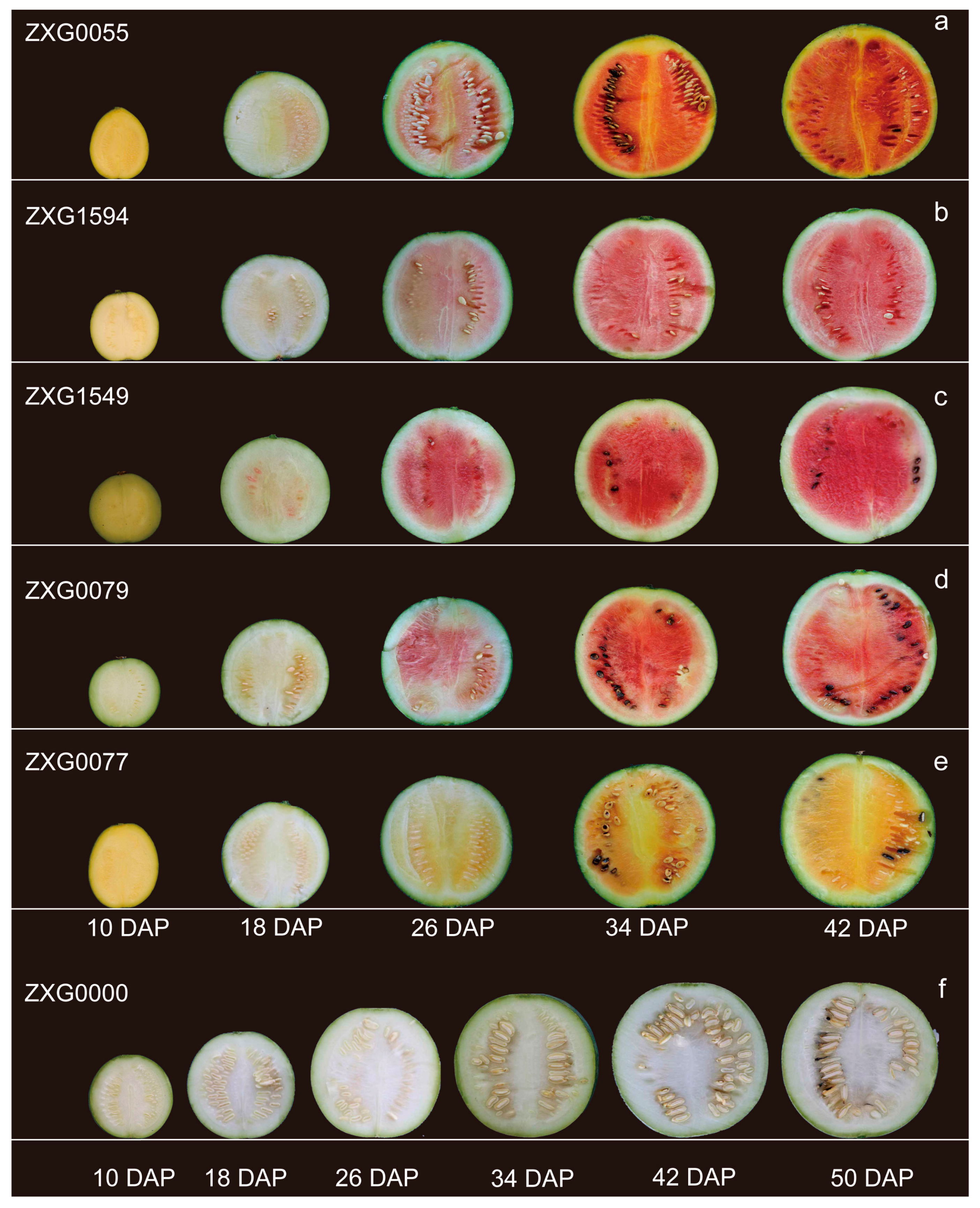
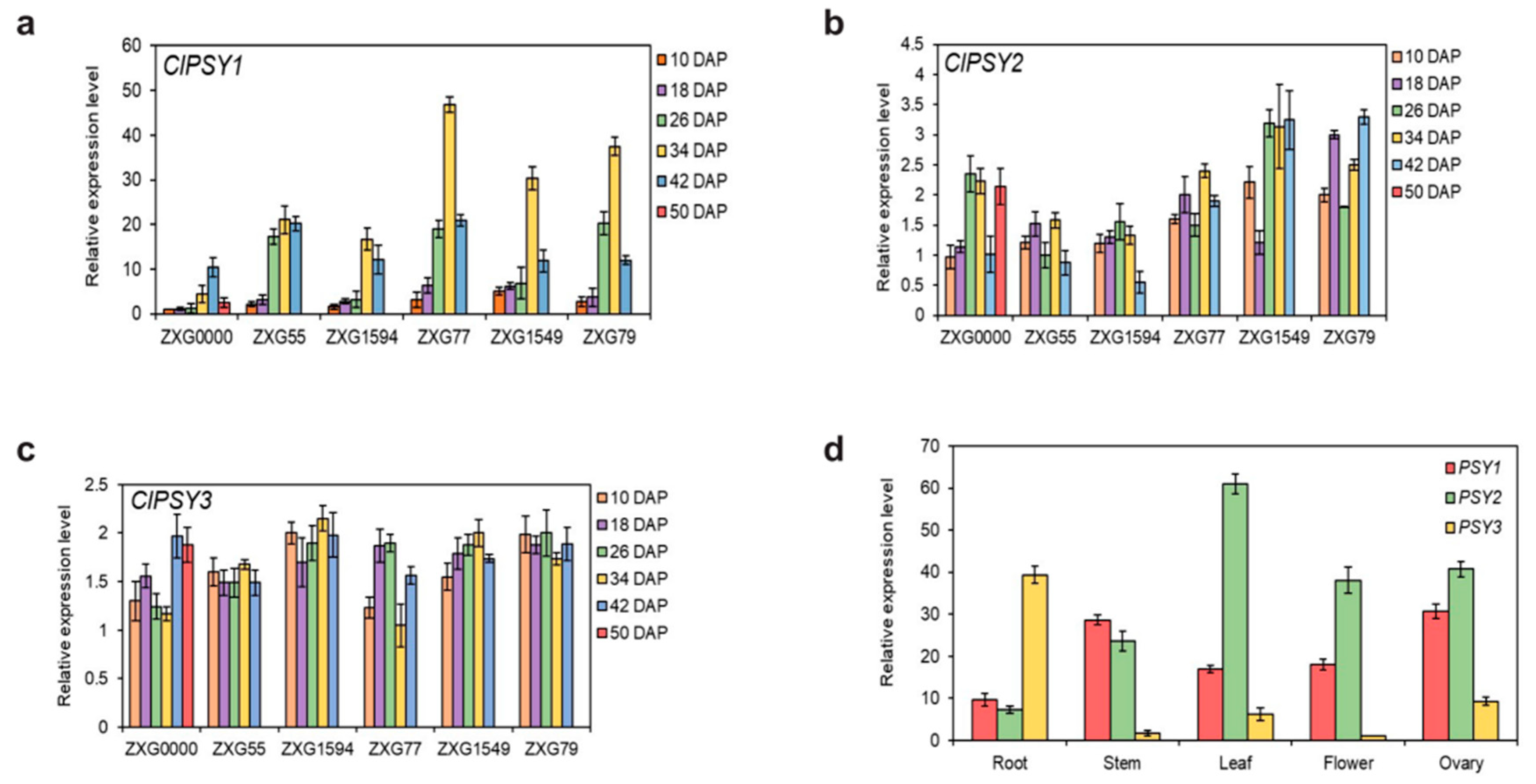
3.1. Sequence Analysis of ClPSY Genes and Genes Expression during Watermelon Flesh Development

Three PSY genes were identified (ClPSY1, ClPSY2 and ClPSY3, Table 1) through the watermelon draft genome from the Cucurbit Genomics Database. We further cloned, characterized and compared the CDS of three ClPSYs and then analyzed the nucleotide and amino acid sequences. As illustrated in Figure 1, ClPSY1, ClPSY2 and ClPSY3 encode polypeptides consisting of 421, 387 and 355 amino acids, respectively, and contain characteristic motifs, including a putative phytoene synthase active site (DXXXD), which is crucial for PSY activity [41]. Protein sequence identity among the three ClPSY homologs ranged from 68 to 75%. At the amino acid level, ClPSY1 showed 75% identity with ClPSY2, ClPSY2 showed 72% identity with ClPSY3, and ClPSY1 showed approximately 68% identity with ClPSY3.
The flesh development and color formation among different watermelon accessions are exhibited in Figure 2. During the early stage (0 to 18 DAP), the six watermelon accessions remain white fleshed; the carotenoids obviously start to generate at 26 DAP, except for the white flesh color accession. From 26 DAP to 42 DAP, the pigments seem to increase constantly until the maturation period. ‘ZXG0000’ (white fleshed) did not change through all the development stages. To explore the relationship between ClPSY genes and flesh color development, the transcript level was analyzed in five or six developmental stages for the six watermelon accessions. For all the three ClPSY genes, ClPSY1 transcripts were much more abundant than ClPSY2 and ClPSY3 transcripts in all varieties through the entire fruit development period (Figure 3a–c). In the first three or four stages, the expression of ClPSY1 was increased continuously and declined in the last stage (50 DAP for ‘ZXG0000’ and 42 DAP for the other five materials) for all the experimental watermelon accessions. The expression of ClPSY1 reached the highest level at 34 DAP for all varieties, except for the white flesh accession ‘ZXG0000’ (reached maximum at 42 DAP) (Figure 3a). ClPSY1 transcripts were highly expressed in red-fleshed ‘ZXG0079’, ‘ZXG1549’ and ‘ZXG0055’, more so than in the pink flesh accession ‘ZXG1594’ when the fruit began to mature and the flesh accumulated the most pigment, implying that the period from 26 to 34 DAP was an important stage for the color formation. Even in the same flesh color group (red group: ‘ZXG0079’, ‘ZXG1549’ and ‘ZXG0055’), the expression levels of ClPSY1 were still different, perhaps due to the accumulation amount of total carotenoid in each stage for different accessions. Interestingly, ClPSY1 transcript levels were a little higher in orange ‘ZXG0077’ than in the red and pink materials, indicating that ‘ZXG0077’ may accumulate more total carotenoids than the other accessions.
For ClPSY2 and ClPSY3, no sensible expression pattern and variation could be detected in all the six accessions, both among different flesh colors and development stages, implying that this gene may not contribute mainly to the flesh color formation (Figure 3b,c). In ClPSY2, the expression pattern could be divided into three groups: two red accessions (‘ZXG1549’ and ‘ZXG0079’) were similar, while ‘ZXG1549’ and ‘ZXG0079’ were expressed more substantially than ‘ZXG0055’ and ‘ZXG1594’. The colorless accession ‘ZXG0000’ performed a medium gene expression, based on our results (Figure 3b).
The expression patterns of three ClPSYs were further analyzed for different organs (leaf, flower, ovary, root and stem) with red-fleshed ‘ZXG0079’ at the mature stage (42 DAP, Figure 3d). All three ClPSYs could express in each organ with different levels. Except in the flesh, the ClPSY1 transcript performed a moderate expressed variation in leaf, flower, ovary and stem, except for the root, with the lowest expression. The transcription patterns of ClPSY1 and ClPSY2 were similar in the stem and ovary but performed significantly differently in the leaf and flower. In particular, the expression of ClPSY2 was markedly higher in the leaf than the ovary and flower, implying that ClPSY2 may be predominantly expressed in the leaf. ClPSY3 may specifically express in roots due to its considerable expression amount and the obviously lower expression of other ClPSYs.

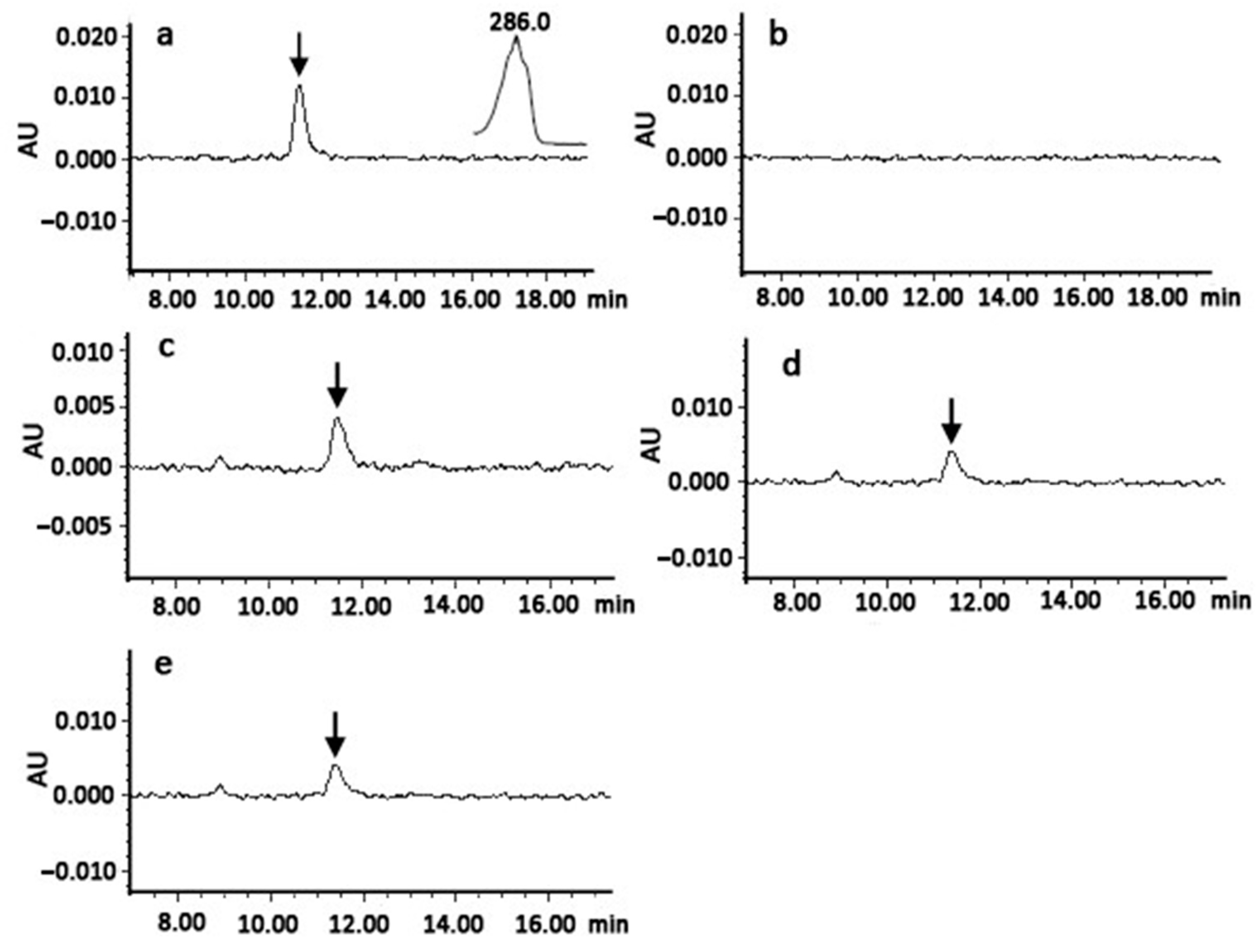
3.2. Functional Analyses of ClPSY Proteins in Watermelon

Standard bacterial complementation method was applied to evaluate catalytic activities of three watermelon ClPSY genes-encoding enzymes. Strain EB, which produced phytoene, was used as a positive control; strain E produced GGPP in bacteria requiring functional ClPSYs to produce phytoene. Plasmids carrying cDNA fragments of the ClPSYs were transformed into strain E. Based on the results of the HPLC analysis, we found peaks with retention time (from 11 to 12 min) and spectral qualities that were the same as the positive control in extracts of cells expressing each ClPSY gene (Figure 4), suggesting that all the ClPSYs encoded enzymes catalyzed the conversion of GGPP to phytoene in the cells. These results indicate that these ClPSYs encoded functional enzymes under these heterologous complementation system conditions.

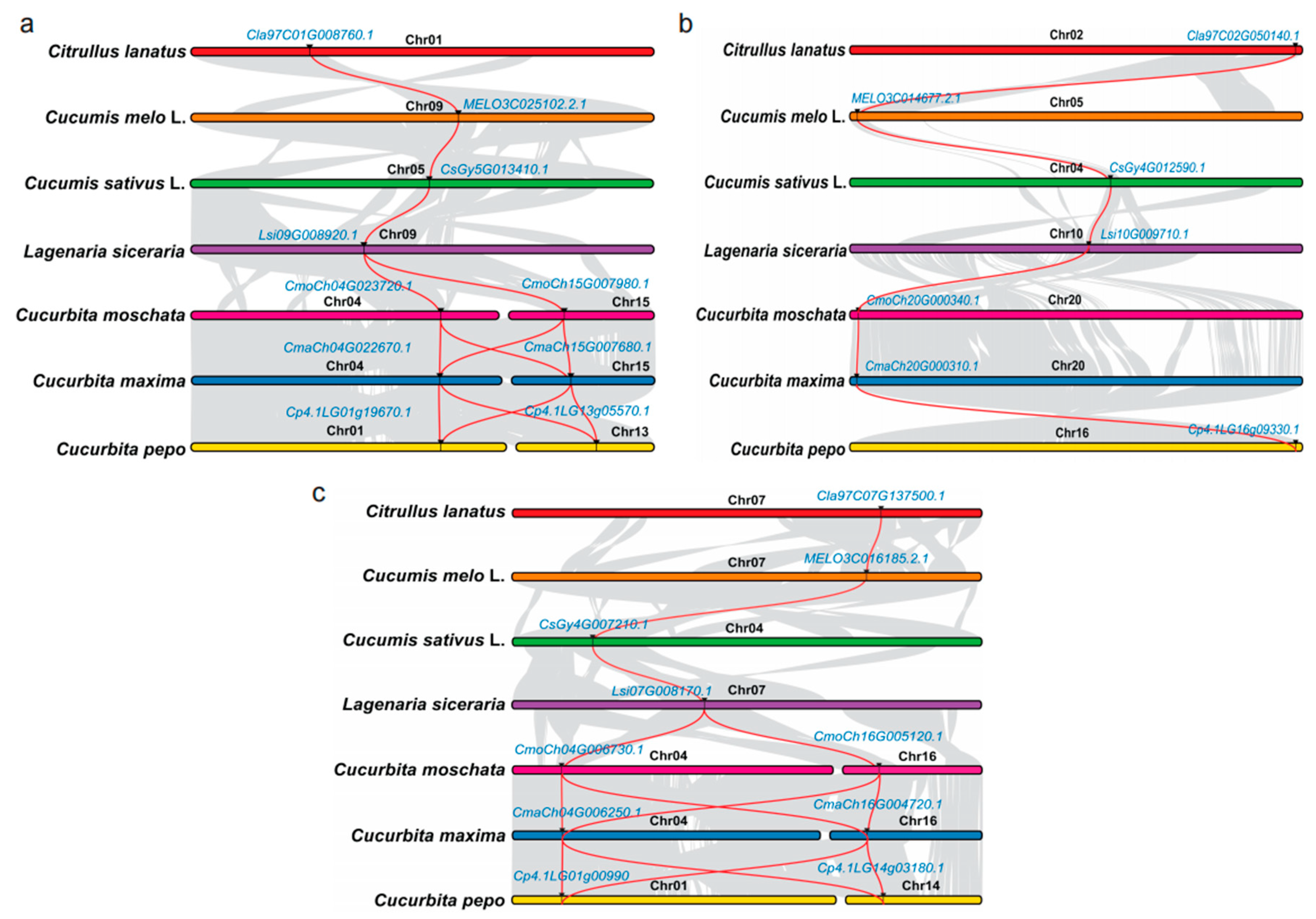
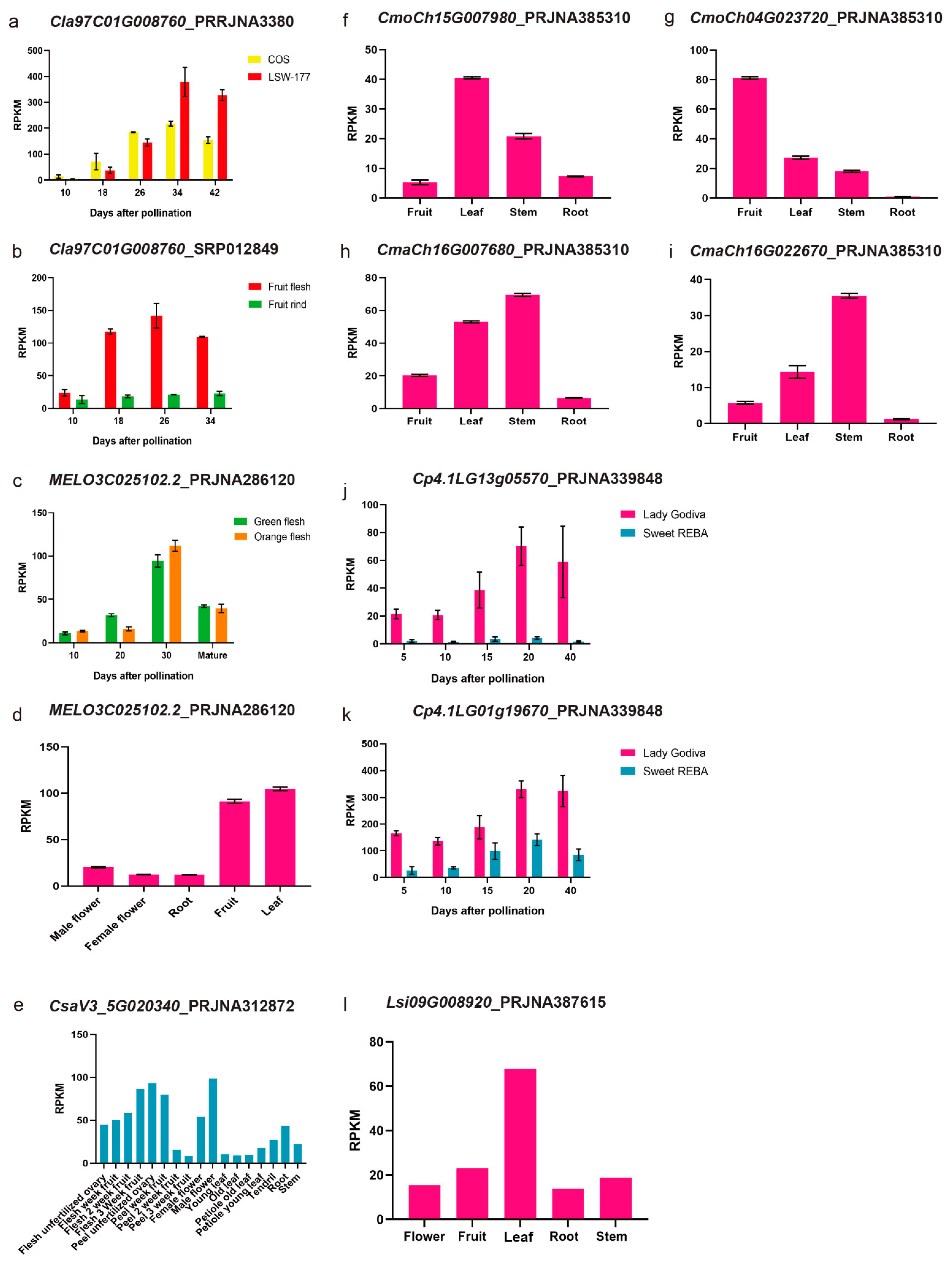
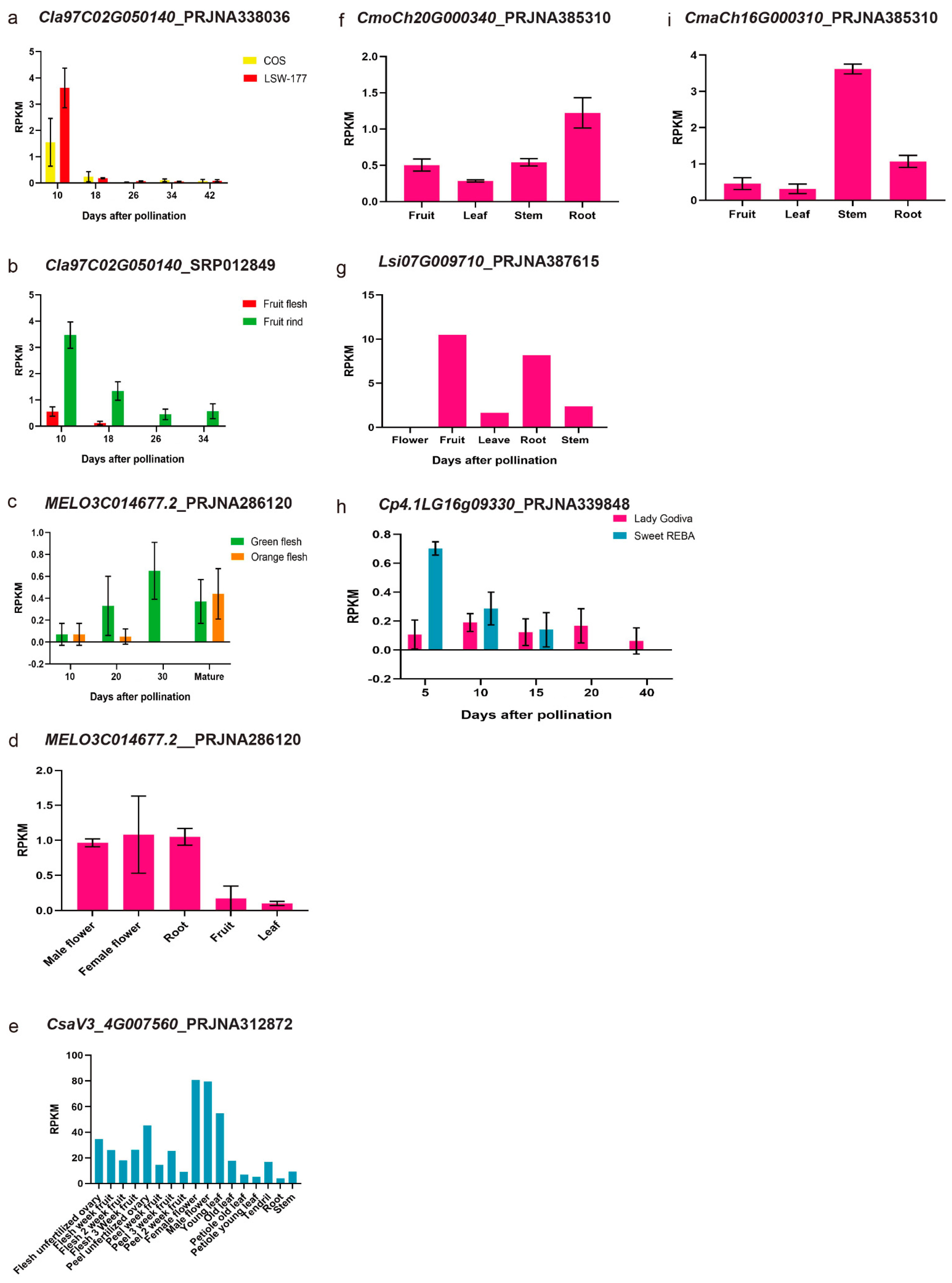
3.3. Synteny Analysis of PSY Members among Different Cucurbit Crops and Their Expression Patterns with the RNA-Seq Data

Three homologous PSYs were detected in melon, cucumber, wild cucumber and bottle gourd, while for the Cucurbita crops, there were five homologous PSYs (Table 3). Compared with the other cucurbit crops, we detected two PSY1 and PSY3 homologous genes through the whole genome only in the three Cucurbita crops. The phylogenetic results indicated three major clades through all the PSYs (Figure 5). For all the cucurbit crops, in each phylogenetic clade, the cucumber (including wild cucumber PI 183967) and melon clustered together, while the watermelon was close to the bottle gourd. PSYs in Cucurbita members were clustered into one sub-branch for each PSY member, and the different copies were also clustered separately for homologous PSY1 and PSY3. We further checked the syntenies of the PSY homologous genes among the cucurbit crops. All the PSY homologous genes were located in the synteny chromosome region (Figure 6).
In order to check the PSYs expression pattern in different cucurbit crops, we used the published RNA-seq data to extend the expressed tendency. Some of the homologous genes clustered together in the phylogenetic tree and located in the synteny chromosome region seemed to have similar expression profiles. For ClPSY1, which we found expressed specifically in the watermelon flesh (Figure 7a,b), the homologous genes in melon (MELO3C025102.2, CmPSY1, Figure 7c,d), cucumber (CsaV3_5G020340, CsPSY1, Figure 7e), Cucurbita moschata var. Rifu (CmoCh04G023720, CmoPSY1-1, Figure 7g) and Cucurbita pepo subsp. pepo (Cp4.1LG01g19670, CpPSY1-1 and Cp4.1LG13g05570, CpPSY1-2, Figure 7j,k) were also mainly expressed in the flesh and increased as the fruit developed, reaching the peak when fruit was mature or near mature. The expression level of ClPSY1 in the flesh was obviously elevated compared with the fruit rind (Figure 7b). In melon, the expression of CmPSY1 was increased with the fruit development until the 30 DAP but did not correspond with the carotenoids accumulation. The orange and green flesh color melon fruits exhibited a similar expression pattern and level. Compared with watermelon, CmPSY1 was expressed in both fruit and leaf (the expression level in leaf was a little higher than the fruit) at the mature stage (Figure 7d). Nonetheless, the gene expression differences were not significant between the green and orange flesh (Figure 7c). In cucumber, the expression of CsPSY1 also exhibited an obvious expression in the male flower and unfertilized ovary peel (Figure 7e). In Cucurbita moschata var. Rifu, only one CmoPSY1 accumulated in abundance at the transcription level in the fruit, while the other one exhibited a relatively high expression level in the leaf (Figure 7f,g). In Cucurbita maxima (Rimu), the two PSY1 homologous genes, CmaCh04G022670 and CmaCh15G007680, showed an abundant expression level in the stem compared to the other tissues. None of the CmaPSY members were specifically expressed in the flesh in Cucurbita maxima (Rimu), based on the published RNA-seq data (Figure 7h,i). In the bottle ground, Lsi09G008920 (LsiPSY1) was highly expressed in the leaf, which may due to the low carotenoid accumulation in its flesh (Figure 7l).
For ClPSY2 homologous genes: MELO3C014677.2 (CmPSY2, Figure 8c,d), CmoCh04G000340 (CmoPSY2, Figure 8f), CmaCh04G000310 (CmaPSY2, Figure 8i), Cp4.1LG16g09330 (CpPSY2, Figure 8h), Lsi07G009710 (LsiPSY2, Figure 8g), CsaV3_4G023380 (CsPSY2, Figure 8e), the expression pattern was quite different. In melon, CmPSY2 mainly expressed in the male flower, female flower and root, while it was obviously low in the fruit and leaf. CmPSY2, CpPSY2 and CsPSY2 were all expressed in the flesh but did not correspond with the fruit development stages. In the bottle gourd, LsiPSY2 was mainly expressed in the fruit and root. The ClPSY3 homologous genes in melon (Figure 9c,d), cucumber (Figure 9e), Cucurbita maxima (Rimu) (Figure 9i), Cucurbita moschata var. Rifu (Figure 9f,g) and bottle gourd (Figure 9l,m) were expressed mainly in the leaf. CsPSY3 was also mainly expressed in the female and male flower. In the flesh, CmPSY3 also gradually declined from 10 DAP to maturation. CpPSY3-1 and CpPSY3-2 could also be detected in the flesh with a low expression level but did not correspond with the fruit development (Figure 9j,k).

4. Discussion

The accumulation of carotenoids in the watermelon plant and fruit may be a result of the three ClPSYs function composition, and ClPSY1 appears to be most important in the flesh, based on our research result. Members of the PSY family might have different effects or might act coordinately in some processes of the carotenoid metabolism regulation during the growth of watermelon and other plants. We also conducted an experiment investigating ClPSY1 expression at different stages of fruit development in watermelon varieties with different flesh colors, and the results were the same as those obtained in previous works [12,13]. ClPSY1 transcript levels were very low initially and reached a maximum with the progression of fruit ripening in colored watermelon flesh (Figure 3a). High expression of ClPSY1 resulted in a large accumulation of phytoene, which provided plenty of primary ingredients for carotenoids synthesis [29]. These demonstrated that ClPSY1 might perform a crucial function of regulating carotenoid synthesis in the watermelon, consistent with many previous studies. According to the Cucurbita pepo genome and RNA-seq data, we identified five PSY homologous genes and two homologous (CpPSY1-1 and CpPSY1-2, clustered with ClPSY1 in one clade) genes exhibited the same trends as the fruit development, which was consistent with the flesh color formation. The RPKM values of the two genes also exhibited an obvious difference between the low and high carotenoids accumulated in the Cucurbita pepo accessions.
The analysis of the ClPSY gene family expression patterns in different organs was performed to show that the three ClPSY genes have specialized roles in watermelon organs. The three members were found to be expressed in all tested tissues but at significantly different levels in different plant organs. Such organ-dependent expression pattern in PSY has also been found in other plants. Our data revealed that, in watermelon, ClPSY2 might regulate the leaf, ovary and flower development, and ClPSY3 is preferentially expressed in the root. This can also be reported in tomato [9], citrus [10] and cassava [4]. For other cucurbit crops, the expression of PSY members could also be detected in different plant organs, but the main tissues were quite different among the crops. In cucurbita crops, two copies of ClPSY1 and ClPSY2 homologous genes could be detected from the genome data. The two copies of each member always showed the same expression pattern. Interestingly, in Cucurbita moschata var. Rifu, the two members of the ClPSY1 homologous genes exhibited different tissue specificity expression patterns. CmoCh15G007980 was in the leaf and CmoCh04G023720 was the in fruit.
Based on the previous publications, the genetic relationships of watermelon and bottle ground were close to each other, and the melon was close to the cucumber [42,43]. In our results, the phylogenetic tree also showed a similar trend, but the gene function did not correspond with the genetic relationship. Genes (or QTLs) located in the collinear chromosome regions always exhibited the same gene function, such as CmCLV3, CsCLV3, and some QTLs were related with the fruit shape and seed size in cucurbit crops [44,45,46]. In our results, the PSY members showed a high collinear relation among all the cucurbit crops, but the function may not have been the same according to the published RNA-seq data. In the high carotenoid accumulation crops, such as watermelon, melon, Cucurbita pepo and Cucurbita maxima (Rimu), the ClPSY1 showed a similar expression pattern and gene function.

5. Conclusions

ClPSY1 is the key gene controlling carotene accumulation in the flesh, while ClPSY2 might regulate the leaf, ovary and flower development, and ClPSY3 is preferentially expressed in the watermelon root. The three ClPSY members in the watermelon could encode active enzymes. Some of the homologous genes clustered together in the phylogenetic tree and located in the synteny chromosome region seemed to have similar expression profiles among different cucurbit crops.

Author Contributions

Conceptualization, S.L.; Formal analysis, X.F.; Investigation, X.F.; Methodology, S.L.; Resources, P.G.; Supervision, F.L.; Validation, X.F.; Visualization, P.G.; Writing—Original draft, X.F.; Writing—Review and editing, S.L. All authors have read and agreed to the published version of the manuscript.

Funding

This work was supported by funding from the National Natural Science Foundation of China [grant numbers 32172577], by the China Agriculture Research System of MOF and MARA [grant number CARS-25].

Data Availability Statement

Not applicable.

Acknowledgments

We are grateful to Misawa (Research Institute for Bioresources and Biotechnology, Ishikawa Prefectural University, Japan) for kindly providing the plasmids pACCRT-E and pACCRT-EB. We thank the National Mid-Term Genbank for providing the watermelon and melon accessions (Zhengzhou Fruit Research Institute, Chinese Academy of Agricultural Sciences, Zhengzhou, China) used in this study.

Conflicts of Interest

The authors declare no conflict of interest.

References

  1. Yuan, H.; Zhang, J.; Nageswaran, D.; Li, L. Carotenoid Metabolism and Regulation in Horticultural Crops. Hortic. Res. 2015, 2, 15036. [Google Scholar] [CrossRef] [PubMed] [Green Version]
  2. Rodriguez-Concepcion, M.; Avalos, J.; Bonet, M.L.; Boronat, A.; Gomez-Gomez, L.; Hornero-Mendez, D.; Limon, M.C.; Meléndez-Martínez, A.J.; Olmedilla-Alonso, B.; Palou, A.; et al. A Global Perspective on Carotenoids: Metabolism, Biotechnology, and Benefits for Nutrition and Health. Prog. Lipid Res. 2018, 70, 62–93. [Google Scholar] [CrossRef] [PubMed] [Green Version]
  3. Cazzonelli, C.I.; Pogson, B.J. Source to Sink: Regulation of Carotenoid Biosynthesis in Plants. Trends Plant Sci. 2010, 15, 266–274. [Google Scholar] [CrossRef] [PubMed]
  4. Welsch, R.; Arango, J.; Bär, C.; Salazar, B.; Al-Babili, S.; Beltrán, J.; Chavarriaga, P.; Ceballos, H.; Tohme, J.; Beyera, P. Provitamin a Accumulation in Cassava (Manihot esculenta) Roots Driven by a Single Nucleotide Polymorphism in a Phytoene Synthase Gene. Plant Cell 2010, 22, 3348–3356. [Google Scholar] [CrossRef] [PubMed] [Green Version]
  5. Giuliano, G. Provitamin A Biofortification of Crop Plants: A Gold Rush with Many Miners. Curr. Opin. Biotechnol. 2017, 44, 169–180. [Google Scholar] [CrossRef] [PubMed]
  6. Fujii, S.; Kobayashi, K.; Nagata, N.; Masuda, T.; Wada, H. Digalactosyldiacylglycerol Is Essential for Organization of the Membrane Structure in Etioplasts. Plant Physiol. 2018, 177, 1487–1497. [Google Scholar] [CrossRef]
  7. Obrero, A.; González-Verdejo, C.I.; Román, B.; Gómez, P.; Die, J.V.; Ampomah-Dwamena, C. Identification, Cloning, and Expression Analysis of Three Phytoene Synthase Genes from Cucurbita Pepo. Biol. Plant 2015, 59, 201–210. [Google Scholar] [CrossRef]
  8. Ampomah-Dwamena, C.; Driedonks, N.; Lewis, D.; Shumskaya, M.; Chen, X.; Wurtzel, E.T.; Espley, R.V.; Allan, A.C. The Phytoene Synthase Gene Family of Apple (Malus x Domestica) and Its Role in Controlling Fruit Carotenoid Content. BMC Plant Biol. 2015, 15, 185. [Google Scholar] [CrossRef] [Green Version]
  9. Fantini, E.; Falcone, G.; Frusciante, S.; Giliberto, L.; Giuliano, G. Dissection of Tomato Lycopene Biosynthesis through Virus-Induced Gene Silencing. Plant Physiol. 2013, 163, 986–998. [Google Scholar] [CrossRef]
  10. Peng, G.; Wang, C.; Song, S.; Fu, X.; Azam, M.; Grierson, D.; Xu, C. The Role of 1-Deoxy-d-Xylulose-5-Phosphate Synthase and Phytoene Synthase Gene Family in Citrus Carotenoid Accumulation. Plant Physiol. Biochem. 2013, 71, 67–76. [Google Scholar] [CrossRef]
  11. Dahan-Meir, T.; Filler-Hayut, S.; Melamed-Bessudo, C.; Bocobza, S.; Czosnek, H.; Aharoni, A.; Levy, A.A. Efficient in Planta Gene Targeting in Tomato Using Geminiviral Replicons and the CRISPR/Cas9 System. Plant J. 2018, 95, 5–16. [Google Scholar] [CrossRef] [PubMed] [Green Version]
  12. Lv, P.; Li, N.; Liu, H.; Gu, H.; Zhao, W.E. Changes in Carotenoid Profiles and in the Expression Pattern of the Genes in Carotenoid Metabolisms during Fruit Development and Ripening in Four Watermelon Cultivars. Food Chem. 2015, 174, 52–59. [Google Scholar] [CrossRef] [PubMed]
  13. Wang, N.; Liu, S.; Gao, P.; Luan, F.; Davis, A.R. Developmental Changes in Gene Expression Drive Accumulation of Lycopene and β-Carotene in Watermelon. J. Am. Soc. Hortic. Sci. 2016, 141, 434–443. [Google Scholar] [CrossRef] [Green Version]
  14. Van Wijk, K.J.; Kessler, F. Plastoglobuli: Plastid Microcompartments with Integrated Functions in Metabolism, Plastid Developmental Transitions, and Environmental Adaptation. Annu. Rev. Plant Biol. 2017, 68, 253–289. [Google Scholar] [CrossRef] [PubMed]
  15. Shumskaya, M.; Bradbury, L.M.T.; Monaco, R.R.; Wurtzela, E.T. Plastid Localization of the Key Carotenoid Enzyme Phytoene Synthase Is Altered by Isozyme, Allelic Variation, and Activity. Plant Cell 2012, 24, 3725–3741. [Google Scholar] [CrossRef] [Green Version]
  16. Lätari, K.; Wüst, F.; Hübner, M.; Schaub, P.; Beisel, K.G.; Matsubara, S.; Beyer, P.; Welsch, R. Tissue-Specific Apocarotenoid Glycosylation Contributes to Carotenoid Homeostasis in Arabidopsis Leaves. Plant Physiol. 2015, 168, 1550–1562. [Google Scholar] [CrossRef]
  17. Martel, C.; Vrebalov, J.; Tafelmeyer, P.; Giovannoni, J.J. The Tomato MADS-Box Transcription Factor RIPENING INHIBITOR Interacts with Promoters Involved in Numerous Ripening Processes in a COLORLESS NONRIPENING-Dependent Manner. Plant Physiol. 2011, 157, 1568–1579. [Google Scholar] [CrossRef] [Green Version]
  18. Xiong, C.; Luo, D.; Lin, A.; Zhang, C.; Shan, L.; He, P.; Li, B.; Zhang, Q.; Hua, B.; Yuan, Z.; et al. A Tomato B-Box Protein SlBBX20 Modulates Carotenoid Biosynthesis by Directly Activating PHYTOENE SYNTHASE 1, and Is Targeted for 26S Proteasome-Mediated Degradation. New Phytol. 2019, 221, 279–294. [Google Scholar] [CrossRef] [Green Version]
  19. Ampomah-Dwamena, C.; Thrimawithana, A.H.; Dejnoprat, S.; Lewis, D.; Espley, R.V.; Allan, A.C. A Kiwifruit (Actinidia deliciosa) R2R3-MYB Transcription Factor Modulates Chlorophyll and Carotenoid Accumulation. New Phytol. 2019, 221, 309–325. [Google Scholar] [CrossRef] [Green Version]
  20. Lu, S.; Zhang, Y.; Zhu, K.; Yang, W.; Ye, J.; Chai, L.; Xu, Q.; Deng, X. The Citrus Transcription Factor CsMADS6 Modulates Carotenoid Metabolism by Directly Regulating Carotenogenic Genes. Plant Physiol. 2018, 176, 2657–2676. [Google Scholar] [CrossRef] [Green Version]
  21. Enfissi, E.M.A.; Nogueira, M.; Bramley, P.M.; Fraser, P.D. The Regulation of Carotenoid Formation in Tomato Fruit. Plant J. 2017, 89, 774–788. [Google Scholar] [CrossRef] [PubMed] [Green Version]
  22. Arango, J.; Jourdan, M.; Geoffriau, E.; Beyer, P.; Welsch, R. Carotene Hydroxylase Activity Determines the Levels of Both α-Carotene and Total Carotenoids in Orange Carrots. Plant Cell 2014, 26, 2223–2233. [Google Scholar] [CrossRef] [PubMed] [Green Version]
  23. Zhou, X.; Welsch, R.; Yang, Y.; Álvarez, D.; Riediger, M.; Yuan, H.; Fish, T.; Liu, J.; Thannhauser, T.W.; Li, L. Arabidopsis OR Proteins Are the Major Posttranscriptional Regulators of Phytoene Synthase in Controlling Carotenoid Biosynthesis. Proc. Natl. Acad. Sci. USA 2015, 112, 3558–3563. [Google Scholar] [CrossRef] [PubMed] [Green Version]
  24. Park, S.; Kim, H.S.; Jung, Y.J.; Kim, S.H.; Ji, C.Y.; Wang, Z.; Jeong, J.C.; Lee, H.S.; Lee, S.Y.; Kwak, S.S. Orange Protein Has a Role in Phytoene Synthase Stabilization in Sweetpotato. Sci. Rep. 2016, 6, 33563. [Google Scholar] [CrossRef] [PubMed]
  25. Bou-Torrent, J.; Toledo-Ortiz, G.; Ortiz-Alcaide, M.; Cifuentes-Esquivel, N.; Halliday, K.J.; Martinez-García, J.F.; Rodriguez-Concepcion, M. Regulation of Carotenoid Biosynthesis by Shade Relies on Specific Subsets of Antagonistic Transcription Factors and Cofactors. Plant Physiol. 2015, 169, 1584–1594. [Google Scholar] [CrossRef]
  26. Tzuri, G.; Zhou, X.; Chayut, N.; Yuan, H.; Portnoy, V.; Meir, A.; Sa’Ar, U.; Baumkoler, F.; Mazourek, M.; Lewinsohn, E.; et al. A “golden” SNP in CmOr Governs the Fruit Flesh Color of Melon (Cucumis melo). Plant J. 2015, 82, 267–279. [Google Scholar] [CrossRef]
  27. Chayut, N.; Yuan, H.; Ohali, S.; Meir, A.; Sa’ar, U.; Tzuri, G.; Zheng, Y.; Mazourek, M.; Gepstein, S.; Zhou, X.; et al. Distinct Mechanisms of the ORANGE Protein in Controlling Carotenoid Flux. Plant Physiol. 2017, 173, 376–389. [Google Scholar] [CrossRef]
  28. Welsch, R.; Zhou, X.; Yuan, H.; Álvarez, D.; Sun, T.; Schlossarek, D.; Yang, Y.; Shen, G.; Zhang, H.; Rodriguez-Concepcion, M.; et al. Clp Protease and OR Directly Control the Proteostasis of Phytoene Synthase, the Crucial Enzyme for Carotenoid Biosynthesis in Arabidopsis. Mol. Plant 2018, 11, 149–162. [Google Scholar] [CrossRef] [Green Version]
  29. Fang, X.; Liu, S.; Gao, P.; Liu, H.; Wang, X.; Luan, F.; Zhang, Q.; Dai, Z. Expression of ClPAP and ClPSY1 in Watermelon Correlates with Chromoplast Differentiation, Carotenoid Accumulation, and Flesh Color Formation. Sci. Hortic. (Amst.) 2020, 270, 109437. [Google Scholar] [CrossRef]
  30. Kong, Q.; Yuan, J.; Gao, L.; Zhao, S.; Jiang, W.; Huang, Y.; Bie, Z. Identification of Suitable Reference Genes for Gene Expression Normalization in QRT-PCR Analysis in Watermelon. PLoS ONE 2014, 9, e90612. [Google Scholar] [CrossRef]
  31. Livak, K.J.; Schmittgen, T.D. Analysis of relative Gene Expression Data Using Real-Time Quantitative PCR and the 2−ΔΔCT Method. Methods 2001, 25, 402–408. [Google Scholar] [CrossRef] [PubMed]
  32. Garcia-Mas, J.; Benjak, A.; Sanseverino, W.; Bourgeois, M.; Mir, G.; Gonźalez, V.M.; Heńaff, E.; Camara, F.; Cozzuto, L.; Lowy, E.; et al. The Genome of Melon (Cucumis melo L.). Proc. Natl. Acad. Sci. USA 2012, 109, 11872–11877. [Google Scholar] [CrossRef] [PubMed] [Green Version]
  33. Li, Q.; Li, H.; Huang, W.; Xu, Y.; Zhou, Q.; Wang, S.; Ruan, J.; Huang, S.; Zhang, Z. A Chromosome-Scale Genome Assembly of Cucumber (Cucumis sativus L.). Gigascience 2019, 8, giz072. [Google Scholar] [CrossRef] [PubMed] [Green Version]
  34. Qi, J.; Liu, X.; Shen, D.; Miao, H.; Xie, B.; Li, X.; Zeng, P.; Wang, S.; Shang, Y.; Gu, X.; et al. A Genomic Variation Map Provides Insights into the Genetic Basis of Cucumber Domestication and Diversity. Nat. Genet. 2013, 45, 1510–1515. [Google Scholar] [CrossRef] [PubMed]
  35. Sun, H.; Wu, S.; Zhang, G.; Jiao, C.; Guo, S.; Ren, Y.; Zhang, J.; Zhang, H.; Gong, G.; Jia, Z.; et al. Karyotype Stability and Unbiased Fractionation in the Paleo-Allotetraploid Cucurbita Genomes. Mol. Plant 2017, 10, 1293–1306. [Google Scholar] [CrossRef] [Green Version]
  36. Montero-Pau, J.; Blanca, J.; Bombarely, A.; Ziarsolo, P.; Esteras, C.; Martí-Gómez, C.; Ferriol, M.; Gómez, P.; Jamilena, M.; Mueller, L.; et al. De Novo Assembly of the Zucchini Genome Reveals a Whole-Genome Duplication Associated with the Origin of the Cucurbita Genus. Plant Biotechnol. J. 2018, 16, 1161–1171. [Google Scholar] [CrossRef] [Green Version]
  37. Wu, S.; Shamimuzzaman, M.; Sun, H.; Salse, J.; Sui, X.; Wilder, A.; Wu, Z.; Levi, A.; Xu, Y.; Ling, K.S.; et al. The Bottle Gourd Genome Provides Insights into Cucurbitaceae Evolution and Facilitates Mapping of a Papaya Ring-Spot Virus Resistance Locus. Plant J. 2017, 92, 963–975. [Google Scholar] [CrossRef] [Green Version]
  38. Barrera-Redondo, J.; Ibarra-Laclette, E.; Vázquez-Lobo, A.; Gutiérrez-Guerrero, Y.T.; Sánchez de la Vega, G.; Piñero, D.; Montes-Hernández, S.; Lira-Saade, R.; Eguiarte, L.E. The Genome of Cucurbita Argyrosperma (Silver-Seed Gourd) Reveals Faster Rates of Protein-Coding Gene and Long Noncoding RNA Turnover and Neofunctionalization within Cucurbita. Mol. Plant 2019, 12, 506–520. [Google Scholar] [CrossRef] [Green Version]
  39. Capella-Gutiérrez, S.; Silla-Martínez, J.M.; Gabaldón, T. TrimAl: A Tool for Automated Alignment Trimming in Large-Scale Phylogenetic Analyses. Bioinformatics 2009, 25, 1972–1973. [Google Scholar] [CrossRef]
  40. Kumar, S.; Stecher, G.; Li, M.; Knyaz, C.; Tamura, K. MEGA X: Molecular Evolutionary Genetics Analysis across Computing Platforms. Mol. Biol. Evol. 2018, 35, 1547–1549. [Google Scholar] [CrossRef]
  41. Marchler-Bauer, A.; Lu, S.; Anderson, J.B.; Chitsaz, F.; Derbyshire, M.K.; DeWeese-Scott, C.; Fong, J.H.; Geer, L.Y.; Geer, R.C.; Gonzales, N.R.; et al. CDD: A Conserved Domain Database for the Functional Annotation of Proteins. Nucleic Acids Res. 2011, 39, 225–229. [Google Scholar] [CrossRef] [PubMed] [Green Version]
  42. Sebastian, P.; Schaefer, H.; Telford, I.R.H.; Renner, S.S. Cucumber (Cucumis sativus) and Melon (C. melo) Have Numerous Wild Relatives in Asia and Australia, and the Sister Species of Melon Is from Australia. Proc. Natl. Acad. Sci. USA 2010, 107, 14269–14273. [Google Scholar] [CrossRef] [PubMed] [Green Version]
  43. Yang, L.; Koo, D.H.; Li, D.; Zhang, T.; Jiang, J.; Luan, F.; Renner, S.S.; Hénaff, E.; Sanseverino, W.; Garcia-Mas, J.; et al. Next-Generation Sequencing, FISH Mapping and Synteny-Based Modeling Reveal Mechanisms of Decreasing Dysploidy in Cucumis. Plant J. 2014, 77, 16–30. [Google Scholar] [CrossRef] [PubMed]
  44. Li, S.; Pan, Y.; Wen, C.; Li, Y.; Liu, X.; Zhang, X.; Behera, T.K.; Xing, G.; Weng, Y. Integrated Analysis in Bi-Parental and Natural Populations Reveals CsCLAVATA3 (CsCLV3) Underlying Carpel Number Variations in Cucumber. Theor. Appl. Genet. 2016, 129, 1007–1022. [Google Scholar] [CrossRef] [PubMed]
  45. Pan, Y.; Wang, Y.; McGregor, C.; Liu, S.; Luan, F.; Gao, M.; Weng, Y. Genetic Architecture of Fruit Size and Shape Variation in Cucurbits: A Comparative Perspective. Theor. Appl. Genet. 2020, 133, 1–21. [Google Scholar] [CrossRef] [PubMed]
  46. Guo, Y.; Gao, M.; Liang, X.; Xu, M.; Liu, X.; Zhang, Y.; Liu, X.; Liu, J.; Gao, Y.; Qu, S.; et al. Quantitative Trait Loci for Seed Size Variation in Cucurbits—A Review. Front. Plant Sci. 2020, 11, 304. [Google Scholar] [CrossRef] [PubMed]
Figure 1. Multiple alignment of watermelon ClPSY genes. The hypothetical active site (DXXXD) is shown in the black box.
Genes 13 01189 g001
Figure 2. Photographs of six watermelon accessions with different flesh colors at five or six time points (10, 18, 26, 34 and 42 or 50 days after pollination) during fruit development. (af): ‘ZXG0055’, ‘ZXG1594’, ‘ZXG1549’, ‘ZXG0079’, ‘ZXG0077’and ‘ZXG0000’, respectively.
Genes 13 01189 g002
Figure 3. Expression of ClPSY members in different watermelon accessions and organs. (ac) Transcription levels of ClPSY1, ClPSY2 and ClPSY3 in six watermelon varieties with different fruit colors at six time points (10, 18, 26, 34, 42, 50 days after pollination) during fruit development. Additionally, the fruit sample ‘ZXG0000’ at 10 DAP was used for calibration. The bars represent the means ± SD (n = 3). (d) Expression patterns of the three ClPSY genes in different organs. Total RNA samples were isolated from various watermelon organs, including 4-week seedling roots, stems, leaves, flowers and ovaries. The y-axis shows the relative expression levels of the three ClPSY genes in different tissues compared to ClPSY3 in flowers. The bars represent the means ± SD (n = 3).
Genes 13 01189 g003
Figure 4. Functional complementation of watermelon ClPSY proteins. (a) E. coli cells harboring EB were used as a positive control. The peak represents phytoene (indicated by an arrow); (b) E+pET (empty vector) was used as a negative control; (c) E+ pET-ClPSY1; (d) E+ pET-ClPSY2; (e) E+ pET-ClPSY3. The peak representing phytoene (indicated by an arrow) was observed in cells expressing ClPSY1, ClPSY2 and ClPSY3.
Genes 13 01189 g004
Figure 5. Phylogenetic tree and gene structure of PSY family proteins from various species retrieved from the Cucurbit Genomics Database (http://www.icugi.org/; accessed on 15 January 2022).
Genes 13 01189 g005
Figure 6. The synteny analysis of PSY members among different cucurbit crops. (ac) for the ClPSY1, ClPSY2 and ClPSY3 homologous genes in different cucurbit crops.
Genes 13 01189 g006
Figure 7. ClPSY1 homologous genes expression pattern analysis with the published RNA-seq data. (al): RPKM of PRRJNA3380, SRP012849, PRJNA286120, PRJNA312872, PRJNA385310, PRJNA339848 and PRJNA387615.
Genes 13 01189 g007
Figure 8. ClPSY2 homologous genes expression pattern analysis with the published RNA-seq data. (ai): RPKM of PRJNA338036, SRP012849, PRJNA286120, PRJNA312872, PRJNA385310, PRJNA387615 and PRJNA339848.
Genes 13 01189 g008
Figure 9. ClPSY3 homologous genes expression pattern analysis with the published RNA-seq data. (am): RPKM of PRJNA338036, SPR012849, PRJNA286120, PRJNA312872, PRJNA385310, PRJNA339848 and PRJNA387615.
Genes 13 01189 g009
Table 1. Description of watermelon PSY family genes and ClYLS8.
GeneGene IDChromosomePositions
ClPSY1Cla97C01G0087601Chr01: 9448426 ~ 9451311 (+)
ClPSY2Cla97C02G0501407Chr07: 25039090 ~ 25042269 (+)
ClPSY3Cla97C07G1375002Chr02: 37419674 ~ 37421665 (−)
ClYLS8Cla97C02G0385902Chr02: 22823405 ~ 22824590 (−)
Table 2. Primers for amplifying the full-length cDNA and primers used for qRT-PCR analysis.
Primer NameAmplicon Size (bp)Primer Sequence (5′-3′)
ClPSY11266F: GGAATTCATGTCTTTTGCTCCTTCGTTGG
R: CCTCGAGAATTCATGAAGGGCCAAG
ClPSY21194F: GGAATTCATGTCTGGTGTGAATGCCAACTCTC
R: CCTCGAGCTATCTTGTTACCAAATCTTT
ClPSY31068F: CGGGATCCATGAGCAAAAGTGGGGTAATT
R: CCTCGAGTCAATGGAAGACTAGACTGGGTGCC
qPSY1179F: TAAGTTTCCAGTTGATATTCAGCCG
R: GTGCTTGCTTGGGAGTCAGG
qPSY2132F: CCTGTCATGGGATTGGCACC
R: CTCTTCCCCTCCTAGCATCTTCTCC
qPSY3118F: GCACCTGATTCTTCACTTCCTACTC
R: ATCCTACCCCTTATAGCATCCTCTC
ClYLS874F: AGAACGGCTTGTGGTCATTC
R: GAGGCCAACACTTCATCCAT
Table 3. PSYs in different Cucurbit family crops.
SpeciesGene IDPosition
Cucumis meloMELO3C025102.2Chr09: 14553133 ~ 14556862 (+)
MELO3C014677.2Chr05: 487434 ~ 489932 (+)
MELO3C016185.2Chr07: 20315779 ~ 20319205 (+)
Cucumis sativusCsaV3_5G020340Chr05: 15246891 ~ 15249568 (+)
CsaV3_4G023380Chr04: 13627834 ~ 13631557 (+)
CsaV3_4G007560Chr04: 5185258 ~ 5189102 (−)
Cucumis sativus var. hardwickiiCSPI05G13040Chr05: 12570152 ~ 12574113 (+)
CSPI04G07340Chr04: 5188671 ~ 5192373 (−)
CSPI04G13030Chr04: 11240846 ~ 11244106 (+)
Lagenaria sicerariaLsi07G008190Chr07: 9123302 ~ 9126554 (−)
Lsi09G008920Chr09: 10525942 ~ 10528939 (+)
Lsi07G008170Chr07: 9114307 ~ 9117400 (−)
Lsi10G009710Chr10: 14062470 ~ 14067420 (+)
Cucurbita maxima var. RimuCmaCh16G004720Chr16: 2390509 ~ 2394475 (+)
CmaCh15G007680Chr15: 3777993 ~ 3782451 (−)
CmaCh04G022670Chr04: 15833307 ~ 15835991 (+)
CmaCh04G006250Chr04: 3203989 ~ 3206323 (−)
CmaCh20G000310Chr20: 140674 ~ 142255 (+)
Cucurbita moschata var. RifuCmoCh04G023720Chr04: 17691476 ~ 17694558 (+)
CmoCh16G005120Chr16: 2472841 ~ 2477006 (+)
CmoCh15G007980Chr15: 3929562 ~ 3934079 (−)
CmoCh04G006730Chr04: 3346882 ~ 3349011 (−)
CmoCh20G000340Chr20: 224511 ~ 225982 (+)
Cucurbita pepo subsp. pepoCp4.1LG14g03180Chr14: 2471728 ~ 2475409 (+)
Cp4.1LG13g05570Chr13: 5416613 ~ 5421307 (+)
Cp4.1LG01g19670Chr01: 16838967 ~ 16841930 (+)
Cp4.1LG01g00990Chr01: 3271567 ~ 3274741 (−)
Cp4.1LG16g09330Chr16: 8547559 ~ 8548969 (−)
Cucurbita argyrospermaCarg18294Scaffold_120: 491768 ~ 494735 (−)
Carg06947Scaffold_065: 466719 ~ 468129 (−)
Carg13119Scaffold_101: 425459 ~ 427786 (+)
Carg01435Scaffold_004: 945560 ~ 949123 (+)
Carg11395Scaffold_025: 739796 ~ 743637 (+)
Publisher’s Note: MDPI stays neutral with regard to jurisdictional claims in published maps and institutional affiliations.

