Article

Peripheral Myelin Protein 22 Gene Mutations in Charcot-Marie-Tooth Disease Type 1E Patients

1
Department of Biological Sciences, Kongju National University, Gongju 32588, Korea
2
Department of Neurology, Samsung Medical Center, Sungkyunkwan University School of Medicine, Seoul 06351, Korea
3
Department of Neurology, Kyung Hee University Hospital at Gangdong, Kyung Hee University School of Medicine, Seoul 05278, Korea
4
Cell & Gene Therapy Institute, Samsung Medical Center, Seoul 06351, Korea
*
Authors to whom correspondence should be addressed.
Genes 2022, 13(7), 1219; https://doi.org/10.3390/genes13071219
Submission received: 2 June 2022 / Revised: 27 June 2022 / Accepted: 4 July 2022 / Published: 8 July 2022
(This article belongs to the Special Issue Genetics and Epigenetics of Neuromuscular Diseases)

Abstract

:
Duplication and deletion of the peripheral myelin protein 22 (PMP22) gene cause Charcot-Marie-Tooth disease type 1A (CMT1A) and hereditary neuropathy with liability to pressure palsies (HNPP), respectively, while point mutations or small insertions and deletions (indels) usually cause CMT type 1E (CMT1E) or HNPP. This study was performed to identify PMP22 mutations and to analyze the genotype–phenotype correlation in Korean CMT families. By the application of whole-exome sequencing (WES) and targeted gene panel sequencing (TS), we identified 14 pathogenic or likely pathogenic PMP22 mutations in 21 families out of 850 CMT families who were negative for 17p12 (PMP22) duplication. Most mutations were located in the well-conserved transmembrane domains. Of these, eight mutations were not reported in other populations. High frequencies of de novo mutations were observed, and the mutation sites of c.68C>G and c.215C>T were suggested as the mutational hotspots. Affected individuals showed an early onset-severe phenotype and late onset-mild phenotype, and more than 40% of the CMT1E patients showed hearing loss. Physical and electrophysiological symptoms of the CMT1E patients were more severely damaged than those of CMT1A while similar to CMT1B caused by MPZ mutations. Our results will be useful for the reference data of Korean CMT1E and the molecular diagnosis of CMT1 with or without hearing loss.

1. Introduction

Charcot-Marie-Tooth disease (CMT), which is the most frequent disorder in the inherited peripheral neuropathies (IPNs), is a genetically and clinically heterogeneous disorder group characterized by distal muscle weakness and sensory loss. CMT is usually categorized into three types mainly according to the electrophysiological properties: demyelinating (CMT1), axonal (CMT2), and intermediate type (Int-CMT). CMT with genetic defects in X-linked genes is exceptionally called CMTX. However, each type is divided into variable subtypes according to the genetic causes and clinical features [1]. In a broad sense, CMT also commonly includes distal hereditary motor neuropathy (dHMN) and hereditary sensory autonomic neuropathy (HSAN).
In the past decade, next-generation sequencing (NGS) technology such as whole-exome sequencing (WES) has far improved the molecular diagnosis of CMT [2]. Mutations in more than 130 genes have been currently reported to cause CMT or CMT-related diseases. Of these, mutations in the peripheral myelin protein 22 (PMP22; MIM 601097) gene, which encodes a major component of myelin in the peripheral nervous system, provide the most frequent genetic causes of CMT. Duplication and deletion of a 1.4 Mbp-length 17p12 region including peripheral myelin protein 22 (PMP22; MIM 601097) cause CMT type 1A (CMT1A; MIM 118220) and hereditary neuropathy with liability to pressure palsies (HNPP; MIM 162500), respectively [3,4]. However, small insertion/deletions (indels) and point mutations in PMP22 are usually associated with CMT1E (MIM 118300) [5,6,7] and HNPP [8]. They have been also reported to cause Dejerine-Sottas syndrome (DSS; MIM 145900) as a severe type of CMT [9,10,11,12,13] and chronic inflammatory demyelinating polyneuropathy (CIDP; MIM 139393) [14]. Some CMT1E patients frequently showed a severe clinical phenotype of wheelchair bounding within the first decade [15]. Most mutations in PMP22 cause IPNs in a dominant manner with a minor exception for recessive inheritance [13,16].
PMP22, as a transmembrane glycoprotein with a four transmembrane (TM) domain, two extracellular (EC) loops, and three intracellular (IC) sequences including N- and C terminals, is mainly expressed in Schwann cells. It consists of 2–5% of the total protein in peripheral compact myelin that provides the electrical insulation of axons and has an essential role in the formation and maintenance of compact myelin [17]. A rat model with PMP22 duplication showed CMT1A-like phenotypes, such as gait abnormalities, Schwann cell hypertrophy, and muscle weakness [18]. Homozygous mouse models with Pmp22 point mutations exhibited a more severe peripheral myelin deficiency compared to heterozygous Pmp22 knockout mice, suggesting gain-of-function or dominant-negative mechanisms of the mutant alleles [19].
This study was performed to identify pathogenic mutations in the PMP22 gene in Korean patients with CMT by WES or targeted gene panel sequencing. As a result, we identified 14 pathogenic or likely pathogenic variants in PMP22 from 21 independent CMT patients and families. Of these, we have previously reported five PMP22 mutations in six Korean IPN families [20,21,22,23,24]. This study also analyzed the clinical phenotypic features and genotype–phenotype correlation for the patients with PMP22 mutations.

2. Materials and Methods

2.1. Patients and Ethics Statements

This study was conducted in a cohort of 1,243 Korean CMT families. By examination of the copy numbers in the 17p12 region, 393 families were determined to have PMP22 duplication. The remaining 850 CMT families were further investigated to detect PMP22 small indels and point mutations. This study was approved by the Institutional Review Boards of Sungkyunkwan University, Samsung Medical Center (2018-05-102-002), and Kongju National University (KNU_IRB_2018-62). All participants were recruited from the Samsung Medical Center (Seoul, Korea) and provided written informed consent according to the Declaration of Helsinki. For minors involved in this study, written consent was provided by their parents.

2.2. Clinical and Electrophysiological Examinations

General clinical and electrophysiological information for affected individuals was obtained using the most recent data by the standard methods described by Park et al. [25]. To measure nerve conductions, the motor and sensory nerve conduction velocities and action potentials of the median, ulnar, peroneal, tibial, and sural nerves were determined. The age at onset was determined as when an individual initially noticed or showed distal dominant sensory-motor impairment. Physical disabilities were measured using several methods: functional disability scale (FDS), the CMT neuropathy score version 2 (CMTNS), Rasch-modified CMTNS (CMTNS-R), CMT examination score (CMTES), and Rasch-modified CMTES (CMTES-R) [26,27,28].

2.3. DNA Purification and Detection of Mutations

Genomic DNA was purified from peripheral blood using the HiGene Genomic DNA Prep Kit (Biofact, Daejeon, Korea). Proband samples with no 17p12 (PMP22) duplication or deletion were examined to find genetic causes by WES or targeted gene panel sequencing. Exome capture was performed using the SureSelect Human All Exon 50M Kit (Agilent Technologies, Santa Clara, CA, USA), and NGS was performed using the HiSeq 2000 or 2500 Genome Analyzer (Illumina, San Diego, CA, USA). Annotation and filtering of single nucleotide variants (SNVs) were performed by the methods of Park et al. with some alteration [25]. As the reference sequence, the UCSC assembly hg19 (GRCh37) was used (http://genome.ucsc.edu/ accessed on 1 May 2022). Rare variant frequencies were obtained from the 1000 Genomes Project (1000G; http://www.1000genomes.org/ accessed on 1 May 2022), the Genome Aggregation Database (gnomAD; https://gnomad.broadinstitute.org/ accessed on 1 May 2022), and the Korean Reference Genome Database (KRGDB; http://coda.nih.go.kr/coda/KRGDB/index.jsp/ accessed on 1 May 2022). Variant nomenclature followed the Human Genome Variation Society (HGVS) recommendations (https://www.hgvs.org/ accessed on 1 May 2022). Pathogenicity of the SNVs was evaluated and categorized into five grades following the American College of Medical Genetics and Genomics (ACMG) and American College of Pathology (AMP) guidelines (https://wintervar.wglab.org/ accessed on 1 May 2022). Pathogenic candidate variants were examined for all available family members by the Sanger sequencing method using the SeqStudio genetic analyzer (Life Technologies-Thermo Fisher Scientific, Foster City, CA, USA).

2.4. In Silico Prediction and Conservation Analysis

In silico analyses were performed to predict the mutation effect using the PROVEAN (http://provean.jcvi.org/ accessed on 15 April 2022), PolyPhen-2 (http://genetics.bwh.harvard.edu/pph2/ accessed on 15 April 2022), and MUpro (http://mupro.proteomics.ics.uci.edu/ accessed on 15 April 2022) programs. Conservation of the mutation sites and their flanking regions was analyzed using MEGA-X ver. 5.05 (http://www.megasoftware.net/ accessed on 15 April 2022). The genomic evolutionary rate profiling (GERP) score was determined by the GERP++ program (http://mendel.stanford.edu/SidowLab/downloads/gerp/ accessed on 15 April 2022). The secondary structure of single-strand DNA was predicted using the mFold algorithm (http://www.unafold.org accessed on 15 April 2022). Three-dimensional (3D) structures of wild-type and mutant PMP22 proteins were simulated by the I-TASSER program (https://zhanglab.ccmb.med.umich.edu/I-TASSER). The 3D structures were visualized using the Mol* feature of the Protein Data Bank (http://www.rcsb.org/ accessed on 15 April 2022).

2.5. Statistical Analysis

A non-parametric Kruskal–Wallis test for one-way ANOVA was used to compare the clinical characteristics of patients grouped according to the three types of CMT1 with different genetic causes. Missing clinical values were excluded from the analysis. Non-parametric Spearman r values were used to analyze the correlation between severity and age of onset. Differences were considered significant at p < 0.05. All statistical analyses and calculations were performed using GraphPad Prism ver. 8.00 (GraphPad Software, San Diego, CA, USA).

3. Results

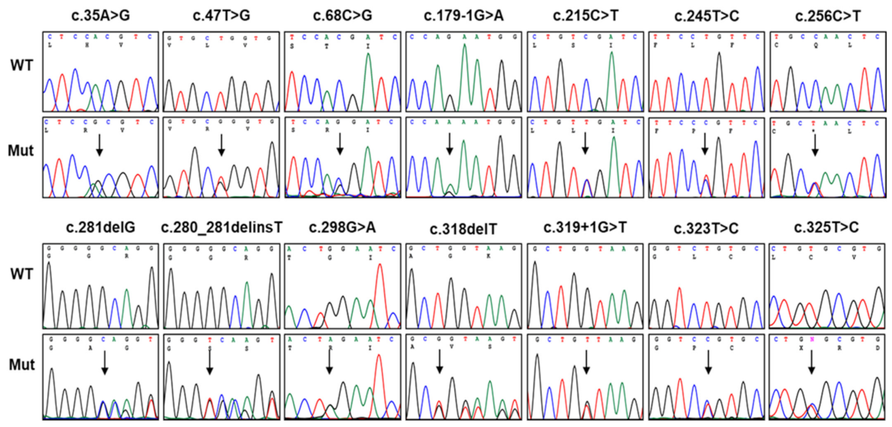
3.1. Identification of Pathogenic Mutations in PMP22

We identified 14 dominant pathogenic or likely pathogenic PMP22 mutations in 21 CMT families which included 72 participants (32 affected and 40 unaffected members) (Figure 1 and Table 1). All these mutations were unreported in the KRGDB and global databases of the 1000G, genomAD, and ESP. The identified mutations were confirmed for all involved family members by Sanger sequencing (Figure 2), and the genotypes for each mutation are provided below the examined individuals in Figure 1. Of these, four mutations (c.35A>G, c.179-1G>A, c.280_281delinsT, and c.319+1G>T) were novel, and another four mutations (c.47T>G, c.245T>C, c.318delT, and c.325T>C) were previously reported by our research group [20,21,22,23,24] but not reported in other population studies. De novo events were presumed in six mutations (c.35A>G, c.215C>T, c.245T>C, c.281delG, c.298G>A, and c.325T>C) involving nine families. For the families with de novo mutations, paternity was confirmed for the corresponding families with the PowerPlex Fusion System (Promega, Wisconsin-Madison, CA, USA).
The c.35A>G (p.H12R), as a novel mutation, was identified in a CMT1 male patient (FC618). His unaffected parents and younger sister did not have the same mutation, presumptively indicating a de novo event. The c.47T>G (p.L16R), which was once reported by Nam et al. [22], was found in a sporadic CMT1 male (FC541). His parents were considered to be unaffected by history taking, but genetic testing was not done. As a mutation of the same amino acid site, a p.L16P was reported in CMT1E with proptosis [29] and Dejerine-Sottas neuropathy patients [12]. The c.68C>G (p.T23R) mutation was found in three CMT1 families (FC50, FC303, and FC680). The FC50 and FC680 families showed an autosomal dominant inheritance, but the patient in the FC303 family was a sporadic case with both parents putatively unaffected (no genetic testing). This mutation has been reported twice including a Korean CMT1 case with deafness [7,30]. A novel c.179-1G>A splicing site mutation was found in a CMT1E family (FC970). In the same splicing acceptor site mutation, c.179-1G>C was reported in an HNPP family [31]. The c.215C>T (p.S72L) mutation was interestingly identified in five CMT1 families (FC285, FC376, FC732, FC829, and FC895). Moreover, four families (FC285, FC376, FC732, and FC829) showed a de novo event which was confirmed by the genetic testing of the father-mother-patient trios. This mutation has been reported many times as the underlying cause of CMT1 and DSS [10,11,21,23,24,32]. Two families (FC285 and FC829) were previously reported by our study group [21,23,24]. The c.245T>C (p.L82P), which was once reported by our study group [23], was identified in a CMT1 female patient (FC416). Her unaffected parents and elder brother did not have the same mutation, suggesting a de novo mutation. The c.256C>T (p.Q86X) stopgain mutation was identified in two CMT1 families (FC1102 and FC1325). In FC1102, the proband’s mother with the same mutation was also affected, while the proband in FC1325 was a sporadic case with unaffected parents (history taking). This mutation was reported twice by Numakura et al. and Liao et al. [33,34]. The c.281delG (p.G94Afs*16) frameshift mutation was identified in a CMT1 family (FC1061). It has been reported twice as an underlying cause of CMT1E [12,35]. The c.280_281delinsT (p.G94Sfs*16) mutation was identified in a sporadic female case with CMT1 (FC1088). It was an unreported novel mutation. The c.298G>A (p.G100R) was identified in a CMT1 female patient (FC1140). Her unaffected parents and elder brother did not have the same mutation, suggesting a de novo mutation. It was reported once as a pathogenic mutation of CMT1 [36]. The c.318delT (p.G107Vfs*3), which was reported once by our study group [20], was identified in a CMT1 family (FC35). His elder brother with the same mutation was also affected. Two affected brothers presumably inherited the mutation from their deceased affected father. Two mutations of c.319+1G>T and c.323T>C (p.L108P) were found in a sporadic CMT1 case, respectively. The c.319+1G>T splicing site mutation was unreported while a c.323T>C mutation was reported once as an underlying cause of Roussy–Levy syndrome (MIM 180800) which is considered to be a type of CMT1 [37]. The c.325T>C (p.C109R) mutation, which was reported by our study group [21,24], was identified in a CMT1 female patient (FC284). Her unaffected parents and younger brother did not have the same mutation, suggesting a de novo mutation.

3.2. In Silico Prediction, Conservation, and Conformational Changes

In silico analyses using the PolyPhen-2, PROVEAN, and MUpro programs predicted the pathogenicity for most of the missense mutations (Table S1). The GERP scores were also calculated and were high values larger than 3.3 at all the missense mutation sites. Amino acids in most of the mutation sites and their flanking regions were highly conserved among vertebrate species from fish to mammals (Figure 3A). Except for two p.G94 frameshift mutations (p.G94Afs*16 and p.G94Sfs*16) positioning in the IC domain, the other mutations were located in the TM domains that are very important for the configuration of the protein interacting with the plasma membrane (Figure 3B).
The TM domains consisting of four α-helices are spatially associated with each other within the plasma membrane (Figure 4A). With the exception of p.T23, all missense mutations are positioned in the α-helices located in the TM domains. For the eight PMP22 mutant proteins with missense mutations, 3D conformational changes were predicted by the I-TASSER program (Figure 4B–I). It was suggested that the substitution of amino acids causes changes in the hydrogen bonds, affinity, or distance between the neighboring helixes. The conformational changes in the transmembrane regions could affect the functions and topology within the membrane.

3.3. Mutational Hotspots and an Atypical Case with Somatic Mutation

The c.68C>G and c.215C>T mutations that were located at the CpG sequences were particularly found in multiple families, suggesting mutational hotspots. Moreover, four families (FC285, FC376, FC732, and FC829) with the c.215C>T showed a de novo event. These mutations have been also reported several times in other studies [7,10,11,30,32]. These frequent mutations may be partly due to the putative palindromic sequences of the corresponding genomic DNA region forming a stem-loop hairpin (Figure 3C) and methylation in the CpG sites.
As an atypical case, the c.281delG (p.G94Afs*16) mutation identified in the FC1061 female patient (III-1) was also found in her father (II-1) but with low mutational loads: rates of 0.19, 0.23, and 0.32 in blood, oral swap, and hair root, respectively. Thus, the results suggest a somatic mutation in an early embryonic stage implicating genetic mosaicism. The proband showed a very severe clinical symptom with a very early infant onset (<1 year) and wheelchair bounding, while her father was clinically and electrophysiologically normal virtually in a recent checkup.

3.4. Prevalence of CMT1E in CMT Patients

The frequencies of CMT1E with PMP22 mutations was calculated as 1.69% in the total examined CMT probands (n = 1243) and 2.47% in the CMT probands (n = 850) excluding CMT1A (Table 2). When the prevalence of total CMT patients was compared with other countries, it was roughly similar with China at 0.94–2.38% [38,39], Italy at 1.36% [40], and Brazil at 2.10% [41] while it was higher than other examined populations, such as in Hungary at 0.37% [42] and Spain at 0.46% [43]. The prevalence of CMT1E in Japanese CMT patients excluding CMT1A ranged from 0.81–1.29%, which is a considerably low frequency compared with that of Korean CMT patients [44,45].

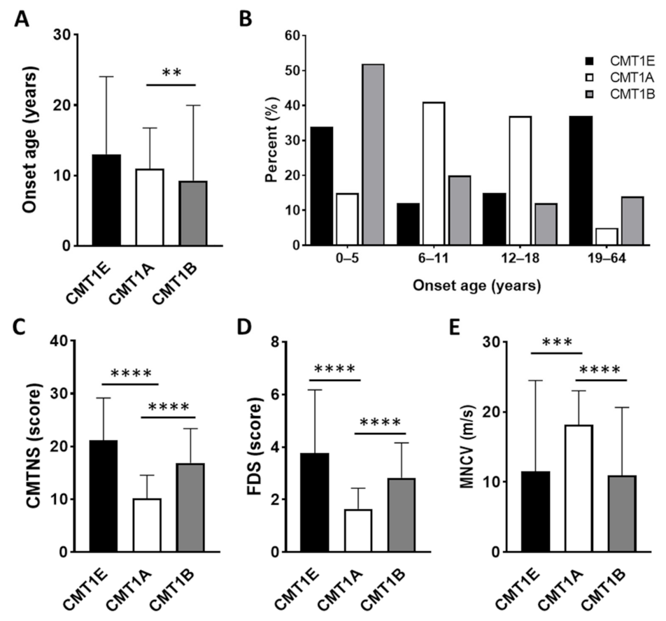
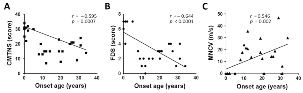
3.5. Characterization of Clinical and Electrophysiological Phenotypes

We analyzed the clinical and electrophysiological phenotypes of the CMT1E patients with PMP22 mutations. Major clinical features are provided in Table 3. Detailed clinical and electrophysiological data are provided in Tables S2 and S3, respectively. Mean onset ages for all the examined CMT1E patients were measured to be 12.8 ± 11.1 years. As the physical disability values, mean CMTNS, CMT-E, CMTES, CMTES-R, and FDS scores were measured as 21.1 ± 8.1, 25.8 ± 9.0, 16.2 ± 5.6, 19.7 ± 6.7, and 3.8 ± 2.4, respectively. Mean median motor nerve conduction velocity (MNCV) was measured to be 11.5 ± 13.0 m/s in CMT1E patients. Most patients showed foot deformity (84.4%), and hearing loss was diagnosed in 40.6% of the patients (13 of 32).
The mean onset age of the CMT1E patients showed no significant difference with those of CMT1A with PMP22 duplication (10.9 ± 5.8) [46] and CMT1B with MPZ mutations (9.3 ± 10.7) (Figure 5A) [46,47]. However, the distribution of onset ages showed a noticeable difference between the CMT1 groups. Infancy onset (<1 year) was only observed in CMT1E with a high frequency of 19.4% (6/32) while the adult-onset (≥19 years) frequency of 37.5% (12/32) in CMT1E was higher than those of CMT1A (5.6%) and CMT1B (14.6%). The CMT1E group roughly showed a U-shaped distribution compared to the bell-shaped distribution of CMT1A and the L-shaped distribution of CMT1B (Figure 5B).
The mean CMTNS score (21.1 ± 8.1) was significantly higher than that of CMT1A (10.2 ± 4.3, p < 0.0001) while it was not significantly different with that of CMT1B (16.9 ± 6.5, p = 0.408) (Figure 5C). For the FDS, the mean value of CMT1E (3.8 ± 2.4) was also significantly higher than that of CMT1A (1.6 ± 0.8, p < 0.0001), but it was similar with that of CMT1B (2.8 ± 1.3, p = 0.999) (Figure 5D). The mean median MNCV of CMT1E (11.5 ± 13.0 m/s) was significantly slower than that of CMT1A (18.2 ± 4.9 m/s, p = 0.0006) or similar with that of CMT1B (10.9 ± 9.7 m/s, p = 0.999) (Figure 5E) [46,47].
As an important feature of the clinical phenotypes in CMT1E patients, a clear correlation between early onset with severe symptoms and late onset with mild symptoms was observed (Figure 6). That is, the physical disability scores measured by CMTNS and FDS showed clearly negative correlations by increasing onset ages (CMTNS: r = −0.595, p = 0.0007; FDS: r = −0.644, p < 0.0001) (Figure 6A,B). The median MNCV, as a major electrophysiological value, showed a positive correlation by increasing onset ages (r = 0.546, p = 0.002) (Figure 6C).

4. Discussion

We identified 14 pathogenic or likely pathogenic point mutations in PMP22 from 21 CMT families or sporadic cases in a Korean CMT cohort genetic study. Of these, four mutations (c.35A>G, c.179-1G>A, c.280_281delinsT, and c.319+1G>T) were unreported novel variants. Another four mutations (c.47T>G, c.245T>C, c.318delT, and c.325T>C) were reported by our research group [20,21,22,23,24] but not reported in other populations. Most mutations were located in the well-conserved TM domains and predicted to be pathogenic at least by one or more in silico analyses. All the mutations were evaluated as pathogenic or likely pathogenic by the ACMG/AMP guidelines.
The prevalence of CMT1E patients with PMP22 mutations was determined to be 1.69% in the total independent patients and 2.47% in the CMT patients excluding CMT1A. This CMT1E prevalence corresponds to the third most common subtype of demyelinated CMT1 after CMT1A with PMP22 duplication and CMT1B with MPZ mutations [47]. The prevalence of CMT1E showed a difference according to the countries studied, but there appears to be no difference by continent. The prevalence of CMT1E in Korean CMT patients was similar to that of Chinese patients [38,39], but it was significantly higher than that of Japanese CMT patients without CMT1A [44,45]. When the prevalence was compared with European populations, it was clearly higher than those of Hungarians [42] and Spaniards [43]. However, it was slightly higher than that of Italians. In Brazilians, the frequency in total patients was similar or slightly higher compared with East Asians, but 7.41% in patients excluding CMT1A is the highest value worldwide [41].
Of note, high frequencies of de novo mutations were observed at a rate of 42.9% in the examined families. The rate increases to over 60% when counting father-mother-child trio families only. Although PMP22 de novo mutations have been frequently reported by other studies [9,15], the rate was higher than those shown in the other CMT gene mutations in Korea compared to the 45.0% and 18.7% of the trio families with the MPZ mutations and PMP22 duplication causing CMT1A [47], respectively. Only two and one de novo cases were observed in 11 families with small heat shock protein (sHSP) gene mutations [48] and 13 families with aminoacyl tRNA-synthetase (ARS) gene mutations [49]. In particular, c.215C>T (p.S72L) de novo mutation, which was observed in 4 of 5 families with the corresponding mutation, is suggested to locate in a mutational hotspot. The c.215C nucleotide is positioned in the single-stranded DNA region forming a stem-loop hairpin. In addition, the c.215C was reported as a strong methylation site in the UCSC Genome Browser (http://genome.ucsc.edu/ accessed on 1 May 2022).
As an important feature of the clinical phenotypes, the CMT1E patients showed a clear correlation between early onset with severe symptoms and late onset with mild symptoms. This aspect is similar to the clinical features of CMT2A patients with MFN2 mutations [50]. More than 40% of CMT1E patients showed hearing loss, which is consistent with previous studies [5,6,7,30]. Regarding onset, more than 1/3 of all CMT1E patients examined in this study showed early onset at the age of 5 years or younger, whereas the rate of patients with late onset after 19 years old was also high with more than 37% which roughly showed a dimorphic distribution. This distribution showed a clear difference from the CMT1A patients with 15% and 5% and CMT1B patients with 52% and 15% of the approximate ratio of early onset and late onset. However, the mean onset age of the CMT1E patients was similar to those of CMT1A and CMT1B [46,47]. When the physical and electrophysiological severities of CMT1E were compared with those of CMT1A and CMT1B in terms of CMTNS, FDS, and MNCV, CMT1E was significantly more severe than CMT1A while showing similar symptoms with CMT1B.
More than 100 genes are associated with CMT in variable inheriting modes. Among them, PMP22 defects are found in more than one-third of CMT patients, and the genomic dosage of PMP22 is often the subject of prenatal diagnosis including the preimplantation genetic diagnosis [51]. In particular, the inhibitory effects of PMP22 overexpression by treatment of ascorbic acid, progesterone antagonist, and siRNA have suggested personalized therapeutic strategies by genetic causes [52,53,54]. Therefore, data for PMP22 mutations could be applied to the early diagnosis and treatment of CMT patients.

5. Conclusions

This genetic cohort study identified 14 PMP22 mutations in 21 Korean families as the underlying causes of CMT1E phenotypes. Of these, eight mutations were not reported in other countries. We carefully analyzed the genotype–phenotype correlations and compared the clinical phenotypes with other frequent CMT1 types of CMT1A and CMT1B. This study particularly provided detailed physical and electrophysiological data for all the examined patients. We believe that our results will be useful for the reference data of Koreans and the molecular diagnosis of CMT1 with or without deafness.

Supplementary Materials

The following supporting information can be downloaded at https://www.mdpi.com/article/10.3390/genes13071219/s1. Table S1: In silico prediction of PMP22 mutations; Table S2: Clinical features of Charcot-Marie-Tooth type 1E patients with mutations in PMP22 gene; Table S3: Electrophysiological values of Charcot-Marie-Tooth type 1E patients with mutations in PMP22 gene.

Author Contributions

Conceptualization, B.-O.C. and K.W.C.; data curation, H.M.K., A.J.L. and S.B.K.; formal analysis, H.M.K. and D.E.N.; funding acquisition, K.W.C.; investigation, N.Y.J., D.E.N., N.T. and A.J.L.; resources, S.B.K.; writing—original draft, N.Y.J. and K.W.C.; writing—review and editing, B.-O.C. All authors have read and agreed to the published version of the manuscript.

Funding

This research was supported by grants from the National Research Foundation (2019R1A2C1087547, 2020M3H4A1A03084600 and 2021R1A4A2001389) and the Korean Health Technology R&D Project, Ministry of Health and Welfare (HI14C3484 and HI20C0039), Republic of Korea.

Institutional Review Board Statement

This study was approved by the Institutional Review Boards of Kongju National University (KNU_IRB_2018-62; 5 July 2018) and Sungkyunkwan University, Samsung Medical Center (2018-05-102-002; 26 June 2018).

Informed Consent Statement

All participants were recruited from the Samsung Medical Center (Seoul, Korea) and provided written informed consent according to the Declaration of Helsinki. For minors under the age of 18 years, written consent was provided by their parents.

Data Availability Statement

The data presented in this study are available in the article. Additional WES and TS data are available upon request to the corresponding authors.

Acknowledgments

We appreciate all the participants of the CMT patients and their family members for providing consent, samples, and clinical information.

Conflicts of Interest

The authors declare no conflict of interest.

References

  1. Pareyson, D.; Scaioli, V.; Laurà, M. Clinical and electrophysiological aspects of Charcot-Marie-Tooth disease. Neuromolecular Med. 2006, 8, 3–22. [Google Scholar] [CrossRef]
  2. Pipis, M.; Rossor, A.M.; Laura, M.; Reilly, M.M. Next-generation sequencing in Charcot-Marie-Tooth disease: Opportunities and challenges. Nat. Rev. Neurol. 2019, 15, 644–656. [Google Scholar] [CrossRef] [PubMed]
  3. Lupski, J.R.; Montes de Oca-Luna, R.; Slaugenhaupt, S.; Pentao, L.; Guzzetta, V.; Trask, B.J.; Saucedo-Cardenas, O.; Barker, D.F.; Killian, J.M.; Garcia, C.A.; et al. DNA duplication associated with Charcot-Marie-Tooth disease type 1A. Cell 1991, 66, 219–232. [Google Scholar] [CrossRef]
  4. Chance, P.F.; Alderson, M.K.; Leppig, K.A.; Lensch, M.W.; Matsunami, N.; Smith, B.; Swanson, P.D.; Odelberg, S.J.; Disteche, C.M.; Bird, T.D. DNA deletion associated with hereditary neuropathy with liability to pressure palsies. Cell 1993, 72, 143–151. [Google Scholar] [CrossRef]
  5. Boerkoel, C.F.; Takashima, H.; Garcia, C.A.; Olney, R.K.; Johnson, J.; Berry, K.; Russo, P.; Kennedy, S.; Teebi, A.S.; Scavina, M.; et al. Charcot-Marie-Tooth disease and related neuropathies: Mutation distribution and genotype-phenotype correlation. Ann. Neurol. 2002, 51, 190–201. [Google Scholar] [CrossRef] [PubMed]
  6. Kovach, M.J.; Lin, J.-P.; Boyadjiev, S.; Campbell, K.; Mazzeo, L.; Herman, K.; Rimer, L.A.; Frank, W.; Llewellyn, B.; Jabs, E.W.; et al. A unique point mutation in the PMP22 gene is associated with Charcot-Marie-Tooth disease and deafness. Am. J. Hum. Genet. 1999, 64, 1580–1593. [Google Scholar] [CrossRef] [Green Version]
  7. Joo, I.S.; Ki, C.S.; Joo, S.Y.; Huh, K.; Kim, J.W. A novel point mutation in PMP22 gene associated with a familial case of Charcot-Marie-Tooth disease type 1A with sensorineural deafness. Neuromuscul. Disord. 2004, 14, 325–328. [Google Scholar] [CrossRef]
  8. Kleopa, K.A.; Georgiou, D.-M.; Nicolaou, P.; Koutsou, P.; Papathanasiou, E.; Kyriakides, T.; Christodoulou, K. A novel PMP22 mutation ser22phe in a family with hereditary neuropathy with liability to pressure palsies and CMT1A phenotypes. Neurogenetics 2004, 5, 171–175. [Google Scholar] [CrossRef]
  9. Valentijn, L.J.; Ouvrier, R.A.; van den Bosch, N.H.A.; Bolhuis, P.A.; Baas, F.; Nicholson, G.A. Dejerine-Sottas neuropathy is associated with a de novo PMP22 mutation. Hum. Mutat. 1995, 5, 76–80. [Google Scholar] [CrossRef]
  10. Roa, B.B.; Dyck, P.J.; Marks, H.G.; Chance, P.F.; Lupski, J.R. Dejerine-Sottas syndrome associated with point mutation in the peripheral myelin protein 22 (PMP22) gene. Nat. Genet. 1993, 5, 269–273. [Google Scholar] [CrossRef]
  11. Ionasescu, V.V.; Searby, C.; Greenberg, S.A. Dejerine-Sottas disease with sensorineural hearing loss, nystagmus, and peripheral facial nerve weakness: De novo dominant point mutation of the PMP22 gene. J. Med. Genet. 1996, 33, 1048–1049. [Google Scholar] [CrossRef] [PubMed] [Green Version]
  12. Ionasescu, V.V.; Searby, C.C.; Ionasescu, R.; Chatkupt, S.; Patel, N.; Koenigsberger, R. Dejerine-Sottas neuropathy in mother and son with same point mutation of PMP22 gene. Muscle Nerve 1997, 20, 97–99. [Google Scholar] [CrossRef]
  13. Parman, Y.; Plante-Bordeneuve, V.; Guiochon-Mantel, A.; Eraksoy, M.; Said, G. Recessive inheritance of a new point mutation of the PMP22 gene in Dejerine-Sottas disease. Ann. Neurol. 1999, 45, 518–522. [Google Scholar] [CrossRef]
  14. Korn-Lubetzki, I.; Argov, Z.; Raas-Rothschild, A.; Wirguin, I.; Steiner, I. Family with inflammatory demyelinating polyneuropathy and the HNPP 17p12 deletion. Am. J. Med. Genet. 2002, 113, 275–278. [Google Scholar] [CrossRef] [PubMed]
  15. Li, J.; Parker, B.; Martyn, C.; Natarajan, C.; Guo, J. The PMP22 gene and its related diseases. Mol. Neurobiol. 2013, 47, 673––698. [Google Scholar] [CrossRef] [Green Version]
  16. Roa, B.B.; Garcia, C.A.; Pentao, L.; Killian, J.M.; Trask, B.J.; Suter, U.; Snipes, G.J.; Ortiz-Lopez, R.; Shooter, E.M.; Patel, P.I.; et al. Evidence for a recessive PMP22 point mutation in Charcot-Marie-Tooth disease type 1A. Nat. Genet. 1993, 5, 189–194. [Google Scholar] [CrossRef] [PubMed]
  17. Watila, M.M.; Balarabe, S.A. Molecular and clinical features of inherited neuropathies due to PMP22 duplication. J. Neurol. Sci. 2015, 355, 18–24. [Google Scholar] [CrossRef]
  18. Sereda, M.; Griffiths, I.; Puhlhofer, A.; Stewart, H.; Rossner, M.J.; Zimmermann, F.; Magyar, J.P.; Schneider, A.; Hund, E.; Meinck, H.M.; et al. A transgenic rat model of Charcot-Marie-Tooth disease. Neuron 1996, 16, 1049–1060. [Google Scholar] [CrossRef] [Green Version]
  19. Tobler, A.R.; Liu, N.; Mueller, L.; Shooter, E.M. Differential aggregation of the Trembler and Trembler J mutants of peripheral myelin protein 22. Proc. Nat. Acad. Sci. USA 2002, 99, 483–488. [Google Scholar] [CrossRef] [Green Version]
  20. Choi, B.O.; Lee, M.S.; Shin, S.H.; Hwang, J.H.; Choi, K.G.; Kim, W.K.; Sunwoo, I.N.; Kim, N.K.; Chung, K.W. Mutational analysis of PMP22, MPZ, GJB1, EGR2 and NEFL in Korean Charcot-Marie-Tooth neuropathy patients. Hum. Mutat. 2004, 24, 185–186. [Google Scholar] [CrossRef]
  21. Choi, B.O.; Koo, S.K.; Park, M.H.; Rhee, H.; Yang, S.J.; Choi, K.G.; Jung, S.C.; Kim, H.S.; Hyun, Y.S.; Nakhro, K.; et al. Exome sequencing is an efficient tool for genetic screening of Charcot-Marie-Tooth disease. Hum. Mutat. 2012, 33, 1610–1615. [Google Scholar] [CrossRef] [PubMed]
  22. Nam, S.H.; Hong, Y.B.; Hyun, Y.S.; Nam, D.E.; Kwak, G.; Hwang, S.H.; Choi, B.O.; Chung, K.W. Identification of genetic causes of inherited peripheral neuropathies by targeted gene panel sequencing. Mol. Cells 2016, 39, 382–388. [Google Scholar] [PubMed] [Green Version]
  23. Kim, J.Y.; Koo, H.; Park, K.D.; Choi, S.S.; Yu, J.S.; Hong, Y.B.; Chung, K.W.; Choi, B.O. Genotype–phenotype correlation of Charcot-Marie-Tooth type 1E patients with PMP22 mutations. Genes Genom. 2016, 38, 659–667. [Google Scholar] [CrossRef]
  24. Kim, J.Y.; Kim, S.H.; Park, J.Y.; Koo, H.; Park, K.D.; Hong, Y.B.; Chung, K.W.; Chot, B.O. A longitudinal clinicopathological study of two unrelated patients with Charcot-Marie-Tooth disease type 1E. Neurol. India 2017, 65, 893–895. [Google Scholar] [PubMed]
  25. Park, J.; Kim, H.S.; Kwon, H.M.; Kim, J.; Nam, S.H.; Jung, N.Y.; Lee, A.J.; Jung, Y.H.; Kim, S.B.; Chung, K.W.; et al. Identification and clinical characterization of Charcot-Marie-Tooth disease type 1C patients with LITAF p.G112S mutation. Genes Genom. 2022, in press. [Google Scholar] [CrossRef]
  26. Murphy, S.M.; Herrmann, D.N.; McDermott, M.P.; Scherer, S.S.; Shy, M.E.; Reilly, M.M.; Pareyson, D. Reliability of the CMT neuropathy score (second version) in Charcot-Marie-Tooth disease. J. Peripher. Nerv. Syst. 2011, 16, 191–198. [Google Scholar] [CrossRef] [Green Version]
  27. Sadjadi, R.; Reilly, M.M.; Shy, M.E.; Pareyson, D.; Laura, M.; Murphy, S.; Feely, S.M.; Grider, T.; Bacon, C.; Piscosquito, G.; et al. Psychometrics evaluation of Charcot-Marie-Tooth Neuropathy Score (CMTNSv2) second version, using Rasch analysis. J. Peripher. Nerv. Syst. 2014, 19, 192–196. [Google Scholar] [CrossRef] [Green Version]
  28. Fridman, V.; Sillau, S.; Acsadi, G.; Bacon, C.; Dooley, K.; Burns, J.; Day, J.; Feely, S.; Finkel, R.S.; Grider, T.; et al. Inherited neuropathies consortium-rare diseases clinical research, a longitudinal study of CMT1A using Rasch analysis based CMT neuropathy and examination scores. Neurology 2020, 94, e884–e896. [Google Scholar] [CrossRef]
  29. Calia, L.; Marques, W., Jr.; Gouvea, S.P.; Lourenço, C.M.; de Oliveira, A.S. Proptosis in a family with the p16 Leuc-to-Prol mutation in the PMP22 gene (CMT 1E). Arq. Neuropsiquiatr. 2013, 71, 332––333. [Google Scholar] [CrossRef] [Green Version]
  30. Luigetti, M.; Zollino, M.; Conti, G.; Romano, A.; Sabatelli, M. Inherited neuropathies and deafness caused by a PMP22 point mutation: A case report and a review of the literature. Neurol. Sci. 2013, 34, 1705–1707. [Google Scholar] [CrossRef]
  31. Meuleman, J.; Pou-Serradell, A.; Löfgren, A.; Ceuterick, C.; Martin, J.J.; Timmerman, V.; Van Broeckhoven, C.; De Jonghe, P. A novel 3’-splice site mutation in peripheral myelin protein 22 causing hereditary neuropathy with liability to pressure palsies. Neuromuscul. Disord. 2001, 11, 400–403. [Google Scholar] [CrossRef]
  32. Simonati, A.; Fabrizi, G.M.; Pasquinelli, A.; Taioli, F.; Cavallaro, T.; Morbin, M.; Marcon, G.; Papini, M.; Rizzuto, N. Congenital hypomyelination neuropathy with Ser72Leu substitution in PMP22. Neuromuscul. Disord. 1999, 9, 257–261. [Google Scholar] [CrossRef]
  33. Numakura, C.; Lin, C.; Ikegami, T.; Guldberg, P.; Hayasaka, K. Molecular analysis in Japanese patients with Charcot-Marie-Tooth disease: DGGE analysis for PMP22, MPZ, and Cx32/GJB1 mutations. Hum. Mutat. 2002, 20, 392–398. [Google Scholar] [CrossRef] [PubMed]
  34. Liao, Y.C.; Tsai, P.C.; Lin, T.S.; Hsiao, C.T.; Chao, N.C.; Lin, K.P.; Lee, Y.C. Clinical and molecular characterization of PMP22 point mutations in Taiwanese patients with inherited neuropathy. Sci. Rep. 2017, 7, 15363. [Google Scholar] [CrossRef] [Green Version]
  35. Takashima, H.; Boerkoel, C.F.; Lupski, J.R. Screening for mutations in a genetically heterogeneous disorder: DHPLC versus DNA sequence for mutation detection in multiple genes causing Charcot-Marie-Tooth neuropathy. Genet. Med. 2001, 3, 335–342. [Google Scholar] [CrossRef] [Green Version]
  36. Bort, S.; Nelis, E.; Timmerman, V.; Sevilla, T.; Cruz-Martínez, A.; Martínez, F.; Millán, J.M.; Arpa, J.; Vílchez, J.J.; Prieto, F.; et al. Mutational analysis of the MPZ, PMP22 and Cx32 genes in patients of Spanish ancestry with Charcot-Marie-Tooth disease and hereditary neuropathy with liability to pressure palsies. Hum. Genet. 1997, 99, 746–754. [Google Scholar] [CrossRef]
  37. Zubair, S.; Holland, N.R.; Beson, B.; Parke, J.T.; Prodan, C.I. A novel point mutation in the PMP22 gene in a family with Roussy-Levy syndrome. J Neurol 2008, 255, 1417–1418. [Google Scholar] [CrossRef]
  38. Hsu, Y.H.; Lin, K.P.; Guo, Y.C.; Tsai, Y.S.; Liao, Y.C.; Lee, Y.C. Mutation spectrum of Charcot-Marie-Tooth disease among the Han Chinese in Taiwan. Ann. Clin. Transl. Neurol. 2019, 6, 1090–1101. [Google Scholar] [CrossRef] [Green Version]
  39. Xie, Y.; Lin, Z.; Liu, L.; Li, X.; Huang, S.; Zhao, H.; Wang, B.; Zeng, S.; Cao, W.; Li, L.; et al. Genotype and phenotype distribution of 435 patients with Charcot-Marie-Tooth disease from central south China. Eur. J. Neurol. 2021, 28, 3774–3783. [Google Scholar] [CrossRef]
  40. Gentile, L.; Russo, M.; Fabrizi, G.M.; Taioli, F.; Ferrarini, M.; Testi, S.; Alfonzo, A.; Aguennouz, M.; Toscano, A.; Vita, G.; et al. Charcot-Marie-Tooth disease: Experience from a large Italian tertiary neuromuscular center. Neurol. Sci. 2020, 41, 1239–1243. [Google Scholar] [CrossRef]
  41. Uchôa Cavalcanti, E.B.; Santos, S.C.L.; Martins, C.E.S.; de Carvalho, D.R.; Rizzo, I.M.P.O.; Freitas, M.C.D.N.B.; da Silva Freitas, D.; de Souza, F.S.; Junior, A.M.; do Nascimento, O.J.M. Charcot-Marie-Tooth disease: Genetic profile of patients from a large Brazilian neuromuscular reference center. J. Peripher. Nerv. Syst. 2021, 26, 290–297. [Google Scholar] [CrossRef] [PubMed]
  42. Milley, G.M.; Varga, E.T.; Grosz, Z.; Nemes, C.; Arányi, Z.; Boczán, J.; Diószeghy, P.; Molnár, M.J.; Gál, A. Genotypic and phenotypic spectrum of the most common causative genes of Charcot-Marie-Tooth disease in Hungarian patients. Neuromuscul. Disord. 2018, 28, 38–43. [Google Scholar] [CrossRef] [PubMed]
  43. Sivera, R.; Sevilla, T.; Vílchez, J.J.; Martínez-Rubio, D.; Chumillas, M.J.; Vázquez, J.F.; Muelas, N.; Bataller, L.; Millán, J.M.; Palau, F.; et al. Charcot-Marie-Tooth disease: Genetic and clinical spectrum in a Spanish clinical series. Neurology 2013, 81, 1617–1625. [Google Scholar] [CrossRef] [PubMed] [Green Version]
  44. Yoshimura, A.; Yuan, J.H.; Hashiguchi, A.; Ando, M.; Higuchi, Y.; Nakamura, T.; Okamoto, Y.; Nakagawa, M.; Takashima, H. Genetic profile and onset features of 1005 patients with Charcot-Marie-Tooth disease in Japan. J. Neurol. Neurosurg. Psychiatry 2019, 90, 195–202. [Google Scholar] [CrossRef] [PubMed] [Green Version]
  45. Higuchi, Y.; Takashima, H. Clinical genetics of Charcot-Marie-Tooth disease. J. Hum. Genet. 2022, in press. [CrossRef]
  46. Lee, A.J.; Nam, D.E.; Choi, Y.J.; Noh, S.W.; Nam, S.H.; Lee, H.J.; Kim, S.J.; Song, G.J.; Choi, B.O.; Chung, K.W. Paternal gender specificity and mild phenotypes in Charcot-Marie-Tooth type 1A patients with de novo 17p12 rearrangements. Mol. Genet. Genom. Med. 2020, 8, e1380. [Google Scholar] [CrossRef]
  47. Kim, H.J.; Nam, S.H.; Kwon, H.M.; Lim, S.O.; Park, J.H.; Kim, H.S.; Kim, S.B.; Lee, K.S.; Lee, J.E.; Choi, B.O.; et al. Genetic and clinical spectrums in Korean Charcot-Marie-Tooth disease patients with myelin protein zero mutations. Mol. Genet. Genom. Med. 2021, 9, e1678. [Google Scholar] [CrossRef]
  48. Lim, S.O.; Jung, N.Y.; Lee, A.J.; Choi, H.J.; Kwon, H.M.; Son, W.; Nam, S.H.; Choi, B.O.; Chung, K.W. Genetic and clinical studies of peripheral neuropathies with three small heat shock protein gene variants in Korea. Genes 2022, 13, 462. [Google Scholar] [CrossRef]
  49. Nam, D.E.; Park, J.H.; Park, C.E.; Jung, N.Y.; Nam, S.H.; Kwon, H.M.; Kim, H.S.; Kim, S.B.; Son, W.S.; Choi, B.O.; et al. Variants of aminoacyl-tRNA synthetase genes in Charcot-Marie-Tooth disease: A Korean cohort study. J. Peripher. Nerv. Syst. 2022, 27, 38–49. [Google Scholar] [CrossRef]
  50. Chung, K.W.; Kim, S.B.; Park, K.D.; Choi, K.G.; Lee, J.H.; Eun, H.W.; Suh, J.S.; Hwang, J.H.; Kim, W.K.; Seo, B.C.; et al. Early onset severe and late-onset mild Charcot-Marie-Tooth disease with mitofusin 2 (MFN2) mutations. Brain 2006, 129, 2103–2118. [Google Scholar] [CrossRef]
  51. Bernard, R.; Boyer, A.; Nègre, P.; Malzac, P.; Latour, P.; Vandenberghe, A.; Philip, N.; Lévy, N. Prenatal detection of the 17p11.2 duplication in Charcot-Marie-Tooth disease type 1A: Necessity of a multidisciplinary approach for heterogeneous disorders. Eur. J. Hum. Genet. 2002, 10, 297–302. [Google Scholar] [CrossRef] [Green Version]
  52. Passage, E.; Norreel, J.C.; Noack-Fraissignes, P.; Sanguedolce, V.; Pizant, J.; Thirion, X.; Robaglia-Schlupp, A.; Pellissier, J.F.; Fontés, M. Ascorbic acid treatment corrects the phenotype of a mouse model of Charcot-Marie-Tooth disease. Nat. Med. 2004, 10, 396–401. [Google Scholar] [CrossRef] [PubMed]
  53. Sereda, M.W.; Meyer zu Hörste, G.; Suter, U.; Uzma, N.; Nave, K.A. Therapeutic administration of progesterone antagonist in a model of Charcot-Marie-Tooth disease (CMT-1A). Nat. Med. 2003, 9, 1533–1537. [Google Scholar] [CrossRef] [PubMed]
  54. Lee, J.S.; Chang, E.H.; Koo, O.J.; Jwa, D.H.; Mo, W.M.; Kwak, G.; Moon, H.W.; Park, H.T.; Hong, Y.B.; Choi, B.O. Pmp22 mutant allele-specific siRNA alleviates demyelinating neuropathic phenotype in vivo. Neurobiol. Dis. 2017, 100, 99–107. [Google Scholar] [CrossRef] [PubMed]
Figure 1. Charcot-Marie-Tooth disease type 1E families with PMP22 mutations. Genotypes of the PMP22 mutations are provided at the bottom of all examined individuals. Arrows indicate the proband and roman numerals at the left side indicate generation within each pedigree (unfilled symbols (□, ○): unaffected individuals; black-filled symbols (■, ●): affected individuals; gray-filled symbol: no symptomatic individual having mutation with mosaic condition; +: wild type allele; −: mutant allele).
Genes 13 01219 g001
Figure 2. Chromatopherograms of the PMP22 mutations. They were obtained by Sanger sequencing, and the mutation sites are indicated by vertical arrows (WT: wild-type allele, Mut: mutant allele).
Genes 13 01219 g002
Figure 3. Conservation and location of mutation sites. (A) Conservation among species. Reference sequences are from NP_000295.1 (Homo sapiens), NP_001289184.1 (Mus musculus), NP_001264000.1 (Gallus gallus), NP_001025552.1 (Xenopus tropicalis), and NP_958468.1 (Danio rerio). (B) Location of mutations in a schematic PMP22 protein (EC: extracellular domain. IC: intracellular domain; TM1~4: transmembrane domain 1~4). Nonpolar, polar, basic, and acidic amino acids are indicated in yellow, blue, violet, and green, respectively. (C) Predicted secondary structures of single-strand DNA for flanking regions of c.68C>G and c.215C>T. Two nucleotides are indicated in yellow.
Genes 13 01219 g003
Figure 4. Predicted conformational changes of the PMP22 protein by mutations. The eight mutation sites are indicated in pink. Hydrogen bonds are indicated by blue dotted lines, and carbon, hydrogen, nitrogen, and oxygen are indicated in green, white, blue, and red, respectively. (A) Whole structure of the wild PMP22. (BI) Predicted conformational changes in the surrounding of the mutated residues. In each panel, the wild structure and mutant are arrayed top and bottom, respectively (B: p.H12R, C: p.L16R, D: p.T23R, E: p.S72L, F: p.L82P, G: p.G100R, H: L108P, and I: p.C109R).
Genes 13 01219 g004
Figure 5. Comparison of clinical phenotypes among the Charcot-Marie-Tooth disease type 1 groups of CMT1E, CMT1A, and CMT1B (**: p ≤ 0.01, ***: p ≤ 0.001, and ****: p ≤ 0.0001). (A) Comparison of onset ages. (B) Distribution of patients by onset ages. (C) Comparison of CMTNS. (D) Comparison of FDS. (E) Comparison of median MNCV.
Genes 13 01219 g005
Figure 6. Correlations of clinical and electrophysiological phenotypes with onset ages. (A) CMTNS vs. onset age, (B) FDS vs. onset age, and (C) MNCV vs. onset age.
Genes 13 01219 g006
Table 1. PMP22 mutations in Charcot-Marie-Tooth disease type 1E patients.
Mutations 1No. of FamiliesFamily IDsMutant Allele Frequencies 2ACMG-AMPNotes and References
Nucleotide Amino Acid1000GgnomADKRGDB
c.35A>Gp.H12R1FC618NRNRNRPNovel, de novo
c.47T>Gp.L16R1FC541NRNRNRLP[22]
c.68C>Gp.T23R3FC50, FC303, FC680NRNRNRP[7,30]
c.179-1G>ASplicing site2FC970NRNRNRLPNovel
c.215C>Tp.S72L5FC285, FC376, FC732, FC829, FC895NRNRNRP[10,11,21,23,24,32], de novo: FC285, FC376, FC732, FC829
c.245T>Cp.L82P1FC416NRNRNRLP[23], de novo
c.256C>Tp.Q86X2FC1102, FC1325NRNRNRP[33,34]
c.281delGp.G94Afs*161FC1061NRNRNRP[12,35], de novo
c.280_281delinsTp.G94Sfs*161FC1088NRNRNRLPNovel
c.298G>Ap.G100R1FC1140NRNRNRP[36], de novo
c.318delTp.G107Vfs*31FC35NRNRNRP[20]
c.319+1G>TSplicing site1FC608NRNRNRPNovel
c.323T>Cp.L108P1FC1060NRNRNRP[37]
c.325T>Cp.C109R1FC284NRNRNRP[21,24], de novo
1 Reference nucleotide and amino acid sequences: NM_000304.4 and NP_000295.1. 2 Minor allele frequencies were obtained from the 1000 Genomes Project (1000G), the Genome Aggregation Database (gnomAD), the Exome Sequencing Project (ESP), and Korean Reference Genome Database (KRGDB). Abbreviations: ACMG-AMP: the American College of Medical Genetics and Genomics-American College of Pathology, CMT1E: Charcot-Marie-Tooth disease type 1E, LP: likely pathogenic, NR: nonreported, P, pathogenic.
Table 2. Prevalence of Charcot-Marie-Tooth disease type 1E patients with PMP22 mutations.
PopulationsExamined NumbersNumber of CMT1E PatientsPrevalence (%)References
TotalCMT1A ExclusionTotalCMT1A Exclusion
Korean1243850211.692.47This study
South Chinese421336102.382.98[39]
Chinese (Taiwan)427 21940.941.83[38]
Japanese-259821-0.81[45]
Japanese-100513-1.29[44]
Italian29523841.361.68[40]
Brazilian286816 2.107.41[41]
Hungarian53132020.370.63[42]
Spanish43825420.460.79[43]
Table 3. Clinical and electrophysiological features of Charcot-Marie-Tooth disease type 1E patient.
Patient IDSexAge (yrs)CMTNSCMTESFDSMRC 1DTR 2 Knee/AnkleHLMedian Nerve Conduction Study
Exam.OnsetUExLExCMAP (mV)MNCV (m/s)SNAP (μV)SNCV (m/s)
FC618 (II-1)M1131237++++++A/ANoAAAA
FC541M1312NDND1+++A/AYesNDNDNDND
FC50 (II-2)F843124204++++A/AYesAAAA
FC50 (III-7)F642520164+++++A/AYes0.714.2AA
FC50 (III-8)M491923183++++A/AYes1.217.7AA
FC50 (III-11)F452621174+++++A/AYes4.114.3AA
FC50 (IV-8)M201215111+++A/AYes6.120.7AA
FC50 (IV-11)M151115112+++A/AYes8.921.4AA
FC303M20101391++D/ANo11.624AA
FC680 (I-1)M702721173++++A/AYes8.118.82.418.2
FC680 (II-1)M34920162++A/AYes3.317.7AA
FC970 (II-3)F4029991++D/DNo19.046.828.635.3
FC970 (II-4)F393414111++D/DYes12.329.99.426.2
FC970 (III-1)F1312760-+N/DNo17.435.520.929.2
FC285 (II-1)M16<131237++++++A/ANoAAAA
FC376 (II-1)F11232247++++++A/ANoAAAA
FC732 (II-2)F7229217+++++A/ANoAAAA
FC829 (III-3)F12<125177+++++A/ANoAA1.820.8
FC895M7<131236++++++A/ANoAAAA
FC416 (II-2)F28<130227+++++A/ANoAAAA
FC1102 (I-2)F4716NDND2+++D/ANoNDNDNDND
FC1102 (II-1)M211415112+++D/ANo6.313.6AA
FC1325 (II-3)M54813153+++A/AYes1.811.4AA
FC1325 (III-1)M31208112++D/ANo6.812.5AA
FC1061 (III-1)F26<131237++++++A/ANoAAAA
FC1088F48530227++++++A/ANoAAAA
FC1140 (II-2)F6122164+++++A/ANoAAAA
FC35 (II-5)M3420NDND3+++A/AYesNDNDNDND
FC35 (II-6)M321915123+++A/AYes10.029.5AA
FC608M393119122+++A/ANo0.56.0AA
FC1060M33211593+++D/ANoAAAA
FC284 (II-1)F15<134267++++++A/ANoAAAA
1 Muscle weakness in upper limbs (UEx): -: normal, +: intrinsic hand weakness of 4/5 on the Medical Research Council (MRC) scale, ++: intrinsic hand weakness < 4/5 on the MRC scale, +++: proximal weakness. Muscle weakness in lower limbs (LEx): +: ankle dorsiflexion of 4/5 on the MRC scale, ++: ankle dorsiflexion < 4/5, +++: proximal weakness. 2 Deep tendon reflex (DTR): A: absent, D: diminished, N: normal reflex. Abbreviations: A: absent action potential, CMAP: compound muscle action potential, CMTNS: Charcot-Marie-Tooth neuropathy score, CMTES: CMT examination score, HL: hearing loss, MNCV: motor nerve conduction velocity, ND: not determined, SNAP: sensory nerve action potential, SNCV: sensory nerve conduction velocity.
Publisher’s Note: MDPI stays neutral with regard to jurisdictional claims in published maps and institutional affiliations.

