Article

The Identification by Exome Sequencing of Candidate Genes in BRCA-Negative Tunisian Patients at a High Risk of Hereditary Breast/Ovarian Cancer

by
Dorra BenAyed-Guerfali
1,
Chamseddine Kifagi
1,†,
Wala BenKridis-Rejeb
2,†,
Nihel Ammous-Boukhris
1,
Wajdi Ayedi
1,
Afef Khanfir
2,
Jamel Daoud
3 and
Raja Mokdad-Gargouri
1,*
1
Center of Biotechnology of Sfax, University of Sfax, Sidi Mansour Street Km 6, BP 1177, Sfax 3038, Tunisia
2
Department of Medical Oncology, Habib Bourguiba Hospital, Sfax 3002, Tunisia
3
Department of Radiotherapy, Habib Bourguiba Hospital, Sfax 3002, Tunisia
*
Author to whom correspondence should be addressed.
These authors contributed equally to this work.
Genes 2022, 13(8), 1296; https://doi.org/10.3390/genes13081296
Submission received: 30 June 2022 / Revised: 16 July 2022 / Accepted: 19 July 2022 / Published: 22 July 2022
(This article belongs to the Special Issue Bioinformatics and Genetics of Human Diseases)

Abstract

:
(1) Background: Germline variants in BRCA1/BRCA2 genes explain about 20% of hereditary breast/ovarian cancer (HBOC) cases. In the present paper, we aim to identify genetic determinants in BRCA-negative families from the South of Tunisia. (2) Methods: Exome Sequencing (ES) was performed on the lymphocyte DNA of patients negative for BRCA mutations from each Tunisian family with a high risk of HBOC. (3) Results: We focus on the canonical genes associated with HBOC and identified missense variants in DNA damage response genes, such as ATM, RAD52, and RAD54; however, no variants in PALB2, Chek2, and TP53 genes were found. To identify novel candidate genes, we selected variants harboring a loss of function and identified 17 stop-gain and 11 frameshift variants in genes not commonly known to be predisposed to HBOC. Then, we focus on rare and high-impact genes shared by at least 3 unrelated patients from each family and selected 16 gene variants. Through combined data analysis from MCODE with gene ontology and KEGG pathways, a short list of eight candidate genes (ATM, EP300, LAMA1, LAMC2, TNNI3, MYLK, COL11A2, and LAMB3) was created. The impact of the 24 selected genes on survival was analyzed using the TCGA data resulting in a selection of five candidate genes (EP300, KMT2C, RHPN2, HSPG2, and CCR3) that showed a significant association with survival. (4) Conclusions: We identify novel candidate genes predisposed to HBOC that need to be validated in larger cohorts and investigated by analyzing the co-segregation of selected variants in affected families and the locus-specific loss of heterozygosity to highlight their relevance for HBOC risk.

1. Introduction

Breast cancer (BC) is the most prevalent cancer worldwide and the second leading cause of death by cancer in women [1,2]. In Tunisia, the incidence of BC is about 3092 new cases/year affecting more often young women (<35 years old), and with more aggressive clinical behavior [3,4]. Ovarian cancer (OC) is less frequent with an incidence of 284 new cases/year [5].
Hereditary breast and ovarian cancer (HBOC) is an inherited disorder in which the risk of breast and ovarian cancers is higher compared to the general population. About 5 to 10% of breast cancers and 10 to 15% of ovarian cancers can be attributed to the hereditary form [6]. Two major predisposition genes, BRCA1 and BRCA2, account for approximately 25% of HBOC cases worldwide [7,8]. Heterozygote individuals for pathogenic variants in BRCA1 have a 72% and 44% increased risk of BC and OC, respectively, by the age of 80 years. Concerning BRCA2, the risk of BC and OC at the age of 80 years was 69% and 17%, respectively [9]. Individuals harboring pathogenic variants in these genes also have an increased risk for developing other malignancies, including melanoma, prostate, and pancreatic cancer, suggesting an important role for these genes in cancer predisposition [10].
It was well documented that the frequency of BRCA mutations varies between populations [11]. In a recent meta-analysis that included 14 Arab countries, the authors reported that the prevalences of BRCA1 and BRCA2 germline mutations were 11% and 17%, respectively [12]. In Tunisia, the prevalence of BRCA1/BRCA2 deleterious mutations varies from 25% to 18% in HBOC patients [13,14,15,16]. In our recent study, using NGS, we found that among 113 patients, 18 (15.9%) harbored germline BRCA mutations [17]. Furthermore, recurrent mutations have been reported in the North African population, such as BRCA2-c.1310_1313delAAGA [17,18,19,20]. However, some mutations are likely to be specific to the Tunisian population and more precisely to patients from the North-East, such as BRCA1-c.211dupA [13,14,15,21], or from the South, such as the BRCA1-c.5030_5033delCTAA and the BRCA2-c.17_20 delAAGA mutations [17].
Despite the extensive research, the genetic etiology of a high percentage of HBOC cases (more than 50%) remains unknown [22,23]. Thanks to next-generation sequencing (NGS), in particular, whole-exome sequencing (WES) or targeted-exome sequencing (ES) approaches, researchers have managed to identify novel variants predisposed to HBOC [24,25]. However, only few WES or ES studies have been conducted on Tunisian families. The study conducted by Hamdi et al. investigated, by WES, two affected sisters from a BRCA-negative Tunisian family with a high risk of HBOC, and showed that MMS19, DNHA3, POLK, and KATB6 were interesting breast cancer candidate genes [26]. Furthermore, a novel truncating mutation in the RCC1 gene was found in Tunisian breast cancer patients using WES associated with genotyping, suggesting that RCC1 is a novel breast cancer susceptibility gene [16].
In the present study, we apply ES to Tunisian HBOC families with an unknown genetic etiology to identify novel candidate genes predisposed to HBOC. The findings are also explored for cancer patients in The Cancer Genome Atlas (TCGA).

2. Materials and Methods

2.1. Materials

We selected 113 families with a high risk of HBOC between 2016–2019 in the Medical Oncology Unit of the CHU Habib Bourguiba of Sfax, Tunisia. The criteria used to include patients in the present study were: (1) the presence of at least three related first- or second-degree breast cancer cases; (2) breast cancer in young patients aged less than 35 years, (3) the presence of male breast cancer among first- or second-degree patients, (4) the presence of at least two cases of breast or ovarian cancer, regardless of age, and at least one case of prostate cancer in a related first- or second-degree patient. Among the 113 patients, 95 were negative for germline BRCA mutations, and we selected 8 unrelated patients with a high risk of HBOC for exome sequencing (ES).

2.2. DNA Extraction and Exome Sequencing

Genomic DNA was isolated from 0.2 mL of peripheral blood obtained from patients from each selected family using the “QIAamp DNA Blood Mini Kit” (Qiagen), following the manufacturer’s instructions. Isolated DNA was quantified by Qubit 3.0 fluorometric quantitation (Thermo Fisher Scientific, USA). An aliquot of 50 ng of DNA was used to prepare the library according to the manufacturer’s instructions (Illumina, USA). Exome capture was performed using the Trusight One Sequencing Panel Kit that provides comprehensive coverage of 4800 disease-associated genes (Illumina, USA). The samples were subjected to paired-end sequencing using the Illumina Miseq platform with a 151-bp read length using a MiSeq Reagent Kit v3 (Illumina, USA).
Exome DNA sequences were mapped to their location in the building of the human genome (hg19/b37) using the Burrows–Wheeler Aligner (BWA) package. The total PF read was 17,979,434 and the Q30 was 90.3%. The mean region coverage depth was 110.9 and the 30X depth of sequencing coverage was 89.5%. Data were analyzed using the BaseSpace Variant Interpreter (https://basespace.illumina.com (accessed on 6 November 2019).

2.3. Functional Annotation and Variants’ Prioritization

To identify pathogenic variants (PVs), we applied the following criteria: (i) variants found in a heterozygous state; (ii) variants located in coding region, 5′-3′UTR, splicing; (iii) rare variants with a minor allele frequency (MAF) ≤ 0.01% in both 1000 genomes and gnomAD; and (iv) missense variants predicted as damaging by SIFT (scores < 0.05), PolyPhen-2 (score > 0.9) and CADD tool (score ≥ 25).
Global Network is built from all the mutated genes reported in our study and protein–protein interaction (PPI) data using Cytoscape software and related plugin “STRING” [27,28]. To detect densely connected regions and clusters in the “Global Network”, a plug-in of Cytoscape v3.9.0 called Molecular Complex Detection (MCODE) was used. Those modules/clusters are often expected to be protein complexes and parts of pathways. The criteria settings were set as follows: node score cutoff = 0.1; K-core = 2; and fluff and degree of cutoff = 2. Gene ontology (GO) and the Kyoto Encyclopedia of Genes and Genomes (KEGG) pathway analyses were realized through another Cytoscape plugin: “ClueGo” [29].

2.4. Survival Analysis

The survival analysis of 8988 breast cancer samples from 13 studies was performed using the cBioPortal (https://www.cbioportal.org/ platform (accessed on 26 August 2021) [30,31]. The log-rank test of the SPSS 20 version was used to determine the significance of the differences in the overall survival (OS) and disease-free survival (DFS) probabilities between the 2 groups; p < 0.05 was considered as statically significant.

3. Results

3.1. Clinical Characteristics of BRCAness Patients

In our previous work, we screened by NGS all exons of the BRCA1 and BRCA2 genes in 113 Tunisian patients with HBOC. Deleterious BRCA mutations were identified in 18 patients (15.9 %) [17]. Among the 95 BRCA-negative cases, we selected eight patients from unrelated families with a high risk of HBOC. The selected families had several affected members with various types of cancers, such as breast, ovarian, prostate, thyroid, and melanoma. We also mentioned that two families (BCF2 and BCF46) had cases of male breast cancer (Figure 1, Table 1). The ages of the selected patients were between 24 and 55 years and two cases presented triple-negative breast cancer (TNBC). The clinical characteristics of the patients are summarized in Table 1.

3.2. Exome Sequencing and Data Analysis

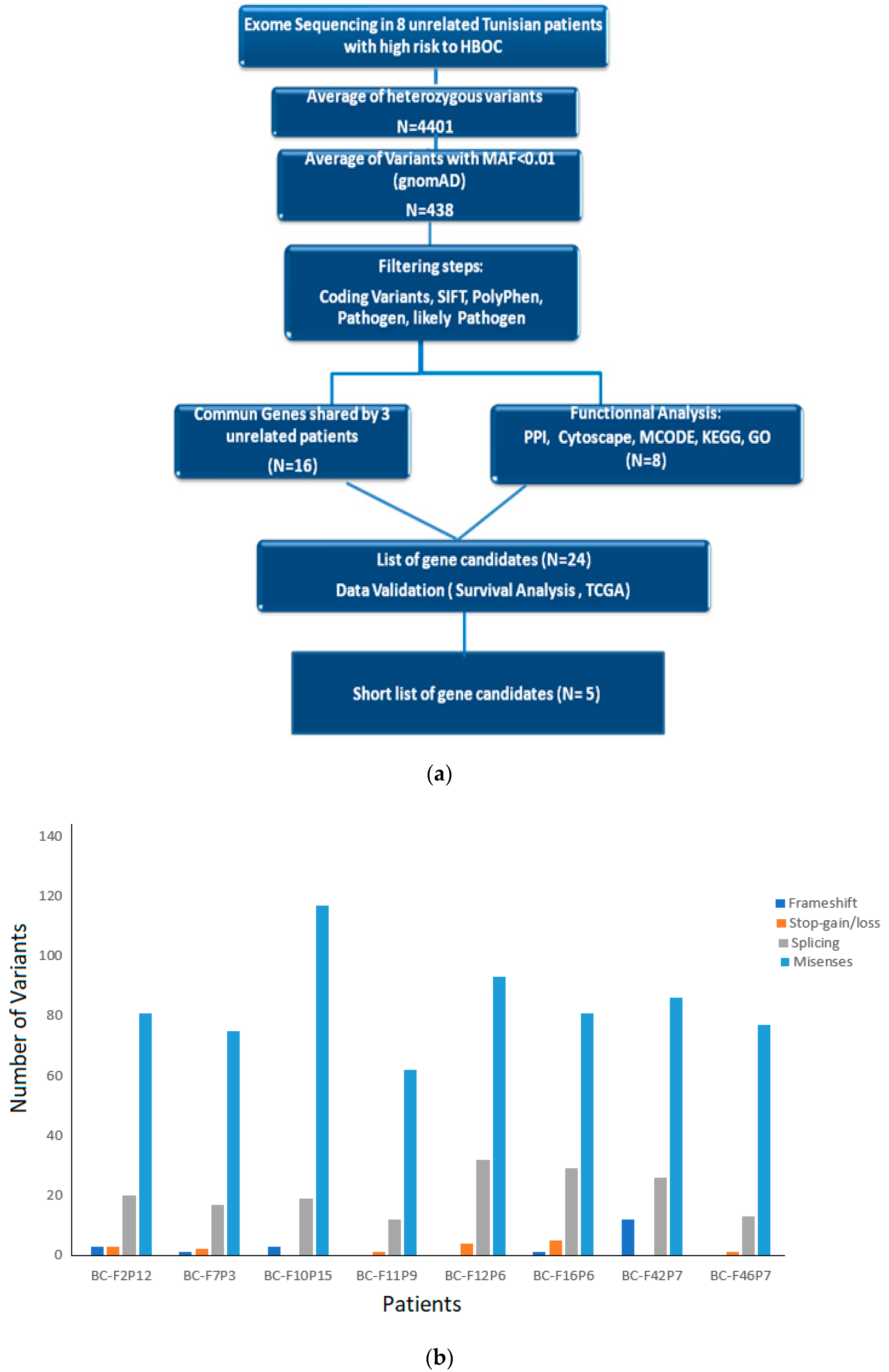
Exome sequencing (ES) was performed for eight BRCAness patients to identify candidate genes associated with the malignancy. Before applying filters, the total number of variants varied between 5607 and 7802 and the heterozygous variants varied between 3413 and 5248. A workflow showing the different steps performed during the analysis of the ES data is presented in Figure 2a. Missenses mutations were the most frequently found compared to frameshift, stop-gain/loss, or splicing (Figure 2b). Splicing variants included donor site, acceptor site, as well as splicing region in intron. For example, the BC-F12 family had the highest number of splicing variants as we identified one splice acceptor variant (c.197-2A>G) in the AIF gene and one splice variant donor (c.4636+1G>T) in the SLX4 gene; both are classified as “likely pathogen” in ClinVar. The other splicing variants are located in the intronic splicing region.
In the first step of our analysis, we focused on the canonical genes predisposed to HBOC, such as ATM, PALB2, Chek2, CDH1, PTEN, and TP53, but no variants in these genes were detected except for two missenses in the ATM gene, c.1810C>T; p.Pro604Ser and c.6115G>A; p.Glu2039Lys, which were identified in two patients from the BC-F15 and BC-F16 families diagnosed with stage-II BC at the ages of 30 and 27 years, respectively.
Evidently, the c.6115G>A variant has been reported as likely pathogenic in the ClinVar database and as VUS according to the ACMG classification, while the c.1810C>T variant has been reported as presenting conflicting interpretations of pathogenicity in ClinVar. Then, we focused on genes traditionally associated with breast/ovarian cancer or other malignancies, and identified 23 missenses, 2 splicing, and 2 UTR variants mainly affecting genes involved in DNA repair, such as RAD51, RAD54B, PMS1, FANCD2, XRCC1, and BLM (Table 2). The same RAD52 variant (c.661C<G, p.Gln221Glu) was identified in two unrelated patients (BC-F10P6 and BC-F16P5; Table 2). About 50% of the identified variants were classified as VUS according to ClinVar and Varsome (Table 2).

3.3. Identification of Potential Gene Candidates Predisposed to HBOC

In the next step of our analysis, we selected genes harboring loss-of-function (LoF) variants (stop-gain, splice site, and frameshift), because these variant types have an impact on protein function and are commonly linked to disease susceptibility (Richards et al., 2015). In this step, we identified 17 stop-gain and 11 frameshift variants affecting genes not commonly known as to be predisposed to HBOC (Table 3). Interestingly, among the high-risk variants listed, two frameshift variants were recurrent in unrelated patients, namely, c.1200delA; p.Lys400AsnfsTer15 in BHMT found in BC-F7P1 and BC-F10P6, and c.2626-2629delTTTG; p.Phe876LysfsTer16 in SH3PXD2B shared by BC-F2P10 and BC-F42P5 patients (Table 3). In addition, several stop-gain variants were identified in different genes, such as APOBEC3B, CERKL, SUMG1, and PIKFYVE (Table 3).
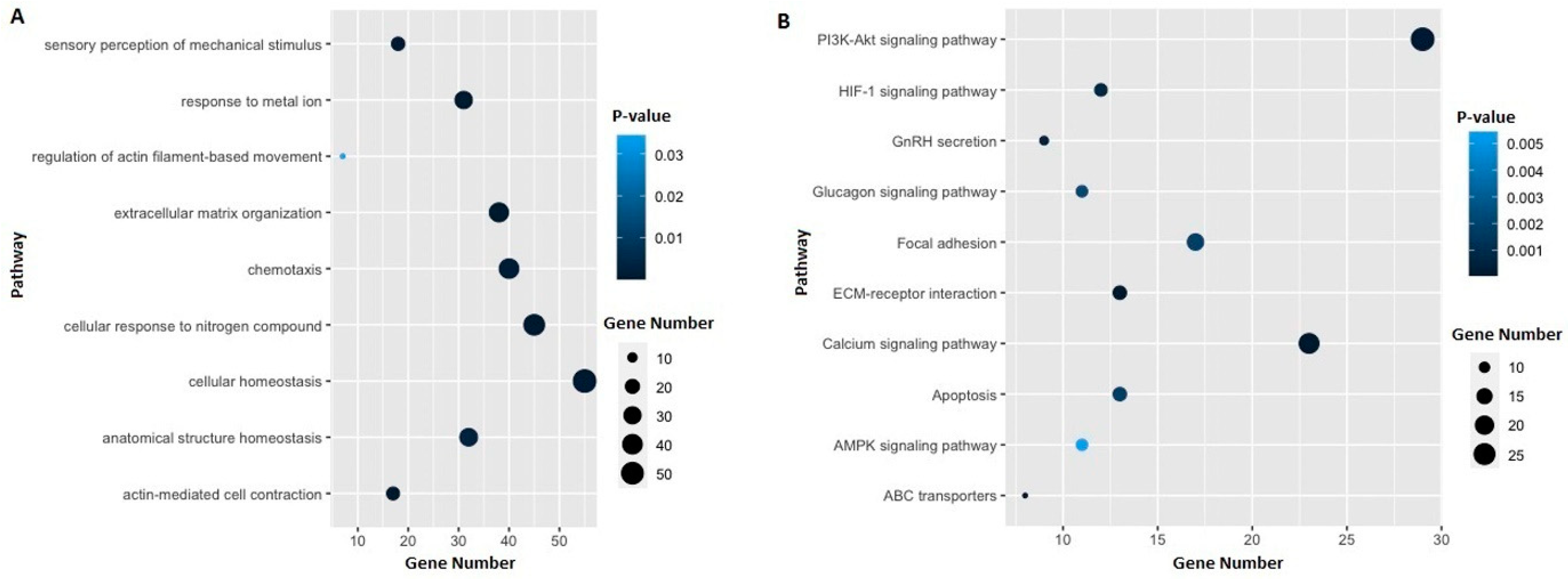
Furthermore, we performed the global network analysis to identify the protein–protein interaction (PPI) between proteins deduced from the ES data. Out of a total of 474 candidate genes, 336 PPIs were identified (Figure 3). Through a functional analysis based on MCODE, we selected the five most significant clusters that were involved in protein complexes and signaling pathways (Figure 4). The list of genes in each cluster is available in the Supplementary Materials, Table 1. Furthermore, gene ontology (GO) and the Kyoto Encyclopedia of Genes and Genomes (KEGG) pathway analysis of the selected genes were performed, and we found that they were mainly involved in extracellular matrix organization, actin-mediated cell contraction, response to metal ions, cellular response to nitrogen compounds, and cellular homeostasis (Figure 5a). Additionally, the results of the KEGG pathway analysis show that gene candidates are distributed in the ECM-receptor interaction, calcium signaling pathway, PI3K-Akt signaling pathway, ABC transporters, GnRH secretion, HIF-1 signaling pathway, apoptosis, focal adhesion, glucagon signaling pathway, and AMPK signaling pathway (Figure 5b). The combined analyses from MCODE with gene ontology and KEGG pathways led us to retain a short list of eight candidate genes (ATM, EP300, LAMA1, LAMC2, TNNI3, MYLK, COL11A2, and LAMB3). The selected genes were involved in at least 2 among the selected top 10 pathways, and the corresponding variants displayed significant Sift and PolyPhen scores.
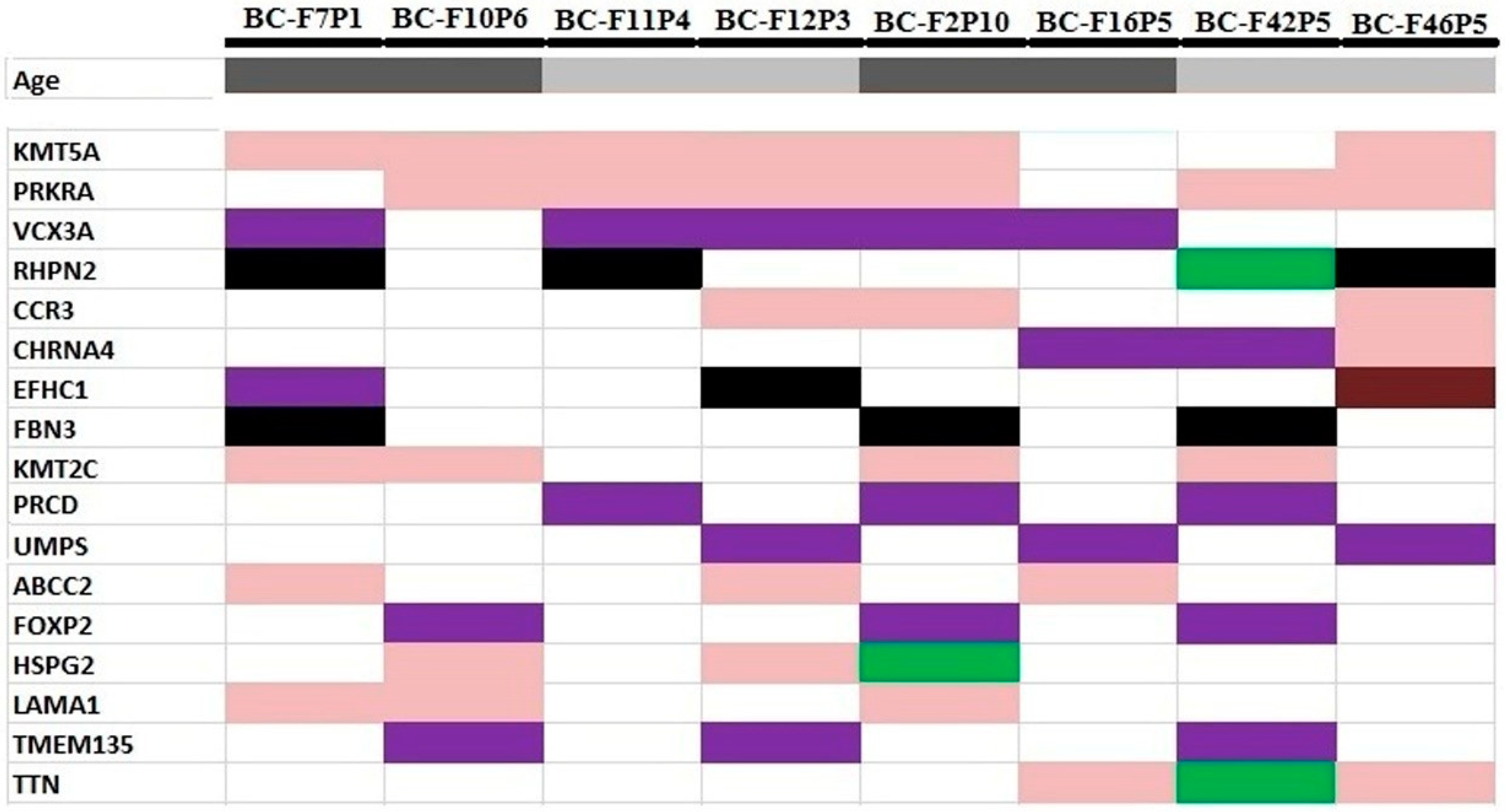
On the other hand, we selected gene variants that were shared by at least 3 patients from unrelated families and listed the 16 common genes: KMT5A, PRKRA, VCX3A, RHPN2, CCR3, CHRNA4, EFHC1, FBN3, KMT2C, PRCD, UMPS ABCC2, FOXP2, HSPG2, TMEM135, and TTN (Figure 6). It is interesting to note that we found the same variant in the KMT5A (c.995T>C), RHPN2 (c.1225+5G>A), CCR3 (c.727A>G), and KMT2C (c.2645T>C) genes in at least three unrelated patients. Based on the TCGA data, the most frequently altered gene in breast cancer was KMT2C (12%), and the TTN gene (17%) in ovarian cancer. A table listing the variants identified in the selected gene candidates is available in the Supplementary Materials (Supplementary Table S2).

3.4. Survival Analysis

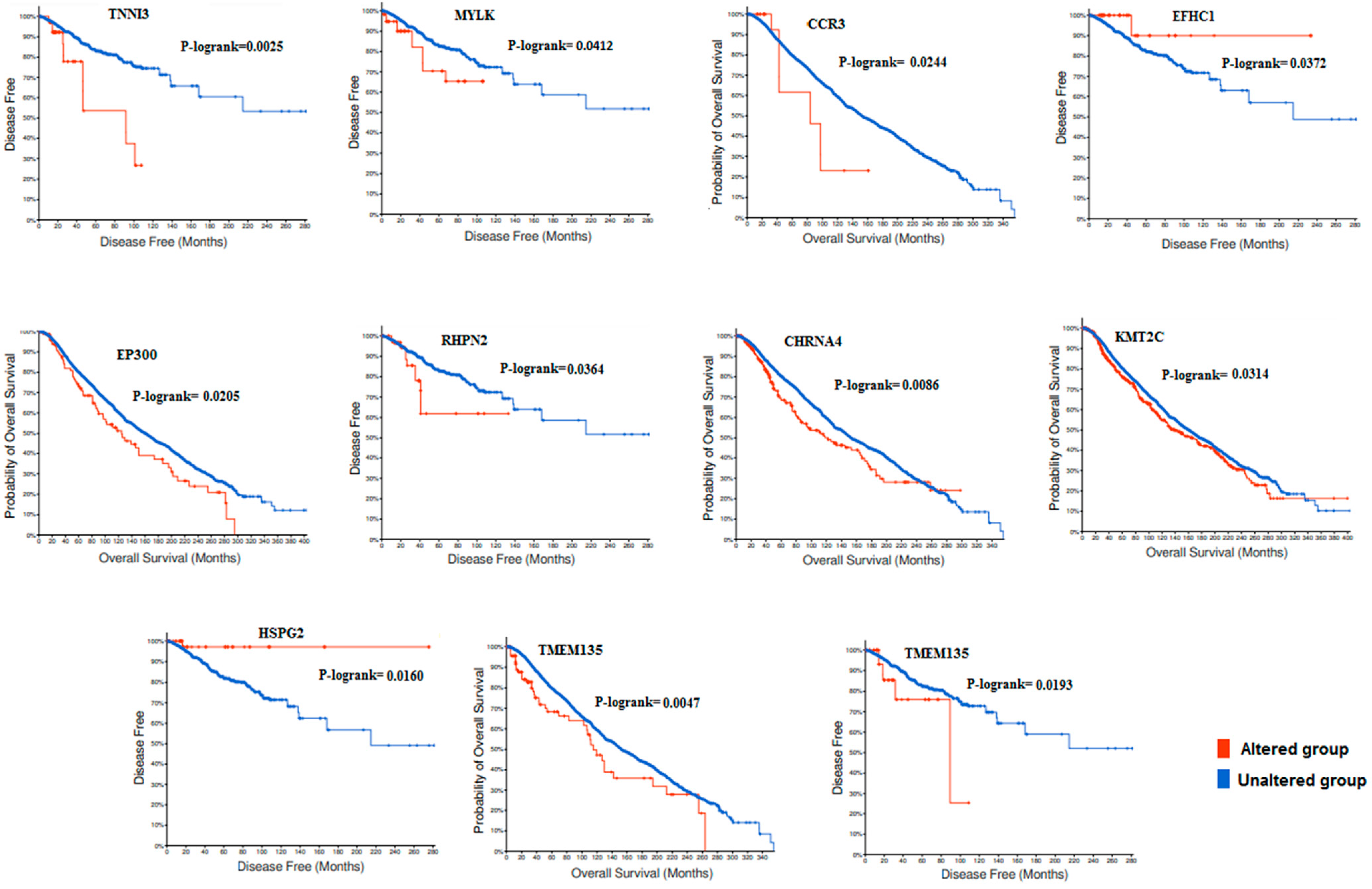
To investigate the impact of the retained genes on the survival rates, we used the TCGA data including 14 studies conducted between 2012 to 2020 to associate the 25 candidate genes with overall survival (OS) and disease-free survival (DFS). Kaplan–Meier curves showed that only 11 genes among 25 were significantly associated with OS or DFS (Figure 7). With concerns of genes related to BOC as reported by the literature data, significant associations were observed between the survival rate and EP300 (P logrank: 0.0205), KMT2C (P logrank: 0.0314), RHPN2 (P logrank: 0.0364), HSPG2 (P logrank: 0.016), and CCR3 (P logrank: 0.0244).

4. Discussion

Next-generation sequencing (NGS) technology has revolutionized the clinical approach for genetic testing in oncology. Whole-exome sequencing (WES) is increasingly used for screening HBOC patients for clinical applications and precision therapy [25,32,33]. Indeed, WES as well as multi-gene panels are powerful methods for the identification of pathogenic variants (PVs) in known HBOC-related genes and novel variants that might be associated with the disease [24,34,35,36,37,38]. Evidently, only few exome sequencing studies were conducted on BRCAness Tunisian families compared to other populations. As an example, Riahi et al. identified by WES a novel frameshift mutation in the RCC1 gene encoding the regulator of chromosome condensation 1. This variant (c.1067_1086del19) has exclusively been found in Tunisian breast cancer patients [16]. Other candidate genes (MMS19, DNHA3, POLK, and KATB6) were identified in a BRCAness family from the North of Tunisia [26]. More recently, and using WES, Mighri et al. identified a rare exonic VUS on an RAD50 gene (c.3647C>G, p.Ala1216Gly), a breast cancer susceptibility gene in a patient originating from the North-Eastern Tunisian region [21].
In our previous study, we reported that the contribution of the predisposing BRCA1/BRCA2 genes was involved in 15.9 % of HBOC patients originating from the South of Tunisia [17]. To further investigate the genetic landscape of HBOC, we performed ES on eight BRCA-negative patients from unrelated families. Initially, we focused on the canonical genes predisposed to HBOC, such as ATM, PALB2, Chek2, CDH1, PTEN, and TP53, but no PV was found, except for two missense variants in the ATM gene that is involved in DNA double-strand-break repair mechanisms and considered as a moderate-penetrance gene [39,40]. Missense variants were identified in DNA repair genes, such as RAD51, RAD54B, PMS1, XRCC1, and BLM. Moreover, we noticed that the same variant in RAD52 (Gln221Glu) was found in two patients from unrelated families.
After investigating known breast cancer genes, we extended our analysis to select rare loss-of-function variants, and we identified 11 frameshift and 17 stop-gain variants affecting genes involved in various cellular functions but not traditionally associated with HBOC. Nevertheless, among these 28 genes, 10 have been previously reported as implicated in breast/ovarian carcinogenesis. Among these variants, the frameshift in SH3PXD2B (c.2626-2629delTTTG) and BHMT (c.1200delA) genes were found in two unrelated patients. SH3PXD2B (Tks4) is a scaffold protein that plays a critical role in the invasion and metastasis of various types of tumors as hepatocarcinoma, melanoma, and breast cancer [41]. In fact, it was well demonstrated that cancer cells develop invadopodia and podosomes composed of structural proteins, such as cortactin, Tks4, and Tks5, to facilitate their migration across the endothelial layer to invade distant tissues [42].
Betaine-homocysteine S-methyltransferase (BHMT) catalyzes the synthesis of methionine from betaine and homocysteine. A previous study showed that choline and betaine are tightly associated with breast carcinogenesis [43,44].
In addition to frameshift variants, stop-gain variants were also identified in several genes involved in various cellular functions, such as the APOBEC3B (Apolipoprotein B mRNA-editing enzyme, catalytic polypeptide-like) belonging to the family of APOBEC enzymes that are strong mutagenic factors in human cancer [45,46]. APOBEC3B has been described as a strong driver of breast cancer and associated with aggressive clinical and pathological features [47,48,49]. A stop-gain variant (c.1166C>T) in APOBEC3B was identified in a BC-F12P6 patient, diagnosed with BC at 47 years of age, and with 3 BC cases and 2 OC cases in this family.
On the other hand, we performed functional analysis based on MCODE and selected the top five significant clusters that are involved in protein complexes and signaling pathways. Furthermore, the gene ontology (GO) and Kyoto Encyclopedia of Genes and Genomes (KEGG) pathway analysis showed that the selected genes were mainly involved in extracellular matrix organization, actin-mediated cell contraction, the PI3K-Akt signaling pathway, ABC transporters, and HIF-1 signaling pathway. Finally, we selected a list of eight candidate genes (ATM, EP300, LAMA1, LAMC2, TNNI3, MYLK, COL11A2, and LAMB3) that were involved in at least two among the selected pathways and showing damaging effects predicted by Sift and PolyPhen tools. LAMA1, LAMC2, and LAMB3 belong to the laminin family, extracellular glycoproteins that are essential components of membranes and involved in tissue differentiation, progression, and invasion [50,51].
In this study, we identified LAMA1 missense variants in three unrelated patients and noted that the BC-F7P1 patients with metastatic BCs harbored missense mutations in LAMA1, LAMC2, and LAMAC5 genes.
Furthermore, we focused on gene variants that were shared by at least three patients from unrelated families and listed the following genes: KMT5A, PRKRA, VCX3A, RHPN2, CCR3, CHRNA4, EFHC1, FBN3, KMT2C, PRCD, UMPS ABCC2, FOXP2, HSPG2, TMEM135, and TTN. We noticed that only a few of them have been previously described as related to breast/ovarian cancer, such as KMT2C, FOXP2, RHPN2, and HSPG2.
Regarding genes related to breast/ovarian cancer, we paid particular attention to those harboring the same variant shared by at least three unrelated patients. For instance, the variant c.2645T>C; p.Ile882Thr in the KMT2C gene was shared by four unrelated patients; three were ER+. This variant was not reported in ClinVar and classified as VUS in Varsome.
The H3K4 methyltransferase KMT2C is a critical regulator of hormone-dependent ERα activity [52]. Additionally, KMT2C is one of the most frequently mutated genes in ER-positive breast cancer and its loss disrupts proliferation through estrogen and conversely promotes tumor outgrowth in hormone-depleted conditions [52]. Based on the TCGA data (https://www.cbioportal.org/ (accessed on 26 August 2021)), we showed that KMT2C was significantly associated with overall survival in patients with BC. Similarly, Gala et al. reported that patients with KMT2C mutations had a lower progression-free survival rate on hormone deprivation therapy than patients with wild-type KMT2C, suggesting the probable contribution of KMT2C to BC hormone resistance [52].
RHPN2, a RhoA-binding protein, promotes malignant cell proliferation in ovarian cancer by activating the JAK2/STAT3 signaling pathway [53]. Here, we identified in four unrelated patients the same splicing variant (c.1225+5G>A) that could impact the splicing as predicted by in silico tools.
FOXP2, a member of the forkhead box transcription factor family, is suggested to regulate the progression of cancer cells through the epithelial–mesenchymal transition [54]. Keeping in mind that the variation in the 3′UTR region plays an important role in gene regulation [55,56], we identified in three unrelated patients, a variant in the 3′UTR region that could affect the expression of FOXP2, but further investigations are needed to better understand the effect of these variants.
HSPG2, also known as perlecan, is a heavily glycosylated protein component of the extra-cellular matrix (ECM) that has been previously observed as part of the surface of malignant cells of various cancers [57]. A strong correlation between HSPG2 expression and survival of TNBC patients was reported, suggesting that HSPG2 might be a promising therapeutic target in metastatic TNBC [58].
Cytokines are a protein family of regulatory factors derived from tumors and their environmental components that contribute to the growth, invasion, and metastasis of cancer. CCR3, a receptor of the C-C motif chemokine ligand (CCL), is identified to be one of the major factors that is involved in the progression and metastasis of some human tumors [59,60,61,62]. Recently, Yamaguchi et al. demonstrated that the CCL5–CCR3 interaction contributed to tumor progression suggesting that this axis may be an important target to improve the prognosis of breast cancer patients [63].
Finally, we associated the 24 selected genes with patients survival using TCGA data. Kaplan–Meier curves showed that EP300, KMT2C, RHPN2, HSPG2, and CCR3 genes correlated significantly with overall or disease-free survival rates.

5. Conclusions

In the present study, we selected 24 candidate genes form Tunisian patients with HBOC through functional and bioinformatic analyses of exome sequencing data. Regarding their role in carcinogenesis and their contribution to key signaling pathways, we retained a short list of five potential new candidate genes associated with HBOC in non-BRCA mutation carriers, namely, EP300, KMT2C, RHPN2, HSPG2, and CCR3. Nevertheless, further studies based on co-segregation analyses of affected families and the locus-specific loss of heterozygosity are needed to highlight the relevance of these candidate genes for HBOC risk.

Supplementary Materials

The following supporting information can be downloaded at: https://www.mdpi.com/article/10.3390/genes13081296/s1, Table S1: List of genes included in each of the 5 clusters identified by MCODE analysis, Table S2: List of 24 gene variants selected by integrative analyses (MCODE, GO, and KEGG) and identified in at least 3 unrelated patients from the selected families.

Author Contributions

D.B.-G. performed the experiments and data analysis. C.K. was responsible for the bioinformatic data analysis. N.A.-B. and W.A. contributed to the data analysis and the writing of the manuscript. W.B.-R. selected the patients and families included in the study and participated in the editing of the manuscript. A.K. and J.D. contributed to the concept and design of the work. R.M.-G. was responsible for the design of the study and writing of the manuscript. All authors have read and agreed to the published version of the manuscript.

Funding

This work was supported by a grant from the CIC (Centre d’Investigation Clinique) and The Ministry of Higher Education and Scientific Research of Tunisia (grant: LR19CBS02).

Institutional Review Board Statement

The study was conducted according to the Declaration of Helsinki and with the approval of the Institutional Review Board of the CHU Habib Bourguiba of Sfax, Tunisie. Written informed consent was obtained from all the patients prior to their participation in the study.

Informed Consent Statement

Informed consent was obtained from all subjects involved in the study.

Data Availability Statement

Data will be available upon request.

Acknowledgments

The authors thank the patients involved in the study.

Conflicts of Interest

The authors declare no conflict of interest.

Abbreviations

ACMGAmerican College of Medical Genetics and Genomics
BBenign
BCBreast Cancer
BWABurrows- Wheeler Aligner
DFSDisease Free Survival
EREostrogen Receptor
ESExome Sequencing
GOGene Ontology
HBOCHereditary Breast Ovarian/Cancer
KEGGKyoto Encyclopedia of Genes and Genomes
LBLikely Benign
LOFLoss of Function
LPLikely Pathogen
MAFMinor Allele Frequency
MCODEMolecular Complex Detection
NANo Available
NGSNext Generation Sequencing
NRNo Reported
OCOvarian Cancer
OSOverall Survival
PPathogen
PFPass Filter
PPIProtein-Protein Interactions
PRProgesteron Receptor
PVsPathogenic Variants
SBR gradeSBR Scarff-Bloom-Richardson (grade)
TCGAThe Cancer Genome Atlas
TNBCTriple Negative Breast Cancer
VUSVariant Unknown Significance
WESWhole Exome Sequencing

References

  1. Bray, F.; Ferlay, J.; Soerjomataram, I.; Siegel, R.L.; Torre, L.A.; Jemal, A. Global cancer statistics 2018: GLOBOCAN estimates of incidence and mortality worldwide for 36 cancers in 185 countries. CA Cancer J. Clin. 2018, 68, 394–424. [Google Scholar] [CrossRef] [PubMed] [Green Version]
  2. Coleman, M.P.; Quaresma, M.; Berrino, F.; Lutz, J.; de Angelis, R.; Capocaccia, R.; Garber, J.E.; Boyd, J.; Lubin, M.B.; DeShano, M.L.; et al. Cancer survival in five continents: A worldwide population-based study (CONCORD). Lancet. Oncol. 2008, 9, 730–756. [Google Scholar] [CrossRef]
  3. Maalej, M.; Hentati, D.; Messai, T.; Kochbati, L.; El May, A.; Mrad, K.; Romdhane, K.B.; Abdallah, M.B.; Zouari, B. Breast cancer in Tunisia in 2004: A comparative clinical and epidemiological study. Bul. Cancer 2008, 95, 10005–10009. [Google Scholar]
  4. Missaoui, N.; Jaidene, L.; Abdelkrim, S.B.; Ben Abdelkader, A.; Beizig, N.; Ben Yaacoub, L.; Yaacoubi, M.T.; Hmissa, S. Breast cancer in Tunisia: Clinical and pathological findings. Asian Pac. J. Cancer Prev. 2011, 121, 69–72. [Google Scholar]
  5. Younes, N.; Zayed, H. Genetic epidemiology of ovarian cancer in the 22 Arab countries: A systematic review. Gene 2019, 684, 154–164. [Google Scholar] [CrossRef]
  6. Claus, E.B.; Schildkraut, J.M.; Thompson, W.D.; Risch, N.J. The genetic attributable risk of breast and ovarian cancer. Cancer 1996, 77, 2318–2324. [Google Scholar] [CrossRef]
  7. Miki, Y.; Swensen, J.; Shattuck-Eidens, D.; Futreal, P.A.; Harshman, K.; Tavtigian, S.; Liu, Q.; Cochran, C.L.; Bennett, M.; Ding, W.; et al. A strong candidate for the breast and ovarian cancer susceptibility gene BRCA1. Science 1994, 266, 66–71. [Google Scholar] [CrossRef] [Green Version]
  8. Wooster, R.; Bignell, G.; Lancaster, J.; Swift, S.; Seal, S.; Mangion, J.; Collins, N.; Gregory, S.; Gumbs, C.; Micklem, G. Identification of the breast cancer susceptibility gene BRCA2. Nature 1995, 378, 789–792. [Google Scholar] [CrossRef]
  9. Antoniou, A.; Pharoah, P.D.; Narod, S.; Risch, H.A.; Eyfjord, J.E.; Hopper, J.L.; Loman, N.; Olsson, H.; Johannsson, O.; Borg, A.; et al. Average risks of breast and ovarian cancer associated with BRCA1 or BRCA2 mutations detected in case series unselected for family history: A combined analysis of 22 studies. Am. J. Hum. Genet. 2003, 7, 1117–1130. [Google Scholar] [CrossRef] [Green Version]
  10. Levy-Lahad, E.; Friedman, E. Cancer risks among BRCA1 and BRCA2 mutation carriers. Br. J. Cancer 2007, 96, 11–15. [Google Scholar] [CrossRef]
  11. Fackenthal, J.D.; Olopade, O.I. Breast cancer risk associated with BRCA1 and BRCA2 in diverse populations. Nat. Rev. Cancer 2007, 7, 937. [Google Scholar] [CrossRef] [PubMed]
  12. Abdulrashid, K.; AlHussaini, N.; Ahmed, W.; Thalib, L. Prevalence of BRCA mutations among hereditary breast and/or ovarian cancer patients in Arab countries: Systematic review and meta-analysis. BMC Cancer 2019, 19, 256–268. [Google Scholar] [CrossRef] [PubMed]
  13. Troudi, W.; Uhrhammer, N.; Sibille, C.; Dahan, C.; Mahfoudh, W.; Bouchlaka Souissi, C.; Jalabert, T.; Chouchane, L.; Bignon, Y.J.; Ben Ayedet, F.; et al. Contribution of the BRCA1 and BRCA2 mutations to breast cancer in Tunisia. J. Hum. Genet. 2007, 52, 915–920. [Google Scholar] [CrossRef] [PubMed] [Green Version]
  14. Fourati, A.; Louchez, M.M.; Fournier, J.; Amor, G.; Rahal, K.; El May, M.V.; El May, A.; Revillion, F.; Peyrat, J.P. Screening for common mutations in BRCA1 and BRCA2 genes: Interest in genetic testing of Tunisian families with breast and/or ovarian cancer. Bull. Cancer 2014, 101, 6–40. [Google Scholar] [CrossRef] [PubMed]
  15. Riahi, A.; Kharrat, M.; Ghourabi, M.E.; Khomsi, F.; Gamoudi, A.; Lariani, I.; May, A.E.; Rahal, K.; Chaabouni-Bouhamed, H. Mutation spectrum and prevalence of BRCA1 and BRCA2 genes in patients with familial and early-onset breast/ovarian cancer from Tunisia. Clin. Genet. 2015, 87, 155–160. [Google Scholar] [CrossRef] [PubMed]
  16. Riahi, A.; Radmanesh, H.; Schürmann, P.; Bogdanova, N.; Geffers, R.; Meddeb, R.; Kharrat, M.; Dörk, T. Exome sequencing and case-control analyses identify RCC1 as a candidate breast cancer susceptibility gene. Int. J. Cancer 2018, 142, 2512–2517. [Google Scholar] [CrossRef] [Green Version]
  17. Ben Ayed-Guerfali, D.; Ben Kridis-Rejab, W.; Ammous-Boukhris, N.; Ayadi, W.; Charfi, S.; Khanfir, A.; Sellami-Boudawara, T.; Frikha, M.; Daoud, J.; Mokdad-Gargouri, R. Novel and recurrent BRCA1/BRCA2 germline mutations in patients with breast/ovarian cancer: A series from the south of Tunisia. J. Transl. Med. 2021, 19, 108. [Google Scholar] [CrossRef]
  18. Bakkach, J.; Mansouri, M.; Derkaoui, T.; Loudiyi, A.; El Fahime, E.M.; Barakat, A.; Ghailani Nourouti, N.; De Villarrea, J.M.; Bringas, C.C.; Mechita, M.B. Contribution of BRCA1 and BRCA2 germline mutations to early onset breast cancer: A series from north of Morocco. BMC Cancer 2020, 20, 859. [Google Scholar] [CrossRef]
  19. Mehemmai, C.; Cherbal, F.; Hamdi, Y.; Guedioura, A.; Benbrahim, W.; Bakour, R.; Abdelhak, S. BRCA1 and BRCA2 Germline Mutation Analysis in Hereditary Breast/Ovarian Cancer Families from the Aures Region (Eastern Algeria): First Report. Pathol. Oncol. Res. 2020, 26, 715–726. [Google Scholar] [CrossRef]
  20. Saied, M.H.; Elkaffash, D.; Fadl, R.; Abdel Haleem, R.; Refeat, A.; Ibrahim, I.; Tahoun, M.; Elkayal, A.; Tayae, E. Preliminary results of targeted sequencing of BRCA1 and BRCA2 in a cohort of breast cancer families: New insight into pathogenic variants in patients and at-risk relatives. Mol. Med. Rep. 2021, 24, 678. [Google Scholar] [CrossRef]
  21. Mighri, N.; Hamdi, Y.; Boujemaa, M.; Othman, H.; Ben Nasr, S.; El Benna, H.; Mejri, N.; Labidi, S.; Ayari, J.; Jaidene, O.; et al. Identification of Novel BRCA1 and RAD50 Mutations Associated with Breast Cancer Predisposition in Tunisian Patients. Front. Genet. 2020, 11, 552971. [Google Scholar] [CrossRef] [PubMed]
  22. Michailidou, K.; Lindström, S.; Beesley, D.J.; Hui, S.; Kar, S.; Lemaçon, A.; Penny, S.; Glubb, D.; Rostamianfar, A.; Bolla, M.K.; et al. Association analysis identifies 65 new breast cancer risk loci. Nature 2017, 551, 92–94. [Google Scholar] [CrossRef] [PubMed] [Green Version]
  23. Mavaddat, N.; Peock, S.; Frost, D.; Ellis, S.; Platte, R.; Fineberg, E.; Gareth Evans, D.; Izatt, L.; AEeles, R.; Adlard, J.; et al. Cancer risks for BRCA1 and BRCA2 mutation carriers: Results from prospective analysis of EMBRACE. J. Natl. Cancer Inst. 2013, 105, 812–822. [Google Scholar] [CrossRef] [PubMed] [Green Version]
  24. Sokolenko, A.P.; Suspitsin, E.N.; Kuligina, E.S.; Bizin, I.V.; Frishman, D.; Imyanitov, E.N. Identification of novel hereditary cancer genes by whole exome sequencing. Cancer Lett. 2015, 369, 274–288. [Google Scholar] [CrossRef] [Green Version]
  25. Zelli, V.; Compagnoni, C.; Cannita, K.; Capelli, R.; Capalbo, C.; Di Vito Nolfi, M.; Alesse, E.; Zazzeroni, F.; Tessitore, A. Applications of Next Generation Sequencing to the Analysis of Familial Breast/Ovarian Cancer. High Throughput 2020, 9, 1. [Google Scholar] [CrossRef] [Green Version]
  26. Hamdi, Y.; Boujemaa, M.; Ben Rekaya, M.; Ben Hamda, C.; Mighri, N.; El Benna, H.; Mejri, N.; Labidi, S.; Daoud, N.; Naouali, C.; et al. Family specific genetic predisposition to breast cancer: Results from Tunisian whole exome sequenced breast cancer cases. J. Trans. Med. 2018, 16, 158. [Google Scholar] [CrossRef] [PubMed] [Green Version]
  27. Shannon, P.; Markiel, A.; Ozier, O.; Baliga, N.S.; Wang, J.T.; Ramage, D.; Amin, N.; Schwikowski, B.; Ideker, T. Cytoscape: A software environment for integrated models of biomolecular interaction networks. Genome Res. 2003, 13, 2498–2504. [Google Scholar] [CrossRef] [PubMed]
  28. Doncheva, N.T.; Morris, J.H.; Gorodkin, J.; Jensen, L.J. Cytoscape StringApp: Network Analysis and Visualization of Proteomics Data. J. Proteome Res. 2019, 18, 623–632. [Google Scholar] [CrossRef]
  29. Bindea, G.; Mlecnik, B.; Hackl, H.; Charoentong, P.; Tosolini, M.; Kirilovsky, A.; Fridman, W.H.; Pagès, F.; Trajanoski, Z.; Galonet, J.; et al. ClueGO: A Cytoscape plug-in to decipher functionally grouped gene ontology and pathway annotation networks. Bioinformatics 2009, 25, 1091–1093. [Google Scholar] [CrossRef] [Green Version]
  30. Cerami, E.; Gao, J.; Dogrusoz, U.; Gross, B.E.; Sumer, S.O.; Aksoy, B.A.; Jacobsen, A.; Byrne, C.J.; Heuer, M.L.; Larsson, E.; et al. The cBio cancer genomics portal: An open platform for exploring multidimensional cancer genomics data. Cancer Discov. 2012, 2, 401–404. [Google Scholar] [CrossRef] [Green Version]
  31. Gao, J.; Aksoy, B.A.; Dogrusoz, U.; Dresdner, G.; Gross, B.; Sumer, S.O.; Sun, Y.; Jacobsen, A.; Sinha, R.; Larsson, E.; et al. Integrative analysis of complex cancer genomics and clinical profiles using the cBioPortal. Sci. Signal 2013, 6, pl1. [Google Scholar] [CrossRef] [PubMed] [Green Version]
  32. Chandler, M.R.; Bilgili, E.P.; Merner, N.D. A review of whole-exome sequencing efforts toward hereditary breast cancer susceptibility gene discovery. Hum. Mutat. 2016, 37, 835–846. [Google Scholar] [CrossRef] [PubMed]
  33. Goodwin, S.; McPherson, J.D.; McCombie, W.R. Coming of age: Ten years of next-generation sequencing technologies. Nat. Rev. Genet. 2016, 17, 333–351. [Google Scholar] [CrossRef] [PubMed]
  34. Zhang, Y.; Cai, Q.; Shu, X.O.; Gao, Y.T.; Li, C.; Zheng, W.; Long, J. Whole-Exome Sequencing Identifies Novel Somatic Mutations in Chinese Breast Cancer Patients. Mol. Genet. Med. 2015, 9, 183. [Google Scholar] [CrossRef] [PubMed]
  35. Jalkh, N.; Chouery, E.; Haidar, Z.; Khater, C.; Atallah, D.; Ali, H. Next-generation sequencing in familial breast cancer patients from Lebanon. BMC Med. Genom. 2017, 10, 8. [Google Scholar] [CrossRef] [Green Version]
  36. Kuligina, E.S.; Sokolenko, A.P.; Bizin, I.V.; Haidar, Z.; Khater, C.; Atallah, D.; Ali, H.; Marafie MJAl-Mulla, M.R.; Al-Mulla, F.; Andre, M. Exome sequencing study of Russian breast cancer patients suggests a predisposing role for USP39. Breast Cancer Res. Treat. 2020, 179, 731–742. [Google Scholar] [CrossRef]
  37. Doddato, G.; Valentino, F.; Giliberti, A.; Papa, F.T.; Tita, R.; Bruno, L.P.; Resciniti, S.; Fallerini, C.; Benetti, E.; Palmieri, M.; et al. Exome sequencing in BRCA1-2 candidate familias: The contribution of other cancer susceptibility genes. Front. Oncol. 2021, 11, 649435. [Google Scholar]
  38. Felicio, P.S.; Grasel, R.S.; Campacci, N.P.; Galvão, A.E.; Torrezan, H.C.R.; Sabato, C.S.; Fernandes, G.C.; Souza, C.P.; Michelli, R.D. Whole-exome sequencing of non-BRCA1/BRCA2 mutation carrier cases at high-risk for hereditary breast/ovarian cancer. Hum. Mutat. 2021, 42, 290–299. [Google Scholar] [CrossRef]
  39. Easton, D.F.; Pharoah, P.D.; Antoniou, A.C.; Tischkowitz, M.; Tavtigian, S.V.; Nathanson, K.L.; Devilee, P.; Meindl, A.; Couch, F.J.; Southey, M.; et al. Gene-panel sequencing and the prediction of breast-cancer risk. N. Engl. J. Med. 2015, 372, 2243–2257. [Google Scholar] [CrossRef] [Green Version]
  40. Southey, M.C.; Goldgar, D.E.; Winqvist, R.; Pylkäs, K.; Couch, F.; Tischkowitz, M.; Foulkes, W.D.; Dennis, J.; Michailidou, K.; Rensburget, E.J.; et al. PALB2, CHEK2 and ATM rare variants and cancer risk: Data from COGS. J. Med. Genet. 2016, 53, 800–811. [Google Scholar] [CrossRef] [Green Version]
  41. Seals, D.F.; Azucena, E.F.; Pass, I.; Tesfay, L.; Gordon, R.; Woodrow, M.; Resau, J.H.; Courtneidge, S.A. The adaptor protein Tks5/Fish isrequired for podosome formation and function, and for the protease-driven invasion of cancer cells. Cancer Cell 2005, 7, 155–165. [Google Scholar] [CrossRef] [PubMed] [Green Version]
  42. Carman, C.V.; Sage, P.T.; Sciuto, T.E.; de la Fuente, M.A.; Geha, R.S.; Ochs, H.D.; Dvorak, H.F.; Dvorak, A.M.; Springer, T.A. Transcellular diapedesis is initiated by invasive podosomes. Immunity 2007, 26, 784–797. [Google Scholar] [CrossRef] [PubMed] [Green Version]
  43. Xu, X.; Gammon, M.D.; Zeisel, S.H.; Lee, Y.L.; Wetmur, J.G.; Teitelbaum, S.L.; Bradshaw, P.T.; Neugut, A.I.; Santella, R.M.; Choline, J.C. Metabolism and risk of breast cancer in a population-based study. FASEB J. 2008, 22, 2045–2052. [Google Scholar] [CrossRef]
  44. Glunde, K.; Bhujwalla, Z.M.; Ronen, S.M. Choline metabolism in malignant transformation. Nat. Rev. Cancer 2011, 11, 835–848. [Google Scholar] [CrossRef] [PubMed] [Green Version]
  45. Swanton, C.; McGranahan, N.; Starrett, G.J.; Harris, R.S. APOBEC enzymes: Mutagenic fuel for cancer evolution and heterogeneity. Cancer Discov. 2015, 5, 704–712. [Google Scholar] [CrossRef] [Green Version]
  46. Asaoka, M.; Santosh, K.P.; Ishikawa, T.; Takabe, K. Different members of the APOBEC3 family of DNA mutators have opposing associations with the landscape of breast cancer. Am. J. Cancer Res. 2021, 11, 5111–5125. [Google Scholar]
  47. Burns, M.B.; Lackey, L.; Carpenter, M.A.; Rathore, A.; Land, A.M.; Leonard, B.; Refsland, E.W.; Kotandeniya, D.; Tretyakova, N. APOBEC3B is an enzymatic source of mutation in breast cancer. Nature 2013, 494, 366–370. [Google Scholar] [CrossRef]
  48. Sieuwerts, A.M.; Willis, S.; Burns, M.B.; Look, M.P.; Meijer-Van Gelder, M.E.; Schlicker, A.; Heideman, M.R.; Jacobs, H.; Wessels, L.; Leyland-Jones, B. Elevated APOBEC3B correlates with poor outcomes for estrogen-receptor-positive breast cancers. Horm. Cancer 2014, 5, 405–413. [Google Scholar] [CrossRef] [Green Version]
  49. Harris, R.S. Molecular mechanism and clinical impact of APOBEC3B-catalyzed mutagenesis in breast cancer. Breast Cancer Res. 2015, 17, 8. [Google Scholar] [CrossRef] [Green Version]
  50. Høye, A.M.; Erler, J.T. Structural ECM components in the premetastatic and metastatic niche. Am. J. Physiol. Cell 2016, 310, 955–967. [Google Scholar] [CrossRef] [Green Version]
  51. Zhao, Y.; Zheng, X.; Zheng, Y.; Chen, Y.; Fei, W.; Wang, F.; Zheng, C. Extracellular Matrix: Emerging Roles and Potential Therapeutic Targets for Breast Cancer. Front. Oncol. 2021, 11, 650453. [Google Scholar] [CrossRef] [PubMed]
  52. Gala, K.; Li, Q.; Sinha, A.; Razavi, P.; Dorso, M.; Sanchez-Vega, F.; Chung, Y.R.; Hendrickson, R.; Hsieh, J.J. KMT2C mediates the estrogen dependence of breast cancer through regulation of ERα enhancer function. Oncogene 2018, 37, 4692–4710. [Google Scholar] [CrossRef] [PubMed] [Green Version]
  53. Yu, F.; Qiao, P.; Yin, G.; Sun, Y.; Yu, X.; Sun, X.; Chu, Y.; Wang, Y. RHPN2 Promotes Malignant Cell Behaviours in Ovarian Cancer by Activating STAT3 Signalling. Onco. Targets Ther. 2020, 10, 11517–11527. [Google Scholar] [CrossRef] [PubMed]
  54. Myatt, S.S.; Lam, E.W. The emerging roles of forkhead box (Fox) proteins in cancer. Nat. Rev. Cancer 2007, 7, 847–859. [Google Scholar] [CrossRef] [PubMed]
  55. López de Silanes, I.; Paz Quesada, M.; Esteller, M. Aberrant regulation of messenger RNA 3’-untranslated region in human cancer. Cell Oncol. 2007, 29, 1–17. [Google Scholar] [CrossRef]
  56. Vislovukh, A.; Rivera Vargas, T.; Polesskaya, A.; Groisman, I. Role of 3′-untranslated region translational control in cancer development, diagnostics and treatment. World J. Biol. Chem. 2014, 26, 40–57. [Google Scholar] [CrossRef] [Green Version]
  57. Theocharis, A.D.; Skandalis, S.S.; Tzanakakis, G.N.; Karamanos, N.K. Proteoglycans in health and disease: Novel roles for proteoglycans in malignancy and their pharmacological targeting. FEBS J. 2010, 277, 3904–3923. [Google Scholar] [CrossRef]
  58. Kalscheuer, S.; Khanna, V.; Kim, H.; Li, S.; Sachdev, D.; DeCarlo, A.; Yang, D.; Panyam, J. Discovery of HSPG2 (Perlecan) as a Therapeutic Target in Triple Negative Breast Cancer. Sci. Rep. 2019, 9, 12492. [Google Scholar] [CrossRef]
  59. Johrer, K.; Zelle-Rieser, C.; Perathoner, A.; Moser, P.; Hager, M.; Ramoner, R.; Gander, H.; Höltl, L.; Bartsch, G.; Greil, R. Up-regulation of functional chemokine receptor CCR3 in human renal cell carcinoma. Clin. Cancer Res. 2005, 11, 2459–2465. [Google Scholar] [CrossRef] [Green Version]
  60. Miyagaki, T.; Sugaya, M.; Fujita, H.; Ohmatsu, H.; Kakinuma, T.; Kadono, T.; Tamaki, K.; Sato, S. Eotxins and CCR3 interaction regulates the Th2 environment of cutaneous T-cell lymphoma. J. Investig. Dermatol. 2010, 130, 2304–2311. [Google Scholar] [CrossRef] [Green Version]
  61. Miyagaki, T.; Sugaya, M.; Murakami, T.; Asano, Y.; Tada, Y.; Kadono, T.; Okochi, H.; Tamaki, K.; Sato, S. CCL11-CCR3 interactions promote survival of anaplastic large cell lymphoma cells via ERK1/2 activation. Cancer Res. 2011, 71, 2056–2065. [Google Scholar] [CrossRef] [PubMed] [Green Version]
  62. Gong, D.H.; Fan, L.; Chen, H.Y.; Ding, K.F.; Yu, K.D. Intratumoral expression of CCR3 in breast cancer is associated with improved relapse-free survival in luminal-like disease. Oncotarget 2016, 7, 28570–28578. [Google Scholar] [CrossRef] [PubMed] [Green Version]
  63. Yamaguchi, M.; Takagi, K.; Narita, K.; Miki, Y.; Onodera, Y.; Miyashita, M.; Sasano, H.; Suzuki, T. Stromal CCL5 Promotes Breast Cancer Progression by Interacting with CCR3 in Tumor Cells. Int. J. Mol. Sci. 2021, 22, 1918. [Google Scholar] [CrossRef] [PubMed]
Figure 1. Pedigrees of the families selected for exome sequencing. The arrow indicates the sequenced patients from each family. Available age at diagnosis of breast cancer cases is indicated.
Genes 13 01296 g001
Figure 2. (a) Workflow showing the different steps performed for ES data analysis. (b) Histogram showing the number of variants identified by exome sequencing in each patient from the selected families.
Genes 13 01296 g002
Figure 3. Interactome showing the protein–protein interactions deduced from the exome sequencing data. The size of the red dots is proportional to the number of times that the gene was identifiedin patients.
Genes 13 01296 g003
Figure 4. MCODE analysis identified 5 clusters: Cluster 1: 20 genes (LAMA1, LAMC2, LAMA5…), Cluster 2: 12 genes (MYH11, MYH13, MYH14…), Cluster 3: 33 genes (EP300, ATM, XRCC1…), Cluster 4: 12 genes (ABCG2, ABCC2, CFTR…), Cluster 5: 12 genes (PDE11A, CNGB1, PRKACA…). For more details, see supplementary data Table S1.
Genes 13 01296 g004
Figure 5. GO and KEGG bubble of mutant genes of analyzed samples. (A) Bubble plot of the Gene Ontology (GO) analysis showing the top 9 significant pathways. Bubble color and size correspond to the P-value and gene number enriched in the pathway. (B) Bubble plot of the KEGG (Kyoto Encyclopedia of Genes and Genomes) analysis showing the top 10 significant pathways. Bubble color and size correspond to the P-value and gene number enriched in the pathway.
Genes 13 01296 g005
Figure 6. Variants identified in the candidate genes selected trough functional analysis and that are shared by at least 3 unrelated patients. In light pink: missense variants; in black: splice acceptor/donor variants; in purple: 3′UTR variants; in brown: 5′UTR variants; and in green: more than one variant. Information about age at diagnosis: in dark gray: diagnosis less than or equal to 40 years of age; in light grey: more than or equal to 40 years of age.
Genes 13 01296 g006
Figure 7. Kaplan–Meier plots showing significant associations between the selected candidate genes and the overall survival or disease-free survival using the TCGA data.
Genes 13 01296 g007
Table 1. Clinico-pathological parameters of patients selected for exome sequencing.
PatientsAge at Diagnosis Family History of BC/OCFamily History of Other CancersHistological TypeSBR GradeTumor SizeTNMERPRHer-2TherapyFollow-Up
BC-F2P10309BC, 2MBC, 3OC-IDCII2I---TAM, FEC, TXT, RTNA
BC-F7P1373BC, 1OCProstateIDCII3IIB++-MCA, TAM, FEC, TXT, RTMetastasis after 3 y of BC. Died with the disease progression
BC-F10P6277BCEndometrialIDCIII4.5IIA++-MCA, TAM, FEC, TXT, RTRemission with OS of 1 y
BC-F11P4304BC, 1OCProstateIDCIII5.3IIB---FEC, RTRemission with OS of 16 y
BC-F12P3494BC, 1OCProstate MelanomaIDCII4.5IIB+--MCA, TAM, RTRemission with OS of 6.5 y
BC-F16P5245BC, 1BC/ThyThyroidIDCII2.5IIA++-TAM, FEC, TXT, RTRemission with OS of 4.5 y
BC-F42P5303BC,
2BC/OC
ProstateIDCII4IIB++-MCA, TAM, FEC, C75, RTNA
BC-F46P5554BC,
1MBC
-IDCII2IA++-TAM, FEC, TXT, RTRemission with OS of 1 y
Table 2. List of genes commonly associated with hereditary breast/ovarian cancer identified by exome sequencing in each patient.
PatientsGenersVariationMAFConsequenceClinVar
BC-F2P10PMS1778185859c.278G>A; p.Arg93His0.000062VUSNR
RAD54B35973866c.247A>G; p.(Ser83Gly)0.0408VUSNR
ATM587781352c.2492A>G; p.(Asp831Gly)0.000016VUSR
RAD51D c.413A>G; p.(Asn138Ser)0.000756VUSR
EP3001268191227c.3976G>A; p.(Val1326Ile) PR
MUTYH140118273c.1544C>T; p.(Ser515Phe)0.0234BR
BC-F7P1ATM2227922c.1810C>T; p.(Pro604Ser)0.0317LBR
RAD52867412462c.1048G>A; p.(Asp350Asn))0.000018VUSNR
RAD54B116312454c.2639A>G; p.(Asp880Gly)0.0043LBR
BC-F10P6FANCE768911543c.298T>A; p.(Ser100Thr)0.00072VUSR
PMS2 c.706-4dup VUSNR
EXO14150001c.2276G>A; p.Gly759Glu0.0338LBR
RAD524987206c.661C>G; p.Gln221Glu0.0325BR
BC-F11P4RAD51DNRc.*1056C>TNALBNR
BC-F12P3FANCL770368316c.288G>T; p.Lys96Asn0.000416VUSR
POLL61757734c.169C>T; p.Arg57Trp0.0026VUSNR
POLH35675573c.986C>T; p.Thr329Ile0.017LBR
BC-F16P5ATM864622251c.6115G>A; p.Glu2039Lys0.000033VUSR
DCC138724679c.527A>G; p.Asn176Ser0.0005VUSR
BARD161754118c.2212A>G; p.Ile738Val0.027LBR
XRCC1143917286c.818C>T; p.Pro273Leu0.00311LBR
RAD524987206c.661C>G; p.Gln221Glu0.0325BR
RAD51DNRc.*106G>ANALBNR
BC-F42P5BLM141503266c.254G>C; p.Arg85Thr0.01LBR
PMS263750055c.1711C>A; p.Leu571Ile0.02LBNR
BC-F46P5EP300 c.227C>G; p.Ser76Cys VUS
RAD54B2919661cc.289G>C; p.Asp97His0.018VUSNR
VUS: variant of unknown significance, LB: likely benign, B: benign, R: reported, NR: not reported, NA: not available, MAF: minor allele frequency, *: stop codon.
Table 3. List of genes with heterozygous rare stop-gain and frameshift variants identified in each patient. Bold characters indicate common variants in unrelated patients.
PatientGenersVariationMAFConsequenceClinVar
BC-F2P10PCK2753706965c.577c>T; p.Arg193*0.000415PR
PDE6BNRc.125_126insTGCGA; p.Asp43Alafs*109NALPNR
PDE6BNRc.120_121insGAGGA; p.Pro41Glufs*111NALPNR
IL31RA144337484c.700C>T; p.Arg234*0.000163LPNR
TTC37768215813c.1708C>T; p.Arg570*0.000054LPNR
SH3PXD2B551498843c.2626_2629del; p.Phe876Lysfs*160.005594PR
BC-F7P1ZnHIT6NRc.1114G>T; p.Glu372*NALPNR
BHMT763726268c.1200del; p.Lys400Asnfs*150.000746LPR
MEGF10NRc.122C>A; p.Ser41*NALPNR
BC-F10P6BHMT763726268c.1200del; p.Lys400AsnfsTer150.000746 R
Cyp3A528383469c.92dup; p.Leu32Thrfs*30.011928LPNR
ALOX15781725832c.316del; p.Leu106*0.000229LPNR
SMUG12233919c.7C>T; p.Gln3*0.054463LBNR
BC-F11P4MAN1B1NRc.383T>G; p.Leu128*NALPNR
BC-F12P3PIKFYVENRc.914C>A; p.Ser305*NALPNR
IL3373251020c.337C>T; p.Arg113*0.000056LPNR
TSSK4200353859c.895A>T; p.Lys299*0.002882LPNR
APOBEC3B199817842c.166C>T; p.Arg56*0.002176LPNR
BC-F16P5CERKL121909398c.847C>T; p.Arg283*0.000538PR
PIKFYVENRc.573C>A; p.Cys191*NALPNR
TM4SF19NRc.273T>A; p.Cys91*NALPNR
ALG1NRc.297_298del; p.Val100Phefs*37NALPNR
BC-F42P5SH3PXD2P551498843c.2626_2629del; p.Phe876Lysfs*160.00559 R
HMSD559021231c.105_120del; p.Asp35Glufs*490.013616LPNR
BC-F46P5SPG11NRc.3235G>T; p.Gly1079*NALPNR
LP: likely pathogen, P: pathogen, LB: likely benign, R: reported, NR: not reported, NA: not available, MAF: minor allele frequency. *: Stop codon, fs: frameshift.
Publisher’s Note: MDPI stays neutral with regard to jurisdictional claims in published maps and institutional affiliations.