Article

Comparison between the Gametophyte and the Sporophyte Transcriptomes of the Endangered Fern Vandenboschia speciosa

by
Rubén Martín-Blázquez
1,
Mohammed Bakkali
2,
Mercedes Ruiz-Estévez
3 and
Manuel A. Garrido-Ramos
2,*
1
Department of Evolutionary Ecology, Estación Biológica de Doñana, Consejo Superior de Investigaciones Científicas (CSIC), Isla de la Cartuja, 41092 Sevilla, Spain
2
Departamento de Genética, Universidad de Granada, 18071 Granada, Spain
3
Corporate Research Materials Laboratory, 3M Center, Saint Paul, MN 55144, USA
*
Author to whom correspondence should be addressed.
Genes 2023, 14(1), 166; https://doi.org/10.3390/genes14010166
Submission received: 7 November 2022 / Revised: 4 January 2023 / Accepted: 5 January 2023 / Published: 7 January 2023
(This article belongs to the Section Plant Genetics and Genomics)

Abstract

:
Genomic resources are essential to understanding the evolution and functional biology of organisms. Nevertheless, generating genomic resources from endangered species may be challenging due to the scarcity of available specimens and sampling difficulties. In this study, we compare the transcriptomes of the sporophyte and the gametophyte of the endangered fern Vandenboschia speciosa. After Illumina sequencing and de novo transcriptome assembly of the gametophyte, annotation proved the existence of cross-species contamination in the gametophyte sample. Thus, we developed an in silico decontamination step for the gametophyte sequences. Once the quality check of the decontaminated reads passed, we produced a de novo assembly with the decontaminated gametophyte reads (with 43,139 contigs) and another combining the sporophyte and in silico decontaminated gametophyte reads (with 42,918 contigs). A comparison of the enriched GO terms from the top 1000 most expressed transcripts from both tissues showed that the gametophyte GO term set was enriched in sequences involved in development, response to stress, and plastid organization, while the sporophyte GO term set had a larger representation of more general metabolic functions. This study complements the available genomic resources on the life cycle of the endangered fern Vandenboschia speciosa.

1. Introduction

In the era of genomics, the amount of high throughput sequencing (HTS) resources for non-model organism has increased significantly in the last decade [1,2,3,4]. This trend eliminates possible artifacts caused by inferring non-model species based on model species and helps us understand the species-specific genomic or transcriptomic dynamics of non-model species. Differences exist even between con-generic species due to peculiarities during the processes of speciation, caused by adaptations to specific environmental conditions [5,6,7,8] or due to genome architecture dynamics [9,10]. Such differences might override characteristics present in a non-model species of interest that are not being captured by the life history of its phylogenetically closest model species. Thus, the genomic characterization of non-model organisms, including endemic/threatened species, is crucial to understanding possible differences between the genetic backgrounds of their more widespread and successful con-generics. As a study case, the endangered fern Vandenboschia speciosa illustrates the need to understand endemic species’ genomics and transcriptomics to better take decisions on their conservation.
Vandenboschia speciosa (Willd.) G. Kunkel (=Trichomanes speciosum Willd.), family Hymenophyllaceae, is considered one of the most vulnerable fern species in Europe. It is threatened by habitat destruction and excessive collection [11,12]. The ecological requirements of this species explain its current distribution, restricted to disjunctive populations in the European Atlantic stripe and the Macaronesian islands (Azores, Madeira, and the Canary islands), constituting a rare Macaronesian-European endemism. This species is the only representative in this area of the genus Vandenboschia, a genus of mainly tropical distribution. Its populations, composed of very few individuals, are found in places considered refuges for tertiary flora, suggesting their relic nature after the glacial cycles that occurred during the Tertiary. The species requires constantly humid and winter-warm conditions and is restricted to extreme low-light environments [13,14,15]. Both phases of the life cycle of V. speciosa, the sporophyte and the gametophyte, are perennial and can reproduce by vegetative propagation [14]. The sporophyte is rhizomatous and can propagate by fragmentation of its rhizome. Fronds are constituted by translucent leaves composed of a single layer of cells, thus having little control over water loss [13,14,15]. This species has adapted to grow in areas with low incidence of light and constant humidity. The gametophyte is epigeous and narrowly filamentous and can live in a wider range of habitats, including those that are darker and less humid. The adaptive responses allowing life in such conditions could be facilitated by morphological and physiological changes in the gametophyte [13,15]. Such sites can provide a microclimate and a stable environment for the long-term survival of independent gametophytes outside the sporophyte distribution range [14]. A mechanism consisting in the production of asexual propagules, called gemmae [15], has evolved in some populations of V. speciosa as well as in a few other species of the family Hymenophyllaceae to perpetuate the gametophyte by vegetative propagation without the intervention of the sporophyte [14,16,17,18].
Currently, there are three publicly available HTS projects for the genus Vandenboschia, two of which belong to V. speciosa. Generating transcriptomic resources for both the sporophyte and gametophyte of V. speciosa might shed light on the genetic regulation of the adaptive response of both life stages of this species [19,20,21,22] and could be of use for its conservation genetics. However, generating genomic resources from endangered species is challenging for some organisms due to sampling difficulties (i.e., low numbers of individuals, difficult access to their habitat, and a lack of high-quality nucleic acid extraction protocols). For instance, V. speciosa samples are rare and difficult to obtain, which makes it difficult to have replicates, especially when even one individual does not provide enough material for one replica—as in this case. In addition, gametophytes of V. speciosa are found in the ground in tight contact with ground and stream water, which makes RNA extraction conceivably contaminated by RNA from other (uni- and pluri-cellular) organisms, even when very high standards of careful and exhaustive isolation and cleaning of the filaments have been applied. In this study, we present a comparison between the V. speciosa gametophyte and sporophyte transcriptomes. The gametophyte transcriptome showed high inter-specific contamination levels due to the difficulty of collecting clean gametophyte tissue [22]. Thus, we performed in silico sequence decontamination steps to extract the most species-specific reads before the de novo transcriptome assembly of the gametophyte. We compared the de novo assemblies of the gametophyte transcriptome (before and after in silico decontamination) with the sporophyte transcriptome [19] and generate an additional de novo assembly using the decontaminated gametophyte and sporophyte reads.

2. Materials and Methods

2.1. Sample Collection and Sequencing

The details of sample collection and sequencing are detailed in [19]. In summary, five sporophytes and five gametophytes of V. speciosa were collected from Valdeinfierno (Cádiz, Spain). RNA was isolated from all ten specimens using the Spectrum™ Plant Total RNA Kit (Sigma, Madrid, Spain), and RNAs were pooled into a sporophyte RNA sample and a gametophyte RNA sample. The five samples were pooled for each life stage, resulting in two pooled samples: one for gametophytes and another for sporophytes. Both pooled sets of RNA were sequenced using Illumina HiSeq 2000 (Illumina Inc., San Diego, CA, USA) at Macrogen Inc. (Macrogen Inc., Seoul, Republic of Korea), generating paired-end reads. Illumina raw reads for sporophyte were already used in [19], and both sporophyte and gametophyte reads can be accessed at the Sequence Read Archive (SRA) of the NCBI under the accession numbers ERX2079928 (sporophyte) and ERX2079929 (gametophyte).

2.2. In Silico Decontamination of the Gametophyte Reads

Vandenboschia speciosa gametophyte reads were retrieved from a sample with non-specific material, which required extra steps of read curation before the analysis. The reads were mapped first to the V. speciosa sporophyte transcriptome using BWA (‘bwtsw’ reference indexing option and ‘sampe’ read alignment option) [23], keeping only reads with 99% identity or more for the downstream analyses. Reads mapped with less than 99% identity were aligned against the non-redundant (NR) NCBI sequence database (accessed on 12 March 2020) using DIAMOND and then analyzed with MEGAN6 to extract their taxonomical information [24]. The reads that matched the taxonomic tag Polypodiidae were kept for further analyses. We did not include “broader” taxonomical categories (i.e., Viridiplantae) nor mapped the remaining reads to other fern genomes to avoid including potentially cross-contaminated sequences from other plant species whose tissues might be present in the sample (pollen, spores, tissue remnants, etc). Both the raw reads and the in silico decontaminated reads were used to perform a de novo transcriptome assembly using Trinity [25].

2.3. V. speciosa De Novo Transcriptome Assembly

We used Trinity v2.13 [25] to perform de novo transcriptome assembly with the gametophyte raw reads alone, the gametophyte in silico decontaminated reads, and an additional de novo transcriptome assembly using both the sporophyte and gametophyte in silico decontaminated reads. To evaluate how the in silico decontamination step went for the gametophyte assembly, we used BLASTx [26,27] against UniProt (accessed on 16 May 2019) to align the contigs of the three unprocessed Trinity assemblies and compared the proportion of plant, animal, fungi, and protozoa from the 50 most represented species BLAST hits between the sporophyte, raw gametophyte, in silico decontaminated gametophyte, and combined assemblies. In addition, we compared the percentage of sequences with a positive BLAST hit against any sequence from Arabidopsis thaliana to show additional evidence for the in silico decontamination step.
The in silico decontaminated gametophyte and combined sporophyte and in silico decontaminated gametophyte reads were then analyzed. We calculated the sequencing depth of each assembled contig and estimated the expression in transcripts per million (TPMs) using Salmon [28]. We calculated N50 and ExN50 statistics of the transcriptomes using the Trinity script contig_ExN50_statistic.pl [25]. Then we removed contigs with TPM < 1 using the Trinity script filter_low_expr_transcripts.pl [25], mapped them to the UniProt sequence database using BLASTx, and used the BLAST results to calculate the BLAST result distribution per contig coverage using the Trinity script analyze_blastPlus_topHit_coverage.pl [25]. We used CD-HIT-EST [29] with c = 0.95 and n = 8 to remove redundant contigs and repeated the quality check steps described above to evaluate the assembly quality after CD-HIT-EST.

2.4. Assessment of Transcriptome Completion, Coding Sequence Presence, and Functional Annotation

We ran BUSCO analyses [30,31] in both transcriptomes, using the lineage databases Eukaryote, Viridiplantae, and Embryophyta from OrthoDB (www.orthodb.org; accessed on 27 of July, 2022), to assess the completeness of the assembly. We used TransDecoder [25] to predict coding domain sequences (CDSs) in the contigs. We compared the transcriptome statistics of both the gametophyte alone and the combination of the gametophyte and sporophyte with the already published sporophyte transcriptome [19]. Gene ontology (GO) term annotation was carried out using the GO term annotation from A. thaliana by running BLASTx with our combined gametophyte and sporophyte transcriptome against the A. thaliana protein set and then retrieving the GO terms (hosted in the Gene Ontology Consortium page, accessed on 28 June 2022) associated to each A. thaliana protein.

2.5. Transcriptome Expression Profile

We used BWA (‘bwtsw’ reference indexing option and ‘sampe’ read alignment option) to align the in silico decontaminated gametophyte and sporophyte reads to the combined gametophyte and sporophyte transcriptome. After editing the resulting files with samtools [32], we summarized the counts with htseq-count [33] (using the “interception non-empty” method). Normalized expression values were calculated using the DESeq2 [34] R package. The logarithm of fold change (logFC) was calculated by dividing each transcript’s gametophyte normalized counts by its sporophyte normalized counts, then taking the logarithm to base two. Lists of transcripts expressed in the gametophyte and the sporophyte were compared using Fisher’s exact test, and the overlap was illustrated in a Venn diagram generated with the online tool hosted at “http://www.interactivenn.net/ (accessed on 27 of July, 2022)”.
GO term enrichment analysis was performed through the Gene Ontology Resource website (“http://geneontology.org/ (accessed on 29 of June, 2022)”), using PANTHER v14 [35,36], selecting A. thaliana as the background dataset, applying the Fisher’s exact test, and using false discovery rate (FDR) corrected p-values [37]. We compared the 1000 most expressed transcripts in the gametophyte and the 1000 most expressed transcripts in the sporophyte through GO term enrichment analysis results. We used ReViGO [38] to remove redundant GO terms from the enriched GO term lists by selecting for a small-sized list of filtered GO terms and searching only in the A. thaliana protein database.

3. Results and Discussion

3.1. Cleaning up Cross-Contamination in the Gametophyte Reads

After mapping the gametophyte reads to the sporophyte transcriptome, a total of 24,733,606 read pairs (50.74%) of them mapped with a 99% identity or more. Regarding the paired reads that showed less than 99% identity, Diamond alignment matched 5.4 million of them (22.5%) to at least one target sequence from the NR database, but only 33.9 thousand reads matched to sequences from Polypodiidae. The total number of reads and total bases sequenced are shown in Table 1.

3.2. De Novo Assembly of the V. speciosa Transcriptome

The de novo assembly generated 203,306 contigs for the raw gametophyte transcriptome, 44,455 contigs for the in silico decontaminated gametophyte transcriptome, and 88,383 contigs for the combined sporophyte and gametophyte transcriptome. The 50 most represented species in the BLAST hits from the raw gametophyte transcriptome showed 30% of plant species, while the in silico decontaminated gametophyte, combined sporophyte and gametophyte, and sporophyte assemblies showed, respectively, 60%, 60%, and 68% of plant species (Table 2). We did not expect a close to 100% plant result in this analysis since the UniProt database includes a selected high-quality annotated protein set, not necessarily including all the proteins from the plant genomes in the database (i.e., transcripts from V. speciosa, whose best hit is a non-plant protein due to a lack of homologous sequences in UniProt). Tracking down the percentage of BLAST hits assigned to A. thaliana, we found that only 30.29% of the BLAST hits in the raw gametophyte assembly belong to that species, while in the in silico decontaminated gametophyte, combined sporophyte and gametophyte, and sporophyte assemblies, the percentages are 55.88%, 55.82%, and 70.00% (Table 2). This trend is also shown in Figure 1, where the ten most represented species in the BLAST results for the transcriptomes assembled in this study show a higher proportion of A. thaliana BLAST hits when comparing the raw gametophyte transcriptome with the others. We discarded the raw gametophyte assembly due to its high species cross-contamination.
After filtering the contigs by sequencing depth and removing redundant contigs, the in silico decontaminated gametophyte and the combined sporophyte and gametophyte transcriptomes contained 43,139 and 42,918 contigs, respectively. Contig length distribution and the N50 and Ex90N50 values are shown for both transcriptomes in Figure 2. Details of transcriptome statistics from both transcriptomes, before and after contig filtering, as well as from the already published sporophyte transcriptome [19], are shown in Table 3. The N50 value was comparable to those of other plant de novo assembly transcriptome projects, including ferns [39,40,41,42,43,44,45]. The difference in contig number between transcriptomes can be attributed to (i) the number of in silico decontaminated gametophyte reads being half that of sporophyte reads, so a transcript with a TPM value close to but higher than one in the in silico decontaminated gametophyte transcriptome could have dropped its TPM value below one in the combined gametophyte and sporophyte transcriptome, thus being filtered after not reaching the expression threshold (TPM > 1) to be considered a valid transcript; and (ii) the gametophyte transcriptome has more fragmentation compared to the combined transcriptome, so sporophyte reads might have contributed to fill in the gaps of these gametophyte partial transcripts, thus reducing the total number of sequences. This last option is supported by the higher values of N50 and Ex90N50 in the combined transcriptome, which indicate higher contig lengths compared to de novo assemblies generated from individual tissues (Table 3). Overall, the net number of transcripts lost between in silico decontaminated gametophyte and combined sporophyte assemblies was lower than 0.4%. Supplemental Figure S1 shows both in silico decontaminated gametophyte and combined gametophyte and sporophyte transcriptome read coverage.
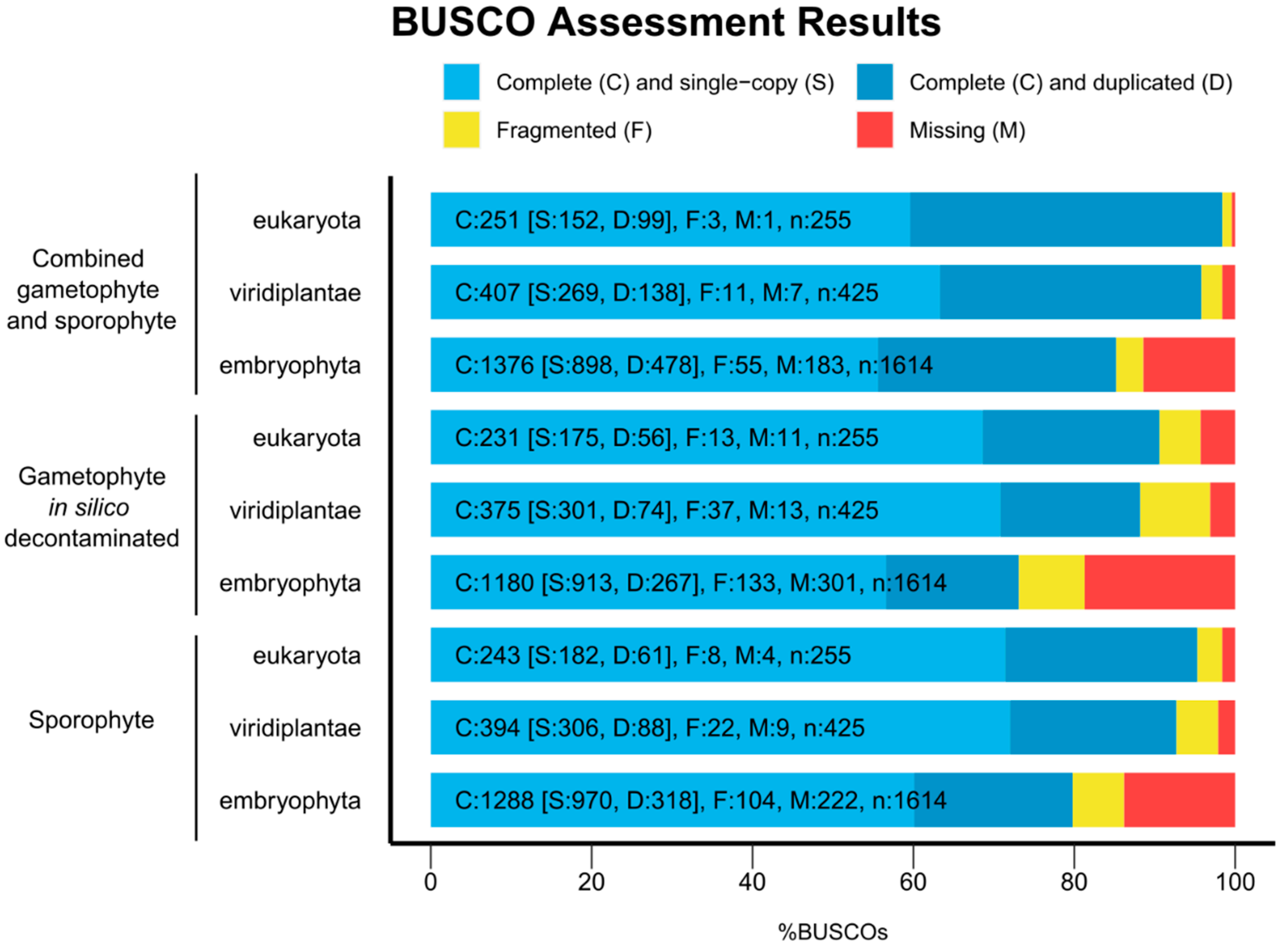
The BUSCO analysis showed always less than 20% (from 18.65% to 0.39%) of missing BUSCOs for all the transcriptomes and for the BUSCO databases Eukaryota, Viridilantae and Embryophyta (Figure 3). The in silico decontaminated gametophyte transcriptome showed the lowest number of complete BUSCOs (90.58% Eukaryota, 88.24% Viridiplantae, 73.11% Embryophyta), whereas the combined sporophyte and gametophyte transcriptome showed a higher number of completed BUSCOs (98.43% Eukaryota, 95.77% Viridiplantae, 85.25% Embryophyta), slightly surpassing the numbers of the sporophyte transcriptome (95.29% Eukaryota, 92.71% Viridiplantae, 79.80% Embryophyta), supporting that complete general species transcriptomes should include sequencing from multiple tissues [31,46]. The proportion of duplicated complete BUSCOs in the combined gametophyte and sporophyte transcriptome increased compared to both single tissue transcriptomes (33.91–39.44% complete duplicated BUSCOs in the combined transcriptome, 19.73–24.24% in the in silico decontaminated gametophyte transcriptome, and 22.34–25.10% in the sporophyte transcriptome). Other transcriptome assemblies from fern species recovered between 53% and 71% of complete Embryophyta BUSCOs, even when including RNA-seq libraries from several tissues [43,44,47]. This new version of the V. speciosa transcriptome completes the transcriptome of the sporophyte [19], increasing the total contig count to 6,488 sequences and increasing the percentage of Eukaryote, Viridiplantae, and Embryophyta BUSCOs to 3.14%, 3.06%, and 5.45%, respectively. Taken together, the BUSCO results and transcript contiguity measures (Table 3) indicate that we have an acceptable transcriptome assembly.
After a search for coding sequences (CDSs) with TransDecoder, the in silico decontaminated gametophyte showed 24,343 CDSs, 14,968 of them complete (61.49%), and the rest being truncated at their 5′ end, 3′ end, or both. The combined gametophyte and sporophyte transcriptome showed 32,726 CDSs, 23,987 of which were completed (73.30%).
BLAST analysis using UniProt as a reference database retrieved 34,405 positive hits for the in silico decontaminated gametophyte transcriptome and 35,712 for the combined gametophyte and sporophyte transcriptome. Table 4 summarizes the number of proteins retrieved before and after filtering the transcriptomes in function of the percentage of identity covered between the query (V. speciosa transcripts) and the target (UniProt database) sequences. As expected, the combined transcriptome surpassed the gametophyte transcriptome in number of assigned proteins for all intervals; however, the combined tissue assembly showed fewer assigned proteins. As mentioned above, this is due to the presence of transcripts with low coverage that passed filtering based on TPM in the gametophyte transcriptome but were purged in the combined tissue transcriptome due to their TPM value readjustment being lower than one. We retrieved 5924 GO terms using the A. thaliana genome protein set (27,136 V. speciosa transcripts with 10,961 BLAST hits from the combined gametophyte and sporophyte transcriptome).

3.3. Differences in Transcript Expression between Tissues

The number of expressed transcripts was 31,821 in the gametophyte and 41,306 in the sporophyte, while 1,083 transcripts did not show mapped reads, according to our read count summary standards, in any tissue. There were 529 transcripts that were expressed in the gametophyte but not in the sporophyte (Supplemental Table S1), while 10,014 transcripts were expressed in the sporophyte but not in the gametophyte (Supplemental Table S2). The overlap between gametophyte and sporophyte-expressed transcripts was not significantly higher than expected by chance (p-value > 0.05, Figure 4). Among the 529 specific transcripts of the gametophyte, 258 were annotated, with 17% of them related to the stress response (defense and disease resistance, abiotic stress, etc.) and 7% being transcription factors, most of them involved in cell growth and differentiation, plant growth and development, as well as stress response. There were also two transcripts derived from transposable elements. Among the 10,014 specific transcripts of the sporophyte, only 3888 could be annotated. Of these, 1.5% of the transcripts were related to stress responses (defense and disease resistance, water deprivation conditions, abiotic stress, including salt and oxidative stress, both clearly related to drought and hydric stress, as well as iron and phosphate starvation). Besides, 3.4% (132 transcripts) were transcription factors, many of them involved in plant growth and development as well as stress responses. Eleven of the transcription factors expressed only in the sporophyte corresponded to different Knotted-like Homeobox genes, key for the distinctive gametophytic and sporophytic developmental programs [48,49,50,51,52], and one transcript corresponded to the Agamous-like MADS-box AGL16 protein that in flowering plants controls flower development [53]. There are also present two transcription factors of the GRAS family, of high importance as regulatory proteins in shoot and root development, stem cell homeostasis, light and hormone signaling, responses to biotic and abiotic stresses, and symbiosis with microorganisms [54]. In addition, 60 transcripts were involved in cell wall formation, including transcripts from genes involved in the synthesis of glucomannans, which constitute the type III primary cell wall in vascular plants and that are exclusively reported in some fern species [55,56]. Curiously, there were also 59 transcripts derived from transposable elements, most of them derived from non-LTR and LTR retrotransposons (43) but also from transposons (16). These elements, which represent 76% of the V. speciosa genome [57], seem to have high and differential activity between the two phases of the life cycle of the species.
Supplemental Tables S3 and S7 show the lists of the most expressed transcripts in the gametophyte and the sporophyte, respectively. The presence of transcription factors involved in development in flowering plants is remarkable among the 1000 most expressed transcripts in the gametophyte. Some of them are involved also in defense response and response to abiotic stress, such as water deprivation conditions. The existence of several transcripts for proteins that control the cell cycle, as well as those involved in the machinery of mRNA splicing, is also remarkable. There are also numerous transcripts related to stress responses (especially defense responses, water deprivation conditions, salt stress, oxidative stress, and osmotic stress) and to chloroplastidial functions. There were less transcription factors among the 1000 transcripts most expressed in the sporophyte, but this set included several transcripts related to cell wall formation, including transcripts from genes involved in the synthesis of glucomannans. The top 1000 most expressed transcripts in the sporophyte also showed transcripts for proteins that control de cell cycle and those involved in the machinery of mRNA splicing, besides many transcripts related to stress response (especially defense response, water deprivation conditions, salt stress, oxidative stress and osmotic stress) and to chloroplastidial functions.
This species is restricted to sheltered, very humid sites and is adapted to extreme low light environments [13,14,15]. Tables S1–S3 and S7 reflect these characteristics since an important fraction of the specific and/or most expressed transcripts are involved in plastid functions and responses to abiotic stresses. In addition, we can find differentiated patterns of gene expression that reflect the ecological, morphological, and physiological differences between the two phases of the life cycle of V. speciosa, such as transcripts from genes involved in cell growth, differentiation, and development, or a greater abundance of transcripts from genes involved in cell wall formation in the sporophyte.
Analysis of enriched GO terms from the most expressed transcripts showed important differences between both the gametophyte and the sporophyte. The most expressed transcripts in the gametophyte (Table S3) showed 240 enriched GO terms from the three different ontologies: 141 from biological process, 27 from molecular function, and 72 from cellular component (Supplemental Tables S4–S6). The most expressed transcripts in the sporophyte (Table S7) showed 416 enriched GO terms: 230 from biological process, 73 from molecular function, and 113 from cellular component ontologies (Supplemental Tables S8–S10). The fold enrichment values from GO terms that were enriched either in the gametophyte or the sporophyte (whose redundancy has been filtered by ReViGO) are shown in Figure 5 and Tables S4–S6,S8–S10. As mentioned above, when comparing these enrichment values of the most expressed transcripts, we can observe differentiated patterns of gene expression between the two phases of the life cycle of V. speciosa. Comparing Tables S4 and S8, we can observe that there are abundant transcripts for proteins involved in metabolic processes, but they are differentiated between the two phases. For example, in the sporophyte, the cinnamic acid and the phenylpropanoid metabolic processes predominates, polysaccharide metabolism, glycine metabolism, and purine metabolism (see Tables S4 and S8). Remarkably, cinnamic acid and phenylpropanoids are central intermediates in the biosynthesis of a set of products, including lignols (precursors to lignin and lignocellulose) among others (flavonoids, isoflavonoids, coumarins, aurones, stilbenes, catechin, and phenylpropanoids). The sporophyte transcripts for proteins involved in metabolic processes related to lignin, cellulose, and glucan biosynthetic pathways and cell wall organization and biogenesis in general are over-represented in this list. Also noteworthy are the transcripts for proteins involved in ATP synthesis, in cytoskeleton organization, or response to stress. However, the latter are much more over-represented in the gametophyte (water, osmotic, heat, salt, ROS, oxidative stress, etc.). Particularly noteworthy are those involved in water control, as highlighted earlier. Both, the sporophyte and the gametophyte have over-representation of transcripts related to plastid assembly, functioning and repair as well as to developmental processes. We can conclude that enriched GO terms related to metabolism and growth are more abundant in the sporophyte, whereas those related to adaptation to extreme conditions and light uptake are more abundant in the gametophyte. Johnson et al. [13] and Makgomol and Sheffield [15] proposed that a very low metabolic rate and effective use of available light are characteristics that allow the gametophyte of V. speciosa to survive in extreme conditions. Our data also support that both phases of the life cycle of V. speciosa are adapted to a constant water supply.
In conclusion, this study complements the previously published transcriptome assembly from V. speciosa sporophyte [19], this time including gametophyte-specific transcripts. Despite its limitations (constrained mostly by the availability of threatened fern individuals and highly contaminated gametophyte samples), the results of this work provide further fern genomic resources and new insights on fern evolution and physiology. The results are even more valuable since the target species is simultaneously a non-model organism and an endangered species. It is also noteworthy that the in silico decontamination method that we apply here can be useful for any heavily contaminated tissue, which should help omics studies of samples whose nature makes them always associated (contaminated) with biological material from other organisms. With the sequencing resources in this and our previous [19] study, we offer a reference transcriptome for the species, unlocking the performance of population genomics and phylogenomics studies on V. speciosa. Being the reproductive success one of the possible causes of the endangered status of the species, the availability of the gene and gene expression data should allow comparative studies on the association between changes in gene sequences or expression and changes of the fitness of individuals and populations of the species. Of course, the gene transcripts that we provide here and in [19] can serve as supporting evidence for gene prediction in a future V. speciosa genome project.

Supplementary Materials

The following supporting information can be downloaded at: https://www.mdpi.com/article/10.3390/genes14010166/s1. Figure S1: Contig coverage distribution per transcriptome; Table S1: Transcripts expressed only in the gametophyte; Table S2: Transcripts expressed only in the sporophyte; Table S3: Top 1000 most expressed transcripts in the gametophyte; Table S4: Biological process GO term enrichment analysis for the gametophyte; Table S5: Molecular function GO term enrichment analysis for the gametophyte; Table S6: Cellular component GO term enrichment analysis for the gametophyte; Table S7: Top 1000 most expressed transcripts in the sporophyte; Table S8: Biological process GO term enrichment analysis for the sporophyte; Table S9: Molecular function GO term enrichment analysis for the sporophyte; Table S10: Cellular component GO term enrichment analysis for the sporophyte.

Author Contributions

Conceptualization, R.M.-B., M.B., and M.A.G.-R.; formal analysis, R.M.-B., M.B., and M.A.G.-R.; funding acquisition, M.A.G.-R.; investigation, R.M.-B., M.B., M.R.-E., and M.A.G.-R.; project administration, M.A.G.-R.; Resources: M.R.-E. and M.A.G.-R.; supervision, M.A.G.-R.; writing—original draft, R.M.-B., M.B., M.R.-E., and M.A.G.-R.; writing—review and editing, R.M.-B., M.B., M.R.-E., and M.A.G.-R. All authors have read and agreed to the published version of the manuscript.

Funding

This research has been financed by the Spanish Ministerio de Economía y Competitividad and FEDER founds, grant: CGL2010-14856 (subprograma BOS).

Data Availability Statement

The resulting database was deposited in FigShare, https://figshare.com/; https://doi.org/10.6084/m9.figshare.21502005, accessed on 4 November 2022.

Acknowledgments

The Dirección General de Gestión del Medio Natural y Espacios Protegidos of the Consejería de Medio Ambiente y Ordenación del Territorio de la Junta de Andalucía (now Consejería de Agricultura, Ganadería, Pesca y Desarrollo Sostenible) authorized and facilitated the sampling of the material. We are highly indebted to Carmen Rodríguez Hiraldo and Jaime Pereña Ortiz who, together with the team of Agentes de Medio Ambiente of the Consejería, helped us with the sampling procedure.

Conflicts of Interest

The authors declare no conflict of interest. The funders had no role in the design of the study; in the collection, analyses, or interpretation of data; in the writing of the manuscript, or in the decision to publish the results.

References

  1. Matz, M.V. Fantastic Beasts and How To Sequence Them: Ecological Genomics for Obscure Model Organisms. Trends Genet. 2018, 34, 121–132. [Google Scholar] [CrossRef]
  2. Ellegren, H. Genome sequencing and population genomics in non-model organisms. Trends Ecol. Evol. 2014, 29, 51–63. [Google Scholar] [CrossRef] [PubMed]
  3. Ekblom, R.; Galindo, J. Applications of next generation sequencing in molecular ecology of non-model organisms. Heredity 2011, 107, 1–15. [Google Scholar] [CrossRef] [Green Version]
  4. da Fonseca, R.R.; Albrechtsen, A.; Themudo, G.E.; Ramos-Madrigal, J.; Sibbesen, J.A.; Maretty, L.; Zepeda-Mendoza, M.L.; Campos, P.F.; Heller, R.; Pereira, R.J. Next-generation biology: Sequencing and data analysis approaches for non-model organisms. Mar. Genom. 2016, 30, 3–13. [Google Scholar] [CrossRef]
  5. Yadav, S.; Stow, A.J.; Dudaniec, R.Y. Microgeographical adaptation corresponds to elevational distributions of congeneric montane grasshoppers. Mol. Ecol. 2021, 30, 481–498. [Google Scholar] [CrossRef]
  6. Hu, S.; Sablok, G.; Wang, B.; Qu, D.; Barbaro, E.; Viola, R.; Li, M.; Varotto, C. Plastome organization and evolution of chloroplast genes in Cardamine species adapted to contrasting habitats. BMC Genom. 2015, 16, 306. [Google Scholar] [CrossRef] [PubMed] [Green Version]
  7. Wang, J.; Li, L.; Zhang, G. A High-Density SNP Genetic Linkage Map and QTL Analysis of Growth-Related Traits in a Hybrid Family of Oysters (Crassostrea gigas × Crassostrea angulata) Using Genotyping-by-Sequencing. G3 Genes|Genomes|Genetics 2016, 6, 1417–1426. [Google Scholar] [CrossRef] [PubMed] [Green Version]
  8. Kubota, S.; Iwasaki, T.; Hanada, K.; Nagano, A.J.; Fujiyama, A.; Toyoda, A.; Sugano, S.; Suzuki, Y.; Hikosaka, K.; Ito, M. A genome scan for genes underlying microgeographic-scale local adaptation in a wild Arabidopsis species. PLoS Genet. 2015, 11, e1005361. [Google Scholar]
  9. Sinha, S.; Schroeder, M.D.; Unnerstall, U.; Gaul, U.; Siggia, E.D. Cross-species comparison significantly improves genome-wide prediction of cis-regulatory modules in Drosophila. BMC Bioinform. 2004, 5, 129. [Google Scholar]
  10. Yang, G.; Mozzicafreddo, M.; Ballarini, P.; Pucciarelli, S.; Miceli, C. An in-silico comparative study of lipases from the Antarctic psychrophilic ciliate Euplotes focardii and the mesophilic congeneric species Euplotes crassus: Insight into Molecular Cold-Adaptation. Mar. Drugs 2021, 19, 67. [Google Scholar] [CrossRef]
  11. Banares Baudet, A.; Blanca, G.; Güemes Heras, J.; Moreno Saiz, J.; Ortiz, S. Red List of Spanish Vascular Flora; Tragsatec: Madrid, Spain, 2008. [Google Scholar]
  12. Moreno, J. Red List of Spanish Vascular Flora; Ministerio de Medio Ambiente, Rural y Marino: Madrid, Spain, 2008. [Google Scholar]
  13. Johnson, G.; Rumsey, F.; Headley, A.; Sheffield, E. Adaptations to extreme low light in the fern Trichomanes speciosum. New Phytol. 2000, 148, 423–431. [Google Scholar] [CrossRef] [Green Version]
  14. Rumsey, F.J.; Vogel, J.C.; Russell, S.J.; Barrett, J.A.; Gibby, M. Population structure and conservation biology of the endangered fern Trichomanes speciosum Willd.(Hymenophyllaceae) at its northern distributional limit. Biol. J. Linn. Soc. 1999, 66, 333–344. [Google Scholar] [CrossRef]
  15. Makgomol, K.; Sheffield, E. Gametophyte morphology and ultrastructure of the extremely deep shade fern, Trichomanes speciosum. New Phytol. 2001, 151, 243–255. [Google Scholar] [CrossRef]
  16. Farrar, D.R. Trichomanes intricatum: The independent Trichomanes gametophyte in the eastern United States. Am. Fern J. 1992, 82, 68–74. [Google Scholar] [CrossRef]
  17. Farrar, D.R.; Mickel, J.T. Vittaria appalachiana: A name for the “Appalachian gametophyte”. Am. Fern J. 1991, 81, 69–75. [Google Scholar] [CrossRef]
  18. Raine, C.A.; Farrar, D.R.; Sheffield, E. A new Hymenophyllum species in the Appalachians represented by independent gametophyte colonies. Am. Fern J. 1991, 81, 109–118. [Google Scholar] [CrossRef]
  19. Bakkali, M.; Martín-Blázquez, R.; Ruiz-Estévez, M.; Garrido-Ramos, M.A. De Novo Sporophyte Transcriptome Assembly and Functional Annotation in the Endangered Fern Species Vandenboschia speciosa (Willd.) G. Kunkel. Genes 2021, 12, 1017. [Google Scholar] [CrossRef]
  20. Ruiz-Estévez, M.; Bakkali, M.; Martín-Blázquez, R.; Garrido-Ramos, M.A. Identification and characterization of TALE homeobox genes in the endangered fern Vandenboschia speciosa. Genes 2017, 8, 275. [Google Scholar] [CrossRef] [Green Version]
  21. Ruiz-Estévez, M.; Bakkali, M.; Martín-Blázquez, R.; Garrido-Ramos, M.A. Differential expression patterns of MIKCC-type MADS-box genes in the endangered fern Vandenboschia speciosa. Plant Gene 2017, 12, 50–56. [Google Scholar] [CrossRef]
  22. Ruiz-Ruano, F.; Navarro-Domínguez, B.; Camacho, J.; Garrido-Ramos, M. Characterization of the satellitome in lower vascular plants: The case of the endangered fern Vandenboschia speciosa. Ann. Bot. 2019, 123, 587–599. [Google Scholar] [CrossRef]
  23. Li, H.; Durbin, R. Fast and accurate short read alignment with Burrows–Wheeler transform. Bioinformatics 2009, 25, 1754–1760. [Google Scholar] [CrossRef] [PubMed] [Green Version]
  24. Bağcı, C.; Patz, S.; Huson, D.H. DIAMOND+ MEGAN: Fast and easy taxonomic and functional analysis of short and long microbiome sequences. Curr. Protoc. 2021, 1, e59. [Google Scholar] [CrossRef] [PubMed]
  25. Haas, B.J.; Papanicolaou, A.; Yassour, M.; Grabherr, M.; Blood, P.D.; Bowden, J.; Couger, M.B.; Eccles, D.; Li, B.; Lieber, M. De novo transcript sequence reconstruction from RNA-seq using the Trinity platform for reference generation and analysis. Nat. Protoc. 2013, 8, 1494–1512. [Google Scholar] [CrossRef] [PubMed]
  26. Altschul, S.F.; Madden, T.L.; Schäffer, A.A.; Zhang, J.; Zhang, Z.; Miller, W.; Lipman, D.J. Gapped BLAST and PSI-BLAST: A new generation of protein database search programs. Nucleic Acids Res. 1997, 25, 3389–3402. [Google Scholar] [CrossRef] [Green Version]
  27. Camacho, C.; Coulouris, G.; Avagyan, V.; Ma, N.; Papadopoulos, J.; Bealer, K.; Madden, T.L. BLAST+: Architecture and applications. BMC Bioinform. 2009, 10, 421. [Google Scholar] [CrossRef] [Green Version]
  28. Patro, R.; Duggal, G.; Love, M.I.; Irizarry, R.A.; Kingsford, C. Salmon provides fast and bias-aware quantification of transcript expression. Nat. Methods 2017, 14, 417–419. [Google Scholar] [CrossRef] [Green Version]
  29. Li, W.; Godzik, A. Cd-hit: A fast program for clustering and comparing large sets of protein or nucleotide sequences. Bioinformatics 2006, 22, 1658–1659. [Google Scholar] [CrossRef] [Green Version]
  30. Simão, F.A.; Waterhouse, R.M.; Ioannidis, P.; Kriventseva, E.V.; Zdobnov, E.M. BUSCO: Assessing genome assembly and annotation completeness with single-copy orthologs. Bioinformatics 2015, 31, 3210–3212. [Google Scholar] [CrossRef] [Green Version]
  31. Waterhouse, R.M.; Seppey, M.; Simão, F.A.; Manni, M.; Ioannidis, P.; Klioutchnikov, G.; Kriventseva, E.V.; Zdobnov, E.M. BUSCO applications from quality assessments to gene prediction and phylogenomics. Mol. Biol. Evol. 2018, 35, 543–548. [Google Scholar] [CrossRef] [Green Version]
  32. Li, H.; Handsaker, B.; Wysoker, A.; Fennell, T.; Ruan, J.; Homer, N.; Marth, G.; Abecasis, G.; Durbin, R. The sequence alignment/map format and SAMtools. Bioinformatics 2009, 25, 2078–2079. [Google Scholar] [CrossRef] [Green Version]
  33. Anders, S.; Pyl, P.T.; Huber, W. HTSeq—A Python framework to work with high-throughput sequencing data. Bioinformatics 2015, 31, 166–169. [Google Scholar] [CrossRef] [PubMed] [Green Version]
  34. Love, M.I.; Huber, W.; Anders, S. Moderated estimation of fold change and dispersion for RNA-seq data with DESeq2. Genome Biol. 2014, 15, 550. [Google Scholar] [CrossRef] [PubMed]
  35. Mi, H.; Muruganujan, A.; Ebert, D.; Huang, X.; Thomas, P.D. PANTHER version 14: More genomes, a new PANTHER GO-slim and improvements in enrichment analysis tools. Nucleic Acids Res. 2019, 47, D419–D426. [Google Scholar] [CrossRef] [PubMed]
  36. Thomas, P.D.; Campbell, M.J.; Kejariwal, A.; Mi, H.; Karlak, B.; Daverman, R.; Diemer, K.; Muruganujan, A.; Narechania, A. PANTHER: A library of protein families and subfamilies indexed by function. Genome Res. 2003, 13, 2129–2141. [Google Scholar] [CrossRef] [PubMed] [Green Version]
  37. Benjamini, Y.; Hochberg, Y. Controlling the false discovery rate: A practical and powerful approach to multiple testing. J. R. Stat. Soc. Ser. B (Methodol.) 1995, 57, 289–300. [Google Scholar] [CrossRef]
  38. Supek, F.; Bošnjak, M.; Škunca, N.; Šmuc, T. REVIGO summarizes and visualizes long lists of gene ontology terms. PLoS ONE 2011, 6, e21800. [Google Scholar] [CrossRef] [Green Version]
  39. Ćuković, K.; Dragićević, M.; Bogdanović, M.; Paunović, D.; Giurato, G.; Filipović, B.; Subotić, A.; Todorović, S.; Simonović, A. Plant regeneration in leaf culture of Centaurium erythraea Rafn. Part 3: De novo transcriptome assembly and validation of housekeeping genes for studies of in vitro morphogenesis. Plant Cell Tissue Organ Cult. (PCTOC) 2020, 141, 417–433. [Google Scholar] [CrossRef]
  40. Niu, S.-C.; Xu, Q.; Zhang, G.-Q.; Zhang, Y.-Q.; Tsai, W.-C.; Hsu, J.-L.; Liang, C.-K.; Luo, Y.-B.; Liu, Z.-J. De novo transcriptome assembly databases for the butterfly orchid Phalaenopsis equestris. Sci. Data 2016, 3, 160083. [Google Scholar] [CrossRef] [Green Version]
  41. Wang, Y.; Li, X.; Zhou, W.; Li, T.; Tian, C. De novo assembly and transcriptome characterization of spruce dwarf mistletoe Arceuthobium sichuanense uncovers gene expression profiling associated with plant development. BMC Genom. 2016, 17, 771. [Google Scholar] [CrossRef] [Green Version]
  42. Hu, R.; Yu, C.; Wang, X.; Jia, C.; Pei, S.; He, K.; He, G.; Kong, Y.; Zhou, G. De novo transcriptome analysis of Miscanthus lutarioriparius identifies candidate genes in rhizome development. Front. Plant Sci. 2017, 8, 492. [Google Scholar] [CrossRef] [Green Version]
  43. Sigel, E.M.; Schuettpelz, E.; Pryer, K.M.; Der, J.P. Overlapping patterns of gene expression between gametophyte and sporophyte phases in the fern Polypodium amorphum (Polypodiales). Front. Plant Sci. 2018, 9, 1450. [Google Scholar] [CrossRef]
  44. Geng, Y.; Cai, C.; McAdam, S.A.; Banks, J.A.; Wisecaver, J.H.; Zhou, Y. A de novo transcriptome assembly of Ceratopteris richardii provides insights into the evolutionary dynamics of complex gene families in land plants. Genome Biol. Evol. 2021, 13, evab042. [Google Scholar] [CrossRef]
  45. Jo, Y.; Choi, H.; Kim, S.-M.; Kim, S.-L.; Lee, B.C.; Cho, W.K. Integrated analyses using RNA-Seq data reveal viral genomes, single nucleotide variations, the phylogenetic relationship, and recombination for Apple stem grooving virus. BMC Genom. 2016, 17, 579. [Google Scholar] [CrossRef] [Green Version]
  46. Torrens-Spence, M.; Fallon, T.; Weng, J. A workflow for studying specialized metabolism in nonmodel eukaryotic organisms. In Methods in Enzymology; Elsevier: Amsterdam, The Netherlands, 2016; Volume 576, pp. 69–97. [Google Scholar]
  47. Aya, K.; Kobayashi, M.; Tanaka, J.; Ohyanagi, H.; Suzuki, T.; Yano, K.; Takano, T.; Yano, K.; Matsuoka, M. De novo transcriptome assembly of a fern, Lygodium japonicum, and a web resource database, Ljtrans DB. Plant Cell Physiol. 2015, 56, e5. [Google Scholar] [CrossRef] [Green Version]
  48. Liu, Y.; You, S.; Taylor-Teeples, M.; Li, W.L.; Schuetz, M.; Brady, S.M.; Douglas, C.J. BEL1-LIKE HOMEODOMAIN6 and KNOTTED ARABIDOPSIS THALIANA7 interact and regulate secondary cell wall formation via repression of REVOLUTA. Plant Cell 2014, 26, 4843–4861. [Google Scholar] [CrossRef] [PubMed] [Green Version]
  49. Gao, J.; Yang, X.; Zhao, W.; Lang, T.; Samuelsson, T. Evolution, diversification, and expression of KNOX proteins in plants. Front. Plant Sci. 2015, 6, 882. [Google Scholar] [CrossRef] [PubMed] [Green Version]
  50. Horst, N.A.; Katz, A.; Pereman, I.; Decker, E.L.; Ohad, N.; Reski, R. A single homeobox gene triggers phase transition, embryogenesis and asexual reproduction. Nat. Plants 2016, 2, 15209. [Google Scholar] [CrossRef]
  51. Sakakibara, K.; Ando, S.; Yip, H.K.; Tamada, Y.; Hiwatashi, Y.; Murata, T.; Deguchi, H.; Hasebe, M.; Bowman, J.L. KNOX2 genes regulate the haploid-to-diploid morphological transition in land plants. Science 2013, 339, 1067–1070. [Google Scholar] [CrossRef]
  52. Furumizu, C.; Alvarez, J.P.; Sakakibara, K.; Bowman, J.L. Antagonistic roles for KNOX1 and KNOX2 genes in patterning the land plant body plan following an ancient gene duplication. PLoS Genet. 2015, 11, e1004980. [Google Scholar] [CrossRef] [PubMed] [Green Version]
  53. Hu, J.-Y.; Zhou, Y.; He, F.; Dong, X.; Liu, L.-Y.; Coupland, G.; Turck, F.; de Meaux, J. miR824-regulated AGAMOUS-LIKE16 contributes to flowering time repression in Arabidopsis. Plant Cell 2014, 26, 2024–2037. [Google Scholar] [CrossRef] [PubMed] [Green Version]
  54. Hirsch, S.; Oldroyd, G.E. GRAS-domain transcription factors that regulate plant development. Plant Signal. Behav. 2009, 4, 698–700. [Google Scholar] [CrossRef] [PubMed] [Green Version]
  55. Silva, G.B.; Ionashiro, M.; Carrara, T.B.; Crivellari, A.C.; Tiné, M.A.; Prado, J.; Carpita, N.C.; Buckeridge, M.S. Cell wall polysaccharides from fern leaves: Evidence for a mannan-rich Type III cell wall in Adiantum raddianum. Phytochemistry 2011, 72, 2352–2360. [Google Scholar] [CrossRef] [PubMed]
  56. Singh, S.; Singh, G.; Arya, S.K. Mannans: An overview of properties and application in food products. Int. J. Biol. Macromol. 2018, 119, 79–95. [Google Scholar] [CrossRef] [PubMed]
  57. Ruiz-Ruano, F.J.; Navarro-Domínguez, B.; Camacho, J.P.M.; Garrido-Ramos, M.A. Transposable element landscapes illuminate past evolutionary events in the endangered fern Vandenboschia speciosa. Genome 2022, 65, 95–103. [Google Scholar] [CrossRef]
Figure 1. Species distribution of BLAST results from V. speciosa transcriptomes. The top 10 most represented species from BLASTx analysis against UniProt are shown for each transcriptome. ARATH: Arabidopsis thaliana. HUMAN: Homo sapiens sapiens. MOUSE: Mus musculus. DICDI: Dictyostelium discoideum. ORYSJ: Oryza sativa subsp. japonica. SCHPO: Schizosaccharomyces pombe. ORYSI: Oryza sativa subsp. indica. DROME: Drosophila melanogaster. BOVIN: Bos taurus. YEAST: Saccharomyces cerevisiae. CAEEL: Caenorhabditis elegans. XENLA: Xenopus laevis. DANRE: Danio rerio. TOBAC: Nicotiana tabacum. MAIZE: Zea mays. SOLLC: Solanum lycopersicon.
Genes 14 00166 g001
Figure 2. Contig length distribution from the gametophyte and the combined transcriptomes. The X axis shows the contig lengths. The Y axis shows the frequency of contigs (transformed to proportion) for each contig length value. The blue vertical line shows the N50 value, and the red vertical line shows the Ex90N50 value.
Genes 14 00166 g002
Figure 3. BUSCO assessment results. The X axis shows the percentage of BUSCOs found for each analysis. The Y axis shows the transcriptome (1st column) and BUSCO taxonomical level (2nd column). Each color-coded bar shows the number of complete (C, including the ones found as single copies, S, and multiple copies, D), fragmented (F), and missing (M) BUSCOs from the total analyzed (n).
Genes 14 00166 g003
Figure 4. Overlapping and uniquely expressed transcripts per tissue. Colored numbers outside the Venn diagram show the total number of transcripts expressed in the gametophyte (orange) and the sporophyte (green).
Genes 14 00166 g004
Figure 5. Enriched GO terms from the 1000 most expressed genes in the gametophyte and sporophyte (top row) and the transcripts expressed only in either the gametophyte or the sporophyte (bottom row). Each bar plot shows the fold enrichment of GO terms from biological process (left column) and molecular function (right column) ontologies. The X axis shows the fold enrichment of each term from either the gametophyte (red) or the sporophyte (blue) gene lists. The Y axis shows the GO term and its GO identifier. For the top 1000 transcript plots, only GO terms retained by the ReViGO analysis and showing the logarithm of the quotient between gametophyte and sporophyte fold enrichments greater than 0.5 are shown.
Genes 14 00166 g005
Table 1. RNA sequencing statistics. The total number of read pairs and the total number of sequenced nucleotides are shown for all sequenced tissues, before and after the quality check (QC).
TissueRaw DataAfter QC
Number of paired-end readsGametophyte48.7 million24.7 million
Combined tissues115 million89.9 million
Sporophyte66.3 million65.2 million
Number of basesGametophyte4900 million2500 million
Combined tissues10,800 million9090 million
Sporophyte6700 million6590 million
Table 2. BLAST analysis species representation. The percentage of plant, animal, fungi and protzoa species detected among the 50 most represented species is shown for the four assemblies. The last row shows the percentage of BLAST hits assigned to A. thaliana for the whole BLASTx analysis.
Percentage of BLAST HitsTaxonRaw
Gametophyte
In Silico Decontaminated GametophyteCombined TissuesSporophyte
50 most represented speciesPlant30606068
Animal40303026
Fungi2410106
Protozoa6000
All speciesA. thaliana30.2955.8855.8270.00
Table 3. De novo transcriptome assembly statistics. The figures shown represent the total number of transcripts, percentage of guanine cytosine, N50, N70, N9, and Ex90N50 statistics (see text for details), number of transcripts with length equal or less to the Ex90N50, sizes of the smallest and largest contigs, number of contigs greater than 1000 and 10,000 base pairs long, median contig length, average contig length, and total number of assembled bases. The columns show the three different approaches (in silico decontaminated gametophyte, combined gametophyte and sporophyte, and sporophyte transcriptomes), before and after quality check and filtering steps. The acronym bp stands for base pairs.
GametophyteCombined TissuesSporophyte
Before
Filtering
After
Filtering
Before
Filtering
After
Filtering
Before
Filtering
After
Filtering
Total transcripts44,45543,13988,38342,91884,75936,430
Percent GC45.4845.4745.2345.1845.1845.18
Contig N50 (bp)210121022264224319552085
Contig N70 (bp)150914001632164013321511
Contig N90 (bp)786659807855479729
Ex90N50 (bp)223622352511261520392299
Number transcripts corresponding to the Ex90 peak11,44511,51913,66513,74314,64521,543
Size of the smallest contig (bp)201201194196201201
Size of the largest contig (bp)95419541167151671513,22513,224
Number of contigs greater than 1 Kb long25,78616,60550,27225,45435,80120,532
Number of contigs greater than 10 Kb long0036201812
Median contig length (bp)12231227123412997221197
Average contig (bp)1462.281465.001504.371534.611144.861437.37
Total number of assembled bases65,005,80063,198,512132,960,36165,862,24697,037,55152,363,571
Table 4. Number of proteins from the UniProt database on which the V. speciosa transcripts align along a percentage of their length. The first column shows the length of the interval of the BLAST results length, in increments of ten. For each de novo assembly, in silico decontaminated gametophyte (Gametophyte) and combined in silico gametophyte and sporophyte (Combined tissues), we showed the “Number of proteins” within a BLAST result length interval, as well as the “Accumulated number of proteins”, for either the assembly “Before filtering” and “After filtering” contigs by expression and clustering by homology. Protein homology was assigned to each contig through a BLASTx analysis against the UniProt database.
GametophyteCombined Tissues
Before FilteringAfter FilteringBefore FilteringAfter Filtering
Percentage of Covered Length IntervalsNumber of ProteinsAccumulated Number of ProteinsNumber of ProteinsAccumulated Number of ProteinsNumber of ProteinsAccumulated Number of ProteinsNumber of ProteinsAccumulated Number of Proteins
91–10030973097309730973739373935853585
81–9013284425132544221564530314935078
71–8093853639375359107063739976075
61–706496012646600576971426906765
51–605896601587659271378556197384
41–506537254648724075386086358019
31–406347888623786377793856238642
21–307698657762862586610,2516539295
11–2084995068449469103611,28772210,017
1–104279933426989558411,87139510,412
Disclaimer/Publisher’s Note: The statements, opinions and data contained in all publications are solely those of the individual author(s) and contributor(s) and not of MDPI and/or the editor(s). MDPI and/or the editor(s) disclaim responsibility for any injury to people or property resulting from any ideas, methods, instructions or products referred to in the content.