Article

The Complete Mitochondrial Genome of Torix tukubana (Annelida: Hirudinea: Glossiphoniidae)

1
Key Laboratory of Livestock Infectious Diseases in Northeast China, Ministry of Education, College of Animal Science and Veterinary Medicine, Shenyang Agricultural University, Shenyang 110866, China
2
College of Science and Engineering, Flinders University, Adelaide 5024, Australia
3
Liaoning Panjin Wetland Ecosystem National Observation and Research Station, Shenyang 110866, China
*
Author to whom correspondence should be addressed.
These authors contributed equally to this work and shared the first authorship.
Genes 2023, 14(2), 388; https://doi.org/10.3390/genes14020388
Submission received: 4 January 2023 / Revised: 29 January 2023 / Accepted: 29 January 2023 / Published: 1 February 2023
(This article belongs to the Section Animal Genetics and Genomics)

Abstract

:
Torix tukubana is a poorly understood proboscidate leech species, generally an ectoparasite on amphibian species. In this study, the complete mitochondrial genome (mitogenome) of T. tukubana was sequenced using next-generation sequencing (NGS), and the essential characteristics, gene arrangement, and phylogenetic relationship were analyzed. The results showed that the T. tukubana mitogenome was 14,814 bp in length, consisting of 13 protein-coding genes (PCGs), 22 tRNAs, 2 rRNAs, and 1 control region (CR). The mitogenome composition presented a strong A + T bias (73.6%). All tRNAs had the typical clover structure except the trnS1 (TCT), whose dihydrouridine (DHU) arm was short, having only one complementary base pair. Additionally, 8 gene order patterns were identified among 25 known Hirudinea species, and T. tukubana was identical to the Hirudinea ground pattern. A phylogenetic analysis based on 13 PCGs indicated that all the studied species clustered into three main clades. The relationships among Hirudinea species were basically consistent with their gene arrangement results, but different from their morphological taxonomy. T. tukubana was in the monophyletic group of Glossiphoniidae, a finding consistent with previous research. Our results provided the essential characteristics of the T. tukubana mitogenome. As the first complete mitogenome of Torix, it could offer valuable information for a systematic understanding of the Hirudinea species.

1. Introduction

Hirudinea, or leeches, are clitellate annelids and are widely distributed in various ecosystems (e.g., aquatic and moist terrestrial) on all continents [1]. Leeches are usually ectoparasites or predators and play an integral role in the freshwater system [2]. The phylogenetic relationship between Hirudinea subclasses has always been disputable in terms of their morphological taxonomy and molecular phylogeny. Molecular technology therefore seems necessary for simplified and accurate identification due to the morphological intraspecific variability exhibited among Hirudinea species [3]. Because of their rapid mutation rate and lack of recombination, mitochondrial DNAs have been extensively applied in population genetics and intra- or inter-specific phylogenetic studies [4,5]. The comparison of complete mitochondrial genomes (mitogenomes) has become increasingly popular in phylogenetic relationship analyses [6,7,8] because complete mitogenomes are more informative than single genes [9]. Additionally, complete mitogenomes can reveal some genome-level characteristics, such as gene rearrangement [10,11,12], which could provide more information for understanding the phylogenetic relationships between species [13,14]. Although they are of significant help in phylogenetic and population genetics, only a few complete mitogenomes of the approximately 680 Hirudinea species have been determined so far [6,15,16,17].
Torix tukubana (Annelida: Hirudinea: Glossiphoniidae) is a poorly understood proboscidate leech species, generally an ectoparasite on amphibian species [3,18]. According to previous records, T. tukubana inhabit mountain streams in Japan, and their host amphibians consist of only three Japanese frogs, Rana japonica, Rana tagoi, and Onychodactylus japonicas [19]. However, individual T. tukubana (morphological identification) specimens have recently been found to parasitize on the body surface of Rana dybowskii (which has great medicinal and economic value) in Dandong, Northern China. Because T. tukubana has been considered endemic to Japan, we amplified the specimen’s cox1 gene and conducted nucleotide alignment to confirm the accuracy of the morphological identification. We found that the cox1 gene of the specimen was identical to that of T. tukubana (GenBank: LC538265). Like other Glossiphoniidae parasites, T. tukubana can cause a range of impairments in their hosts, including tissue damage and weight loss [20]. In addition, infestation of T. tukubana is also associated with Rickettsia infection [20]. The outbreak of T. tukubana is doubtlessly a potential menace to the aquaculture industry and the wild resource of R. dybowskii.
In previous studies, the understanding of phylogenetic relationships among leech species, including T. tukubana, were mainly based on short fragments, such as mitochondrial (12S rRNA, cox1, nad1) and nuclear ribosomal internal transcribed spacer (ITS) gene sequences [17,21,22,23,24]. Therefore, the complete mitogenome might provide new insight into the phylogenetic relationships between leech species. Currently, the research on T. tukubana consists mainly of morphological descriptions, with few molecular studies based on the mitochondrial cox1 gene [18]. There has been no report of the complete mitogenome of T. tukubana. In this study, the complete mitogenome of T. tukubana was obtained utilizing next-generation sequencing (NGS). As this is the first mitogenome in the genus Torix, the results will enrich the Hirudinea molecular database. Furthermore, the phylogenetic relationships among 25 complete Hirudinea mitogenomes were analyzed, the results of this analysis are of great importance to the future study of their phylogenetic relationship and will improve our understanding of the genetic biodiversity of Hirudinea.

2. Materials and Methods

2.1. Sample Collection and DNA Extraction

Twenty-six T. tukubana specimens were collected from the body surface of an R. dybowskii, which inhabited the mountain of Dandong City (40.884° N; 124.762° E), Liaoning Province, China, in October 2020. The body sizes were from 0.56 cm to 0.89 cm (0.765 cm ± 0.034 SD). The T. tukubana specimens were identified based on the external morphological features described by Kambayashi et al. [19] and cox1 gene alignment.
Before DNA isolation, the specimen was immediately stored in liquid nitrogen. A TIANamp Marine Animals DNA Kit (Tiangen, China) was used to extract the total genomic DNA from one T. tukubana, and the degree of concentration and purity of the DNA was evaluated using a Thermo Scientific NanoDrop 2000. The integrity was evaluated using an Agilent 2100 Bioanalyzer and electrophoresis on a 1% agarose gel.

2.2. Genome Assembly and Annotation

Briefly, after the genomic DNA was randomly broken into fragments using a Covairs ultrasonic breaker, a PE400 genomic DNA library was constructed based on the Whole Genome Shotgun (WGS) strategy and sequenced utilizing an Illumina NovaSeq 6000 instrument (Illumina, USA). More than 2.58 Gb of raw reads were obtained, and at least 1.67 Gb of clean reads were obtained from the raw reads after removing adaptor sequences using AdapterRemoval v2 [25] and filtering low-quality sequences using SOAPdenovo2 [26]. The obtained clean data were assembled using A5-miseq v20150522 [27] and SPAdesv3.9.0 [28] to construct contigs and scaffolds. A colinear analysis for splicing sequences was conducted using mummer v3.1 [29] and the complete mitogenome sequence was then revised and verified using pilon v1.18 [30]. All these procedures were performed by Shanghai Personal Biotechnology Co., Ltd., China.
The locations of protein-coding genes (PCGs) and rRNA genes were preliminarily predicted using GeSeq [31] and MITOS [32], and the precise locations were confirmed using related Hirudinea mitogenomes from GenBank. The initiation and termination codons were identified using ORF finder and Blastn of NCBI, according to their alignment with other related species, and the rearrangements of mitochondrial genes in the Hirudinea species were examined simply by observation. MITOS [32] was used to predict and annotate the locations and secondary structures of tRNA genes, and these were then visualized using the online software TBI-forna (http://rna.tbi.univie.ac.at/forna/, accessed on 4 January 2022). The nucleotide composition and the relative synonymous codon usage (RSCU) were determined using MEGA 7 [33].
AT skew = (A − T)/(A + T) and GC skew = (G − C)/(G + C) were analyzed to describe base composition [34]. A graphical diagram of the complete mitogenome was drawn using the online mitochondrial visualization tool mtviz (http://pacosy.informatik.uni-leipzig.de/mtviz/mtviz, accessed on 4 January 2022). Finally, the complete mitochondrial DNA sequence was uploaded to the GenBank database with accession number OL779256.

2.3. Phylogenetic Analysis

The mitogenome sequences of 24 Hirudinea species other than T. tukubana were obtained from GenBank for phylogenetic analysis (Table 1). The nucleotide sequences of 13 PCGs were aligned using the online software Clustal Omega (https://www.ebi.ac.uk/Tools/msa/clustalo/, accessed on 4 January 2022). Moreover, the poorly aligned regions and divergent sites were removed utilizing the online software Gblocks (http://molevol.cmima.csic.es/castresana/Gblocks_server.html, accessed on 4 January 2022) [35]. The online software IQ-TREE (http://iqtree.cibiv.univie.ac.at/, accessed on 6 January 2022) [36] was used to build a maximum likelihood (ML) phylogenetic tree which was visualized using iTOL (https://itol.embl.de, accessed on 6 January 2022) [37]. In addition, the mitogenomes of Duplodicodrilus schmardae (KT429015.1) and Tubifex tubifex (MW690579.1) were employed as an outgroup taxon.

3. Results and Discussion

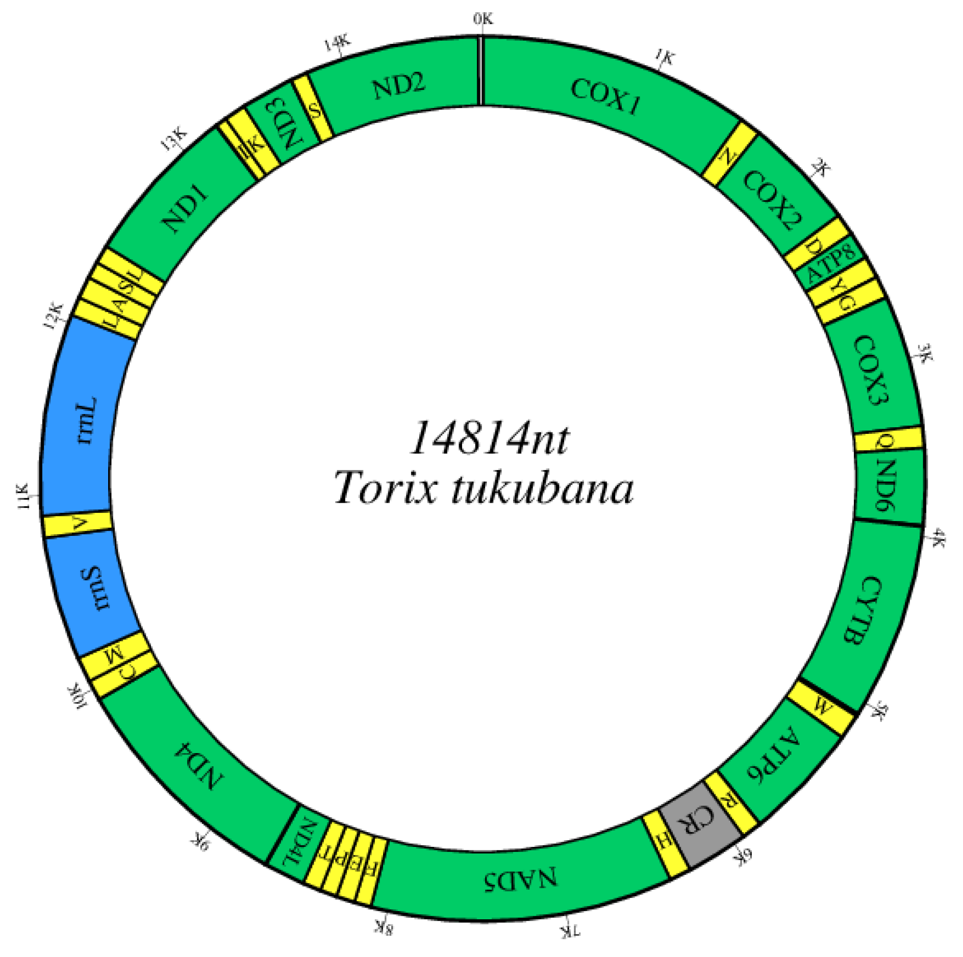
3.1. Genome Composition

After assembly, a circular mitogenome with a length of 14,814 bp was ultimately generated (Figure 1), and this was in the range of the known Hirudinea mitogenomes (14,407–16,161 bp). There were strong similarities between the whole T. tukubana mitogenome and those of closely related species (84.75% with Hemiclepsis yangtzenensis, 82.45% with Theromyzon tessulatum, and 78.62% with Helobdella robusta).
The gene content of the T. tukubana mitogenome, including 13 PCGs, two rRNA genes, 22 tRNA genes, and a control region (CR; Table 2 and Figure 1), was similar to that of all known Hirudinea. All 37 genes were coded on the heavy chain, as in all known Hirudinea. Like most Hirudinea mitogenomes, the mitogenome of T. tukubana was closely aligned, with only a small number of bases overlapping adjacent genes, indicating efficient RNA transcription and protein translation (Table 2).
The genome composition (A: 35.2%, G: 11.9%, T: 38.4%, C: 14.5%) presented a strong A + T bias (73.6%), and showed a negative AT skew (−0.043) and GC skew (−0.098). The AT skew was higher than that of most Hirudinea mitogenomes in this study, but lower than that of Acanthobdella peledina (−0.007), Codonobdella sp. IK-2021 (−0.030), Erpobdella japonica (−0.025), Erpobdella sp. JP-2021 (−0.032), Erpobdella testacea (−0.004), H. yangtzenensis (−0.034), and Ozobranchus jantseanus (−0.033) (Table 1). The GC skew of T. tukubana was lower than that of most of the other previously sequenced species (Table 1). However, different regions of the T. tukubana mitogenome had similar A + T contents, ranging from 73.1% (CR) to 74.5% (tRNAs; Table S1).

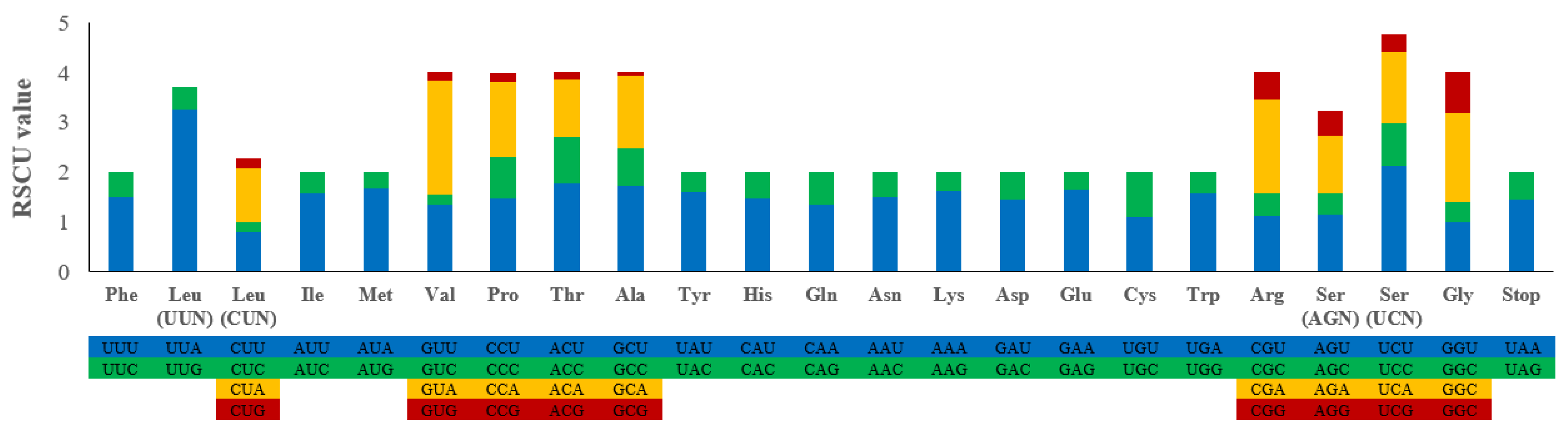
3.2. Protein-Coding Genes

The PCG region was 11,097 bp in length and accounted for 74.91% of the T. tukubana mitogenome. The size of the 13 PCGs ranged between 159 bp and 1698 bp (Table 2). Each PCG was initiated by a canonical ATN codon, and the termination codons were TAA and TAG. Of the 13 PCGs, three (atp8, nad4, and nad3) terminated with TAG, and the other 10 PCGs used a typical TAA termination codon (Table 2). The number of bases in the 13 PCGs followed the pattern T > A > C > G, and they showed a strong A + T bias (73.3%; Table S1). The bias of T and C was strong, and the AT skew and GC skew were −0.080 and −0.143, respectively. The T. tukubana mitogenome had a slightly negative AT skew, indicating a higher incidence of the T than of the A nucleotide, a feature observed in other mitogenomes where the AT skew is negative. In addition, the T. tukubana mitogenome had lower GC skew values than most other mitogenomes, while its AT skew values were higher (Table S1).
The average codon frequencies of the PCGs were calculated, and the results showed that the RSCU values of the preference codons were all greater than 1 (Figure 2). The results of this study showed that the codons of all the PCGs had a strong preference. The RSCU values of NNU and NNA (namely the codon of the third site, U or A) were mostly higher than 1, and the frequency of usage was higher, which was consistent with Whitmania spp. [15].

3.3. tRNAs, rRNAs, and CR

The T. tukubana mitogenome contained 22 tRNA genes, all located on the H strand (Figure 1, Table 2), a characteristic shared by most Hirudinea. The tRNA sequences ranged from 59 bp to 69 bp, with strong A + T bias (73.9%) and positive AT skew (0.034; Table S1). All of the 22 tRNAs showed the typical cloverleaf structure except trnS1 (TCT), whose dihydrouridine (DHU) arm was short, having only one complementary base pair (Figure S1), a characteristic not seen in Whitmania spp. [15].
The rrnS and rrnL genes were located between trnM and trnV and between trnV and trnL1, and they were 735 bp and 1160 bp in length, respectively. The A+T content of this rRNA region was 74.5% and showed a positive AT skew (0.090) and GC skew (0.043). The CR was 402 bp, situated between trnR and trnH, and had a positive AT skew (0.130) and negative GC skew (−0.056; Table S1). The CR length of Hirudinea, ranging from 77 bp to 1670 bp, showed significant length differences [6,15,17,38]. The CR of the T. tukubana mitogenome was of a moderate size. That is a common phenomenon, as the CR is generally considered to have the most significant length variation in the mitogenome [10].

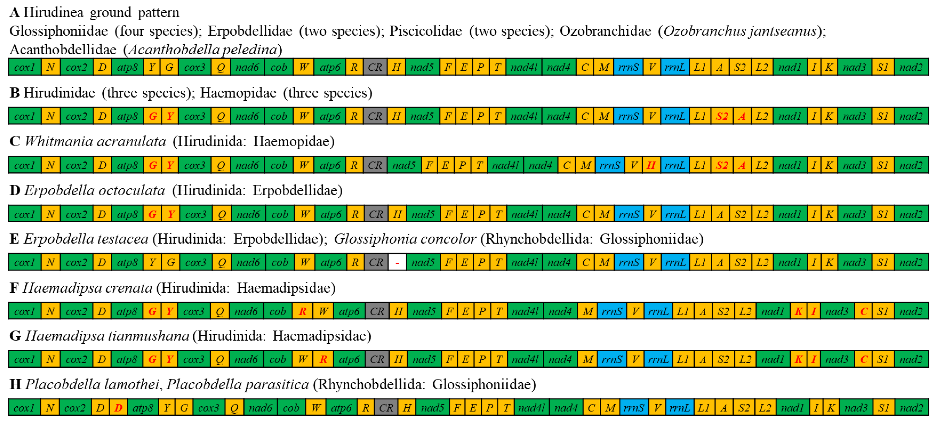
3.4. Gene Arrangement

All 37 genes of all the studied Hirudinea species, including T. tukubana, were transcribed from the same strand (Table 2, Figure 1), which was identical to that of all known Hirudinea species in previous studies [6,15,17]. The gene order and orientation of the 13 PCGs in T. tukubana were consistent with those of all previously sequenced annelids. However, tRNAs are generally believed to change their relative position much faster than the PCGs and rRNAs. The rearrangement of tRNAs has been identified in known annelids sequences [6]. Among all 25 known Hirudinea mitogenomes, eight different gene order patterns (A to H) have been identified (Figure 3). The gene order of the T. tukubana mitogenome was the same as the ground pattern (A) which was found in most of the studied Hirudinea mitogenomes. Another representative gene order pattern (B) was identified in three Hirudinidae species and three Haemopidae species. The main differences between these two patterns were translocations between trnG and trnY and trnA and trnS2 (TGA). Patterns E and H were found in two species. Compared with the ground pattern (A), pattern E included a deletion of trnH between CR and nad5, while pattern H included an extra trnD between cox2 and atp8. The other four patterns were identified in one species. In comparison with pattern B, pattern C included a relocation of trnH, while pattern D included relocations of trnA and trnS2 (TGA). Patterns F and G included translocations of trnR and trnC and a rearrangement between trnK and trnI which differed from the other six patterns. The difference between patterns F and G was the gene order of trnR and trnW.

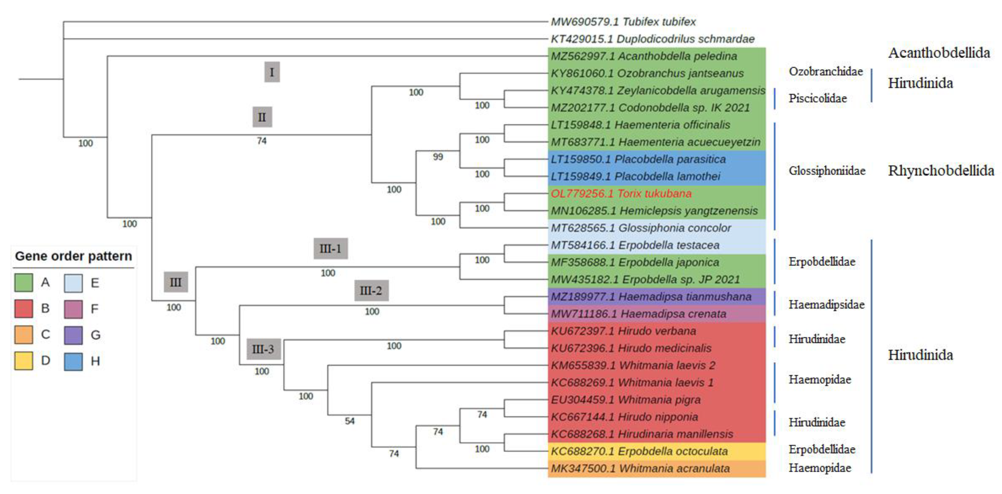
3.5. Phylogenetic Analysis

In this study, the concatenated PCGs derived from the 25 Hirudinea mitogenomes belonging to 8 families of 14 genera were used for the phylogenetic relationship analysis (Table 1). The results of an ML analysis showed that each cluster established a phylogenetic tree with high support values (Figure 4). All species clustered into three main clades, except for the outgroups. As a primitive parasitic seawater species, A. peledina, which belongs to Acanthobdellida, was exclusive to one main clade (Ⅰ). Three species of Oceanobdelliformes formed a monophyletic group, and seven species of Rhynchobdellida: Glossiphoniidae, including T. tukubana, formed a monophyletic group, and then these groups clustered into one main clade (Ⅱ). In another main clade (Ⅲ), three Erpobdelliformes species formed a monophyletic group (Ⅲ-1), and two Haemadipsa species formed another branch (Ⅲ-2). They then clustered with the other eight species of Hirudiniformes and one of Erpobdelliformes (Erpobdella octoculata) in clade Ⅲ-3. The phylogenetic relationship within the Glossiphoniidae family showed that the genus Torix was more similar to Hemiclepsis, an observation which conforms with previous analyses by Kambayashi et al. [19] and Xu et al. [39].
The phylogenetic relationships within the Hirudinea class have always been subject to debate in studies of their morphological cladistics and molecular phylogeny [40]. Most of the previously published studies on phylogenetic relationships among leech species, including T. tukubana, were based on short barcode sequences like mitochondrial (rrnS, cox1, and nad1) or nuclear ITS genes [17,21,22,23,24]. The phylogenetic tree in this study was similar to those of previous studies using 13 PCGs or complete mitogenomes [16,17,41]. Overall, we found that Glossiphoniidae was monophyletic, but that the Erpobdellidae, Haemadipsidae, Haemopidae, and Hirudinidae families were not. For example, E. octoculata clustered with the Hirudiniformes species in clade Ⅲ-3, whereas the other three Erpobdella species formed a monophyletic branch. This result matches those of previous studies based on cox1 sequences [15] and complete mitogenomes [41]. It is worth noting that their gene order patterns also indicated that they were probably not genetically homologous.
The phylogenetic tree in this study exhibited a certain degree of consistency in its gene order rearrangement results. As shown in Figure 3, the ground gene order (pattern A) occurred widely in clades Ⅰ, Ⅱ, and Ⅲ-1. This result confirmed that this pattern was the basic pattern among Hirudinea mitogenomes. Patterns E and H, which had a single tRNA deletion or addition compared with pattern A, also occurred in clades Ⅱ and Ⅲ-1. An important feature was the translocation of trnG and trnY (patterns B, C, D, F, and G), distinguishing the above clades from clades Ⅲ-2 and Ⅲ-3. Another representative pattern, B, only occurred in clade Ⅲ-3. Patterns C and D, which are similar, also existed in the same clade. As mentioned above, patterns F and G significantly differed from patterns A and B. They existed in clade Ⅲ-2, which also diverged early in clade Ⅲ. We are certain that since there exist only a tiny number of archived Hirudinea mitogenomes, more mitogenomes will be needed to reveal their phylogenetic relationships and the correlations between gene rearrangement and molecular phylogeny.

4. Conclusions

The first complete mitogenome of T. tukubana was determined in this study. All 37 genes of the T. tukubana mitogenome were encoded on the heavy chain, and the gene order of 13 PCGs was completely consistent with those of all known Hirudinea sequences. The T. tukubana mitogenome composition presented a strong A + T bias (73.6%). The 22 tRNAs had the typical clover structure, except trnS1 (TCT), whose dihydrouridine (DHU) arm was short, having only one complementary base pair. All 25 known Hirudinea mitogenomes can be divided into eight gene order patterns based on the rearrangement of tRNAs. The main differences between these two representative patterns were rearrangements between trnG and trnY and between trnA and trnS2 (TGA). The gene order of the T. tukubana mitogenome was identical to that of the Hirudinea ground pattern. The phylogenetic analysis based on 13 PCGs indicated that the 25 studied Hirudinea mitogenomes clustered into three main clades in a manner basically consistent with their gene rearrangement. T. tukubana and six other Glossiphoniidae species formed a monophyletic group. The analysis of the complete T. tukubana mitogenome could help us to understand this species’ basic mitogenome characteristics. It has also provided valuable molecular information for parasitological and systematic studies of Hirudinea. However, considering the tiny number of archived mitogenomes, more complete mitogenomes are essential to reveal the phylogenetic relationships within Hirudinea species.

Supplementary Materials

The following supporting information can be downloaded at: https://www.mdpi.com/article/10.3390/genes14020388/s1, Figure S1: Predicted secondary structures of the 22 tRNA genes of the T. tukubana mitogenome; Table S1: Composition and skewness in PCGs, tRNAs, rRNAs, and CR of different Hirudinea.

Author Contributions

Conceptualization, X.Z., Y.Z. and Y.L.; sample collection and species identification, N.H. and Q.H.; experiments, H.W. and Q.H.; data analysis, X.Z. and Y.Z.; writing—original draft preparation, X.Z. and Y.Z.; reagents and materials, X.Z., Y.Z., H.W. and Y.L.; writing—review and editing, X.Z., Y.Z., H.W. and Y.L. All authors have read and agreed to the published version of the manuscript.

Funding

This research was supported by grants from the National Natural Science Foundation of China (No. 32002314), Liaoning Province’s “The Open Competition Mechanism to Select the Best Candidates” Project (2021JH1/10400040), and the Earmarked Fund for Modern Agro-industry Technology Research System (CARS-48).

Institutional Review Board Statement

Ethical review and approval were waived for this study because T. tukubana is an annelid.

Informed Consent Statement

Not applicable.

Data Availability Statement

All data sets used in this study were provided by GenBank.

Acknowledgments

We thank three anonymous reviewers for comments on the earlier version of this article.

Conflicts of Interest

The authors declare no conflict of interest. The funders had no role in the design of the study, in the collection, analyses, or interpretation of data, in the writing of the manuscript, or in the decision to publish the results.

References

  1. Apakupakul, K.; Siddall, M.E.; Burreson, E.M. Higher Level Relationships of Leeches (Annelida: Clitellata: Euhirudinea) Based on Morphology and Gene Sequences. Mol. Phylogenet. Evol. 1999, 12, 350–359. [Google Scholar] [CrossRef] [PubMed]
  2. Moser, W.E.; Govedich, F.R.; Klemm, D.J. Annelida, Hirudinida (Leeches). In Encyclopedia of Inland Waters; Elsevier: Amsterdam, The Netherlands, 2009; pp. 116–123. ISBN 978-0-12-370626-3. [Google Scholar]
  3. Oka, A. Notices Sur Les Hirudinées d’Extreme Orient. Anno. Zool. Jpn 1925, 10, 311–326. [Google Scholar]
  4. Simon, C.; Frati, F.; Beckenbach, A.; Crespi, B.; Liu, H.; Flook, P. Evolution, Weighting, and Phylogenetic Utility of Mitochondrial Gene Sequences and a Compilation of Conserved Polymerase Chain Reaction Primers. Ann. Entomol. Soc. Am. 1994, 87, 651–701. [Google Scholar] [CrossRef]
  5. Boore, J.L. Animal Mitochondrial Genomes. Nucleic Acids Res. 1999, 27, 1767–1780. [Google Scholar] [CrossRef]
  6. Shen, X.; Wu, Z.; Sun, M.; Ren, J.; Liu, B. The Complete Mitochondrial Genome Sequence of Whitmania pigra (Annelida, Hirudinea): The First Representative from the Class Hirudinea. Comp. Biochem. Physiol. Part D Genom. Proteom. 2011, 6, 133–138. [Google Scholar] [CrossRef] [PubMed]
  7. Dabney, J.; Knapp, M.; Glocke, I.; Gansauge, M.-T.; Weihmann, A.; Nickel, B.; Valdiosera, C.; García, N.; Pääbo, S.; Arsuaga, J.L.; et al. Complete Mitochondrial Genome Sequence of a Middle Pleistocene Cave Bear Reconstructed from Ultrashort DNA Fragments. Proc. Natl. Acad. Sci. USA 2013, 110, 15758–15763. [Google Scholar] [CrossRef] [PubMed]
  8. Zhao, Y.; Zhu, X.; Li, Y.; Han, Z.; Xu, W.; Dong, J.; Wei, H.; Li, X. Mitochondrial Genome of Chinese Grass Shrimp, Palaemonetes sinensis and Comparison with Other Palaemoninae Species. Sci. Rep. 2019, 9, 17301. [Google Scholar] [CrossRef] [PubMed]
  9. Roe, A.D.; Sperling, F.A.H. Patterns of Evolution of Mitochondrial Cytochrome c Oxidase I and II DNA and Implications for DNA Barcoding. Mol. Phylogenet. Evol. 2007, 44, 325–345. [Google Scholar] [CrossRef]
  10. Boore, J.L.; Brown, W.M. Big Trees from Little Genomes: Mitochondrial Gene Order as a Phylogenetic Tool. Curr. Opin. Genet. Dev. 1998, 8, 668–674. [Google Scholar] [CrossRef]
  11. Boore, J.L.; Macey, J.R.; Medina, M. Sequencing and Comparing Whole Mitochondrial Genomes of Animals. In Methods in Enzymology; Elsevier: Amsterdam, The Netherlands, 2005; Volume 395, pp. 311–348. ISBN 978-0-12-182800-4. [Google Scholar]
  12. Boore, J.L.; Lavrov, D.V.; Brown, W.M. Gene Translocation Links Insects and Crustaceans. Nature 1998, 392, 667–668. [Google Scholar] [CrossRef]
  13. Sankoff, D.; Leduc, G.; Antoine, N.; Paquin, B.; Lang, B.F.; Cedergren, R. Gene Order Comparisons for Phylogenetic Inference: Evolution of the Mitochondrial Genome. Proc. Natl. Acad. Sci. USA 1992, 89, 6575–6579. [Google Scholar] [CrossRef] [PubMed] [Green Version]
  14. Huang, W.; Feng, H.; Tu, W.; Xiong, C.; Jin, X.; Li, P.; Wang, X.; Li, Q. Comparative Mitogenomic Analysis Reveals Dynamics of Intron Within and Between Tricholoma Species and Phylogeny of Basidiomycota. Front. Genet. 2021, 12, 534871. [Google Scholar] [CrossRef] [PubMed]
  15. Ye, F.; Liu, T.; Zhu, W.; You, P. Complete Mitochondrial Genome of Whitmania laevis (Annelida, Hirudinea) and Comparative Analyses within Whitmania Mitochondrial Genomes. Belg. J. Zool. 2015, 145, 114–128. [Google Scholar] [CrossRef]
  16. Liu, X.; Luo, D.; Zhao, Y.; Zhang, Q.; Zhang, J. Complete Mithochondrial Genome of Ozobranchus jantseanus (Hirudinida: Arhychobdellida: Ozobranchidae). Mitochondrial DNA Part B 2017, 2, 232–233. [Google Scholar] [CrossRef] [PubMed]
  17. Wang, M.; Tong, X.; Su, Y.; Meng, F.; Liu, Z. Characterization of the Complete Mitogenome of a Land Leech, Haemadipsa crenata Ngamprasertwong (Arhynchobdellida: Haemadipsidae). Mitochondrial DNA Part B 2021, 6, 2069–2070. [Google Scholar] [CrossRef]
  18. Kambayashi, C.; Kurabayashi, A.; Nakano, T. Topotype-Based Redescription of the Leech Torix tukubana (Hirudinida: Glossiphoniiformes: Glossiphoniidae). Proc. Biol. Soc. Wash. 2020, 133, 59. [Google Scholar] [CrossRef]
  19. Kambayashi, C.; Kurabayashi, A.; Nakano, T. Evaluating the Ontogenetic External Morphology of an Ectoparasitic Torix tukubana (Hirudinida: Glossiphoniidae), with Records of Its New Host Amphibian Species. Parasitol. Res. 2019, 118, 663–666. [Google Scholar] [CrossRef]
  20. Kikuchi, Y.; Fukatsu, T. Rickettsia Infection in Natural Leech Populations. Microb. Ecol. 2005, 49, 265–271. [Google Scholar] [CrossRef]
  21. Siddall, M.E.; Burreson, E.M. Phylogeny of Leeches (Hirudinea) Based on Mitochondrial Cytochromec Oxidase Subunit I. Mol. Phylogenet. Evol. 1998, 9, 156–162. [Google Scholar] [CrossRef]
  22. Borda, E.; Siddall, M.E. Insights into the Evolutionary History of Indo-Pacific Blood feeding Terrestrial Leeches (Hirudinida: Arhynchobdellida: Haemadipisdae). Invert. Syst. 2010, 24, 456. [Google Scholar] [CrossRef]
  23. Schnell, I.B.; Bohmann, K.; Schultze, S.E.; Richter, S.R.; Murray, D.C.; Sinding, M.-H.S.; Bass, D.; Cadle, J.E.; Campbell, M.J.; Dolch, R.; et al. Debugging Diversity—A Pan-Continental Exploration of the Potential of Terrestrial Blood-Feeding Leeches as a Vertebrate Monitoring Tool. Mol. Ecol. Resour. 2018, 18, 1282–1298. [Google Scholar] [CrossRef] [PubMed] [Green Version]
  24. Utevsky, S.; Kovalchuk, A.; Kovalchuk, N.; Utevsky, A.; Chernyshev, A.V. A New Species of the Genus Johanssonia selensky, 1914 (Hirudinea: Piscicolidae) Collected in the Kuril-Kamchatka Trench at the Greatest Depth Ever Recorded for Fish Leeches. Prog. Oceanogr. 2019, 176, 102133. [Google Scholar] [CrossRef]
  25. Schubert, M.; Lindgreen, S.; Orlando, L. AdapterRemoval v2: Rapid Adapter Trimming, Identification, and Read Merging. BMC Res. Notes 2016, 9, 88. [Google Scholar] [CrossRef] [PubMed]
  26. Luo, R.; Liu, B.; Xie, Y.; Li, Z.; Huang, W.; Yuan, J.; He, G.; Chen, Y.; Pan, Q.; Liu, Y.; et al. SOAPdenovo2: An Empirically Improved Memory-Efficient Short-Read de Novo Assembler. Gigascience 2012, 1, 18. [Google Scholar] [CrossRef] [PubMed]
  27. Coil, D.; Jospin, G.; Darling, A.E. A5-Miseq: An Updated Pipeline to Assemble Microbial Genomes from Illumina MiSeq Data. Bioinformatics 2015, 31, 587–589. [Google Scholar] [CrossRef]
  28. Bankevich, A.; Nurk, S.; Antipov, D.; Gurevich, A.A.; Dvorkin, M.; Kulikov, A.S.; Lesin, V.M.; Nikolenko, S.I.; Pham, S.; Prjibelski, A.D.; et al. SPAdes: A New Genome Assembly Algorithm and Its Applications to Single-Cell Sequencing. J. Comput. Biol. 2012, 19, 455–477. [Google Scholar] [CrossRef]
  29. Kurtz, S.; Phillippy, A.; Delcher, A.L.; Smoot, M.; Shumway, M.; Antonescu, C.; Salzberg, S.L. Versatile and Open Software for Comparing Large Genomes. Genome Biol. 2004, 5, R12. [Google Scholar] [CrossRef]
  30. Walker, B.J.; Abeel, T.; Shea, T.; Priest, M.; Abouelliel, A.; Sakthikumar, S.; Cuomo, C.A.; Zeng, Q.; Wortman, J.; Young, S.K.; et al. Pilon: An Integrated Tool for Comprehensive Microbial Variant Detection and Genome Assembly Improvement. PLoS ONE 2014, 9, e112963. [Google Scholar] [CrossRef]
  31. Tillich, M.; Lehwark, P.; Pellizzer, T.; Ulbricht-Jones, E.S.; Fischer, A.; Bock, R.; Greiner, S. GeSeq—Versatile and Accurate Annotation of Organelle Genomes. Nucleic Acids Res. 2017, 45, W6–W11. [Google Scholar] [CrossRef]
  32. Bernt, M.; Donath, A.; Jühling, F.; Externbrink, F.; Florentz, C.; Fritzsch, G.; Pütz, J.; Middendorf, M.; Stadler, P.F. MITOS: Improved de Novo Metazoan Mitochondrial Genome Annotation. Mol. Phylogenet. Evol. 2013, 69, 313–319. [Google Scholar] [CrossRef]
  33. Kumar, S.; Stecher, G.; Tamura, K. MEGA7: Molecular Evolutionary Genetics Analysis Version 7.0 for Bigger Datasets. Mol. Biol. Evol. 2016, 33, 1870–1874. [Google Scholar] [CrossRef] [PubMed] [Green Version]
  34. Perna, N.T.; Kocher, T.D. Patterns of Nucleotide Composition at Fourfold Degenerate Sites of Animal Mitochondrial Genomes. J. Mol. Evol. 1995, 41, 353–358. [Google Scholar] [CrossRef]
  35. Castresana, J. Selection of Conserved Blocks from Multiple Alignments for Their Use in Phylogenetic Analysis. Mol. Biol. Evol. 2000, 17, 540–552. [Google Scholar] [CrossRef]
  36. Trifinopoulos, J.; Nguyen, L.T.; von Haeseler, A.; Minh, B.Q. W-IQ-TREE: A Fast Online Phylogenetic Tool for Maximum Likelihood Analysis. Nucleic Acids Res. 2016, 44, W232–W235. [Google Scholar] [CrossRef] [PubMed]
  37. Letunic, I.; Bork, P. Interactive Tree Of Life (ITOL) v5: An Online Tool for Phylogenetic Tree Display and Annotation. Nucleic Acids Res. 2021, 49, W293–W296. [Google Scholar] [CrossRef] [PubMed]
  38. Oceguera-Figueroa, A.; Manzano-Marín, A.; Kvist, S.; Moya, A.; Siddall, M.E.; Latorre, A. Comparative Mitogenomics of Leeches (Annelida: Clitellata): Genome Conservation and Placobdella-Specific TrnD Gene Duplication. PLoS ONE 2016, 11, e0155441. [Google Scholar] [CrossRef]
  39. Xu, Z.; Yang, C.; Gofarov, M.Y.; Eliseeva, T.A.; Kondakov, A.V.; Yuan, H.; Bolotov, I.N.; Yang, D. A New Freshwater Leech Species from Asian Swamp Eel Stocks in China. Parasitol. Res. 2021, 120, 2769–2778. [Google Scholar] [CrossRef]
  40. Borda, E.; Siddall, M.E. Arhynchobdellida (Annelida: Oligochaeta: Hirudinida): Phylogenetic Relationships and Evolution. Mol. Phylogenet. Evol. 2004, 30, 213–225. [Google Scholar] [CrossRef]
  41. Nikitina, A.; Babenko, V.; Akopian, T.; Shirokov, D.; Manuvera, V.; Kurdyumov, A.; Kostryukova, E.; Lazarev, V. Draft Mitochondrial Genomes of Hirudo medicinalis and Hirudo verbana (Annelida, Hirudinea). Mitochondrial DNA Part B 2016, 1, 254–256. [Google Scholar] [CrossRef] [Green Version]
Figure 1. Graphical map of the mitogenome of T. tukubana (generated by mtviz). PCGs and rRNA genes are shown using standard abbreviations. PCGs are green, tRNAs are yellow, rRNAs are blue, and the CR is shown in gray. Genes for tRNAs are abbreviated using a single letter (N = Asn; D = Asp; Y = Tyr; G = Gly; Q = Gln; W = Trp; R = Arg; H = His; F = Phe; E = Glu; P = Pro; T = Thr; C = Cys; M = Met; V = Val; L = Leu; A = Ala; S = Ser; I = Ile; K = Lys). CR = control region. The outside line and inside line indicate the heavy chain and the light chain, respectively. The bold line represents the transcribed strand, and all genes are encoded on the same strand.
Genes 14 00388 g001
Figure 2. RSCU in the T. tukubana mitogenome. Codon families for amino acids and stop codons are shown on the x-axis and RSCU values are shown on the y-axis.
Genes 14 00388 g002
Figure 3. Gene order patterns of Hirudinea mitogenomes. Gene segments are not drawn to scale. All genes are encoded on the same strand. The rearrangements of tRNAs are shown in red.
Genes 14 00388 g003
Figure 4. Topology derived from 13 concatenated mitochondrial PCGs from Hirudinea mitogenomes using ML analysis (generated by iTOL). The Roman numerals represent main clades. The numbers below the branches are the % bootstrap support values from the ML analysis (values below 54 are not shown).
Genes 14 00388 g004
Table 1. Genomic characteristics of Hirudinea mitogenome acquired from GenBank.
SpeciesAccession No.Size
(bp)
Nucleotide Composition/%AT SkewGC Skew
T(U)CAGA + T (U)
T. tukubanaOL77925614,81438.414.535.211.973.6−0.043−0.098
Acanthobdella peledinaMZ562997.118,52835.219.034.711.169.9−0.007−0.262
Codonobdella sp. IK-2021MZ202177.114,48638.912.336.612.275.5−0.030−0.004
Erpobdella japonicaMF358688.114,72537.014.935.213.072.2−0.025−0.068
Erpobdella octoculataKC688270.114,40740.612.530.915.971.5−0.1360.120
Erpobdella sp. JP-2021MW435182.115,46936.017.433.812.869.8−0.032−0.152
Erpobdella testacea *MT584166.114,49536.714.536.412.473.1−0.004−0.078
Glossiphonia concolor *MT628565.114,54839.213.135.512.274.7−0.050−0.036
Haemadipsa crenataMW711186.114,72542.410.934.412.376.8−0.1040.060
Haemadipsa tianmushanaMZ189977.114,62542.810.535.111.677.9−0.0990.050
Haementeria acuecueyetzinMT683771.114,98538.914.934.811.473.7−0.056−0.133
Haementeria officinalis *LT159848.114,84939.015.134.311.773.3−0.064−0.127
Hemiclepsis yangtzenensisMN106285.114,98437.715.035.212.172.9−0.034−0.107
Hirudinaria manillensisKC688268.114,47040.712.231.415.872.1−0.1290.129
Hirudo medicinalis *KU672396.114,72942.810.733.413.276.2−0.1230.105
Hirudo nipponiaKC667144.114,41440.911.931.715.572.6−0.1270.131
Hirudo verbana *KU672397.114,60443.210.533.712.776.9−0.1240.095
Ozobranchus jantseanusKY861060.114,86437.414.935.012.672.4−0.033−0.084
Placobdella lamotheiLT159849.115,19036.418.031.713.968.1−0.069−0.129
Placobdella parasitica *LT159850.114,90937.815.53412.771.8−0.053−0.099
Whitmania acranulataMK347500.114,46840.812.130.816.371.6−0.1400.148
Whitmania laevis 1KC688269.114,43341.212.030.8160.72.0−0.1440.143
Whitmania laevis 2KM655839.114,44241.911.131.115.973.0−0.1480.178
Whitmania pigraEU304459.114,42641.311.830.816.172.1−0.1460.154
Zeylanicobdella arugamensisKY474378.116,16143.710.435.510.379.2−0.104−0.005
*: Incomplete mitogenome sequence.
Table 2. Annotation of T. tukubana mitogenome.
GenePositionGene Length/bpStart
Codon
Stop
Codon
Anti
Codon
H/L
Strand
Intergenic/Overlapping Region Length/bp
cox11–15371537ATGT-- +
trnN1538–160164 GTT+
cox21602–2279678ATGTAA +1
trnD2281–234464 GTC+
atp82345–2503159ATGTAG +−2
trnY2502–256766 GTA+−2
trnG2566–262661 TCC+−3
cox32624–3404781ATAT-- +
trnQ3405–347369 TTG+1
nad63475–3945471ATGTAA +−8
cob3938–50741137ATGTAA +16
trnW5091–515464 TCA+1
atp65156–5860705ATGTAA +−2
trnR5859–592668 TCG+
CR5927–6328402
trnH6329–639264 GTG+
nad56393–80941702ATGT-- +
trnF8095–815763 GAA+
trnE8158–821659 TTC+1
trnP8218–828164 TGG+
trnT8282–834362 TGT+
nad4l8344–8637294ATGTAA +−7
nad48631–99651335ATGTAG +−2
trnC9964–10,02764 GCA+
trnM10,028–10,08962 CAT+
rrnS10,090–10,824735 +
trnV10,825–10,88763 TAC+
rrnL10,888–12,0471160 +
trnL112,048–12,10760 TAG+
trnA12,108–12,16760 TGC+−1
trnS212,167–12,23367 TGA+
trnL212,234–12,29461 TAA+
nad112,295–13,233939ATGTAA +5
trnI13,239–13,30062 GAT+
trnK13,301–13,36565 TTT+
nad313,366–13,719354ATGTAG +−3
trnS113,717–13,78266 TCT+1
nad213,784–14,7881005ATTTAA +25
Disclaimer/Publisher’s Note: The statements, opinions and data contained in all publications are solely those of the individual author(s) and contributor(s) and not of MDPI and/or the editor(s). MDPI and/or the editor(s) disclaim responsibility for any injury to people or property resulting from any ideas, methods, instructions or products referred to in the content.

