Article

Genomic Insights of Alnus-Infective Frankia Strains Reveal Unique Genetic Features and New Evidence on Their Host-Restricted Lifestyle

1
Université de Lyon, F-69361 Lyon, France, Université Claude Bernard Lyon 1, CNRS, UMR 5557, INRA UMR 1418, Ecologie Microbienne, F-69622 Villeurbanne, France
2
UMR CNRS 5557 Ecologie Microbienne, INRA UMR 1418, Centre d’Etude des Substances Naturelles, Université Claude Bernard Lyon 1, F-69622 Villeurbanne, France
*
Author to whom correspondence should be addressed.
Genes 2023, 14(2), 530; https://doi.org/10.3390/genes14020530
Submission received: 30 January 2023 / Revised: 10 February 2023 / Accepted: 12 February 2023 / Published: 20 February 2023
(This article belongs to the Special Issue Evolution of Root Nodule Symbioses)

Abstract

:
The present study aimed to use comparative genomics to explore the relationships between Frankia and actinorhizal plants using a data set made of 33 Frankia genomes. The determinants of host specificity were first explored for “Alnus-infective strains” (i.e., Frankia strains belonging to Cluster Ia). Several genes were specifically found in these strains, including an agmatine deiminase which could possibly be involved in various functions as access to nitrogen sources, nodule organogenesis or plant defense. Within “Alnus-infective strains”, Sp+ Frankia genomes were compared to Sp− genomes in order to elucidate the narrower host specificity of Sp+ strains (i.e., Sp+ strains being capable of in planta sporulation, unlike Sp− strains). A total of 88 protein families were lost in the Sp+ genomes. The lost genes were related to saprophytic life (transcriptional factors, transmembrane and secreted proteins), reinforcing the proposed status of Sp+ as obligatory symbiont. The Sp+ genomes were also characterized by a loss of genetic and functional paralogs, highlighting a reduction in functional redundancy (e.g., hup genes) or a possible loss of function related to a saprophytic lifestyle (e.g., genes involved in gas vesicle formation or recycling of nutrients).

1. Introduction

Despite its abundance in the atmosphere, nitrogen is the main element limiting plant growth. This is known as the nitrogen paradox. Actually, atmospheric nitrogen (N2) is not directly available to plants, as only diazotrophic bacteria are able to fix N2 through the action of nitrogenase, a metalloenzyme reducing N2 to ammonia (NH3). The symbiotic association with such diazotrophic bacteria allows the plant to benefit from an abundant nitrogen source. In return, the plant provides photosynthates to bacteria. This exchange benefits both partners and thus defines the symbiotic relationship between plant and bacteria. This symbiosis between plants and diazotrophic soil bacteria is found in a very limited number of plants and with two types of bacteria: Rhizobium and Frankia, defining the legume–Rhizobium symbiosis and the plant–Frankia symbiosis (i.e., actinorhizal symbiosis), respectively.
In both symbiotic models, the microbial symbiotic partner can show a variable degree of host specificity (resulting from multiple interactions involving signaling among bacteria and host plants): some strains establish highly specific interactions with their host, while others are versatile and infect a large spectrum of host plants [1,2]. This host specificity concept has long formed the basis of Rhizobia and Frankia strain classification into host specificity groups (HSGs, i.e., set of strains nodulating the same compatible host plants). For instance, until the early 1980s, all symbiotic nitrogen-fixing bacteria from leguminous plants were classified in the single genus Rhizobium, including six species, Rhizobium leguminosarum, R. meliloti, R. trifolii, R. phaseoli, R. lupini and R. japonicum, matching the cross-inoculation groups [3,4,5,6]. However, many exceptions to this “host specificity rule” have been revealed, and the classification of legume-infective rhizobial strains has undergone great changes based on their characterization by polyphasic taxonomy [5]. On the other hand, host specificity still remains a strong criterion which allows the division of Frankia strains into large groups. Strong correlations indeed exist between the taxonomy of Frankia strains and their host range [7,8,9]. Frankia strains are more precisely classified into four HSGs, three of which contain symbiotic strains. Among them, Cluster I groups Frankia strains nodulating plants into three actinorhizal families of the order Fagales: Betulaceae, Casuarinaceae and Myricaceae [7]. This cluster is subdivided into two main subclusters: Ia (often referred to as “Alnus strains”) including Alnus-infective strains (cultivated or directly identified from Alnus nodules) and few strains isolated from Myrica or Comptonia nodules, and Ic for the narrow host range “Casuarina strains” that, under natural conditions, nodulate only Casuarina and Allocasuarina species in the Casuarinaceae [10]. “Alnus-infective strains” from Cluster Ia were long thought to share the same host range (this specificity group concept was confirmed for most Alnus-cultured strains, even for strains isolated from Myrica and Comptonia—Refs. [1,7,11]), until cross-inoculation experiments using crushed nodules as inocula suggested the existence of particular Alnus-infective Frankia strains with a narrower host range [12,13,14,15,16]). These strains, named “Sp+”, are distinguished from others by their ability to profusely sporulate within the host root nodules (unlike Sp− strains, unable of in planta sporulation) [13]. Described in 1978 by Van Dijk [17], they are still culture recalcitrant (none are available in pure culture despite many isolation attempts) [18]. Their narrower host specificity was recently confirmed based on plant-trapping experiments, suggesting a strong host dependence [19]. Recently, Sp+ genomes were obtained directly from Frankia spores isolated from nodules of different Alnus species and revealed that Alnus-infective Sp+ strains represent distinct species within the Cluster Ia, strongly correlated to the Alnus species [20,21,22].
The strong influence of host specificity in Frankia strain classification and the existence of especial Sp+ Alnus-infective strains make the Frankia genus, and particularly Cluster Ia, a relevant model to investigate the decisive factors controlling host specificity. To date, little is known about these factors, despite numerous studies. Host specificity in actinorhizal symbioses is in part controlled by the production of an extracellular root hair deforming factor by the bacterial partner. Interestingly, the results obtained by Cérémonie et al. [23] suggest that Frankia root hair deforming factor is structurally different from Rhizobium nod factors: biochemical bioassays showed that Frankia root hair deforming factor is heat-stable, hydrophilic and chitinase resistant. These results were later comforted by the sequencing of Frankia genomes, highlighting the absence of nod genes similar to the ones found in Rhizobium (only some putative nod-like genes were detected in Frankia genomes, without any organized clusters) [24,25], except for the genome of Frankia datiscae Dg1 from the Cluster II which expressed a nodABC in its host plant [26].
Over the past few years, more than thirty Frankia strains covering the diversity of the Frankia genus have been sequenced, including uncultured strains, with a large number of them in Cluster I (more than half of published genomes). All these genomes allowed researchers to name at least 15 Frankia species, with representatives in each major cluster of the genus, including two Sp+ Frankia species in Cluster Ia [22]. Comparative genomic studies have also revealed (i) the metabolic diversity and natural product biosynthesis pathways in Frankia strains [27,28,29], (ii) a strong correlation between genome sizes in frankiae and strain saprotrophic capabilities [22,24,30], (iii) the absence of any nitrogen fixation genes within the genome of ineffective Frankia strains (i.e., atypical non-nodulating or non-nitrogen-fixing strains) [31,32] and (iv) variable numbers of Horizontal Gene Transfers (HGT) and Insertion Sequence (IS) elements (an indication of the genome plasticity) according to Frankia strains [32,33]. However, all these studies generally include no Sp+ genome (whereas a total of 5 Sp+ genomes have been sequenced, [20,22]). In this context, the present study aims to use comparative genomics to investigate Cluster Ia Alnus-infective Frankia strains, for which several genomes are available, including the only Sp+ genomes described so far, in order to:
(i) identify candidate molecules responsible for host specificity by comparing genomes of Cluster Ia Frankia strains to Cluster Ic, Cluster II (the phylogenetically basal cluster of Frankia, including strains infective on actinorhizal Cucurbitales, Rosaceae and the Rhamnaceae genus Ceanothus), Cluster III (grouping strains nodulate Elaeagnaceae, Rhamnaceae except for Ceanothus, and Gymnostoma and Morella, two outlier genera of the Fagales) and Cluster IV (containing atypical non-nodulating or non-effective strains) [7,8]. We hypothesized that within the shared and specific genes of Cluster Ia strains (i.e., genes shared by all Frankia belonging to Cluster Ia and absent in Frankia belonging to other clusters Ic, II, III and IV) will be present genes explaining the host specificity.
(ii) investigate for the first time Sp+ Frankia genomes in comparison with available Sp− genomes, in order to elucidate original traits, such as their ability to sporulate in planta or their non-culturability, and more largely their specific relationships with the host plant. This part of the present work will be reinforced by sequencing a new Sp+ Frankia strain, infective on Alnus cordata (given that previous sequenced Sp+ strains were infective on A. glutinosa, A. incana and A. alnobetula formerly A. viridis) [20,21,22].

2. Materials and Methods

2.1. Collection of Frankia Genomes from Databases

A total of 32 Frankia genomes were collected from databases (Table 1). These genomes included eleven genomes from Alnus-infective strains (Cluster Ia), seven genomes from Cluster Ic, four genomes from Cluster II (obligate symbiont, small genome), six genomes from Cluster III and four genomes from Cluster IV (saprophytic strains, including CN3 for the largest genomes). Within Cluster Ia, five Frankia genomes belonged to Sp+ (isolated from A. glutinosa, A. viridis and A. incana) and six to Sp− strains.

2.2. Genome Sequencing of a New Sp+ Frankia Strain Infective of Alnus cordata

In the present study, we sequenced a new Sp+ genome from nodules collected on a different Alnus species: A. cordata, endemic to Corsica. Nodules were sampled in November 2011 at the Col de Prato in Corsica (42.426022 latitude, 9.335868 longitude and 920 m elevation) [34]. The Frankia genome was sequenced using DNA extracted from a spore suspension isolated from a crushed nodule, as previously described [20]. Genome assembly was realized using Unicycler v0.8.4.0 [35], after reads sorting by nucleotide frequencies to remove potential plant contamination (G+C content ≤ 54%; [20]), and the annotation was conducted on MicroScope platform version 3.10.0 [36]. This new Sp+ Frankia genome was named AcoPra (the whole-genome shotgun project has been deposited in DDBJ/EMBL/GenBank under the accession no. PRJEB58754).
Average Nucleotide Identity (ANI) calculations were performed in order to accurately distinguish between strains at the species level in Cluster Ia, using the threshold of 95% for species delineation [37]. The analysis was performed for nine representative Frankia genomes of species previously described in Cluster Ia (Table 1), including Candidatus Frankia nodulisporulans and Candidatus Frankia alpina Sp+ species, using EDGAR 2.0 [38].

2.3. Comparative Genome Analyses between Frankia Strains

The identification of homologous protein families between Frankia strains was performed with HOGENOM, an automated procedure allowing massive all-against-all similarity searches, gene clustering, multiple alignments computation and phylogenetic trees construction and reconciliation [39]. In the present study, this procedure was used from the nucleic sequences of the 33 Frankia genomes to provide high quality homologous families between these genomes. The coding sequences (CDS) were first translated from nucleic genome sequences to generate the corresponding protein sequences. To build families, a similarity search of all proteins against themselves was performed with the BLASTP2 program, the BLOSUM62 amino-acid similarity matrix and a threshold of 10−4 for BLAST E-values. The Build_Fam program was used to cluster protein sequences into families. Two protein sequences were included in the same family if remaining HSPs (high-scoring segment = segment with a high level of similarity) covered at least 80% of the protein length and if their similarity was over 50% (two amino-acids are considered similar if their BLOSUM62 similarity score is positive).
COG (Clusters of Orthologous Groups) assignment for each protein was performed using Microscope pipeline from Genoscope (https://mage.genoscope.cns.fr/microscope/home (accessed on 17 November 2022)) and completed through manual annotation using several other softwares. Pfam/InterPro motifs were researched to determine catalytic domains (https://www.ebi.ac.uk/interpro/about/interpro/ (accessed on 17 November 2022)). Signal and transmembrane sequences were identified using signalP6 (https://dtu.biolib.com/SignalP-6 (accessed on 15 December 2022); [40]) and DeepTMHMM (https://dtu.biolib.com/DeepTMHMM (accessed on 15 December 2022), [41]), respectively.
Paralogs were identified using two approaches. The first approach was via KEGG (https://www.genome.jp/kegg/ (accessed on 17 November 2022)) by searching if several enzymes were present in the same metabolic pathways. The second one is based on protein similarity using BlastP in Frankia alni ACN14a genome as a query (with full protein length aligned >50% and a % of identity >30%).

3. Results and Discussion

3.1. Genome Sequencing of a New Alnus cordata-Infective Sp+ Frankia Strain

The final draft assembly for AcoPra consisted of 118 contigs (>500 pb). The maximum length and N50 values of the contigs were 402.97 kb and 142.15 kb, respectively.
Genome completeness was estimated at 98.1%, using CheckM software that assesses the presence of a specific number of markers depending on the studied organism (307 markers for Frankia genomes) [42]. The total genome size was 6,392,990 bp, with an overall G + C content of 71.34%. Although this size is slightly larger than that of other Alnus-infective Sp+ strains [21,22], it remains among the smallest genomes in the Cluster Ia (generally around 7.5 Mb) and sustains the hypothesis of genome reduction in Sp+ strains.
The AcoPra genome showed median average nucleotide identity (ANI) values higher than 97% with Frankia nodulisporulans AgTrS, and equal to or below 78.5% with other Alnus-infective Frankia species (Table 2). These results suggest that the A. cordata-infective Sp+ strain AcoPra from Corsica would belong to Candidatus Frankia nodulisporulans sp. nov., previously described as including Sp+ strains infective on A. glutinosa from France and Sweden.
The new Sp+ AcoPra genome therefore enriches the genomic data already available for Cluster Ia, including the only Sp+ genomes described so far. We then searched for 33 Frankia genomes, among them 12 genomes belonging to Cluster Ia (including the new genome AcoPra), in order (i) to identify candidate molecules responsible for Cluster Ia Frankia strain host specificity and (ii) to investigate Sp+ Frankia genomes in comparison with Sp− genomes.

3.2. Identification of Candidate Molecules Responsible for Host Specificity in Cluster Ia

In order to identify genes specific to Cluster Ia (Alnus-infective), the genomes of the 12 strains belonging to this Cluster Ia were compared to the 21 genomes of strains from Clusters Ic, II, III and IV (Figure 1). The results of the HOGENOM analysis showed the strains belonging to the Cluster Ia had on average 3112 genes (number of unique CDS); this number varied from 2369 for AgUmASH1 to 3744 for CpI1-S. The 12 strains have a conserved core of about 1404 genes (Figure 1a).
Not surprisingly, the number of specific genes (found in only one strain) decreased with the increasing number of representatives within one species. Indeed, Candidatus Frankia alpina, Frankia alni and Frankia torreyi were each represented by two strains and the number of specific genes varied from 5 to 41 (with a mean of 18 specific genes per species); while Frankia canadensis and Frankia sp. were only represented by one strain and the number of specific genes varied from 86 to 154. Interestingly, the decrease in the number of specific genes with the increase in strains within one species was not observed for Candidatus Frankia nodulisporulans. There are four strains belonging to this species (AgUmASH1, AgUmASt1, AgTrs and AcoPra), but the number of specific genes reached 81 for AcoPra.

3.2.1. Specific Core Genome of Frankia Belonging to Cluster Ia

Comparing the core genome of Frankia belonging to Cluster Ia (pink circle) and the pan genome of Frankia belonging to Clusters Ic, II, III and IV, only nine proteins were both present in the core genome of Frankia belonging to the Cluster Ia and absent in the pan genome of the Frankia belonging to the Clusters Ic, II, III and IV (specific core Ia, orange section) (Figure 1b). Out of these nine proteins, analyses based on sequence similarities allowed us to identify either the structure or the function for six proteins (Table 3).
Frankia ACN14a was used as a reference genome since this genome is annotated on KEEG. FRAAL2448 was annotated as a flavodoxin domain-containing protein, FRAAL6541 as a putative signal peptide, FRAAL0164 as an agmatine deiminase, FRAAL0169 as a putative esterase/acetylhydrolase domains-containing protein, FRAAL4245 as a hypothetical integral membrane protein and FRAAL4244 as a sulfite exporter TauE/SafE family protein. FRAAL4245 and FRAAL4244 are located one next to the other in the Frankia genome. Protein structure prediction (i.e., DeepTMHMM) identified both proteins as transmembrane proteins; moreover, FRAAL4244 was proposed as a sulfite exporter involved in taurine metabolism (TauE/SafE). As reviewed by Mosier et al. [43], taurine is involved in numerous physiological functions across various lineages; it is a particularly effective osmoregulator and is used as a compatible solute by a variety of microorganisms; moreover, some microbes can use taurine as a source of carbon, nitrogen and sulfur. The use of taurine as a nutrient source was highlighted in Actinobacteria in a recent study where the growth of Marmoricola sp. TYQ2 (a deep-sea actinobacteria) was significantly promoted by the supplement of taurine [44].
FRAAL0164 and FRAAL0169 are two other genes located close to each other on the Frankia genome, indicating that they could be involved in the same metabolic function. While little can be said about FRAAL0169 (i.e., annotated as a putative esterase/acetylhydrolase domains-containing protein), FRAAL0164 caught our attention since it was annotated as an agmatine deiminase and, consequently, due to its potential action in the degradation of agmatine.

3.2.2. Agmatine Deiminase

Among the nine genes found in Frankia belonging to Cluster Ia and absent in the pan genome of the Frankia belonging to Clusters Ic, II, III and IV, the FRAAL0164 was annotated as an agmatine deiminase (AgD). The lowest percentage of similarity (Clustal Omega alignment tool; [45]) for AgD was observed when comparing ARgP5 and the strains belonging to the species Candidatus Frankia nodulisporulans (77.1–77.2% similarity) while the percentage of similarity was on average 85.35% for the 12 strains from Cluster Ia (Table 4).
These results show that AgD is a high conserved protein within Frankia strains belonging to Cluster Ia. High conserved proteins carry a very important function, which we hypothesized was our case.
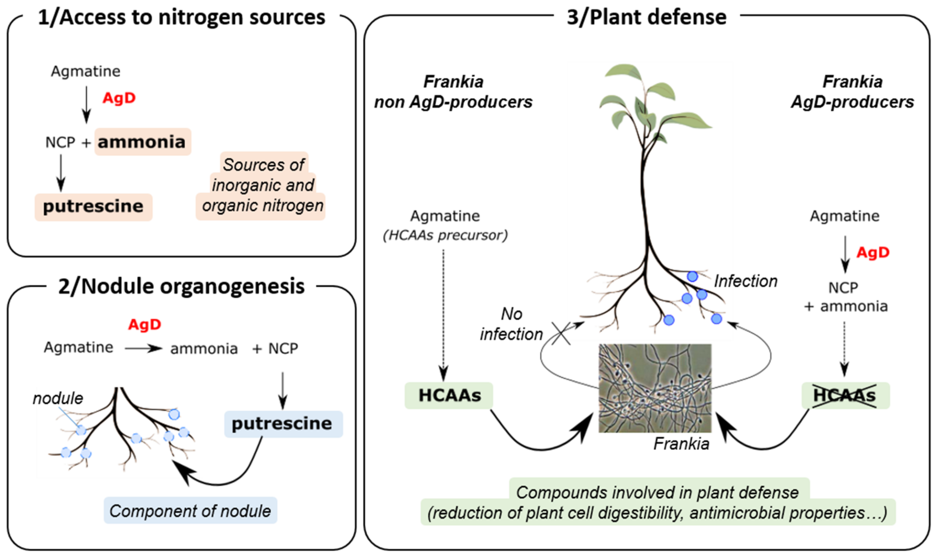
The AgDs catalyze the deimination of agmatine (i.e., decarboxylated arginine) to form N-carbamoyl putrescine (NCP) and ammonia [46]. We can hypothesize that the AgD produced by Frankia could be used in order to degrade agmatine found in the plant (Figure 2). The enzyme could thus allow Frankia to produce putrescine (via the conversion of NCP into putrescine) and use ammonia as sources of nitrogen. Actually, studies have shown that Frankia strains can use a variety of organic and inorganic sources of nitrogen for growth [10], including putrescine [47]. Moreover, putrescine was identified as one of the three main polyamine (together with spermidine and spermine) in roots and nodules of legumes and of actinorhizals [48,49,50], suggesting an association between polyamines and nodule development [47].
We could also hypothesize that AgD plays a crucial role in Frankia infection to circumvent plant defense. Actually, agmatine is a precursor of several secondary metabolites, such as hydroxycinnamic acid amides (HCAAs) produced by plants [51]. HCAAs are a widely distributed group of plant secondary metabolites with a role in several growth and developmental processes (including floral induction, flower formation, sexual differentiation, tuberization, cell division and cytomorphogenesis); they are also involved in plant defense against pathogens [52]. The HCAAs structure is characterized by the association of at least one hydroxycinnamic acid derivative (e.g., p-Coumaroyl-CoA, caffeoyl-CoA, Feruloy-CoA…), which is linked through an amide bond to an aromatic monoamine (e.g., tyramine, dopamine, serotonin…) or an aliphatic polyamine (e.g., agmatine, putrescine, spermidine…) [53]. The combination of different hydroxycinnamic acid and amine moieties together with the possibility of one to four N-substitutions on aliphatic polyamines are responsible for the broad structural diversity in phenolamides. Muroi et al. [51] have shown that mutants of Arabidopsis thaliana that do not accumulate HCAAs derived from agmatine and putrescine (p-Coumaroylagmatine, Feruloylagmatine, p-Coumaroylputrescine and Feruloylputrescine) were much more sensitive to Altenaria brassicicola infection compared to wild-type, suggesting that these four HCAAs play a crucial role in the infection process.
Regarding their roles as secondary metabolites involved in plant defense against pathogens, we hypothesized that HCAAs derived from agmatine and putrescine potentially produced by Alnus prevent infection by Frankia non-AgD producers, as illustrated in Figure 2.
On the contrary, Frankia AgD producers would have the capability to degrade agmatine into NCP and ammonia; this degradation would prevent the production of HCAAs or strongly reduce HCAAS biosynthesis. In both cases, the decrease in HCAAs production allows the infection by Frankia and the subsequent formation of root nodules (Figure 2b).
HCAAs are involved in plant defense by reducing plant cell digestibility by deposition in cell walls [52] and/or by having antimicrobial effects such as the suppression of or reduction in hyphal elongation [51,54,55,56]. We hypothesize the HCAAs produced by Alnus will have similar effects on Frankia (reduction in the elongation of hyphae), hence preventing Frankia infection.
In conclusion, nine genes were specifically found in Frankia from Cluster Ia. Among them, FRAAL0164 was annotated as an AgD. This enzyme could play a central role in the Frankia/Alnus relationship by degrading agmatine into NCP and ammonia. These roles could concern: 1. access to nitrogen sources by providing putrescine (via NCP) and ammonia to Frankia and/or 2. nodule organogenesis by using putrescine (i.e., one on the main polyamines in roots and nodules of legumes and of actinorhizals), as well as 3. plant defense by stopping the production of HCAAs derived from agmatine and putrescine.

3.3. What Genome Comparison Tells Us about Sp+ Alnus-Infective Frankia Strains

In addition to identifying candidate molecules responsible for Cluster Ia Frankia strain host specificity, the second objective was to investigate Sp+ Frankia genomes in comparison with Sp− genomes. The narrower host specificity observed in Sp+ strains [19], combined with the fact that they have never been cultured despite numerous attempts, suggests they could be dependent on the host plant for a large part of their life cycle. Several hypotheses have been proposed regarding the in planta sporulation strategy of Sp+ Frankia strains, among them a possible evolution of Sp+ Frankia strains towards an obligatory symbiont status [12,21,22,57]. Under this hypothesis, the early abundant production of spores into host plant cells could allow a massive spore release into the soil during nodule decay and promote the subsequent root vicinity invasion. Indeed, the sporulation in planta would enable Sp+ strains to survive and disseminate outside the host, and to infect new roots without the need for saprophytic growth.
A substantial genomic purge of Sp+ strains in Cluster Ia was previously reported, supporting the obligate symbiont scenario previously discussed [21,22]. In the present study, we sequenced a new Sp+ Frankia genome from Cluster Ia. Although its size was slightly larger than that of other Alnus-infective Sp+ strains [21,22], it remains among the smallest genomes in Cluster Ia (generally around 7.5 Mb), sustaining the hypothesis of a genome reduction in Sp+ strains in two independent lineages. At this stage in the work, it remains crucial to elucidate lost genomic regions in Sp+ strains. Analyzing lost genes could, for instance, comfort the hypothesis that Sp+ strains would have evolved into obligate symbionts.
In the present study, a comparison between Sp+ and Sp− genomes from Cluster Ia Alnus-infective Frankia strains was performed with HOGENOM. This analysis allowed us to identify 88 protein sequence families found especially in the six Sp− genomes without orthologs in the six Sp+ genomes (Table 5) (it should be noted that the analysis did not reveal any sequence family present in Sp+ genomes without orthologs in Sp− genomes).
These 88 sequence families were characterized based on their COG affiliation or their cellular localization (Table 5). This analysis revealed four major pieces of information that could support the hypothesis of Sp+ strain evolution towards an obligatory symbiont status:

3.3.1. The Loss of Transcription-Associated Protein Sequences in Sp+ Frankia Genomes

Based on COG affiliation, we observed 15.9% of lost protein sequences in Sp+ genomes (14 out of 88 sequences) were associated with the “Transcription” category (COG K) (Table 5). For example, several genes encoding transcriptional regulators, including LuxR (e.g., FRAAL4738), MarR (e.g., FRAAL3611), TetR (e.g., FRAAL3977) or putative two-component system response regulators (e.g., FRAAL1658) were observed only in Sp− genomes. Such a purge in genes encoding transcriptional factors and particularly activators has already been reported in the genome reduction bacteria. This phenomenon was hypothesized to reflect a host-restricted lifestyle that requires the symbiont to less finely regulate its gene expression to respond and adapt to changing environmental conditions (e.g., biotic and abiotic stresses) [58,59]. In the case of Sp+ strains, a reduction in the number of transcription-associated sequences could therefore indicate a narrower interaction with the host plant compared to Sp− strains, comforting the hypothesis of the evolution towards an obligate symbiotic status.

3.3.2. A Reduced Secretome in Sp+ Frankia Strains

The 88 protein families without orthologs in Sp+ genomes were analyzed regarding their localization in the cell (Table 5). Twenty-six percent of these families were predicted as transmembrane proteins or secreted proteins (indicated in Table 5 as “TM” and “SP”, respectively), including various receptors, transporters and secreted enzymes. For example, orthologs to FRAAL3906 and FRAAL3907 organized in a synton (encoding transporters) were observed only in Sp− genomes. In other words, a significant part of lost sequences in Sp+ genomes would be related to the secretome. A previous comparison of predicted secretomes between plant symbiotic bacteria, in this case Frankia strains, and soil bacteria reported a secretome size reduction in the symbiotic bacteria [60]. This reduction was discussed as a consequence of the bacterial adaptation to plant endosymbiotic lifestyle that may require fewer secreted proteins [60,61]. Such a reduction in Sp+ Frankia genomes could therefore be additional evidence in favor of the hypothesis of their obligate status.

3.3.3. The Potential Loss of Saprophytic Functions in Sp+ Frankia Strains

Interestingly, several protein encoding sequences lost in Sp+ genomes did not present paralogs in Sp− genomes. The loss of these sequences could lead to the loss of functions in Sp+ Frankia strains, in absence of divergent genes ensuring the same function.
Genes encoding putative gas vesicles illustrate this situation. Gas vesicles are intracellular air-filled organelles of around two nanometers, composed solely of proteins to trap gas to provide buoyancy to cells in a watery environment. Our analysis revealed that protein encoding sequences involved in the formation of gas vesicle formation (FRAAL3025 annotated “gvpA” and FRAAL3026 annotated “gvpF”) were absent in Sp+ genomes compared to Sp− genomes. Their absence was recently reported based on the first three sequenced Sp+ genomes [22], and it is supported in the present study including double Sp+ genomes. Gas vesicle proteins could possibly be used for floatation of free-living bacteria on the soil watertable. We hypothesize that obligate plant endosymbionts would not require such a function, and thus the absence of gvp genes in Sp+ strains could evidence their high dependance on the host.
In addition to gas vesicle formation, another striking example of encoding protein sequences present in single copies in Sp− genomes, which was lost in Sp+ genomes is FRAAL3502 putatively encoding a 3-ketosteroid 9alpha-monooxygenase. This gene is involved in the cholesterol degradation pathway [62,63]. This pathway could allow Sp− Frankia strains to metabolize cholesterol as a carbon and energy source, and it could be involved in strain ability to scavenge nutrients in soil. Steroid degradation is indeed a critical process for biomass decomposition in soil and plant rhizosphere, and it has been found mostly due to actinobacteria, to which the genus Frankia belongs [64]. The loss of this gene in Sp+ genomes could suggest a loss of saprophytic abilities in Sp+ strains: Sp+ strains would have lost the ability to metabolize cholesterol in soil but, as obligate symbionts, they would still require host cholesterol for intracellular survival (as previously reported for Mycobacterium leprae) [65].
More anecdotally, other sequences encoding proteins with potential functions in the use of soil nutrient and energy resources were also missing in Sp+ genomes, with among them one sequence coding acid phosphatase (SurE, FRAAL0277), considered a predominant form of extracellular phosphatases in soils [4,66].

3.3.4. The Loss of Genetic and Functional Redundancy in Sp+ Genomes

Interestingly, 27% of genes lost in Sp+ genomes present paralogs in Sp− genomes, suggesting a functional redundancy. We can hypothesize that their absence in Sp+ genomes could have few or no effects on their phenotype.
The hup genes are the striking example of the presence of paralogs in Frankia genomes, some of which have been lost in Sp+ strains. The hupS and hupL genes encode the hydrogenase structural subunits. With the other hupABCDEF genes encoding enzymes involved in the recruitment and incorporation of metallic groups, they form the hup gene cluster. Uptake hydrogenases catalyze the oxidation of hydrogen to protons and electrons in order to supply them to the respiratory chain to produce energy. In diazotrophic bacteria, the nitrogen-fixing activity produces hydrogen that can be consumed to yield energy for other metabolic pathways in the cell [67]. Two sets of uptake hydrogenase genes, organized in synton #1 and synton #2, have been described in Frankia [68,69]. The uptake hydrogenase synton #1 was described as more expressed under free-living conditions, whereas hydrogenase synton #2 was mainly involved in symbiotic interactions [68]. In our analysis, hupDSL genes belonging to the synton #1 were not present in Sp+ genomes. This could suggest that synton #1 would be no longer needed or useful for the Sp+ strain lifestyle, converging with the hypothesis of their obligate status. Under this hypothesis, they would have lost synton #1, but still require synton #2 to take up hydrogen inside host cells.
In addition to hydrogenase function improving nitrogen fixation, we found gene redundancy assigned to functions involved in the metabolization of different sources that could be associated to life in cell free conditions. Several genes belonging to the “Energy production and conversion” COG were, for example, recovered from the list of lost genes in Sp+ genomes, such as FRAAL3448 or FRAAL4787 encoding a putative Glycerophosphoryl diester phosphodiesterase (indicated as “GlpQ” in Table 5) and putative N-glycosyltransferase, respectively. GlpQ is a protein able to hydrolyze glycerophosphodiester bonds [70] of phospholipid fatty acids, composing cell membranes in all organisms other than archaea, in order to access carbon and phosphate sources [71]. In parallel, the glycosyltransferases classified as GT1 according to the Cazy database (http://www.cazy.org/ (accessed on 17 November 2022) [72]) catalyze the transfer of a sugar moiety from an activated donor sugar onto acceptor molecules such as glycolipids, flavonoids or macrolides [73]. The important role of this enzyme is to resist toxic products produced by bacteria in the environment [74,75,76]. Thus, those enzymes could participate in the bacterial homeostasis to reduce biotic stress or to access new nutrients.

4. Conclusions

The present study aimed to use comparative genomics to explore the host specificity of both “Alnus-infective strains” and Sp+ Frankia. Several genes were specifically found in “Alnus-infective strains”, including an agmatine deiminase which could possibly be involved in various functions such as access to nitrogen sources, nodule organogenesis or plant defense. In order to test these functions, the heterologous expression of AgD could be used in future studies to produce this agmatine deiminase to confirm its biochemical function. Its deletion in the Frankia genome is a striking demonstration of this, provided that the technique is developed in this model, which is not yet the case.
A total of 88 protein families were lost in the Sp+ genomes. This loss included (i) transcriptional factors, (ii) transmembrane and secreted proteins, (ii) genetic and functional paralogs highlighting a reduction in functional redundancy (genes that copy number decreased, e.g., hup genes) and (iv) a possible loss of function (genes with loss of all copies, e.g., genes involved in gas vesicle formation or recycling of nutrients). It highlights a purge of genes related to saprophytic life and comforts the hypothetical status of obligatory symbiont of Sp+ strains. At this stage in the work, it could be interesting to test if lost genes could indeed play a role in Frankia saprophytic life. The comparison of their expression when Frankia is free-living in soil (e.g., in inoculated soil with Frankia Sp− strains) versus under a symbiotic state (e.g., in Sp− nodules) through transcriptomic-based analyses (e.g., qPCR or RNAseq analysis) could, for example, be tested.
To date, we still do not know what explains the ability of Sp+ strains to sporulate in planta. Our comparative genomic analysis did not provide new clues to this question (no protein sequence family specific to Sp+ genomes (i.e., without orthologs in Sp− genomes) was revealed). Remember, however, that based on Sp− Frankia strains’ ability to sporulate in vitro, it was hypothesized that both Sp+ and Sp− strains have sporulation-associated genes in their genomes, but molecular factors (e.g., transcriptional factors) could suppress the sporulation capacity of Sp− Frankia strains in planta and allow in Sp+ strains the expression of sporulation inside nodules [77]. To elucidate the question of in planta sporulation ability, it would therefore be more worthwhile to follow the expression of Frankia genes identified as involved in sporulation in Sp+ versus Sp− nodules [77].

Author Contributions

Conceptualization, S.K.T., H.B. and A.H.-B.; formal analysis, S.K.T., H.B. and A.H.-B.; investigation, S.K.T., H.B. and A.H.-B.; methodology, D.A.; resources, L.B. and P.F.; writing—original draft, S.K.T., H.B. and A.H.-B.; writing—review and editing, S.K.T., H.B. and A.H.-B. All authors have read and agreed to the published version of the manuscript.

Funding

This research was supported by the University of Lyon (BQR Grant).

Acknowledgments

The authors thank P. Normand for his careful proofreading of the manuscript.

Conflicts of Interest

The authors declare no conflict of interest.

References

  1. Baker, D.D. Relationships among pure cultured strains of Frankia based on host specificity. Physiol. Plant. 1987, 70, 245–248. [Google Scholar] [CrossRef]
  2. Bosco, M.; Fernandez, M.; Simonet, P.; Materassi, R.; Normand, P. Evidence that some Frankia sp. strains are able to cross boundaries between Alnus and Elaeagnus Host Specificity Groups. Appl. Environ. Microbiol. 1992, 58, 1569–1576. [Google Scholar] [CrossRef] [PubMed] [Green Version]
  3. De Ley, J.; Rassel, A. DNA base composition, flagellation and taxonomy of the genus Rhizobium. J. Gen. Microbiol. 1965, 41, 85–91. [Google Scholar] [CrossRef] [PubMed] [Green Version]
  4. Sharma, P.; Kundu, B.; Dogra, R. Molecular mechanism of host specificity in Legume-Rhizobium symbiosis. Biotechnol. Adv. 1993, 11, 741–779. [Google Scholar] [CrossRef]
  5. Zakhia, F.; de Lajudie, P. Taxonomy of Rhizobia. Agronomie 2001, 21, 569–576. [Google Scholar] [CrossRef]
  6. Krieg, N.R.; Holt, J.G. Bergey’s Manual of Systematic Bacteriology; Yi Hsien Publishing Co.: Taipei, Taiwan, 1984; ISBN 0-683-04108-8. [Google Scholar]
  7. Normand, P.; Orso, S.; Cournoyer, B.; Jeannin, P.; Chapelon, C.; Dawson, J.; Evtushenko, L.; Misra, A.K. Molecular phylogeny of the genus Frankia and related genera and emendation of the family Frankiaceae. Int. J. Syst. Evol. Microbiol. 1996, 46, 1–9. [Google Scholar] [CrossRef]
  8. Pozzi, A.C.; Bautista-Guerrero, H.H.; Abby, S.S.; Herrera-Belaroussi, A.; Abrouk, D.; Normand, P.; Menu, F.; Fernandez, M.P. Robust Frankia Phylogeny, species delineation and intraspecies diversity based on Multi-Locus Sequence Analysis (MLSA) and Single-Locus Strain Typing (SLST) adapted to a large sample size. Syst. Appl. Microbiol. 2018, 41, 311–323. [Google Scholar] [CrossRef]
  9. Nguyen, T.V.; Wibberg, D.; Vigil-Stenman, T.; Berckx, F.; Battenberg, K.; Demchenko, K.N.; Blom, J.; Fernandez, M.P.; Yamanaka, T.; Berry, A.M.; et al. Frankia-enriched metagenomes from the earliest diverging symbiotic Frankia Cluster: They come in teams. Genome Biol. Evol. 2019, 11, 2273–2291. [Google Scholar] [CrossRef] [Green Version]
  10. Benson, D.R.; Silvester, W.B. Biology of Frankia strains, Actinomycete symbionts of Actinorhizal plants. Microbiol. Mol. Biol. Rev. 1993, 57, 293–319. [Google Scholar] [CrossRef]
  11. Dawson, J.O. Ecology of actinorhizal plants. In Nitrogen-Fixing Actinorhizal Symbioses-Nitrogen Fixation Research: Origins and Progress; Pawlowski, K., Newton, W.E., Eds.; Springer: New York, NY, USA, 2008; Volume 6, pp. 199–234. [Google Scholar]
  12. Cotin-Galvan, L.; Pozzi, A.C.; Schwob, G.; Fournier, P.; Fernandez, M.P.; Herrera-Belaroussi, A. In-Planta Sporulation Capacity enhances infectivity and rhizospheric competitiveness of Frankia strains. Microbes Environ. 2016, 31, 11–18. [Google Scholar] [CrossRef] [Green Version]
  13. Schwintzer, C.R. Spore-Positive and Spore-Negative nodules. In The Biology of Frankia and Actinorhizal Plants; Academic Press: Cambridge, MA, USA, 1990; pp. 177–193. [Google Scholar]
  14. Weber, A.; Nurmiaho-Lassila, E.; Sundman, V. Features of the intrageneric Alnus-Frankia specificity. Physiol. Plant. 1987, 70, 289–296. [Google Scholar] [CrossRef]
  15. Kurdali, F.; Domenach, A.-M.; Fernandez, M.D.L.P.; Capellano, A.; Moiroud, A. Compatibility of Frankiae Spore Positive and Spore Negative inocula with Alnus glutinosa and Alnus incana. Soil Sci. Plant Nutr. 1988, 34, 451–459. [Google Scholar] [CrossRef] [Green Version]
  16. Markham, J.H. Variability of nitrogen-fixing Frankia on Alnus species. Botany 2008, 86, 501–510. [Google Scholar] [CrossRef]
  17. Van Dijk, C. Spore Formation and endophyte diversity in root nodules of Alnus Glutinosa (L.) Vill. New Phytol. 1978, 81, 601–615. [Google Scholar] [CrossRef]
  18. Torrey, J.G. Endophyte sporulation in root nodules of Actinorhizal plants. Physiol. Plant. 1987, 70, 279–288. [Google Scholar] [CrossRef]
  19. Schwob, G.; Roy, M.; Pozzi, A.; Herrera-Belaroussi, A.; Fernandez, M. In Planta Sporulation of Frankia Spp. as a Determinant of Alder-Symbiont Interactions. Appl. Environ. Microbiol. 2018, 84, e01737-18. [Google Scholar] [CrossRef] [Green Version]
  20. Bethencourt, L.; Vautrin, F.; Taib, N.; Dubost, A.; Castro-Garcia, L.; Imbaud, O.; Abrouk, D.; Fournier, P.; Briolay, J.; Nguyen, A.; et al. Draft genome sequences for three unisolated Alnus-infective Frankia Sp+ strains, AgTrS, AiOr and AvVan, the first sequenced Frankia strains able to sporulate in-planta. J. Genom. 2019, 7, 50–55. [Google Scholar] [CrossRef] [Green Version]
  21. Pozzi, A.C.M.; Herrera-Belaroussi, A.; Schwob, G.; Bautista-Guerrero, H.H.; Bethencourt, L.; Fournier, P.; Dubost, A.; Abrouk, D.; Normand, P.; Fernandez, M.P. Proposal of ‘Candidatus Frankia alpina’, the uncultured symbiont of Alnus alnobetula and A. incana that forms spore-containing nitrogen-fixing root nodules. Int. J. Syst. Evol. Microbiol. 2020, 70, 5453–5459. [Google Scholar] [CrossRef]
  22. Herrera-Belaroussi, A.; Normand, P.; Pawlowski, K.; Fernandez, M.P.; Wibberg, D.; Kalinowski, J.; Brachmann, A.; Berckx, F.; Lee, N.; Blom, J.; et al. Candidatus Frankia nodulisporulans sp. nov., an Alnus glutinosa-infective Frankia species unable to grow in pure culture and able to sporulate in-planta. Syst. Appl. Microbiol. 2020, 43, 126134. [Google Scholar] [CrossRef]
  23. Cérémonie, H.; Debellé, F.; Fernandez, M.P. Structural and functional comparison of Frankia root hair deforming factor and Rhizobia Nod factor. Can. J. Bot. 1999, 77, 1293–1301. [Google Scholar]
  24. Normand, P.; Lapierre, P.; Tisa, L.S.; Gogarten, J.P.; Alloisio, N.; Bagnarol, E.; Bassi, C.A.; Berry, A.M.; Bickhart, D.M.; Choisne, N.; et al. Genome characteristics of facultatively symbiotic Frankia Sp. strains reflect Host Range and Host Plant Biogeography. Genome Res. 2007, 17, 7–15. [Google Scholar] [CrossRef] [PubMed] [Green Version]
  25. Franche, C.; Lindström, K.; Elmerich, C. Nitrogen-fixing bacteria associated with Leguminous and non-Leguminous plants. Plant Soil 2009, 321, 35–59. [Google Scholar] [CrossRef]
  26. Persson, T.; Battenberg, K.; Demina, I.V.; Vigil-Stenman, T.; Vanden Heuvel, B.; Pujic, P.; Facciotti, M.T.; Wilbanks, E.G.; O’Brien, A.; Fournier, P.; et al. Candidatus Frankia datiscae Dg1, the Actinobacterial microsymbiont of Datisca glomerata, expresses the Canonical Nod genes NodABC in symbiosis with its host plant. PLoS ONE 2015, 10, e0127630. [Google Scholar] [CrossRef] [PubMed] [Green Version]
  27. Udwary, D.W.; Gontang, E.A.; Jones, A.C.; Jones, C.S.; Schultz, A.W.; Winter, J.M.; Yang, J.Y.; Beauchemin, N.; Capson, T.L.; Clark, B.R.; et al. Significant natural product biosynthetic potential of Actinorhizal symbionts of the genus Frankia, as revealed by comparative genomic and proteomic analyses. Appl. Environ. Microbiol. 2011, 77, 3617–3625. [Google Scholar] [CrossRef] [PubMed] [Green Version]
  28. Deicke, M.; Mohr, J.F.; Roy, S.; Herzsprung, P.; Bellenger, J.-P.; Wichard, T. Metallophore profiling of Nitrogen-Fixing Frankia Spp. to understand metal management in the rhizosphere of Actinorhizal plants. Metallomics 2019, 11, 810–821. [Google Scholar] [CrossRef]
  29. Nouioui, I.; Cortés-Albayay, C.; Carro, L.; Castro, J.F.; Gtari, M.; Ghodhbane-Gtari, F.; Klenk, H.-P.; Tisa, L.S.; Sangal, V.; Goodfellow, M. Genomic insights into Plant-Growth-Promoting potentialities of the genus Frankia. Front. Microbiol. 2019, 10, 1457. [Google Scholar] [CrossRef]
  30. Carlos-Shanley, C.; Guerra, T.; Hahn, D. Draft genomes of non-nitrogen-fixing Frankia strains. J. Genom. 2021, 9, 68–75. [Google Scholar] [CrossRef]
  31. Beauchemin, N.; Gtari, M.; Ghodhbane-Gtari, F.; Furnholm, T.; Sen, A.; Wall, L.; Tisa, L.S. What can the genome of an infective ineffective (Fix-) Frankia strain (EuI1c) that is able to form nodules with its host plant tell us about Actinorhizal symbiosis and Frankia evolution. In Proceedings of the 112th General Meeting of the American Society for Microbiology, San Francisco, CA, USA, 16–19 June 2012. [Google Scholar]
  32. Tisa, L.S.; Beauchemin, N.; Gtari, M.; Sen, A.; Wall, L.G. What Stories Can the Frankia Genomes Start to Tell Us? J. Biosci. 2013, 38, 719–726. [Google Scholar] [CrossRef]
  33. Tisa, L.S.; Oshone, R.; Sarkar, I.; Ktari, A.; Sen, A.; Gtari, M. Genomic approaches toward understanding the Actinorhizal symbiosis: An update on the status of the Frankia genomes. Symbiosis 2016, 70, 5–16. [Google Scholar] [CrossRef]
  34. Pozzi, A.C.; Roy, M.; Nagati, M.; Schwob, G.; Manzi, S.; Gardes, M.; Moreau, P.-A.; Fernandez, M.P. Patterns of diversity, endemism and specialization in the root symbiont communities of Alder species on the island of Corsica. New Phytol. 2018, 219, 336–349. [Google Scholar] [CrossRef] [Green Version]
  35. Wick, R.R.; Judd, L.M.; Gorrie, C.L.; Holt, K.E. Unicycler: Resolving bacterial genome assemblies from short and long sequencing reads. PLoS Comput. Biol. 2017, 13, e1005595. [Google Scholar] [CrossRef] [Green Version]
  36. Vallenet, D.; Calteau, A.; Cruveiller, S.; Gachet, M.; Lajus, A.; Josso, A.; Mercier, J.; Renaux, A.; Rollin, J.; Rouy, Z.; et al. MicroScope in 2017: An expanding and evolving integrated resource for community expertise of microbial genomes. Nucleic Acids Res. 2017, 45, D517–D528. [Google Scholar] [CrossRef]
  37. Goris, J.; Konstantinidis, K.T.; Klappenbach, J.A.; Coenye, T.; Vandamme, P.; Tiedje, J.M. DNA–DNA hybridization values and their relationship to whole-genome sequence similarities. Int. J. Syst. Evol. Microbiol. 2007, 57, 81–91. [Google Scholar] [CrossRef] [Green Version]
  38. Blom, J.; Kreis, J.; Spänig, S.; Juhre, T.; Bertelli, C.; Ernst, C.; Goesmann, A. EDGAR 2.0: An enhanced software platform for comparative gene content analyses. Nucleic Acids Res. 2016, 44, W22–W28. [Google Scholar] [CrossRef] [Green Version]
  39. Penel, S.; Arigon, A.-M.; Dufayard, J.-F.; Sertier, A.-S.; Daubin, V.; Duret, L.; Gouy, M.; Perrière, G. Databases of homologous gene families for comparative genomics. BMC Bioinform. 2009, 10, S3. [Google Scholar] [CrossRef] [Green Version]
  40. Petersen, T.N.; Brunak, S.; von Heijne, G.; Nielsen, H. SignalP 4.0: Discriminating signal peptides from transmembrane regions. Nat. Methods 2011, 8, 785–786. [Google Scholar] [CrossRef]
  41. Krogh, A.; Larsson, B.; von Heijne, G.; Sonnhammer, E.L.L. Predicting transmembrane protein topology with a hidden Markov Model: Application to complete genomes. J. Mol. Biol. 2001, 305, 567–580. [Google Scholar] [CrossRef] [Green Version]
  42. Parks, D.H.; Imelfort, M.; Skennerton, C.T.; Hugenholtz, P.; Tyson, G.W. CheckM: Assessing the quality of microbial genomes recovered from isolates, single cells, and metagenomes. Genome Res. 2015, 25, 1043–1055. [Google Scholar] [CrossRef] [Green Version]
  43. Annika, C.M.; Justice, N.B.; Bowen, B.P.; Baran, R.; Thomas, B.C.; Northern, T.R.; Banfield, J.F. Metabolites associated with adaptation of microorganisms to an acidophilic, metal-rich environment identified by Stable-Isotope-enabled metabolomics. mBio 2013, 4, e00484-12. [Google Scholar] [CrossRef] [Green Version]
  44. Tan, Y.; Shan, Y.; Zheng, R.; Liu, R.; Sun, C. Characterization of a Deep-Sea Actinobacterium Strain Uncovers Its Prominent Capability of Utilizing Taurine and Polyvinyl Alcohol. Front. Microbiol. 2022, 13, 868728. [Google Scholar] [CrossRef]
  45. Madeira, F.; Park, Y.M.; Lee, J.; Buso, N.; Gur, T.; Madhusoodanan, N.; Basutkar, P.; Tivey, A.R.N.; Potter, S.C.; Finn, R.D.; et al. The EMBL-EBI Search and sequence analysis tools APIs in 2019. Nucleic Acids Res. 2019, 47, W636–W641. [Google Scholar] [CrossRef] [PubMed] [Green Version]
  46. Jones, J.E.; Dreyton, C.J.; Flick, H.; Causey, C.P.; Thompson, P.R. Mechanistic studies of Agmatine Deiminase from multiple bacterial species. Biochemistry 2010, 49, 9413–9423. [Google Scholar] [CrossRef] [PubMed] [Green Version]
  47. Wheeler, C.T.; Tonin, G.S.; Sutcliffe, A. Poly Amines of Frankia in relation to nitrogen nutrition. Soil Biol. Biochem. 1994, 26, 577–581. [Google Scholar] [CrossRef]
  48. Smith, T.A. Homospermidine in Rhizobium and Legume root nodules. Phytochemistry 1977, 16, 278–279. [Google Scholar] [CrossRef]
  49. Chatterjee, S.; Choudhuri, M.; Ghosh, B. Changes in Polyamine contents during root and nodule growth of Phaseolus Mungo. Phytochemistry 1983, 22, 1553–1556. [Google Scholar] [CrossRef]
  50. Tonin, G.; Wheeler, C.; Crozier, A. Effect of changes in Nitrogen nutrition on the polyamine content of Alnus glutinosa. Plant Cell Environ. 1991, 14, 415–421. [Google Scholar] [CrossRef]
  51. Muroi, A.; Ishihara, A.; Tanaka, C.; Ishizuka, A.; Takabayashi, J.; Miyoshi, H.; Nishioka, T. Accumulation of Hydroxycinnamic Acid Amides induced by pathogen infection and identification of Agmatine Coumaroyltransferase in Arabidopsis thaliana. Planta 2009, 230, 517–527. [Google Scholar] [CrossRef]
  52. Facchini, P.J.; Hagel, J.; Zulak, K.G. Hydroxycinnamic Acid Amide metabolism: Physiology and biochemistry. Can. J. Bot. 2002, 80, 577–589. [Google Scholar] [CrossRef]
  53. Roumani, M.; Duval, R.E.; Ropars, A.; Risler, A.; Robin, C.; Larbat, R. Phenolamides: Plant specialized metabolites with a wide range of promising pharmacological and health-promoting interests. Biomed. Pharmacother. 2020, 131, 110762. [Google Scholar] [CrossRef]
  54. Mayama, S.; Tani, T.; Matsuura, Y.; Ueno, T.; Fukami, H. The production of Phytoalexins by Oat in response to Crown Rust, Puccinia coronata f. sp. avenae. Physiol. Plant Pathol. 1981, 19, 217–226. [Google Scholar] [CrossRef]
  55. Mayama, S.; Matsuura, Y.; Iida, H.; Tani, T. The role of Avenalumin in the Resistance of Oat to Crown Rust, Puccinia coronata f. sp. avenae. Physiol. Plant Pathol. 1982, 20, 189–199. [Google Scholar] [CrossRef]
  56. Miyagawa, H.; Ishihara, A.; Nishimoto, T.; Ueno, T.; Mayama, S. induction of Avenanthramides in Oat leaves inoculated with Crown Rust fungus, Puccinia coronata f. sp. avenae. Biosci. Biotechnol. Biochem. 1995, 59, 2305–2306. [Google Scholar] [CrossRef] [Green Version]
  57. Pozzi, A.C.; Bautista-Guerrero, H.H.; Nouioui, I.; Cotin-Galvan, L.; Pepin, R.; Fournier, P.; Menu, F.; Fernandez, M.P.; Herrera-Belaroussi, A. In-planta Sporulation Phenotype: A Major life history trait to understand the evolution of Alnus-infective Frankia strains. Environ. Microbiol. 2015, 17, 3125–3138. [Google Scholar] [CrossRef]
  58. Galan-Vasquez, E.; Sanchez-Osorio, I.; Martinez-Antonio, A. Transcription factors exhibit differential conservation in bacteria with reduced genomes. PLoS ONE 2016, 11, e0146901. [Google Scholar] [CrossRef] [Green Version]
  59. Miravet-Verde, S.; Lloréns-Rico, V.; Serrano, L. Alternative transcriptional regulation in genome-reduced bacteria. Curr. Opin. Microbiol. 2017, 39, 89–95. [Google Scholar] [CrossRef]
  60. Mastronunzio, J.E.; Tisa, L.S.; Normand, P.; Benson, D.R. Comparative secretome analysis suggests low plant cell wall degrading capacity in Frankia Symbionts. BMC Genom. 2008, 9, 47. [Google Scholar] [CrossRef] [Green Version]
  61. Mastronunzio, J.; Huang, Y.; Benson, D. Diminished exoproteome of Frankia spp. in culture and symbiosis. Appl. Environ. Microbiol. 2009, 75, 6721–6728. [Google Scholar] [CrossRef] [Green Version]
  62. Capyk, J.K.; D’Angelo, I.; Strynadka, N.C.; Eltis, L.D. Characterization of 3-Ketosteroid 9α-Hydroxylase, a rieske oxygenase in the Cholesterol Degradation Pathway of Mycobacterium tuberculosis. J. Biol. Chem. 2009, 284, 9937–9946. [Google Scholar] [CrossRef] [Green Version]
  63. Rohman, A.; Dijkstra, B.W. The Role and Mechanism of Microbial 3-Ketosteroid Δ1-Dehydrogenases in steroid breakdown. J. Steroid Biochem. Mol. Biol. 2019, 191, 105366. [Google Scholar] [CrossRef]
  64. Holert, J.; Cardenas, E.; Bergstrand, L.H.; Zaikova, E.; Hahn, A.S.; Hallam, S.J.; Mohn, W.W. Metagenomes reveal global distribution of bacterial steroid catabolism in natural, engineered, and host environments. mBio 2018, 9, e02345-17. [Google Scholar] [CrossRef] [Green Version]
  65. Marques, M.A.M.; Berrêdo-Pinho, M.; Rosa, T.L.; Pujari, V.; Lemes, R.M.; Lery, L.M.; Silva, C.A.M.; Guimarães, A.C.R.; Atella, G.C.; Wheat, W.H.; et al. The essential role of Cholesterol metabolism in the intracellular survival of Mycobacterium leprae is not coupled to central carbon metabolism and energy production. J. Bacteriol. 2015, 197, 3698–3707. [Google Scholar] [CrossRef] [PubMed] [Green Version]
  66. Park, Y.; Solhtalab, M.; Thongsomboon, W.; Aristilde, L. Strategies of organic Phosphorus recycling by soil bacteria: Acquisition, metabolism, and regulation. Environ. Microbiol. Rep. 2022, 14, 3–24. [Google Scholar] [CrossRef] [PubMed]
  67. Dixon, R. Hydrogenases and efficiency of nitrogen fixation in Aerobes. Nature 1976, 262, 173. [Google Scholar] [CrossRef]
  68. Leul, M.; Normand, P.; Sellstedt, A. The organization, regulation and phylogeny of uptake Hydrogenase genes in Frankia. Physiol. Plant. 2007, 130, 464–470. [Google Scholar] [CrossRef]
  69. Leul, M.; Normand, P.; Sellstedt, A. The phylogeny of uptake Hydrogenases in Frankia. Int. Microbiol. 2009, 12, 23–28. [Google Scholar]
  70. Corda, D.; Mosca, M.G.; Ohshima, N.; Grauso, L.; Yanaka, N.; Mariggiò, S. The emerging physiological roles of the Glycerophosphodiesterase family. FEBS J. 2014, 281, 998–1016. [Google Scholar] [CrossRef]
  71. De Carvalho, C.C.; Caramujo, M.J. The various roles of Fatty Acids. Molecules 2018, 23, 2583. [Google Scholar] [CrossRef] [Green Version]
  72. Drula, E.; Garron, M.-L.; Dogan, S.; Lombard, V.; Henrissat, B.; Terrapon, N. The Carbohydrate-Active Enzyme Database: Functions and literature. Nucleic Acids Res. 2022, 50, D571–D577. [Google Scholar] [CrossRef]
  73. Zhang, P.; Zhang, Z.; Zhang, L.; Wang, J.; Wu, C. Glycosyltransferase GT1 Family: Phylogenetic distribution, substrates coverage, and representative structural features. Comput. Struct. Biotechnol. J. 2020, 18, 1383–1390. [Google Scholar] [CrossRef]
  74. Bolam, D.N.; Roberts, S.; Proctor, M.R.; Turkenburg, J.P.; Dodson, E.J.; Martinez-Fleites, C.; Yang, M.; Davis, B.G.; Davies, G.J.; Gilbert, H.J. The Crystal Structure of Two Macrolide Glycosyltransferases Provides a Blueprint for Host Cell Antibiotic Immunity. Proc. Natl. Acad. Sci. USA 2007, 104, 5336–5341. [Google Scholar] [CrossRef] [Green Version]
  75. Wang, C.; Liu, X.; Zhang, P.; Wang, Y.; Li, Z.; Li, X.; Wang, R.; Shang, Z.; Yan, J.; He, H.; et al. Bacillus licheniformis escapes from Myxococcus xanthus predation by deactivating Myxovirescin a through enzymatic glucosylation. Environ. Microbiol. 2019, 21, 4755–4772. [Google Scholar] [CrossRef] [PubMed]
  76. Yakovlieva, L.; Fülleborn, J.A.; Walvoort, M.T. Opportunities and challenges of bacterial Glycosylation for the development of novel antibacterial strategies. Front. Microbiol. 2021, 12, 745702. [Google Scholar] [CrossRef] [PubMed]
  77. Bethencourt, L.; Boubakri, H.; Taib, N.; Normand, P.; Armengaud, J.; Fournier, P.; Brochier-Armanet, C.; Herrera-Belaroussi, A. Comparative genomics and proteogenomics highlight key molecular players involved in Frankia sporulation. Res. Microbiol. 2019, 170, 202–213. [Google Scholar] [CrossRef] [PubMed]
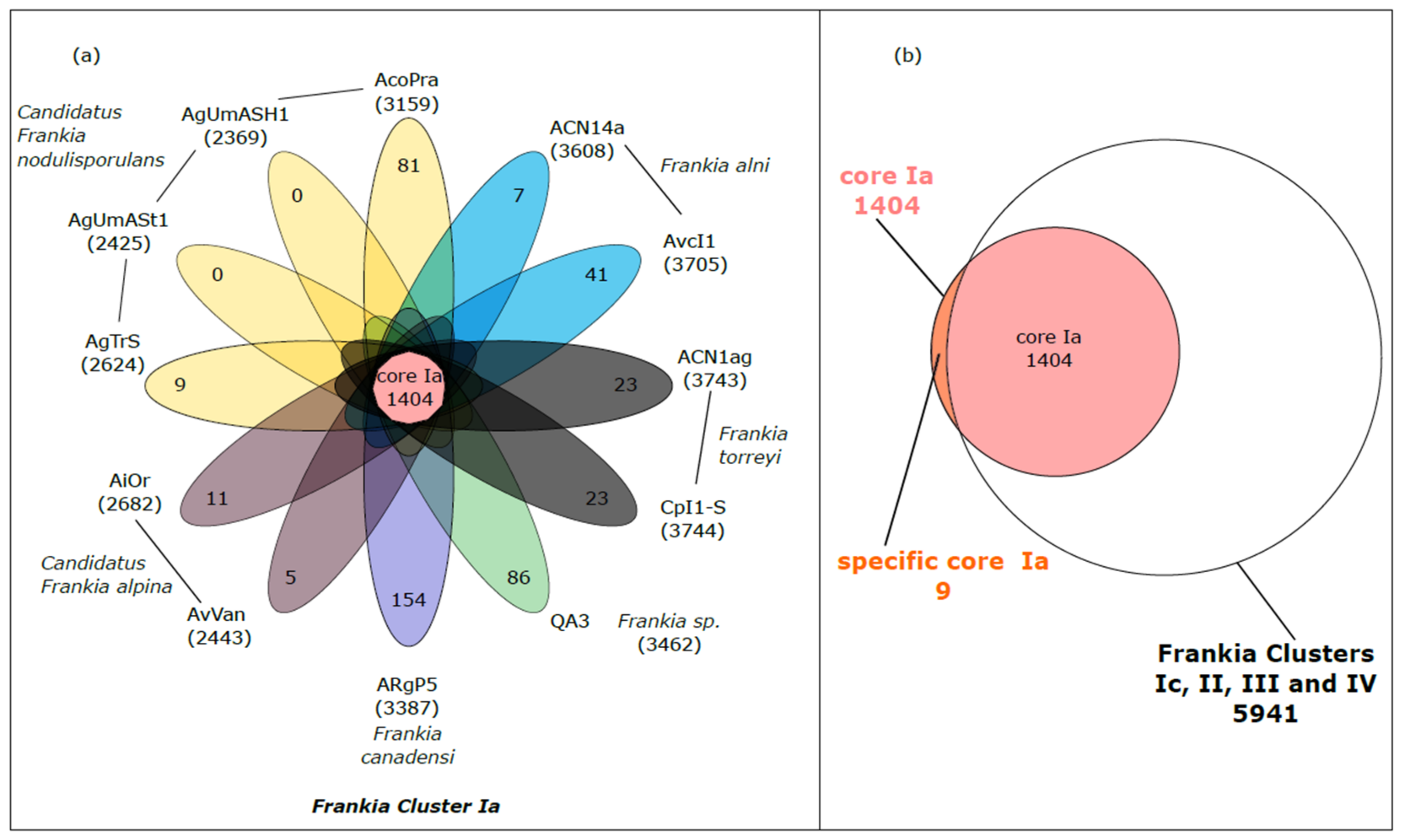
Figure 1. Multigenomic analysis permitted to identify the core genome of Frankia belonging to the Cluster Ia and the specific core Ia. (a) Flower plot diagram of strains from Cluster Ia. The number within brackets associated to each strain indicates the number of genes in the genome (number of unique CDS). The central circle shows the number of genes common to all strains while the petals show the number of genes specific to each strain. The strains belonging to the same species are shaded with the same color. (b) Venn diagram showing the specific core Ia (genes both present in the core-genome of Frankia belonging to the Cluster Ia and absent in the pan genome of the Frankia belonging to the Clusters Ic, II, III and IV).
Genes 14 00530 g001
Figure 2. Proposed roles for agmatine deiminase (AgD) in the relationship Frankia/Alnus regarding: 1. Access to nitrogen: AgD produced by Frankia could be used in order to degrade agmatine found in the plant. The enzyme could thus allow Frankia to access putrescine (via the conversion of NCP into putrescine) and ammonia as sources of nitrogen, 2. Nodule organogenesis: putrescine obtained from NCP after degradation of agmatine by AgD could be used in nodule development, as putrescine is one of the main polyamine in roots and nodules of actinorhizal and 3. Plant defense: Hydroxycinnamic acid amides (HCAAs) produced by Alnus from agmatine are secondary metabolites involved in the defense of plants against pathogens. Production of AgD by Frankia AgD producers leads to the degradation of agmatine into N-carbomoyl-putrescine and ammonium and puts a stop to the production of HCAAs. The absence of HCAAs makes possible the infection by Frankia and the subsequent formation of nodules.
Genes 14 00530 g002
Table 1. List of the 32 Frankia genomes collected.
Frankia StrainClusterGenome Size (pb)Number of ContigCheckM Completeness (%)GC%Total Number of CDS *Accession Number
Candidatus Frankia nodulisporulans AgTrSIa4,943,75261298.0971.615178NZ_CADCWS010000612.1
Candidatus Frankia nodulisporulans AgUmASt1Ia4,311,76330498.6371.343665CADDZU010000001
Candidatus Frankia nodulisporulans AgUmASH1Ia4,285,76323197.5471.233652CADDZW010000001
Candidatus Frankia alpina AiOrIa5,571,61666999.3871.576192GCA_902806485
Candidatus Frankia alpina AvVanIa5,009,155123398.1071.345157GCA_004803575
Frankia alni ACN14aIa7,497,934110072.836714NC_008278.1
Frankia alni AvcI1Ia7,741,9027799.6572.607255LJFZ01000001.1
Frankia sp. QA3Ia7,590,85312010072.597307CM001489.1
Frankia torreyi CpI1-SIa7,639,95815399.3872.437201JYFN00000000.1
Frankia torreyi ACN1agIa7,521,04710899.3772,505687LJPA01000001.1
Frankia canadensi ARgP5Ia7,730,28556899.7372.397500OESX01000001
Frankia casuarinae CcI3Ic5,433,628199.5970.085593CP000249.1
Frankia casuarinae CcI6Ic5,592,32313899.5969.995837GCA_000503735.2
Frankia casuarinae ThrIc5,298,125184__4654NZ_JENI00000000.1
Frankia casuarinae Allo2Ic5,352,110110_70.004368GCA_000733325.1
Frankia casuarinae BRIc5,227,240180__6478NZ_LRTJ00000000.1
Frankia casuarinae CeDIc5,004,600120_70.103937GCA_000732115.1
Frankia casuarinae KB5Ic5,455,56442097.4070.104915NZ_MRUJ00000000.1
Candidatus Frankia datiscae Dg1II5,341,139198.3670.045472CP002801
Frankia coriaria BMG5.1II5,806,76311695.3170.246487JWIO00000000
Candidatus Frankia californiensis Dg2II6,180,138274289.3967.997838FLUV00000000
Frankia meridionalis Cppng1II4,858,2601_68,104968PRJEB19438
Frankia discariae BCU110501III7,907,74120010072.397567ARDT00000000
Frankia sp. EAN1pecIII8,982,042110071.159063NC_009921.1
Frankia sp. EUN1fIII9,392,24039698.3770.819728ADGX01000001.1
Frankia elaeagni BMG5.12III7,602,43613698.6271.676977ARFH00000000
Frankia irregularis G2III9,538,4048399.4670.958663FAOZ00000000
Frankia soli NRRL B-16219III8,032,739289_71.707114MN238860.1
Frankia inefficax EuI1cIV8,815,781110072.318099CP002299.1
Frankia sp. DC12IV6,884,3361210071.936630KQ031391.1
Frankia saprophytica CN3IV9,978,692298.3571.819262AGJN00000000
Frankia asymbiotica M16386IV9,453,06417410071.978884MOMC00000000
* CDS = Coding sequences.
Table 2. Median ANI values between AcoPra and other Alnus-infective Frankia species in Cluster Ia.
Frankia sp. QA3Candidatus Frankia Alpina AiOrCandidatus Frankia Alpina AvVanFrankia alni ACN14aFrankia alni AvcI1Frankia torreyi ACN1agFrankia torreyi CpI1Frankia canadensis ARgP5Frankia sp. AcoPraCandidatus Frankia Nodulisporulans AgTrs
Frankia sp. QA3100.090.591.189.989.890.890.779.978.078.9
Candidatus Frankia alpina AiOr90.1100.099.388.388.389.389.279.677.778.8
Candidatus Frankia alpina AvVan90.699.3100.088.888.889.689.680.578.779.4
Frankia alni ACN14a90.088.989.5100.099.792.192.179.677.878.9
Frankia alni AvcI189.988.889.499.7100.092.092.079.477.778.9
Frankia torreyi ACN1ag90.889.890.392.192.0100.099.979.577.778.8
Frankia torreyi CpI190.989.890.392.292.099.9100.079.477.778.8
Frankia canadensis ARgP579.980.481.379.379.479.679.4100.078.579.4
Frankia sp. AcoPra77.778.078.977.577.377.377.378.3100.098.0
Candidatus Frankia nodulisporulans AgTrs78.078.679.378.077.877.877.778.597.9100.0
Table 3. Genes both present in the core genome of Frankia belonging to the Cluster Ia and absent in the pan genome of the Frankia belonging to the Clusters Ic, II, III and IV (specific core Ia). Label, Begin, End and Length are given for Frankia ACN14a as a reference genome. “SP” and “TM” for Secreted Proteins and Transmembrane Proteins, respectively.
ProductLocalization EC NumberPathwayFrankia alni ACN14a
N° AccessionBeginEndLength (pb)
Flavodoxin domain-containing protein FRAAL24482,667,1692,667,918750
Putative signal peptideSP FRAAL65417,118,0527,118,477426
Hypothetical protein FRAAL47615,156,2165,157,130915
Hypothetical proteinSP FRAAL16491,769,4111,769,734324
Hypothetical protein FRAAL0667728,649729,065417
Agmatine deiminase EC:3.5.3.12arginine catabolismFRAAL0164158,747159,8021056
Putative esterase/acetylhydrolase domains-containing proteinSP FRAAL0169163,780164,418639
Hypothetical integral membrane proteinTM FRAAL42454,608,0834,608,523441
Sulfite exporter TauE/SafE family proteinTM FRAAL42444,607,1814,608,086906
Table 4. Percent Identity Matrix calculated from AgD protein sequences of the 12 Frankia strains from Cluster Ia using the Clustal Omega alignment tool.
SpeciesStrainAcoPraAgTrSAgUmASt1AgUmASH1ACN14aAvcI1CpI1-SACN1agAvVanAiOrARgP5QA3
Candidatus Frankia nodulisporulansAcoPra
AgTrS98.82
AgUmASt198.82100
AgUmASH198.82100100
Frankia alniACN14a79.0179.1779.1779.17
AvcI178.478.5778.5778.5799.43
Frankia torreyiCpI1-S79.3279.4679.4679.4693.1692.59
ACN1ag79.6379.7679.7679.7693.4592.8899.72
Candidatus Frankia alpinaAvVan79.3279.4679.4679.4690.8890.3190.0390.31
AiOr79.9480.0680.0680.0690.8890.3190.0390.3198.86
Frankia canadensiARgP577.2377.1577.1577.1580.1279.5479.8380.1278.178.1
Frankia sp.QA380.5680.3680.3680.3691.4590.8890.8891.1793.4593.4580.98
Table 5. List of specific genes found in Sp− genomes and absent in Sp+ genome from Cluster 1.
COGN° Accession in Frankia alni ACN14aGene NameProductGenetic or Functional Paralog *Localization #
Several Copies
CEnergy production and conversionFRAAL2393hupL1Uptake hydrogenase large subunitFRAAL1829
FRAAL2391hupD1Hydrogenase maturation proteinFRAAL1828
FRAAL2392hupS1Uptake hydrogenase small subunit precursorFRAAL1830SP
FRAAL3522 Putative Formyl-CoA transferaseFRAAL4675
FRAAL3876 Putative acyl-CoA transferases/carnitine dehydrataseFRAAL4764
FRAAL2565 Putative polyketide oxygenase/hydroxylaseFRAAL4792, FRAAL2325, FRAAL3051, FRAAL3395
FRAAL3041 Putative Dihydrolipoamide acyltransferasesFRAAL5152
EAmino acid transport and metabolismFRAAL6516 Putative membrane proteinFRAAL1256TM
ILipid transport and metabolismFRAAL2505atoDAcetoacetyl-CoA transferaseFRAAL2504, FRAAL3148, FRAAL3149
FRAAL4765 Putative enoyl-CoA hydrataseFRAAL2509, FRAAL2514, FRAAL3092, FRAAL3517, FRAAL3973, FRAAL5910, FRAAL6774
FRAAL1660 Putative Acyl-CoA dehydrogenaseFRAAL6459
JTranslation, ribosomal structure and biogenesisFRAAL4260 Putative glutamyl-tRNA(Gln) amidotransferase, subunit AFRAAL0363, FRAAL3665, FRAAL6013, FRAAL6173
KTranscriptionFRAAL2359 Putative tetR family transcriptional regulatorFRAAL4751
FRAAL1892 Putative HTH-type transcriptional regulatorFRAAL4821
FRAAL6046 Transcriptional regulator (MerR-family)FRAAL6751
FRAAL1282 Putative merR family transcriptional regulatorFRAAL6823
LReplication, recombination and repairFRAAL5342 Hypothetical protein; putative DNA helicase IIhomologFRAAL0267
FRAAL6137 Putative ribosylglycoyhydrolaseFRAAL0303, FRAAL5802, FRAAL6736
PInorganic ion transport and metabolismFRAAL1452 Putative ABC transporter, permease proteinFRAAL1453, FRAAL1557TM
QSecondary metabolites biosynthesis, transport and catabolismFRAAL3901 Putative Phytoene dehydrogenaseFRAAL2168
RGeneral function prediction onlyFRAAL0277surEAcid phosphatase SurE, survival protein.FRAAL6200SP
TSignal transduction mechanismsFRAAL3898 Hypothetical proteinFRAAL6520
NIFRAAL6489 Hypothetical proteinFRAAL1398TM
FRAAL1769 Hypothetical proteinFRAAL5611
Single copy
CEnergy production and conversionFRAAL1457 Putative Xanthine dehydrogenase
FRAAL4787 Putative N-glycosyltransferase
FRAAL3448glpQGlycerophosphoryl diester phosphodiesterase SP
DCell cycle control, cell division, chromosome partitioningFRAAL2959 ATP/GTP binding protein TM
EAmino acid transport and metabolismFRAAL5354 Hypothetical protein
FRAAL4450 Putative Monomeric sarcosine oxidase (MSOX)
FRAAL1891 Putative sarcosine oxidase subunit β
FRAAL4839 ABC peptide transporter SP
FNucleotide transport and metabolismFRAAL3674 Uridine kinase
GCarbohydrate transport and metabolismFRAAL0592 Putative ROK family transcriptional regulator
HCoenzyme transport and metabolismFRAAL6157 Conserved hypothetical protein; putative Pantothenate kinase
ILipid transport and metabolismFRAAL2810 Hypothetical protein
KTranscriptionFRAAL0335 Putative LuxR family transcriptional regulator
FRAAL1455 Hypothetical protein
FRAAL1658 Putative two-component system response regulator
FRAAL2338 Hypothetical protein
FRAAL2354 Putative DNA-binding protein
FRAAL3054 Hypothetical protein
FRAAL3611 Putative MarR family transcriptional regulator
FRAAL3970 Putative repressor
FRAAL3977 Putative TetR-family transcriptional regulator
FRAAL4738 Putative LuxR-family transcriptional regulator
LReplication, recombination and repairFRAAL0558 Conserved hypothetical protein; putative DNA-glycosylase domain
LReplication, recombination and repairFRAAL4221 Hypothetical protein
OPosttranslational modification, protein turnover, chaperonesFRAAL1895 Putative heat shock protein 16
FRAAL2394 Thioredoxin-like protein
FRAAL5033 Putative alkaline serine protease SP
PInorganic ion transport and metabolismFRAAL3036 Hypothetical protein
FRAAL3387 Cyclohexanone monooxygenase SP
FRAAL3502 Hypothetical protein; putative Rieske [2Fe-2S] domain
RGeneral function prediction onlyFRAAL0327 Putative amidohydrolase
FRAAL5340 Hypothetical protein
FRAAL3906 Putative integral membrane transport protein TM
FRAAL3907 Putative ABC-type uncharacterized transport system TM
SFunction unknownFRAAL1385 Hypothetical protein
FRAAL3029 Hypothetical protein
FRAAL1789 Hypothetical protein TM
TSignal transduction mechanismsFRAAL1745 Tellurium resistance protein terE
UIntracellular trafficking, secretion, and vesicular transportFRAAL4430 Putative signal peptide SP
NIFRAAL0290 Hypothetical protein
FRAAL1186 Hypothetical protein
FRAAL6274 Hypothetical protein
FRAAL6706 Hypothetical protein
FRAAL3025gvpAGas vesicle synthesis-like protein
FRAAL3026gvpFGas vesicle protein F
FRAAL1685 Putative IMP dehydrogenase/ GMP reductase domain
FRAAL1686 Putative P-loop containing nucleotide triphosphate hydrolase domain
FRAAL2305 Hypothetical protein
FRAAL2306 Hypothetical protein
FRAAL2795 Hypothetical protein
FRAAL3310 Hypothetical protein
FRAAL3311 Hypothetical protein
FRAAL3894 Hypothetical protein
FRAAL4437 Hypothetical protein
FRAAL4895 Hypothetical protein
FRAAL4893 Putative N-acetylmuramoyl-L-alanine amidase domains SP
FRAAL0360 Putative signal peptide SP
FRAAL5030 Putative signal peptide SP
FRAAL5032 Putative signal peptide SP
FRAAL4294 Putative signal peptide SP
FRAAL4721 Putative signal peptide SP
FRAAL5515 Putative lipoprotein SP
FRAAL6270 Putative signal peptide TM
FRAAL3669 Hypothetical protein TM
* Paralog proteins were identified by BlastP (obtaining a coverage > 50% and a percent of identity > 30%) or by KEGG (found in the same metabolic function). # Localization was performed using SignalP6 and DeepTMHMM. “SP” and “TM” for Secreted Proteins and Transmembrane Proteins, respectively.
Disclaimer/Publisher’s Note: The statements, opinions and data contained in all publications are solely those of the individual author(s) and contributor(s) and not of MDPI and/or the editor(s). MDPI and/or the editor(s) disclaim responsibility for any injury to people or property resulting from any ideas, methods, instructions or products referred to in the content.