Article

Identification by Exome Sequencing of Predisposing Variants in Familial Cases of Autoinflammatory Recurrent Fevers

1
Dipartimento di Scienze della Vita e Sanità Pubblica, Sezione di Medicina Genomica, Università Cattolica del Sacro Cuore–Fondazione Policlinico Universitario A. Gemelli IRCCS, 00168 Roma, Italy
2
Research Unit of Medical Genetics, Department of Medicine and Surgery, Università Campus Bio-Medico di Roma, 00128 Rome, Italy
3
Dipartimento di Scienze Geriatriche e Ortopediche, Università Cattolica del Sacro Cuore–Fondazione Policlinico Universitario A. Gemelli IRCCS, 00168 Roma, Italy
4
Bioinformatics Core Facility, Gemelli Science and Technology Park (G-STeP), Fondazione Policlinico Universitario A. Gemelli IRCCS, 00168 Roma, Italy
5
Operative Research Unit of Medical Genetics, Fondazione Policlinico Universitario Campus Bio-Medico, 00128 Rome, Italy
6
Periodic Fevers Research Center, Università Cattolica del Sacro Cuore–Fondazione Policlinico Universitario A. Gemelli IRCCS, 00168 Roma, Italy
*
Author to whom correspondence should be addressed.
Genes 2023, 14(7), 1310; https://doi.org/10.3390/genes14071310
Submission received: 29 May 2023 / Revised: 14 June 2023 / Accepted: 19 June 2023 / Published: 21 June 2023
(This article belongs to the Special Issue Autoimmunity and Autoinflammatory Genetic Syndromes)

Abstract

:
Periodic fever syndromes include autoinflammatory disorders (AID) that involve innate immunity. These disorders are characterized by recurrent fevers and aberrant multi-organ inflammation, without any involvement of T or B cells or the presence of autoantibodies. A complex genetic architecture has been recognized for many AID. However, this complexity has only been partially uncovered for familial Mediterranean fever and other conditions that have a classical monogenic origin and Mendelian transmission. Several gene panels are currently available for molecular diagnosis in patients suspected of having AID. However, even when an extensive number of genes (up to 50–100) are tested in a cohort of clinically selected patients, the diagnostic yield of AID ranges between 15% and 25%, depending on the clinical criteria used for patient selection. In the remaining 75–85% of cases, it is conceivable that the causative gene or genes responsible for a specific condition are still elusive. In these cases, the disease could be explained by variants, either recessive or dominant, that have a major effect on unknown genes, or by the cumulative impact of different variants in more than one gene, each with minor additive effects. In this study, we focused our attention on five familial cases of AID presenting with classical autosomal dominant transmission. To identify the probable monogenic cause, we performed exome sequencing. Through prioritization, filtering, and segregation analysis, we identified a few variants for each family. Subsequent bioinformatics evaluation and pathway analysis helped to narrow down the best candidate genes for each family to FCRL6, PKN1, STAB1, PTDGR, and VCAM1. Future studies on larger cohorts of familial cases will help confirm the pathogenic role of these genes in the pathogenesis of these complex disorders.

1. Introduction

Autoinflammatory conditions (AID) are a group of diseases with a strong genetic component [1,2]. The identification of the MEFV gene in familial Mediterranean fever families in 1997 [3,4] led to discoveries of many other genes in different AID conditions. Along with monogenic entities, there are other mixed autoinflammatory disorders, such as Behçet’s disease, Kawasaki syndrome, and recurrent pericarditis, with a more complex genetic architecture requiring the intervention of multiple variants in more than one gene, along with other endogenous and exogenous factors [5,6]. For this reason, they are rarely transmitted in a Mendelian way. When AIDs present in a sporadic patient, it can be difficult to determine whether they have monogenic or more complex origins. Many autoinflammatory conditions are often diagnosed based solely on the presence of recurrent fever and, after ruling out variants in major genes through the analysis of a gene panel, it can be challenging to molecularly define a specific diagnosis [7]. These cases are now labeled as syndromes of undefined recurrent fever (SURF), where a polygenic or complex genetic-environmental architecture can be suspected [8,9,10]. A subgroup of SURF patients presents a well-defined autosomal dominant Mendelian transmission, highlighting a major gene variant in those cases.
The diagnosis of SURF overlaps with the clinical diagnosis of PFAPA. This syndrome, first described in 1987 [11], is characterized by febrile attacks that occur with “clockwork” periodicity and are accompanied by stereotyped symptoms in the oral cavity, such as tonsillitis/oral aphthosis and/or lymph node enlargement. It is predominantly observed in children, and approximately 10% of cases have an autosomal dominant transmission, highlighting its strong genetic origin. Consequently, most genetic studies have focused on this condition. In a comprehensive genetic study involving 14 families, a locus on chromosome 8 was identified through linkage analysis [12]. However, subsequent whole-exome sequencing failed to reveal any significant variants in the same locus or elsewhere in the exome. The same research group conducted targeted sequencing of 32 genes involved in innate immunity in 82 patients diagnosed with PFAPA [13]. This analysis revealed a frameshift variant in CARD8, which displayed a significant association with the phenotype. Additionally, other susceptibility loci were identified by searching for common predisposing variants of other oropharyngeal ulcerative disorders [14]. Our research group identified rare variants in the ALPK1 gene in a few individuals exhibiting PFAPA-like symptoms [15]. Nevertheless, the majority of patients diagnosed with PFAPA or SURF still lack a genetic diagnosis. For this reason, we focused our attention on those cases that presented a documented familial history of recurrent fever along with other symptoms. In order to investigate the responsible gene in SURF/PFAPA families, we undertook whole-exome sequencing analysis in two affected individuals from each of the five families we identified. This analysis, followed by the bioinformatics characterization of the variants and genes, allowed us to identify different putative predisposing variants in different genes involved in autoinflammation, highlighting the broad genetic heterogeneity of these conditions.

2. Material and Methods
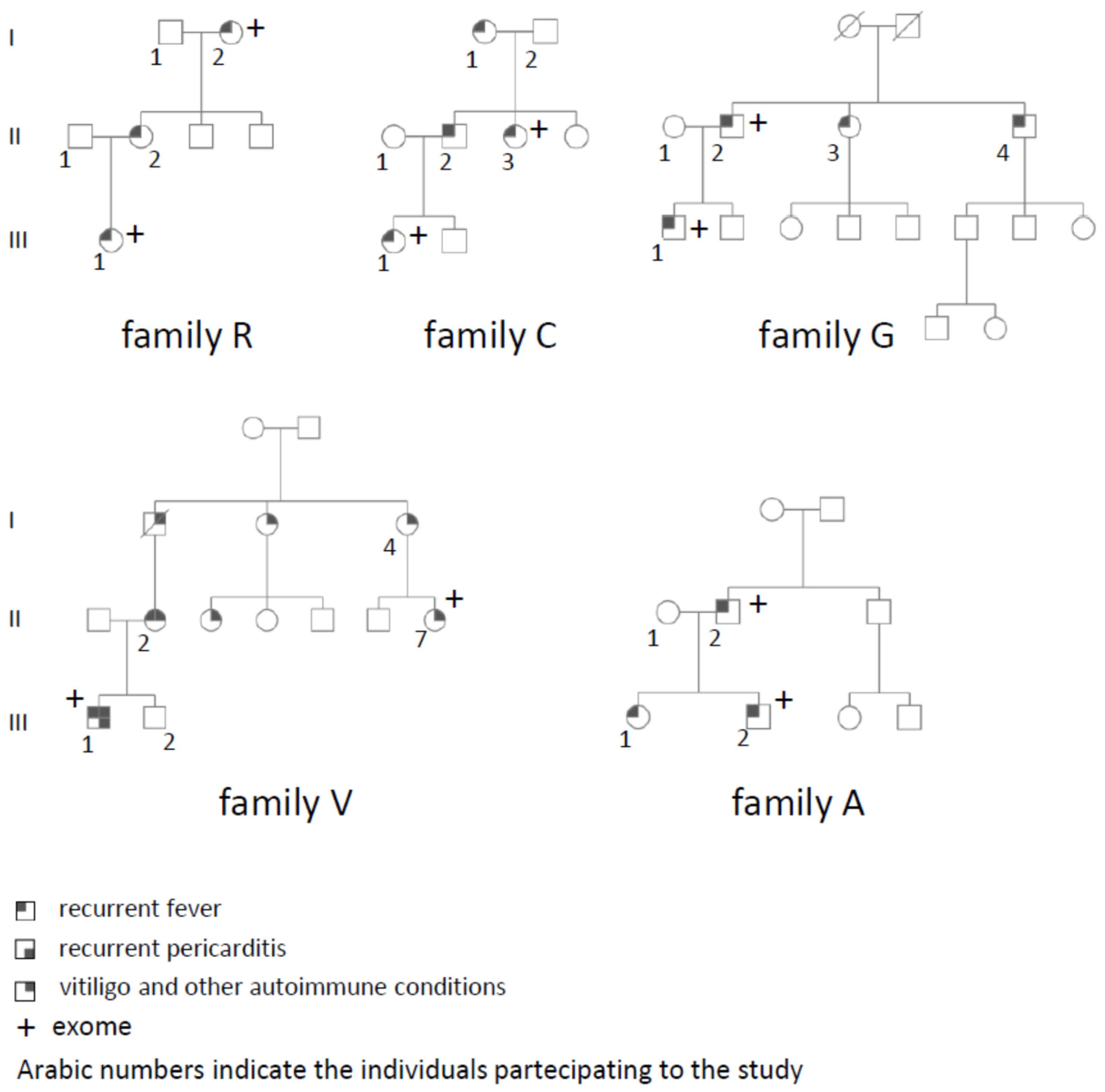
2.1. Families

All patients and families were recruited at our outpatient clinics, “Centro per lo studio delle Febbri Periodiche,” at the Fondazione Policlinico Gemelli. For each proband, their clinical history was evaluated, and biochemical and blood values were recorded during each visit. Biochemical tests and blood analyses were conducted during and after fever recurrences as part of the clinical management.
During the enrollment process, a comprehensive familial history was obtained. When additional individuals with similar symptoms were reported, we contacted and recruited them. We extended the genetic analysis to all available family members whenever possible. Five families were recruited (Figure 1). All individuals provided informed consent, which was approved in 2020 by the Comitato di Bioetica of Fondazione Policlinico Gemelli. All five families had at least two or more affected individuals and tested negative for variants in a gene panel of 13 genes involved in autoinflammatory diseases. Families R, C, G, and A had a clinical diagnosis of PFAPA, SURF, or AIDs not otherwise specified. Family V had a much more complex phenotype with autoinflammatory and autoimmune symptoms. Individual III1 had recurrent fever along with Henoch–Schoenlein purpura and two episodes of pericarditis. His main problem was represented by demyelinating lesions in his brain. His mother, II1, had a series of autoimmune conditions (vitiligo, Hashimoto’s thyroiditis, autoimmune gastritis, and recurrent sterile tonsillitis), her father, I1, died as a consequence of a polyneuritis and transverse myelitis, while the remaining individuals were reported to have only vitiligo.

2.2. Sequencing

Whole-exome sequencing was performed by the Galseq SRL company (Bresso, Milan, Italy) using Illumina technology. An average coverage of 60X was obtained, and paired fastq files were processed on the Galaxy server following standard procedures [16]. After variant calling, we isolated high-quality variants shared by the two affected individuals in each family and annotated them. We selected variants in exonic/splicing regions and filtered out synonymous variants, common variants (MAF > 1/1000), and homozygous variants, considering the apparent autosomal dominant transmission. From the Infevers database (https://infevers.umai-montpellier.fr/ accessed on 20 June 2023) the list of genes involved in autoinflammatory conditions was downloaded. Variants were then prioritized based on keywords such as “autoinflammatory condition,” “inflammasome,” “innate immunity,” “IL1,” “MEFV,” “PYRIN,” and “autoinflammation” using the Varelect software (https://varelect.genecards.org/ accessed on 20 June 2023) [17]. Variants in the top 20 genes according to Varelect were then segregated in the remaining affected individuals of the family, and those not segregating were no longer considered for bioinformatics evaluation.
Sanger sequencing was performed on each variant that passed the filtering/prioritization procedure using standard methods. The variants were analyzed in the affected individuals of the same family.

2.3. Bioinformatics Analysis

A bioinformatics analysis of the genes was conducted using the COXPRESdb (https://coxpresdb.jp/ accessed on 20 June 2023) database [18], which investigates genes co-expressed across different cell types, tissues, and experimental settings to identify genes co-expressed in different gene networks. Gene co-expression represents the synchronization of gene expression across various cellular and environmental conditions and is widely used to infer the biological function of genes. We uploaded our genes along with genes from the Infevers database to identify which genes from our families were co-expressed with genes already identified in other autoinflammatory conditions.
The STRING database (https://string-db.org/ accessed on 20 June 2023) [19], one of the main resources dedicated to organism-wide protein association networks, was used to create a protein–protein interaction network based on the genes detected in the five families, which could be the hypothetical cause of recurrent chronic fevers with inheritance characteristics, and the genes derived from the Infevers database. The minimum required interaction score was lowered to 0.40 (medium confidence) to allow the connection between all the inserted genes/nodes in the multiple proteins selection.

3. Results

We recruited five families with a clinical history characterized by recurrent fever attacks in two or more individuals (Figure 1). Based on pedigree analysis, putative autosomal dominant transmission was suspected. For each family, we selected the two most distantly related individuals for exome sequencing. To prioritize variants in genes already involved in autoinflammatory conditions, we used a gene list for autoinflammatory diseases derived from the Infevers database. However, no pathogenic variants were identified in any of those genes. We then used the Varelect analysis to identify genes prioritized based on keywords related to autoinflammatory conditions. The variants in the top 20 genes with the highest score from this analysis were confirmed using Sanger sequencing and were segregated among all the other affected individuals in the family. Variants shared by all the affected individuals had their frequency evaluated in the gnomAD database (https://gnomad.broadinstitute.org/ accessed on 20 June 2023) and their in silico impact was analyzed using the CADD score [20] and other common software (Table 1).
In family R, four variants in four different genes were identified to segregate with the phenotype (Table 1). As one of the genes was the C-reactive protein (CRP) gene, we investigated whether rare variants in this gene were responsible for a similar phenotype in other sporadic or familial cases of autoinflammatory diseases (AID) in our database. This family had fever periodicity characterized by episodes lasting 7–10 days every 2 months, similar to TNF receptor-associated periodic syndrome (TRAPS) [21,22]. We screened 150 patients, 40 of whom had suspected TRAPS, but none of them carried any rare variant in the CRP gene. In family G, four candidate variants were segregating, while in families C, V, and A, three different variants in each family segregated with the phenotype. Families A and V shared two variants in the same gene PNN (Table 1). To sort the strongest candidate among the new genes identified in these five families, we used bioinformatics tools to determine the role and expression of those genes with respect to genes already involved in autoinflammatory diseases. Firstly, we determined the expression domain in cells and organs involved in the pathogenesis of the disease using the online server COXPRESdb. Three genes, FCRL6, TCTEX1D4, and PBK, were not found to be highly expressed in the hematopoietic system. From this analysis, eight genes (GBP3, CCNG2, STAB1, PTGDR, PNN, VCAM1, PKN1, GNAI2) showed a high level of expression in whole blood, spleen, tonsils, lymph nodes, or bone marrow (Table 2). Using the same database, we investigated whether the proteins obtained from our exome analysis were interacting with the proteins in the Infevers database. From this analysis, GNAI2, PKN1, and VCAM1 were found to directly interact with proteins in the Infevers datasets (Table 3).
To further extend our evaluation of the protein–protein interaction network, we interrogated the STRING database. We uploaded the genes obtained from our exome analysis in node 1 and the genes from the Infevers database in node 2. Using a threshold for the combined score of above 0.4, the five genes with the strongest interactions with one or multiple proteins were GNAI2, PKN1, CRP, SLC15A1, and GBP3 (Table 4).

4. Discussion

In this study, we performed exome sequencing on five families diagnosed with SURF [8,9,10], PFAPA [23,24,25], or atypical PFAPA syndrome. Due to the limited size of each family, we were unable to restrict the number of segregating variants to a single one through segregation analysis among the affected individuals. However, we utilized bioinformatics tools, expression data, and a literature search to infer stronger candidates for each gene. We prioritized variants with a lower allelic frequency and a higher CADD score, and then evaluated their expression level, involvement in the innate immune system, and strong interaction in the autoinflammatory pathway through co-expression and direct protein-protein interactions.
In family R, the CRP gene, initially considered a potential candidate, was partly excluded as we did not find any additional variants in a large cohort of familial and sporadic AID patients. Moreover, the identified variant had a relatively high frequency in the gnomAD database. Consequently, we directed our focus towards three remaining variants in the MROH9, FCRL6, and KIF26B genes. Among these, FCRL6 emerged as the most promising candidate due to its strong expression and role in the immune system [26]. The encoded protein is an Fc-receptor-like protein highly expressed on cytotoxic T and NK cells [27]. It is upregulated on the expanded population of terminally differentiated CD8+ T cells in patients with HIV and B-cell chronic lymphocytic leukemia. However, its role in the autoinflammatory process remains to be determined, as no direct or indirect interactions with proteins/pathways involved in autoinflammation were retrieved from the bioinformatics analyses using the Infevers genes.
In family G, we identified four variants in four different genes, with the strongest candidate being the PKN1 gene. The p.(Gly884Ser) variant in PKN1 is novel with a high CADD score, placing it in the top 1% of deleterious variants. This variant is located in the AGC-kinase C-terminal domain of the protein [28]. PKN1 is a kinase that phosphorylates PYRIN at serine positions 242 and 244, blocking its activation [29,30]. Consequently, PKN1 emerged as the strongest candidate based on bioinformatics evaluations from different databases. Its role should be further evaluated in other familial and sporadic patients with autoinflammatory conditions to validate its significance.
In family C, three variants were identified, among which the variants in STAB1 and CCNG2 looked promising. On the other hand, the GNAI2 variant had a low CADD score, indicating a more benign role. Both variants in STAB1 and CCNG2 had CADD scores above 20, placing them in the top 1% of deleterious variants. CCNG2 is a cyclin with a role in cell-cycle regulation in normal and cancer cells, but no specific role has been described in regulating inflammation or the immune system thus far [31,32]. STAB1 is a scavenger receptor expressed in many immune cells, especially macrophages, with a central role in the innate immune system [33]. Knockout (KO) mice for Stab1 are more susceptible to the lethal effects of Coxsackievirus B3-induced myocarditis, sepsis, or Listeria infection [34,35]. Additionally, KO mice spontaneously develop an autoimmune phenotype as they age. The presumed mechanism is through blocking the fibronectin-mediated STAB1+ monocyte recruitment and differentiation into anti-inflammatory macrophages, resulting in an increased T cell response. Loss-of-function mutations could lead to a dysregulated inflammatory response both in animal models and in humans.
In family V, we observed two interesting variants among three genes, both with CADD scores close to 30 and functions in the innate immune system. The first gene, PNN, was the only gene found to be mutated in two different families in our cohort of five families. This gene, called PININ, is involved in desmosome junctions and splicing mechanisms [36]. In mice, its mutation leads to early embryonic lethality in the homozygous state, while a hypomorphic allele causes widespread defects in neural crest derivatives and consequent malformations in different organs and tissues [37]. Therefore, it is an unlikely candidate for autoinflammatory diseases. The stronger candidate in this family is the PTGDR gene, encoding a receptor for Prostaglandin D2. Polymorphisms in this gene have been controversially associated with predisposition to asthma and allergic reactions. The p.(Met228Ile) variant is very rare in the gnomAD database and has a CADD score of 29. The PGA2/PTGDR axis is considered to have an anti-inflammatory role. PTGDR has an inhibitory role in PYDC3, a protein that negatively regulates inflammasome activation [38]. Ptgdr−/− mice exhibit increased IL-1β expression with increased mortality when challenged with a neurotropic virus. This effect was mediated by increased activation of the inflammasome and reduced by blocking IL1β [39]. It is interesting to observe that this protein is considered critical for the pathogenesis of experimental autoimmune encephalitis, an animal model for multiple sclerosis. In family V, individuals III1 and I2, along with other symptoms have signs and symptoms of a demyelinating disease.
In family A, after excluding PNN due to its role in development, we focused our attention on the VCAM1 variant. This variant has a high CADD score and has never been reported before in the gnomAD database. It falls within the first Ig-like domain of the protein, adjacent to a cysteine required to form disulfide bonds. VCAM1 is an adhesion molecule initially identified as an endothelial cell surface glycoprotein [40]. It binds α4β1 and α4β7 integrins on leukocytes through Ig-like domains 1 and 4, facilitating rolling, adhesion, and transmigration. VCAM1 expression is driven by TNFα through the NFKB and MAPK pathway, and its expression facilitates leukocyte migration through endothelial junctions. This protein is at a crossroads with most of the pathways involved in autoinflammatory conditions and in the innate immune system [40].
In summary, through exome sequencing, we identified potential candidate genes for each family, selected based on the variant and their involvement in the innate immunity. These five genes, namely FCRL6, PKN1, STAB1, PTGDR, and VCAM1, hold significant promise as candidates for further investigation in future studies involving a larger cohort of patients with autoinflammatory diseases.

Author Contributions

Conceptualization, E.S. and R.M.; Methodology, A.A., R.R., I.C. and F.G.; Software, L.G. and M.A.T.; Validation, R.R.; Formal analysis, E.S., A.A., I.C., E.V., F.G. and R.M.; Investigation, I.C. and E.V.; Resources, R.M.; Data curation, L.G.; Writing—original draft, E.S.; Writing—review & editing, A.A.; Supervision, E.S.; Funding acquisition, R.M. All authors have read and agreed to the published version of the manuscript.

Funding

Periodic Fevers Research Center, Università Cattolica del Sacro Cuore.

Institutional Review Board Statement

The study was conducted in accordance with the Declaration of Helsinki, and approved by the Institutional Review Board (or Ethics Committee) of Università Cattolica del Sacro Cuore-Fondazione Policlinico Gemelli (protocol code 3343, 23 July 2023).

Informed Consent Statement

Informed consent was obtained from all subjects involved in the study.

Data Availability Statement

Data sharing not applicable.

Conflicts of Interest

The authors declare no conflict of interest.

References

  1. Caso, F.; Rigante, D.; Vitale, A.; Lucherini, O.M.; Costa, L.; Atteno, M.; Compagnone, A.; Caso, P.; Frediani, B.; Galeazzi, M.; et al. Monogenic Autoinflammatory Syndromes: State of the Art on Genetic, Clinical, and Therapeutic Issues. Int. J. Rheumatol. 2013, 2013, 513782. [Google Scholar] [CrossRef] [PubMed]
  2. David, T.; Ling, S.F.; Barton, A. Genetics of Immune-Mediated Inflammatory Diseases. Clin. Exp. Immunol. 2018, 193, 3–12. [Google Scholar] [CrossRef] [Green Version]
  3. The International FMF Consortium. Ancient Missense Mutations in a New Member of the RoRet Gene Family Are Likely to Cause Familial Mediterranean Fever. Cell 1997, 90, 797–807. [Google Scholar] [CrossRef]
  4. Consortium, T.F.F.; Bernot, A.; Clepet, C.; Dasilva, C.; Devaud, C.; Petit, J.-L.; Caloustian, C.; Cruaud, C.; Samson, D.; Pulcini, F.; et al. A Candidate Gene for Familial Mediterranean Fever. Nat. Genet. 1997, 17, 25–31. [Google Scholar] [CrossRef]
  5. Sangiorgi, E.; Rigante, D. The Clinical Chameleon of Autoinflammatory Diseases in Children. Cells 2022, 11, 2231. [Google Scholar] [CrossRef] [PubMed]
  6. Ombrello, M.J. Advances in the Genetically Complex Autoinflammatory Diseases. Semin. Immunopathol. 2015, 37, 403–406. [Google Scholar] [CrossRef] [Green Version]
  7. Rama, M.; Mura, T.; Kone-Paut, I.; Boursier, G.; Aouinti, S.; Touitou, I.; Sarrabay, G. Is Gene Panel Sequencing More Efficient than Clinical-Based Gene Sequencing to Diagnose Autoinflammatory Diseases? A Randomized Study. Clin. Exp. Immunol. 2021, 203, 105–114. [Google Scholar] [CrossRef]
  8. Papa, R.; Penco, F.; Volpi, S.; Sutera, D.; Caorsi, R.; Gattorno, M. Syndrome of Undifferentiated Recurrent Fever (SURF): An Emerging Group of Autoinflammatory Recurrent Fevers. J. Clin. Med. 2021, 10, 1963. [Google Scholar] [CrossRef]
  9. Luu, I.; Nation, J.; Page, N.; Carvalho, D.; Magit, A.; Jiang, W.; Leuin, S.; Bliss, M.; Bothwell, M.; Brigger, M.; et al. Undifferentiated Recurrent Fevers in Pediatrics Are Clinically Distinct from PFAPA Syndrome but Retain an IL-1 Signature. Clin. Immunol. 2021, 226, 108697. [Google Scholar] [CrossRef]
  10. Sutera, D.; Bustaffa, M.; Papa, R.; Matucci-Cerinic, C.; Matarese, S.; D’Orsi, C.; Penco, F.; Prigione, I.; Palmeri, S.; Bovis, F.; et al. Clinical Characterization, Long-Term Follow-up, and Response to Treatment of Patients with Syndrome of Undifferentiated Recurrent Fever (SURF). Semin. Arthritis Rheum. 2022, 55, 152024. [Google Scholar] [CrossRef]
  11. Marshall, G.S.; Edwards, K.M.; Butler, J.; Lawton, A.R. Syndrome of Periodic Fever, Pharyngitis, and Aphthous Stomatitis. J. Pediatr. 1987, 110, 43–46. [Google Scholar] [CrossRef] [PubMed]
  12. Di Gioia, S.A.; Bedoni, N.; von Scheven-Gête, A.; Vanoni, F.; Superti-Furga, A.; Hofer, M.; Rivolta, C. Analysis of the Genetic Basis of Periodic Fever with Aphthous Stomatitis, Pharyngitis, and Cervical Adenitis (PFAPA) Syndrome. Sci. Rep. 2015, 5, 10200. [Google Scholar] [CrossRef] [Green Version]
  13. Cheung, M.S.; Theodoropoulou, K.; Lugrin, J.; Martinon, F.; Busso, N.; Hofer, M. Periodic Fever with Aphthous Stomatitis, Pharyngitis, and Cervical Adenitis Syndrome Is Associated with a CARD8 Variant Unable to Bind the NLRP3 Inflammasome. J. Immunol. 2017, 198, 2063–2069. [Google Scholar] [CrossRef] [PubMed] [Green Version]
  14. Manthiram, K.; Preite, S.; Dedeoglu, F.; Demir, S.; Ozen, S.; Edwards, K.M.; Lapidus, S.; Katz, A.E.; Feder, H.M.; Genomic Ascertainment Cohort; et al. Common Genetic Susceptibility Loci Link PFAPA Syndrome, Behçet’s Disease, and Recurrent Aphthous Stomatitis. Proc. Natl. Acad. Sci. USA 2020, 117, 14405–14411. [Google Scholar] [CrossRef] [PubMed]
  15. Sangiorgi, E.; Azzarà, A.; Molinario, C.; Pietrobono, R.; Rigante, D.; Verrecchia, E.; Sicignano, L.L.; Genuardi, M.; Gurrieri, F.; Manna, R. Rare Missense Variants in the ALPK1 Gene May Predispose to Periodic Fever, Aphthous Stomatitis, Pharyngitis and Adenitis (PFAPA) Syndrome. Eur. J. Hum. Genet. EJHG 2019, 27, 1361–1368. [Google Scholar] [CrossRef] [PubMed]
  16. Goecks, J.; Nekrutenko, A.; Taylor, J.; Team, T.G. Galaxy: A Comprehensive Approach for Supporting Accessible, Reproducible, and Transparent Computational Research in the Life Sciences. Genome Biol. 2010, 11, R86. [Google Scholar] [CrossRef] [Green Version]
  17. Stelzer, G.; Plaschkes, I.; Oz-Levi, D.; Alkelai, A.; Olender, T.; Zimmerman, S.; Twik, M.; Belinky, F.; Fishilevich, S.; Nudel, R.; et al. VarElect: The Phenotype-Based Variation Prioritizer of the GeneCards Suite. BMC Genom. 2016, 17 (Suppl. S2), 444. [Google Scholar] [CrossRef] [Green Version]
  18. Obayashi, T.; Kodate, S.; Hibara, H.; Kagaya, Y.; Kinoshita, K. COXPRESdb v8: An Animal Gene Coexpression Database Navigating from a Global View to Detailed Investigations. Nucleic Acids Res. 2023, 51, D80–D87. [Google Scholar] [CrossRef]
  19. Szklarczyk, D.; Gable, A.L.; Nastou, K.C.; Lyon, D.; Kirsch, R.; Pyysalo, S.; Doncheva, N.T.; Legeay, M.; Fang, T.; Bork, P.; et al. The STRING Database in 2021: Customizable Protein-Protein Networks, and Functional Characterization of User-Uploaded Gene/Measurement Sets. Nucleic Acids Res. 2021, 49, D605–D612. [Google Scholar] [CrossRef]
  20. Rentzsch, P.; Witten, D.; Cooper, G.M.; Shendure, J.; Kircher, M. CADD: Predicting the Deleteriousness of Variants throughout the Human Genome. Nucleic Acids Res. 2019, 47, D886–D894. [Google Scholar] [CrossRef]
  21. Hull, K.M.; Drewe, E.; Aksentijevich, I.; Singh, H.K.; Wong, K.; McDermott, E.M.; Dean, J.; Powell, R.J.; Kastner, D.L. The TNF Receptor-Associated Periodic Syndrome (TRAPS): Emerging Concepts of an Autoinflammatory Disorder. Medicine 2002, 81, 349–368. [Google Scholar] [CrossRef] [PubMed]
  22. Gaggiano, C.; Vitale, A.; Obici, L.; Merlini, G.; Soriano, A.; Viapiana, O.; Cattalini, M.; Maggio, M.C.; Lopalco, G.; Montin, D.; et al. Clinical Features at Onset and Genetic Characterization of Pediatric and Adult Patients with TNF-α Receptor-Associated Periodic Syndrome (TRAPS): A Series of 80 Cases from the AIDA Network. Mediat. Inflamm. 2020, 2020, 8562485. [Google Scholar] [CrossRef] [PubMed]
  23. Cantarini, L.; Vitale, A.; Sicignano, L.L.; Emmi, G.; Verrecchia, E.; Patisso, I.; Cerrito, L.; Fabiani, C.; Cevenini, G.; Frediani, B.; et al. Diagnostic Criteria for Adult-Onset Periodic Fever, Aphthous Stomatitis, Pharyngitis, and Cervical Adenitis (PFAPA) Syndrome. Front. Immunol. 2017, 8, 1018. [Google Scholar] [CrossRef] [PubMed] [Green Version]
  24. Gattorno, M.; Caorsi, R.; Meini, A.; Cattalini, M.; Federici, S.; Zulian, F.; Cortis, E.; Calcagno, G.; Tommasini, A.; Consolini, R.; et al. Differentiating PFAPA Syndrome From Monogenic Periodic Fevers. Pediatrics 2009, 124, e721–e728. [Google Scholar] [CrossRef] [PubMed]
  25. Sicignano, L.L.; Rigante, D.; Moccaldi, B.; Massaro, M.G.; Delli Noci, S.; Patisso, I.; Capozio, G.; Verrecchia, E.; Manna, R. Children and Adults with PFAPA Syndrome: Similarities and Divergences in a Real-Life Clinical Setting. Adv. Ther. 2021, 38, 1078–1093. [Google Scholar] [CrossRef]
  26. Kulemzin, S.V.; Zamoshnikova, A.Y.; Yurchenko, M.Y.; Vitak, N.Y.; Najakshin, A.M.; Fayngerts, S.A.; Chikaev, N.A.; Reshetnikova, E.S.; Kashirina, N.M.; Peclo, M.M.; et al. FCRL6 Receptor: Expression and Associated Proteins. Immunol. Lett. 2011, 134, 174–182. [Google Scholar] [CrossRef]
  27. Schreeder, D.M.; Pan, J.; Li, F.J.; Vivier, E.; Davis, R.S. FCRL6 Distinguishes Mature Cytotoxic Lymphocytes and Is Upregulated in Patients with B-Cell Chronic Lymphocytic Leukemia. Eur. J. Immunol. 2008, 38, 3159–3166. [Google Scholar] [CrossRef] [PubMed] [Green Version]
  28. Schnappauf, O.; Chae, J.J.; Kastner, D.L.; Aksentijevich, I. The Pyrin Inflammasome in Health and Disease. Front. Immunol. 2019, 10, 1745. [Google Scholar] [CrossRef] [Green Version]
  29. Park, Y.H.; Wood, G.; Kastner, D.L.; Chae, J.J. Pyrin Inflammasome Activation and RhoA Signaling in the Autoinflammatory Diseases FMF and HIDS. Nat. Immunol. 2016, 17, 914–921. [Google Scholar] [CrossRef] [Green Version]
  30. Magnotti, F.; Lefeuvre, L.; Benezech, S.; Malsot, T.; Waeckel, L.; Martin, A.; Kerever, S.; Chirita, D.; Desjonqueres, M.; Duquesne, A.; et al. Pyrin Dephosphorylation Is Sufficient to Trigger Inflammasome Activation in Familial Mediterranean Fever Patients. EMBO Mol. Med. 2019, 11, e10547. [Google Scholar] [CrossRef]
  31. Sun, G.G.; Zhang, J.; Hu, W.N. CCNG2 Expression Is Downregulated in Colorectal Carcinoma and Its Clinical Significance. Tumour Biol. 2014, 35, 3339–3346. [Google Scholar] [CrossRef] [PubMed]
  32. Sun, G.G.; Hu, W.N.; Cui, D.W.; Zhang, J. Decreased Expression of CCNG2 Is Significantly Linked to the Malignant Transformation of Gastric Carcinoma. Tumour Biol. 2014, 35, 2631–2639. [Google Scholar] [CrossRef] [PubMed]
  33. Cabral, F.; Al-Rahem, M.; Skaggs, J.; Thomas, T.A.; Kumar, N.; Wu, Q.; Fadda, P.; Yu, L.; Robinson, J.M.; Kim, J.; et al. Stabilin Receptors Clear LPS and Control Systemic Inflammation. iScience 2021, 24, 103337. [Google Scholar] [CrossRef]
  34. Carai, P.; Papageorgiou, A.P.; Van Linthout, S.; Deckx, S.; Velthuis, S.; Lutgens, E.; Wijnands, E.; Tschöpe, C.; Schmuttermaier, C.; Kzhyshkowska, J.; et al. Stabilin-1 Mediates Beneficial Monocyte Recruitment and Tolerogenic Macrophage Programming during CVB3-Induced Viral Myocarditis. J. Mol. Cell. Cardiol. 2022, 165, 31–39. [Google Scholar] [CrossRef] [PubMed]
  35. Pombinho, R.; Pinheiro, J.; Resende, M.; Meireles, D.; Jalkanen, S.; Sousa, S.; Cabanes, D. Stabilin-1 Plays a Protective Role against Listeria Monocytogenes Infection through the Regulation of Cytokine and Chemokine Production and Immune Cell Recruitment. Virulence 2021, 12, 2088–2103. [Google Scholar] [CrossRef]
  36. Ouyang, P.; Sugrue, S.P. Characterization of Pinin, a Novel Protein Associated with the Desmosome-Intermediate Filament Complex. J. Cell. Biol. 1996, 135, 1027–1042. [Google Scholar] [CrossRef]
  37. Joo, J.-H.; Lee, Y.J.; Munguba, G.C.; Park, S.; Taxter, T.J.; Elsagga, M.Y.; Jackson, M.R.; Oh, S.P.; Sugrue, S.P. Role of Pinin in Neural Crest, Dorsal Dermis, and Axial Skeleton Development and Its Involvement in the Regulation of Tcf/Lef Activity in Mice. Dev. Dyn. 2007, 236, 2147–2158. [Google Scholar] [CrossRef]
  38. Lee, Y.H.; Choi, S.J.; Ji, J.D.; Song, G.G. PTGDR Polymorphisms and Susceptibility to Asthma: A Meta-Analysis. Mol. Biol. Rep. 2013, 40, 2195–2203. [Google Scholar] [CrossRef]
  39. Zheng, J.; Sariol, A.; Meyerholz, D.; Zhang, Q.; Abrahante Lloréns, J.E.; Narumiya, S.; Perlman, S. Prostaglandin D2 Signaling in Dendritic Cells Is Critical for the Development of EAE. J. Autoimmun. 2020, 114, 102508. [Google Scholar] [CrossRef]
  40. Kong, D.-H.; Kim, Y.K.; Kim, M.R.; Jang, J.H.; Lee, S. Emerging Roles of Vascular Cell Adhesion Molecule-1 (VCAM-1) in Immunological Disorders and Cancer. Int. J. Mol. Sci. 2018, 19, 1057. [Google Scholar] [CrossRef] [Green Version]
Figure 1. Pedigrees of the families participating in this study. In each family, individuals with an Arabic numeral participated in this study, providing their DNA upon signing the informed consent form. Individuals with «+» had their exome sequenced.
Genes 14 01310 g001
Table 1. List of the variants identified in different family segregating among the affected individuals. The allelic frequency of each variant was obtained from the gnomAD database. In silico evaluations of each variant were performed using CADD, Mutation Taster, SIFT, Polyphen and GERP.
GenesVariantAllelic Frequency gnomADCADD Mutation TasterSIFTPolyphenGERP
MROH9p.Leu671Ser6/17901823.6PolymorphismDamagingProbably damaging5.91family
R
CRPp.Arg206Trp77/28268019.27PolymorphismDamaging Possibly damaging−2.18
FCRL6p.Gly350Arg23/28283414.48PolymorphismToleratedPossibly damaging−1.12
KIF26Bp.Ala277Val47/26991014.42PolymorphismToleratedBenign3.9
NUBP1p.Pro5Arg46/20808632Disease causingDeleteriousProbably damaging3.96family
G
SLC15A1p.Ile631Thr313/28222025.9Disease causingDeleteriousBenign5.91
PKN1p.Gly884Ser-23.6Disease causingToleratedBenign3.82
GBP3p.Glu457Asp223/28274414.52PolymorphismDamagingBenign0.45
CCNG2p.Ala67Val-23.7PolymorphismDamagingBenign4.55family
C
STAB1p.Arg1305Gln19/25064221.6PolymorphismToleratedBenign0.26
GNAI2p.Thr11Lys-6.59Disease causingDamagingBenign−0.36
PTGDRp.Met228Ile2/24574629.3Disease causingDamagingProbably damaging4.14family
V
PNNp.Ala74Pfs*43-28.6Disease causing---
TCTEX1D4p.Gly115Glu-11.28PolymorphismToleratedBenign3.5
PNNp.Asp680Gly1/25126228.6Disease causingDamagingProbably damaging6.13family
A
VCAM1p.Thr49Ile-23.6PolymorphismDamagingPossibly damaging4.73
PBKp.Asp178Asn7/27990823Disease causingToleratedPossibly damaging2.24
Table 2. Gene expression values obtained from the COXPRESdb, using the GSE3526 and GTEx data. The replicates for each of the tissues were averaged in base-2 logarithm. Different gray shades indicates higher expression values.
GSE3526 GTEx
GeneBone MarrowLymph NodesSpleenTonsilSpleenWhole Blood
MROH90.0930−0.1060−0.2327−0.0957−11.815−11.815
CRP−0.0024−0.1586−0.2677−0.05310.3596−0.6280
KIF26B0.3755−0.3633−0.42750.1002−0.5859−54.054
NUBP1−0.02190.16590.09320.61140.7842−0.4391
SLC15A1−0.1079−0.2001−0.1035−0.2850−23.668−68.529
PKN10.44060.65240.86330.606312.9130.6124
GBP3−11.97713.19419.2940.706613.878−11.340
CCNG2−0.15320.643212.25412.6140.6959−0.7247
STAB1−0.047320.65127.4690.597440.3820.9264
GNAI20.70990.72810.99060.258111.30519.680
PTGDR0.41300.962333.963−0.098537.45320.216
PNN0.219312.60018.7400.90200.9617−17.914
VCAM117.45232.88040.50026.20354.978−52.102
Color scale−3~−2−2~−1−1~+1+1~+2+2~+3+3~
Table 3. Query genes from our exome analysis, interacting genes are from the Infevers database using the COXPRESdb.
Query GeneInteract GeneData SourceExperiment TypePubmed
GNAI2UBA1HPRD_complexin vivo16263121
PKN1PSMB4IntAct(hsa)validated two hybrid32296183
PKN1PSMB4IntAct(hsa)two hybrid prey pooling approach32296183
VCAM1PSMA3IntAct(hsa)cross-linking study22623428
VCAM1TRAP1IntAct(hsa)cross-linking study22623428
Table 4. Analysis from the STRING database. Node 1 has genes from the exomes, node 2 has the genes from Infevers database. The threshold for the combined score was set to 0.4.
Node1Node2Co-ExpressionExperimentally
Determined Interaction
Database
Annotated
Automated
Text Mining
Combined
Score
GNAI2CDC420.2490.3520.9000.1670.954
PKN1MEFV0.05700.8000.5170.901
CRPIL100000.8380.838
CRPVCAM10000.8020.802
VCAM1IL100.053000.7080.712
SLC15A1SLC29A30.066000.6790.688
CRPIL1RN0.062000.6810.687
PKN1CDC420.0620.30000.5040.646
CRPTNFRSF1A0000.6010.601
VCAM1TNFRSF1A0000.5950.595
GBP3NLRP30.072000.5480.563
CRPMEFV0000.5560.556
CRPNLRP30000.5560.556
CRPIL36RN0.062000.5410.551
GBP3PSMB80.2410.05800.3450.491
VCAM1ADAM170.049000.4620.466
SLC15A1NOD20000.4620.462
GNAI2ALPK10000.4550.455
VCAM1NLRP30.052000.4280.435
VCAM1IL36RN0000.4270.427
VCAM1IL1RN0.062000.4100.423
CRPNOD20000.4070.407
VCAM1CDC420.089000.3720.403
Disclaimer/Publisher’s Note: The statements, opinions and data contained in all publications are solely those of the individual author(s) and contributor(s) and not of MDPI and/or the editor(s). MDPI and/or the editor(s) disclaim responsibility for any injury to people or property resulting from any ideas, methods, instructions or products referred to in the content.

