Case Report

A Study of Polish Family with Scoliosis and Limb Contractures Expands the MYH3 Disease Spectrum

by
Justyna Frasuńska
1,
Agnieszka Pollak
2,*,
Paweł Turczyn
3,
Anna Kutkowska-Kaźmierczak
4,
Jakub Pepłowski
5,
Rafał Płoski
2 and
Beata Tarnacka
1
1
Department of Rehabilitation, Medical University of Warsaw, 02-091 Warsaw, Poland
2
Department of Medical Genetics, Medical University of Warsaw, 02-091 Warsaw, Poland
3
Clinic of Early Arthritis, National Institute of Geriatrics, Rheumatology and Rehabilitation, 02-637 Warsaw, Poland
4
Department of Medical Genetics, Institute of Mother and Child, 01-211 Warsaw, Poland
5
The Rare Diseases Laboratory, Laboratory of Genetics, University Center for Laboratory Medicine, University Clinical Centre of the Medical University of Warsaw, 02-097 Warsaw, Poland
*
Author to whom correspondence should be addressed.
Genes 2024, 15(1), 125; https://doi.org/10.3390/genes15010125
Submission received: 20 November 2023 / Revised: 31 December 2023 / Accepted: 16 January 2024 / Published: 19 January 2024
(This article belongs to the Section Genetic Diagnosis)

Abstract

:
A disease associated with malfunction of the MYH3 gene is characterised by scoliosis, contractures of the V fingers, knees and elbows, dysplasia of the calf muscles, foot deformity and limb length asymmetry. The aim of this study was to identify the cause of musculoskeletal deformities in a three-generation Polish family by exome sequencing. The segregation of the newly described c.866A>C variant of the MYH3 gene in the family indicates an autosomal dominant model of inheritance. The detected MYH3 variant segregates the disease within the family. The presented results expand the MYH3 disease spectrum and emphasize the clinical diagnostic challenge in syndromes harbouring congenital spine defects and joint contractures.

1. Introduction

The causes underlying congenital spine defects are heterogeneous and comprise a variety of environmental and genetic factors. Environmental factors have been proven to have a strong influence on the development of congenital scoliosis. Among the most important are hypoxia, vitamin A deficiency, alcohol use, smoking, valproic acid, boric acid and hyperthermia. The rank of environmental factors is significantly lower for confirmed genetic disease with spinal defects as a symptom [1]. Furthermore, spine defects may be a component of various disease entities such as hemivertebra, Klippel–Feil syndrome, Chiari malformation, tethered cord, spina bifida, diastematomyelia and many others [2]. Congenital curvature of the spine in the form of scoliosis occurs in 11.4% of all cases of congenital spine defects [2]. Some of these deformities are caused by genetic syndromes related to pathogenic variants in many genes, i.e., the fibrilin-1 gene (FBN1) in 5q23, the fibriline–2 gene (FBN2) in 5q23 or the myosin heavy chain (MYH3) in 17p13.
The MYH3 gene encodes the protein known as the embryonic myosin heavy chain. The role of MYH3 in the development of the skeletal and muscular systems is of significant importance. This protein is composed of various components, including a globular head, a neck domain, a hinge and a long coiled-coil rod domain. The head contains an actin filament binding site and is crucial for ATP hydrolysis, which is essential for muscle contraction. Within the neck, there are IQ motifs that have the ability to bind to calmodulin. The coiled-coil rod plays a mechanical role in the formation of stiff fibre bundles [3,4]. During normal human development, expression is restricted to stages of embryonic development and rapidly declines after birth [5,6]. In humans, MYH3 expression predominates in myotubes fated to become fast myofibers, subsequently replaced by the expression of different myosins (MYH1, MYH2 and MYH4). While one or more of these myosins may contribute to the development of structurally normal skeletal muscle in individuals with MYH3 pathogenic variants, the function of foetal muscles rich in fast-twitch myofibers is functionally impaired [3].
Congenital diseases associated with the FBN1, FBN2 and MYH3 genes include, respectively, Marfan syndrome, Beals syndrome and distal arthrogryposis type 8. These disorders share many similar defects, such as scoliosis, joint limpness, joint contractures, arachnodactyly and muscle hypoplasia, often making their accurate diagnosis difficult [2]. Many of these defects can be diagnosed prenatally. Ultrasonography plays an important role in this process. The precise detection of the cause of a defect is possible by applying comprehensive genetic testing such as whole-exome sequencing (WES). In many cases, comprehensive genetic tests are not performed, and the molecular cause of a deformity is not established; thus, the disorder causing the deformity is classified as idiopathic. The difficulty of identifying and preventing these diseases is also related to their sporadic as well as familial occurrence. Despite significant improvement in prenatal testing, there is still no fully effective treatment, and unfortunately, severe disability and impaired quality of life for affected children are common.

2. Materials and Methods

The following study was conducted in cooperation with the Department of Medical Genetics, Medical University of Warsaw. A total of 11 family members of the proband were recruited for genetic testing. Informed consent for the presentation of clinical and medical data was obtained from all subjects involved in the study.

2.1. The Proband

A 41-year-old male was admitted to our Department of Rehabilitation, Medical University of Warsaw, for the diagnosis and treatment of severe scoliosis and intermittent significant spinal pain. During the examination, a number of dysmorphic features besides scoliosis were noted, which suggested the presence of a genetic syndrome.

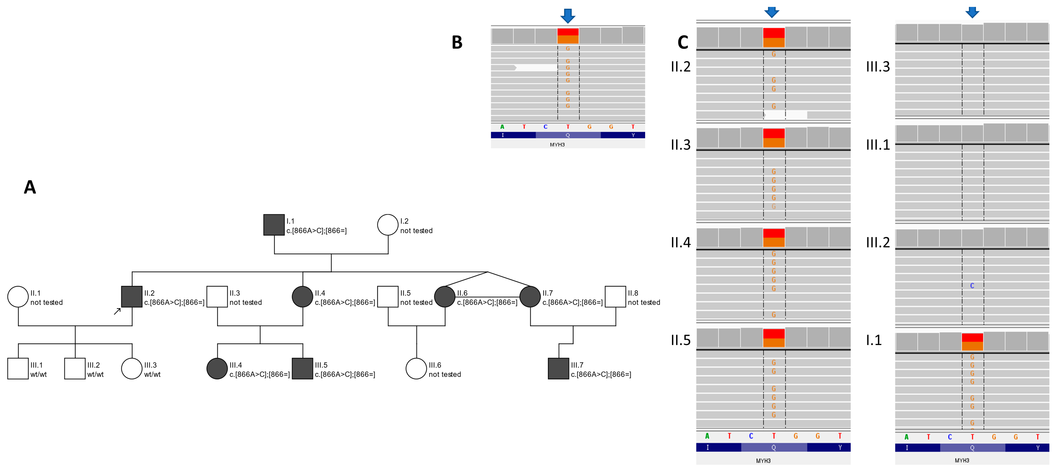
2.2. The Proband’s Family

After interviewing the patient, it was determined that other family members also presented clinical abnormalities (the proband’s father and three of the proband’s siblings); thus, they underwent a detailed clinical evaluation. The presence of scoliosis, camptodactyly and other deviations was confirmed. All the aforementioned family members were referred for genetic testing. Three of the proband’s children, who do not present characteristic phenotypic features, also underwent additional genetic testing. Initially, no genetic testing was performed on the children of the remaining siblings of the proband. Recently, genetic testing was performed on three children of the proband’s sisters (III.4, III.5, III.7) who have similar clinical symptoms.

2.3. Genetic Testing

DNA from the proband and his family (Figure 1A) was obtained from peripheral blood and extracted with a standard protocol. Library preparation for WES was performed on the proband’s DNA sample with SeqCap EZ MedExome probes (Roche, Basel, Switzerland) targeting the human exome with enhanced coverage for clinically relevant genes. Subsequently, paired-end index sequencing (2 × 100) was performed on the Illumina sequencer (Illumina Inc., San Diego, CA, USA) to obtain 85,896,495 reads (89.5% of the target bases were covered at a minimum of 20×, whereas 97.5% had a coverage of minimum 10×). The raw data were processed with the pipeline described previously [7]. Variants were inspected with an Integrative Genomics Viewer (IGV). The heterozygous variants within the MYH3 gene (NM_002470.4:c.866A>C, p.Gln289Pro) were prioritised for further verification by amplicon deep sequencing (ADS) in the proband and his relatives (i.e., all kids, siblings and father) (Figure 1B). ADS confirmed the presence of the MYH3 variant in the proband’s DNA and revealed that it is also present in affected all of his family members (namely, his father and siblings) (Figure 1A,C). The population frequency for the variant c.866A>C was 0 in gnomAD (database, v.3, https://gnomad.broadinstitute.org/, accessed on 19 November 2023) and 0.0001 in the in-house datasets of >4500 WES of Polish individuals. To date, the c.866A>C variant within MYH3 has not been described in The Human Gene Mutation Database Professional v.2023.1. All applied bioinformatics algorithms marked it as pathogenic/damaging/disease-causing (the Combined Annotation Dependent Depletion (CADD) score was 26.6; the Rare Exome Variant Ensemble Learner (REVEL) score was 0.923). According to ACMG criteria [8], the c.866A>C variant in the MYH3 gene is finally rated as likely pathogenic (total score of 7 points, class 4 in accordance with IARC recommendations [9]).

3. Results

3.1. Clinical Features

Upon physical examination, three siblings (two of them were monozygotic twins) and the subject’s father were found to have similar symptoms. The detailed clinical characteristics of the affected family members are presented in Table 1. Based on the symptoms and skeletal deformities in the twins, Beals syndrome was initially diagnosed in early childhood, which was revised after the WES results in the adult patients. The clinical diagnostic score for Beals syndrome was 10 points for all the adult family members [10].

3.2. Clinical Characteristics of Patients

The presence of scoliosis, camptodactyly, joint contractures, dysplasia of the calf muscles, asymmetry of the limb lengths, short stature, short neck and dysmorphic changes in the head (downslating palpebral fissures) were found. The clinical features of the five patients are show in Table 1. No intellectual disability was found, and all the adults in the family have tertiary education.
All the family members suffered from scoliosis, contractures of several joints, camptodactyly, drooping outer corners of the eyes, low posture and a short neck.
The greatest severity of defects was observed in the twins studied (subjects II.6 and II.7). Both sisters had advanced scoliosis (operated on at 15 and 16 years of age), divergent strabismus (operated on at 5 years of age) and one of the twins also had surgery for a cleft soft palate (at 2 years of age). The third sibling sister also had strabismus surgery at the age of 5 years.
In all the family members, we observed right-convex thoracic scoliosis, whose secondary lumbar scoliosis, convex to the left, was compensated. The greatest degree for scoliosis was presented by the twins (the Cobb angle was above 40 degrees [11]). Both sisters had surgical treatment of scoliosis: subject II.7 at 16 years of age, posterior stabilisation of the spine between Th2 and L3, and subject II.6 at 15 years, posterior stabilisation of the Th3 and L2 spinal nerves. In the other family members, the severity of scoliosis was less and did not require surgical treatment.
The skeletal changes differed slightly between the different family members. Imaging studies showed spinal defects located primarily in the upper cervical and lower lumbar regions, and they mainly involved the location of the occipitovertebral and lumbosacral transitions. These spinal defects included:
-
Bony blocks or partial bony blocks between the occipitocervical junctions; the C1 vertebra and C2 vertebra; the L4 vertebra; the L5 vertebra; and the lumcosacral junctions (all subjects);
-
Unconjoined/hypoplasia of the posterior arch of the C1 vertebra (subjects II.4 and II.7);
-
Spina bifida of the L5 and S1 vertebra (subject II.4);
-
Narrow vertebra canal (subjects I.1. and II.2);
-
Lumbar vertebrae with atypical morphology (all subjects);
-
Congenital lower hypoplastic intervertebral discs (all subjects).
Images of phenotypic and radiographic features of the family are presented in Figure 2.
All the patients studied had contractures in the shoulder, elbow, interphalangeal, hip and ankle joints. A lack of full extension at the elbows, hips and knees included values below 20 degrees of joint mobility. Camptodactyly in four of the family members was most advanced in the V fingers, and in one person (subject II.7), in the II-IV fingers. In two of the family members harbouring the MYH3 variant, three additional isolated foot defects were observed (Table 1).
Subject II.6 (a woman) presented the most severe symptoms. She was born with the lowest weight and height compared to the other siblings. Furthermore, she was the only one of her siblings to be diagnosed with other medical problems. She had a cleft palate operated on at two years old and was burdened with severe bronchial asthma. Additionally, in childhood, she had multiple stays in pulmonology clinics. Due to asthma and deformation of the chest as a result of severe scoliosis, the patient still has a breathing disorder. She was not diagnosed with any cardiac defects or other severe heart diseases.
In subject II.6, small mitral and tricuspid regurgitations with a history of sinus tachycardia were described in childhood with an echo-controlled heart. Also, subject II.4 has small mitral, aortic and tricuspid regurgitations.
The children’s father (subject I.1) has additionally been diagnosed with asthma, diabetes, glaucoma, hypertension and myasthenia gravis. Subjects II.2 and II.6 were also diagnosed with hypertension. Some of the family members (subjects II.4 and II.7) also have autoimmune thyroiditis.
The children of patient II.2 were developing correctly. Patients III.4 (13 years), III.5 (6 years) and III.7 (2 years) had similar clinical symptoms. Patient III.6 had no clinical symptoms, and therefore the MYH3 gene was not tested in her.

4. Discussion

The genetic testing of eleven members of a three-generation family detected a causative variant within the MYH3 gene in eight individuals (Figure 1). Here, we present the first description of the c.866A>C variant within the MYH3 gene. To date, four disease syndromes caused by pathogenic variants in the MYH3 gene have been described (MIM*160720). Table 2 shows the differential phenotypic features of other pathogenic variants of this gene [4,12,13,14,15,16], including the one in our study.
During a study conducted on our family in the 1980s, an initial diagnosis of Beals syndrome was proposed based on the characteristic clinical features of Beals syndrome observed in subject II.2 [17], which have never been reported in the literature so far in patients harbouring MYH3 pathogenic variants (Table 2). Thus, the presented phenotype expands the phenotypic spectrum of MYH3-related disorders. It should be emphasised that the clinical presentations in patients harbouring pathogenic variants within the MYH3 gene vary from a complex syndrome (i.e., Freeman–Sheldon or Sheldon–Hall) to early-onset scoliosis [18]; every novel distinctive finding in MYH3 is important for more precise and faster diagnosis.
The novel c.866A>C MYH3 missense variant described in this study is located within the motor domain of the embryonic myosin heavy chain, adjacent to the previously described c.875C>G and c.859T>G variants. Both variants are established causes of Arthrogryposis distal type 2B [3,19] and Sheldon–Hall syndrome with vertebral fusions [20], respectively. The phenotypes have many overlapping features (short stature, multiple contractures, etc.), but elongation of the phalanges and abnormal pigmentation of the skin have never been reported to date.

5. Conclusions

Our case supports and expands the unusual phenotypic variability in MYH3-related disorders. Further studies are required to understand this phenomenon.

Author Contributions

Conceptualization, B.T. and R.P.; methodology, J.F, A.P., P.T., A.K.-K. and J.P.; software, R.P.; validation, J.F. and A.P., formal analysis, B.T., R.P., J.F. and A.P.; investigation, J.F., A.P., P.T., A.K.-K. and J.P.; resources, J.F., P.T., A.K.-K. and J.P., data curation, J.F. and A.P.; writing—original draft preparation J.F., A.P. and P.T.; writing—review and editing J.F., A.P., A.K.-K., R.P. and B.T.; visualization, J.F., A.P. and P.T.; supervision, B.T. and R.P.; project administration, B.T. and R.P.; funding acquisition, B.T., R.P. and A.K.-K. All authors have read and agreed to the published version of the manuscript.

Funding

This research received no external funding.

Institutional Review Board Statement

The study was conducted in accordance with the Declaration of Helsinki and approved by the Ethics Committee of the Medical University of Warsaw (protocol code: KB/142/2021).

Informed Consent Statement

Informed consent was obtained from all subjects involved in the study.

Data Availability Statement

Raw data are available upon request to the researchers. The data are not publicly available due to privacy restrictions.

Conflicts of Interest

The authors declare no conflicts of interest.

References

  1. Li, Z.; Yu, X.; Shen, J. Environmental aspects of congenital scoliosis. Environ. Sci. Pollut. Res. Int. 2015, 22, 5751–5755. [Google Scholar] [CrossRef] [PubMed]
  2. Passias, P.G.; Poorman, G.W.; Jalai, C.M.; Diebo, B.G.; Vira, S.; Horn, S.R.; Baker, J.F.; Shenoy, K.; Hasan, S.; Buza, J.; et al. Incidence of Congenital Spinal Abnormalities Among Pediatric Patients and Their Association with Scoliosis and Systemic Anomalies. J. Pediatr. Orthop. 2019, 39, e608–e613. [Google Scholar] [CrossRef] [PubMed]
  3. Toydemir, R.M.; Rutherford, A.; Whitby, F.G.; Jorde, L.B.; Carey, J.C.; Bamshad, M.J. Mutations in embryonic myosin heavy chain (MYH3) cause Freeman-Sheldon syndrome and Sheldon-Hall syndrome. Nat. Genet. 2006, 38, 561–565. [Google Scholar] [CrossRef] [PubMed]
  4. Zieba, J.; Zhang, W.; Chong, J.X.; Forlenza, K.N.; Martin, J.H.; Heard, K.; Grange, D.K.; Butler, M.G.; Kleefstra, T.; Lachman, R.S.; et al. A postnatal role for embryonic myosin revealed by MYH3 mutations that alter TGFbeta signaling and cause autosomal dominant spondylocarpotarsal synostosis. Sci. Rep. 2017, 7, 41803. [Google Scholar] [CrossRef] [PubMed]
  5. Eller, M.; Stedman, H.H.; Sylvester, J.E.; Fertels, S.H.; Wu, Q.L.; Raychowdhury, M.K.; Rubinstein, N.A.; Kelly, A.M.; Sarkar, S. Human embryonic myosin heavy chain cDNA. Interspecies sequence conservation of the myosin rod, chromosomal locus and isoform specific transcription of the gene. FEBS Lett. 1989, 256, 21–28. [Google Scholar] [CrossRef] [PubMed]
  6. Karsch-Mizrachi, I.; Travis, M.; Blau, H.; Leinwand, L.A. Expression and DNA sequence analysis of a human embryonic skeletal muscle myosin heavy chain gene. Nucleic. Acids Res. 1989, 17, 6167–6179. [Google Scholar] [CrossRef] [PubMed]
  7. Rydzanicz, M.; Wachowska, M.; Cook, E.C.; Lisowski, P.; Kuzniewska, B.; Szymanska, K.; Diecke, S.; Prigione, A.; Szczaluba, K.; Szybinska, A.; et al. Novel calcineurin A (PPP3CA) variant associated with epilepsy, constitutive enzyme activation and downregulation of protein expression. Eur. J. Hum. Genet. 2019, 27, 61–69. [Google Scholar] [CrossRef]
  8. Richards, S.; Aziz, N.; Bale, S.; Bick, D.; Das, S.; Gastier-Foster, J.; Grody, W.W.; Hegde, M.; Lyon, E.; Spector, E.; et al. Standards and guidelines for the interpretation of sequence variants: A joint consensus recommendation of the American College of Medical Genetics and Genomics and the Association for Molecular Pathology. Genet. Med. 2015, 17, 405–424. [Google Scholar] [CrossRef]
  9. Plon, S.E.; Eccles, D.M.; Easton, D.; Foulkes, W.D.; Genuardi, M.; Greenblatt, M.S.; Hogervorst, F.B.; Hoogerbrugge, N.; Spurdle, A.B.; Tavtigian, S.V.; et al. Sequence variant classification and reporting: Recommendations for improving the interpretation of cancer susceptibility genetic test results. Hum. Mutat. 2008, 29, 1282–1291. [Google Scholar] [CrossRef] [PubMed]
  10. Meerschaut, I.; De Coninck, S.; Steyaert, W.; Barnicoat, A.; Bayat, A.; Benedicenti, F.; Berland, S.; Blair, E.M.; Breckpot, J.; de Burca, A.; et al. A clinical scoring system for congenital contractural arachnodactyly. Genet. Med. 2020, 22, 124–131. [Google Scholar] [CrossRef] [PubMed]
  11. Vrtovec, T.; Pernus, F.; Likar, B. A review of methods for quantitative evaluation of spinal curvature. Eur. Spine J. 2009, 18, 593–607. [Google Scholar] [CrossRef] [PubMed]
  12. Bamshad, M.; Van Heest, A.E.; Pleasure, D. Arthrogryposis: A review and update. J. Bone Joint. Surg. Am. 2009, 91, 40–46. [Google Scholar] [CrossRef] [PubMed]
  13. Cameron-Christie, S.R.; Wells, C.F.; Simon, M.; Wessels, M.; Tang, C.Z.N.; Wei, W.; Takei, R.; Aarts-Tesselaar, C.; Sandaradura, S.; Sillence, D.O.; et al. Recessive Spondylocarpotarsal Synostosis Syndrome Due to Compound Heterozygosity for Variants in MYH3. Am. J. Hum. Genet. 2018, 102, 1115–1125. [Google Scholar] [CrossRef] [PubMed]
  14. Carapito, R.; Goldenberg, A.; Paul, N.; Pichot, A.; David, A.; Hamel, A.; Dumant-Forest, C.; Leroux, J.; Ory, B.; Isidor, B.; et al. Protein-altering MYH3 variants are associated with a spectrum of phenotypes extending to spondylocarpotarsal synostosis syndrome. Eur. J. Hum. Genet. 2016, 24, 1746–1751. [Google Scholar] [CrossRef] [PubMed]
  15. Tajsharghi, H.; Kimber, E.; Kroksmark, A.K.; Jerre, R.; Tulinius, M.; Oldfors, A. Embryonic myosin heavy-chain mutations cause distal arthrogryposis and developmental myosin myopathy that persists postnatally. Arch. Neurol. 2008, 65, 1083–1090. [Google Scholar] [CrossRef] [PubMed]
  16. Xu, Y.; Kang, Q.L.; Zhang, Z.L. A MYH3 mutation identified for the first time in a Chinese family with Sheldon-Hall syndrome (DA2B). Neuromuscul. Disord. 2018, 28, 456–462. [Google Scholar] [CrossRef] [PubMed]
  17. Tuncbilek, E.; Alanay, Y. Congenital contractural arachnodactyly (Beals syndrome). Orphanet. J. Rare Dis. 2006, 1, 20. [Google Scholar] [CrossRef] [PubMed]
  18. Zhao, S.; Zhang, Y.; Chen, W.; Li, W.; Wang, S.; Wang, L.; Zhao, Y.; Lin, M.; Ye, Y.; Lin, J.; et al. Diagnostic yield and clinical impact of exome sequencing in early-onset scoliosis (EOS). J. Med. Genet. 2021, 58, 41–47. [Google Scholar] [CrossRef] [PubMed]
  19. Vivante, A.; Ityel, H.; Pode-Shakked, B.; Chen, J.; Shril, S.; van der Ven, A.T.; Mann, N.; Schmidt, J.M.; Segel, R.; Aran, A.; et al. Exome sequencing in Jewish and Arab patients with rhabdomyolysis reveals single-gene etiology in 43% of cases. Pediatr. Nephrol. 2017, 32, 2273–2282. [Google Scholar] [CrossRef] [PubMed]
  20. Scala, M.; Accogli, A.; De Grandis, E.; Allegri, A.; Bagowski, C.P.; Shoukier, M.; Maghnie, M.; Capra, V. A novel pathogenic MYH3 mutation in a child with Sheldon-Hall syndrome and vertebral fusions. Am. J. Med. Genet. A 2018, 176, 663–667. [Google Scholar] [CrossRef] [PubMed]
Figure 1. Family pedigree accompanied by a graphical presentation of the c.866A>C variant in MYH3 gene created in Integrative Genomics Viewer v.2.9.4. Family pedigree—(A) panel. Circles represent females and squares represent males. The filled symbols show affected individuals. Proband is marked with a black arrow. Results of WES in proband—(B) panel, results of amplicon deep sequencing in proband and relatives—(C) panel. Members of the family are marked in accordance with the numbering on the pedigree. Arrows indicates position of the c.866A>C variant.
Genes 15 00125 g001
Figure 2. Phenotypic and radiological features of a family harbouring a pathogenic variant in the MYH3 gene.
Genes 15 00125 g002
Table 1. Clinical (phenotypic and radiological) features of presented patients with pathogenic variant within the MYH3 gene.
SubjectI.1II.2II.4II.6II.7III.4III.5III.7
Biological attributessexMaleMaleFemaleFemaleFemaleFemaleMaleFemale
age (years)69413831311362
Stature and trunkPhysical findingshort stature+++++−25c25c75c
short neck++++++
Radiological findingscoliosis+++++mildmild
vertebral fusion+++++?
FacePhysical findingdownslating palpebral fissures++++++++
Upper limbPhysical findingjoint contractures+++++++
camptodactyly++++++
elongation of the proximal and middle phalanges of the hands+
Radiological findingcarpal fusion++???
Lower limbPhysical findingdysplasia of the calf muscles+++++++
joint contractures+++++++mild
hallux vagus+++
partial syndactyly of the II and III toes+
Elongation of the proximal phalanges and metatarsal bones++
Radiological findingperipheral instability with plantar deviation of the toes at the IP I joints with predominance of the right foot++
tarsal fusion???
+ feature present, − feature absent, ?-unknown, c-centile.
Table 2. Differential phenotypes of MYH3-gene-related diseases; abbreviations: n.d.—no data.
PhenotypeArthrogryposis, Distal, Type 2A (Freeman–Sheldon) [4]Arthrogryposis, Distal, Type 2B3 (Sheldon–Hall) [5,6]Contractures, Pterygia and Spondylocarpotarsal Fusion Syndrome 1A [7,8,9]Contractures, Pterygia and Spondylocarpotarsal Fusion Syndrome 1B [9]Family Described within the Presented Study
inheritanceADADADARAD
facevery small mouth,
pinched lips,
H-shaped dimpling of the chin
Triangular face
Facial contractures that result in deep nasolabial folds
Attached earlobes
Downslating palpebral fissures
Broad bridge of nose
Small mouth
Microcephaly (in some patients)
Ptosis cleft palate
Downslating palpebral fissures
Low-set posteriorly rotted ears
Hearing loss (in some patients)
Dysmorphic features
Cleft palate (rare)
Downslating palpebral fissures
neckShort neckn.d.Short neck
Webbed neck
Short neck
Webbed neck
Short neck
contracturesMultiple (shoulders, elbows, thumbs, hips, knees, toes)Multiple (shoulders, elbows, fingers, hips, knees, feet)Elbows, knees, hips (in same patients)Variable (neck, shoulders, elbows, fingers, hips and/or knees)Multiple (elbows, hands, fingers, hips, knees, feet)
spineKyphoscoliosis (frequently develops)
Spina bifida occulta
Scoliosis (rare)Scoliosis
Vertebral fusion
Hemivertebrae
Spondylolisthesis (rare)
Scoliosis
Vertebral fusion
Scoliosis
handsCortical thumbs
Camptodactyly
Ulnar deviation
Camptodactyly
Ulnar deviation
Palmar position of the thumb
Overlapping finger
Carpal fusion
Camptodactyly
V finger clinodactyly
Carpal fusionCamptodactyly
and isolated defects, e.g.,
elongation of the phalanges
feetTalipes equinovarus
Contracted toes
Hallux vagus
Talipes equinovarus
Tarsal fusionTarsal fusion
Club foot (rare)
Isolated defects, e.g.,
hallux vagus,
partial syndactyly, peripheral instability with plantar deviation of the toes
skinSkin thickening on fingersHypoplastic or absent flexion creases of palmsVariable pterygia (neck, elbows and/or fingers)Variable pterygia (neck, elbows, fingers and/ or knees)Abnormal pigmentation
Skin dryness
hightShort statureShort statureShort staturen.d.Short stature
Disclaimer/Publisher’s Note: The statements, opinions and data contained in all publications are solely those of the individual author(s) and contributor(s) and not of MDPI and/or the editor(s). MDPI and/or the editor(s) disclaim responsibility for any injury to people or property resulting from any ideas, methods, instructions or products referred to in the content.

