Article

Genome-Wide Identification, Characterization, and Transcriptional Profile of the HECT E3 Ubiquitin Ligase Gene Family in the Hard-Shelled Mussel Mytilus coruscus Gould

1
National Engineering Research Center for Marine Aquaculture, Zhejiang Ocean University, Zhoushan 316022, China
2
Marine Science and Technology College, Zhejiang Ocean University, Zhoushan 316022, China
*
Author to whom correspondence should be addressed.
These authors contributed equally to this work.
Genes 2024, 15(8), 1085; https://doi.org/10.3390/genes15081085
Submission received: 21 July 2024 / Revised: 11 August 2024 / Accepted: 14 August 2024 / Published: 16 August 2024
(This article belongs to the Section Animal Genetics and Genomics)

Abstract

:
The homologous E6-AP carboxy-terminal structural domain (HECT) contained in E3 ubiquitin ligases (E3s) is a key factor in protein degradation and maintenance of cellular homeostasis in animals. However, the functional roles and evolutionary aspects of the HECT gene family in bivalve mussels remain unclear and warrant further investigation. In this study, we identified 22 HECT genes within the genome of Mytilus coruscus Gould, all containing a conserved HECT structural domain derived from dispersed repeats, distributed unevenly across 11 chromosomes. Phylogenetic analysis classified M. coruscus HECT genes into six major classes, with amino acid sequences within the same evolutionary clade displaying similar conserved motifs. Homology analysis with HECT genes of four bivalve species revealed that M. coruscus and Mytilus galloprovincialis possessed the largest number of homologous gene pairs, showing a significant correlation between the two in the evolution of the HECT gene family. Homology analysis with HECT genes of four bivalve species revealed that M. coruscus and M. galloprovincialis possessed the largest number of homologous gene pairs, showing a significant correlation between the two in the evolution of the HECT gene family. M. coruscus exhibited pronounced and specific expression in gills and blood tissues. Notably, Mco_UPL3 gene expression was significantly upregulated after 12 h of acute heat stress (33 °C) and 24 h of Vibrio injection (0.4 OD). Gene ontology analysis of the HECT genes in M. coruscus revealed that it is primarily enriched in protein modification and degradation functions. This suggests that HECT genes may play a key role in protein degradation and immunomodulation in M. coruscus. These findings offer valuable insights for the breeding of stress-tolerant traits in M. coruscus. In summary, our data shed light on the potential functions of HECT E3 ligases in response to heat stress and Vibrio infection, providing practical guidance for enhancing resilience through breeding in M. coruscus.

1. Introduction

Ubiquitination represents a fundamental biological process highly prevalent in eukaryotes. It encompasses a diverse array of essential functions, encompassing not only its well-established regulatory role at the protein level but also precise and ancillary functions in endocytosis, cellular signaling, DNA repair, and the regulation of gene expression [1,2,3]. Upon the binding of ubiquitin molecules to specific substrate proteins, the ubiquitinated proteins undergo degradation orchestrated by the 26S proteasome complex. These intricate processes involve a three-step enzymatic reaction catalyzed by ubiquitin-activating enzyme (E1), ubiquitin-conjugating enzyme (E2), and ubiquitin ligase (E3). Specifically, the activation of the ubiquitin molecule by E1, facilitated by ATP, precedes its transfer to E2. Subsequently, the ubiquitin ligase E3 binds and facilitates the transfer of the ubiquitin moiety to its substrate, culminating in the ATP-dependent degradation of the 26S proteasome [4,5,6]. Notably, the E3 ubiquitin ligase assumes a pivotal role in determining the ubiquitination and subsequent degradation of the substrate. Given the direct binding capability of E3 ligases to substrates and their role in specifying the ubiquitin system’s specificity, a substantial number of E3 ligases exist across various organisms, in stark contrast to the relatively limited number of E1 and E2 ligases [1,7]. Classifying E3 ligases based on their structure and function reveals four main types: HECT-type, U-box-type, RING-finger-type, and RBR-type. Intriguingly, these diverse E3 ligase types exhibit low sequence homology, with significant differences in their compositions [7].
The HECT (homologous to E6AP carboxy-terminal) E3 ligase family stands as one of the earliest and extensively studied E3 ligase families [2]. Based on the N-terminal structural domain, HECT E3 ligases can be categorized into three groups: the Nedd4 family (9 members), the HERC family (6 members), and another HECT family (13 members) [1]. Alongside the shared HECT C-terminal domain, the Nedd4 subfamily distinguishes itself with the presence of the WW and C2 domains, which have been subjects of intensive investigation. The C2 structural domain at the N-terminal end exhibits an ability to bind Ca2+ and phospholipids. This feature is crucial not only for directing proteins to phospholipid membranes but also for contributing to the ubiquitination of targeted substrate proteins [8,9,10].
The HERC subfamily is characterized by the inclusion of one or more rcc-like domains (RLDs) [11]. Depending on the number of RLDs, the HERC subfamily further divides into two large HERCs and four small HERCs. Additionally, several other HECT ligases, such as E6AP and HUWEI, exist. E6AP, the inaugural member of the family, incorporates a zinc-binding folding structural domain, AZUL (the Zn-finger structural domain of the amino-terminus of Ube3a ligases). Conversely, HUWE1 features a WWE structural domain and a ubiquitin-associated (UBA) structural domain, with a primary focus on cancer development [12,13].
The HECT E3 ubiquitin ligases play pivotal roles in animals, actively participating in processes such as protein degradation and the maintenance of cellular homeostasis [13,14,15,16,17]. Exploration of the evolutionary patterns of HECT E3 ubiquitin ligases in animals has enriched our comprehension of cellular and protein regulatory mechanisms. Moreover, it has been evidenced that animals employ the ubiquitin–proteasome system as a selective mechanism for degrading specific proteins, acting as an adaptive response to environmental stresses [18,19,20,21,22]. This system effectively governs intracellular signaling and metabolic processes by selectively degrading proteins. Particularly in aquatic animals, this regulatory mechanism assumes significance in coping with stresses such as temperature changes and microbial infections [23].
Significant strides have been achieved in ubiquitination investigations within the realm of aquaculture animals, particularly in fish, with a notable focus on antiviral immune modulation. Notably, TRIM (tripartite motif) family proteins, acting as E3 ubiquitin ligases, have emerged as pivotal players in the innate antiviral immune response in crucian carp [24]. The E3 ubiquitin ligase ring finger protein 114 (RNF114) in sea perch (Lateolabrax japonicus) [25] acts as an inhibitor of the RLR signaling pathway during infection with red grouper neuron necrosis virus (RGNNV) [26]. In the domain of shellfish research, studies encompassing Philippine clams (Ruditapes philippinarum) [23], hard-shelled mussels (M. coruscus) [18], Pacific oysters (Crassostrea gigas) [27], and abalone (Haliotis Genus) [28] under heat stress have unveiled the significant mediation of relevant gene expression by ubiquitin. Furthermore, ubiquitination significantly influences homeostatic repair mechanisms in vivo through signal transduction and gene regulation when Artemia franciscana is subjected to salt stress [29]. Lin et al. elucidated the crucial role of the HECT gene in immune regulation under Vibrio eel infection by identifying and analyzing the HECT gene in Philippine clams [23]. Additionally, the study conducted by Song et al. demonstrated that CgWWP1, functioning as a ubiquitin protein ligase, actively participates in the regulation of granulocyte proliferation.
The hard-shelled mussel (M. coruscus Gould), classified under the phylum Mollusca (Mollusca), the class Bivalvia (Bivalvia), the order Mytilida (Mytilidae), the family Mytilidae, and the genus Mytilus, primarily inhabit temperate waters in East Asia, including China’s East China Sea, the Yellow Sea, and the Bohai Sea. The primary concentration of aquaculture activities for M. coruscus is observed in Shengsi County, Zhoushan City, Zhejiang Province. Recognized as one of the economically valuable shellfish in marine aquaculture in China [30,31,32,33], understanding the mechanisms underlying intertidal shellfish adaptation to elevated temperatures has garnered significant attention in aquatic biology research. This interest arises from the escalating summer temperatures, the heightened frequency of extreme heat waves, and the observed collective mortality events in species such as mussels and barnacles caused by high temperatures [34,35,36,37]. Notably, bivalve shellfish show ubiquitin binding as an indicator of heat damage, emphasizing the significant effect of heat exposure on ubiquitinated protein levels [38]. In addition, the problem of diseases caused by Vibrio-like bacterial stress during bivalve production has become a major bottleneck affecting the industry [39].
To date, the sequence characterization and function of HECT have been elucidated in R. philippinarum [23]. However, in the context of bivalve mussels, the functionality and evolutionary aspects of the HECT gene family remain relatively obscure, demanding immediate and thorough investigation. In this study, we identified and characterized the HECT genes from the genome of M. coruscus. Our investigation encompassed a comprehensive analysis of gene structures and chromosomal locations of HECT, along with an exploration of the evolutionary relationships among different bivalve species. This analysis involved the construction of phylogenetic trees, examination of motif composition, and covariance analysis. Furthermore, we delved into the expression patterns of HECT genes in M. coruscus under both abiotic stress (high-temperature stress) and biotic stress (Vibrio stress). The meticulous examination of the M. coruscus genome and its transcriptome furnishes crucial insights for analyzing the function and evolution of the mussel HECT gene family.

2. Materials and Methods

2.1. Identification of HECTs in M. coruscus

The chromosomal-level genome of M. coruscus was derived from proprietary data annotated by our laboratory assembly (unpublished data). We obtained the Hidden Markov model (HMM) profile for the HECT domain (PF00632) from the Pfam protein family database (http://pfam.xfam.org) (accessed on 7 March 2024) [40]. Utilizing HMMER software (v3.0) with default parameters [41], we extracted protein sequences of the HECT gene family from the genomic data of M. coruscus. Following the manual elimination of redundant sequences, we validated the conserved HECT domains of candidate proteins through scrutiny with the Conservative Domain Databases (CDD) (https://www.ncbi.nlm.nih.gov/Structure/cdd/wrpsb.cgi) (accessed on 12 March 2024) [42], SMART (http://smart.embl.de/) (accessed on 13 March 2024) [43], and the PANTHER classification system (http://pantherdb.org/) (accessed on 13 March 2024) [44]. Protein sequences lacking the HECT domain were subsequently excluded. Additionally, we predicted key physicochemical properties of the HECT protein, including amino acid count, theoretical molecular weight (kDa), and isoelectric point (pI), employing calculations through the online program ExPASyProtParam (https://web.expasy.org/protparam/) (accessed on 18 March 2024) [45].

2.2. Bioinformatics Analysis of HECT Genes

Utilizing the genome annotation file, the TBtools tool (https://www.tbtools.com/) (accessed on 1 April 2024) [46] was employed to analyze the chromosomal location and gene structure of HECT genes in M. coruscus. Furthermore, TBtools was employed for the generation of diagrams illustrating the motif, chromosomal location, and gene structure. The identification of PFAM domains was conducted using the SMART program. CDD was employed for the prediction of conserved domains in HECTs (E-value < 0.001), followed by protein structure visualization using DOG 2.0 software (http://dog.biocuckoo.org) (accessed on 5 April 2024) with default parameters [47]. Multiple Expectation Maximization for Motif Elicitation (MEME) software (v5.5.3) (https://meme-suite.org/meme/) (accessed on 5 April 2024) [48] was employed to scrutinize conserved motifs within HECT sequences. This investigation incorporated the following parameters: the maximum number of motifs set to 10, and the preferred motif width spanning from 6 to 50 amino acid residues.

2.3. Construction of Phylogenetic Tree

The construction of the phylogenetic tree relied on HECT gene sequences identified from M. coruscus and various other selected species. Representative model organisms encompassed nematode (Caenorhabditis elegans), fruit fly (Drosophila melanogaster), zebrafish (Danio rerio), and humans (Homo sapiens). Within the Mytilidae family, M. coruscus and M. galloprovincialis stood out as notable species. Other bivalve species included R. philippinarum, Patinopecten yessoensis, and C. gigas (Table S1). Protein sequences for these species were obtained from the NCBI database, the UniProt database [49] (https://www.uniprot.org/) (accessed on 13 April 2024), MolluscDB [50] (http://mgbase.qnlm.ac/home) (accessed on 13 April 2024), and previously published articles (Table S2). Subsequently, the protein sequences were named based on their respective entries in the genome file. Multiple protein sequences underwent alignment using the MUSCLE technique with default parameters. The results were subjected to analysis utilizing the JTT matrix model, and a maximum likelihood (ML) phylogenetic tree was constructed through MEGAX (version 10.2) [51] with 1000 bootstrap replicates. Finally, the Evolview website (https://evolgenius.info/) (accessed on 25 April 2024) [52] was employed for the classification and visualization of the phylogenetic tree.

2.4. Collinearity Analysis

Gene duplication identification between M. coruscus and other selected bivalves was executed using the MCScanX program [53]. In this procedure, the DIAMOND software (v2.1.9) [54] was employed with specific parameters: a maxtarget-seqs set to 5 and an E-value threshold of 8 × 10⁻¹⁰. To achieve this, bidirectional BLASTP sequencing was conducted using whole-genome protein sequences of M. coruscus against those of M. galloprovincialis, M. yessoensis, and C. gigas. The outcomes of the BLASTP analysis served as input data for the MCScanX software (v0.8), alongside GFF files. Subsequently, the results obtained were visualized and synthesized under the guidance of JCVI.

2.5. Subcellular Localization and Protein Structure Prediction

We conducted subcellular localization analysis using WoLF PSORT (https://wolfpsort.hgc.jp/) (accessed on 27 April 2024) [55]. Protein secondary structure prediction employed SOPMA (http://npsa-pbil.ibcp.fr/cgi-bin/npsa_automat.pl?page=npsa_sopma.html) (accessed on 29 April 2024) [56]. To identify protein models with homology to the HECT gene, we queried the PDB database (http://www.rcsb.org/) (accessed on 29 April 2024) [57]. Subsequently, we predicted tertiary structures of the proteins through homology modeling, utilizing the default parameters of the Swiss Model (https://www.swissmodel.expasy.org/) (accessed on 30 April 2024) [58]. The quality of the resulting models was assessed using SAVES v.6.0 (http://servicesn.mbi.ucla.edu/SAVES/) (accessed on 1 May 2024) [59].

2.6. Characterization of HECT Gene Expression

All RNA-seq data used in this study are fully summarized in the Supplementary file (Table S3). Our dataset covers RNA-seq data collected from five different organs or tissues of M. coruscus: blood, gill, mantle, gonad, and foot. In addition, transcriptome data were obtained from species subjected to abiotic (temperature) and biotic (Vibrio alginolyticus) stresses. We scrutinized the expression profile of the HECT gene using these different data sets.
Adult M. coruscus were collected from Shengsi Island, Zhoushan City, Zhejiang Province, China, and acclimated in the laboratory at 18 °C for one week. Before the experiment, the mussels were starved for 12 h and then randomly assigned to two groups in a temperature-controlled incubator set at 18 °C. Each group was placed in three 16 L temperature-controlled incubators, with nine mussels per incubator. The experimental group was exposed to high-temperature stress at 33 °C, while the control group was maintained at 18 °C. Samples were collected from both groups at 0 and 12 h, with nine mussels from each incubator per group at each time point, totaling 18 samples. Collected gill, mantle, and adductor muscle tissues underwent RNA extraction for subsequent transcriptome analysis.
For biotic stress, adult M. coruscus were collected from Gao Yun Market in Zhoushan City, Zhejiang Province, China, and temporarily stored in a temperature-controlled incubator at 20 °C, where they were acclimated for 7 days. After acclimation, 20 mussels were selected for infection with V. alginolyticus. The bacteria were cultured in LB medium at 37 °C for 4 h until the OD600 reached 0.4–0.6. The culture was then centrifuged, and the resulting pellet was resuspended in sterile PBS. The bacterial concentration was adjusted to an OD600 of 0.4, and 100 μL of the suspension was injected into the adductor muscle of each mussel. The remaining 10 mussels, which were not injected, served as controls. Twenty-four hours post-injection, three mussels from each group were randomly selected for dissection, and tissues from the adductor muscle, gills, and mantle were collected for transcriptome sequencing.
Additionally, all HECT genes were functionally annotated and analyzed through gene ontology (GO) using the online database eggNOG-mapper (http://eggnog-mapper.embl.de/) (accessed on 8 August 2024) [60], with the aim of classifying these genes for functional enrichment based on the annotation results.

3. Results

3.1. Identification and Characterization of HECT Gene Family Members in Mussel

In this investigation, a total of 22 HECT family members homologous to M. coruscus were identified. Table 1 provides comprehensive information for all members of the HECT gene family, including details such as name, ID, chromosomal location, chromosome length, intron count, protein length (aa), HECT domain, theoretical PI, and protein molecular weight (kDa). Notably, within this group, Mco_UPL2 featured the shortest conserved structural domain, encompassing 75 amino acids, while Mco_HECTD1 exhibited the longest conserved structural domain, spanning 507 amino acids. The theoretical PI and molecular weight of the HECT proteins ranged from 4.6 to 9.29 and 15,283.79 to 427,315.87 kDa, respectively. These 22 members of the HECT gene family are distributed across 11 chromosomes, with chromosome 1 hosting the highest number, containing four family members.

3.2. Phylogenetic Analysis of the HECT Gene Family among Species

We utilized the MEGA tool to generate a Maximum Likelihood (ML) tree for the analysis of interrelationships among HECT genes. A total of 148 amino acid sequences from nine different species were compared. The HECT genes of these species were annotated using genomes from the NCBI database, protein sequences from the mentioned research paper (R. philippinarum) [23], and our unpublished laboratory genome files (M. coruscus and M. galloprovincialis). The protein sequences are designated according to the branches of the HECT gene family phylogenetic tree and are distinguished by different colors and shapes (Figure 1). We identified six major groups of proteins with different structural domains based on prior studies [5], which include IQ structural domains (class IV), armadillo sequences (class V), EDD structural domains (class I), NEDD4 subfamily structural domains (class II), and HERC subfamily structural domains (class VI). The NEDD4 subfamily significantly contributes to the breadth and size of the HECT gene family. Our investigation revealed 22 HECT sequences in the M. coruscus genome distributed across 15 subfamilies (Table 2). Notably, the UPL1-3, UBE3B/3C, and LARGE HERCs subfamilies contain three, two, and two sequences, respectively, in the M. coruscus genome. The NEDD4 subfamily harbors five sequences in the M. coruscus genome, while four sequences are found in other bivalves, with only one sequence in R. philippinarum. It is noteworthy that G2E3 or HECTD4 is absent in M. coruscus, and the remaining subfamilies in M. coruscus have only one sequence each.

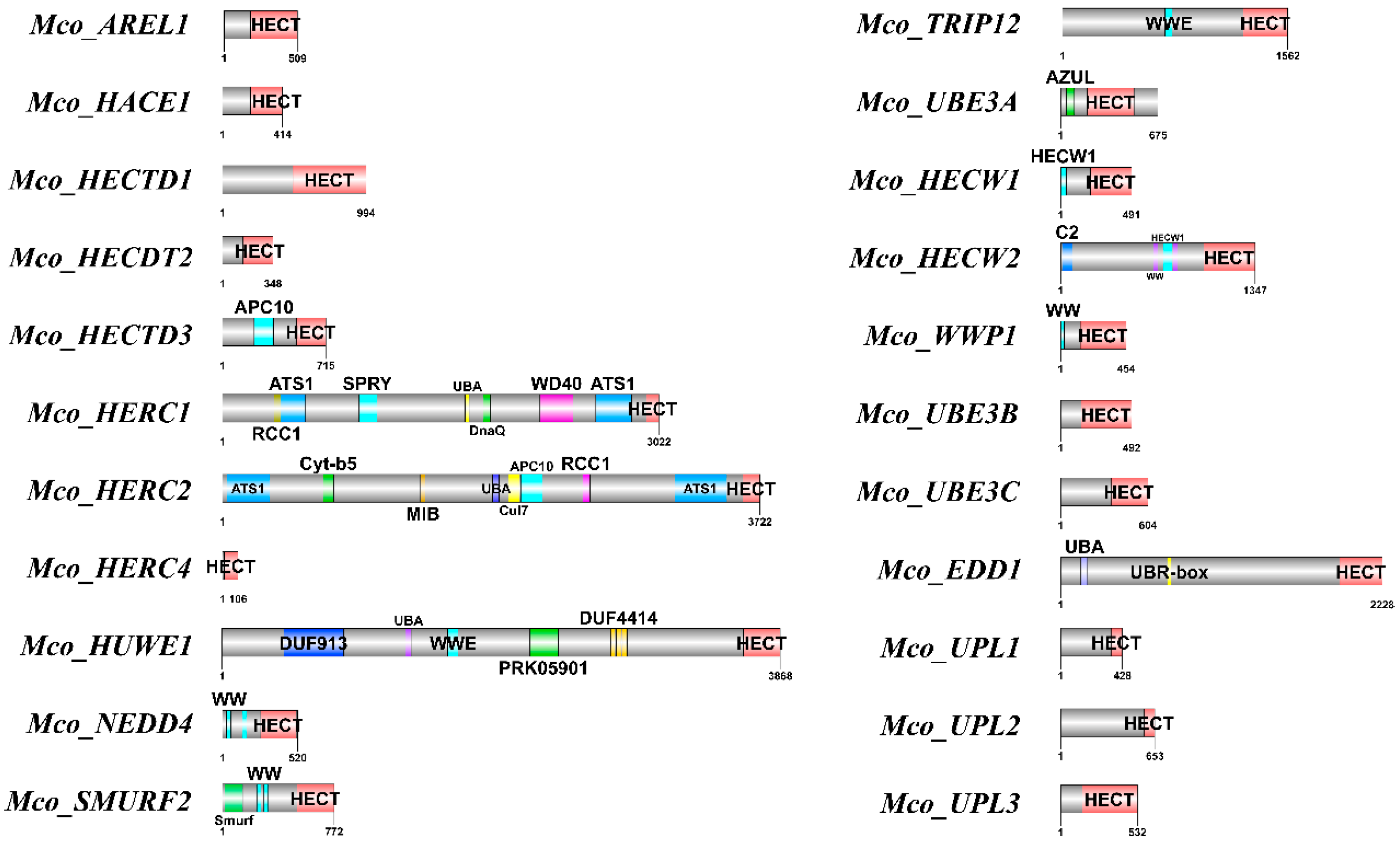
3.3. Analysis of Conserved Domains, Motif Discovery, and Gene Structures

The analysis revealed variability in motif numbers within the HECT gene family in the M. coruscus genome, ranging from 2 to 10 motifs. Notably, sequences such as Mco_HERC1, Mco_HERC2, Mco_HECTD3, Mco_UBE3A, Mco_HERC4, Mco_UPL1, Mco_UPL2, and Mco_UPL3 exhibited a limited count of 1 to 3 motifs, sharing similarities with motifs found in other sequences. Duplicated homologs, such as Mco_HECW and Mco_UPL, displayed comparable motif arrangement structures within their protein configurations (Figure 2A).
To understand the evolutionary conservation of this gene family, we scrutinized the gene structure of HECT genes using M. coruscus genome annotation files (Figure 2B). The disparity in coding sequences (CDS) versus intron structures among these HECT genes suggests potential diversity in their biological functions. Results indicated a spectrum of CDS counts ranging from 3 to 58, with approximately one-third of the sequences showing over 20 CDS counts. Notably, Mco_HERC1, Mco_HERC4, Mco_HECDT2, Mco_UPL1, Mco_UPL2, Mco_UPL3, and Mco_TRIP12 lacked untranslated regions (UTR).
Further characterization of the HECT proteins’ structures was conducted using the NCBI Conservative Domain Database (https://www.ncbi.nlm.nih.gov/cdd/) (accessed on 3 May 2024). The investigation identified 26 proteins in M. coruscus potentially containing HECT structural domains (Figure 3). These structural domains ranged in length from 23 to 507 amino acids and included significant domains such as the WW domain, SPRY domain, ATS1 domain, C2_Smurf-like domain, UBR-box domain, DUF domain, HECW1_helix domain, and UBA_HERC2 domain.

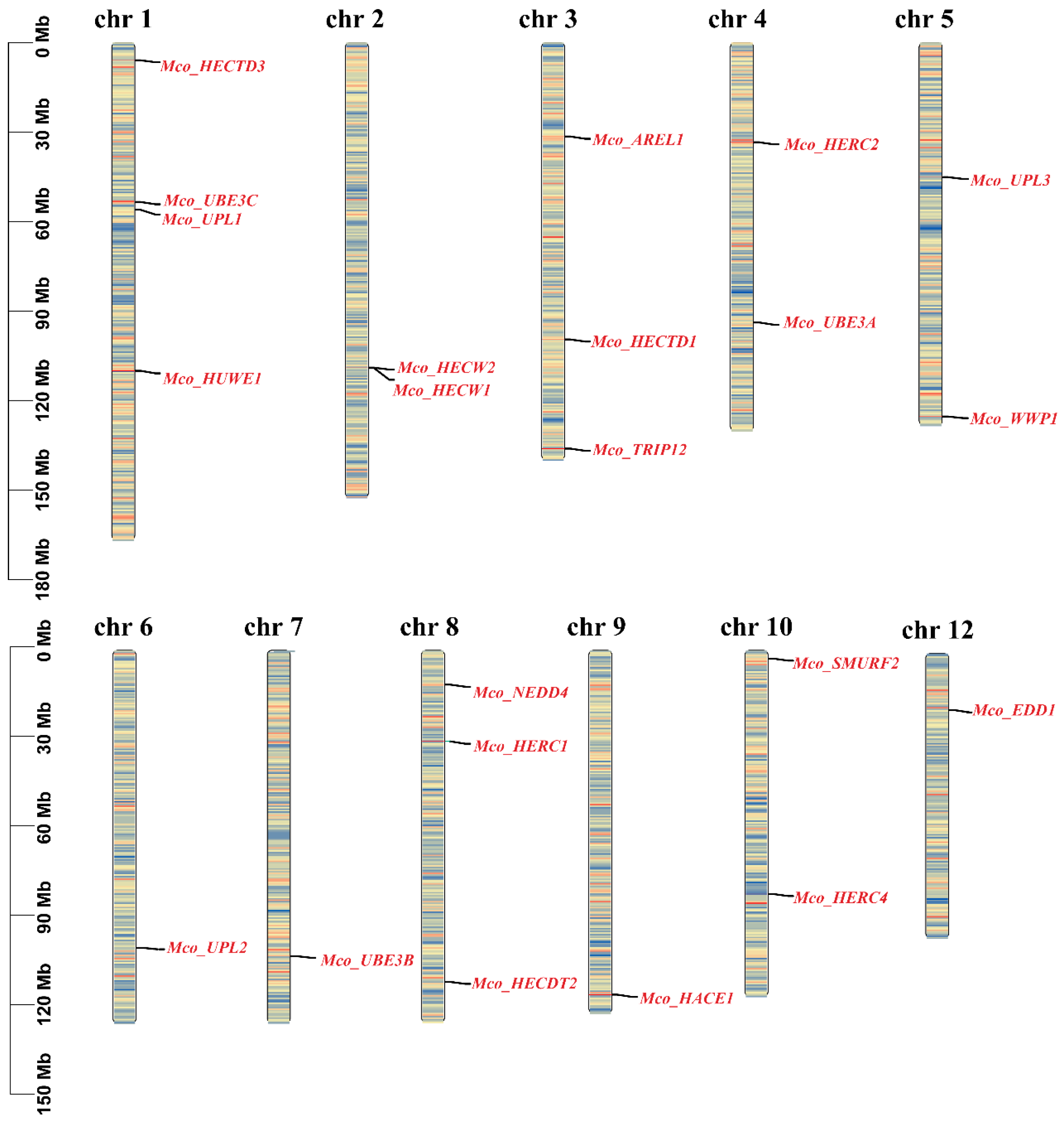
3.4. Chromosomal Localization of the HECT Gene Family

The TBtools software (v2.096) was employed to visualize the chromosomal distribution of HECT genes in the M. coruscus genome, revealing an uneven distribution of 22 HECT genes across 11 out of the 14 chromosomes (Figure 4 and Table 1). Duplicated HECT genes were observed on distinct scaffolds, with Mco_UPL1, Mco_UPL2, and Mco_UPL3 situated on chromosome 1, chromosome 6, and chromosome 5, respectively.

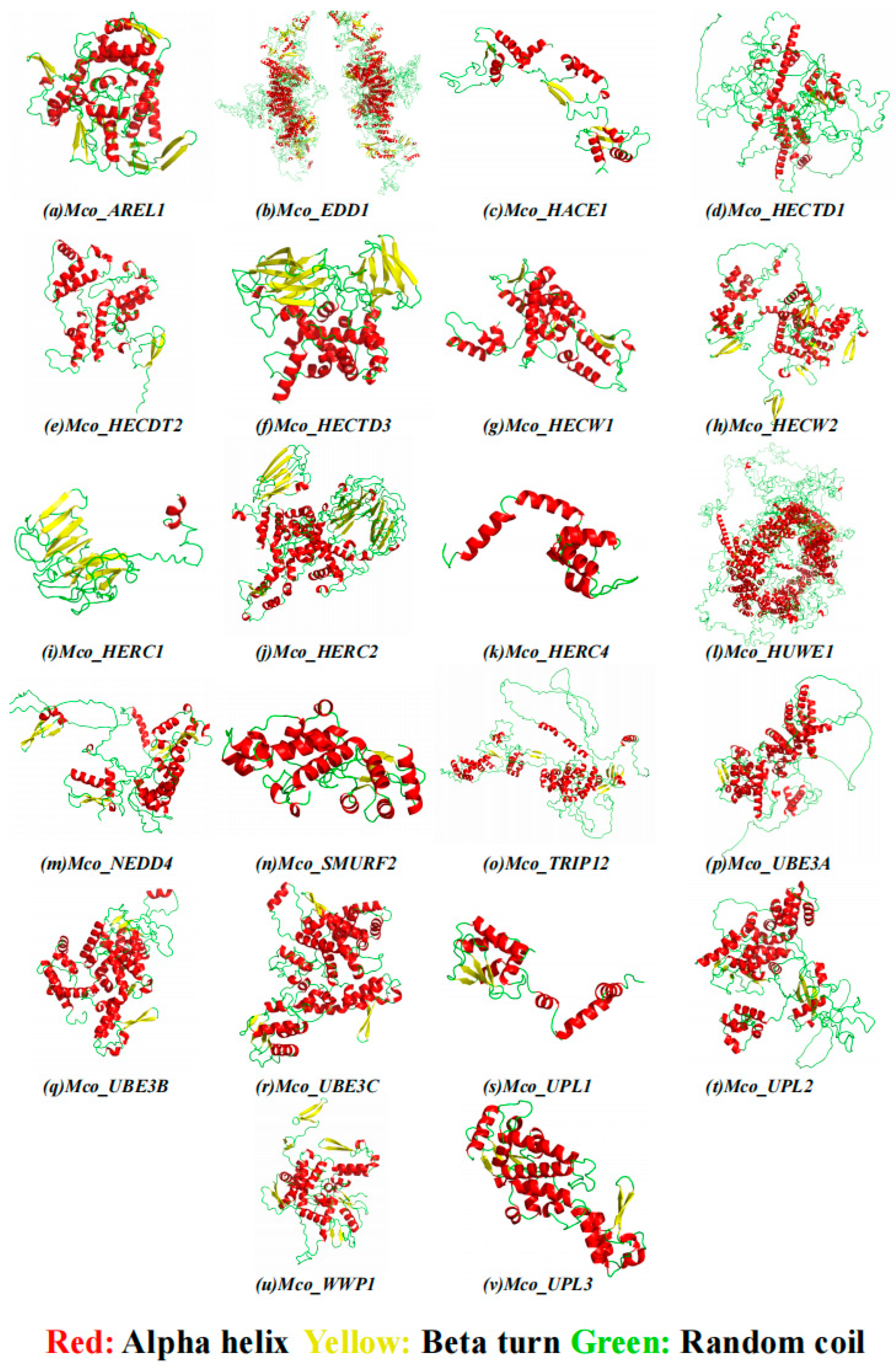
3.5. Secondary Homology Modeling of HECT Protein Structures and Subcellular Localization

The M. coruscus HECT gene family exhibits diverse subcellular localizations encompassing the nucleus, cytoplasm, plasma membrane, and mitochondria (Table 3). Regarding the secondary structure of the 22 encoded proteins by M. coruscus HECT genes, they predominantly consist of alpha helices and random helices. Specifically, alpha helices ranged from 28.54% to 53.54%, beta turns from 4.10% to 7.49%, random helices from 28.88% to 51.22%, and elongated chains from 8.79% to 21.16% (Table 3 and Figure 5). Notably, proteins from distinct groups within the same species exhibited substantial differences, highlighting the structural diversity prevalent within the HECT family.

3.6. HECT Gene Colinearity Analysis

To thoroughly investigate the phylogenetic mechanisms within bivalve HECT genes, we examined the homology of HECT genes across four bivalve species (M. coruscus, M. galloprovincialis, M. yessoensis, and C. gigas) (Figure 6). The HECT genes of M. coruscus exhibited homology with those of the selected bivalve species, demonstrating conserved homologous relationships in M. galloprovincialis (11 homologous pairs of genes across Chr1, Chr2, Chr3, Chr4, Chr5, Chr8, and Chr14), M. yessoensis (2 homologous gene pairs on Chr3 and Chr5), and C. gigas (1 homologous gene pair on Chr5) chromosomes, as depicted in Figure 6. In the analysis of HECT gene homology between M. coruscus and M. yessoensis, we identified AREL1 and HACE1 associated with a homologous gene pair. Additionally, in the covariance analysis of HECT genes between M. coruscus and C. gigas, HACE1 was linked with a covariant gene pair. These findings suggest the potential pivotal role of HACE1 in the evolutionary trajectory of the HECT family among bivalve species.

3.7. Analysis of HECT Gene Expression Patterns

Our investigation explored the expression patterns of HECT genes across various mussel tissues and developmental stages, utilizing a combination of published and unpublished RNA-seq datasets. Analysis of RNA-seq data from five distinct adult mussel tissues—blood, gill, mantle, gonad, and foot—facilitated the characterization of HECT expression profiles in M. coruscus. Notably, the analysis revealed that, on average, HECT genes exhibited elevated expression levels in the blood and gill, while displaying lower expression in the gonad and foot. Heat maps generated using FPKM values illustrated the tissue-specific expression patterns of HECT genes across various tissues (Figure 7A). Specifically, Mco_HERC4 and Mco_HECDT2 demonstrated robust expression primarily in the gill, while Mco_UPL1 and Mco_HERC1 showcased elevated levels in the blood. Furthermore, heightened expression was observed for Mco_HERC2 and Mco_UPL2 in the mantle. However, it is noteworthy that most HECT genes displayed expression in at least one tissue, suggesting their involvement across diverse physiological domains.
To investigate the expression dynamics of HECT genes in M. coruscus under abiotic and biotic stresses, we analyzed RNA-seq datasets from different tissues (adductor muscle, gill, and mantle) subjected to heat and V. alginolyticus stress. Following heat stress, four genes—Mco_UPL3, Mco_HUWE1, Mco_NEDD4, and Mco_UBE3C—exhibited upregulation in the gill, while only Mco_HUWE1 and Mco_UPL3 showed upregulation in the mantle and adductor tissues, respectively (Figure 7B). Remarkably, under V. alginolyticus stress, HECT genes were prominently expressed in gill tissues. However, only Mco_UPL3 displayed significant upregulation, while the expression of Mco_HECTD3, Mco_UBE3A, Mco_HERC1, Mco_HACE1, Mco_AREL1, Mco_EDD1, and Mco_TRIP12 was significantly downregulated. Additionally, Mco_HECW1 exhibited notable upregulation, and Mco_HECDT2 and Mco_UPL2 showed significant downregulation in the mantle. Conversely, the expression in the adductor muscle remained largely unaltered (Figure 7C).

3.8. Gene Ontology Analysis of the HECT Gene

To further investigate the functions and metabolic pathways of the identified HECT E3 ligases in M. coruscus, we conducted a gene ontology (GO) enrichment analysis. The results indicated that 22 genes were significantly enriched in 51 GO terms (p-adjust < 0.05). These genes were predominantly associated with protein modification and degradation mechanisms, with a particular focus on pathways involving ubiquitination and ubiquitin-like modifications. The five most enriched GO terms were ubiquitin protein ligase activity (GO:0061630), ubiquitin-like protein transferase activity (GO:0019787), ubiquitin-like protein ligase activity (GO:0061659), acyltransferase activity (GO:0016746), and ubiquitin-protein transferase activity (GO:0004842) (Figure 8 and Table S4).

4. Discussion

Gene duplication is a pivotal factor in the evolution of gene families, contributing to the enhancement of gene structure and functional diversity through the generation of new family members [61,62]. In this investigation, we identified 22 HECT genes in the genome of M. coruscus, distributed across 11 chromosomes. Comparative analysis with other bivalves such as M. galloprovincialis, M. yessoensis, C. gigas, and R. philippinarum revealed a higher number of HECTs in M. coruscus, suggesting diverse functional roles for HECT genes in this species. While tandem duplications and segmental duplications are common types of gene duplications [63], our study did not uncover a substantial presence of these duplications in the M. coruscus HECT gene family. Notably, the HECW HECT gene originated from tandem duplication, and the Mco_UPL1, Mco_UPL2, and Mco_UPL3 genes stemmed from segmental duplication, while the remainder originated from dispersed duplication. The observation that HECT genes in M. coruscus exhibit a high number of introns may indicate conservation, akin to early eukaryotes possessing genes with abundant introns [64]. Phylogenetic analysis of HECT genes in bivalves (M. galloprovincialis, M. yessoensis, R. philippinarum), and model organisms (H. sapiens, D. melanogaster, D. rerio, and Drosophila), along with the corresponding HECT homology classification, revealed six major categories (Figure 1) [61]. This classification aligns with the HECT classification observed in R. philippinarum [23], underscoring the relative evolutionary conservatism of the HECT E3 gene family in M. coruscus. Based on the phylogenetic tree, the NEDD4 subfamily and UPL subfamily HECT genes were most abundant among bivalves, featuring five and three genes, respectively, in M. coruscus. Conversely, R. philippinarum exhibited the fewest NEDD4 subfamily HECT genes, with only one identified among the bivalve sequences. Additionally, we noted that Class II harbors the most numerous and diverse types of HECT genes, known as key regulators of membrane proteins [65,66]. Numerous studies have demonstrated the crucial role of genes in this family in balancing tolerance and immunity [66]. Consideration of sequence, structure, and functional relationships suggests potential interactions with proteins and ligands.
Intertidal shellfish are subjected to a diverse array of environmental factors, with gills playing a pivotal role in facilitating gas exchange [67]. They facilitate oxygen uptake during submersion and prevent dehydration upon exposure to air. Beyond their respiratory function, the gills of shellfish serve as crucial organs for filtration and ingestion, essential for survival and adaptation to aquatic habitats. In response to environmental stressors such as temperature fluctuations and high salinity, gills undergo changes that impact respiratory efficiency, osmoregulation, and immune defense mechanisms [68]. Studies suggest that these adaptive changes in gill function enable shellfish to effectively manage stress, although chronic or severe stress can adversely affect physiological processes and overall survival [69,70,71]. Furthermore, in bivalves, blood plays an equally crucial role in adaptation to the intertidal environment. This blood transports nutrients, gases, and immune cells, contributing to osmoregulation, maintaining internal stability during tidal fluctuations, and preventing salinity changes [72]. Research on clams, oysters, and mussels underscores the significant role of hemocytes, immune cells in shellfish blood, in responding to environmental stressors, particularly pathogen exposure [73,74,75]. In the present investigation, M. coruscus HECT genes exhibited common expression in the examined tissues, notably in gills and blood. The specific high expression of HECT genes in the hemolymph and gill tissues of M. coruscus implies their potential involvement in regulating homeostatic tolerance and immune response in these organisms.
After being passed from ubiquitin-activating enzymes (E1) to ubiquitin-conjugating enzymes (E2), ubiquitin ligases co-ubiquitinate proteins (E3). The ubiquitin–proteasome system is one of the many intricate processes of ubiquitin-mediated protein degradation that is highly selective and degrades a significant amount of proteins in cells [76]. Abiotic stresses can induce the accumulation of misfolded or unfolded proteins, thereby triggering the generation of reactive oxygen species (ROS) [77]. Extensive evidence has demonstrated that intertidal shellfish endure high-temperature stress during the summer, a circumstance that may induce protein misfolding or denaturation, resulting in protein aggregation and the loss of physiological function. The E3 ligase ubiquitin is recognized as a pivotal factor in the proteasome pathway (UPP) [78], participating in the repair of damaged proteins under abiotic stresses [79,80]. We observed a significant upregulation of several HECT genes in the gill tissues of M. coruscus under heat stress conditions, including Mco_UPL3, Mco_HUWE1, Mco_NEDD4, and Mco_UBE3C. It has been proposed that UPL3 is involved in protein ubiquitination, although studies on its exact physiological role in animals are more limited [81,82]. HUWE1 is a key member of the E3 ligase family with a eukaryotic HECT structural domain. HUWE1 regulates the turnover of various substrates, including MCL-1, P53, and c-MYC, thus participating in a broad spectrum of biological processes, including but not limited to apoptosis, autophagy, proliferation, differentiation, DNA damage repair, and stress responses [83,84,85]. NEDD4 primarily participates in the regulation of ubiquitin-mediated protein degradation, contributing to various cellular pathways that impact growth, development, and immunity [86]. UBE3C is associated with HECT and is involved in protein degradation pathways that affect the cell cycle and DNA repair in animals [87].
In terms of biotic stress, Vibrio vulnificus emerges as a significant pathogen in cultivated shellfish, causing a mortality rate of up to 90–100% within 24 h in infected bivalves, with associated industrial losses typically reaching 60% [88,89]. In our study, the expression of the HECT gene family in gill tissues decreased following stress induced by V. alginolyticus. This observation suggests a potential impact of these genes on the regulation of immune responses in M. coruscus. A prior investigation by YI et al. revealed a significant downregulation of HUWE1 and TRAF6 expression during white spot syndrome virus (WSSV) infection. This downregulation disrupted the ubiquitination of p53, resulting in apoptosis and reactive oxygen species (ROS) signaling through the accumulation of p53. Ultimately, this cascade inhibited viral invasion in mud crabs, offering a novel molecular mechanism for invertebrates to resist viral infections [90]. Our study detected an upregulation of Mco_UPL3 under both heat and Vibrio stress, highlighting their pivotal roles in cellular processes such as protein ubiquitination and degradation. The functions of the UPL3 gene, encompassing mediation of protein stability, cellular signaling, and immune regulation, suggest a crucial role in regulating the thermoimmune response, particularly post-stress heat exposure. The diminished expression of HECT genes in mud crabs was found to be correlated with a reduction in UPL3 gene expression. The interaction between the diminished expression of HECT genes and the selective upregulation of UPL3 genes potentially orchestrated an adaptive immune response, enhancing resistance to stress. In conclusion, genes associated with the HECT structural domain may possess a regulatory role in the thermal immune response in bivalves. Nevertheless, further studies are imperative to comprehensively elucidate the specific functions of these genes and their roles in both biotic and abiotic stresses.

5. Conclusions

In this investigation, we characterized the HECT genes within the genome of M. coruscus, elucidating their molecular features and phylogenetic relationships. A total of 22 HECT genes were identified and categorized into six major types, all containing conserved HECT structural domains. Furthermore, we examined the expression patterns of HECT genes in M. coruscus exposed to both abiotic (high-temperature) and biotic (V. alginolyticus) stresses and performed functional annotation and enrichment analysis of these genes in the mussels. The findings revealed that the identified HECT genes in M. coruscus were enriched for molecular functions, including ubiquitination, degradation, and protein modification. These genes were significantly expressed in gill and blood tissues, with some showing marked upregulation in response to high-temperature and Vibrio vulnificus stress. This suggests potential roles for HECT genes in essential functions such as protein degradation and immunomodulation in mussels. Our results provide useful information for breeding for resilience in M. coruscus.

Supplementary Materials

The following supporting information can be downloaded at: https://www.mdpi.com/article/10.3390/genes15081085/s1 [18,50,91,92,93], Table S1: Statistics of all genome project numbers; Table S2: A total of 148 HECT sequences in 10 species; Table S3: The BioProject number of RNA-seq datasets; Table S4: Gene ontology (GO) annotation results for the HECT gene of M. coruscus.

Author Contributions

Writing—original draft, conceptualization, investigation, methodology, formal analysis, validation, F.G. and Z.X.; investigation, methodology, data curation, Z.D.; funding acquisition, project administration, Y.Y. All authors have read and agreed to the published version of the manuscript.

Funding

The present manuscript was financially supported by the Project of Bureau of Science and Technology of Zhoushan (No. 2021C21017) and the National Key R&D Program of China (2019YFD0901204).

Institutional Review Board Statement

Not applicable.

Informed Consent Statement

Not applicable.

Data Availability Statement

The original contributions presented in the study are included in the article/Supplementary Material, further inquiries can be directed to the corresponding author.

Conflicts of Interest

The authors declare no conflicts of interest.

References

  1. Rotin, D.; Kumar, S. Physiological functions of the HECT family of ubiquitin ligases. Nat. Rev. Mol. Cell Biol. 2009, 10, 398–409. [Google Scholar] [CrossRef] [PubMed]
  2. Huibregtse, J.M.; Scheffner, M.; Beaudenon, S.; Howley, P.M. A family of proteins structurally and functionally related to the E6-AP ubiquitin-protein ligase. Proc. Natl. Acad. Sci. USA 1995, 92, 2563–2567. [Google Scholar] [CrossRef]
  3. Marín, I. Evolution of plant HECT ubiquitin ligases. PLoS ONE 2013, 8, e68536. [Google Scholar] [CrossRef]
  4. Nakayama, K.I.; Nakayama, K. Ubiquitin ligases: Cell-cycle control and cancer. Nat. Rev. Cancer 2006, 6, 369–381. [Google Scholar] [CrossRef]
  5. Grau-Bové, X.; Sebé-Pedrós, A.; Ruiz-Trillo, I. A Genomic Survey of HECT Ubiquitin Ligases in Eukaryotes Reveals Independent Expansions of the HECT System in Several Lineages. Genome Biol. Evol. 2013, 5, 833–847. [Google Scholar] [CrossRef] [PubMed]
  6. Pickart, C.M. Mechanisms Underlying Ubiquitination. Annu. Rev. Biochem. 2001, 70, 503–533. [Google Scholar] [CrossRef] [PubMed]
  7. Li, W.; Bengtson, M.H.; Ulbrich, A.; Matsuda, A.; Reddy, V.A.; Orth, A.; Chanda, S.K.; Batalov, S.; Joazeiro, C.A.P. Genome-Wide and Functional Annotation of Human E3 Ubiquitin Ligases Identifies MULAN, a Mitochondrial E3 that Regulates the Organelle’s Dynamics and Signaling. PLoS ONE 2008, 3, e1487. [Google Scholar] [CrossRef]
  8. Dunn, R.; Klos, D.A.; Adler, A.S.; Hicke, L. The C2 domain of the Rsp5 ubiquitin ligase binds membrane phosphoinositides and directs ubiquitination of endosomal cargo. J. Cell Biol. 2004, 165, 135–144. [Google Scholar] [CrossRef]
  9. Tian, M.; Bai, C.; Lin, Q.; Lin, H.; Liu, M.; Ding, F.; Wang, H.R. Binding of RhoA by the C2 domain of E3 ligase Smurf1 is essential for Smurf1-regulated RhoA ubiquitination and cell protrusive activity. FEBS Lett. 2011, 585, 2199–2204. [Google Scholar] [CrossRef]
  10. Rizo, J.; Südhof, T.C. C2-domains, Structure and Function of a Universal Ca2+-binding Domain*. J. Biol. Chem. 1998, 273, 15879–15882. [Google Scholar] [CrossRef]
  11. Bischoff, F.R.; Ponstingl, H. Catalysis of guanine nucleotide exchange on Ran by the mitotic regulator RCC1. Nature 1991, 354, 80–82. [Google Scholar] [CrossRef]
  12. Lemak, A.; Yee, A.; Bezsonova, I.; Dhe-Paganon, S.; Arrowsmith, C.H. Zn-binding AZUL domain of human ubiquitin protein ligase Ube3A. J. Biomol. NMR 2011, 51, 185. [Google Scholar] [CrossRef] [PubMed]
  13. Scheffner, M.; Kumar, S. Mammalian HECT ubiquitin-protein ligases: Biological and pathophysiological aspects. Biochim. Biophys. Acta (BBA) Mol. Cell Res. 2014, 1843, 61–74. [Google Scholar] [CrossRef] [PubMed]
  14. Dielen, A.-S.; Badaoui, S.; Candresse, T.; German-Retana, S. The ubiquitin/26S proteasome system in plant–pathogen interactions: A never-ending hide-and-seek game. Mol. Plant Pathol. 2010, 11, 293–308. [Google Scholar] [CrossRef] [PubMed]
  15. Marín, I. Animal HECT ubiquitin ligases: Evolution and functional implications. BMC Evol. Biol. 2010, 10, 56. [Google Scholar] [CrossRef]
  16. Qian, H.; Zhang, Y.; Wu, B.; Wu, S.; You, S.; Zhang, N.; Sun, Y. Structure and Function of HECT E3 Ubiquitin Ligases and their Role in Oxidative Stress. J. Transl. Intern. Med. 2020, 8, 71–79. [Google Scholar] [CrossRef]
  17. Wang, Y.; Argiles-Castillo, D.; Kane, E.I.; Zhou, A.; Spratt, D.E. HECT E3 ubiquitin ligases-emerging insights into their biological roles and disease relevance. J. Cell Sci. 2020, 133, jcs228072. [Google Scholar] [CrossRef]
  18. Wang, Y.-X.; Lin, S.-R.; Xu, L.-Z.; Ye, Y.-Y.; Qi, P.-Z.; Wang, W.-F.; Buttino, I.; Li, H.-F.; Guo, B.-Y. Comparative transcriptomic analysis revealed changes in multiple signaling pathways involved in protein degradation in the digestive gland of Mytilus coruscus during high-temperatures. Comp. Biochem. Physiol. Part D Genom. Proteom. 2023, 46, 101060. [Google Scholar] [CrossRef] [PubMed]
  19. Sun, Y.; Liang, X.; Chen, J.; Tang, R.; Li, L.; Li, D. Change in Ubiquitin Proteasome System of Grass Carp Ctenopharyngodon idellus Reared in the Different Stocking Densities. Front. Physiol. 2018, 9, 837. [Google Scholar] [CrossRef]
  20. Xu, C.; Li, E.; Liu, Y.; Wang, X.; Qin, J.G.; Chen, L. Comparative proteome analysis of the hepatopancreas from the Pacific white shrimp Litopenaeus vannamei under long-term low salinity stress. J. Proteom. 2017, 162, 1–10. [Google Scholar] [CrossRef]
  21. Bayİr, A.; Polat, M.; Aras, N.; Nalbantoğlu, B.J.A.J.o.A.; Advances, V. Effects of high temperature stress on mRNA levels of ubiquitin-activating enzyme in ovarian tissue of goldfish, Carassius auratus. Asian J. Anim. Vet. Adv. 2012, 7, 356–360. [Google Scholar] [CrossRef]
  22. Yum, S. Ubiquitin expression in soft coral (Scleronephthya gracillimum) exposed to environmental stresses. Korean J. Genet. 2006, 28, 149–156. [Google Scholar]
  23. Lin, Z.; Nie, H.; Zhang, Y.; Yin, Z.; Yan, X. Genome-wide identification and analysis of HECT E3 ubiquitin ligase gene family in Ruditapes philippinarum and their involvement in the response to heat stress and Vibrio anguillarum infection. Comp. Biochem. Physiol. Part D Genom. Proteom. 2022, 43, 101012. [Google Scholar] [CrossRef]
  24. Qu, Z.-L.; Li, Y.-L.; Gong, X.-Y.; Zhao, X.; Sun, H.-Y.; Dan, C.; Gui, J.-F.; Zhang, Y.-B. A finTRIM Family Protein Acquires RNA-Binding Activity and E3 Ligase Activity to Shape the IFN Response in Fish. J. Immunol. 2022, 209, 1335–1347. [Google Scholar] [CrossRef]
  25. Xu, F.; Shi, Y.-H.; Chen, J. Characterization and immunologic functions of the macrophage migration inhibitory factor from Japanese sea bass, Lateolabrax japonicus. Fish Shellfish Immunol. 2019, 86, 947–955. [Google Scholar] [CrossRef]
  26. Xiang, Y.; Zhang, W.; Jia, P.; Lu, X.; Liu, W.; Yi, M.; Jia, K. E3 Ubiquitin Ligase RNF114 Inhibits Innate Immune Response to Red-Spotted Grouper Nervous Necrosis Virus Infection in Sea Perch by Targeting MAVS and TRAF3 to Mediate Their Degradation. J. Immunol. 2021, 206, 77–88. [Google Scholar] [CrossRef]
  27. Song, Y.; Song, X.; Zhang, D.; Yang, Y.; Wang, L.; Song, L. An HECT domain ubiquitin ligase CgWWP1 regulates granulocytes proliferation in oyster Crassostrea gigas. Dev. Comp. Immunol. 2021, 123, 104148. [Google Scholar] [CrossRef]
  28. Kyeong, D.; Kim, J.; Shin, Y.; Subramaniyam, S.; Kang, B.C.; Shin, E.H.; Park, E.H.; Noh, E.S.; Kim, Y.O.; Park, J.Y.; et al. Expression of Heat Shock Proteins in Thermally Challenged Pacific Abalone Haliotis discus hannai. Genes 2019, 11, 22. [Google Scholar] [CrossRef] [PubMed]
  29. De Vos, S.; Van Stappen, G.; Sorgeloos, P.; Vuylsteke, M.; Rombauts, S.; Bossier, P. Identification of salt stress response genes using the Artemia transcriptome. Aquaculture 2019, 500, 305–314. [Google Scholar] [CrossRef]
  30. Guo, F.; Ye, Y.; Zhu, K.; Lin, S.; Wang, Y.; Dong, Z.; Yao, R.; Li, H.; Wang, W.; Liao, Z.; et al. Genetic Diversity, Population Structure, and Environmental Adaptation Signatures of Chinese Coastal Hard-Shell Mussel Mytilus coruscus Revealed by Whole-Genome Sequencing. Int. J. Mol. Sci. 2023, 24, 13641. [Google Scholar] [CrossRef]
  31. Kulikova, V.A.; Lyashenko, S.A.; Kolotukhina, N.K. Seasonal and interannual dynamics of larval abundance of Mytilus coruscus Gould, 1861 (Bivalvia: Mytilidae) in Amursky Bay (Peter the Great Bay, Sea of Japan). Russ. J. Mar. Biol. 2011, 37, 342–347. [Google Scholar] [CrossRef]
  32. Dong, W.; Chen, Y.; Lu, W.; Wu, B.; Qi, P. Transcriptome analysis of Mytilus coruscus hemocytes in response to Vibrio alginnolyficus infection. Fish Shellfish Immunol. 2017, 70, 560–567. [Google Scholar] [CrossRef] [PubMed]
  33. Li, R.; Zhang, W.; Lu, J.; Zhang, Z.; Mu, C.; Song, W.; Migaud, H.; Wang, C.; Bekaert, M. The Whole-Genome Sequencing and Hybrid Assembly of Mytilus Coruscus. Front. Genet. 2020, 11, 440. [Google Scholar] [CrossRef] [PubMed]
  34. Zippay, M.L.; Helmuth, B. Effects of temperature change on mussel, Mytilus. Integr. Zool. 2012, 7, 312–327. [Google Scholar] [CrossRef] [PubMed]
  35. Burge, C.A.; Mark Eakin, C.; Friedman, C.S.; Froelich, B.; Hershberger, P.K.; Hofmann, E.E.; Petes, L.E.; Prager, K.C.; Weil, E.; Willis, B.L.; et al. Climate Change Influences on Marine Infectious Diseases: Implications for Management and Society. Annu. Rev. Mar. Sci. 2014, 6, 249–277. [Google Scholar] [CrossRef]
  36. Moyen, N.E.; Somero, G.N.; Denny, M.W. Mussel acclimatization to high, variable temperatures is lost slowly upon transfer to benign conditions. J. Exp. Biol. 2020, 223, jeb222893. [Google Scholar] [CrossRef] [PubMed]
  37. Liao, Z.; Wang, X.-c.; Liu, H.-h.; Fan, M.-h.; Sun, J.-j.; Shen, W. Molecular characterization of a novel antimicrobial peptide from Mytilus coruscus. Fish Shellfish Immunol. 2013, 34, 610–616. [Google Scholar] [CrossRef]
  38. Hofmann, G.E.; Somero, G.N. Evidence for Protein Damage at Environmental Temperatures: Seasonal Changes in Levels of Ubiquitin Conjugates and Hsp70 in the Intertidal Mussel Mytilus Trossulus. J. Exp. Biol. 1995, 198, 1509–1518. [Google Scholar] [CrossRef]
  39. Willer, D.F.; Aldridge, D.C. Microencapsulated diets to improve bivalve shellfish aquaculture for global food security. Glob. Food Secur. 2019, 23, 64–73. [Google Scholar] [CrossRef]
  40. Finn, R.D.; Bateman, A.; Clements, J.; Coggill, P.; Eberhardt, R.Y.; Eddy, S.R.; Heger, A.; Hetherington, K.; Holm, L.; Mistry, J.; et al. Pfam: The protein families database. Nucleic Acids Res. 2014, 42, D222–D230. [Google Scholar] [CrossRef]
  41. Finn, R.D.; Clements, J.; Eddy, S.R. HMMER web server: Interactive sequence similarity searching. Nucleic Acids Res 2011, 39, W29–W37. [Google Scholar] [CrossRef]
  42. Lu, S.; Wang, J.; Chitsaz, F.; Derbyshire, M.K.; Geer, R.C.; Gonzales, N.R.; Gwadz, M.; Hurwitz, D.I.; Marchler, G.H.; Song, J.S.; et al. CDD/SPARCLE: The conserved domain database in 2020. Nucleic Acids Res. 2020, 48, D265–D268. [Google Scholar] [CrossRef]
  43. Letunic, I.; Khedkar, S.; Bork, P. SMART: Recent updates, new developments and status in 2020. Nucleic Acids Res. 2021, 49, D458–D460. [Google Scholar] [CrossRef] [PubMed]
  44. Mi, H.; Muruganujan, A.; Thomas, P.D. PANTHER in 2013: Modeling the evolution of gene function, and other gene attributes, in the context of phylogenetic trees. Nucleic Acids Res. 2013, 41, D377–D386. [Google Scholar] [CrossRef]
  45. Wilkins, M.R.; Gasteiger, E.; Bairoch, A.; Sanchez, J.C.; Williams, K.L.; Appel, R.D.; Hochstrasser, D.F. Protein identification and analysis tools in the ExPASy server. Methods Mol. Biol. 1999, 112, 531–552. [Google Scholar] [CrossRef]
  46. Chen, C.; Chen, H.; Zhang, Y.; Thomas, H.R.; Frank, M.H.; He, Y.; Xia, R. TBtools: An Integrative Toolkit Developed for Interactive Analyses of Big Biological Data. Mol. Plant 2020, 13, 1194–1202. [Google Scholar] [CrossRef]
  47. Ren, J.; Wen, L.; Gao, X.; Jin, C.; Xue, Y.; Yao, X. DOG 1.0: Illustrator of protein domain structures. Cell Res. 2009, 19, 271–273. [Google Scholar] [CrossRef]
  48. Bailey, T.L.; Johnson, J.; Grant, C.E.; Noble, W.S. The MEME Suite. Nucleic Acids Res. 2015, 43, W39–W49. [Google Scholar] [CrossRef]
  49. The UniProt, C. UniProt: The Universal Protein Knowledgebase in 2023. Nucleic Acids Res. 2023, 51, D523–D531. [Google Scholar] [CrossRef] [PubMed]
  50. Liu, F.; Li, Y.; Yu, H.; Zhang, L.; Hu, J.; Bao, Z.; Wang, S. MolluscDB: An integrated functional and evolutionary genomics database for the hyper-diverse animal phylum Mollusca. Nucleic Acids Res. 2021, 49, D1556. [Google Scholar] [CrossRef] [PubMed]
  51. Kumar, S.; Stecher, G.; Li, M.; Knyaz, C.; Tamura, K. MEGA X: Molecular Evolutionary Genetics Analysis across Computing Platforms. Mol. Biol. Evol. 2018, 35, 1547–1549. [Google Scholar] [CrossRef]
  52. Subramanian, B.; Gao, S.; Lercher, M.J.; Hu, S.; Chen, W.-H. Evolview v3: A webserver for visualization, annotation, and management of phylogenetic trees. Nucleic Acids Res. 2019, 47, W270–W275. [Google Scholar] [CrossRef]
  53. Wang, Y.; Tang, H.; DeBarry, J.D.; Tan, X.; Li, J.; Wang, X.; Lee, T.-h.; Jin, H.; Marler, B.; Guo, H.; et al. MCScanX: A toolkit for detection and evolutionary analysis of gene synteny and collinearity. Nucleic Acids Res. 2012, 40, e49. [Google Scholar] [CrossRef] [PubMed]
  54. Buchfink, B.; Xie, C.; Huson, D.H. Fast and sensitive protein alignment using DIAMOND. Nat. Methods 2015, 12, 59–60. [Google Scholar] [CrossRef]
  55. Horton, P.; Park, K.J.; Obayashi, T.; Fujita, N.; Harada, H.; Adams-Collier, C.J.; Nakai, K. WoLF PSORT: Protein localization predictor. Nucleic Acids Res. 2007, 35, W585–W587. [Google Scholar] [CrossRef] [PubMed]
  56. Geourjon, C.; Deléage, G. SOPMA: Significant improvements in protein secondary structure prediction by consensus prediction from multiple alignments. Bioinformatics 1995, 11, 681–684. [Google Scholar] [CrossRef]
  57. Fermi, G.; Perutz, M.F.; Shaanan, B.; Fourme, R. The crystal structure of human deoxyhaemoglobin at 1.74 A resolution. J. Mol. Biol. 1984, 175, 159–174. [Google Scholar] [CrossRef] [PubMed]
  58. Waterhouse, A.; Bertoni, M.; Bienert, S.; Studer, G.; Tauriello, G.; Gumienny, R.; Heer, F.T.; de Beer, T.A.P.; Rempfer, C.; Bordoli, L.; et al. SWISS-MODEL: Homology modelling of protein structures and complexes. Nucleic Acids Res. 2018, 46, W296–w303. [Google Scholar] [CrossRef]
  59. Benkert, P.; Biasini, M.; Schwede, T. Toward the estimation of the absolute quality of individual protein structure models. Bioinformatics 2011, 27, 343–350. [Google Scholar] [CrossRef] [PubMed]
  60. Cantalapiedra, C.P.; Hernández-Plaza, A.; Letunic, I.; Bork, P.; Huerta-Cepas, J. eggNOG-mapper v2: Functional Annotation, Orthology Assignments, and Domain Prediction at the Metagenomic Scale. Mol. Biol. Evol. 2021, 38, 5825–5829. [Google Scholar] [CrossRef] [PubMed]
  61. Roth, C.; Rastogi, S.; Arvestad, L.; Dittmar, K.; Light, S.; Ekman, D.; Liberles, D.A. Evolution after gene duplication: Models, mechanisms, sequences, systems, and organisms. J. Exp. Zool. Part B Mol. Dev. Evol. 2007, 308, 58–73. [Google Scholar] [CrossRef]
  62. Zhang, Y.; Zhang, J.; Yang, D.; Jin, Y.; Liu, X.; Zhang, Z.; Gu, L.; Zhang, H. Genome-Wide Identification of NAP1 and Function Analysis in Moso Bamboo (Phyllostachys edulis). Int. J. Mol. Sci. 2022, 23, 6491. [Google Scholar] [CrossRef]
  63. Cannon, S.B.; Mitra, A.; Baumgarten, A.; Young, N.D.; May, G. The roles of segmental and tandem gene duplication in the evolution of large gene families in Arabidopsis thaliana. BMC Plant Biol. 2004, 4, 10. [Google Scholar] [CrossRef]
  64. Irimia, M.; Penny, D.; Roy, S.W. Coevolution of genomic intron number and splice sites. Trends Genet. TIG 2007, 23, 321–325. [Google Scholar] [CrossRef]
  65. Harvey, K.F.; Kumar, S. Nedd4-like proteins: An emerging family of ubiquitin-protein ligases implicated in diverse cellular functions. Trends Cell Biol. 1999, 9, 166–169. [Google Scholar] [CrossRef]
  66. Gay, D.L.; Ramón, H.; Oliver, P.M. Cbl- and Nedd4-family ubiquitin ligases: Balancing tolerance and immunity. Immunol. Res. 2008, 42, 51–64. [Google Scholar] [CrossRef]
  67. Cerdeira-Arias, J.D.; Otero, J.; Álvarez-Salgado, X.A.; Mena RodrÍguez, A.; Nombela, M.Á. Multivariate substrate characterization: The case of shellfish harvesting areas in the Rías Altas (north-west Iberian Peninsula). Sedimentology 2021, 68, 697–712. [Google Scholar] [CrossRef]
  68. Chi, C.; Giri, S.S.; Jun, J.W.; Kim, S.W.; Kim, H.J.; Kang, J.W.; Park, S.C. Detoxification- and Immune-Related Transcriptomic Analysis of Gills from Bay Scallops (Argopecten irradians) in Response to Algal Toxin Okadaic Acid. Toxins 2018, 10, 308. [Google Scholar] [CrossRef]
  69. Arredondo-Espinoza, R.; Ibarra, A.M.; Roberts, S.B.; Sicard-González, M.T.; Escobedo-Fregoso, C. Transcriptome profile in heat resilient Pacific oyster Crassostrea gigas families under thermal challenge. Comp. Biochem. Physiol. Part D Genom. Proteom. 2023, 47, 101089. [Google Scholar] [CrossRef] [PubMed]
  70. Kim, B.M.; Kim, K.; Choi, I.Y.; Rhee, J.S. Transcriptome response of the Pacific oyster, Crassostrea gigas susceptible to thermal stress: A comparison with the response of tolerant oyster. Mol. Cell. Toxicol. 2017, 13, 105–113. [Google Scholar] [CrossRef]
  71. Jiang, W.; Lin, F.; Fang, J.; Gao, Y.; Du, M.; Fang, J.; Li, W.; Jiang, Z. Transcriptome analysis of the Yesso scallop, Patinopecten yessoensis gills in response to water temperature fluctuations. Fish Shellfish Immunol. 2018, 80, 133–140. [Google Scholar] [CrossRef] [PubMed]
  72. de la Ballina, N.R.; Maresca, F.; Cao, A.; Villalba, A. Bivalve Haemocyte Subpopulations: A Review. Front. Immunol. 2022, 13, 826255. [Google Scholar] [CrossRef]
  73. Meng, J.; Zhang, G.; Wang, W.X. Functional heterogeneity of immune defenses in molluscan oysters Crassostrea hongkongensis revealed by high-throughput single-cell transcriptome. Fish Shellfish Immunol. 2022, 120, 202–213. [Google Scholar] [CrossRef] [PubMed]
  74. Xie, Y.; Hu, B.; Wen, C.; Mu, S. Morphology and phagocytic ability of hemocytes from Cristaria plicata. Aquaculture 2011, 310, 245–251. [Google Scholar] [CrossRef]
  75. Liu, J.; Zhao, Y. Morphological and functional characterization of clam Ruditapes philippinarum haemocytes. Fish Shellfish Immunol. 2018, 82, 136–146. [Google Scholar] [CrossRef] [PubMed]
  76. Pickart, C.M.; Eddins, M.J. Ubiquitin: Structures, functions, mechanisms. Biochim. Biophys. Acta (BBA) Mol. Cell Res. 2004, 1695, 55–72. [Google Scholar] [CrossRef] [PubMed]
  77. Zhu, J.-K. Abiotic Stress Signaling and Responses in Plants. Cell 2016, 167, 313–324. [Google Scholar] [CrossRef] [PubMed]
  78. Hou, C.C.; Yang, W.X. New insights to the ubiquitin-proteasome pathway (UPP) mechanism during spermatogenesis. Mol. Biol. Rep. 2013, 40, 3213–3230. [Google Scholar] [CrossRef]
  79. Gerdol, M.; Gomez-Chiarri, M.; Castillo, M.G.; Figueras, A.; Fiorito, G.; Moreira, R.; Novoa, B.; Pallavicini, A.; Ponte, G.; Roumbedakis, K.; et al. Immunity in Molluscs: Recognition and Effector Mechanisms, with a Focus on Bivalvia. In Advances in Comparative Immunology; Cooper, E.L., Ed.; Springer International Publishing: Cham, Switzerland, 2018; pp. 225–341. [Google Scholar]
  80. Eissa, N.; Wang, H.-P. Transcriptional stress responses to environmental and husbandry stressors in aquaculture species. Rev. Aquac. 2016, 8, 61–88. [Google Scholar] [CrossRef]
  81. Furniss, J.J.; Grey, H.; Wang, Z.; Nomoto, M.; Jackson, L.; Tada, Y.; Spoel, S.H. Proteasome-associated HECT-type ubiquitin ligase activity is required for plant immunity. PLoS Pathog. 2018, 14, e1007447. [Google Scholar] [CrossRef]
  82. Long, X.; Zhao, L.; Li, G.; Wang, Z.; Deng, Z. Identification of GSPT1 as prognostic biomarker and promoter of malignant colon cancer cell phenotypes via the GSK-3β/CyclinD1 pathway. Aging 2021, 13, 10354–10368. [Google Scholar] [CrossRef] [PubMed]
  83. Gong, X.; Du, D.; Deng, Y.; Zhou, Y.; Sun, L.; Yuan, S. The structure and regulation of the E3 ubiquitin ligase HUWE1 and its biological functions in cancer. Investig. New Drugs 2020, 38, 515–524. [Google Scholar] [CrossRef] [PubMed]
  84. He, G.Q.; Xu, W.M.; Liao, H.J.; Jiang, C.; Li, C.Q.; Zhang, W. Silencing Huwe1 reduces apoptosis of cortical neurons exposed to oxygen-glucose deprivation and reperfusion. Neural Regen. Res. 2019, 14, 1977–1985. [Google Scholar] [CrossRef] [PubMed]
  85. Grabarczyk, D.B.; Petrova, O.A.; Deszcz, L.; Kurzbauer, R.; Murphy, P.; Ahel, J.; Vogel, A.; Gogova, R.; Faas, V.; Kordic, D.; et al. HUWE1 employs a giant substrate-binding ring to feed and regulate its HECT E3 domain. Nat. Chem. Biol. 2021, 17, 1084–1092. [Google Scholar] [CrossRef] [PubMed]
  86. Wang, Z.-w.; Hu, X.; Ye, M.; Lin, M.; Chu, M.; Shen, X. NEDD4 E3 ligase: Functions and mechanism in human cancer. Semin. Cancer Biol. 2020, 67, 92–101. [Google Scholar] [CrossRef]
  87. Pan, S.-J.; Zhan, S.-K.; Ji, W.-Z.; Pan, Y.-X.; Liu, W.; Li, D.-Y.; Huang, P.; Zhang, X.-X.; Cao, C.-Y.; Zhang, J.; et al. Ubiquitin-protein ligase E3C promotes glioma progression by mediating the ubiquitination and degrading of Annexin A7. Sci. Rep. 2015, 5, 11066. [Google Scholar] [CrossRef]
  88. Dubert, J.; Barja, J.L.; Romalde, J.L. New Insights into Pathogenic Vibrios Affecting Bivalves in Hatcheries: Present and Future Prospects. Front. Microbiol. 2017, 8, 762. [Google Scholar] [CrossRef] [PubMed]
  89. Elston, R.A.; Hasegawa, H.; Humphrey, K.L.; Polyak, I.K.; Häse, C.C. Re-emergence of Vibrio tubiashii in bivalve shellfish aquaculture: Severity, environmental drivers, geographic extent and management. Dis. Aquat. Org. 2008, 82, 119–134. [Google Scholar] [CrossRef] [PubMed]
  90. Gong, Y.; Kong, T.; Aweya, J.J.; Ma, H.; Zhang, Y.; Li, S. p53 Ubiquitination Comediated by HUWE1 and TRAF6 Contributes to White Spot Syndrome Virus Infection in Crustacean. J. Virol. 2022, 96, e0202921. [Google Scholar] [CrossRef]
  91. Peñaloza, C.; Gutierrez, A.P.; Eöry, L.; Wang, S.; Guo, X.; Archibald, A.L.; Bean, T.P.; Houston, R.D. A chromosome-level genome assembly for the Pacific oyster Crassostrea gigas. Gigascience 2021, 10, giab020. [Google Scholar] [CrossRef]
  92. Yan, X.; Nie, H.; Huo, Z.; Ding, J.; Li, Z.; Yan, L.; Jiang, L.; Mu, Z.; Wang, H.; Meng, X.; et al. Clam Genome Sequence Clarifies the Molecular Basis of Its Benthic Adaptation and Extraordinary Shell Color Diversity. iScience 2019, 19, 1225–1237. [Google Scholar] [CrossRef] [PubMed]
  93. Li, H.; Zhao, J.; Li, Y.; Dong, Z.; Lin, S.; Guo, B.; Qi, P. Transcriptome analysis reveals tissue-specific responses of Mytilus unguiculatus to Vibrio alginolyticus infection. Fish Shellfish Immunol. 2024, 144, 109301. [Google Scholar] [CrossRef] [PubMed]
Figure 1. Phylogenetic maximum likelihood (ML) tree analysis of HECT family members of M. coruscus. The HECT gene family is subdivided into six categories represented by six colors, with different colored symbols representing different species.
Genes 15 01085 g001
Figure 2. Conserved motifs and gene structure of HECT in M. coruscus based on phylogenetic relationships. (A) Phylogenetic analysis (left) and motif analysis (right) of the HECT genes in M. coruscus, (B) gene structure analysis.
Genes 15 01085 g002
Figure 3. Twenty-two sequences of HECT proteins with predicted conserved structural domains. The gray bar indicates the length of each protein sequence, and the conserved domains are indicated by the pink boxes, while the other colors indicate other conserved domains.
Genes 15 01085 g003
Figure 4. Chromosomal localization and gene density of the HECT genes in M. coruscus. The red color on the chromosome represents high-density regions, the blue indicates low-density regions, and the black line marks the location of the gene on the chromosome.
Genes 15 01085 g004
Figure 5. Three-dimensional structure of 22 HECT proteins from M. coruscus. Secondary structure elements include α-helices (red), β-turns (yellow) and random coils (green).
Genes 15 01085 g005
Figure 6. Covariance analysis of M. coruscus HECT genes with M. galloprovincialis (Mga), M. yessoensis (Mye), and C. gigas (Cgi). Gray lines in the background indicate colinear blocks in the genomes of M. coruscus and other bivalves, while red lines highlight colinear HECT gene pairs.
Genes 15 01085 g006
Figure 7. Heat map of the expression of HECT family members in different tissues of adult M. coruscus under abiotic and biotic stresses. (A) Expression of five tissues of M. coruscus in different tissues in the natural state. (B) Expression of three tissues of thick-shelled mussels under an abiotic stress (heat stress). (C) Expression of three tissues of M. coruscus under a biotic stress (V. lysogeneticus stress).
Genes 15 01085 g007
Figure 8. Gene ontology (GO) annotation results for HECT genes. Different functional categories are represented by different colors.
Genes 15 01085 g008
Table 1. Summary of twenty-two HECT genes identified in M. coruscus.
NoGene NameGene IDCDSa LengthProtein LengthHECT DomainMolecular WeightTheoreticalChromosomeLocation
(bp)(aa)Location (aa)(kDa)pI
1Mco_NEDD4Maker000240651545514264–51059,629.238.25chr811,448,585–11,509,784
2Mco_EDD1Maker00026964669622311935–2228245,120.185.43chr1218,904,970–19,014,437
3Mco_UPL3Maker000350721608535450–53260,513.884.62chr545,075,511–45,107,379
4Mco_HERC4Maker0000253938412711–10615,283.795.63chr1081,621,876–81,717,176
5Mco_HERC1Maker00017428918630612936–3022338,818.365.84chr830,444,670–30,540,646
6Mco_AREL1Maker000236311536511183–50958,122.035.58chr331,450,908–31,484,837
7Mco_UBE3AMaker000271422118705556–67579,286.275.69chr493,795,246–93,823,387
8Mco_HACE1Maker000303011266421194–41447,852.625.63chr9115,378,105–115,430,703
9Mco_SMURF2Maker000311302328775516–77286,570.706.24chr102,742,196–2,791,928
10Mco_UPL1Maker000348621332443350–42850,020.414.6chr156,026,608–56,033,160
11Mco_HUWE1Maker0003559011,61638713613–3868427,315.875.16chr1109,925,874–110,051,336
12Mco_UPL2Maker000018122172723578–65382,200.815.26chr699,678,904–99,683,847
13Mco_UBE3CMaker000015021821606350–60469,595.055.36chr153,415,654–53,447,223
14Mco_HECTD1Maker000050022991996487–994112,627.935.02chr399,488,382–99,510,834
15Mco_HECDT2Maker000051911056351139–34839,778.409.29chr8111,081,717–11,114,3368
16Mco_TRIP12Maker00005211469215631254–1562171,572.528.14chr3135,995,715–136,168,882
17Mco_HECW1Maker000072921494497208–49158,686.158.3chr2109,062,131–109,097,211
18Mco_HECW2Maker0000730340501349992–1347151,476.026.19chr2108,953,278–108,963,278
19Mco_UBE3BMaker000081331485494144–49256,659.976.49chr7102,429,555–102,469,658
20Mco_HECTD3Maker000116662274757511–71587,274.835.54chr15,886,440–5,977,704
21Mco_WWP1Maker000130081371456137–45454,119.196.37chr5125,313,868–125,339,147
22Mco_HERC2Maker0001530211,24137463603–3722412,033.615.92chr433,350,025–33,456,457
Table 2. The E3 ubiquitin ligase-like subfamily of the HECT structural domain of M. coruscus.
HECT SubfamilyGene NameAcc. No.Presence in Bivalvia Species *
NEDD4Mco_NEDD4Maker00024065M. coruscus(5), M. galloprovincialis(4),
C. gigas(4), R. philippinarum(1), M. yessoensis(4)
Mco_SMURF2Maker00031130
Mco_WWP1Maker00013008
Mco_HECW1Maker00007292
Mco_HECW2Maker00007303
UPL1-3Mco_UPL1Maker00035072M. coruscus(3), M. galloprovincialis(0),
C. gigas(0), R. philippinarum(2), M. yessoensis(0)
Mco_UPL2Maker00034862
Mco_UPL3Maker00001812
HACE1Mco_HACE1Maker00030301M. coruscus(1), M. galloprovincialis(1),
C. gigas(1), R. philippinarum(1), M. yessoensis(1)
HUWE1Mco_HUWE1Maker00035590M. coruscus(1), M. galloprovincialis(1),
C. gigas(1), R. philippinarum(1), M. yessoensis(1)
KIAA0317Mco_AREL1Maker00023631M. coruscus(1), M. galloprovincialis(1),
C. gigas(1), R. philippinarum(1), M. yessoensis(1)
UBE3A/E6-APMco_UBE3AMaker00027142M. coruscus(1), M. galloprovincialis(1),
C. gigas(1), R. philippinarum(1), M. yessoensis(1)
SMALL HERCsMco_HERC4Maker00002539M. coruscus(1), M. galloprovincialis(1),
C. gigas(1), R. philippinarum(1), M. yessoensis(1)
UBE3B/3CMco_UBE3BMaker00008133M. coruscus(2), M. galloprovincialis(1),
C. gigas(2), R. philippinarum(1), M. yessoensis(2)
Mco_UBE3CMaker00001502
TRIP12Mco_TRIP12Maker00005211M. coruscus(1), M. galloprovincialis(0),
C. gigas(1), R. philippinarum(1), M. yessoensis(1)
HECTD1Mco_HECTD1Maker00005002M. coruscus(1), M. galloprovincialis(1),
C. gigas(1), R. philippinarum(1), M. yessoensis(1)
HECTD2Mco_HECDT2Maker00005191M. coruscus(1), M. galloprovincialis(1),
C. gigas(1), R. philippinarum(0), M. yessoensis(1)
HECTD3Mco_HECTD3Maker00011666M. coruscus(1), M. galloprovincialis(1),
C. gigas(1), R. philippinarum(1), M. yessoensis(1)
EDD/UBR5Mco_EDD1Maker00026964M. coruscus(1), M. galloprovincialis(1),
C. gigas(1), R. philippinarum(1), M. yessoensis(1)
G2E3Not foundNot foundM. coruscus(0), M. galloprovincialis(0),
C. gigas(1), R. philippinarum(0), M. yessoensis(1)
LARGE HERCsMco_HERC1Maker00017428M. coruscus(1), M. galloprovincialis(1),
C. gigas(1), R. philippinarum(1), M. yessoensis(1)
LARGE HERCsMco_HERC2Maker00015302M. coruscus(1), M. galloprovincialis(0),
C. gigas(1), R. philippinarum(1), M. yessoensis(1)
Not in any subfamily M. coruscus(0), M. galloprovincialis(1),
C. gigas(1), R. philippinarum(1), M. yessoensis(1)
* Only the genomes specifically mentioned for use in this study were selected; not all bivalve genomes were included.
Table 3. Secondary structure prediction and subcellular localization prediction of HECT proteins in M. coruscus.
ProteinsAlphaBetaRandomExtendedSubcellular
HelixTurnCoilStandLocation Prediction
Mco_NEDD434.82%6.03%48.83%10.31%Nucleus
Mco_EDD134.02%6.28%42.94%16.76%Cytoplasm
Mco_UPL350.47%5.05%35.70%8.79%Nucleus
Mco_HERC453.54%5.51%25.98%14.96%Cytoplasm
Mco_HERC136.92%8.95%35.41%18.72%Nucleus
Mco_AREL140.90%4.89%34.44%19.77%Cytoplasm
Mco_UBE3A42.55%4.82%40.43%12.20%Cytoplasm
Mco_HACE144.66%4.51%35.87%14.96%Cytoplasm
Mco_SMURF219.23%8.26%51.35%21.16%Nucleus
Mco_UPL138.83%6.77%42.44%11.96%Nucleus
Mco_HUWE144.33%4.55%40.02%11.11%Nucleus
Mco_UPL241.63%4.98%40.39%13.00%Plasma membrane
Mco_UBE3C48.68%7.10%28.88%15.35%Plasma membrane
Mco_HECTD136.55%4.62%44.08%14.76%Cytoplasm
Mco_HECDT240.17%6.55%35.90%17.38%Cytoplasm
Mco_TRIP1239.60%7.49%38.20%14.72%Nucleus
Mco_HECW150.30%5.43%33.60%10.66%Mitochondria
Mco_HECW228.54%5.86%51.22%14.38%Nucleus
Mco_UBE3B48.38%5.26%33.00%13.36%Cytoplasm
Mco_HECTD335.40%4.10%43.73%16.78%Nucleus
Mco_WWP137.28%6.80%41.01%14.91%Cytoplasm
Mco_HERC238.49%6.19%38.60%16.71%Nucleus
Disclaimer/Publisher’s Note: The statements, opinions and data contained in all publications are solely those of the individual author(s) and contributor(s) and not of MDPI and/or the editor(s). MDPI and/or the editor(s) disclaim responsibility for any injury to people or property resulting from any ideas, methods, instructions or products referred to in the content.

