Review

Advances in the Application of Protein Language Modeling for Nucleic Acid Protein Binding Site Prediction

Institute for Advanced Study, Shenzhen University, Shenzhen 518061, China
*
Author to whom correspondence should be addressed.
Genes 2024, 15(8), 1090; https://doi.org/10.3390/genes15081090
Submission received: 22 July 2024 / Revised: 13 August 2024 / Accepted: 14 August 2024 / Published: 18 August 2024
(This article belongs to the Section Bioinformatics)

Abstract

:
Protein and nucleic acid binding site prediction is a critical computational task that benefits a wide range of biological processes. Previous studies have shown that feature selection holds particular significance for this prediction task, making the generation of more discriminative features a key area of interest for many researchers. Recent progress has shown the power of protein language models in handling protein sequences, in leveraging the strengths of attention networks, and in successful applications to tasks such as protein structure prediction. This naturally raises the question of the applicability of protein language models in predicting protein and nucleic acid binding sites. Various approaches have explored this potential. This paper first describes the development of protein language models. Then, a systematic review of the latest methods for predicting protein and nucleic acid binding sites is conducted by covering benchmark sets, feature generation methods, performance comparisons, and feature ablation studies. These comparisons demonstrate the importance of protein language models for the prediction task. Finally, the paper discusses the challenges of protein and nucleic acid binding site prediction and proposes possible research directions and future trends. The purpose of this survey is to furnish researchers with actionable suggestions for comprehending the methodologies used in predicting protein–nucleic acid binding sites, fostering the creation of protein-centric language models, and tackling real-world obstacles encountered in this field.

1. Introduction

The interactions between proteins and nucleic acids form the cornerstone of the functionality of numerous proteins across diverse biological activities and processes, including gene expression, DNA replication, signal transduction, chromatin remodeling, DNA repair, and cellular metabolism, all of which are essential for living organisms [1,2,3,4]. Identifying the nucleic acid binding sites of proteins is vital for comprehending biomolecular mechanisms, elucidating protein functionalities, and facilitating the research into and design of innovative drugs. This effort supports advancements in understanding cellular processes and developing targeted therapies [5]. Contemporary experimental methodologies like X-ray crystallography, nuclear magnetic resonance (NMR) spectroscopy, Cryo-EM [6], and laser Raman spectroscopy have been adapted to decode the complex structures of biomolecular assemblies. These methods excel at resolving intricate molecular structures, each offering unique advantages in understanding complex assemblies. However, experimentally identifying nucleic acid–protein binding sites is labor-intensive and time-consuming. In addition, studies in recent years have emphasized the key role of intrinsically disordered protein (IDP) or region (IDR) in protein–nucleic acid interactions. This includes RNA maturation, ribosome assembly, etc. [7,8]. Unlike structural proteins, IDPs and IDRs lack a fixed three-dimensional structure under physiological conditions. This is also difficult to study by means of experimental assays. Despite the generation of extensive protein data through next-generation sequencing, many proteins still lack nucleic acid binding site annotations. Thus, developing novel, fast, and accurate computational methods for the large-scale identification of nucleic acid-binding residues in proteins is highly desirable [9].
Computational methods for protein–nucleic acid binding site prediction (PNBP) can generally be categorized into two major types: sequence-based and structure-based approaches. In the early stage, sequence-based methods encompass a variety of tools, including NCBRPred [10], DNAPred [11], DNAgenie [12], RNABindRPlus [13], ProNA2020 [14], ConSurf [15], TargetDNA [16], SCRIBER [17], and TargetS [18], which predict residues at nucleic acid binding sites using information solely from protein sequences. While sequence data are abundant, binding sites, despite some spatial configuration conservation, are not always easily identifiable at the sequence level, limiting prediction accuracy [19]. Conversely, structure-based methods, including COACH-D [20], NucBind [21], DNABind [22], DeepSite [23], aaRNA [24], NucleicNet [25], GraphBind [9], and GraphSite [26], tend to achieve higher prediction accuracy by incorporating structural information [27].
Despite many attempts and some progress in computational methods, there are still constraints in the utilization of protein information. First, structure-based methods rely on the Protein Data Bank (PDB) [28], which contains the crystal structures of target proteins. However, many protein structures remain unknown, making structural data much scarcer than sequence data. Moreover, the process of experimentally determining protein structures is both time-intensive and labor-intensive. Sequence-based methods heavily rely on evolutionary insights, requiring extensive comparison and alignment with large protein databases. However, these methods perform poorly when predicting orphan proteins that lack similar entries in the database. The extraction of evolutionary features from proteins necessitates a substantial investment of time. IDP and IDR bring unique challenges for PNBP due to their lack of stable structure and high sequence variability [7]. Lastly, it is crucial to note that current methodologies heavily rely on manually curated features to encapsulate structural information and construct predictive models. This approach requires extensive domain knowledge and may fail to capture essential biological features for specific tasks [24].
Notably, the realm of protein structure prediction has undergone significant advancements, largely fueled by the groundbreaking application of deep learning techniques. For example, in the structure prediction competition CASP 14 [29], AlphaFold2 [30], and RoseTTAFold [31] made a major breakthrough in protein tertiary structure prediction, providing raw structural data for large-scale PNBP as a reliable alternative to experimental methods. In addition to understanding protein structure, nucleic acid structure is equally critical for elucidating the mechanisms of protein–nucleic acid interactions. Accurate nucleic acid structures can reveal important binding sites and conformational changes that occur upon binding. Significant progress has been made in structure prediction by deploying large-scale pre-trained biological language models through the attention-based Transformer network. Traditional computational methods for nucleic acid structure prediction play a crucial role in this field. Thermodynamic models predict the secondary structure of nucleic acids based on sequence information, calculate the minimum free energy of possible structures, and determine the most stable conformation. Physics-based modeling methods, on the other hand, use fragment assembly and energy minimization to predict nucleic acid structures with high accuracy. The conformational space of nucleic acid molecules can be explored through Monte Carlo simulations, thus enabling the modeling of large and complex nucleic acid structures [32,33,34,35]. In CASP 15 [36], which focuses more on protein complex and RNA structure prediction, Alchemy RNA learns richer sequence information through pre-trained RNA language models (RNA-FM [37]), ranking first among all AI methods. In addition, protein language modeling (pLM) has also achieved great results. ESMFold [38], for example, differs from previous methods by generating position-specific scoring matrices (PSSMs) from multiple sequence alignments (MSAs) using only protein sequences as inputs. This improves the speed of prediction while maintaining high accuracy at the atomic level. In summary, ESMFold [38] surpasses other methods in handling proteins with limited homologous sequences. Besides protein structure prediction, there is evidence that pLMs also perform well in various other predictive modeling tasks, including protein function annotation [39,40], protein design [41,42], and ligand binding prediction [43,44]. This undoubtedly indicates that pLMs have significant potential in the downstream study of protein function and structure. Consequently, numerous researchers are now devoting their efforts to leveraging the capabilities of pLMs for the large-scale and accurate prediction of protein–nucleic acid binding sites.
PLM has demonstrated numerous advantages over traditional methodologies in PNBP. In pLM, self-supervised learning is utilized to obtain protein representations capable of resolving long-range sequence dependencies and better capturing protein structural information [45]. Moreover, these models can learn rich feature representations directly from large-scale protein sequences, eliminating the need for manual feature extraction. Through sequence-level pre-training, pLMs can capture correlations and binding patterns among nucleic acid binding residues in proteins, encoding them as distinctive feature embeddings. To date, several pre-trained pLMs have been proposed, including ESM [38], TAPE [46], ProtTrans [47], SeqVec [48], and ESM-MSA [49]. These models construct a generalizable architecture through large-scale pre-training on protein sequences and extract diverse and complementary features as embeddings. Numerous prediction models utilizing pLM embeddings have been reported in current literature, such as bindEmbed21DL [39], DeepProSite [50], EquiPNAS [27], ULDNA [51], ESM-NBR [52], and CLAPE [53]. These models consistently outperform those without language model embeddings. Furthermore, extensive studies have demonstrated the robustness of pLM embeddings, their ability to reduce dependence on evolutionary information, and their significant improvement in PNBP accuracy. The inclusion of pLM embeddings in feature combinations significantly enhances overall model performance.

2. Biological Language Model
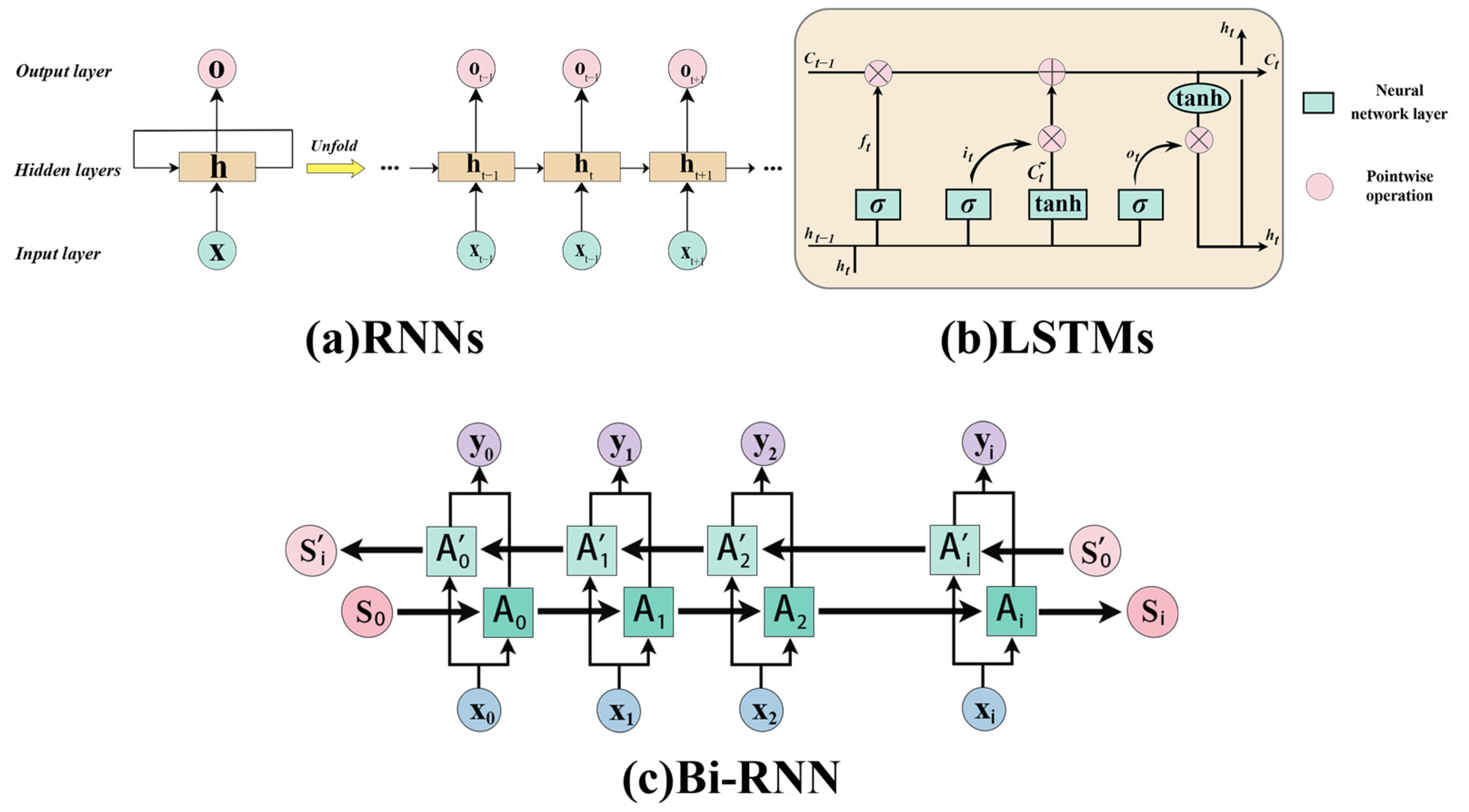
Language models (LMs) excel in content-aware data representation, from sequential databases, making them widely utilized in machine translation, question-answering systems, and even extended to applications in computer vision [54]. Encoder architectures for LMs are typically categorized into two main types: examples of recurrent neural networks (RNNs) encompass the long short-term memory (LSTM) architecture [55], while attention-based mechanisms, exemplified by Transformers [56], offer an alternative approach, both renowned for their powerful capabilities. The Hidden Markov Model (HMM), a cornerstone linguistic framework, is widely utilized in the realms of protein homology modeling and searching. Given the parallels between human language and biological languages, LMs have evolved into biological language models. Through transfer learning, biological language models are effectively applied to characterize the downstream structure and function of biological substances [57].

2.1. RNNs and LSTM

RNNs use a cyclic structure as compared to the traditional model where the nodes are not connected within the network layer and hence can handle temporal data. This was first applied in natural language modeling to capture language context and dependencies. RNNs allow the previously hidden layer to be used as an input and will share the parameters of each step and hence can be used to process variable-length sequences of inputs [58]. The fundamental architecture of a neural network comprises distinct layers: input, hidden, and output, as depicted in Figure 1a, offering a structured approach to data processing. At each timestep t, the input xt ∈ Rl, the hidden state ht ∈ Rd, and the output state vector ot ∈ Rd are formulated as follows, with superscripts l and d representing the dimensions of input features and hidden units, respectively, as outlined in [57]:
h t = f ( U x t + W h t 1 )
o t = g ( V h t )
Equation (1) represents the formula for the recurrent hidden layer, where U denotes the input weight matrix, W represents the weight matrix for the feedback connection from the previous hidden state, and f serves as the activation function that introduces nonlinearity into the model. Equation (2) represents the formula for the output layer, where V represents the output weight matrix and g is the activation function applied to the layer. The hidden layer has two inputs, the first is the product of U and the xt vector, and the second is the product of the states ht−1 and W output by the previous hidden layer, and finally together they output the final ot. By iteratively substituting Equation (1) into Equation (2), we derive an expression:
o t = g ( V h t )
= V f ( U x t + W h t 1 )
= V f ( U x t + W f ( U x t 1 + W h t 2 ) )
= V f ( U x t + W f ( U x t 1 + W ( f ( U x t 2 + W h t 3 ) ) ) )
= V f ( U x t + W f ( U x t 1 + W ( f ( U x t 2 + W ( f ( U x t 3 + ) ) ) ) )
From the aforementioned, it becomes evident that the output value ot of the recurrent neural network is intricately influenced by the sequence of preceding input values xt−1, xt−2, xt−3, xt−4, and so on, for successive input values. This is the reason why the RNN can consider any number of input values in its computations.
For many language models, bi-directional sequence information is required, and a bi-directional recurrent neural network is needed. As shown in Figure 1c, the hidden layer of a bidirectional neural network maintains two distinct values: A, which participates in the forward computation, and A′, which contributes to the reverse computation. The ultimate output value ot is a synthesis of both At and A′t. Its calculation is:
o t = g ( V h t + V h t )
h = f ( U x t + W h t 1 )
h = f ( U x t + W h t + 1 )
From the three formulas mentioned above, it is evident that the forward and reverse calculations do not share weights, indicating that U and U′, V and V′, as well as V and V′ are all distinct weight matrices.
However, in practice, RNNs do not handle longer sequences well, and the training process is susceptible to issues such as gradient explosion and gradient vanishing. These problems can prevent the gradient from being successfully propagated through longer sequences, ultimately hindering the RNNs’ ability to capture information over long distances. Therefore, LSTM networks was developed to solve this problem [55].
The LSTM network has three primary inputs: the current input value xt, the output value ht-1 from the previous timestep, and the vector of memory cells ct−1 from the preceding moment. The LSTM has two outputs: the vector of hidden states at the current moment, ht, and the vector of states of the memory cells at the current moment, ct. Furthermore, Figure 1b illustrates the utilization of σ and tanh, which signify the sigmoid and hyperbolic tanh layers, respectively, within the neural network architecture. The forget gate layer plays a pivotal role in determining which information from the cell state at timestep t should be discarded, and the remaining features are used to calculate ct. xt ∈ Rl, ht ∈ (−1,1)d, and ft ∈ (0,1)d, and sigmoid is usually used as the activation function.
f t = σ ( U x ( f ) x t + W h 1 ( f ) h ( t 1 ) + b ( f ) )
The input gate decides which values to update. This decision is made based on the input data xt and the hidden state ht−1, which are processed through a neural network layer. C t ~ ∈ (−1,1)d represents the candidate cell state update, and it ∈ (0,1)d is the input gate’s activation vector
i t = σ ( U x ( i ) x t + W h 1 h t 1 ( i ) + b ( i ) )
C ~ t = t a n h ( U x ( C ~ t ) x t + W h 1 ( C ~ t ) h t 1 + b ( C ~ t ) )
Compared to traditional RNNs, LSTM networks incorporate a unique gate mechanism, known as the cell state, which enables precise control over the flow and retention of features. This cell state is represented by the horizontal line at the top of Figure 1b, which runs through the entire chain-like system. To update the cell state Ct to its new value, we combine the old cell state Ct-1 with the new candidate state.
C t = f t C t 1 + i t C ~ t
To obtain the prediction value and prepare the input for the subsequent time step, the hidden state’s output ht is computed by passing it through the output gate, where ot ∈ (0,1)d.
o t = σ ( U x ( o ) x t + W h 1 ( o ) h t 1 + b ( o ) )
h t = o t t a n h ( C t )

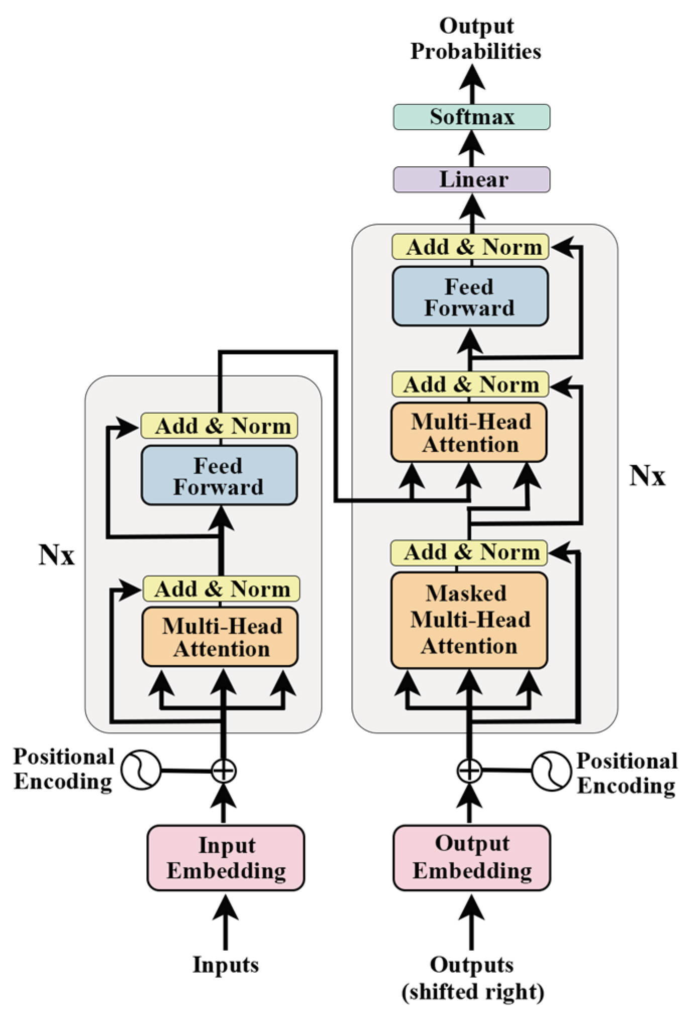
2.2. Attention Mechanism and Transformer

The traditional Sequence-to-Sequence (Seq2Seq) model uses RNN or LSTM as an encoder or decoder to process the sequence and extract features [59]. However, it is difficult for RNN or LSTM as an encoder to fully retain input sequence information in their final state. In addition, the computation of RNN and LSTM is time-dependent and therefore difficult to compute in parallel, which leads to very slow computation. Vaswani et al. [56] proposed the Transformer model to compensate for these shortcomings. The Transformer model utilizes self-attention, enabling it to individually access and weight all prior states. Furthermore, the Transformer abandons the traditional horizontal RNN transmission and only transmits vertically, requiring only the stacking of self-attention layers. This approach enables parallel computation within each layer and can be accelerated using a GPU. The proposed BERT [60] and GPT [61,62] models based on this perform exceptionally well in natural language processing tasks. Unlike RNNs, the Transformer simultaneously processes the entire input sequence using stacked self-attentive layers in both its encoder and decoder. Each layer includes a multi-head attention module, allowing a residual connection for preventing network degradation, and layer normalization modules for normalizing the activation values of each layer (Figure 2). Within the Transformer, the fundamental single-head attention mechanism is termed “Scaled Dot-Product Attention”, where the self-attention output is derived through a specific computational process.
A t t e n t i o n ( Q , K , V ) = s o f t m a x ( Q K T d K ) V
Self-Attention is a fundamental component of the Transformer, as illustrated in Figure 3a. The inputs Q (queries), K (keys), and V (values) for self-attention are linearly transformed from the input matrix X, which comprises vectors x or the output from the preceding encoder layer. Multiple attention consists of combinations of multiple self-attention and allows parallel attention to information from different subspaces (Figure 3b). By directing X through h distinct self-attention layers, h output matrices Z are generated. These matrices are then concatenated and further processed by a linear layer, yielding the final output matrix Z, which mirrors the input matrix X’s dimensions.
M u l t i H e a d ( Q , K , V ) = C o n c a t ( h e a d 1 , , h e a d h ) W O
w h e r e   h e a d i = A t t e n t i o n ( Q W i Q K W i K V W i V )
In the context of multi-head attention, projections are executed utilizing specific parameter matrices WiQ R d × d i , WiK R d × d i , WiV R d × d i , and WO R h d i × d , di = d/h. This configuration comprises h parallel attention layers, or ‘heads’, each independently processing the input data.
The formula for the Add and Norm layer is as follows:
L a y e r N o r m ( X + M u l t i H e a d A t t e n t i o n ( X ) )
L a y e r N o r m ( X + F e e d F o r w a r d ( X ) )
where X signifies the input data fed into either the Multi-Head Attention module or the Feed Forward module. The respective outputs of these two components are denoted by MultiHeadAttention(X) and FeedForward(X). The operation add refers to adding the input X to the output of the Multi-Head Attention module, resulting in X + MultiHeadAttention(X). This is a form of residual connection, commonly used to address the challenges of training deep neural networks by allowing the network to focus on learning the differences in the current layer. Layer Normalization is a technique commonly employed in RNN and other neural network architectures. It serves to regulate the input to each layer of neurons by ensuring they share a consistent mean and variance. This standardization process aids in accelerating the training convergence by mitigating the issue of internal covariate shift, where the distribution of layer inputs changes during training.
Furthermore, the feed-forward module comprises a two-layer fully connected network. The first layer employs ReLU as its activation function, while the second layer does not utilize any activation function. Furthermore, in addition to embedding sequence information, the Transformer model requires the embedding of positional information at the start of both the encoder and decoder to represent the position of each element in the sequence.

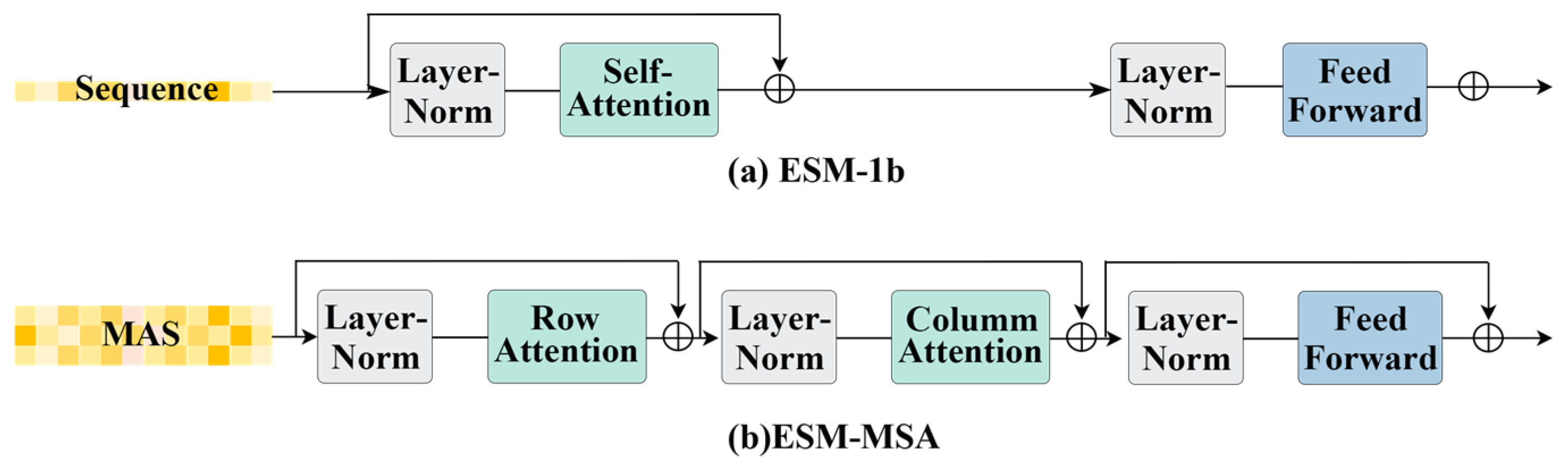
2.3. Protein Language Models

DeepCNF, proposed by Wang et al. [63], utilizes a deep neural network architecture, incorporating LSTM, to predict protein secondary structure (SS). This approach effectively addresses the challenge of long-range dependencies in sequence processing. This method consistently matches or exceeds state-of-the-art models in SS prediction. SPIDER3-Single [64] innovatively eliminates the dependency on evolutionary information, enabling predictions based solely on individual sequences. Other models, such as OCLSTM [65] and LSTM-BRNN [66], share similar research objectives and architectural frameworks. Additionally, models like GLTM [67] and LSTMCNNsucc [68] utilize pre-trained pLM embeddings to tackle diverse downstream tasks, including protein polymer motif prediction and post-translational modification prediction. These studies collectively demonstrate the efficacy of LSTM within pLMs in capturing the intricate biological features of proteins.
Recent advancements have introduced a range of deep neural language models specifically for protein sequences. Notable examples include ESM [69], TAPE-Transformer [46], ProtTrans [47], UDSMProt [70], UniRep [71], and ESM-MSA [49], each demonstrating remarkable progress in the field. ESM2 [38], a key component of the ESM framework advanced by the DeepMind team (Table 1 details the ESM family of models), builds upon the powerful Transformer architecture. This unsupervised deep attention neural network boasts a multi-layered architecture, with each layer integrating multiple attention heads alongside a feed-forward network (FFN). ESM2, with its 15 billion parameters, is trained on approximately 43 million protein sequences, leveraging mask pre-training to learn sequence–structure–function relationships and apply these learned features to downstream tasks.
ESM-MSA [49], as depicted in Figure 4, excels in unsupervised learning by encoding input MSA knowledge into feature embedding matrices. Each module integrates row-attention and column-attention layers to capture co-evolutionary relationships among amino acids at the sequence and positional levels. Another notable model, ProtTrans [47] (see Table 2), shares a similar architecture to ESM2. Both models demonstrate the capability of pLMs to extract syntactic content from large-scale protein sequences, even without relying on MSA information alone. Furthermore, integrating multiple sources of information such as structure, function, MSA, and other biological priors enhances protein characterization [72].
ProteinBERT [73], another significant model, incorporates Gene Ontology (GO) annotations during pre-training, enriching protein characterization by combining sequence data with GO annotation information to predict protein functions effectively.
Table 1. ESM family.
HyperparameterESM-1bESM-MSA-1bESM-1vESM-2
DatasetUniRef50UniRef50MSAUniRef90
Number of layers33123348
Params650 M100 M650 M15 B
Embedding Dim102876810285120
InputSingle-sequenceMSASingle-sequenceSingle-sequence
UniversalityFamily-specificFew-shotZero-shortZero-short
ModelTransformerTwo rows of attention mechanisms have been addedTransformerTransformer
References[69][49][74][75]
Table 2. ProtTrans family.
HyperparameterProtTXLProtBertProtXLNetProtALbertProtElectraProtT5-XLProtT5-XXL
DatasetBFD 100BFD 100UniRef 100UniRef 100UniRef 100UniRef 100UniRef 100UniRef 50BFD 100UniRef 50BFD 100
Number of layers3230303012302424
Params562 M420 M409 M409 M224 M420 M3 B11 B
Hidden layers size1024102410281024102410241024
Figure 4. Core modules of ESM-1b and ESM-MSA.
Genes 15 01090 g004

2.4. Nucleic Acid Language Models

In addition to pLM, there have been good advances in language models designed specifically for nucleic acids, including DNABERT [76] and RNA-TorsionBERT [77]. These models are based on the BERT [60] (Bidirectional Encoder Representation from Transformers) architecture and are tailored to capture the unique features and sequence patterns of DNA and RNA.
DNABERT [76] and RNA-TorsionBERT [77] adapt BERT models for DNA and RNA sequences. They are trained on large-scale genomic data to learn the underlying patterns of nucleotide sequences. These models have been successfully applied to a variety of tasks and have helped to deepen the understanding of the PNBP mechanism. It provides a powerful approach to understanding the complex dynamics of protein–nucleic acid interactions. For instance, DNABERT has been used in tasks like identifying transcription factor binding sites and predicting methylation patterns, while RNA-TorsionBERT has been applied to understand RNA conformational dynamics and to predict RNA–protein interactions.
However, in comparison to pLMs, nucleic acid language models have limited training data, which restricts the generalization ability of the models. In addition, the progress of pLMs is due to the rich evolutionary information contained in the protein sequences themselves, whereas nucleic acids themselves may not contain similarly rich information. Especially in non-coding regions and species-specific regulatory elements, it is difficult for nucleic acid language models to obtain better access to evolutionary information [35]. Nucleic acid language modeling is at an early stage of development and needs further validation, but it still has a very promising future.
Looking forward, the future of nucleic acid language models is undoubtedly promising. Continued advancements in genomic sequencing technologies and the accumulation of more comprehensive datasets could potentially address the current limitations. Additionally, integrating nucleic acid models with other types of biological data, such as epigenetic marks, chromatin accessibility, and transcriptional activity, could enhance their ability to make accurate predictions. As these models evolve, they are expected to play an increasingly crucial role in decoding the intricacies of genetic regulation, gene expression, and the broader mechanisms underlying protein–nucleic acid interactions.

3. Methods of Nucleic Acid Protein Binding Sites Prediction

This section showcases cutting-edge models designed to predict nucleic acid and protein binding residues, reflecting the latest advancements in current research endeavors. It distinguishes these models and compares them based on their use of pLMs as feature embeddings, highlighting the clear advantages of pLMs in this prediction task. Figure 5 categorizes the features that are currently in common use, and the methods mentioned in the text involve a wide range of features at the sequence and structural level.

3.1. Overview of Methods Framework

A typical generic framework for PNBP consists of three primary modules: the protein feature embedding unit, the backbone network module, and the loss computation module. The embedding unit constructs representations of input proteins leveraging evolutionary data or crafting discriminative embeddings through pLM, while also incorporating structural attributes as salient features. These features are subsequently fed into the backbone network, which then performs PNBP within the input protein. Deep learning has demonstrated significant advantages in predicting nucleic acid–protein binding sites, with many models discussed in this review utilizing various mainstream neural networks such as MLPs, CNNs, RNNs, GNNs, and LSTMs, in addition to traditional machine learning approaches like SVMs.
Finally, the loss calculation module performs backpropagation using diverse loss functions such as binary classification loss, contrastive loss, cross-entropy loss [78], class-balanced focal loss [74,75], and triple center loss (TCL) [79], among others. These functions guide the updating of model parameters based on the calculated loss.

3.2. Benchmark Datasets

To ensure fair comparisons between different methods, most models utilize benchmark datasets derived from previous works, such as GraphBind [9], which predominantly builds upon BioLiP [80]. BioLiP is a database of biologically pertinent ligand–protein interactions meticulously curated from the Protein Data Bank (PDB) [28]. It meticulously curates interactions through a blend of computational validation and manual verification, ensuring biological relevance by filtering out non-biologically significant ligands.
Within BioLiP, a residue qualifies as a binding residue if its minimum atomic distance from a nucleic acid molecule falls below a threshold calculated as 0.5 Å added to the combined van der Waals radii of the two closest atoms. This criterion is used to complement experimental data. Each entry in BioLiP is replete with annotations, encompassing details like ligand-binding residues, binding affinities, catalytic sites, enzyme classifications, gene ontology terms, and hyperlinks to related databases, offering a holistic view of the ligand–protein interactions [80].
The GraphBind method, for instance, leverages BioLiP’s comprehensive annotation of nucleic acid binding residues. It achieves this by aggregating binding residues across multiple similar or identical protein complexes, where proteins may interact with different DNA or RNA fragments in the collated data. As of 15 June 2024, BioLiP comprised 43,648 DNA–protein complexes and 153,190 RNA–protein complexes.
Xia et al. utilized the BioLiP database for their work, focusing on DNA and RNA binding proteins. They excluded DNA–RNA–protein complexes and divided proteins based on reporting dates, using sequences reported before 6 January 2016, for training. To tackle the imbalance between binding and non-binding residues in the data, they augmented the training set by increasing the number of binding residues using bl2seq [81] and TM-align [82] algorithms for sequence and structure comparisons. They also reduced sequence redundancy using CD-HIT [75] (threshold 30%). Ultimately, their final training set comprised a robust collection of 573 DNA-binding protein chains and 495 RNA-binding protein chains. Similarly, their test set, subjected to comparable processing measures, encompassed 129 DNA-binding proteins and 117 RNA-binding proteins.
Other studies similarly rely on BioLiP-based datasets, employing various data processing techniques such as CD-HIT [83] for sequence redundancy reduction and TM-align [82] for dataset partitioning into training and test sets. Some studies also create validation sets to ensure model robustness and avoid overfitting during hyperparameter tuning.

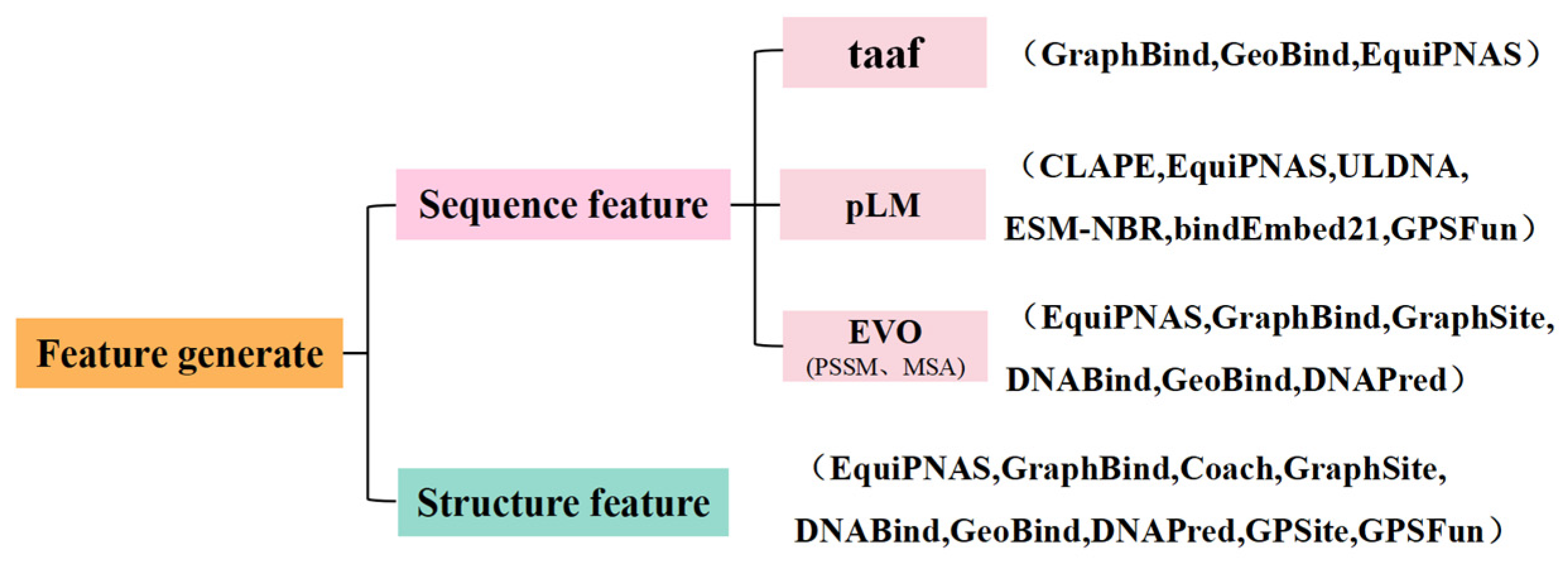
3.3. Feature Extraction

Extracting discriminative features is crucial for accurately predicting nucleic acid–protein binding sites, leading researchers to explore innovative approaches to characterize protein sequences and structures. The widely held conviction underscores the notion that a protein’s sequence serves as the blueprint for its three-dimensional (3D) structure and functionality, thereby fueling the adoption of diverse sequence-level features in relevant analyses. Commonly employed features include amino acid species encoding, residue propensity calculations, and physical properties. Evolutionary information derived from sequence comparisons is also frequently utilized to describe interactions. Moreover, pLMs, highlighted in this paper, effectively extract information from protein sequences as features.
At the structural level, proteins are often characterized by encoding 3-state and 8-state secondary structures (SS), while relative solvent accessibility (RSA) data have proven significant in predicting nucleic acid binding sites [84]. Local geometric features, residue orientation, and other structural attributes further enhance feature extraction in many models. In addition, geometric deep learning is well practiced in protein structure modeling and can be used to extract advanced biophysical–chemical knowledge of the structure.
In summary, a diverse array of features can be harnessed to accurately predict nucleic acid–protein binding sites, combining features from different sources and assigning appropriate weights enriches feature representation, enhancing model performance. By integrating multiple features, both protein structure and sequence information can be fully utilized to achieve more accurate PNBP, thereby improving model robustness and generalization.

3.3.1. Features Based on Amino Acids

Identifying nucleic acid binding residues entails employing various methods that draw upon amino acid composition, residue preferences, and physicochemical characteristics. Amino acid composition analysis determines the relative abundance of each amino acid type around DNA binding sites, providing insights into their prevalence in nucleic acid interactions. Residue propensity calculation assesses the likelihood of specific residues being involved in nucleic acid binding, revealing which amino acids are preferred in these sites. Biochemical composition analysis examines the physicochemical properties of residues—such as polarity, hydrophobicity, and charge—to understand their functional roles in nucleic acid interactions. Patiyal et al. utilized the Pfeature [85] package to compute these features comprehensively [86]. In contrast, GeoBind [87] employs a lightweight neural network that avoids hand-crafted physicochemical descriptors. Instead, it adopts an atomic point cloud approach similar to dMaSIF, where chemical features are succinctly represented as a 1 × 6 vector. This vector employs one-hot encoding to capture the presence of specific atoms, including Carbon (C), Hydrogen (H), Oxygen (O), Nitrogen (N), Sulfur (S), and others, offering a precise and efficient way to encode chemical information. This approach enables GeoBind to effectively analyze protein surfaces by distinguishing atom types, providing crucial information for subsequent prediction tasks.
Pseudo-positions capture the center of mass for each residue, taking into account both main chain and side chain atoms to represent their positions in nucleic acid interactions. This feature type is essential for modeling interactions involving both main and side chain atoms.
Additionally, atomic features describe the physicochemical properties and structural characteristics of residues. Xia et al. [9] extracted seven features for each residue, which encompass atom mass, B-factor, a flag indicating whether it belongs to a side-chain, its electronic charge, the number of bonded hydrogen atoms, ring status, and van der Waals radius. These features were averaged across residues to create an atomic feature matrix (L × 7) for each query protein, where L represents the number of residues.
For simplicity, approaches such as Roche et al. [27] have adopted one-hot encoding as a means to represent the 20 distinct amino acid residue types, facilitating the straightforward integration of amino acid type information in predictive modeling tasks.

3.3.2. Features Based on Evolutionary Information

The PSSMs serve as a valuable tool for representing residue conservation in protein sequences. PSSMs are typically generated using the PSI-BLAST tool [88], which employs heuristic and dynamic programming algorithms to search for homologous sequences in databases like NCBI’s non-redundant (NR) database or Swiss-Prot, with an output size of L × 20.
Research by Xia et al. has demonstrated that different backend algorithms and databases, such as PSI-BLAST and HHblits, yield complementary results [9]. HHblits utilizes a Hidden Markov Model (HMM) to search the uniclust30 database, generating an HMM matrix of size L × 30 [89]. The HMM matrix is structured to encapsulate crucial information pertaining to amino acid sequences. It comprises 20 columns that mirror the observed frequencies of each of the 20 amino acids within homologous sequences. Additionally, seven columns are dedicated to representing transition frequencies and three columns are utilized to encapsulate local diversities.
MSA information plays a crucial role in assessing protein homology. Tools like Clustal Omega [90], MAFFT [91], and MUSCLE [92] offer various algorithms and parameters to enhance accuracy and robustness in comparing protein sequences. MSAs provide insights into structural and functional relationships among proteins, revealing conserved and variable regions and aiding in understanding evolutionary and structural features. Several pipelines facilitate MSA generation, such as the ColabFold [93] pipeline utilized by Roche et al. [25], which employs MMseq2 [94] to generate MSAs from amino acid sequences.

3.3.3. Feature Based on Structure

Protein 3D structures are crucial for revealing nucleic acid–protein binding sites and are frequently employed to predict their characteristics. Solvent accessibility, introduced by Lee and Richards [95], plays a pivotal role in this process because it identifies the protein surface regions likely to interact with other molecules. Solvent accessibility categorizes residues as buried (B), intermediate (I), or exposed (E), and can be accurately predicted using programs like SANN [96]. This method constructs an RSA (Relative Solvent Accessibility) feature for each residue using a sliding window of size 9, resulting in an RSA feature vector of dimension 27. Additionally, RSA can also be computed using the DSSP program [97]. Apart from RSA, calculating the protein’s monothermal secondary structure profile deriving the sine and cosine values of the protein backbone torsion angle ϕ are effective methods for representing the protein’s 3D structure. By leveraging one-hot encoding to represent SS in either a 3-state or 8-state format, each amino acid residue within a protein sequence can be transformed into a multidimensional feature vector, where each dimension corresponds to a specific type of secondary structure. The torsion angles, including the C=O cosine angle between consecutive residues, the torsion angle between consecutive Cα atoms, and the normalized backbone torsion angle, provide critical local geometric information about the protein’s backbone. These angles offer insights into local spatial relationships, torsion, and curvature of the protein backbone, enhancing our understanding of its structural functions.
Predicted protein structures can also serve as features for PNBP, as demonstrated in studies like DNABind [22]. These studies used predicted secondary structures and RSA to characterize residue structures, with secondary structures generated using programs such as SPINE [98] based solely on input protein sequences.
Geometric deep learning plays a crucial and impactful role in extracting features from protein structures. GPSite [99] and GPSFun [100] utilize a geometric featurizer to extract atomic and inter-atomic features, treating residues as nodes and constructing protein radius maps to represent protein structures. Experimental findings have convincingly demonstrated that this approach captures and represents the intricate three-dimensional structural features of proteins.

3.3.4. Feature Representation Extraction from pLMs

While traditional hand-designed features have been effective in predicting nucleic acid–protein binding sites, they are constrained by a priori knowledge and may not capture diverse patterns, limiting their feature extraction capabilities. Moreover, these features often lack generalization ability and struggle to adapt to different prediction tasks. In contrast, pLM embeddings offer a more comprehensive, accurate, and adaptable feature representation. pLM embeddings are learned through large-scale pre-training, making them highly transferable and effective even with limited data samples. When used in analysis, pLM embeddings can be directly integrated into models for end-to-end learning. This simplifies the modeling process, circumvents the complexities of manual feature design, and enhances flexibility and versatility. Furthermore, pLM embeddings can be further harnessed by fine-tuning them for specific tasks or domains, improving their characterization capabilities and overall performance.
ESM2, for example, was trained on the Uniref50 [101] database, leveraging evolutionary insights from millions of sequences. It offers a range of pre-trained models with varying sizes, each providing feature matrices of different dimensions (e.g., 320, 480, 640, 1280, 2560, 5120) for protein sequences of length L. However, due to GPU limitations, processing long sequences (e.g., length > 1000) with even smaller models can still lead to memory issues, necessitating sequence segmentation while retaining sufficient evolutionary information.
Additionally, other methods like ProtT5-XL-U50 have been pre-trained on datasets like BFD [102], yielding 1024-dimensional sequence embeddings normalized to scores between 0 and 1. This normalization ensures consistent scaling across embeddings, facilitating straightforward integration into predictive models using tools like HuggingFace’s Transformers(v4.44.0) [103].

3.4. Performance Evaluation

Here we evaluate several methods for predicting nucleic acid binding sites using pLMs as feature embeddings: CLAPE [53], ESM-NBR [52], DeepProSite [50], EquiPNAS [27], ULDNA [51], GPSite [99], and GPSFun [100], which are generally summarized in Table 3. We compare them to cutting-edge methods that rely on manual feature extraction, evaluating their performance side-by-side.
Firstly, compared to sequence-based models, pLM embeddings significantly enhance model performance due to pre-training. Notably, some of these methods outperform most structure-based methods, such as CLAPE [53], even without leveraging any structural information. Meanwhile, the accuracy of protein structures is crucial for prediction tasks, as predicted structures are often less useful for PNBP. When using experimental protein structures, CLAPE’s performance is slightly lower than GraphBind [9]. However, when replacing with predicted protein structures, CLAPE [53] outperforms GraphBind [9], highlighting the superiority of pLMs in the absence of precise structural data. In experiments with the EquiPNAS [27] method using structural features and pLM embeddings, EquiPNAS [27] outperforms other methods even when using AlphaFold2-predicted structures as inputs, and the performance degradation is small compared to using experimental structures. The embeddings from pLMs undoubtedly contributed significantly to this performance improvement. GPSite [99] and GPSFun [100] extract features from structures predicted by ESMFold using geometric characterizers and incorporate pLM as embeddings. This approach leverages the accuracy and computational efficiency of language models, resulting in prediction accuracies that surpass most experimental structure-based methods. These results indicate that using pLMs as embeddings provides robustness and performance elasticity, achieving high prediction accuracy and significantly enhancing the scalability of PNBP without compromising accuracy.
Secondly, these models demonstrate excellent robustness and generalization capabilities, as evidenced by the EquiPNAS [27] experiments. GPSite [99] can predict the binding residues for ten different molecules, while GPSFun [100] is capable of annotating protein sequences with Gene Ontology (GO) terms in addition to predicting molecule interactions. As another good example, DeepProSite [50] excels in pinpointing protein–protein/peptide binding sites, and its application has been broadened to encompass the prediction of binding residues for a diverse range of ligands (e.g., Mg2+, Ca2+, and Mn2+) to verify its generalization ability. Results demonstrate that DeepProSite [50] outperforms its competitors across the majority of evaluation metrics, further confirming the robust and generalized capabilities of pLMs.
Thirdly, pLM embedding methods significantly accelerate PNBP by eliminating the time required to compute evolutionary information. ESM-NBR predicts a 500 bp protein sequence in just 5.52 s, approximately 16 times faster than the second-ranking DRNAPred [104], with other methods not even in the same order of magnitude. This demonstrates the clear computational speed advantage of pLMs.
Finally, the combined application of different pLMs offers complementary advantages for predicting nucleic acid binding sites, further enhancing prediction accuracy. ULDNA [51] demonstrates that not only can learned information be complementary across pre-trained models, but failures in one method’s predictions can be corrected by the other two methods. Despite the potential overlap in true positive predictions, ULDNA’s overall accuracy surpasses that of single pre-trained model methods.

3.5. Ablation Studies

The relative importance of features in a model can be assessed through ablation experiments, where different pLMs or different combinations with other features are formed. This allows for the investigation of the impact of various feature combinations on model performance. The ablation study allows us to further understand the contribution made by pLMs in PNBP.
Extensive experiments have demonstrated that evolutionary information has difficulty in replacing the role of pLMs in predictions. This suggests that pLMs may already encapsulate protein evolutionary information and possess richer data, thereby enhancing prediction quality [50]. Table 4 shows the classification of features taken by some of the methods. EquiPNAS [27] shows that even completely discarding evolutionary features results in only a negligible decrease in prediction accuracy for protein–nucleic acid binding sites. This underscores the importance of using pLMs as predictive embeddings, while features derived from pLMs significantly contribute to model performance.
Moreover, combining pLMs with appropriate features is not redundant but positively impacts model performance. Features such as structural features (DSSP), GO annotations, etc., are also important to improve model performance. Therefore, identifying and utilizing suitable combined features is essential.
We already know that different pLMs can complement each other in terms of information. Further ablation studies have investigated three significant pLMs: ESM-MSA, ProtTrans, and ESM2. ULDNA [51] combined these different pLMs as feature embeddings, and results showed that the inclusion of ESM2 brought the most significant performance improvement. Thus, among the three pLMs, ESM2 contributed the most.
In conclusion, feature ablation studies have demonstrated the powerful impact of pLM embeddings on PNBP. Compared to traditional feature extraction methods, pLMs can learn more effective discriminative features, reducing the reliance on conventional sequence- and structure-based features. Additionally, the embeddings from pre-trained pLMs decrease the model’s dependency on evolutionary information, enabling feature extraction for orphan proteins or rapidly evolving proteins with sparse evolutionary data. This also avoids the time-consuming task of generating MSA and PSSM features. Furthermore, even when using only pLMs, the performance of EquiPNAS [27] is comparable to or better than the current best-in-class for PNBP. This indicates that pLMs can effectively learn usable evolutionary information embedded within the protein sequences themselves. All these points illustrate that methods utilizing pLMs offer robustness and can be used to develop a versatile and scalable model, standing out against other advanced approaches.

4. Discussion

With the emergence of multi-million protein sequence databases, pLMs are becoming increasingly larger (e.g., ESM2 has 1.5 billion parameters). However, training such large-scale pLMs is often impractical for academic research teams. Therefore, it is advisable for academic researchers to leverage existing pre-trained language model embeddings with good generalization abilities for downstream tasks. In the case of PNBP, the pLMs are not readily usable due to the presence of the nucleic acid ligands and need to be customized to at least take the interplay between protein and the nucleic acids into account.
Moreover, it is important to note that increasing the size of pLMs does not always result in better model performance. Nijkamp et al. [105] found that larger models do not necessarily yield better zero-shot fitness performance. Similarly, ESM2 [75] points out that the improvement of small-scale pLMs tends to saturate when dealing with proteins that have a high evolutionary depth. However, for proteins with low evolutionary depth, increasing the model size significantly improves performance. This suggests that integrating appropriate biological or physical prior knowledge (e.g., PSSM, MSA, DSSP, GO) with pLMs can not only reduce the size of the pLM but also enhance the performance of downstream tasks. The involvement of multi-source data in task design implies that multi-task or multi-modal learning is worth exploring. On the other hand, the language models built upon nucleic acids, such as the DNA language model [76], RNA language model [106], and the combined biological language models [107], could be integrated into the tasks of PNBP for residue-level properties with high accuracy and efficiency. Since nucleic acids can bind to different sites on the surface of a protein, the structural features of proteins, especially local structural information, would not lose their importance if not play an increasingly important role in the near future. As RNA molecules are highly flexible, RNA language models could be very useful in guiding the related structure prediction tasks. How the local structural information can be extracted to well complement existing pre-trained pLMs is challenging. To note a promising direction, Zheng et al. proposed a multi-view graph embedding fusion of two networks that capture the global and local embedding representations, respectively [108].
In the case of PNBP, many methods have opted to create new datasets to meet the demand. This has introduced discrepancies in assessing the efficacy of varied approaches, eliciting apprehensions over potential data prejudices and ethical implications. Hence, the establishment of comprehensive, credible, and impartial benchmarks becomes imperative for assessing diverse models and promoting the advancement of dependable methodologies. Due to the existence of many approaches, a systematic assessment of these approaches on plenty of datasets could benchmark the performance and the potential problems (such as dataset bias) to guide the efforts to improve them in the coming years.
As the progress of PNBP with pLMs continues, it is of great possibility that unknown facets of protein–nucleic acid binding could be revealed and novel nucleic acid-binding proteins of desired properties could be designed. One challenge to this endeavor is that the complex embedding representation in pLMs can hardly be interpretable by any human. Understanding how protein sequences are processed and represented is crucial to identifying how models predict nucleic acid binding sites, which is helpful in protein design. On the one hand, pLMs can be linked to interpretable molecular representations such as physicochemical properties. On the other hand, model interpretability can be improved by machine learning approaches that are self-explainable [109]. In addition, biological knowledge and physical principles could be integrated within the frameworks of machine learning models to develop easily comprehensible models [110]. For most of the deep learning models in PNBP, their applicability domains are not unambiguously discussed and there are risks that such models could be applied to certain applications in which the underlying assumptions are not satisfied. Proteins are usually flexible and undergo conformational changes upon ligand binding. One is thus cautioned when applying pLMs to PNBP applications where changes in conformation and allosteric effects play a role, although successful applications of pLMs are noted in other related tasks such as drug discovery and protein engineering [111].
In addition, the role of IDP or IDR in protein–nucleic acid interactions complicates binding site prediction. Essentially, the structure of IDPs or IDRs is highly flexible and capable of dynamically interacting with nucleic acid binding. This structural plasticity allows them to participate in a wide range of binding events and often modulate binding affinity and specificity. Many intrinsic disorder predictors for IDRs that interact with proteins and nucleic acids such as DisoRDPbind [112] and DeepDISOBind [113] have been proposed and can be found, for example, in a recent survey [114]. Some of these predictors including the ones for disordered nucleic acids–binding proteins have also been accessed recently based on a novel benchmark dataset with reduced similarity to existing datasets [115]. Very recently, HybridDBRpred was developed to improve sequence-based prediction of DNA-binding residues for both the structure-annotated proteins and the disorder-annotated proteins and thus reduced prediction biases from different annotation types [116]. Both amino acid level and structural level information have been used in existing intrinsic disorder predictors; however, the utilization of pLMs in such tasks has not been reported yet. One possible reason could be that the flexibility of IDRs makes it difficult for pLMs to accurately predict binding sites, as conventional models may not fully capture the transient and dynamic nature of these interactions. Given this complexity, it is critical to develop and refine computational methods that better account for protein flexibility and the unique properties of IDPs or IDRs in nucleic acid binding. Future development of pLMs should focus on incorporating structural dynamics and disorder into their predictions to improve the accuracy and reliability of PNBP [117].

5. Conclusions

This paper systematically reviews the recent advancements in protein–nucleic acid binding site prediction. It covers the background, prediction challenges, the development of pLMs, and their application in this field. We highlight several successful cases that demonstrate the superior prediction quality achieved by pLM embeddings, further emphasizing the advantages of pLMs. Additionally, we discuss current limitations, potential directions, and future trends. Ultimately, we anticipate that language modeling will play a significant and convincing role in specific biological domains in the future.

Funding

This research was funded by the Shenzhen Science and Technology Innovation Commission (Grant No. 20220809164213001) and the Natural Science Foundation of Guangdong Province, China (Grant No. 2023A1515010471).

Conflicts of Interest

The authors declare no conflicts of interest.

References

  1. Charoensawan, V.; Wilson, D.; Teichmann, S.A. Genomic Repertoires of DNA-Binding Transcription Factors across the Tree of Life. Nucleic Acids Res. 2010, 38, 7364–7377. [Google Scholar] [CrossRef]
  2. Stormo, G.D.; Zhao, Y. Determining the Specificity of Protein–DNA Interactions. Nat. Rev. Genet. 2010, 11, 751–760. [Google Scholar] [CrossRef]
  3. Zhang, Q.C.; Petrey, D.; Deng, L.; Qiang, L.; Shi, Y.; Thu, C.A.; Bisikirska, B.; Lefebvre, C.; Accili, D.; Hunter, T.; et al. Structure-Based Prediction of Protein–Protein Interactions on a Genome-Wide Scale. Nature 2012, 490, 556–560. [Google Scholar] [CrossRef]
  4. Yu, B.; Pettitt, B.M.; Iwahara, J. Dynamics of Ionic Interactions at Protein–Nucleic Acid Interfaces. Acc. Chem. Res. 2020, 53, 1802–1810. [Google Scholar] [CrossRef]
  5. Schmidtke, P.; Barril, X. Understanding and Predicting Druggability. A High-Throughput Method for Detection of Drug Binding Sites. J. Med. Chem. 2010, 53, 5858–5867. [Google Scholar] [CrossRef]
  6. Yu, Y.; Li, S.; Ser, Z.; Kuang, H.; Than, T.; Guan, D.; Zhao, X.; Patel, D.J. Cryo-EM Structure of DNA-Bound Smc5/6 Reveals DNA Clamping Enabled by Multi-Subunit Conformational Changes. Proc. Natl. Acad. Sci. USA 2022, 119, e2202799119. [Google Scholar] [CrossRef]
  7. Dyson, H.J. Roles of Intrinsic Disorder in Protein–Nucleic Acid Interactions. Mol. BioSyst. 2012, 8, 97–104. [Google Scholar] [CrossRef]
  8. Järvelin, A.I.; Noerenberg, M.; Davis, I.; Castello, A. The New (Dis)Order in RNA Regulation. Cell Commun. Signal. 2016, 14, 9. [Google Scholar] [CrossRef] [PubMed]
  9. Xia, Y.; Xia, C.-Q.; Pan, X.; Shen, H.-B. GraphBind: Protein Structural Context Embedded Rules Learned by Hierarchical Graph Neural Networks for Recognizing Nucleic-Acid-Binding Residues. Nucleic Acids Res. 2021, 49, e51. [Google Scholar] [CrossRef] [PubMed]
  10. Zhang, J.; Chen, Q.; Liu, B. NCBRPred: Predicting Nucleic Acid Binding Residues in Proteins Based on Multilabel Learning. Brief. Bioinform. 2021, 22, bbaa397. [Google Scholar] [CrossRef] [PubMed]
  11. Zhu, Y.-H.; Hu, J.; Song, X.-N.; Yu, D.-J. DNAPred: Accurate Identification of DNA-Binding Sites from Protein Sequence by Ensembled Hyperplane-Distance-Based Support Vector Machines. J. Chem. Inf. Model. 2019, 59, 3057–3071. [Google Scholar] [CrossRef] [PubMed]
  12. Zhang, J.; Ghadermarzi, S.; Katuwawala, A.; Kurgan, L. DNAgenie: Accurate Prediction of DNA-Type-Specific Binding Residues in Protein Sequences. Brief. Bioinform. 2021, 22, bbab336. [Google Scholar] [CrossRef]
  13. Walia, R.R.; Xue, L.C.; Wilkins, K.; El-Manzalawy, Y.; Dobbs, D.; Honavar, V. RNABindRPlus: A Predictor That Combines Machine Learning and Sequence Homology-Based Methods to Improve the Reliability of Predicted RNA-Binding Residues in Proteins. PLoS ONE 2014, 9, e97725. [Google Scholar] [CrossRef]
  14. Qiu, J.; Bernhofer, M.; Heinzinger, M.; Kemper, S.; Norambuena, T.; Melo, F.; Rost, B. ProNA2020 Predicts Protein–DNA, Protein–RNA, and Protein–Protein Binding Proteins and Residues from Sequence. J. Mol. Biol. 2020, 432, 2428–2443. [Google Scholar] [CrossRef]
  15. Armon, A.; Graur, D.; Ben-Tal, N. ConSurf: An Algorithmic Tool for the Identification of Functional Regions in Proteins by Surface Mapping of Phylogenetic Information. J. Mol. Biol. 2001, 307, 447–463. [Google Scholar] [CrossRef]
  16. Hu, J.; Li, Y.; Zhang, M.; Yang, X.; Shen, H.-B.; Yu, D.-J. Predicting Protein-DNA Binding Residues by Weightedly Combining Sequence-Based Features and Boosting Multiple SVMs. IEEE/ACM Trans. Comput. Biol. Bioinform. 2017, 14, 1389–1398. [Google Scholar] [CrossRef]
  17. Zhang, J.; Kurgan, L. SCRIBER: Accurate and Partner Type-Specific Prediction of Protein-Binding Residues from Proteins Sequences. Bioinformatics 2019, 35, i343–i353. [Google Scholar] [CrossRef] [PubMed]
  18. Yu, D.-J.; Hu, J.; Yang, J.; Shen, H.-B.; Tang, J.; Yang, J.-Y. Designing Template-Free Predictor for Targeting Protein-Ligand Binding Sites with Classifier Ensemble and Spatial Clustering. IEEE/ACM Trans. Comput. Biol. Bioinform. 2013, 10, 994–1008. [Google Scholar] [CrossRef] [PubMed]
  19. Chen, J.; Xie, Z.-R.; Wu, Y. Understand Protein Functions by Comparing the Similarity of Local Structural Environments. Biochim. Biophys. Acta 2017, 1865, 142–152. [Google Scholar] [CrossRef]
  20. Wu, Q.; Peng, Z.; Zhang, Y.; Yang, J. COACH-D: Improved Protein–Ligand Binding Sites Prediction with Refined Ligand-Binding Poses through Molecular Docking. Nucleic Acids Res. 2018, 46, W438–W442. [Google Scholar] [CrossRef]
  21. Su, H.; Liu, M.; Sun, S.; Peng, Z.; Yang, J. Improving the Prediction of Protein–Nucleic Acids Binding Residues via Multiple Sequence Profiles and the Consensus of Complementary Methods. Bioinformatics 2019, 35, 930–936. [Google Scholar] [CrossRef]
  22. Liu, R.; Hu, J. DNABind: A Hybrid Algorithm for Structure-Based Prediction of DNA-Binding Residues by Combining Machine Learning- and Template-Based Approaches: DNA-Binding Residue Prediction. Proteins 2013, 81, 1885–1899. [Google Scholar] [CrossRef] [PubMed]
  23. Jiménez, J.; Doerr, S.; Martínez-Rosell, G.; Rose, A.S.; De Fabritiis, G. DeepSite: Protein-Binding Site Predictor Using 3D-Convolutional Neural Networks. Bioinformatics 2017, 33, 3036–3042. [Google Scholar] [CrossRef] [PubMed]
  24. Li, S.; Yamashita, K.; Amada, K.M.; Standley, D.M. Quantifying Sequence and Structural Features of Protein–RNA Interactions. Nucleic Acids Res. 2014, 42, 10086–10098. [Google Scholar] [CrossRef]
  25. Lam, J.H.; Li, Y.; Zhu, L.; Umarov, R.; Jiang, H.; Héliou, A.; Sheong, F.K.; Liu, T.; Long, Y.; Li, Y.; et al. A Deep Learning Framework to Predict Binding Preference of RNA Constituents on Protein Surface. Nat. Commun. 2019, 10, 4941. [Google Scholar] [CrossRef] [PubMed]
  26. Yuan, Q.; Chen, S.; Rao, J.; Zheng, S.; Zhao, H.; Yang, Y. AlphaFold2-Aware Protein-DNA Binding Site Prediction Using Graph Transformer. Brief. Bioinform. 2022, 23, bbab564. [Google Scholar] [CrossRef]
  27. Roche, R.; Moussad, B.; Shuvo, M.H.; Tarafder, S.; Bhattacharya, D. EquiPNAS: Improved Protein–Nucleic Acid Binding Site Prediction Using Protein-Language-Model-Informed Equivariant Deep Graph Neural Networks. Nucleic Acids Res. 2024, 52, e27. [Google Scholar] [CrossRef]
  28. Abola, E.E.; Bernstein, F.C.; Koetzle, T.F. The Protein Data Bank. In Neutrons in Biology; Schoenborn, B.P., Ed.; Springer: Boston, MA, USA, 1984; p. 441. ISBN 978-1-4899-0377-8. [Google Scholar]
  29. Jumper, J.; Evans, R.; Pritzel, A.; Green, T.; Figurnov, M.; Ronneberger, O.; Tunyasuvunakool, K.; Bates, R.; Žídek, A.; Potapenko, A.; et al. Applying and Improving AlphaFold at CASP14. Proteins 2021, 89, 1711–1721. [Google Scholar] [CrossRef] [PubMed]
  30. Jumper, J.; Evans, R.; Pritzel, A.; Green, T.; Figurnov, M.; Ronneberger, O.; Tunyasuvunakool, K.; Bates, R.; Žídek, A.; Potapenko, A.; et al. Highly Accurate Protein Structure Prediction with AlphaFold. Nature 2021, 596, 583–589. [Google Scholar] [CrossRef]
  31. Baek, M.; DiMaio, F.; Anishchenko, I.; Dauparas, J.; Ovchinnikov, S.; Lee, G.R.; Wang, J.; Cong, Q.; Kinch, L.N.; Schaeffer, R.D.; et al. Accurate Prediction of Protein Structures and Interactions Using a Three-Track Neural Network. Science 2021, 373, 871–876. [Google Scholar] [CrossRef]
  32. Wang, X.; Yu, S.; Lou, E.; Tan, Y.-L.; Tan, Z.-J. RNA 3D Structure Prediction: Progress and Perspective. Molecules 2023, 28, 5532. [Google Scholar] [CrossRef] [PubMed]
  33. Li, J.; Chiu, T.-P.; Rohs, R. Predicting DNA Structure Using a Deep Learning Method. Nat. Commun. 2024, 15, 1243. [Google Scholar] [CrossRef] [PubMed]
  34. Ou, X.; Zhang, Y.; Xiong, Y.; Xiao, Y. Advances in RNA 3D Structure Prediction. J. Chem. Inf. Model. 2022, 62, 5862–5874. [Google Scholar] [CrossRef] [PubMed]
  35. Schneider, B.; Sweeney, B.A.; Bateman, A.; Cerny, J.; Zok, T.; Szachniuk, M. When Will RNA Get Its AlphaFold Moment? Nucleic Acids Res. 2023, 51, 9522–9532. [Google Scholar] [CrossRef] [PubMed]
  36. Kryshtafovych, A.; Antczak, M.; Szachniuk, M.; Zok, T.; Kretsch, R.C.; Rangan, R.; Pham, P.; Das, R.; Robin, X.; Studer, G.; et al. New Prediction Categories in CASP15. Proteins 2023, 91, 1550–1557. [Google Scholar] [CrossRef] [PubMed]
  37. Chen, J.; Hu, Z.; Sun, S.; Tan, Q.; Wang, Y.; Yu, Q.; Zong, L.; Hong, L.; Xiao, J.; Shen, T.; et al. Interpretable RNA Foundation Model from Unannotated Data for Highly Accurate RNA Structure and Function Predictions. arXiv 2022, arXiv:2204.00300. [Google Scholar]
  38. Lin, Z.; Akin, H.; Rao, R.; Hie, B.; Zhu, Z.; Lu, W.; Smetanin, N.; Verkuil, R.; Kabeli, O.; Shmueli, Y.; et al. Evolutionary-Scale Prediction of Atomic-Level Protein Structure with a Language Model. Science 2023, 379, 1123–1130. [Google Scholar] [CrossRef]
  39. Littmann, M.; Heinzinger, M.; Dallago, C.; Weissenow, K.; Rost, B. Protein Embeddings and Deep Learning Predict Binding Residues for Various Ligand Classes. Sci. Rep. 2021, 11, 23916. [Google Scholar] [CrossRef]
  40. Zhu, Y.-H.; Zhang, C.; Yu, D.-J.; Zhang, Y. Integrating Unsupervised Language Model with Triplet Neural Networks for Protein Gene Ontology Prediction. PLoS Comput. Biol. 2022, 18, e1010793. [Google Scholar] [CrossRef]
  41. Madani, A.; Krause, B.; Greene, E.R.; Subramanian, S.; Mohr, B.P.; Holton, J.M.; Olmos, J.L.; Xiong, C.; Sun, Z.Z.; Socher, R.; et al. Large Language Models Generate Functional Protein Sequences across Diverse Families. Nat. Biotechnol. 2023, 41, 1099–1106. [Google Scholar] [CrossRef]
  42. Ferruz, N.; Schmidt, S.; Höcker, B. ProtGPT2 Is a Deep Unsupervised Language Model for Protein Design. Nat. Commun. 2022, 13, 4348. [Google Scholar] [CrossRef] [PubMed]
  43. Song, Y.; Yuan, Q.; Zhao, H.; Yang, Y. Accurately Identifying Nucleic-Acid-Binding Sites through Geometric Graph Learning on Language Model Predicted Structures. Brief. Bioinform. 2023, 24, bbad360. [Google Scholar] [CrossRef] [PubMed]
  44. Jiang, Z.; Shen, Y.-Y.; Liu, R. Structure-Based Prediction of Nucleic Acid Binding Residues by Merging Deep Learning- and Template-Based Approaches. PLoS Comput. Biol. 2023, 19, e1011428. [Google Scholar] [CrossRef] [PubMed]
  45. Baek, M.; McHugh, R.; Anishchenko, I.; Jiang, H.; Baker, D.; DiMaio, F. Accurate Prediction of Protein–Nucleic Acid Complexes Using RoseTTAFoldNA. Nat. Methods 2024, 21, 117–121. [Google Scholar] [CrossRef] [PubMed]
  46. Rao, R.; Bhattacharya, N.; Thomas, N.; Duan, Y.; Chen, X.; Canny, J.; Abbeel, P.; Song, Y.S. Evaluating Protein Transfer Learning with TAPE. In Proceedings of the Advances in Neural Information Processing Systems 32 (NeurIPS 2019), Vancouver, BC, Canada, 8–14 December 2019. [Google Scholar]
  47. Elnaggar, A.; Heinzinger, M.; Dallago, C.; Rehawi, G.; Wang, Y.; Jones, L.; Gibbs, T.; Feher, T.; Angerer, C.; Steinegger, M.; et al. ProtTrans: Toward Understanding the Language of Life Through Self-Supervised Learning. IEEE Trans. Pattern Anal. Mach. Intell. 2021, 44, 7112–7127. [Google Scholar] [CrossRef] [PubMed]
  48. Heinzinger, M.; Elnaggar, A.; Wang, Y.; Dallago, C.; Nechaev, D.; Matthes, F.; Rost, B. Modeling Aspects of the Language of Life through Transfer-Learning Protein Sequences. BMC Bioinform. 2019, 20, 723. [Google Scholar] [CrossRef]
  49. Rao, R.; Liu, J.; Verkuil, R.; Meier, J.; Canny, J.F.; Abbeel, P.; Sercu, T.; Rives, A. MSA Transformer. In Proceedings of the International Conference on Machine Learning, Virtual, 18–24 July 2021. [Google Scholar]
  50. Fang, Y.; Jiang, Y.; Wei, L.; Ma, Q.; Ren, Z.; Yuan, Q.; Wei, D.-Q. DeepProSite: Structure-Aware Protein Binding Site Prediction Using ESMFold and Pretrained Language Model. Bioinformatics 2023, 39, btad718. [Google Scholar] [CrossRef] [PubMed]
  51. Zhu, Y.-H.; Liu, Z.; Liu, Y.; Ji, Z.; Yu, D.-J. ULDNA: Integrating Unsupervised Multi-Source Language Models with LSTM-Attention Network for High-Accuracy Protein–DNA Binding Site Prediction. Brief. Bioinform. 2024, 25, bbae040. [Google Scholar] [CrossRef]
  52. Zeng, W.; Lv, D.; Liu, X.; Chen, G.; Liu, W.; Peng, S. ESM-NBR: Fast and Accurate Nucleic Acid-Binding Residue Prediction via Protein Language Model Feature Representation and Multi-Task Learning. In Proceedings of the 2023 IEEE International Conference on Bioinformatics and Biomedicine (BIBM), Istanbul, Turkiye, 5–8 December 2023; pp. 76–81. [Google Scholar]
  53. Liu, Y.; Tian, B. Protein–DNA Binding Sites Prediction Based on Pre-Trained Protein Language Model and Contrastive Learning. Brief. Bioinform. 2023, 25, bbad488. [Google Scholar] [CrossRef] [PubMed]
  54. Bepler, T.; Berger, B. Learning the Protein Language: Evolution, Structure, and Function. Cell Syst. 2021, 12, 654–669.e3. [Google Scholar] [CrossRef]
  55. Lample, G.; Ballesteros, M.; Subramanian, S.; Kawakami, K.; Dyer, C. Neural Architectures for Named Entity Recognition. arXiv 2016, arXiv:1603.01360. [Google Scholar]
  56. Vaswani, A.; Shazeer, N.; Parmar, N.; Uszkoreit, J.; Jones, L.; Gomez, A.N.; Kaiser, L.; Polosukhin, I. Attention Is All You Need. arXiv 2017, arXiv:1706.03762. [Google Scholar]
  57. Shen, Y.; Chen, Z.; Mamalakis, M.; He, L.; Xia, H.; Li, T.; Su, Y.; He, J.; Wang, Y.G. A Fine-Tuning Dataset and Benchmark for Large Language Models for Protein Understanding. arXiv 2024, arXiv:2406.05540. [Google Scholar]
  58. Graves, A.; Liwicki, M.; Fernandez, S.; Bertolami, R.; Bunke, H.; Schmidhuber, J. A Novel Connectionist System for Unconstrained Handwriting Recognition. IEEE Trans. Pattern Anal. Mach. Intell. 2009, 31, 855–868. [Google Scholar] [CrossRef] [PubMed]
  59. Hu, B.; Xia, J.; Zheng, J.; Tan, C.; Huang, Y.; Xu, Y.; Li, S.Z. Protein Language Models and Structure Prediction: Connection and Progression. arXiv 2022, arXiv:2211.16742. [Google Scholar]
  60. Devlin, J.; Chang, M.-W.; Lee, K.; Toutanova, K. BERT: Pre-Training of Deep Bidirectional Transformers for Language Understanding. arXiv 2018, arXiv:1810.04805. [Google Scholar]
  61. Radford, A.; Narasimhan, K.; Salimans, T.; Sutskever, I. Improving Language Understanding by Generative Pre-Training. Available online: https://cdn.openai.com/research-covers/language-unsupervised/language_understanding_paper.pdf (accessed on 22 July 2018).
  62. Brown, T.B.; Mann, B.; Ryder, N.; Subbiah, M.; Kaplan, J.; Dhariwal, P.; Neelakantan, A.; Shyam, P.; Sastry, G.; Askell, A.; et al. Language Models Are Few-Shot Learners. In Proceedings of the Advances in Neural Information Processing Systems 33 (NeurIPS 2020), Virtual, 6–12 December 2020. [Google Scholar]
  63. Wang, S.; Peng, J.; Ma, J.; Xu, J. Protein Secondary Structure Prediction Using Deep Convolutional Neural Fields. Sci. Rep. 2016, 6, 18962. [Google Scholar] [CrossRef] [PubMed]
  64. Heffernan, R.; Paliwal, K.; Lyons, J.; Singh, J.; Yang, Y.; Zhou, Y. Single-sequence-based Prediction of Protein Secondary Structures and Solvent Accessibility by Deep Whole-sequence Learning. J. Comput. Chem. 2018, 39, 2210–2216. [Google Scholar] [CrossRef] [PubMed]
  65. Zhao, Y.; Liu, Y. OCLSTM: Optimized Convolutional and Long Short-Term Memory Neural Network Model for Protein Secondary Structure Prediction. PLoS ONE 2021, 16, e0245982. [Google Scholar] [CrossRef]
  66. Heffernan, R.; Yang, Y.; Paliwal, K.; Zhou, Y. Capturing Non-Local Interactions by Long Short-Term Memory Bidirectional Recurrent Neural Networks for Improving Prediction of Protein Secondary Structure, Backbone Angles, Contact Numbers and Solvent Accessibility. Bioinformatics 2017, 33, 2842–2849. [Google Scholar] [CrossRef] [PubMed]
  67. Ma, Q.; Zou, K.; Zhang, Z.; Yang, F. GLTM: A Global-Local Attention LSTM Model to Locate Dimer Motif of Single-Pass Membrane Proteins. Front. Genet. 2022, 13, 854571. [Google Scholar] [CrossRef] [PubMed]
  68. Huang, G.; Shen, Q.; Zhang, G.; Wang, P.; Yu, Z.-G. LSTMCNNsucc: A Bidirectional LSTM and CNN-Based Deep Learning Method for Predicting Lysine Succinylation Sites. BioMed Res. Int. 2021, 2021, 1–10. [Google Scholar] [CrossRef] [PubMed]
  69. Rives, A.; Meier, J.; Sercu, T.; Goyal, S.; Lin, Z.; Liu, J.; Guo, D.; Ott, M.; Zitnick, C.L.; Ma, J.; et al. Biological Structure and Function Emerge from Scaling Unsupervised Learning to 250 Million Protein Sequences. Proc. Natl. Acad. Sci. USA 2021, 118, e2016239118. [Google Scholar] [CrossRef] [PubMed]
  70. Strodthoff, N.; Wagner, P.; Wenzel, M.; Samek, W. UDSMProt: Universal Deep Sequence Models for Protein Classification. Bioinformatics 2020, 36, 2401–2409. [Google Scholar] [CrossRef]
  71. Alley, E.C.; Khimulya, G.; Biswas, S.; AlQuraishi, M.; Church, G.M. Unified Rational Protein Engineering with Sequence-Based Deep Representation Learning. Nat. Methods 2019, 16, 1315–1322. [Google Scholar] [CrossRef] [PubMed]
  72. Chatzou, M.; Magis, C.; Chang, J.-M.; Kemena, C.; Bussotti, G.; Erb, I.; Notredame, C. Multiple Sequence Alignment Modeling: Methods and Applications. Brief. Bioinform. 2016, 17, 1009–1023. [Google Scholar] [CrossRef] [PubMed]
  73. Brandes, N.; Ofer, D.; Peleg, Y.; Rappoport, N.; Linial, M. ProteinBERT: A Universal Deep-Learning Model of Protein Sequence and Function. Bioinformatics 2022, 38, 2102–2110. [Google Scholar] [CrossRef] [PubMed]
  74. Meier, J.; Rao, R.; Verkuil, R.; Liu, J.; Sercu, T.; Rives, A. Language Models Enable Zero-Shot Prediction of the Effects of Mutations on Protein Function. In Proceedings of the Advances in Neural Information Processing Systems 34 (NeurIPS 2021), Virtual, 6–14 December 2021. [Google Scholar]
  75. Lin, Z.; Akin, H.; Rao, R.; Hie, B.; Zhu, Z.; Lu, W.; Fazel-Zarandi, M.; Sercu, T.; Candido, S.; Rives, A. Language Models of Protein Sequences at the Scale of Evolution Enable Accurate Structure Prediction. BioRxiv 2022, 2022, 500902. [Google Scholar] [CrossRef]
  76. Ji, Y.; Zhou, Z.; Liu, H.; Davuluri, R.V. DNABERT: Pre-Trained Bidirectional Encoder Representations from Transformers Model for DNA-Language in Genome. Bioinformatics 2021, 37, 2112–2120. [Google Scholar] [CrossRef]
  77. Bernard, C.; Postic, G.; Ghannay, S.; Tahi, F. RNA-TorsionBERT: Leveraging Language Models for RNA 3D Torsion Angles Prediction. bioRxiv 2024, 597803. [Google Scholar] [CrossRef]
  78. Zhang, Z.; Sabuncu, M. Generalized Cross Entropy Loss for Training Deep Neural Networks with Noisy Labels. In Proceedings of the Advances in Neural Information Processing Systems 31 (NeurIPS 2018), Montréal, QC, Canada, 3–8 December 2018. [Google Scholar]
  79. He, X.; Zhou, Y.; Zhou, Z.; Bai, S.; Bai, X. Triplet-Center Loss for Multi-View 3D Object Retrieval. In Proceedings of the 2018 IEEE/CVF Conference on Computer Vision and Pattern Recognition, Salt Lake City, UT, USA, 18–23 June 2018; pp. 1945–1954. [Google Scholar]
  80. Yang, J.; Roy, A.; Zhang, Y. BioLiP: A Semi-Manually Curated Database for Biologically Relevant Ligand–Protein Interactions. Nucleic Acids Res. 2012, 41, D1096–D1103. [Google Scholar] [CrossRef] [PubMed]
  81. McGinnis, S.; Madden, T.L. BLAST: At the Core of a Powerful and Diverse Set of Sequence Analysis Tools. Nucleic Acids Res. 2004, 32, W20–W25. [Google Scholar] [CrossRef] [PubMed]
  82. Zhang, Y. TM-Align: A Protein Structure Alignment Algorithm Based on the TM-Score. Nucleic Acids Res. 2005, 33, 2302–2309. [Google Scholar] [CrossRef] [PubMed]
  83. Fu, L.; Niu, B.; Zhu, Z.; Wu, S.; Li, W. CD-HIT: Accelerated for Clustering the next-Generation Sequencing Data. Bioinformatics 2012, 28, 3150–3152. [Google Scholar] [CrossRef] [PubMed]
  84. Ahmad, S.; Gromiha, M.M.; Sarai, A. Real Value Prediction of Solvent Accessibility from Amino Acid Sequence. Proteins 2003, 50, 629–635. [Google Scholar] [CrossRef]
  85. Pande, A.; Patiyal, S.; Lathwal, A.; Arora, C.; Kaur, D.; Dhall, A.; Mishra, G.; Kaur, H.; Sharma, N.; Jain, S.; et al. Computing Wide Range of Protein/Peptide Features from Their Sequence and Structure. BioRxiv 2019, 599126. [Google Scholar] [CrossRef]
  86. Patiyal, S.; Dhall, A.; Raghava, G.P.S. A Deep Learning-Based Method for the Prediction of DNA Interacting Residues in a Protein. Brief. Bioinform. 2022, 23, bbac322. [Google Scholar] [CrossRef]
  87. Li, P.; Liu, Z.-P. GeoBind: Segmentation of Nucleic Acid Binding Interface on Protein Surface with Geometric Deep Learning. Nucleic Acids Res. 2023, 51, e60. [Google Scholar] [CrossRef]
  88. Schaffer, A.A. Improving the Accuracy of PSI-BLAST Protein Database Searches with Composition-Based Statistics and Other Refinements. Nucleic Acids Res. 2001, 29, 2994–3005. [Google Scholar] [CrossRef]
  89. Remmert, M.; Biegert, A.; Hauser, A.; Söding, J. HHblits: Lightning-Fast Iterative Protein Sequence Searching by HMM-HMM Alignment. Nat. Methods 2012, 9, 173–175. [Google Scholar] [CrossRef] [PubMed]
  90. Sievers, F.; Wilm, A.; Dineen, D.; Gibson, T.J.; Karplus, K.; Li, W.; Lopez, R.; McWilliam, H.; Remmert, M.; Söding, J.; et al. Fast, Scalable Generation of High-quality Protein Multiple Sequence Alignments Using Clustal Omega. Mol. Syst. Biol. 2011, 7, 539. [Google Scholar] [CrossRef]
  91. Katoh, K. MAFFT: A Novel Method for Rapid Multiple Sequence Alignment Based on Fast Fourier Transform. Nucleic Acids Res. 2002, 30, 3059–3066. [Google Scholar] [CrossRef] [PubMed]
  92. Edgar, R.C. MUSCLE: Multiple Sequence Alignment with High Accuracy and High Throughput. Nucleic Acids Res. 2004, 32, 1792–1797. [Google Scholar] [CrossRef]
  93. Mirdita, M.; Schütze, K.; Moriwaki, Y.; Heo, L.; Ovchinnikov, S.; Steinegger, M. ColabFold: Making Protein Folding Accessible to All. Nat. Methods 2022, 19, 679–682. [Google Scholar] [CrossRef] [PubMed]
  94. Steinegger, M.; Söding, J. MMseqs2 Enables Sensitive Protein Sequence Searching for the Analysis of Massive Data Sets. Nat. Biotechnol. 2017, 35, 1026–1028. [Google Scholar] [CrossRef] [PubMed]
  95. Lee, B.; Richards, F.M. The Interpretation of Protein Structures: Estimation of Static Accessibility. J. Mol. Biol. 1971, 55, 379-IN4. [Google Scholar] [CrossRef]
  96. Joo, K.; Lee, S.J.; Lee, J. Sann: Solvent Accessibility Prediction of Proteins by Nearest Neighbor Method. Proteins 2012, 80, 1791–1797. [Google Scholar] [CrossRef]
  97. Kabsch, W.; Sander, C. Dictionary of Protein Secondary Structure: Pattern Recognition of Hydrogen-bonded and Geometrical Features. Biopolymers 1983, 22, 2577–2637. [Google Scholar] [CrossRef]
  98. Faraggi, E.; Zhang, T.; Yang, Y.; Kurgan, L.; Zhou, Y. SPINE X: Improving Protein Secondary Structure Prediction by Multistep Learning Coupled with Prediction of Solvent Accessible Surface Area and Backbone Torsion Angles. J. Comput. Chem. 2012, 33, 259–267. [Google Scholar] [CrossRef]
  99. Yuan, Q.; Tian, C.; Yang, Y. Genome-Scale Annotation of Protein Binding Sites via Language Model and Geometric Deep Learning. eLife 2024, 13, RP93695. [Google Scholar] [CrossRef]
  100. Yuan, Q.; Tian, C.; Song, Y.; Ou, P.; Zhu, M.; Zhao, H.; Yang, Y. GPSFun: Geometry-Aware Protein Sequence Function Predictions with Language Models. Nucleic Acids Res. 2024, 52, W248–W255. [Google Scholar] [CrossRef] [PubMed]
  101. Suzek, B.E.; Huang, H.; McGarvey, P.; Mazumder, R.; Wu, C.H. UniRef: Comprehensive and Non-Redundant UniProt Reference Clusters. Bioinformatics 2007, 23, 1282–1288. [Google Scholar] [CrossRef]
  102. Steinegger, M.; Mirdita, M.; Söding, J. Protein-Level Assembly Increases Protein Sequence Recovery from Metagenomic Samples Manyfold. Nat. Methods 2019, 16, 603–606. [Google Scholar] [CrossRef]
  103. Wolf, T.; Debut, L.; Sanh, V.; Chaumond, J.; Delangue, C.; Moi, A.; Cistac, P.; Rault, T.; Louf, R.; Funtowicz, M.; et al. HuggingFace’s Transformers: State-of-the-Art Natural Language Processing. arXiv 2019, arXiv:1910.03771. [Google Scholar]
  104. Yan, J.; Kurgan, L. DRNApred, Fast Sequence-Based Method That Accurately Predicts and Discriminates DNA- and RNA-Binding Residues. Nucleic Acids Res. 2017, 45, e84. [Google Scholar] [CrossRef]
  105. Nijkamp, E.; Ruffolo, J.; Weinstein, E.N.; Naik, N.; Madani, A. ProGen2: Exploring the Boundaries of Protein Language Models. Cell Syst. 2023, 14, 968–978.e3. [Google Scholar] [CrossRef]
  106. Zhang, Y.; Lang, M.; Jiang, J.; Gao, Z.; Xu, F.; Litfin, T.; Chen, K.; Singh, J.; Huang, X.; Song, G.; et al. Multiple Sequence Alignment-Based RNA Language Model and Its Application to Structural Inference. Nucleic Acids Res. 2024, 52, e3. [Google Scholar] [CrossRef] [PubMed]
  107. Li, H.-L.; Pang, Y.-H.; Liu, B. BioSeq-BLM: A Platform for Analyzing DNA, RNA and Protein Sequences Based on Biological Language Models. Nucleic Acids Res. 2021, 49, e129. [Google Scholar] [CrossRef]
  108. Zheng, M.; Sun, G.; Li, X.; Fan, Y. EGPDI: Identifying Protein–DNA Binding Sites Based on Multi-View Graph Embedding Fusion. Brief. Bioinform. 2024, 25, bbae330. [Google Scholar] [CrossRef]
  109. Minh, D.; Wang, H.X.; Li, Y.F.; Nguyen, T.N. Explainable Artificial Intelligence: A Comprehensive Review. Artif. Intell. Rev. 2022, 55, 3503–3568. [Google Scholar] [CrossRef]
  110. Jiménez-Luna, J.; Grisoni, F.; Schneider, G. Drug Discovery with Explainable Artificial Intelligence. Nat. Mach. Intell. 2020, 2, 573–584. [Google Scholar] [CrossRef]
  111. Nerín-Fonz, F.; Cournia, Z. Machine Learning Approaches in Predicting Allosteric Sites. Curr. Opin. Struct. Biol. 2024, 85, 102774. [Google Scholar] [CrossRef] [PubMed]
  112. Peng, Z.; Kurgan, L. High-Throughput Prediction of RNA, DNA and Protein Binding Regions Mediated by Intrinsic Disorder. Nucleic Acids Res. 2015, 43, e121. [Google Scholar] [CrossRef] [PubMed]
  113. Zhang, F.; Zhao, B.; Shi, W.; Li, M.; Kurgan, L. DeepDISOBind: Accurate Prediction of RNA-, DNA- and Protein-Binding Intrinsically Disordered Residues with Deep Multi-Task Learning. Brief. Bioinform. 2022, 23, bbab521. [Google Scholar] [CrossRef] [PubMed]
  114. Basu, S.; Kihara, D.; Kurgan, L. Computational Prediction of Disordered Binding Regions. Comput. Struct. Biotechnol. J. 2023, 21, 1487–1497. [Google Scholar] [CrossRef] [PubMed]
  115. Katuwawala, A.; Kurgan, L. Comparative Assessment of Intrinsic Disorder Predictions with a Focus on Protein and Nucleic Acid-Binding Proteins. Biomolecules 2020, 10, 1636. [Google Scholar] [CrossRef]
  116. Zhang, J.; Basu, S.; Kurgan, L. HybridDBRpred: Improved Sequence-Based Prediction of DNA-Binding Amino Acids Using Annotations from Structured Complexes and Disordered Proteins. Nucleic Acids Res. 2024, 52, e10. [Google Scholar] [CrossRef]
  117. Wright, P.E.; Dyson, H.J. Intrinsically Disordered Proteins in Cellular Signalling and Regulation. Nat. Rev. Mol. Cell Biol. 2015, 16, 18–29. [Google Scholar] [CrossRef]
Figure 1. The structure of RNN, Bi-RNN, LSTM.
Genes 15 01090 g001
Figure 2. The Transformer model architecture.
Genes 15 01090 g002
Figure 3. Scaled Dot-Product Attention (a). Multi-Head Attention consists of several attention layers running in parallel (b).
Genes 15 01090 g003
Figure 5. Classification of protein characteristics. At the sequence level, features are categorized into traditional amino acid features (taaf), enriched features extracted from pLM, and evolutionary features containing PSSM and MSA. At the structural level, they lack a concise classification and are elaborated in Section 3.3.3 in detail. Methods that utilize such features are listed in the parentheses. Some methods such as DNABind and GeoBind utilize both sequence features and structural ones.
Genes 15 01090 g005
Table 3. Introduction to pLM extraction of features.
MethodFeature GenerationFeature RepresentationKey Learning Architecture
CLAPE [53]ProtBertTensorConcatenated ACNNs
ESM-NBR [52]ESM2TensorLSTM
DeepProSite [50]ProtBert, DSSPGraphsGNN
EquiPNAS [27]ESM2, DSSP, PSSM, MSA, taaf, SS, RSA, et al.GraphsGNN
ULDNA [51]ESM, ESM-MSA, ProtBertTensorLSTM
GPSite [99]ProtBert, ESMFoldGraphsGNN
GPSFun [100]ProtBert, ESMFoldGraphsGNN
Table 4. Feature combinations of different methods for ablation studies. Bolded text is the combination of features used in the full version of the methods.
ULDNA [51]DeepProsite [50]EquiPNAS [27]
ProtTrans + ESM-MSA
ESM2 + ESM-MSA
ESM2 + ProtTrans
ESM2 + ProtTrans + ESM-MSA (ULDNA)
EVO
DSSP
ProtT5
ProtT5 + EVO + DSSP
EVO + DSSP
ProtT5 + EVO
ProtT5 + DSSP (DeepProSite)
No ESM2
No (PSSM + MSA)
No MSA
No PSSM
ESM2, DSSP, PSSM, MSA, taaf, SS, RSA, et al. (EquiPNAS)
Disclaimer/Publisher’s Note: The statements, opinions and data contained in all publications are solely those of the individual author(s) and contributor(s) and not of MDPI and/or the editor(s). MDPI and/or the editor(s) disclaim responsibility for any injury to people or property resulting from any ideas, methods, instructions or products referred to in the content.

