Brief Report

Schistosoma japonicum sja-let-7 Inhibits the Growth of Hepatocellular Carcinoma Cells via Cross-Species Regulation of Col1α2

1
National Reference Laboratory for Animal Schistosomiasis, Key Laboratory of Animal Parasitology of Ministry of Agriculture and Rural Affairs, Shanghai Veterinary Research Institute, Chinese Academy of Agricultural Sciences, Shanghai 200241, China
2
State Key Laboratory for Animal Disease Control and Prevention, Key Laboratory of Veterinary Parasitology of Gansu Province, Lanzhou Veterinary Research Institute, Chinese Academy of Agricultural Science, Lanzhou 730046, China
*
Author to whom correspondence should be addressed.
Genes 2024, 15(9), 1165; https://doi.org/10.3390/genes15091165
Submission received: 14 August 2024 / Revised: 29 August 2024 / Accepted: 2 September 2024 / Published: 4 September 2024
(This article belongs to the Section Human Genomics and Genetic Diseases)

Abstract

:
Liver fibrosis, a critical precursor to hepatocellular carcinoma (HCC), results from chronic liver injury and significantly contributes to HCC progression. Schistosomiasis, a neglected tropical disease, is known to cause liver fibrosis; however, this process can be modulated by schistosome-derived miRNAs. Previous studies from our laboratory have demonstrated that Schistosoma japonicum extracellular vesicles (EVs) deliver sja-let-7 to hepatic stellate cells, leading to the inhibition of Col1α2 expression and alleviation of liver fibrosis. Given the well-documented antifibrotic and antiproliferative properties of the let-7 miRNA family, this study aims to preliminarily investigate the effects of the sja-let-7/Col1α2 axis on BALB/c mice and HCC cell line SNU387, providing a basis for the potential application of parasite-derived molecules in HCC therapy. In the present study, schistosome-induced fibrosis datasets were analyzed to identify the role of Col1α2 in extracellular matrix organization. Pan-cancer analysis revealed that Col1α2 is upregulated in various cancers, including HCC, with significant associations with immune cell infiltration and clinical parameters, highlighting its diagnostic importance. Functional assays demonstrated that transfection with sja-let-7 mimics significantly reduced Col1α2 expression, inhibited HCC cell proliferation, migration, and colony formation. These findings suggest that sja-let-7, by targeting Col1α2, has the potential to serve as a therapeutic agent in HCC treatment. This study indicates the pivotal role of Col1α2 in liver fibrosis and HCC, and the promising therapeutic application of helminth-derived miRNAs.

1. Introduction

Liver fibrosis, a pathological process resulting from persistent liver injury, is crucial for the development of hepatocellular carcinoma (HCC) [1]. HCC is a highly invasive and lethal cancer with high incidence and mortality rates worldwide [2]. Despite advances in current treatments such as surgery, radiotherapy, and chemotherapy, their effectiveness remains limited and is often accompanied by high recurrence rates [3]. Therefore, identifying new therapeutic targets and strategies is crucial for reversing liver fibrosis and improving HCC treatment outcomes.
Schistosomiasis is a neglected parasitic disease that impacts over 250 million individuals in tropical and subtropical regions globally, which is mainly caused by Schistosoma mansoni, Schistosoma japonicum, and Schistosoma haematobium, the latter of which is related to bladder cancer [4]. Chronic infection with S. japonicum can result in liver fibrosis and liver cirrhosis, both of which are significant risk factors for HCC. However, the correlation does not seem to apply in the case of schistosomiasis japonica and the association between S. japonicum -induced liver fibrosis and HCC is less clear and remains debated [5]. Case–control studies conducted in regions endemic for schistosomiasis japonica have suggested that infection with S. japonicum does not have a direct association with HCC [6,7,8].
During the course of schistosome-induced liver fibrosis, microRNAs (miRNAs) from parasite-derived extracellular vesicles (EVs) can transfer to host cells via EVs and regulate gene expression within the host cells by binding to the 3′ untranslated regions (3′UTRs) of target mRNAs [9,10]. Interestingly, several miRNAs derived from SjEVs have demonstrated anti-fibrotic effects in cross-species regulation in previous studies [9,11]. Thus, we propose that certain factors, such as miRNAs and proteins derived from S. japonicum infection, could be advantageous to the host by enhancing resistance to specific diseases, including cancer.
Pan’s lab has identified several Sj-miRNAs that exhibits anti-tumor effects. Based on their report, sja-miR-7-5p targets the host S-phase kinase-associated protein 2 gene, significantly inhibiting the growth, migration, and colony formation of both mouse and human hepatoma cell lines, while also result in G1/G0 cell cycle arrest [12]. Additionally, sja-miR-61 [13] and sja-miR-3096 [14] target the phosphoglycerate mutase 1 and phosphatidylinositol 3-kinase C2α genes, respectively, significantly inhibiting the migration and angiogenesis of hepatoma cells. In vivo, transplantation of hepatoma cells with sja-miR-61 or sja-miR-3096 mimics into mouse models resulted in significantly reduced tumor volume and weight. Furthermore, an anti-fibrotic miRNA, sja-miR-71a, was also shown to have anti-tumor effects via targeting frizzled class receptor 4 gene [15].
Our previous studies found that SjEV-derived sja-let-7 can target the host collagen type I α 2 chain (col1α2) gene, thereby inhibiting the progression of both schistosome- and carbon tetrachloride-induced liver fibrosis [11,16]. Since col1α2 is an important marker for both liver fibrosis and HCC, it is intriguing to explore whether the targeting relationship between sja-let-7 and col1α2 can inhibit the proliferation of hepatoma cells. Hence, a preliminary investigation was conducted to assess the effect of this targeting relationship in hepatoma cell line, with the aim of providing a theoretical foundation for the potential application of parasite-derived molecules and the treatment of HCC.

2. Materials and Methods

2.1. Data Collection and DEGs Identification

The microarray data were retrieved from the Gene Expression Omnibus [17]. The platform for GSE14367 was GPL6105 (Illumina mouse-6 v1.1 expression beadchip), which contained two liver control samples not infected with S. japonicum and two liver samples infected with S. japonicum for seven weeks. The platform for GSE25713 was GPL6887 (Illumina MouseWG-6 v2.0 expression beadchip), which contained six liver control samples not infected with S. japonicum and six liver samples infected with S. japonicum for seven weeks. The platform for GSE41941 was GPL6885 (Illumina MouseRef-8 v2.0 expression beadchip), which contained two liver control samples not infected with S. japonicum and two liver granuloma samples with S. japonicum infection for seven weeks. The data were processed through UMAP analysis, which was performed using the R package “umap”. Gene symbols were identified by converting all probes based on the platform’s normalized annotation information. The data were then processed on the GEO2R website [17]. The differentially expressed genes (DEGs) were identified at a cutoff |logFC| > 1 and adjusted p value < 0.05.

2.2. Enrichment Analysis

The gene set enrichment analysis (GSEA) [18] was employed to rank the genome a thousand times, identifying pathways enriched in liver fibrosis. For GSEA analysis, the threshold value for statistically significant findings was defined by an adjusted p < 0.05 and a false discovery rate (FDR) of <0.25. Enrichment results were evaluated using normalized enrichment scores (NESs) and adjusted p-values. Additionally, Gene ontology (GO) analysis [19] was performed to assess the molecular function (MF), cellular component (CC) and biological process (BP) of DEGs, and Kyoto Encyclopedia of Genes (KEGG) analysis [20] was also performed to categorize genes into relevant metabolic and regulatory pathways.

2.3. Protein-Protein Interaction Network Construction

To identify the potential interacting molecules of col1α2, the STRING v11.5 online tool [21] was utilized to construct and visualize a protential interaction network.

2.4. Construction of Liver Fibrosis Mice Model

The liver fibrosis mice model was established based on previous studies [11,16]. Briefly, 6 male BALB/c mice (6–8 weeks old; weight 18 ± 2 g) were purchased from Shanghai Jiesijie Laboratory Animal Co., Ltd. (Shanghai, China) and were percutaneously infected with 20 ± 2 S. japonicum cercariae (maintained in Shanghai Veterinary Research Institute) to establish the schistosome-induced liver fibrosis mouse model and named as the infected group. Another 6 BALB/c mice served as the uninfected control group. Mice were sacrificed at 6 weeks post infection (wpi). Additionally, 4 BALB/c mice were intraperitoneally injected with CCL4 (0.1 mL 10% CCL4 diluted in peanut oil) (Sinopharm, Shanghai, China) twice a week for 4 weeks. Mice were sacrificed at 4 wpi. Liver samples from each mouse were collected for further studies.

2.5. Pan-Cancer View and Clinical Correlation Analysis of the Cancer Genome Atlas Program (TCGA) Platform

The expression of col1α2 were obtained from TCGA pan-cancer view using the Xiantao platform (https://www.xiantao.love/, accessed 20 June 2024) [22]. Clinical correlation, such as the relevance of col1α2 between histological type, pathologic stage, tumor status, fibrosis ishak score and adjacent hepatic tissue inflammation were analyzed using patient data from the TCGA through the clinical significance module of the Xiantao platform. The analyses were performed using the Welch one-way ANOVA method.

2.6. Analysis of Immune Cell Infiltration

The single sample gene enrichment analysis (ssGSEA) method was employed to examine tumor infiltration across 24 distinct immune cell types [23]. The link between the expression of col1α2 and immune cell infiltration was analyzed via Spearman correlation, and the Wilcoxon signed-rank sum.

2.7. Diagnostic Value Analysis

The receiver operating characteristic (ROC) curve was used to assess the diagnostic potential of col1α2 in HCC patients, conducted via the “pROC” package (version 1.18.0). Then, the area under the curve (AUC) was calculated, with higher AUC value indicating the better diagnostic accuracy. Typically, an AUC value of 0.5–0.7 indicates a low diagnostic effect, 0.7–0.9 suggests a moderate effect, and an AUC value above 0.9 indicates a high diagnostic effect [24].

2.8. Cell Culture and Treatments

Based on the expression of col1α2 in various human hepatoma cell line as documented in The Human Protein Atlas (HPA) database [25], we have selected SNU387 and Li7 cell lines for further study, which were kindly donated by Dr. Changlong Liu and Dr. Lilei Lv from Shanghai Veterinary Research Institute, Chinese Academy of Agricultural Sciences and cultured in the RPMI-1640 culture medium (Corning, NY, USA), supplemented with 10% heat-inactivated fetal bovine serum (FBS, Gibco, Grandlsland, NY, USA), 1% penicillin-streptomycin (Thermo Fisher Scientific, Waltham, MA, USA) in a humidified incubator at 37 °C with 5% CO2.
For transfection, cells were grown to a density of 1 × 10⁶ cells/well in a 6-well plate and were then transfected with 100 pmol/well Sja-let-7 mimics (GenePharma, Shanghai, China) or the corresponding negative control (NC) mimic with Lipofectamine 3000 (Invitrogen, Waltham, MA, USA) for 48 h, following the manufacturer’s instructions. An additional group, referred to as the Mock group, received only the liposomal transfection reagent and phosphate buffer solution (PBS, Corning, USA). The detailed sequences of miRNA mimics are provided in Supplementary Table S1. For transfection efficiency observation, FAM-labeled NC/Sja-let-7 mimics were used. After transfection for 24 h, the culture medium was discarded, and then the cells were fixed with 4% formaldehyde solution (Servicebio, Wuhan, China) for 15 min. Cells were then incubated with TRITC phalloidin (Yeasen, Shanghai, China) for 30 min, followed by staining of the nuclei with 4′,6-diamidino-2-phenylindole (DAPI, Sigma Aldrich, Waltham, MA, USA) for 3 min. Between each step, cells were washed three times with PBS for 15 min. After the remaining DAPI was removed, the cells were examined via a fluorescence microscopy (Olympus, Ishikawa-machi, Japan).

2.9. Quantitative Real-Time PCR

To evaluate the level of mRNAs and miRNAs in SNU387 and Li7 cells and liver tissues, total RNA was extracted from cells and liver tissues using TRIzol reagent (Invitrogen, USA) according to the manufacturer’s instructions [26]. A Nanodrop 2000 spectrophotometer (Thermo Fisher Scientific, Waltham, MA, USA) was used to detect the quantity and purity of the extracted RNA. Each sample was tested three times, and the OD260 nm/OD280 nm ratio was between 1.8 and 2.1. For reverse-transcription and mRNA/miRNA quantification, the cDNA was synthesized from 900 ng (for mRNA)/2 μg (for miRNA) extracted RNA from each sample. The protocol was performed as previous described [11]. The fold change in the expression of all mRNAs and miRNAs was calculated using the 2−ΔΔCt method [27], with all samples being assessed in triplicate. The primers used in this study are listed in Supplementary Table S2.

2.10. Cell Proliferation Assay

SNU387 cells (2 × 10⁵) were seeded in a 6-well plate and allowed to incubate overnight. The following day, the cells were transfected with either NC or Sja-let-7mimics. After 24 h, cells were harvested and seeded in a 96-well plate (1.5 × 10³) for 1, 2, 3, and 4 d, with six replicates per group. At each specified time point, 10 µL Cell Counting Kit-8 (CCK-8, Sangon biotech, Shanghai, China) was added to each well, and cells were incubated for 1 h at 37 °C. Absorbance at 450 nm was then measured using a microplate reader (Bio-Tek, Winooski, VT, USA).

2.11. Colony Formation Assay

SNU387 cells (2 × 10⁵) were seeded in a 6-well plate and incubated overnight. The following day, the cells were transfected with either NC or Sja-let-7 mimics. After 24 h, the cells were harvested, and 500 cells were reseeded in a 6-well plate with 1500 µL of complete medium, with three replicates per group. Following a 14-day incubation period, the cells were fixed in methanol for 30 min and then stained with crystal violet for 15 min. Colonies containing more than 50 cells were counted under a light microscope.

2.12. Wound Healing Assay

SNU387 cells (1 × 10⁶) were seeded in a 6-well plate and incubated overnight. The following day, the cells were transfected with either NC or Sja-let-7 mimics as previously described. Once the cells reached confluence, a straight-line scratch was made using a 200 µL sterile pipette tip. The detached cells were washed away with PBS, and the remaining cells were cultured in RPMI-1640 with 1% (v/v) FBS. The scratched area was photographed at 0 and 48 h, respectively. The relative area of migration formula A/B, where A represents the area of migrated cells in the experimental group after 48 h, and B represents the area of migrated cells in the control group after 48 h). The area of cell migration was measured using ImageJ software version 1.52a (National Institutes of Health, Bethesda, MD, USA).

2.13. Statistical Analysis

Data were analyzed with SPSS software version 25.0 (SPSS Inc., Chicago, IL, USA) and were presented as mean ± standard deviation (SD) of three independent biological replicates. Data were statistically analyzed with Student’s t-tests. A p-value of <0.05 was considered statistically significant in statistical analysis.

3. Results

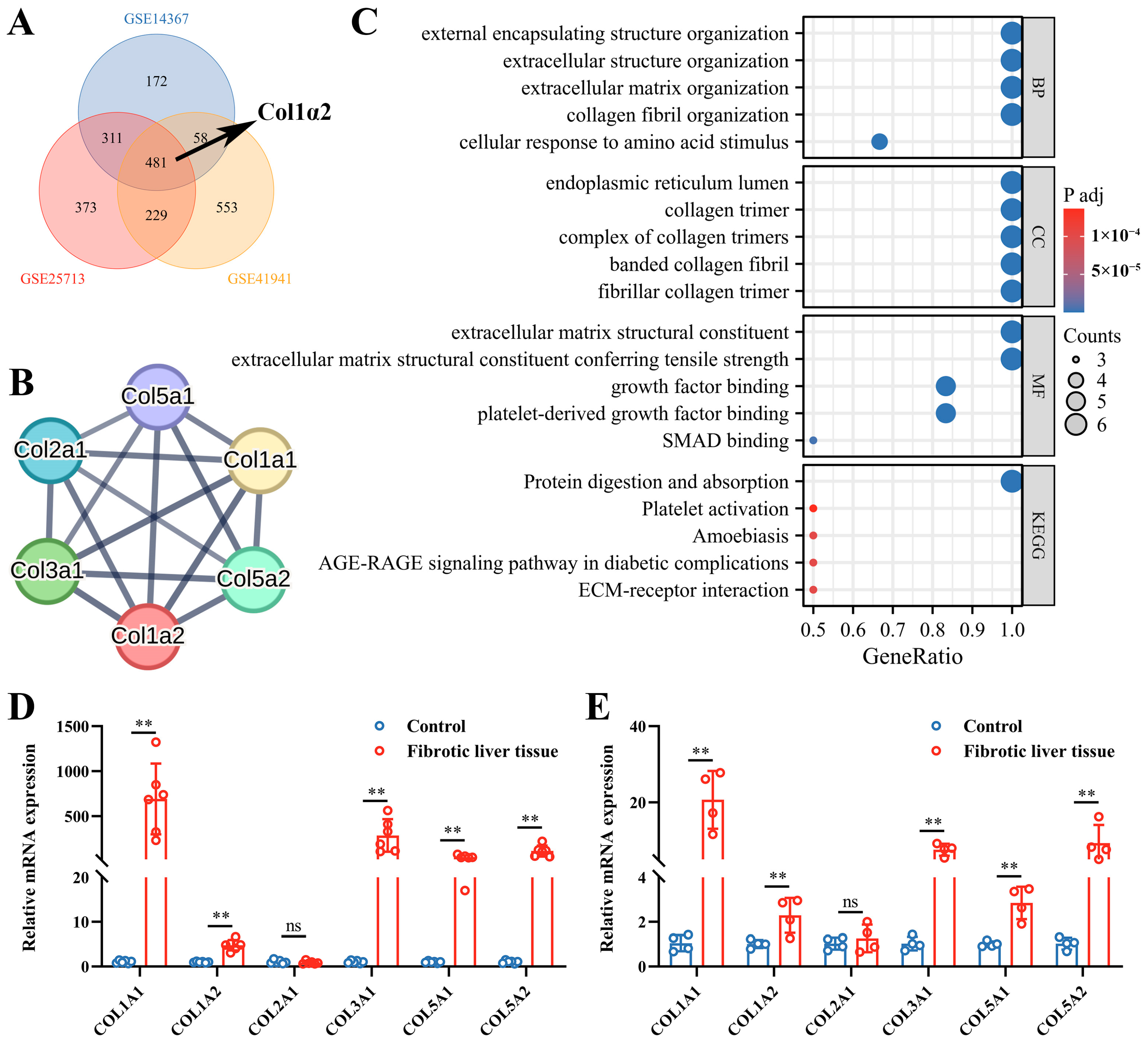
3.1. Col1α2 Expression and Its Functional Interactions in Fibrosis Datasets
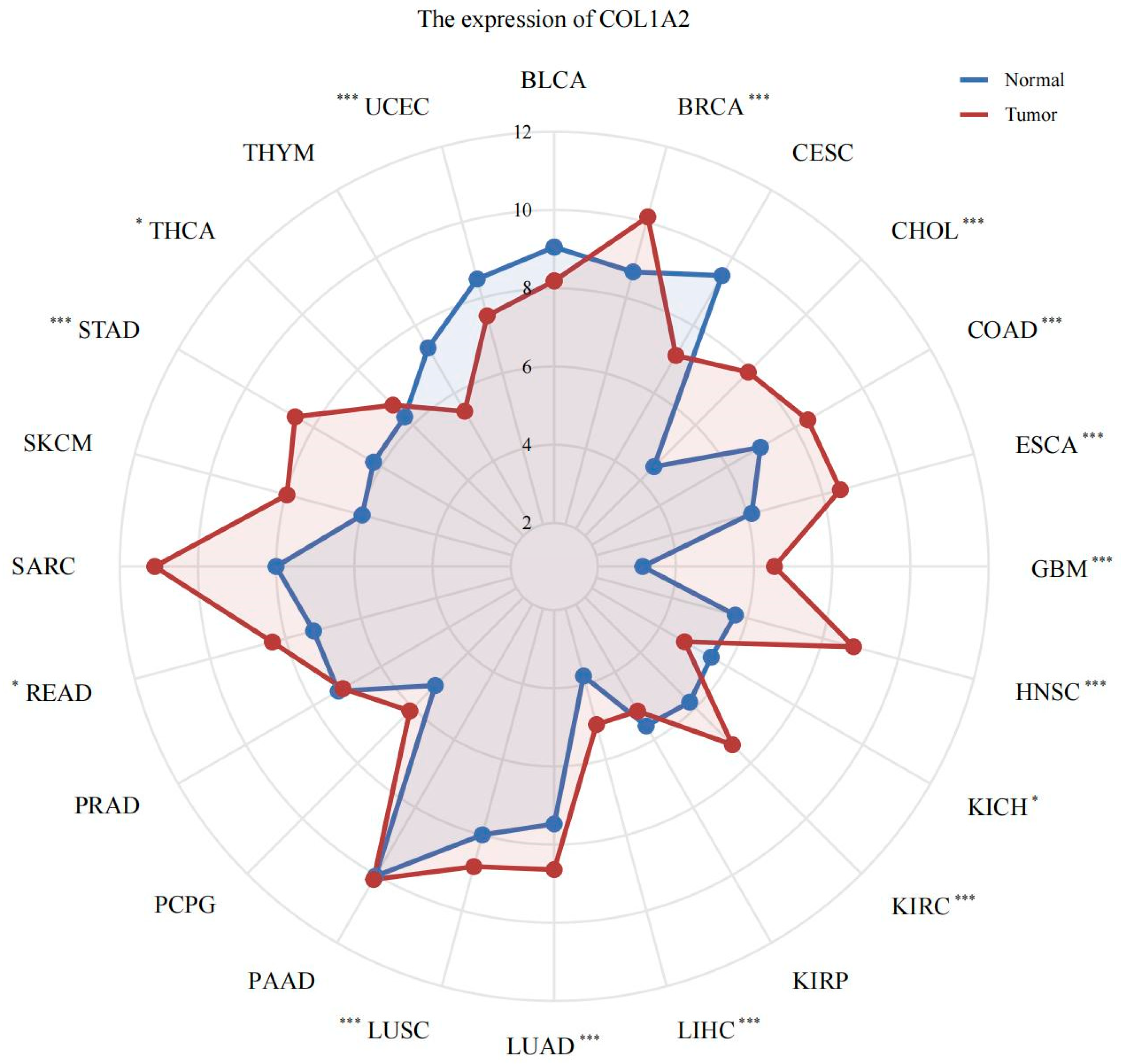
To investigate the significant role of Col1α2 in schistosomiasis-induced liver fibrosis, three schistosome-induced fibrosis datasets were selected and analyzed the expression and potential biological functions of Col1α2. The UMAP results showed clear clustering differences among the liver samples in each dataset, making them suitable for further analysis (Supplementary Figure S1A–C). GSEA enrichment analysis indicated that pathways related to collagen biosynthesis, collagen degradation, collagen formation, and extracellular matrix organization pathways were significantly enriched in the infected samples of all three datasets (Supplementary Figure S1A–C). Col1α2 was found to be among the upregulated genes across all datasets (Figure 1A).
Subsequently, the PPI network of Col1α2 was analyzed, highlighting Col1α2 and its interacting molecules (Col1α1, Col3α1, Col5α1, and Col5α2) as central nodes (Figure 1B). GO enrichment analysis demonstrated that Col1α2 and its interacting molecules were predominantly involved in extracellular matrix organization and collagen fibril organization (Figure 1C). In both schistosome-induced liver fibrosis models and CCL4-induced liver fibrosis model, quantitative mRNA expression analysis showed that, except for Col2α1, the levels of Col1α2 and other collagen genes in fibrotic liver tissues were significantly higher when compared to control groups, further supporting their role in liver fibrosis (Figure 1D,E).

3.2. Pan-Cancer Analysis of col1α2 Expression

The expression of Col1α2 across various cancer types was examined using the TCGA pan-cancer data. The radar chart indicated that Col1α2 was upregulated in multiple cancer types, including LIHC (liver hepatocellular carcinoma), compared to normal tissues (Figure 2). This widespread overexpression in various cancers suggests the potential significance of Col1α2 as a biomarker and therapeutic target.

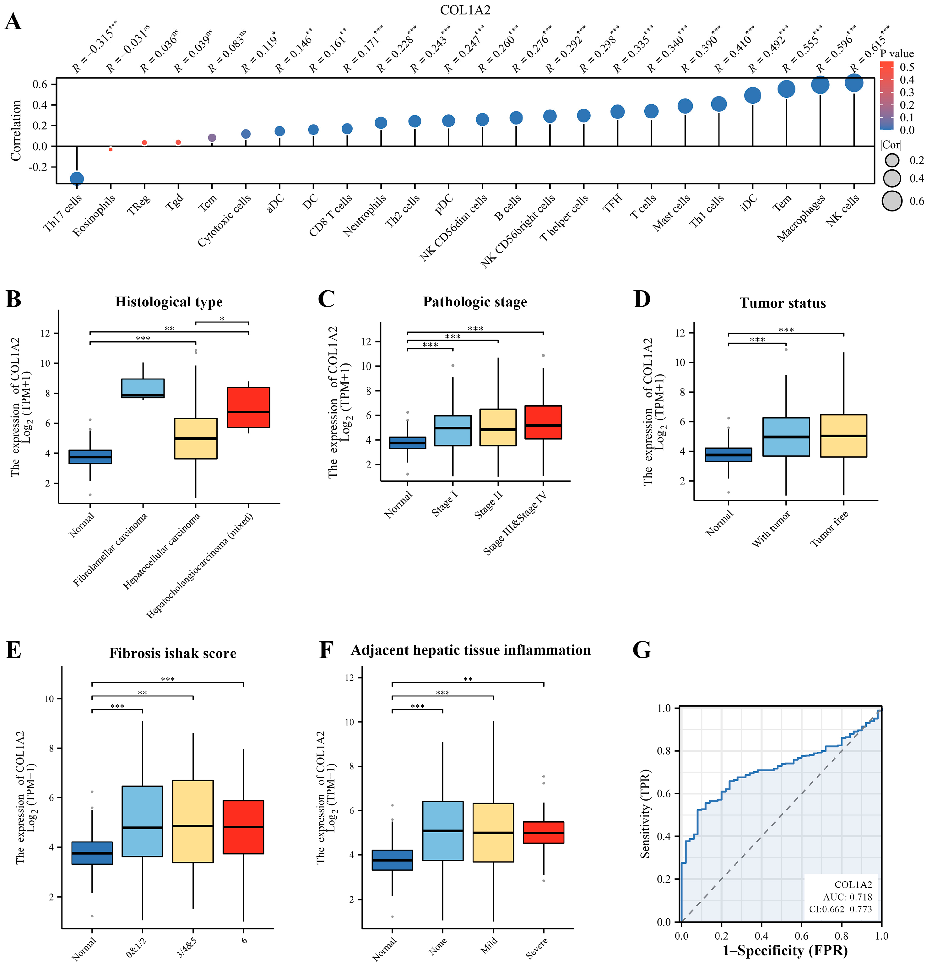
3.3. Immune Infiltration and Clinical Correlation Analysis of Col1α2 in HCC

To understand the role of Col1α2 in the tumor microenvironment of HCC, the correlation between Col1α2 expression and immune cell infiltration was analyzed using TCGA data. Results indicated that Col1α2 expression positively correlated with the infiltration of immune cells such as natural killer (NK) cells, macrophages, and T effector memory (Tem) cells which suggests that Col1α2 may play a role in modulating the immune response within the HCC microenvironment, potentially impacting tumor progression (Figure 3A).
Clinical correlation analysis further demonstrated the significance of Col1α2 expression in HCC. Results indicated that the Col1α2 expression was higher in patient with fibrolamellar carcinoma or hepatocholangiocarcinoma (Figure 3B), and patients with later stages of HCC also exhibited increased Col1α2 levels, indicating its potential role in cancer progression (Figure 3C). Additionally, Col1α2 expression was significantly higher in both tumor tissues and non-tumorous tissues compared to healthy liver tissues, suggesting that Col1α2 is involved in both the tumorigenic process and the surrounding microenvironment, potentially contributing to the overall pathophysiology of HCC (Figure 3D). Moreover, Col1α2 expression was positively correlated with the fibrosis Ishak score, suggesting that it could be a marker for liver fibrosis severity in HCC patients (Figure 3E). Increased Col1α2 levels were also observed in patients with significant inflammation in adjacent hepatic tissues, which might indicate an ongoing inflammatory response contributing to tumor progression (Figure 3F).
Besides, to evaluate the diagnostic value of Col1α2 in HCC, a ROC curve analysis was performed, yielding an AUC value of 0.718, which suggests that Col1α2 has a moderate diagnostic value for HCC, indicating its potential utility as a biomarker for identifying patients at risk for HCC or for monitoring disease progression.
Overall, these findings highlight the multifaceted role of Col1α2 in HCC, including its involvement in immune cell infiltration, association with critical clinical parameters, and potential as a diagnostic biomarker. The significant correlations with immune infiltration and clinical features emphasize the importance of Col1α2 not only in the pathogenesis of HCC but also in its potential application in clinical diagnostics and prognostics.

3.4. Inhibition of Hepatoma Cells Growth by sja-let-7 Targeting Col1α2

In our previous work, it was demonstrated that sja-let-7 can bind to the 3′ UTR of Col1α2 and inhibit its expression [11]. Given the crucial role of Col1α2 in HCC, the effects of sja-let-7 on Col1α2 expression and HCC cell proliferation in vitro were investigated. First, Col1α2 expression levels across various hepatoma cell lines were compared using the HPA database, and the SNU387 (high Col1α2 expression) and the Li7 (low Col1α2 expression) cell line, were selected for further experiments (Figure 4A). The expression levels of Col1α2 in these cell lines were validated using qPCR, and the results were consistent with the HPA database (Figure 4B).
Next, sja-let-7 mimics were transfected into SNU387 and Li7 cells to increase the sja-let-7 levels, which was then verified by qPCR. Additionally, mimics labeled with FAM fluorescence were used for transfection to visually confirm transfection efficiency. Results shown that both SNU387 and Li7 cells exhibited numerous green fluorescent spots after transfection with FAM-labeled mimics (Supplementary Figure S2A), and sja-let-7 levels were significantly elevated, while Col1α2 expression was significantly reduced, indicating successful transfection and inhibition of the target gene (Figure 4C,D and Figure S2B–D). However, due to the relatively low expression of Col1α2 and lower transfection efficiency in the Li7 cell line, subsequent experiments were conducted using only the SNU387 cell line.
Subsequently, the CCK-8 assay presented that sja-let-7 mimics significantly reduced the proliferation of SNU387 cells (Figure 4E). Colony formation assays were conducted, demonstrating that sja-let-7 inhibited colony formation in SNU387 cells to a greater extent than in the NC group or Mock group (Figure 4F,G). Additionally, we also showed that transfection of the sja-let-7 mimics significantly suppressed cell migration, as assessed by the wound healing assay when compared with the NC group or Mock group (Figure 4H,I). Given that the TGF-β/SMAD signaling pathway is closely associated with cell proliferation and has been implicated in the regulation of the sja-let-7/COL1A2 axis, we examined the impact of sja-let-7 on this pathway [11]. Our results revealed that sja-let-7 mimics led to a significant downregulation of TGF-β, TGF-βR1, and SMAD2, while the antagonist SMAD7 was upregulated, suggesting that the anti-tumor effects of sja-let-7 may involve modulation of the TGF-β/SMAD pathway (Figure 4J).
In summary, these data indicated that the growth, migration, and colony formation of human hepatoma cells were inhibited by sja-let-7, suggesting that sja-let-7 has the potential to be a tumor suppressor.

4. Discussion

Col1α2 is a critical component of the extracellular matrix, playing a significant role in maintaining the structural integrity of various tissues [28]. It is involved in the formation of collagen fibrils, which provide tensile strength to tissues and are crucial for tissue repair and remodeling [29,30]. In mammals, dysregulation of Col1α2 expression is a hallmark of fibrotic diseases [31]. Overproduction of Col1α2 leads to excessive collagen deposition, contributing to the development of fibrosis in organs such as the liver, lungs, kidneys, and heart and can progress to organ failure [31]. Hence, understanding the regulation of Col1α2 is essential for developing therapeutic strategies to combat fibrotic diseases. The ECM, including components like Col1α2, plays a dual role in cancer biology [32]. While it acts as a physical barrier that can suppress tumor cell migration and limit immune cell infiltration, its remodeling by tumor cells can facilitate tumor growth and metastasis [33]. Understanding this dynamic interplay is essential for developing therapeutic strategies targeting ECM remodeling in HCC treatment.
Liver fibrosis, resulting from chronic liver injury, is a critical precursor to HCC [34]. Studies have shown that high Col1α2 expression levels correlate with poor prognosis and lower overall survival rates in HCC patients [35]. Additionally, increased Col1α2 expression is often associated with higher tumor grade and stage, as well as enhanced metastatic potential [36]. These associations highlight the role of Col1α2 not only as a biomarker for fibrosis but also as a critical factor in HCC pathogenesis. In the present study, the role of Col1α2 in the tumor microenvironment, particularly in relation to immune cell infiltration, was thoroughly analyzed using TCGA data. Col1α2 expression positively correlated with the infiltration of various immune cells, including NK cells (R = 0.615, p < 0.001), macrophages (R = 0.596, p < 0.001), and Tem cells (R = 0.555, p < 0.001). NK cells are known for the ability to recognize and kill tumor cells without prior sensitization, making them critical for early defense against tumors [37]. Macrophages can exhibit both pro-tumor and anti-tumor activities depending on their polarization state, with tumor-associated macrophages often promoting tumor growth and metastasis [38]. Tem cells play a key role in immune memory and can mount rapid responses to tumor antigens [39]. The high correlation between Col1α2 expression and these immune cells suggests that Col1α2 may potentially impacting tumor progression by influencing immune cell infiltration and activity within the HCC microenvironment.
Schistosome-induced liver fibrosis is a significant health issue worldwide, caused primarily by S. japonicum infection [4]. This chronic parasitic disease leads to the formation of granulomas and subsequent fibrosis in the liver due to the immune response against schistosome eggs [40]. Although liver fibrosis is closely linked to the development of HCC, epidemiological data indicate that patients with schistosome-induced liver fibrosis do not exhibit a significantly higher incidence of HCC compared to those with other causes of liver fibrosis [6]. Previous studies have revealed that schistosome-derived EVs contain miRNAs that can modulate the host’s fibrotic response [10]. For example, sja-miR-71 has been shown to alleviate fibrosis by targeting host genes involved in collagen synthesis, suggesting that schistosomes might mitigate disease progression through the delivery of regulatory miRNAs, enabling their prolonged survival within the host [9].
Based on the hypothesis that schistosome-derived miRNAs could play a role in modulating fibrosis and potentially have anti-tumor effect, our previous work identified sja-let-7 as a miRNA with anti-fibrotic properties, targeting the 3′UTR of Col1α2, thereby inhibiting its expression and alleviating fibrosis [11]. Mammalian let-7 family members are well-known for their tumor suppressor functions and their role in various cancers [41,42]. Specifically, let-7g has been shown to reduce proliferation and migration of HCC cells by inhibiting Col1α2 expression, which indicates the therapeutic potential of the let-7/Col1α2 signaling axis in HCC [36]. To investigate whether sja-let-7, a member of the let-7 family from schistosomes, has similar effects, we conducted experiments that confirmed its ability to inhibit HCC cell proliferation and migration through Col1α2. Our results showed that sja-let-7 significantly reduced HCC cell proliferation by 26.02% (p < 0.01) and migration by 42.25% (p < 0.01) compared to the NC group. Additionally, we observed a corresponding decrease in Col1α2 mRNA levels by 61.78% (p < 0.01), supporting the hypothesis that sja-let-7 exerts its anti-tumor effects via the let-7/Col1α2 signaling axis. Given the well-established connection between the TGF-β/SMAD signaling pathway and cell proliferation [11], our findings indicated that sja-let-7 mimics not only downregulate TGF-β, TGF-βR1, and SMAD2 but also upregulate the antagonist SMAD7, suggesting a potential involvement of the TGF-β/SMAD pathway in regulating sja-let-7/Col1α2 axis, which may contribute to anti-tumor activities.
The “hygiene hypothesis” posits that exposure to helminths and their derivatives may have beneficial effects on immune regulation and disease prevention [43]. Increasing attention is being given to the therapeutic potential of helminth-derived substances. Helminthic therapy has been explored for autoimmune diseases and allergies, with some promising results [44,45]. Our study provides a promising example, demonstrating that sja-let-7 can inhibit HCC cell proliferation and migration by targeting Col1α2. However, our study has several limitations that warrant further investigation. First, the findings are primarily based on in vitro experiments, which may not fully explain the complexity of the tumor microenvironment in vivo. Future research should focus on validating these results in animal models of HCC to determine if the anti-tumor effects of sja-let-7 observed in vitro can be replicated in a living organism. Additionally, the potential off-target effects of sja-let-7 were not extensively explored, and further studies should employ comprehensive transcriptomic or proteomic analyses to identify additional targets and understand its broader impact. Moreover, the therapeutic potential of combining sja-let-7 with existing cancer therapies remains unexplored, and investigating such combinations could enhance treatment efficacy [46]. Finally, a deeper understanding of the molecular mechanisms underlying sja-let-7′s anti-tumor effects will be crucial for developing effective treatments, including elucidating the signaling pathways involved and exploring its regulation within schistosomes.
In summary, this study highlights the significant role of Col1α2 in liver fibrosis and HCC, the potential therapeutic application of sja-let-7, and the broader implications of helminth-derived miRNAs in disease modulation. These findings open new avenues for research and therapeutic development in HCC and other fibrosis-related diseases.

Supplementary Materials

The following supporting information can be downloaded at: https://www.mdpi.com/article/10.3390/genes15091165/s1, Figure S1: Overview of schistosome-induced fibrosis datasets UMAP analysis, GSEA enrichment pathways, and gene expression details of GSE14367. (A), GSE25713 (B) and GSE41941 (C) datasets. Figure S2: Transfection efficiency and validation. (A) Fluorescence microscopy observation of SNU387 and Li7 cells after transfection with FAM-labeled NC/sja-let-7 mimics (Scale bar = 200 μm). (B) The binding site of sja-let-7 on the 3′UTR of Col1α2; (C) Sja-let-7 transfection efficiency in Li7 cell line (n = 3); (D) Relative expression of Col1α2 in Li7 cell line (n = 3). All graph data are expressed as the mean ± SD of at least three biological replicates per group. * p< 0.05, ** p< 0.01, ns, not significant. Table S1: The sequence of miRNA mimics; Table S2: Primers used in the experiment.

Author Contributions

Conceptualization, Y.J. and H.Z.; methodology, H.Z., B.D. and D.Z.; software, H.Z.; validation, H.Z.; formal analysis, H.Z.; investigation, Y.J. and H.Z.; resources, Y.J.; data curation, Y.J.; writing—original draft preparation, H.Z.; writing—review and editing, Y.J.; visualization, Z.F.; supervision, J.L. and G.G.; project administration, Y.J.; funding acquisition, Y.J. All authors have read and agreed to the published version of the manuscript.

Funding

This research was funded by the Agricultural Science and Technology Innovation Program (ASTIP) (no. CAAS-ASTIP-2021-SHVRI), the Natural Science Foundation of Shanghai (no. 20ZR1469300) and partially funded by the Key Laboratory of Veterinary Etiological Biology, Ministry of Agricultural and Rural Affairs, China (no. XKQ2024002). The funders had no role in study design, data collection and analysis, decision to publish, or preparation of the manuscript.

Institutional Review Board Statement

All protocols involved in the animal experiments were performed according to the guidelines of the Committee for the Care and Use of Laboratory Animals of the Shanghai Veterinary Research Institute (Shanghai, China, permit no. SYXK-20160010) and were approved by the Ethics and Animal Welfare Committee of the Shanghai Veterinary Research Institute, Chinese Academy of Agricultural Sciences (Shanghai, China, experiment no. SV-20230505-03).

Informed Consent Statement

Not applicable.

Data Availability Statement

The datasets presented in this study can be found in online repositories. The names of the repository/repositories and accession number(s) can be found in the article. Further inquiries can be directed to the corresponding author.

Acknowledgments

We thank Changlong Liu and Lilei Lv from Shanghai Veterinary Research Institute, Chinese Academy of Agricultural Sciences for donation of the SNU387 and Li7 cell lines. We also thank Ke Lu and Hao Li from Shanghai Veterinary Research Institute of Chinese Academy Agricultural of Sciences for their contribution to the parasite maintain, cercariae isolation and animal experiments.

Conflicts of Interest

The authors declare no conflicts of interest.

References

  1. Baglieri, J.; Brenner, D.A.; Kisseleva, T. The Role of Fibrosis and Liver-Associated Fibroblasts in the Pathogenesis of Hepatocellular Carcinoma. Int. J. Mol. Sci. 2019, 20, 1723. [Google Scholar] [CrossRef] [PubMed]
  2. Toh, M.R.; Wong, E.Y.T.; Wong, S.H.; Ng, A.W.T.; Loo, L.H.; Chow, P.K.; Ngeow, J. Global Epidemiology and Genetics of Hepatocellular Carcinoma. Gastroenterology 2023, 164, 766–782. [Google Scholar] [CrossRef] [PubMed]
  3. Sidali, S.; Trépo, E.; Sutter, O.; Nault, J.C. New concepts in the treatment of hepatocellular carcinoma. United Eur. Gastroenterol. J. 2022, 10, 765–774. [Google Scholar] [CrossRef]
  4. Colley, D.G.; Bustinduy, A.L.; Secor, W.E.; King, C.H. Human schistosomiasis. Lancet 2014, 383, 2253–2264. [Google Scholar] [CrossRef] [PubMed]
  5. He, X.; Pan, W. Host-parasite interactions mediated by cross-species microRNAs. Trends Parasitol 2022, 38, 478–488. [Google Scholar] [CrossRef] [PubMed]
  6. Tokoro, Y.; Koganezawa, S. Metrical and histopathological studies on Japanese schistosomiasis of the colon associated with cancer. Nihon Shokakibyo Gakkai Zasshi—Jpn. J. Gastro-Enterol. 1976, 73, 972–985. [Google Scholar]
  7. Inaba, Y.; Maruchi, N.; Matsuda, M.; Yoshihara, N.; Yamamoto, S. A case-control study on liver cancer with special emphasis on the possible aetiological role of schistosomiasis. Int. J. Epidemiol. 1984, 13, 408–412. [Google Scholar] [CrossRef]
  8. El-Tonsy, M.M.; Hussein, H.M.; Helal Tel, S.; Tawfik, R.A.; Koriem, K.M.; Hussein, H.M. Schistosoma mansoni infection: Is it a risk factor for development of hepatocellular carcinoma? Acta Trop. 2013, 128, 542–547. [Google Scholar] [CrossRef]
  9. Wang, L.; Liao, Y.; Yang, R.; Yu, Z.; Zhang, L.; Zhu, Z.; Wu, X.; Shen, J.; Liu, J.; Xu, L.; et al. Sja-miR-71a in Schistosome egg-derived extracellular vesicles suppresses liver fibrosis caused by schistosomiasis via targeting semaphorin 4D. J. Extracell. Vesicles 2020, 9, 1785738. [Google Scholar] [CrossRef]
  10. He, X.; Wang, Y.; Fan, X.; Lei, N.; Tian, Y.; Zhang, D.; Pan, W. A schistosome miRNA promotes host hepatic fibrosis by targeting transforming growth factor β receptor III. J. Hepatol. 2020, 72, 519–527. [Google Scholar] [CrossRef]
  11. Zhong, H.; Dong, B.; Zhu, D.; Fu, Z.; Liu, J.; Jin, Y. Sja-let-7 suppresses the development of liver fibrosis via Schistosoma japonicum extracellular vesicles. PLoS Pathog. 2024, 20, e1012153. [Google Scholar] [CrossRef]
  12. Hu, C.; Zhu, S.; Wang, J.; Lin, Y.; Ma, L.; Zhu, L.; Jiang, P.; Li, Z.; Pan, W. Schistosoma japonicum MiRNA-7-5p Inhibits the Growth and Migration of Hepatoma Cells via Cross-Species Regulation of S-Phase Kinase-Associated Protein 2. Front. Oncol. 2019, 9, 175. [Google Scholar] [CrossRef]
  13. Hu, C.; Li, Y.; Pan, D.; Wang, J.; Zhu, L.; Lin, Y.; Zhu, S.; Pan, W. A Schistosoma japonicum MicroRNA Exerts Antitumor Effects Through Inhibition of Both Cell Migration and Angiogenesis by Targeting PGAM1. Front. Oncol. 2021, 11, 652395. [Google Scholar] [CrossRef] [PubMed]
  14. Lin, Y.; Zhu, S.; Hu, C.; Wang, J.; Jiang, P.; Zhu, L.; Li, Z.; Wang, S.; Zhang, Y.; Xu, X.; et al. Cross-Species Suppression of Hepatoma Cell Growth and Migration by a Schistosoma japonicum MicroRNA. Mol. Ther. Nucleic Acids 2019, 18, 400–412. [Google Scholar] [CrossRef]
  15. Jiang, P.; Wang, J.; Zhu, S.; Hu, C.; Lin, Y.; Pan, W. Identification of a Schistosoma japonicum MicroRNA That Suppresses Hepatoma Cell Growth and Migration by Targeting Host FZD4 Gene. Front. Cell. Infect. Microbiol. 2022, 12, 786543. [Google Scholar] [CrossRef] [PubMed]
  16. Zhong, H.; Dong, B.; Zhu, D.; Li, H.; Lu, K.; Fu, Z.; Liu, J.; Jin, Y. Sja-Let-7 Attenuates Carbon Tetrachloride-Induced Liver Fibrosis in a Mouse Model via Col1α2. Biology 2023, 12, 1465. [Google Scholar] [CrossRef] [PubMed]
  17. Edgar, R.; Domrachev, M.; Lash, A.E. Gene Expression Omnibus: NCBI gene expression and hybridization array data repository. Nucleic Acids Res. 2002, 30, 207–210. [Google Scholar] [CrossRef]
  18. Subramanian, A.; Tamayo, P.; Mootha, V.K.; Mukherjee, S.; Ebert, B.L.; Gillette, M.A.; Paulovich, A.; Pomeroy, S.L.; Golub, T.R.; Lander, E.S.; et al. Gene set enrichment analysis: A knowledge-based approach for interpreting genome-wide expression profiles. Proc. Natl. Acad. Sci. USA 2005, 102, 15545–15550. [Google Scholar] [CrossRef]
  19. Conesa, A.; Götz, S.; García-Gómez, J.M.; Terol, J.; Talón, M.; Robles, M. Blast2GO: A universal tool for annotation, visualization and analysis in functional genomics research. Bioinformatics 2005, 21, 3674–3676. [Google Scholar] [CrossRef]
  20. Kanehisa, M.; Goto, S. KEGG: Kyoto encyclopedia of genes and genomes. Nucleic Acids Res. 2000, 28, 27–30. [Google Scholar] [CrossRef]
  21. Szklarczyk, D.; Kirsch, R.; Koutrouli, M.; Nastou, K.; Mehryary, F.; Hachilif, R.; Gable, A.L.; Fang, T.; Doncheva, N.T.; Pyysalo, S.; et al. The STRING database in 2023: Protein-protein association networks and functional enrichment analyses for any sequenced genome of interest. Nucleic Acids Res. 2023, 51, D638–D646. [Google Scholar] [CrossRef] [PubMed]
  22. Weinstein, J.N.; Collisson, E.A.; Mills, G.B.; Shaw, K.R.; Ozenberger, B.A.; Ellrott, K.; Shmulevich, I.; Sander, C.; Stuart, J.M. The Cancer Genome Atlas Pan-Cancer analysis project. Nat. Genet. 2013, 45, 1113–1120. [Google Scholar] [CrossRef] [PubMed]
  23. Bindea, G.; Mlecnik, B.; Tosolini, M.; Kirilovsky, A.; Waldner, M.; Obenauf, A.C.; Angell, H.; Fredriksen, T.; Lafontaine, L.; Berger, A.; et al. Spatiotemporal dynamics of intratumoral immune cells reveal the immune landscape in human cancer. Immunity 2013, 39, 782–795. [Google Scholar] [CrossRef] [PubMed]
  24. Zhou, X.; Zhang, Y.; Wang, N. Systematic identification of key extracellular proteins as the potential biomarkers in lupus nephritis. Front. Immunol. 2022, 13, 915784. [Google Scholar] [CrossRef]
  25. Pontén, F.; Jirström, K.; Uhlen, M. The Human Protein Atlas--a tool for pathology. J. Pathol. 2008, 216, 387–393. [Google Scholar] [CrossRef]
  26. Rio, D.C.; Ares, M., Jr.; Hannon, G.J.; Nilsen, T.W. Purification of RNA using TRIzol (TRI reagent). Cold Spring Harb. Protoc. 2010, 2010, pdb.prot5439. [Google Scholar] [CrossRef]
  27. Livak, K.J.; Schmittgen, T.D. Analysis of relative gene expression data using real-time quantitative PCR and the 2(-Delta Delta C(T)) Method. Methods 2001, 25, 402–408. [Google Scholar] [CrossRef]
  28. Sawamura, S.; Makino, K.; Ide, M.; Shimada, S.; Kajihara, I.; Makino, T.; Jinnin, M.; Fukushima, S. Elevated α 1(I) to α 2(I) Collagen Ratio in Dermal Fibroblasts Possibly Contributes to Fibrosis in Systemic Sclerosis. Int. J. Mol. Sci. 2022, 23, 6811. [Google Scholar] [CrossRef]
  29. Le Saux, O.; Teeters, K.; Miyasato, S.; Choi, J.; Nakamatsu, G.; Richardson, J.A.; Starcher, B.; Davis, E.C.; Tam, E.K.; Jourdan-Le Saux, C. The role of caveolin-1 in pulmonary matrix remodeling and mechanical properties. Am. J. Physiol.-Lung Cell. Mol. Physiol. 2008, 295, L1007–L1017. [Google Scholar] [CrossRef]
  30. Shoulders, M.D.; Raines, R.T. Collagen structure and stability. Annu. Rev. Biochem. 2009, 78, 929–958. [Google Scholar] [CrossRef]
  31. Ghosh, A.K. Factors involved in the regulation of type I collagen gene expression: Implication in fibrosis. Exp. Biol. Med. 2002, 227, 301–314. [Google Scholar] [CrossRef] [PubMed]
  32. Cho, Y.; Doh, J. The extracellular matrix in solid tumor immunotherapy. Trends Immunol. 2024, S1471-4906(24)00184-4. [Google Scholar] [CrossRef]
  33. Li, X.; Shepard, H.M.; Cowell, J.A.; Zhao, C.; Osgood, R.J.; Rosengren, S.; Blouw, B.; Garrovillo, S.A.; Pagel, M.D.; Whatcott, C.J.; et al. Parallel Accumulation of Tumor Hyaluronan, Collagen, and Other Drivers of Tumor Progression. Clin. Cancer Res. Off. J. Am. Assoc. Cancer Res. 2018, 24, 4798–4807. [Google Scholar] [CrossRef] [PubMed]
  34. Affo, S.; Yu, L.X.; Schwabe, R.F. The Role of Cancer-Associated Fibroblasts and Fibrosis in Liver Cancer. Annu. Rev. Pathol. 2017, 12, 153–186. [Google Scholar] [CrossRef]
  35. Romualdo, G.R.; Grassi, T.F.; Goto, R.L.; Tablas, M.B.; Bidinotto, L.T.; Fernandes, A.A.H.; Cogliati, B.; Barbisan, L.F. An integrative analysis of chemically-induced cirrhosis-associated hepatocarcinogenesis: Histological, biochemical and molecular features. Toxicol. Lett. 2017, 281, 84–94. [Google Scholar] [CrossRef] [PubMed]
  36. Ji, J.; Zhao, L.; Budhu, A.; Forgues, M.; Jia, H.-L.; Qin, L.-X.; Ye, Q.-H.; Yu, J.; Shi, X.; Tang, Z.-Y.; et al. Let-7g targets collagen type I α2 and inhibits cell migration in hepatocellular carcinoma. J. Hepatol. 2010, 52, 690–697. [Google Scholar] [CrossRef]
  37. Zhou, Y.; Cheng, L.; Liu, L.; Li, X. NK cells are never alone: Crosstalk and communication in tumour microenvironments. Mol. Cancer 2023, 22, 34. [Google Scholar] [CrossRef]
  38. Qian, B.Z.; Pollard, J.W. Macrophage diversity enhances tumor progression and metastasis. Cell 2010, 141, 39–51. [Google Scholar] [CrossRef]
  39. Liu, Q.; Sun, Z.; Chen, L. Memory T cells: Strategies for optimizing tumor immunotherapy. Protein Cell 2020, 11, 549–564. [Google Scholar] [CrossRef]
  40. Andrade, Z.A. Schistosomiasis and liver fibrosis. Parasite Immunol. 2009, 31, 656–663. [Google Scholar] [CrossRef]
  41. De Santis, C.; Gotte, M. The Role of microRNA Let-7d in Female Malignancies and Diseases of the Female Reproductive Tract. Int. J. Mol. Sci. 2021, 22, 7359. [Google Scholar] [CrossRef]
  42. Lee, H.; Han, S.; Kwon, C.S.; Lee, D. Biogenesis and regulation of the let-7 miRNAs and their functional implications. Protein Cell 2016, 7, 100–113. [Google Scholar] [CrossRef]
  43. Pfefferle, P.I.; Keber, C.U.; Cohen, R.M.; Garn, H. The Hygiene Hypothesis—Learning from but Not Living in the Past. Front. Immunol. 2021, 12, 635935. [Google Scholar] [CrossRef] [PubMed]
  44. Acharya, S.; Da’dara, A.A.; Skelly, P.J. Schistosome immunomodulators. PLoS Pathog. 2021, 17, e1010064. [Google Scholar] [CrossRef]
  45. Mu, Y.; McManus, D.P.; Hou, N.; Cai, P. Schistosome Infection and Schistosome-Derived Products as Modulators for the Prevention and Alleviation of Immunological Disorders. Front. Immunol. 2021, 12, 619776. [Google Scholar] [CrossRef] [PubMed]
  46. Keck, J.; Chambers, J.P.; Yu, J.-J.; Cheng, X.; Christenson, L.K.; Guentzel, M.N.; Gupta, R.; Arulanandam, B.P. Modulation of Immune Response to Chlamydia muridarum by Host miR-135a. Front. Cell. Infect. Microbiol. 2021, 11, 638058. [Google Scholar] [CrossRef] [PubMed]
Figure 1. Col1α2 expression and biological function in schistosome-induced liver fibrosis. (A) Venn diagram of upregulated genes in the GSE14367, GSE25713 and GSE41941 datasets; (B) PPI network analysis of Col1α2; (C) GO and KEGG enrichment analysis of Col1α2 and its interacting molecules; (D) Relative mRNA expression of Col1α2 and its interacting molecules in schistosome-induced liver fibrosis model (n = 6); (E) Relative mRNA expression of Col1α2 and its interacting molecules in CCL4-induced liver fibrosis model (n = 4). All graph data are expressed as the mean ± SD of at least three biological replicates per group. ** p < 0.01, ns, not significant.
Genes 15 01165 g001
Figure 2. Radar chart showing the expression levels of Col1α2 across various cancer types using TCGA pan-cancer data. * p < 0.05, *** p < 0.001.
Genes 15 01165 g002
Figure 3. Immune infiltration and clinical correlation analysis of Col1α2 in HCC. (A) Correlation analysis of Col1α2 expression with various immune cell infiltrations in HCC; (BF) Clinical correlation analysis of advanced histological types, pathological stages, tumor status, fibrosis Ishak scores and adjacent hepatic tissue inflammation associated with Col1α2 expression; (G) ROC curve analysis of Col1α2 expression in HCC patients. * p < 0.05, ** p < 0.01, *** p < 0.001, ns, not significant.
Genes 15 01165 g003
Figure 4. Inhibition of hepatoma cells growth by sja-let-7 targeting Col1α2. (A) Comparison of Col1α2 expression levels across various hepatoma cell lines using the HPA database; (B) Validation of Col1α2 expression levels in SNU387 and Li7 cell lines using qPCR (n = 3); (C) Sja-let-7 transfection efficiency in SNU387 cell line (n = 3); (D) Relative expression of Col1α2 in SNU387 cell line (n = 3); (E) CCK-8 assay of SNU387 cell line (n = 6); (F) Representative clone photo of SNU387 cells (Scale bar = 200 μm); (G) Number of clones (n = 3); (H) Representative cell wound photo of SNU387 cells (100×); (I) Wound healing rate of SNU387 cells (n = 3); (J) Relative expression of TGF-β, SMAD2, SMAD7 and TGF-βRI in SNU387 cell line (n = 3). All graph data are expressed as the mean ± SD of at least three biological replicates per group. * p < 0.05, ** p < 0.01, ns, not significant, compared to Mock; ## p < 0.01, compared to NC mimics.
Genes 15 01165 g004
Disclaimer/Publisher’s Note: The statements, opinions and data contained in all publications are solely those of the individual author(s) and contributor(s) and not of MDPI and/or the editor(s). MDPI and/or the editor(s) disclaim responsibility for any injury to people or property resulting from any ideas, methods, instructions or products referred to in the content.

