Article

Genome-Wide Identification and Tissue-Specific Expression Profiling of Goji CER Gene Family

1
Gansu Analysis and Research Center, Lanzhou 730070, China
2
College of Forestry, Gansu Agricultural University, Lanzhou 730070, China
3
Wolfberry Harmless Cultivation Engineering Research Center of Gansu Province, Lanzhou 730070, China
4
College of Horticulture, Gansu Agricultural University, Lanzhou 730070, China
*
Author to whom correspondence should be addressed.
Genes 2025, 16(11), 1257; https://doi.org/10.3390/genes16111257 (registering DOI)
Submission received: 23 September 2025 / Revised: 17 October 2025 / Accepted: 23 October 2025 / Published: 24 October 2025
(This article belongs to the Section Plant Genetics and Genomics)

Abstract

Background: Goji berry, known as a “superfood”, is widely distributed in northwest China and possesses significant medicinal and health value. The CER gene family serves as a key regulator of cuticular wax synthesis, which plays important roles in enhancing plant drought resistance and disease tolerance. However, genome-wide identification of the goji CER gene family and its expression analysis across different varieties and organs have not been reported. Methods: Based on SEM observations and wax load measurements, this study identified CER gene family members using whole genome data of the goji berry. Representative genes were selected and their expression patterns in different varieties and organs were validated by qRT–PCR. Results: The stem wax load was significantly higher than that in other organs, while the leaf wax load of ‘Ningqi I’ goji was significantly higher than that in other varieties, consistent with SEM observations. A total of 113 CER gene family members were identified in goji berry, which were unevenly distributed on 12 chromosomes. The goji CER proteins mainly localized in the cell membrane, cytoplasm, chloroplast, and nucleus and clustered into five subfamilies. Ten conserved motifs were identified in CER proteins, with Motif5 and Motif7 being the most widely distributed. The LbaCER10-1 gene contained the highest number of exons (39). Cis-acting elements related to light-responsiveness, MeJA-responsiveness, and ABA-responsiveness showed high frequencies. Goji berry shared more homologous CER genes with tomato, potato, and tobacco than with Arabidopsis, with chr3 and chr9 being most conserved while chr7 showed greater variation. Conclusions: Integrating SEM, wax load, and qRT–PCR results, LbaCER1-1 was identified as a candidate gene responsible for the higher wax load on goji stems, while LbaCER2-5 and LbaCER3-12 were candidate genes for greater wax load on ‘Ningqi I’ leaves.

1. Introduction

Goji berry (Lycium barbarum), a perennial shrub species belonging to the Solanaceae family, exhibits remarkable cold resistance [1], drought tolerance [2], and saline–alkali endurance [3], endowing it with significant ecological importance. As a predominant economic crop cultivated in Northwest China, this species possesses substantial economic and medicinal value [4]. Globally recognized by consumers, it has been widely acclaimed as a “superfood” [5,6].
The plant cuticular wax is a hydrophobic layer covering the epidermal cells of all terrestrial plant aerial parts, serving as a crucial physical barrier that plants have developed to adapt to terrestrial environments [7]. It plays important roles in protecting against UV damage [8,9], maintaining plant surface cleanliness [10], resisting pathogen and pest attacks [11,12], and preventing non-stomatal water loss [13,14]. Plant cuticular wax is primarily composed of very-long-chain fatty acids (VLCFAs) and their derivatives, including both aliphatic compounds (alkanes, alkenes, aldehydes, alcohols, ketones, and terpenoids) and aromatic compounds (phenylpropanoids, polyphenols, and flavonoids) [15,16]. The eceriferum (CER) gene family is one of the major gene families regulating cuticular wax biosynthesis. To date, CER gene family members have been reported in several plant species, including Arabidopsis thaliana [17], maize (Zea mays) [18], cucumber (Cucumis sativus) [19], wheat (Triticum aestivum) [20], tomato (Solanum lycopersicum) [21], sunflower (Helianthus annuus) [22], jujube (Ziziphus jujuba) [23], chestnut (Castanea mollissima) [24], barley (Hordeum vulgare) [25], and pepper (Capsicum annuum) [26]. Extensive functional studies have been conducted on the CER gene family in Arabidopsis. The AtCER1 and AtCER3 complexes serve as key enzymes in the alkane biosynthesis pathway, jointly catalyzing the formation of alkanes from VLCFA-CoA [27]. AtCER2 is a regulatory protein of the fatty acid elongase complex, acting on C28 elongation and showing epidermis-specific expression [28,29]. Inactivation of AtCER16 suppresses AtCER3 expression, thereby affecting alkane production [30]; AtWBC11 interacts with AtCER5 to facilitate the secretion of surface waxes [31]. Other members studied include AtCER6 [32], AtCER7 [33,34], AtCER8 [35], AtCER10 [36], AtCER11 [37], and AtCER17 [38]. In other plant species, functional studies have revealed diverse roles for CER genes. For example, CsCER1 in cucumber influences VLC alkane biosynthesis and drought resistance [39]. The apple (Malus pumila) MdCER gene family responds to PEG-induced drought stress [40]. In tomato, the multicellular trichome regulator Woolly and SlMYB31 coordinately regulate SlCER6 expression to control cuticular wax biosynthesis [21]. The SlCER1-1 gene can regulate n-alkane and branched alkane content and enhance adaptation to drought stress [41]. In Populus × canescens, CER6 gene mutation leads to significant changes in wax composition but not in total wax quantity [42].
The functional roles of goji cuticular wax have been investigated in several aspects including drought resistance in leaves [43], powdery mildew resistance [44], and postharvest fruit storage [45]. Several functional genes, such as CER1, CER6, FAR, MAH1, and MYB96, have been identified. However, fundamental information regarding the entire goji CER gene family remains unclear, particularly concerning the number of family members, their genomic localization, structural characteristics, and expression patterns across different cultivars and organs. In this study, we utilized whole-genome sequencing data of goji berry and employed Arabidopsis CER gene family protein structures as reference models to identify all CER gene family members. Through comprehensive bioinformatics analyses, we characterized various features of these family members, including protein physicochemical properties, chromosomal localization, synteny relationships, conserved motifs, cis-acting elements, and evolutionary relationships. Finally, by integrating domain architecture analysis with transcriptome data, we selected five representative genes for expression profiling across different cultivars and organs. These expression patterns were correlated with wax load measurements and SEM observations, aiming to elucidate the relationship between goji CER gene family expression and cuticular wax deposition. This research provides theoretical support for multiple applications, including goji berry processing and storage, exploration of drought resistance mechanisms, and breeding of disease-resistant cultivars.

2. Materials and Methods

2.1. Plant Materials

The plant materials were obtained from ‘Ningqi I’, ‘Ningnongqi XVI’, and ‘Huangguo’ goji, three cultivars planted in spring 2022 in the field of the Economic Forest Training Base of the College of Forestry, Gansu Agricultural University, located in Anning District, Lanzhou City, Gansu Province. All three cultivars received identical cultivation management, including fertilization, irrigation, pruning, and pest control, during their growth period. In mid-July 2025, samples of stems, leaves, flowers, and fruits were collected from each cultivar for scanning electron microscopy (SEM) observation, cuticular wax load measurement, and quantitative real-time PCR (qRT–PCR) analysis. Three individual plant samples from each variety were collected. For qRT–PCR analysis, the collected materials were immediately frozen in liquid nitrogen and stored at −80 °C until use.

2.2. The Cuticular Wax Crystallization Patterns

SEM imaged the cuticular wax crystallization patterns. The plant materials were stored overnight in a 5% glutaraldehyde fixation solution at 4 °C and then transferred to a supercritical dryer to dehydrate for more than 4 h [46]. The dried samples were coated with gold for 2 min in a sputter coater (MC1000, Hitachi, Tokyo, Japan) and observed via SEM (S3400N, Hitachi, Tokyo, Japan). The plant materials were placed facing up and subjected to SEM, at an accelerating voltage of 5 kV; afterwards, the leaves were observed, and photos were taken with six replicates.

2.3. The Cuticular Wax Load Analysis

The wax load of plant materials was determined by the chloroform method [47]. Firstly, the wax-extracted materials were scanned using a scanner (Perfection V700 Photo, Epson, Suwa, Japan), and the area was calculated using Image J software (version 1.54). The materials were eluted with chloroform for 45 s. The chloroform had to completely submerge the extraction materials. We weighed the empty beaker and the beaker after extraction and drying to constant weight. The difference was the weight of the wax. The wax load was expressed as μg/cm2. Each sample analysis was repeated three times.

2.4. Identification and Physicochemical Characterization of CER Gene Family Members

The annotated CER sequences of A. thaliana were downloaded from the Arabidopsis genome annotation website (https://www.arabidopsis.org/, accessed on 9 August 2025). The goji plants’ genome data were downloaded from the goji (L. barbarum) genome database (https://figshare.com/articles/dataset/Goji_genomes_and_the_evolution_of_Lycium_Solanaceae_/20416593, accessed on 6 April 2023) and blasted using BioEdit software (version 7.7), with an E-value threshold of 0.001 for the candidate gene family. Subsequently, using the protein domain architecture of Arabidopsis CER gene family members as reference, all preliminarily identified goji CER genes were methodically validated through the SMART database (https://www.embl.org/sites/heidelberg/, accessed on 10 August 2025) for structural confirmation. Sequences failing to meet the canonical CER protein domain specifications were eliminated.
Online analysis software, ExPASy (https://web.expasy.org/protparam/, accessed on 11 August 2025, version 3.0), was used for the analysis of CER genes, encoding amino acid numbers, molecular weight, isoelectric points, and other physical and chemical properties. Then, the protein subcellular localization prediction software Wolfpsort (https://www.genscript.com/, accessed on 11 August 2025) was used for the LbaCER family protein sequence analysis to predict subcellular localization.

2.5. Chromosome Localization and Synteny Relationship Analysis

The chromosomal localization information of goji CER gene family members was extracted from the goji genome annotation file. Chromosomal distribution mapping of goji CER genes was subsequently performed using the MG2C online tool (http://mg2c.iask.in/mg2c_v2.1/, accessed on 11 August 2025) [48]. Synteny relationships, including both intraspecies and interspecies analyses, were investigated using TBtools software (version 2.056). The data for the tomato, potato, and tobacco genomes were downloaded from the Sol genomics network (https://solgenomics.net/, accessed on 8 August 2025).

2.6. Conserved Domain and Motif Analysis

The goji CER gene protein sequences were submitted to the NCBI-CDD database website (https://www.ncbi.nlm.nih.gov/Structure/cdd/wrpsb.cgi, accessed on 11 August 2025) to obtain the results file. The MEME (https://web.mit.edu/meme/current/share/doc/overview.html, accessed on 11 August 2025, version 4.11.4) website was used for the analysis of goji CER genes encoding protein sequences with conservative base sequence analysis. The number of motifs was set to 15. The conserved domain and motifs of goji CER genes were visualized using the TBtools (v2.056) software gene structure view (advanced) program.

2.7. Cis-Acting Element Analysis

To analyze the cis-acting elements contained in the promoter region, the upstream 2000 bp sequences of 113 goji CER genes were extracted using TBtools (v2.056). The online software PlantCARE version 2.0 (http://bioinformatics.psb.ugent.be/webtools/plantcare/html/, accessed on 12 August 2025) was used to predict the cis function components. The TBtools (v2.056) software gene structure view (advanced) was used for visualization.

2.8. Phylogenetic Analysis

Multiple sequence alignments of CER amino acid sequences for A. thaliana and L. barbarum were performed using DNAMAN software (version 9.0), and the repeated sequences were removed. Then, MEGA11.0 software was used to determine the optimal conformational tree model of the CER gene family for A. thaliana and L. barbarum. Bootstrap = 1000 to construct the evolutionary tree. The iTOL online website (http://www.evolgenius.info/evolview, accessed on 12 August 2025) was used to beautify the evolutionary tree.

2.9. qRT–PCR Assay

RNA was extracted with an RNA Extraction Kit and then diluted to 1000 ng/μL after reverse transcription to obtain cDNA. Primers were designed in the conserved region of the gene by using Primer 5.0 and Oligo 7.0 software and were synthesized by Shanghai Bioengineering Technology Service, Shanghai, China. The gene information and primers used are shown in Table 1. The RH37 gene was used as a reference gene. The two-step reaction method was used to perform qRT–PCR with a SYBR Green Pro Taq HS qPCR Kit. The reaction system was prepared on ice with 20 μL of 2×SYBR Green Pro Taq HS Premix (10.0 μL), 1 μL each of the upstream and downstream primers (0.4 μmol/mL), 2 μL of cDNA (1000 ng/μL), and 7.2 μL of ddH2O. The reaction conditions were as follows: 95 °C predenaturation for 30 s, 95 °C denaturation for 5 s, based on the Tm value of the primer, and 60 °C annealing for 30 s, with a total of 40 cycles. A Light Cycler 96 SW 1.1 (Roche, Basel, Switzerland) was used for qRT–PCR. The number of repeats was n = 4. The reaction specificity was determined by analyzing the melting curve, and the cycle threshold (CQ) value of each sample was obtained. The relative expression level of the target gene was calculated using the 2−ΔΔCt method [49,50].

2.10. Data Processing

Statistical analysis was performed using GraphPad Prism software (version 10.1.2). One-way analysis of variance (ANOVA) followed by Tukey’s multiple-comparisons test was conducted to determine significant differences among groups. Data are presented as the mean ± SD, with statistical significance defined at p < 0.05. Significant differences (p < 0.05) are indicated by distinct lowercase letters above each corresponding column in the bar charts.

3. Results

3.1. Differences in Wax Ultrastructure and Load Across Organs of Different Goji Berry Cultivars

Consistent with previous findings, substantial interspecific variations in both wax load and crystal morphology were documented among the examined goji cultivars. Consequently, we conducted SEM analysis of aerial organs (including stems, leaves, flowers, and fruits) from three representative cultivars, ‘Ningqi I’, ‘Ningnongqi XVI’, and ‘Huangguo’ goji. As shown in Figure 1, the SEM analysis revealed that wax crystals formed dense sheet-like structures on stems, while appearing as granular deposits on leaves, flowers, and fruits across all cultivars. Notably, the wax crystal density on leaf and petal surfaces of ‘Ningqi I’ was significantly higher compared to that of the other two cultivars. No obvious differences in wax ultrastructure were observed among the three cultivars in either stems or fruits.
Quantitative analysis of cuticular wax load revealed substantial differences among different cultivars and organs (Figure 2). The stems exhibited the highest wax load (ranging from 279.12 to 520.22 μg/cm2), while the flowers showed the lowest wax load (ranging from 30.38 to 65.76 μg/cm2). The fruits displayed intermediate wax load (ranging from 115.35 to 151.35 μg/cm2), which remained relatively stable. The ‘Huangguo’ goji stems contained 520.22 μg/cm2 of wax, significantly greater than that of other cultivars. Notably, the wax load on ‘Ningqi I’ leaves reached 281.95 μg/cm2, significantly exceeding that of other cultivars, being 2.02 times greater than that of ‘Huangguo’ goji leaves and 1.56 times greater than that of ‘Ningnongqi XVI’ leaves. Moreover, ‘Ningqi I’ was the only cultivar where leaf wax load surpassed stem wax. The wax load on ‘Ningqi I’ flowers was also significantly greater than that of other cultivars, while its fruit wax content was significantly lower than that of others. These results were largely consistent with observations from SEM.

3.2. Identification and Physicochemical Properties of the Goji CER Gene Family Members

To more accurately screen and investigate the possible reasons for the differences in wax load on the goji, we conducted bioinformatics analysis of the CER gene family members.
Through HMMER searches and comparison with the Arabidopsis CER gene family, a total of 113 CER genes were identified in the goji genome. As shown in Supplementary Table S1, the physicochemical properties of the CER proteins in the goji exhibited notable variation, with the number of amino acids ranging from 127 to 2829, molecular weight between 14,413.92 and 320,972.08 kDa, theoretical pI from 4.62 to 9.66, an instability index between 22.69 and 56.53, an aliphatic index from 75.43 to 113.69, and a grand average of hydropathicity (GRAVY) ranging from −0.528 to 0.364. Subcellular localization prediction of the 113 CER proteins revealed their predominant localization in the plasma membrane, cytoplasm, and chloroplasts, among nine organelles. Of these, 101 CER proteins were localized to a single organelle, while 12 proteins exhibited dual localization. Specifically, 54 CER proteins were localized to the plasma membrane, 29 to the cytoplasm, 18 to chloroplasts, 9 to the nucleus, and 7 to peroxisomes. These results indicate that the amino acid length and relative molecular weight of the goji CER proteins exhibit significant variation, and their subcellular localization is highly diverse across different organelles.

3.3. Chromosomal Localization and Intra-Genomic Synteny Analysis of the Goji CER Gene Family Members

The chromosomal localization of the goji CER gene family members is shown in Figure 3. The 113 genes were unevenly distributed across 12 pairs of chromosomes. Among them, chr6 contained the highest number (20 genes), followed by chr4 (17 genes), while chr8 had the fewest (only 3 genes). Most CER genes in the goji were arranged in clusters, displaying tandem duplication patterns, with the majority located near the 5′ or 3′ regions.
To investigate gene duplication events, intragenomic synteny analysis of the CER gene family was performed. The results revealed 23 syntenic gene pairs within the goji CER gene family. Specifically, chr6, chr7, chr11, and chr12 had one syntenic gene pair; chr2, chr3, and chr9 had two syntenic gene pairs; chr1 had three syntenic gene pairs; chr4 and chr5 had five syntenic gene pairs; and chr8 and chr10 had no syntenic gene pairs (Figure 3).

3.4. Protein Conserved Motifs and Domain Analysis of Goji CER Gene Family Members

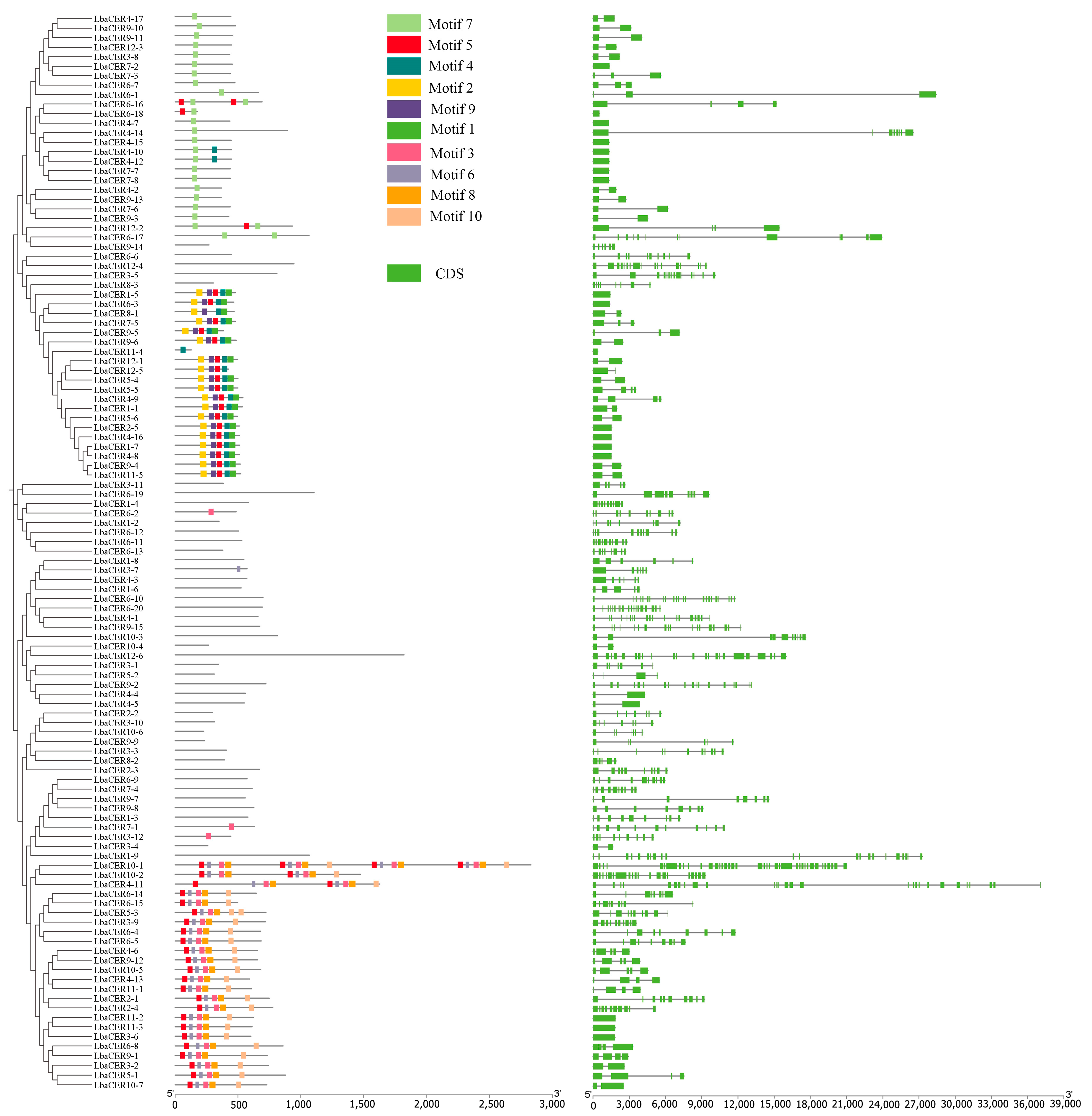
As shown in Figure 4, 10 conserved motifs were identified in goji CER proteins. Among these, Motif5 and Motif7 showed relatively widespread distribution, present in 46 and 27 proteins, respectively. Motif1 was found in only 18 proteins, representing the smallest distribution. From specific observations, 23 CER proteins possessed 1 motif and 4 CER proteins had 2 motifs, among which LbaCER10-1 contained 18 motifs, while the remaining proteins contained 3-9 motifs. However, 41 CER proteins lacked any motifs, suggesting that the goji CER genes may have experienced loss or acquisition of conserved motifs during evolution, potentially indicating the emergence of novel functions in evolutionary process.
In Figure 4, exon/intron structure analysis of the goji CER genes revealed that 17 genes contained a single exon, 19 genes contained two exons, and 6 genes contained three exons. LbaCER10-1 possessed 39 exons, representing the highest number, while the remaining genes contained 1-26 exons. The goji CER genes exhibited substantial variation in exon/intron numbers, indicating that these members may possess differentiated biological functions.

3.5. Cis-Acting Elements Analysis of Goji CER Gene Family Members

As shown in Figure 5 and Supplementary Table S2, the analysis of 2000 bp promoter regions from 113 members of the goji CER gene family identified 32 types of cis-acting elements. They mainly included light-responsiveness, MeJA-responsiveness, abscisic acid-responsiveness, and anaerobic induction. Among these, cis-acting elements associated with light-responsiveness, MeJA-responsiveness, and abscisic acid-responsiveness showed relatively high abundance, appearing 353, 316, and 254 times, respectively, accounting for 18.15%, 16.25%, and 13.06% of total occurrences. Notably, root specific appeared only once, representing the lowest abundance. Therefore, the goji CER gene family may regulate these elements to enable active responses to various environmental stresses such as strong light, drought, and pest/disease conditions.

3.6. Synteny Analysis of Goji CER Gene Family Members

To elucidate the evolutionary relationships of the goji CER gene family, interspecies synteny analysis was conducted between goji and the model plant Arabidopsis, as well as Solanaceae plants including tomato, potato, and tobacco. The results (Figure 6) demonstrated that there were 56 homologous gene pairs between goji and Arabidopsis, 110 homologous gene pairs between goji and tomato, 108 homologous gene pairs between goji and potato, and 97 homologous gene pairs between goji and tobacco, indicating that closer phylogenetic relationships correspond to more homologous gene pairs. Statistical analysis of homologous genes among these five species revealed that (Supplementary Table S3) chr3 contained the highest number of homologous gene pairs (54 pairs), while chr9 showed the second highest number (53 pairs), and chr7 contained the fewest homologous gene pairs (18 pairs). Therefore, goji chromosomes 3 and 9 appear to be the most evolutionarily conserved, whereas chromosome 7 exhibits the most significant variation.

3.7. Phylogenetic Analysis of Goji CER Proteins with Arabidopsis

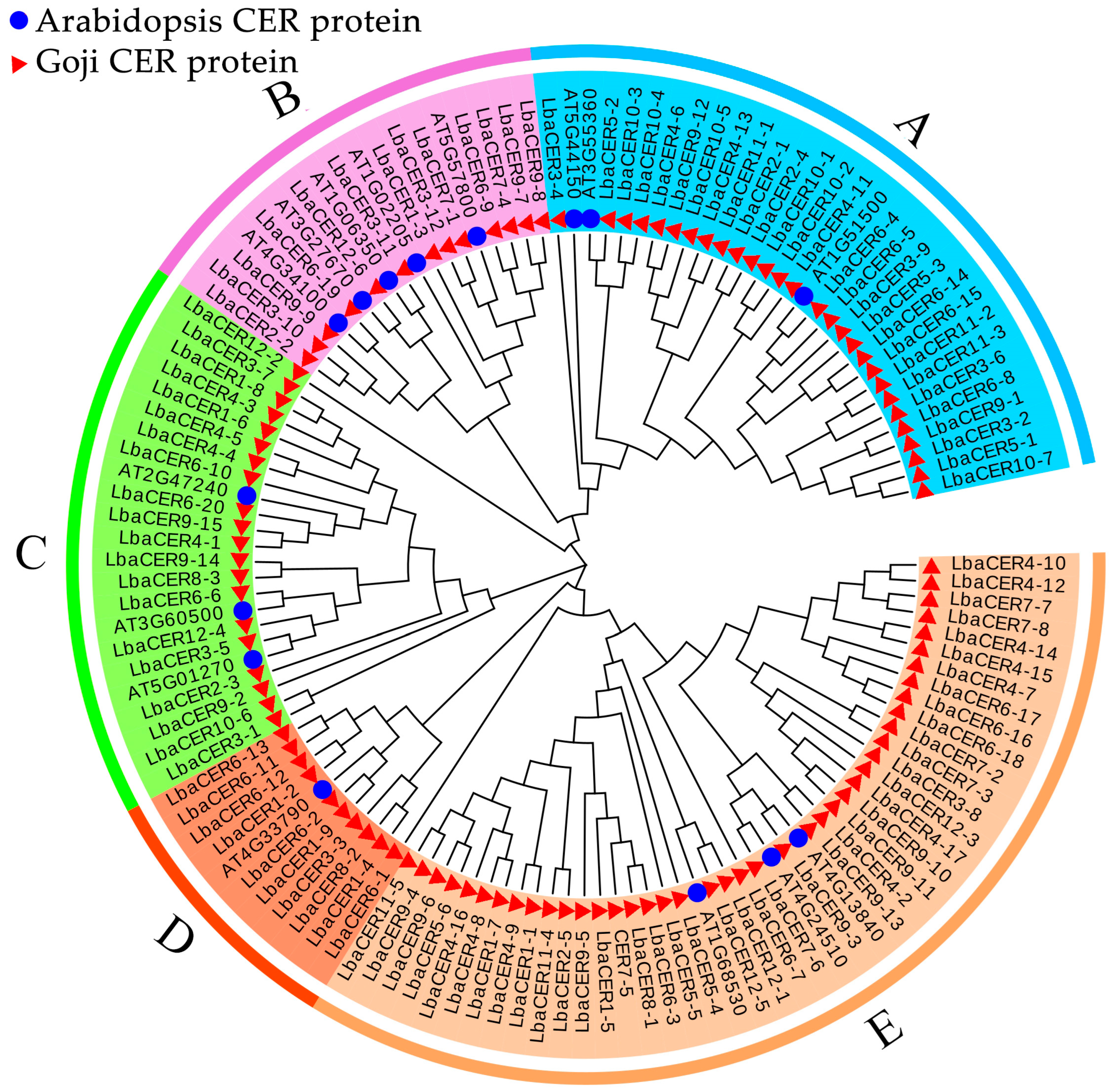
As shown in Figure 7, based on phylogenetic relationships, 15 structurally diverse Arabidopsis proteins and 113 goji CER proteins were classified into five subgroups (A–E). Subgroup A consisted of 3 Arabidopsis CER proteins (AT1G51500, AT3G55360, and AT5G44150) and 28 goji CER gene family member proteins. Subgroup B comprised 5 Arabidopsis proteins and 15 goji proteins. Subgroup C contained Arabidopsis AT4G33790 and 9 goji proteins. Subgroup D included Arabidopsis AT1G68530 and 22 goji proteins. Subgroup E was composed of 5 Arabidopsis proteins and 39 goji CER proteins. All goji CER gene family members showed phylogenetic relationships with their Arabidopsis counterparts, demonstrating the accuracy of the family member selection.

3.8. Expression Analysis of Five CER Genes in Different Goji Cultivars and Organs

To further validate the expression patterns of goji CER gene family members in different cultivars and organs, five genes with distinct domains were selected for expression analysis at the transcriptional level based on previous transcriptome data, as shown in Figure 8. The LbaCER1-1 gene exhibited significantly greater expression in stems than in other organs across three cultivars. Both LbaCER2-5 and LbaCER3-12 showed significantly greater expression in leaves of ‘Ningqi I’ and ‘Ningnongqi XVI’ compared to other organs. LbaCER3-11 displayed notably greater expression in flowers of ‘Ningnongqi XVI’, while LbaCER1-5 was highly expressed in flowers of both ‘Ningqi I’ and ‘Huangguo’ goji. LbaCER3-12 also showed elevated expression in flowers of ‘Huangguo’ goji. Additionally, both LbaCER2-5 and LbaCER3-11 were highly expressed in the fruits of ‘Ningqi I’ and ‘Huangguo’ goji. Combined with results from Figure 1 and Figure 2, we hypothesize that the high expression of LbaCER1-1 makes it a candidate gene for the greater cuticular wax load on stems of ‘Huangguo’ goji, and the elevated expression of both LbaCER2-5 and LbaCER3-12 suggests that they are candidate genes for the greater wax load on leaves of ‘Ningqi I’; additionally, LbaCER1-1, LbaCER2-5, and LbaCER3-12 show closer regulatory relationships with cuticular wax deposition in vegetative organs (stems and leaves), and LbaCER1-5 and LbaCER3-11 appear more closely associated with wax regulation in reproductive organs (flowers and fruits). Notably, some variations were observed among different cultivars in these expression patterns.

4. Discussion

The cuticular wax layer serves as a crucial barrier covering the aerial epidermis of terrestrial plants, protecting them from external environmental stresses [16]. Wax load is often used as an important indicator for screening drought-resistant and pest-resistant germplasms [51]. The production of cuticular wax is regulated by multiple genes, including CER [52], KCS [53], and FAR [54], among others. The CER gene family represents key regulators of cuticular wax biosynthesis in plants [55] and has been studied in various species, including apple [56], sunflower [22], jujube [23], passion fruit (Passiflora edulis) [57], chestnut [24], cotton (Gossypium hirsutum) [55], barley [25], and pepper [26].
The numbers of CER gene family exhibit considerable variation among different species [25]. Species with fewer members include apple (10 members) [58], barley (12 members) [25], and cotton (16 members) [55]. Moderately represented species include jujube (29 members) [23], cabbage (Brassica oleracea) (32 members) [59], chestnut (34 members) [60], and sunflower (37 members) [22]. Species with abundant members include pepper (79 members) [26]. In the present study, the goji CER gene family reached 113 members, substantially exceeding the numbers in the aforementioned species. This discrepancy may be attributed to variations in genome size and various tandem and segmental duplications. In preliminary investigations, we attempted to identify the gene using PF12076 [25] and PF04116 [23] values; however, the results proved unsatisfactory. The PF12076 screening identified only eight members that containing both FA_hydroxylase and Wax2_C domains, while PF04116 failed to identify any relevant members, contradicting current understanding of the CER gene family. Consequently, this study employed Arabidopsis CER protein structures as templates, initially conducting virtual alignment of similar protein domains, followed by individual manual verification to obtain family members. This technical approach demonstrated accurate and reliable screening of goji CER gene family members.
The variations in conserved motifs among gene family members can be used to explain differences in protein functions [60]. Within the same evolutionary clade, LbaCER genes exhibited similar motif compositions and exon–intron structures. As shown in Figure 4, LbaCER4-17 through LbaCER9-3 all contained the conserved Motif7, while LbaCER1-5 to LbaCER11-5 shared multiple conserved motifs. Notably, 36 proteins from LbaCER3-11 to LbaCER1-9 showed unique motif losses, potentially related to functional divergence or emergence of novel functions. These motif structural characteristics were largely consistent with CER gene families in pepper [26], apple [40], and sunflower [22]. Variations in exon and intron numbers represent an important source of gene family diversity, influencing gene function and expression [61]. Differential exon usage and alternative splicing have been demonstrated to be crucial for functional diversification of CER genes [17]. In this study, LbaCER genes exhibited substantial variation in intron numbers, ranging from 1 to 39, exceeding the ranges observed in cotton (3–13) [55], sunflower (1–19) [22], and passion fruit (1-28) [57]. Specifically, 17 genes contained a single exon, while LbaCER10-1 possessed the maximum number of 39 exons. Notably, members with identical or similar motifs and exons clustered well in the phylogenetic tree. These results suggest that LbaCER genes may have undergone multiple rounds of intron loss and gain during evolution, promoting alternative splicing and generating messenger RNAs with diverse functions [62], thereby enhancing functional diversification.
Cis-acting elements serve as crucial factors regulating gene expression levels, providing molecular insights into species evolution and elucidating the interactions between environmental conditions and gene expression [63]. In Brassica napus [64] and maize [65], cis-acting elements of CER genes have been associated with wax accumulation and drought tolerance signaling pathways. The involvement of ABRE (abscisic acid-responsive element) cis-acting elements in Arabidopsis confers enhanced drought stress tolerance [66]. The GARE motif in rice exhibits responsiveness to gibberellins, particularly under conditions of salt and water-deficit stress [67]. The promoters of CalCER genes are enriched with hormone-responsive elements and stress-related elements, consistent with their roles in hormone-mediated stress responses [26]. In this study, analysis of cis-acting elements in the goji CER gene family identified 353 elements associated with light-responsiveness, 316 elements involved in MeJA-responsiveness, and 254 elements participating in abscisic acid-responsiveness. These findings suggest that goji likely possesses strong tolerance to stresses such as intense light, drought, and pests/diseases.
The relative expression levels of five CER gene family members in stems, leaves, flowers, and fruits of the three goji cultivars were detected by qRT–PCR. LbaCER1-1 exhibited the highest expression in stems across all three cultivars, with stable expression trends (Figure 8). Concurrently, the cuticular wax content in stems of these cultivars was significantly greater than that in other organs (Figure 2), a finding further confirmed by SEM observations (Figure 1). Integrating these results, the LbaCER1-1 gene likely plays a crucial role in cuticular wax biosynthesis and secretion in goji stems, which may correlate with its drought tolerance characteristics [2]. Combining the relative expression levels of LbaCER2-5 and LbaCER3-12 with SEM images and leaf wax load data, these two genes may account for the higher wax accumulation in leaves of ‘Ningnongqi XVI’ and ‘Ningqi I’. The elevated leaf wax load suggests these cultivars may possess enhanced resistance to atmospheric drought [43] and disease susceptibility [44].
Certainly, this study still has some limitations. For instance, the functions of the identified LbaCER genes were all predicted based on computational analysis and have not been functionally validated through gene knockout and/or overexpression approaches. In goji cultivation practice, leaves represent critical organs for water loss and susceptibility to pests and diseases. Higher leaf wax load helps reduce water dissipation and pest attachment. Conversely, fruits are economically valuable parts, and a lower fruit wax load can improve fresh fruit palatability and enhance drying efficiency. Therefore, leaf wax and fruit wax load represent two distinct breeding objectives. In future research, CRISPR/Cas9 technology could be employed to generate LbaCER mutants, combined with metabolic network analysis to examine changes in wax content or composition. This would provide a theoretical foundation for improving fresh goji processing and storage, elucidating drought resistance mechanisms, and developing disease-resistant cultivars.

5. Conclusions

The wax load on leaf surfaces of ‘Ningqi I’ was significantly greater than that of other cultivars, while the wax load on stem surfaces of ‘Huangguo’ goji was markedly greater than that of both other cultivars and plant organs, which aligned with SEM observations. A total of 113 CER gene family members were identified in the goji plants, unevenly distributed across 12 chromosomes, with primary localizations in organelles including the plasma membrane, cytoplasm, chloroplasts, and nucleus, clustering into five subgroups. Among goji CER proteins, Motif5 and Motif7 demonstrated the widest distribution. High-frequency occurrences were observed for cis-acting elements associated with light-responsiveness, MeJA-responsiveness, and abscisic acid-responsiveness. Chr3 and 9 exhibited the highest conservation, while chr7 showed the greatest variation. The elevated expression of LbaCER1–1 gene potentially explains the higher wax load on ‘Huangguo’ goji stems; similarly, the high expression of LbaCER2-5 and LbaCER3-12 genes likely accounts for the increased wax load on ‘Ningqi I’ leaves.

Supplementary Materials

The following supporting information can be downloaded at https://www.mdpi.com/article/10.3390/genes16111257/s1, Table S1: Information and physicochemical properties of the goji CER gene family members; Table S2: Cis-acting elements of the goji CER gene family members; Table S3: Synteny analysis of CER gene family members in goji berries and other species.

Author Contributions

Conceptualization, J.L. and L.G.; methodology, Q.Y. and J.L.; software, B.L.; formal analysis, Q.Y.; investigation, L.J., F.Z. and L.G.; resources, B.L.; data curation, Q.Y. and J.L.; writing—original draft preparation, Q.Y.; writing—review and editing, Q.Y. and J.L.; visualization, Q.Y. and J.L.; supervision, J.L. and F.Z.; funding acquisition, Q.Y. and J.L. All authors have read and agreed to the published version of the manuscript.

Funding

This research was funded by Gansu Province Natural Science Foundation [24JRRA650] and National Natural Science Foundation of China [32060341].

Institutional Review Board Statement

Not applicable.

Informed Consent Statement

Not applicable.

Data Availability Statement

The goji plants genome data were downloaded from the goji (Lycium barbarum) genome database (https://figshare.com/articles/dataset/Goji_genomes_and_the_evolution_of_Lycium_Solanaceae_/20416593, accessed on 6 April 2023). The genome data for the tomato, potato, and tobacco genomes were downloaded from the Sol genomics network (https://solgenomics.net/, accessed on 8 August 2025).

Acknowledgments

We thank Zhongjian Liu from Fujian A&F University for sharing the reference genome.

Conflicts of Interest

The authors declare no conflicts of interest.

Abbreviations

The following abbreviations are used in this manuscript:
CEREceriferum
UVUltraviolet Radiation
VLCFAsVery-long-chain fatty acids
FARFatty acyl reductase
MAHMedium chain alkane hydroxylase
SEMScanning electron microscopy
qRT–PCRQuantitative real-time polymerase chain reaction
MeJAMethyl jasmonate
ABAAbscisic acid
PEGPolyethylene glycol
VLCVery long chain

References

  1. Yang, Z.; Dai, G.; Qin, K.; Wu, J.; Wang, Z.; Wang, C. Comprehensive Evaluation of Germplasm Resources in Various Goji Cultivars Based on Leaf Anatomical Traits. Forests 2025, 16, 187. [Google Scholar] [CrossRef]
  2. Hõrak, H. Learning from the Experts: Drought Resistance in Desert Plants. New Phytol. 2017, 216, 5–7. [Google Scholar] [CrossRef] [PubMed]
  3. Li, W.; Rao, S.; Du, C.; Liu, L.; Dai, G.; Chen, J. Strategies Used by Two Goji Species, Lycium ruthenicum and Lycium barbarum, to Defend against Salt Stress. Sci. Hortic. 2022, 306, 111430. [Google Scholar] [CrossRef]
  4. Zhang, Z.; He, K.; Zhang, T.; Tang, D.; Li, R.; Jia, S. Physiological Responses of Goji Berry (Lycium barbarum L.) to Saline-Alkaline Soil from Qinghai Region, China. Sci. Rep. 2019, 9, 12057. [Google Scholar] [CrossRef]
  5. Teixeira, S.; Luís, I.; Oliveira, M.; Abreu, I.; Batista, R. Goji Berries Superfood—Contributions for the Characterisation of Proteome and Ige-Binding Proteins. Food Agric. Immunol. 2019, 30, 262–280. [Google Scholar] [CrossRef]
  6. Amagase, H.; Farnsworth, N.R. A Review of Botanical Characteristics, Phytochemistry, Clinical Relevance in Efficacy and Safety of Lycium barbarum Fruit (Goji). Food Res. Int. 2011, 44, 1702–1717. [Google Scholar] [CrossRef]
  7. Jeffree, C.E. The Fine Structure of the Plant Cuticle. In Annual Plant Reviews Volume 23: Biology of the Plant Cuticle; Wiley-Blackwell: Hoboken, NJ, USA, 2006; pp. 11–125. [Google Scholar]
  8. Steinmüller, D.; Tevini, M. Action of Ultraviolet Radiation (UV-B) Upon Cuticular Waxes in Some Crop Plants. Planta 1985, 164, 557–564. [Google Scholar] [CrossRef]
  9. Moreno, A.G.; Cózar, A.; Prieto, P.; Domínguez, E.; Heredia, A. Radiationless Mechanism of Uv Deactivation by Cuticle Phenolics in Plants. Nat. Commun. 2022, 13, 1786. [Google Scholar] [CrossRef]
  10. Barthlott, W.; Neinhuis, C. Purity of the Sacred Lotus, or Escape from Contamination in Biological Surfaces. Planta 1997, 202, 1–8. [Google Scholar] [CrossRef]
  11. Khanal, B.P.; Grimm, E.; Finger, S.; Blume, A.; Knoche, M. Intracuticular Wax Fixes and Restricts Strain in Leaf and Fruit Cuticles. N. Phytol. 2013, 200, 134–143. [Google Scholar] [CrossRef] [PubMed]
  12. Jiang, Y.; Peng, Y.; Hou, G.; Yang, M.; He, C.; She, M.; Li, X.; Li, M.; Chen, Q.; Lin, Y. A High Epicuticular Wax Strawberry Mutant Reveals Enhanced Resistance to Tetranychus urticae Koch and Botrytis cinerea. Sci. Hortic. 2024, 324, 112636. [Google Scholar] [CrossRef]
  13. Buschhaus, C.; Jetter, R. Composition and Physiological Function of the Wax Layers Coating Arabidopsis Leaves: Β-amyrin Negatively Affects the Intracuticular Water Barrier. Plant Physiol. 2012, 160, 1120–1129. [Google Scholar] [CrossRef]
  14. Jiang, B.; Liu, R.; Fang, X.; Tong, C.; Gao, H. Effects of Salicylic Acid Treatment on Fruit Quality and Wax Composition of Blueberry (Vaccinium virgatum Ait). Food Chem. 2021, 5, 130757. [Google Scholar] [CrossRef] [PubMed]
  15. Lee, S.B.; Suh, M.C. Advances in the Understanding of Cuticular Waxes in Arabidopsis thaliana and Crop Species. Plant Cell Rep. 2015, 34, 4. [Google Scholar] [CrossRef]
  16. Yeats, T.H.; Rose, J.K. The Formation and Function of Plant Cuticles. Plant Physiol. 2013, 163, 5–20. [Google Scholar] [CrossRef] [PubMed]
  17. Aarts, M.G.; Keijzer, C.J.; Stiekema, W.J.; Pereira, A. Molecular Characterization of the Cer1 Gene of Arabidopsis Involved in Epicuticular Wax Biosynthesis and Pollen Fertility. Plant Cell 1995, 7, 2115–2127. [Google Scholar] [CrossRef] [PubMed]
  18. Sturaro, M.; Hartings, H.; Schmelzer, E.; Velasco, R.; Salamini, F.; Motto, M. Cloning and Characterization of Glossy1, a Maize Gene Involved in Cuticle Membrane and Wax Production. Plant Physiol. 2005, 138, 478–489. [Google Scholar] [CrossRef]
  19. Wang, W.; Zhang, Y.; Xu, C.; Ren, J.; Liu, X.; Black, K.; Gai, X.; Wang, Q.; Ren, H. Cucumber Eceriferum1 (Cscer1), Which Influences the Cuticle Properties and Drought Tolerance of Cucumber, Plays a Key Role in Vlc Alkanes Biosynthesis. Plant Mol. Biol. 2015, 87, 219–233. [Google Scholar] [CrossRef]
  20. Li, T.; Sun, Y.; Liu, T.; Wu, H.; An, P.; Shui, Z.; Wang, J.; Zhu, Y.; Li, C.; Wang, Y.; et al. Tacer1-1a Is Involved in Cuticular Wax Alkane Biosynthesis in Hexaploid Wheat and Responds to Plant Abiotic Stresses. Plant Cell Environ. 2019, 42, 3077–3091. [Google Scholar] [CrossRef]
  21. Xiong, C.; Xie, Q.; Yang, Q.; Sun, P.; Gao, S.; Li, H.; Zhang, J.; Wang, T.; Ye, Z.; Yang, C. Woolly, Interacting with MYB Transcription Factor MYB31, Regulates Cuticular Wax Biosynthesis by Modulating Cer6 Expression in Tomato. Plant J. 2020, 103, 323–337. [Google Scholar] [CrossRef]
  22. Muhammad Ahmad, H.; Wang, X.; Fiaz, S.; Mahmood Ur, R.; Azhar Nadeem, M.; Aslam Khan, S.; Ahmar, S.; Azeem, F.; Shaheen, T.; Mora-Poblete, F. Comprehensive Genomics and Expression Analysis of Eceriferum (Cer) Genes in Sunflower (Helianthus annuus). Saudi J. Biol. Sci. 2021, 28, 6884–6896. [Google Scholar] [CrossRef]
  23. Li, N.; Li, X.Z.; Song, Y.Q.; Yang, S.T.; Li, L.L. Genome-Wide Identification, Characterization, and Expression Profiling of the Eceriferum (Cer) Gene Family in Ziziphus jujube. Russ. J. Plant Physiol. 2021, 68, 828–837. [Google Scholar] [CrossRef]
  24. Zhao, S.; Nie, X.; Liu, X.; Wang, B.; Liu, S.; Qin, L.; Xing, Y. Genome-Wide Identification of the Cer Gene Family and Significant Features in Climate Adaptation of Castanea mollissima. Int. J. Mol. Sci. 2022, 23, 16202. [Google Scholar] [CrossRef] [PubMed]
  25. Panahi, B.; Hamid, R.; Jacob, F. Genome-Wide Characterization of the Eceriferum (Cer) Gene Family in Barley (Hordeum vulgare L.). Sci. Rep. 2025, 15, 20674. [Google Scholar] [CrossRef] [PubMed]
  26. Yang, F.; Wei, K.; Zhang, Y.; Chang, X.; Yang, W.; Yao, Q.; Xiao, H. Genome-Wide Identification of the Eceriferum Gene Family and Analysis of Gene Expression Patterns under Different Treatments in Pepper (Capsicum annuum L.). Horticulturae 2025, 11, 571. [Google Scholar] [CrossRef]
  27. Bernard, A.; Joubès, J. Arabidopsis Cuticular Waxes: Advances in Synthesis, Export and Regulation. Prog. Lipid Res. 2013, 52, 110–129. [Google Scholar] [CrossRef]
  28. Zhong, M.-S.; Jiang, H.; Cao, Y.; Wang, Y.-X.; You, C.-X.; Li, Y.-Y.; Hao, Y.-J. Mdcer2 Conferred to Wax Accumulation and Increased Drought Tolerance in Plants. Plant Physi Biochem. 2020, 149, 277–285. [Google Scholar] [CrossRef]
  29. Haslam, T.M.; Haslam, R.; Thoraval, D.; Pascal, S.; Delude, C.; Domergue, F.; Fernández, A.M.; Beaudoin, F.; Napier, J.A.; Kunst, L.; et al. Eceriferum2-Like Proteins Have Unique Biochemical and Physiological Functions in Very-Long-Chain Fatty Acid Elongation. Plant Physiol. 2015, 167, 682–692. [Google Scholar] [CrossRef]
  30. Yang, X.; Feng, T.; Li, S.; Zhao, H.; Zhao, S.; Ma, C.; Jenks, M.A.; Lü, S. Cer16 Inhibits Post-Transcriptional Gene Silencing of Cer3 to Regulate Alkane Biosynthesis. Plant Physiol. 2020, 182, 1211–1221. [Google Scholar] [CrossRef]
  31. Bird, D.; Beisson, F.; Brigham, A.; Shin, J.; Greer, S.; Jetter, R.; Kunst, L.; Wu, X.; Yephremov, A.; Samuels, L. Characterization of Arabidopsis ABCg11/WBC11, an Atp Binding Cassette (ABC) Transporter That Is Required for Cuticular Lipid Secretion. Plant J. 2007, 52, 485–498. [Google Scholar] [CrossRef]
  32. Fiebig, A.; Mayfield, J.A.; Miley, N.L.; Chau, S.; Fischer, R.L.; Preuss, D. Alterations in Cer6, a Gene Identical to Cut1, Differentially Affect Long-Chain Lipid Content on the Surface of Pollen and Stems. Plant Cell 2000, 12, 2001–2008. [Google Scholar] [CrossRef]
  33. Lü, S.; Zhao, H.; Des Marais, D.L.; Parsons, E.P.; Wen, X.; Xu, X.; Bangarusamy, D.K.; Wang, G.; Rowland, O.; Juenger, T.; et al. Arabidopsis Eceriferum9 Involvement in Cuticle Formation and Maintenance of Plant Water Status. Plant Physiol. 2012, 159, 930–944. [Google Scholar] [CrossRef] [PubMed]
  34. Zhao, L.; Kunst, L. Superkiller Complex Components Are Required for the Rna Exosome-Mediated Control of Cuticular Wax Biosynthesis in Arabidopsis Inflorescence Stems. Plant Physiol. 2016, 171, 960–973. [Google Scholar] [CrossRef] [PubMed]
  35. Lü, S.; Song, T.; Kosma, D.K.; Parsons, E.P.; Rowland, O.; Jenks, M.A. Arabidopsis Cer8 Encodes Long-Chain Acyl-Coa Synthetase 1 (Lacs1) That Has Overlapping Functions with Lacs2 in Plant Wax and Cutin Synthesis. Plant J. 2009, 59, 553–564. [Google Scholar] [CrossRef] [PubMed]
  36. Fukuda, N.; Oshima, Y.; Ariga, H.; Kajino, T.; Koyama, T.; Yaguchi, Y.; Tanaka, K.; Yotsui, I.; Sakata, Y.; Taji, T. Eceriferum 10 Encoding an Enoyl-Coa Reductase Plays a Crucial Role in Osmotolerance and Cuticular Wax Loading in Arabidopsis. Front. Plant Sci. 2022, 13, 898317. [Google Scholar] [CrossRef]
  37. Shi, L.; Dean, G.H.; Zheng, H.; Meents, M.J.; Haslam, T.M.; Haughn, G.W.; Kunst, L. Eceriferum11/C-Terminal Domain Phosphatase-Like2 Affects Secretory Trafficking. Plant Physiol. 2019, 181, 901–915. [Google Scholar] [CrossRef]
  38. Yang, X.; Zhao, H.; Kosma, D.K.; Tomasi, P.; Dyer, J.M.; Li, R.; Liu, X.; Wang, Z.; Parsons, E.P.; Jenks, M.A.; et al. The Acyl Desaturase Cer17 Is Involved in Producing Wax Unsaturated Primary Alcohols and Cutin Monomers. Plant Physiol. 2017, 173, 1109–1124. [Google Scholar] [CrossRef]
  39. Zhang, Q.; Han, L.; Jia, J.; Song, L.; Wang, J. Management of Drought Risk under Global Warming. Theor. Appl. Climatol. 2016, 125, 187–196. [Google Scholar] [CrossRef]
  40. Qi, C.; Jiang, H.; Zhao, X.; Mao, K.; Liu, H.; Li, Y.; Hao, Y. The Characterization, Authentication, and Gene Expression Pattern of the Mdcer Family in Malus domestica. Hortic. Plant J. 2019, 5, 1–9. [Google Scholar] [CrossRef]
  41. Wu, H.; Liu, L.; Chen, Y.; Liu, T.; Jiang, Q.; Wei, Z.; Li, C.; Wang, Z. Tomato Slcer1–1 Catalyzes the Synthesis of Wax Alkanes, Increasing Drought Tolerance and Fruit Storability. Hortic. Res. 2022, 9, uhac004. [Google Scholar] [CrossRef]
  42. Grünhofer, P.; Herzig, L.; Zhang, Q.; Vitt, S.; Stöcker, T.; Malkowsky, Y.; Brügmann, T.; Fladung, M.; Schreiber, L. Changes in Wax Composition but Not Amount Enhance Cuticular Transpiration. Plant Cell Environ. 2024, 47, 91–105. [Google Scholar] [CrossRef] [PubMed]
  43. Wang, X.; Li, S.; Zhang, X.; Wang, J.; Hou, T.; He, J.; Li, J. Integration of Transcriptome and Metabolome Reveals Wax Serves a Key Role in Preventing Leaf Water Loss in Goji (Lycium barbarum). Int. J. Mol. Sci. 2024, 25, 10939. [Google Scholar] [CrossRef] [PubMed]
  44. Li, J.; Wen, X.; Zhang, S.-D.; Zhang, X.; Feng, L.-D.; He, J. An Increased Wax Load on the Leaves of Goji Plants (Lycium barbarum) Results in Increased Resistance to Powdery Mildew. Chem. Biol. Technol. Agric. 2024, 11, 62. [Google Scholar] [CrossRef]
  45. Wang, P.; Wang, J.; Zhang, H.; Wang, C.; Zhao, L.; Huang, T.; Qing, K. Chemical Composition, Crystal Morphology, and Key Gene Expression of the Cuticular Waxes of Goji (Lycium barbarum L.) Berries. J. Agri. Food Chem. 2021, 69, 7874–7883. [Google Scholar] [CrossRef]
  46. Li, N.; Song, Y.; Li, J.; Hao, R.; Feng, X.; Li, L. Transcriptome and Genome Re-Sequencing Analysis Reveals Differential Expression Patterns and Sequence Variation in Pericarp Wax Metabolism-Related Genes in Ziziphus jujuba (Chinese Jujube). Sci. Hortic. 2021, 288, 110415. [Google Scholar] [CrossRef]
  47. Wang, Y.; Zeng, J.; Xia, X.; Xu, Y.; Chen, S. Comparative Analysis of Leaf Trichomes, Epidermal Wax and Defense Enzymes Activities in Response to Puccinia horiana in Chrysanthemum and Ajania Species. Hortic. Plant J. 2020, 6, 8. [Google Scholar] [CrossRef]
  48. Chao, J.; Li, Z.; Sun, Y.; Aluko, O.O.; Wu, X.; Wang, Q.; Liu, G. Mg2c: A User-Friendly Online Tool for Drawing Genetic Maps. Mol. Hortic. 2021, 1, 16. [Google Scholar] [CrossRef]
  49. Livak, K.J.; Schmittgen, T.D. Analysis of Relative Gene Expression Data Using Real-Time Quantitative Pcr. Methods 2002, 25, 402–408. [Google Scholar] [CrossRef]
  50. Tao, Y.X.; Peer, A.F.V.; Huang, Q.H.; Shao, Y.P.; Xie, B.G. Identification of Novel and Robust Internal Control Genes from Volvariella volvacea That Are Suitable for RT-qPCR in Filamentous Fungi. Sci. Rep. 2016, 6, 29236. [Google Scholar] [CrossRef]
  51. Zhao, Y.; Liu, X.; Wang, M.; Bi, Q.; Cui, Y.; Wang, L. Transcriptome and Physiological Analyses Provide Insights into the Leaf Epicuticular Wax Accumulation Mechanism in Yellowhorn. Hortic. Res. 2021, 8, 134. [Google Scholar] [CrossRef]
  52. Lee, S.B.; Suh, M.C. Regulatory Mechanisms Underlying Cuticular Wax Biosynthesis. J. Exp. Bot. 2022, 73, 2799–2816. [Google Scholar] [CrossRef]
  53. Yang, H.; Mei, W.; Wan, H.; Xu, R.; Cheng, Y. Comprehensive Analysis of Kcs Gene Family in Citrinae Reveals the Involvement of Cskcs2 and Cskcs11 in Fruit Cuticular Wax Synthesis at Ripening. Plant Sci. 2021, 310, 110972. [Google Scholar] [CrossRef]
  54. Wang, Y.; Wang, M.; Sun, Y.; Wang, Y.; Li, T.; Chai, G.; Jiang, W.; Shan, L.; Li, C.; Xiao, E.; et al. Far5, a Fatty Acyl-Coenzyme a Reductase, Is Involved in Primary Alcohol Biosynthesis of the Leaf Blade Cuticular Wax in Wheat (Triticum aestivum L.). J. Exp. Bot. 2015, 66, 1165–1178. [Google Scholar] [CrossRef] [PubMed]
  55. Hamid, R.; Ghorbanzadeh, Z.; Jacob, F.; Nekouei, M.K.; Zeinalabedini, M.; Mardi, M.; Sadeghi, A.; Ghaffari, M.R. Decoding Drought Resilience: A Comprehensive Exploration of the Cotton Eceriferum (Cer) Gene Family and Its Role in Stress Adaptation. BMC Plant Biol. 2024, 24, 468. [Google Scholar] [CrossRef]
  56. Finocchiaro, F.; Terzi, V.; Delbono, S. Barley: From Molecular Basis of Quality to Advanced Genomics-Based Breeding. In Compendium of Crop Genome Designing for Nutraceuticals; Springer Nature: Singapore, 2023; pp. 1–38. [Google Scholar]
  57. Rizwan, H.M.; Waheed, A.; Ma, S.; Li, J.; Arshad, M.B.; Irshad, M.; Li, B.; Yang, X.; Ali, A.; Ahmed Ma, A.; et al. Comprehensive Genome-Wide Identification and Expression Profiling of Eceriferum (Cer) Gene Family in Passion Fruit (Passiflora edulis) under Fusarium kyushuense and Drought Stress Conditions. Front. Plant Sci. 2022, 13, 898307. [Google Scholar] [CrossRef] [PubMed]
  58. Waqas, M.; Azhar, M.T.; Rana, I.A.; Azeem, F.; Ali, M.A.; Nawaz, M.A.; Chung, G.; Atif, R.M. Genome-Wide Identification and Expression Analyses of Wrky Transcription Factor Family Members from Chickpea (Cicer arietinum L.) Reveal Their Role in Abiotic Stress-Responses. Genes Genom. 2019, 41, 467–481. [Google Scholar] [CrossRef] [PubMed]
  59. Cao, W.; Sun, H.; Wang, C.; Yang, L.; Zhang, Y.; Zhuang, M.; Lv, H.; Wang, Y.; Liu, F.; Ji, J. Genome-Wide Identification of the Eceriferum (Cer) Gene Family in Cabbage and Critical Role of Bocer4.1 in Wax Biosynthesis. Plant Physiol Biochem. 2025, 222, 109718. [Google Scholar] [CrossRef]
  60. Faraji, S.; Filiz, E.; Kazemitabar, S.K.; Vannozzi, A.; Palumbo, F.; Barcaccia, G.; Heidari, P. The Ap2/Erf Gene Family in Triticum durum: Genome-Wide Identification and Expression Analysis under Drought and Salinity Stresses. Genes 2020, 11, 1464. [Google Scholar] [CrossRef]
  61. Xu, G.; Guo, C.; Shan, H.; Kong, H. Divergence of Duplicate Genes in Exon-Intron Structure. Proc. Natl. Acad. Sci. USA 2012, 109, 1187–1192. [Google Scholar] [CrossRef]
  62. Xu, B.; Shi, Y.; Wu, Y.; Meng, Y.; Jin, Y. Role of Rna Secondary Structures in Regulating Dscam Alternative Splicing. Biochim. Biophys. Acta Gene Regul. Mech. 2019, 1862, 194381. [Google Scholar] [CrossRef]
  63. Wittkopp, P.J.; Kalay, G. Cis-Regulatory Elements: Molecular Mechanisms and Evolutionary Processes Underlying Divergence. Nat. Rev. Genet. 2012, 13, 59–69. [Google Scholar] [CrossRef] [PubMed]
  64. Wang, S.; Bai, C.; Luo, N.; Jiang, Y.; Wang, Y.; Liu, Y.; Chen, C.; Wang, Y.; Gan, Q.; Jin, S.; et al. Brassica Napus Bnac9.Dewax1 Negatively Regulates Wax Biosynthesis Via Transcriptional Suppression of Bncer1-2. Int. J. Mol. Sci. 2023, 24, 4287. [Google Scholar] [CrossRef]
  65. Xu, L.; Hao, J.; Lv, M.; Liu, P.; Ge, Q.; Zhang, S.; Yang, J.; Niu, H.; Wang, Y.; Xue, Y.; et al. A Genome-Wide Association Study Identifies Genes Associated with Cuticular Wax Metabolism in Maize. Plant Physiol. 2024, 194, 2616–2630. [Google Scholar] [CrossRef]
  66. Yoshida, T.; Fujita, Y.; Sayama, H.; Kidokoro, S.; Maruyama, K.; Mizoi, J.; Shinozaki, K.; Yamaguchi-Shinozaki, K. Areb1, Areb2, and Abf3 Are Master Transcription Factors That Cooperatively Regulate Abre-Dependent Aba Signaling Involved in Drought Stress Tolerance and Require Aba for Full Activation. Plant J. 2010, 61, 672–685. [Google Scholar] [CrossRef]
  67. De Silva, W.S.I.; Perera, M.M.N.; Perera, K.L.N.S.; Wickramasuriya, A.M.; Jayasekera, G.A.U. In Silico Analysis of Osr40c1 Promoter Sequence Isolated from Indica Variety Pokkali. Rice Sci. 2017, 24, 228–234. [Google Scholar] [CrossRef]
Figure 1. Scanning electron micrographs of the epicuticular wax on the above-ground organs of three goji varieties.
Genes 16 01257 g001
Figure 2. Wax load on above-ground organs of three goji varieties. Note: (A) is the wax load of stems; (B) is the wax load of leaves; (C) is the wax load of flowers; (D) is the wax load of young fruit. The letters above bars represents the differences between data. The same letters indicate non-significant differences, while different letters indicate significant differences.
Genes 16 01257 g002
Figure 3. Chromosomal localization and intra-group collinearity analysis of goji CER gene family members.
Genes 16 01257 g003
Figure 4. Analysis of conserved motifs and domains in goji CER gene family members.
Genes 16 01257 g004
Figure 5. Analysis of cis-acting elements in the goji CER gene family members.
Genes 16 01257 g005
Figure 6. Synteny analysis of goji CER gene family members with other species.
Genes 16 01257 g006
Figure 7. Phylogenetic evolution of the goji CER proteins.
Genes 16 01257 g007
Figure 8. Tissue-specific expression patterns of 5 goji CER genes. Note: (A) is the relative expression of the LbaCER1-1 gene; (B) is the relative expression of the LbaCER1-5 gene; (C) is the relative expression of the LbaCER2-5 gene; (D) is the relative expression of the LbaCER3-11 gene; (E) is the relative expression of the LbaCER3-12 gene. The letters above bars represents the differences between data. The same letters indicate non-significant differences, while different letters indicate significant differences.
Genes 16 01257 g008
Table 1. Primer sequences of five goji CER genes and reference gene.
Gene IDGene NamePrimer TypePrimer Sequences (5′ to 3′)
Lba01g00543LbaCER1–1FCATCACCACGCTAGGACCATTG
RTTTCACCTTGGCTTTCAACACTTTC
Lba01g01071LbaCER1–5FCCTAGCCCTTCTCTTTCTTCCATTATC
RACAACCCATTCCACTTATCGTATAGC
Lba02g02625LbaCER2–5FCTGGTGTGATAGCGGTTGATCTTG
RTCTCCGTACTAACAACAACAGCATAC
Lba03g02510LbaCER3–11FCAAGTTGAATTGTTATGGACTGGAAGG
RGCATTCACAGTTGGTATTGGCATTAC
Lba03g02612LbaCER3–12FTCGTGCTTATTATGTGGCTGAAGTC
RGTTTGGTTGAGTTTCCCTCTTATGTTG
Loc132636280RH37F
R
GCAGGCAAGTCAGGATTAGCA
CGCATAACGAGTCAACCATTCAG
Disclaimer/Publisher’s Note: The statements, opinions and data contained in all publications are solely those of the individual author(s) and contributor(s) and not of MDPI and/or the editor(s). MDPI and/or the editor(s) disclaim responsibility for any injury to people or property resulting from any ideas, methods, instructions or products referred to in the content.