Review

Chlamydia trachomatis: From Urogenital Infections to the Pathway of Infertility

1
PerMed Research Group, RISE-Health, Faculty of Medicine, University of Porto, Alameda Professor Hernâni Monteiro, 4200-319 Porto, Portugal
2
Molecular Diagnostics Laboratory, Unilabs Portugal, Centro Empresarial Lionesa Porto, Rua Lionesa, Leça do Balio, 4465-671 Matosinhos, Portugal
3
RISE-Health, Department of Pathology, Faculty of Medicine, University of Porto, Alameda Professor Hernâni Monteiro, 4200-319 Porto, Portugal
4
Centre for Reproductive Genetics Alberto Barros, 4100-012 Porto, Portugal
5
RISE-Health, Department of Community Medicine, Health Information and Decision (MEDCIDS), Faculty of Medicine, University of Porto, Rua Doutor Plácido da Costa, 4200-450 Porto, Portugal
*
Author to whom correspondence should be addressed.
Genes 2025, 16(2), 205; https://doi.org/10.3390/genes16020205
Submission received: 10 January 2025 / Revised: 2 February 2025 / Accepted: 5 February 2025 / Published: 7 February 2025
(This article belongs to the Special Issue Genetics of Multifactorial Diseases: 2nd Edition)

Abstract

:
Chlamydia trachomatis (CT) is a major cause of sexually transmitted infections (STIs) worldwide, with significant implications for reproductive health. The bacterium’s genome contains highly polymorphic regions, influencing both the type and severity of infections. These genetic variations, particularly those occurring in the major outer membrane protein (MOMP) gene, are critical for classifying the bacterium into distinct serovars and enable CT to adapt to diverse host environments, contributing to its immune evasion, persistence, and pathogenicity. Persistent or untreated urogenital infections can lead to chronic inflammation, tissue damage, and pelvic inflammatory disease, ultimately increasing the risk of ectopic pregnancy, spontaneous abortion, and infertility. This review consolidates current knowledge on the genetic diversity of CT, its potential role in modulating infection outcomes, and its immune evasion mechanisms. By integrating scientific evidence linking chlamydial infections to infertility, we underscore the urgent need for targeted research to address this critical public health challenge.

1. Introduction

Sexually transmitted infections (STIs) are a major public health concern in developed countries due to their significant impact on communities, particularly because of the complications that can arise if they are not diagnosed and treated in a timely manner. Among these, chlamydial infections stand out as the most common bacterial STI. According to the European Centre for Disease Prevention and Control (ECDC), 216,508 confirmed cases of Chlamydia trachomatis infection were reported in 27 EU/EEA countries in 2022. Moreover, the ECDC reports a steady increase in these rates since at least 2018, highlighting the growing prevalence of this pathogen in recent years [1]. CT is an obligate intracellular pathogen of eukaryotic cells capable of infecting several different cell types. It is a simple parasite, composed of DNA, RNA, and ribosomes, and enclosed by a cell wall [2]. This Gram-negative pathogen is capable of causing a diversity of pathogenicity in humans, as it exhibits different tissue tropisms depending on certain genetic characteristics [3]. Specifically, the bacterium could cause extragenital or urogenital tract infections; however, in this manuscript, we will give emphasis to the urogenital infections, which will be explored in greater detail. The majority of CT urogenital infections are clinically asymptomatic, and the infection has no specific symptoms [4]. As a result, the infection transmission rate is high and, when diagnosed, it is often already associated with urogenital and reproductive tract complications, influencing patient morbidity [5]. In terms of cell biology, obviously, this obligate intracellular bacterium is totally dependent on host cell biosynthetic machinery for its development [2,6]. The developmental cycle is biphasic, constituted by two different morphological stages of CT: the elementary body (EB) and the reticulated body (RB) [7]. In detail, in terms of time, after infecting one cell, within 8 h, the bacterium is able to initiate replication within the host cell, and usually, in 48 to 72 h, the development cycle is completed, and the bacteria are ready to disseminate from this first host cell to other surrounding cells [8,9]. The first step of its biphasic development cycle begins with the EB, the infectious form, which is not able to replicate but is essential to promoting bacterium survival. It can bind to the host cell receptor through bacterial adhesins, and the invasion process starts [10]. After the contact, EBs are internalized into the inclusion bodies, a type of cellular vacuole, and gene transcription is initiated, ensuring all conditions for the bacteria lifecycle to occur and establishing an anti-apoptotic state in the host cell [7,8]. With the bacterial synthesis initiation, EBs differentiate into the reticulated body (RB), an actively replicating form, which then initiates replication at an alarming rate within the inclusion bodies (Figure 1) [9]. After completing these replication cycles, RBs re-differentiate back into EBs, which then can be released through host cell lysis or through extrusion to infect other cells, continuing the infection process [11]. Importantly, the RBs can enter a persistent non-replicative state under adverse conditions and cellular stress, dedifferentiated into aberrant bodies (ABs), leading to a chronic infection that is difficult to diagnose and treat [12]. In this state of persistence, the pathogen evades immune detection because it is less metabolically active and less susceptible to antibiotics [13].
In fact, without an effective vaccine development, antibiotics are the only treatment used to treat these infections [14]. There are many classes of antibiotics, among which macrolide and tetracycline are the most prescribed ones—specifically, the drugs azithromycin and doxycycline, respectively. These most effective anti-chlamydial drugs act by inhibiting bacterial protein synthesis through distinct mechanisms [13,15]. In detail, macrolide drug action occurs through its binding to a specific region of the 50S ribosomal subunit of the ribosome, which physically blocks the channel, inhibiting protein synthesis [15,16,17]. Tetracycline drugs can interfere in two different pathways: it not only can increase the CT membrane permeability, leading to bacterium death, but also it can bind to the 30S subunit of the ribosome, blocking tRNA binding and ultimately disrupting protein synthesis [15,16]. The proper use of antibiotics is a crucial factor in preventing bacteria from entering a protective state [18]. Importantly, in the persistent form, CT can be reactivated when intracellular conditions change, or in other words, when the factors that have triggered persistence are eliminated. For example, CT reactivation can occur upon the removal of stress conditions, such as nutrient deprivation, oxidative stress, or the cessation of antibiotic treatment [8,12,18]. Also, this chronic condition in the female genital tract causes long-term exposure of the reproductive organs to the bacterium, leading to inflammation and scarring [19]. This is the leading cause of tubal factor infertility, a condition in which the fallopian tubes are congested, avoiding spermatozoa from reaching an egg for fertilization or preventing an embryo from reaching the uterus for pregnancy [20]. Also, chronic CT infection can be associated with pelvic inflammatory disease (PID), preterm birth, endometritis, and perihepatitis [21,22]. Indeed, PID, which is a female heterogenous syndrome possibly associated with infertility, ectopic pregnancy, and chronic pelvic pain, is also a well-established risk factor for infertility [23,24,25]. The fact that CT is one of the leading causes of PID and that pelvic inflammatory disease is a syndrome known to contribute to infertility, are other scientifically based validations to hypothesize this infection as a potentially significant risk factor for infertility [24].
It is also important to emphasize that the diagnose of CT infection in men is very relevant, not only because they provide an important reservoir of bacteria for new and recurrent infections in women but also because they may experience serious adverse outcomes as well [26]. In detail, the most common outcomes of prolonged CT urogenital infection in men are urethritis, epididymis, prostatitis, and Reither’s syndrome [27,28]. Infertility could also be a consequence in this gender, as we will further explore later [29,30].
In this review, we aim to present a comprehensive analysis of the current understanding of the relationship between chlamydial infection and the pathways to infertility in males and females. Additionally, we seek to elucidate whether the bacterium’s genetic characteristics contribute to differing variation in clinical outcomes, including the heterogeneity of the infection’s worst consequences. At some point, we try to understand whether each different infection itself with a particular strain of CT may provide some clues regarding the subsequent infection outcome, seeking the possibility of a personalized approach depending on each infection in a specific individual.

2. C. trachomatis Genome

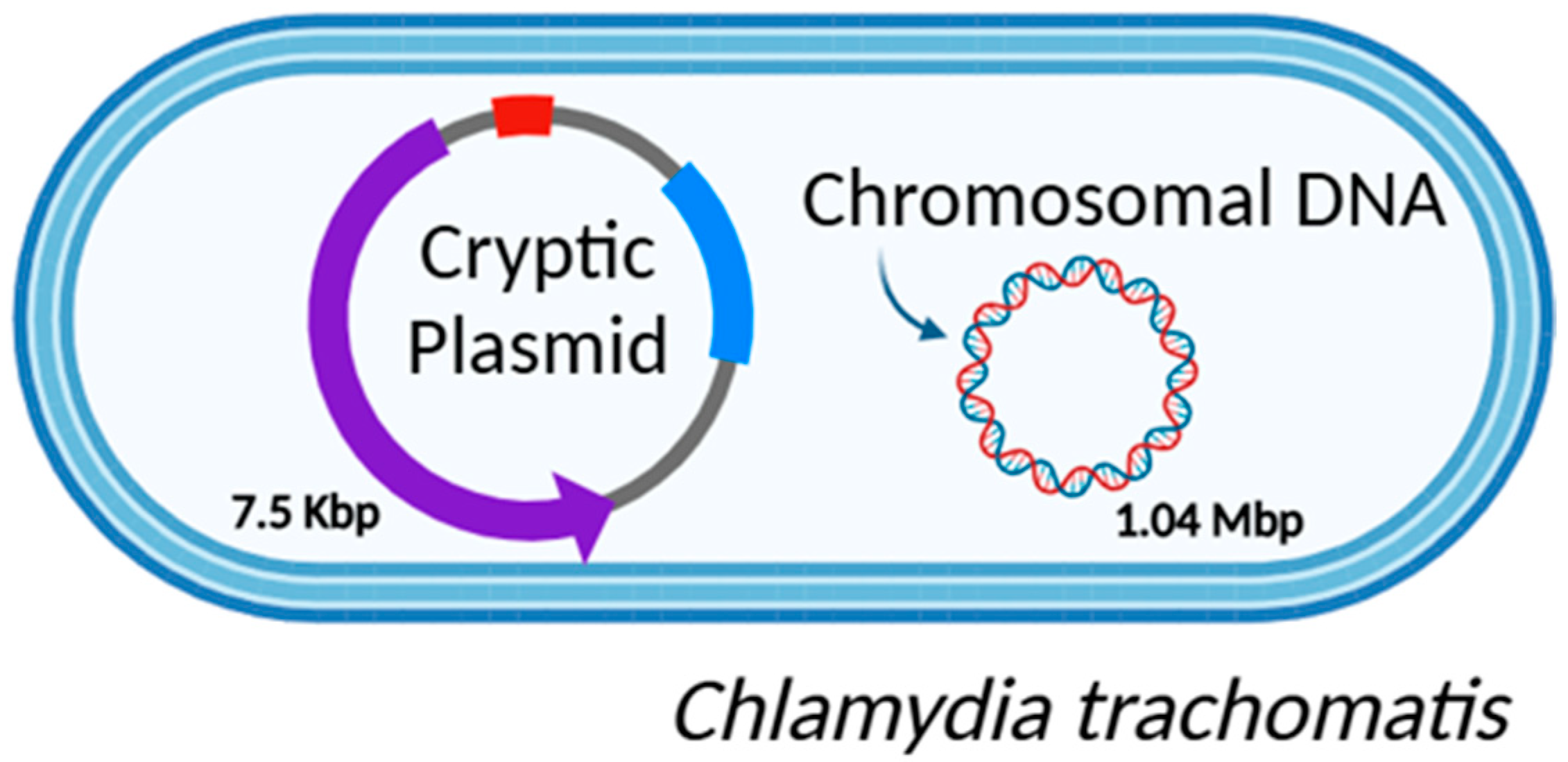
As detailed in our previous work, early research on CT biology was rudimentary, relying primarily on basic cell culture, serology, and immunohistochemistry techniques [31]. However, recent technological advances, including high-resolution microscopy and the development of omics approaches, have significantly advanced the field of research into the genetic diversity of bacteria [32]. In particular, the advent of new sequencing methodologies using molecular techniques has been crucial for studying pathogens like CT, which is challenging to manipulate in vitro, mostly due to its unique biology and developmental lifecycle [33]. These molecular advancements have enabled researchers to thoroughly characterize this pathogen, as well as many others. Researchers demonstrated that CT has a small genome (Figure 2) due to the long-term adaptation to an obligate intracellular lifestyle constituted by a circular chromosome of 1.04 Mbp containing 900 to 1500 coding sequences. Additionally, it is constituted by a highly conserved chlamydial virulent plasmid of approximately 7.5 Kbp, which may be present in several copies [34,35,36].
Of note, most of the bacterial variability is attributed to single nucleotide polymorphisms (SNPs), influencing virulence and tissue tropism and leading to CT infections in particular regions of the human body. Therefore, CT is classified into different biovars: trachoma, epithelial ano-urogenital, and LGV biovars [37]. This distinction is extremely important for the correct treatment and management of CT infection. In detail, the trachoma biovar infects conjunctival epithelial cells, causing trachoma, the leading cause of preventable blindness. The ano-urogenital biovar, the most frequent infection of both sexes, is responsible for severe complications related to the reproductive tract, and it is able to infect many other epithelial cells, such as those in the respiratory, gastrointestinal, and ocular (non-trachoma) tracts. Also, it can cause neonatal infections through vertical transmission. Finally, the LGV biovar is much more invasive than the others, and it can cause lymphogranuloma venereum (LGV), a systemic infection that starts in the lymphatic tissue and can cause irreversible sequelae if not treated adequately [38,39,40]. These biovars are further divided into different serovars based on the variability of the ompA gene, which confers specific characteristics to the bacterium [40]. The ompA gene encodes the major outer membrane protein (MOMP) of the bacterium and exhibits polymorphism through single nucleotide polymorphisms (SNPs) [32,41]. Based on these polymorphisms, the trachoma biovar includes serovars A to C. Serovars D to K are the most frequent and, as mentioned before, can infect all the following tracts: urogenital, pharyngeal, conjunctiva, and anorectal [42,43]. Lastly, the LGV biovar comprises serovars L1 to L3 (Table 1) [9].
It is interesting to understand that from the molecular point of view, there is genomic conservation between all the different CT serovars in more than 98%. Notwithstanding, despite these genotypes’ similarity, phenotypically there are evident differences, specifically in terms of infection outcomes and bacteria tissue tropism [32]. Therefore, is it clear that minor genetic variations provide key characteristics of the bacterium, influencing infection pathobiology. In detail, to date, these key variations are described as occurring due to SNPs, insertion or deletions (INDELs) in genomic DNA, or bacterium plasmid variations. Also, epigenetic factors could contribute to the differences in infection outcome, specially through a mechanism of miRNA regulation [44].

2.1. CT Genomic DNA

The small, circularized genome of CT is composed of some important elements—specifically, the plasticity zone (PZ), T3SS, Incs, Pmps, ompA, tarP, and Trp operons [32,45]. Some of these are represented in Table 2 and have great significance in the bacterium virulence and tissue tropism, especially the PZ genomic region, which is highly variable, containing more than 45 genes, partial Trp operons, and the cytotoxin gene (tox) [46]. This particular tox gene is informative, identifying CT tissue tropisms. Tox open reading frames (ORFs) can be intact in urogenital strains, truncated in ocular strains, or absent in LGV strains [47]. Additionally, the T3SS region is one of the most crucial elements for the translocation of effector proteins into the host cell. It contains 20 to 25 genes encoding for highly conserved apparatus scaffolding, inner membrane components, extracellular needles, chaperons, and various effectors [9,48]. TarP is one of these T3SS effectors. It is responsible for actin cytoskeleton remodeling, contributing to the entry of CT into the host cell. Variations in this gene sequence, translated into distinct N-terminal tyrosine-repeat units and C-terminal binding domains, are theorized to contribute to bacterium virulence and tissue tropisms [49]. Another relevant region is the one encoding for Inc proteins, which may be associated with tissue tropisms and disease severity between LGV and trachoma biovars of CT [9]. In detail, when some variations in amino acids occur in regions of Inc proteins, which are exposed to the host cytoplasm during the infection, they may influence host–pathogen interactions and immune recognition [49,50]. Almeida and colleagues found that LGV strains present a characteristic gene expression pattern in CT058, CT192, and CT214 of the Inc region, mediated by specific gene mutations in CT192 and CT214, and by post-transcriptional regulation of CT058 transcripts, which may be associated with tissue tropism targeted more towards macrophages [50]. In addition, pmp genes encode for a group of potential virulence factors: polymorphic membrane proteins (Pmps), a family of surface-exposed membrane proteins. To the best of our knowledge, the exact function of Pmps remains unclear. Notwithstanding, it is hypothesized that they are implicated in attachment, invasion, and immune evasion processes [51]. Interestingly, although genome reduction is obvious for almost every class of proteins of the distinct species of Chlamydia (genus), there is expansion of the Pmp family, which holds distinctive significance [52,53]. The OmpA gene also plays a key role in the bacterium genome, as it is highly polymorphic and encodes for the chlamydial major outer membrane protein (MOMP), a surface-exposed porin protein. This gene is composed of four highly polymorphic variable sequences (VSs), which are flanked by five constant sequences (CSs) [54]. As described before, it is used for CT serovar classification [55].
Indeed, there are several genomic regions where occur variation could occur, resulting in distinct infection and outcomes.

2.2. CT Plasmid Diversity

Chlamydial plasmid contains eight coding sequences (CDSs), known as plasmid glycoproteins (pGPs 1 to 8), whose role is to facilitate the infection ascension, induce inflammation, and contribute to the extrusion process, as well as four tandem repeat sequences [56,57,58]. Each CDS encodes distinct proteins, including integrase (for plasmid replication), helicase, a secreted virulence protein (associated with persistent infection), a transcriptional regulator for genes located both on the plasmid and on the chromosome, glycogen synthase, infectivity factors, and partitioning proteins [56,59,60,61].
In the diagnosis of C. trachomatis infection, the CDSs of the bacterium’s plasmid were valuable targets for testing due to their stability and high copy number [62]. Nonetheless, the emergence of a mutant strain from Sweden, which contains a 377 bp deletion in CDS1 of the plasmid, has led to false negative test results, making this target unsuitable for reliable detection [63]. Of note, this variant was characterized using whole genome sequencing (WGS) and has since been occasionally reported in other countries [64]. Additionally, other variants, resulting from mutations in CT plasmid, such as the Finnish New Variant, have been reported recently, emphasizing the need for more sensible diagnosis using not only plasmid-based targets for bacterium detection [62].
Despite CT plasmid not being essential for bacterium survival, it is crucial to attribute distinct virulence. In vivo studies with a CT strain carrying plasmid deletion have showed its significance to pathology and inflammation [65]. Sigar et al. demonstrated that without the plasmid, there was a virulence attenuation and a lower inflammatory response, concluding that the type of plasmid influences the disease outcome [66]. Additionally, it is known that in stress conditions, the copy number of plasmids increases; however, not all the functions of this bacterial entity are yet understood [58].

2.3. Patho-Epigenetics in Chlamydial Infection

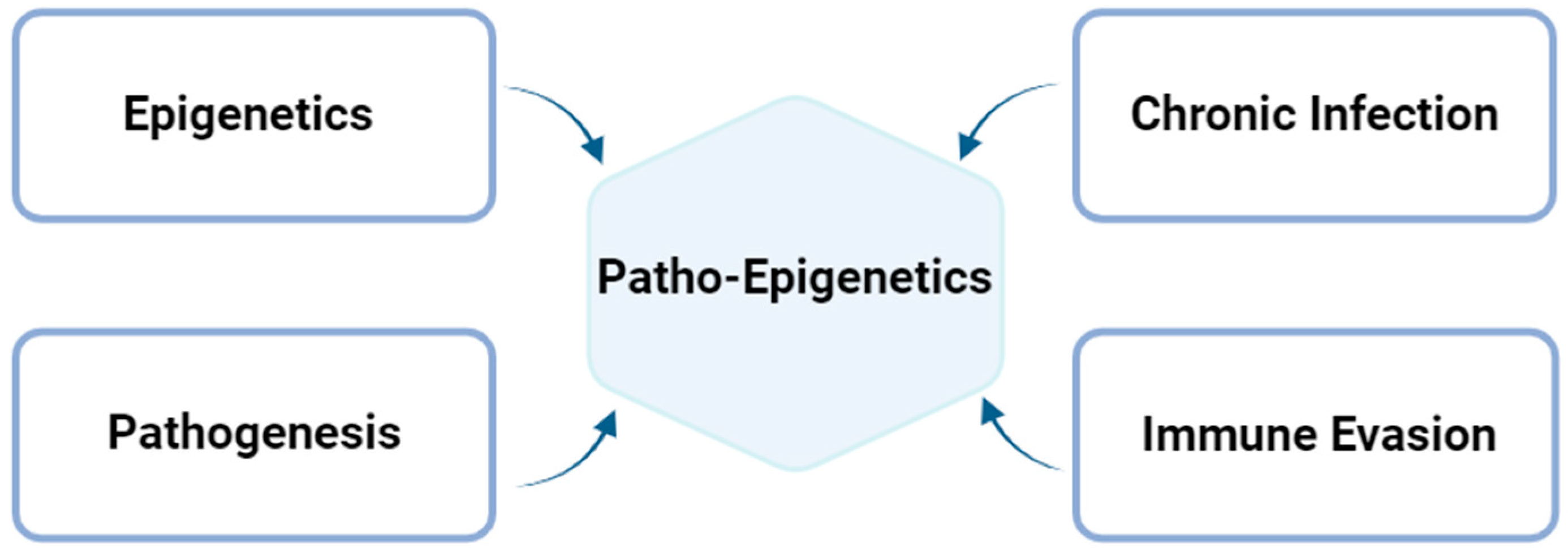
Epigenetic modifications are alterations affecting gene expression that occur in the genome without changing the DNA sequence [66]. The most common types of these modifications include DNA methylation, post-translational modifications of the histones, and gene silencing via non-coding RNA, such as miRNAs or siRNAs [67]. These modifications can be influenced by environmental factors, lifestyle, or infections caused by specific pathogens, such as viruses and intracellular bacteria. Specifically, patho-epigenetics is a term that encompasses the development of a disease (pathogenesis) prompted by epigenetic dysregulation [68]. In the specific case of prolonged chlamydial infection, this designation arises from distinct factors that contribute to the patho-epigenetic establishment, as represented below (Figure 3). Where CT invades the host, it is well known that there is a possibility that it could be a long-term infection [7]. This chronic infection could potentially increase the epigenetic changes, as well as the bacterium’s ability to evade the immune system [69]. Altogether, these dysregulations contribute to epigenetic changes associated with the pathogenesis created by the bacterium itself [68].
In fact, there is evidence that intracellular bacteria can trigger epigenetic changes in the host cells to guarantee their maintenance, replication, and transmission [70,71]. CT has been reported to trigger specific epigenetic modifications in the host cell that are linked to its pathogenic effects [72]. One key role of the bacterium is the regulation of miRNAs, which are small non-coding RNA molecules that play a critical function in controlling gene expression by binding to messenger RNA (mRNA) and preventing the production of certain proteins [73]. miRNAs are essential for processes such as cell proliferation, immune response, and tissue development [74]. CT infection disrupts normal miRNA processing, leading to dysregulation of several important pathways [75]. This disruption occurs through RNA interference (RNAi), a cellular mechanism where miRNAs and other small RNAs silence specific genes after transcription [75,76]. RNAi can result in either mRNA degradation or the inhibition of protein translation, depending on the pathway involved [77]. This disruption of gene regulation is significant and requires careful exploration. In detail, two distinct mechanisms are thought to be involved in this process [73]. The first is the inactivation of the Dicer, an essential component in generating miRNAs and whose role is to cleave precursor RNA molecules to mature miRNAs, which are crucial for several cellular processes, such as cell proliferation and immune response [78,79,80]. Indeed, there are some reports showing that Dicer modifications are involved in infertility in in vivo mouse models [81]. Additionally, the second mechanism involves Argonaute 2 (Ago2), a key protein in RNAi [73]. During CT infection, Ago2 levels are reduced, which significantly impacts fertility by affecting key processes in the female reproductive tract. This dysregulation may lead to infertility and other pathological conditions, such as tumorigenesis [80]. Thus, it could be interesting to focus some future studies on the epigenetic landscape to further understand the potential of these modifications in human infections in order to uncover potential biomarkers associated with chlamydial infection severity and infertility.

3. Long-Term Chlamydial Urogenital Infection and Infertility

Despite the significant rate of spontaneously resolved chlamydial infections, specifically with 50% of cases resolving within one year, 82% resolving within two years, and 95% of cases resolving within three years, there is still a risk of developing severe complications [82,83]. One of the potentially most significant long-term consequences of chlamydial infection is infertility [84]. The repercussions of infertility extend beyond the individual, affecting psychological well-being, the couple’s stability, societal dynamics, and even the economy of countries [85].
According to the World Health Organization (WHO), infertility affects approximately one in six individuals globally [86]. This fact underscores the urgent need for research to understand the factors contributing to this trend of increasing rate, highlighted by Centers for Disease Control and Prevention (CDC) [87]. The prevention of infertility is extremely important; thus, given the far-reaching implications of chlamydial infection on reproductive health, it is crucial to examine its role in infertility to better understand the extent of its influence [88,89]. In the literature, there is accordance that if this bacterium goes undiagnosed or if there are repeated infections, it can lead to severe consequences in the female upper genital tract [90,91,92]. Also, in males, evidence suggests that the infection may influence their reproductive capability [93]. Notwithstanding, it is important to reinforce that the association between chlamydial infection and infertility is still under scientific debate, as a direct causal relationship is not well documented in the literature, and the majority of the studies have significant biases. Furthermore, the ECDC has published a document defending that a significant proportion of chlamydial infections in women do not lead to PID, and that even when PID does occur, it does not always result in infertility [94].
Chlamydial genital infections are often asymptomatic, meaning that the infected individuals are not diagnosed and treated properly, thus transmitting the infection to their sexual partners and becoming more susceptible to long-term complications from the infection [95]. Importantly, this infection affects mostly younger adults (<25 years old), having a potentially high impact on their fertility capability if not detected and treated in a timely manner [96]. This persistent presence of the pathogen in the genital organs leads to a chronic immune response, characterized by the increased production of pro-inflammatory cytokines, creating a pro-inflammatory milieu favorable to epithelial cell damage [91,97,98]. Subsequently, this pro-inflammatory response gradient could trigger serious repercussions for women’s reproductive health, including PID, chronic pelvic pain, adverse pregnancy outcomes, infertility, and even tumorigenesis [5,99].
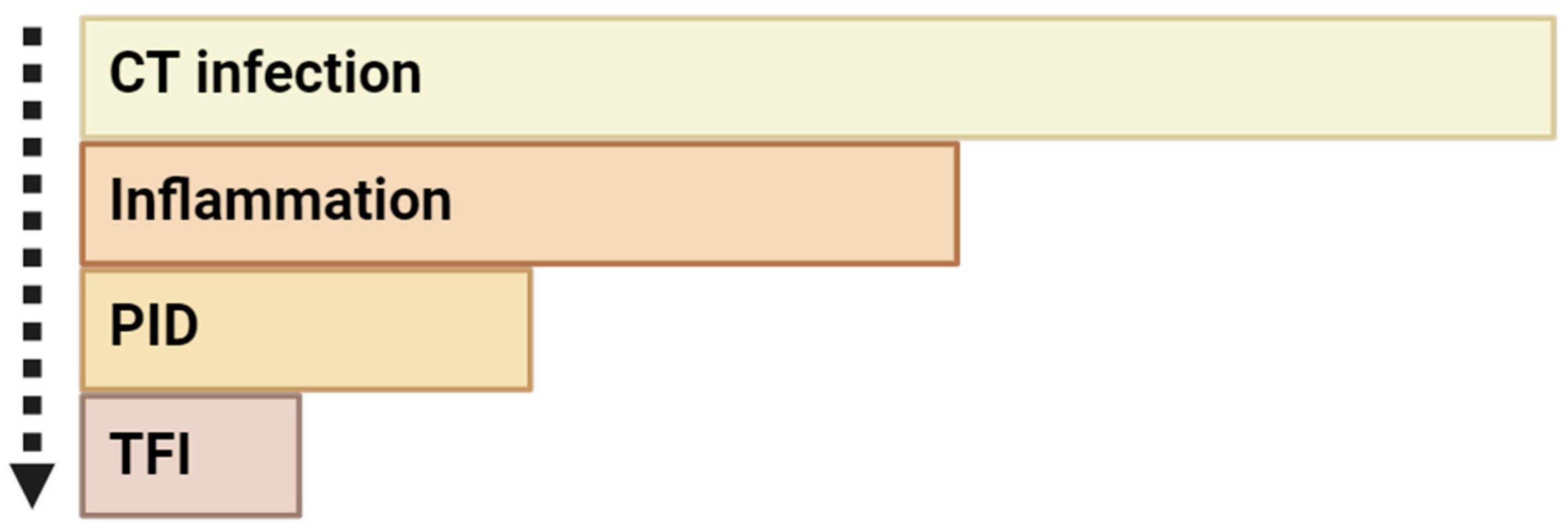
Indeed, infertility may result from scar formation and occlusion of the fallopian tubes, a possible consequence of chlamydial infection (Figure 4) [20,100]. In detail, during this infection, the bacterium can ascend from the lower genital tract, reaching the uterus, fallopian tubes, and ovaries [101]. This process triggers an exacerbated immune response, responsible for creating a pro-inflammatory cascade that causes significant tissue damage and fibrosis, ultimately leading to pelvic inflammatory disease (PID) [102,103,104]. Then, scarring and blocking of the fallopian tubes leads to a condition referred to as tubal factor infertility (TFI) [105]. In most cases, TFI arises from inflammation of the epithelial tissues of the fallopian tubes and pelvic–peritoneal adhesions, which results in the inability for fertilization or embryo implantation [106]. This means that one of the possible underlying mechanisms of CT infection causing infertility is the intensified host immune response, which establishes an inflammatory milieu favorable to tissue damage in the reproductive organs [92].
In fact, first, it is important to understand how the immune system of the host reacts when the chlamydial infection occurs, as it must be our “defense shield” against external agents such as CT [107,108]. Briefly, through cell surface and endosomal receptors, the immune system is aware that an infection occurred. In the cervicovaginal mucosa, microbiota—specifically, lactobacillus—confer an important defense to this region [109]. Therefore, imbalance in women’s vaginal microbiota confers a predisposition to vaginal infections [110]. If conditions are reunited, CT surpasses this barrier and the infection process begins. Host immune responses against the bacterium primarily occur through the innate immune system, the first line of defense, providing a rapid, non-specific response to pathogens. No prior exposure is required to recognize the pathogens, and mast cells are among the first barrier that CT has to surpass [111,112]. In addition, dendritic cells play a crucial role, not only by capturing and processing antigens but also by triggering the adaptative immune response, presenting the antigen to T cells through the molecules of major histocompatibility complex class I or class II (MHC-I and MHC-II, respectively) [113]. Subsequently, activated T cells (effector and memory cells) and NK cells produce IFN-γ to activate macrophages, whose role is to clear the infection [114,115,116,117]. Of note, antigen-specific immunity, producing immunity via B-cell involvement, also forms part of the adaptative immune response, which is also important for fighting against this pathogen on a secondary level [111,117].
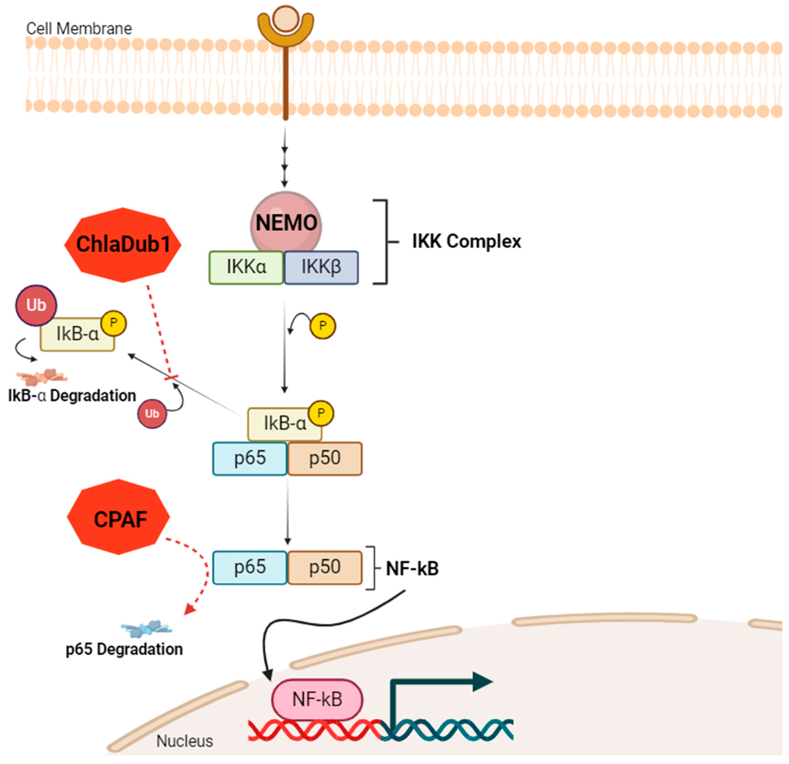
The mechanisms of CT to evade the host immune system are diverse. There is recent in vitro evidence that NF-kB, a transcription factor that plays an important role in the regulation of inflammatory response, immune cell activation, and the production of some cytokines, is actively inhibited during CT infection [118]. Of note, NF-kB transcription factor is a complex formed by two distinct units: p65 and p50 [119]. Le Negrate and colleagues described two possible mechanisms underlying NF-kB pathway dysregulation (Figure 5) [120]. In their study, the authors demonstrated that a deubiquitinating protease from the bacterium ChlaDub1, which is expressed by CT in infected cells, has the ability of NF-kB pathway inhibition, acting downstream of the IKK complex, which is composed of three different units: NEMO, IKKα, and IKKβ [119]. In detail, ChlaDub1 blocks IkB-α degradation through the impairment of its ubiquitination [120]. Therefore, the transcription factor NF-kB is not able to migrate to the cell nucleus. Another mechanism is p65 degradation through a chlamydial protease or proteasome-like activity factor (CPAF), destroying a subunit of NF-kB [121].
Moreover, CT can also evade the host immune system using other mechanisms, such as inhibiting the cytokines and chemotactic molecules produced by epithelial cells, interfering with antigen-presenting cell (APC) functions, or downregulating MHC class I and II molecules [122]. Therefore, by reducing the antigen-presenting capacity of the APCs and subsequently reducing immune cell recruitment to the region of the infection, CT leads to lower immune system activity and capacity to clear the infection [122,123].
CT intracellular lifecycle allows the bacterium to remain protected within the inclusion body, shielding it from humoral immunity [111]. Additionally, recent findings showed that CT has another way to escape the immune system: by RBs, which can invade other host cells through nanotubes (TNTs), avoiding the extracellular microenvironment, which is in reach of immune cells and molecules, and thus increasing the bacterium’s survival chances [124]. In addition, the bacterium could enter a state of persistence and latency, helping it survive under less favorable conditions in the microenvironment [12]. CT also evades host cell apoptosis, which is crucial for its replication and survival [111]. Moreover, the bacterium can modulate the immune response, leading to an exacerbated inflammatory response that may cause organ damage. During this process, symptoms are often absent, which can result in irreversible reproductive damage, such as infertility [125,126].
In fact, currently, the mechanisms of host immune evasion by CT still need to be further explored to understand the interaction between the immune cells and the bacterium to better combat the infection [111]. It is also important to highlight that pathogens other than CT can cause reproductive organs damage and that these infections can co-occur in the same host, such as Neisseria gonorrhoeae infection, HPV, HIV, syphilis, Trichomonas vaginalis, bacterial vaginosis, and Mycoplasma genitalium [5,125,127,128,129]. Therefore, these co-infections could potentially increase the risk of severe sequelae, such as tumorigenesis and infertility, highlighting the relevant role of multi-test diagnosis of STIs [5].
In men, the association between chlamydial infection and infertility it is not as well studied as it is in women. Notwithstanding, CT infection in males can evidently lead to inflammatory conditions such as urethritis, epididymitis, or, less commonly, prostatitis [130,131,132,133]. Furthermore, evidence suggests that CT infection may affect spermatogenesis—in detail, the bacterium can penetrate inside the spermatozoa, affecting the quantity and quality of sperm [82,83,134]. Additionally, some authors argue that the bacterium interacts with sperm, inducing oxidative stress that leads to apoptosis of the cells [135]. Indeed, CT could also cause obstructive azoospermia, interfering with spermatozoid motility [135,136].
Recent studies have provided additional insights into these mechanisms. Stojanov and colleagues have proposed the “intracellular bacterial hypothesis”, which suggests that intracellular bacteria like C. trachomatis can impair male fertility by interacting directly with sperm cells, inducing oxidative stress and triggering apoptosis. This aligns with findings that CT infection can alter sperm motility and viability, potentially affecting overall fertility [137]. In line with this, Wang et al. (2021) highlighted the role of oxidative stress and autophagy in the pathogenesis of bacterial infections, including CT, as contributors to male infertility. These findings suggest that prolonged CT infections may interfere with sperm function beyond the standard sperm parameters assessed in routine analysis, potentially affecting processes like fertilization or implantation [138].
Moreover, Moazenchi 2018 conducted a comprehensive study demonstrating the significant impacts of CT infection on sperm parameters, including motility and morphology. Importantly, through flow cytometry, the authors also assessed the DNA fragmentation index of the sperm. Finally, the authors suggest that DNA damage in sperm and seminal parameter alterations (concentration, morphology, viability, and motility) might be directly associated with CT infection and infertility [139,140]. Interestingly, last year, Haratian and colleagues identified an SNP in the DEFB126 gene (rs11467417), which is primarily expressed in the principal epididymal epithelial cells. This genetic variant may increase susceptibility to CT infection and subsequent infertility in men [141].
Therefore, based on cumulative evidence, it is possible to affirm that there may be an association between chlamydial long-term infections and male fertility impairment, although contradictory findings in the literature highlight the need for further investigation [142].
It is important to highlight that infertility is indeed a huge global health issue that carries profound economic and societal consequences [143]. According to Inhorn and Patrizio, a country’s economic burden associated with infertility includes direct healthcare costs, such as fertility treatments, and indirect costs, such as lost productivity and the psychological health impairment of individuals and their families [144]. As a consequence, infertility may also hinder the ability to maintain employment, potentially resulting in prolonged economic or financial challenges [145]. Given the significant personal and societal impacts, it is critical that countries prioritize investment in preventive measures for infertility cases, beginning with preventable factors such as STIs—particularly chlamydial infections. Preventive measures such as increased screening, early diagnosis, and treatment of chlamydial infections are cost-effective solutions that could alleviate both the financial burden on healthcare systems and the emotional suffering of those affected [146].

4. Conclusions and Future Directions

CT, the most common cause of bacterial STIs globally, has become a growing concern due to the sequelae that prolonged urogenital infection can leave [147]. One of the primary consequences of chlamydial urogenital infection is the activation of the host’s immune response, leading to the establishment of a pro-inflammatory milieu during prolonged infection. As a result, several complications may arise in both sexes, including chronic pain, urethritis, pelvic inflammatory disease (PID), infertility, pregnancy complications, epididymitis, prostatitis, and an increased risk of contracting other STIs. Moreover, there is evidence suggesting that these prolonged infections may be associated with events of oncogenesis. Of note, following the ECDC’s most recent data, CT infection rates are increasing. Therefore, these trends highlight the importance of primary and secondary prevention of STIs, with education on safe sex practices, and due to their asymptomatic presentation, the establishment of routine screening tests in all countries to detect infections in a timely manner and reduce their spread.
Throughout this article, we have reviewed the key biological and genetic aspects of CT, focusing on its genetic variations and their impact on infection outcomes. Indeed, genetic polymorphisms of CT are key determinants in the variability of bacterium virulence and tissue tropisms. Its unique lifecycle, characterized by the alternation between elementary bodies and reticulate bodies, allows CT to evade the immune system, contributing to persistent infections and making the clinical outcome of infection worse. These long-term urogenital infections influence the immune microenvironment and exacerbate inflammatory responses, contributing to tissue scarring and damage to the reproductive organs. Additionally, there is evidence in the literature suggesting that chlamydial infection could be associated with the development of infertility in both men and women. Notwithstanding, while the association between infection and reproductive sequelae is already demonstrated, the mechanisms underlying the infection and fertility impairment in both sexes remain unclear.
The analysis of the literature reveals that the association between infertility and CT infections has not been thoroughly investigated. Complications from infection may vary depending on the bacterial serovar, as well as individual factors of the infected person, such as the immune status and the vaginal flora. Notwithstanding, several important aspects remain unclear, including the self-clearance of CT infections and the interactions between the bacterium and immune system cells. There is lack of fundamental studies, and some of the findings came from research where numerous other variables, including co-infections, may act as confounding factors, influencing the conclusions drawn. It is also important to highlight that some studies exploring this association have some limitations that prevent an accurate generalization of their results. Additionally, biases may arise from differences in CT detection methods and infertility criteria across studies. Regarding the mechanisms underlying the infection, some findings are based on in vivo or in vitro models, which do not fully replicate the complexity of human infection. These limitations highlight the need for further studies with larger sample sizes and standardized criteria for bacterial detection and infertility assessment. Moreover, laboratory findings should be integrated with clinical studies and longitudinal data analysis to better understand the long-term outcomes of infection.
To address the gaps in knowledge, future research should also focus on the development of clinical applications aimed at improving the prevention, diagnosis, and treatment of this bacterial infection. In order to better manage the infection worldwide, vaccines against CT represent a promising avenue, particularly when administered to adolescents before their first sexual intercourse, as this could reduce the burden of asymptomatic infections and prevent complications such as infertility. Notwithstanding, despite the enormous efforts of the scientific community to develop a vaccine against this pathogen, no viable solutions of this type are currently available. In the absence of a vaccination plan, efforts can shift toward a more personalized approach to managing CT infections. While vaccines may one day be adapted to individual immune profiles or genetic markers, current advances in personalized medicine can already offer valuable alternatives. For instance, identifying genetic polymorphisms in CT and the host immune system can pave the way for targeted treatments based on host genetic markers or immune profiles. This approach enables a better understanding of individual susceptibility to CT infection and allows for therapies that improve treatment efficacy and reduce adverse effects.
These advances, coupled with a deeper understanding of CT pathogenesis, will be crucial in addressing the global burden of CT infection and improving reproductive health outcomes.

Author Contributions

Conceptualization N.V.; methodology, R.R. and N.V.; validation, N.V.; formal analysis, R.R. and N.V.; investigation, R.R., C.S., A.B. and N.V.; resources, N.V.; data curation, R.R.; writing—original draft preparation, R.R.; writing—review and editing, C.S., A.B. and N.V.; visualization, N.V.; supervision, N.V.; project administration, N.V.; funding acquisition, N.V. All authors have read and agreed to the published version of the manuscript.

Funding

This work was financed by the FEDER—Fundo Europeu de Desenvolimento Regional through COMPETE 2020—Operational Programme for Competitiveness and Internationalization (POCI), Portugal 2020, and by Portuguese funds through the FCT—Fundação para a Ciência e a Tecnologia, in a framework of the projects in CINTESIS, R&D Unit (reference UIDB/4255/2020), and within the scope of the project “RISE—LA/P/0053/2020”. N.V. would also like to thank the support from the FCT and FEDER (European Union), award number IF/00092/2014/CP1255/CT0004, and the Chair in Onco-Innovation at the FMUP.

Institutional Review Board Statement

Not applicable.

Informed Consent Statement

Not applicable.

Acknowledgments

RR thanks FCT for her Ph.D. grant (2022.11755.BDANA).

Conflicts of Interest

The authors declare no conflict of interest.

References

  1. ECDC STI Cases on the Rise Across Europe. Available online: https://www.ecdc.europa.eu/en/news-events/sti-cases-rise-across-europe (accessed on 10 November 2024).
  2. Becker, Y. Chlamydia. In Medical Microbiology, 4th ed.; University of Texas Medical Branch at Galveston: Galveston, TX, USA, 1996. [Google Scholar]
  3. Rodrigues, R.; Sousa, C.; Vale, N. Chlamydia trachomatis as a Current Health Problem: Challenges and Opportunities. Diagnostics 2022, 12, 1795. [Google Scholar] [CrossRef] [PubMed]
  4. Huai, P.; Li, F.; Chu, T.; Liu, D.; Liu, J.; Zhang, F. Prevalence of genital Chlamydia trachomatis infection in the general population: A meta-analysis. BMC Infect. Dis. 2020, 20, 589. [Google Scholar] [CrossRef] [PubMed]
  5. Rodrigues, R.; Sousa, C.; Vale, N. Deciphering the Puzzle: Literature Insights on Chlamydia trachomatis-Mediated Tumorigenesis, Paving the Way for Future Research. Microorganisms 2024, 12, 1126. [Google Scholar] [CrossRef]
  6. Stelzner, K.; Vollmuth, N.; Rudel, T. Intracellular lifestyle of Chlamydia trachomatis and host–pathogen interactions. Nat. Rev. Microbiol. 2023, 21, 448–462. [Google Scholar] [CrossRef]
  7. Witkin Steven, S.; Minis, E.; Athanasiou, A.; Leizer, J.; Linhares Iara, M. Chlamydia trachomatis: The Persistent Pathogen. Clin. Vaccine Immunol. 2017, 24, e00203-17. [Google Scholar] [CrossRef]
  8. Elwell, C.; Mirrashidi, K.; Engel, J. Chlamydia cell biology and pathogenesis. Nat. Rev. Microbiol. 2016, 14, 385–400. [Google Scholar] [CrossRef]
  9. Bugalhão, J.N.; Mota, L.J. The multiple functions of the numerous Chlamydia trachomatis secreted proteins: The tip of the iceberg. Microb. Cell 2019, 6, 414–449. [Google Scholar] [CrossRef] [PubMed]
  10. Zhang, J.P.; Stephens, R.S. Mechanism of C. trachomatis attachment to eukaryotic host cells. Cell 1992, 69, 861–869. [Google Scholar] [CrossRef]
  11. Rodrigues, R.; Vieira-Baptista, P.; Catalão, C.; Borrego, M.J.; Sousa, C.; Vale, N. Chlamydial and Gonococcal Genital Infections: A Narrative Review. J. Pers. Med. 2023, 13, 1170. [Google Scholar] [CrossRef] [PubMed]
  12. Di Pietro, M.; Filardo, S.; Romano, S.; Sessa, R. Chlamydia trachomatis and Chlamydia pneumoniae Interaction with the Host: Latest Advances and Future Prospective. Microorganisms 2019, 7, 140. [Google Scholar] [CrossRef] [PubMed]
  13. Rodrigues, R.; Marques, L.; Vieira-Baptista, P.; Sousa, C.; Vale, N. Therapeutic Options for Chlamydia trachomatis Infection: Present and Future. Antibiotics 2022, 11, 1634. [Google Scholar] [CrossRef] [PubMed]
  14. Sahu, R.; Verma, R.; Dixit, S.; Igietseme, J.U.; Black, C.M.; Duncan, S.; Singh, S.R.; Dennis, V.A. Future of human Chlamydia vaccine: Potential of self-adjuvanting biodegradable nanoparticles as safe vaccine delivery vehicles. Expert Rev. Vaccines 2018, 17, 217–227. [Google Scholar] [CrossRef]
  15. Yang, S.; Zeng, J.; Yu, J.; Sun, R.; Tuo, Y.; Bai, H. Insights into Chlamydia Development and Host Cells Response. Microorganisms 2024, 12, 1302. [Google Scholar] [CrossRef] [PubMed]
  16. Hammerschlag, M.R.; Kohlhoff, S.A. Treatment of chlamydial infections. Expert Opin. Pharmacother. 2012, 13, 545–552. [Google Scholar] [CrossRef]
  17. Riska Paul, F.; Kutlin, A.; Ajiboye, P.; Cua, A.; Roblin Patricia, M.; Hammerschlag Margaret, R. Genetic and Culture-Based Approaches for Detecting Macrolide Resistance in Chlamydia pneumoniae. Antimicrob. Agents Chemother. 2004, 48, 3586–3590. [Google Scholar] [CrossRef] [PubMed]
  18. Panzetta, M.E.; Valdivia, R.H.; Saka, H.A. Chlamydia Persistence: A Survival Strategy to Evade Antimicrobial Effects in-vitro and in-vivo. Front. Microbiol. 2018, 9, 3101. [Google Scholar] [CrossRef]
  19. Jury, B.; Fleming, C.; Huston, W.M.; Luu, L.D.W. Molecular pathogenesis of Chlamydia trachomatis. Front. Cell. Infect. Microbiol. 2023, 13, 1281823. [Google Scholar] [CrossRef] [PubMed]
  20. Ambildhuke, K.; Pajai, S.; Chimegave, A.; Mundhada, R.; Kabra, P. A Review of Tubal Factors Affecting Fertility and its Management. Cureus 2022, 14, 30990. [Google Scholar] [CrossRef]
  21. den Heijer, C.D.J.; Hoebe, C.; Driessen, J.H.M.; Wolffs, P.; van den Broek, I.V.F.; Hoenderboom, B.M.; Williams, R.; de Vries, F.; Dukers-Muijrers, N. Chlamydia trachomatis and the Risk of Pelvic Inflammatory Disease, Ectopic Pregnancy, and Female Infertility: A Retrospective Cohort Study Among Primary Care Patients. Clin. Infect. Dis. 2019, 69, 1517–1525. [Google Scholar] [CrossRef]
  22. Garcia, M.R.; Leslie, S.W.; Wray, A.A. Sexually Transmitted Infections; StatPearls Publishing LLC: Treasure Island, FL, USA, 2024. [Google Scholar]
  23. Hunt, S.; Vollenhoven, B. Pelvic inflammatory disease and infertility. Aust. J. Gen. Pract. 2023, 52, 215–218. [Google Scholar] [CrossRef]
  24. Jennings, L.K.; Krywko, D.M. Pelvic Inflammatory Disease; StatPearls Publishing LLC: Treasure Island, FL, USA, 2024. [Google Scholar]
  25. Mitchell, C.M.; Anyalechi, G.E.; Cohen, C.R.; Haggerty, C.L.; Manhart, L.E.; Hillier, S.L. Etiology and Diagnosis of Pelvic Inflammatory Disease: Looking Beyond Gonorrhea and Chlamydia. J. Infect. Dis. 2021, 224 (Suppl. 2), 29–35. [Google Scholar] [CrossRef] [PubMed]
  26. Chen, M.Y.; Donovan, B. Screening for genital Chlamydia trachomatis infection: Are men the forgotten reservoir? Med. J. Aust. 2003, 179, 124–125. [Google Scholar] [CrossRef]
  27. Mohseni, M.; Sung, S.; Takov, V. Chlamydia; StatPearls Publishing LLC: Treasure Island, FL, USA, 2024. [Google Scholar]
  28. Gaydos, C.A.; Ferrero, D.V.; Papp, J. Laboratory aspects of screening men for Chlamydia trachomatis in the new millennium. Sex. Transm. Dis. 2008, 35, 45–50. [Google Scholar] [CrossRef] [PubMed]
  29. Cunningham, K.A.; Beagley, K.W. Male genital tract chlamydial infection: Implications for pathology and infertility. Biol. Reprod. 2008, 79, 180–189. [Google Scholar] [CrossRef] [PubMed]
  30. Joki-Korpela, P.; Sahrakorpi, N.; Halttunen, M.; Surcel, H.M.; Paavonen, J.; Tiitinen, A. The role of Chlamydia trachomatis infection in male infertility. Fertil. Steril. 2009, 91, 1448–1450. [Google Scholar] [CrossRef]
  31. Rodrigues, R.; Silva, A.R.; Sousa, C.; Vale, N. Addressing Challenges in Chlamydia trachomatis Detection: A Comparative Review of Diagnostic Methods. Medicina 2024, 60, 1236. [Google Scholar] [CrossRef] [PubMed]
  32. Luu, L.D.W.; Kasimov, V.; Phillips, S.; Myers, G.S.A.; Jelocnik, M. Genome organization and genomics in Chlamydia: Whole genome sequencing increases understanding of chlamydial virulence, evolution, and phylogeny. Front. Cell. Infect. Microbiol. 2023, 13, 1178736. [Google Scholar] [CrossRef]
  33. Meyer, T. Diagnostic Procedures to Detect Chlamydia trachomatis Infections. Microorganisms 2016, 4, 25. [Google Scholar] [CrossRef]
  34. Stephens, R.S.; Kalman, S.; Lammel, C.; Fan, J.; Marathe, R.; Aravind, L.; Mitchell, W.; Olinger, L.; Tatusov, R.L.; Zhao, Q.; et al. Genome Sequence of an Obligate Intracellular Pathogen of Humans: Chlamydia trachomatis. Science 1998, 282, 754–759. [Google Scholar] [CrossRef] [PubMed]
  35. Seth-Smith, H.M.; Harris, S.R.; Skilton, R.J.; Radebe, F.M.; Golparian, D.; Shipitsyna, E.; Duy, P.T.; Scott, P.; Cutcliffe, L.T.; O’Neill, C.; et al. Whole-genome sequences of Chlamydia trachomatis directly from clinical samples without culture. Genome Res. 2013, 23, 855–866. [Google Scholar] [CrossRef] [PubMed]
  36. Ouellette Scot, P.; Lee, J.; Cox John, V. Division without Binary Fission: Cell Division in the FtsZ-Less Chlamydia. J. Bacteriol. 2020, 202, e00252-20. [Google Scholar] [CrossRef] [PubMed]
  37. Grygiel-Górniak, B.; Folga, B.A. Chlamydia trachomatis—An Emerging Old Entity? Microorganisms 2023, 11, 1283. [Google Scholar] [CrossRef] [PubMed]
  38. Ceovic, R.; Gulin, S.J. Lymphogranuloma venereum: Diagnostic and treatment challenges. Infect. Drug Resist. 2015, 8, 39–47. [Google Scholar] [CrossRef] [PubMed]
  39. Ausar, S.F.; Larson, N.R.; Wei, Y.; Jain, A.; Middaugh, C.R. Subunit-based vaccines: Challenges in developing protein-based vaccines. Pract. Asp. Vaccine Dev. 2022, 4, 79–135. [Google Scholar]
  40. Roe, S.K.; Zhu, T.; Slepenkin, A.; Berges, A.; Fairman, J.; de la Maza, L.M.; Massari, P. Structural Assessment of Chlamydia trachomatis Major Outer Membrane Protein (MOMP)-Derived Vaccine Antigens and Immunological Profiling in Mice with Different Genetic Backgrounds. Vaccines 2024, 12, 789. [Google Scholar] [CrossRef]
  41. Thomson, N.R.; Clarke, I.N. Chlamydia trachomatis: Small genome, big challenges. Future Microbiol. 2010, 5, 555–561. [Google Scholar] [CrossRef]
  42. Holló, P.; Jókai, H.; Herszényi, K.; Kárpáti, S. Genitourethral infections caused by D-K serotypes of Chlamydia trachomatis. Orvosi Hetil. 2015, 156, 19–23. [Google Scholar] [CrossRef]
  43. Dunlop, E.M.; Harris, R.J.; Darougar, S.; Treharne, J.D.; Al-Egaily, S.S. Subclinical pneumonia due to serotypes D-K of Chlamydia trachomatis: Case reports of two infants. Br. J. Vener. Dis. 1980, 56, 337–340. [Google Scholar] [CrossRef] [PubMed]
  44. Nunes, A.; Borrego, M.J.; Gomes, J.P. Genomic features beyond Chlamydia trachomatis phenotypes: What do we think we know? Infect. Genet. Evol. 2013, 16, 392–400. [Google Scholar] [CrossRef] [PubMed]
  45. Byrne, G.I. Chlamydia trachomatis strains and virulence: Rethinking links to infection prevalence and disease severity. J. Infect. Dis. 2010, 201, 126–133. [Google Scholar] [CrossRef] [PubMed]
  46. Bachmann, N.L.; Polkinghorne, A.; Timms, P. Chlamydia genomics: Providing novel insights into chlamydial biology. Trends Microbiol. 2014, 22, 464–472. [Google Scholar] [CrossRef] [PubMed]
  47. Dimond, Z.E.; Suchland, R.J.; Baid, S.; LaBrie, S.D.; Soules, K.R.; Stanley, J.; Carrell, S.; Kwong, F.; Wang, Y.; Rockey, D.D.; et al. Inter-species lateral gene transfer focused on the Chlamydia plasticity zone identifies loci associated with immediate cytotoxicity and inclusion stability. Mol. Microbiol. 2021, 116, 1433–1448. [Google Scholar] [CrossRef]
  48. da Cunha, M.; Pais, S.V.; Bugalhão, J.N.; Mota, L.J. The Chlamydia trachomatis type III secretion substrates CT142, CT143, and CT144 are secreted into the lumen of the inclusion. PLoS ONE 2017, 12, 178856. [Google Scholar] [CrossRef] [PubMed]
  49. Borges, V.; Nunes, A.; Ferreira, R.; Borrego, M.J.; Gomes, J.P. Directional evolution of Chlamydia trachomatis towards niche-specific adaptation. J. Bacteriol. 2012, 194, 6143–6153. [Google Scholar] [CrossRef] [PubMed]
  50. Almeida, F.; Borges, V.; Ferreira, R.; Borrego, M.J.; Gomes, J.P.; Mota, L.J. Polymorphisms in inc proteins and differential expression of inc genes among Chlamydia trachomatis strains correlate with invasiveness and tropism of lymphogranuloma venereum isolates. J. Bacteriol. 2012, 194, 6574–6585. [Google Scholar] [CrossRef]
  51. Nunes, A.; Gomes, J.P.; Karunakaran, K.P.; Brunham, R.C. Bioinformatic Analysis of Chlamydia trachomatis Polymorphic Membrane Proteins PmpE, PmpF, PmpG and PmpH as Potential Vaccine Antigens. PLoS ONE 2015, 10, 131695. [Google Scholar] [CrossRef] [PubMed]
  52. Debrine Abigail, M.; Karplus, P.A.; Rockey Daniel, D. A structural foundation for studying chlamydial polymorphic membrane proteins. Microbiol. Spectr. 2023, 11, e03242-23. [Google Scholar] [CrossRef] [PubMed]
  53. Vasilevsky, S.; Stojanov, M.; Greub, G.; Baud, D. Chlamydial polymorphic membrane proteins: Regulation, function and potential vaccine candidates. Virulence 2016, 7, 11–22. [Google Scholar] [CrossRef] [PubMed]
  54. Zhao, J.; Shui, J.; Luo, L.; Ao, C.; Lin, H.; Liang, Y.; Wang, L.; Wang, H.; Chen, H.; Tang, S. Identification and characterization of mixed infections of Chlamydia trachomatis via high-throughput sequencing. Front. Microbiol. 2022, 13, 1041789. [Google Scholar] [CrossRef]
  55. Millman, K.; Black, C.M.; Stamm, W.E.; Jones, R.B.; Hook, E.W.; Martin, D.H.; Bolan, G.; Tavaré, S.; Dean, D. Population-based genetic epidemiologic analysis of Chlamydia trachomatis serotypes and lack of association between ompA polymorphisms and clinical phenotypes. Microbes Infect. 2006, 8, 604–611. [Google Scholar] [CrossRef]
  56. Pawlikowska-Warych, M.; Śliwa-Dominiak, J.; Deptuła, W. Chlamydial plasmids and bacteriophages. Acta Biochim. Pol. 2015, 62, 1–6. [Google Scholar] [CrossRef]
  57. Versteeg, B.; Bruisten, S.M.; Pannekoek, Y.; Jolley, K.A.; Maiden, M.C.J.; van der Ende, A.; Harrison, O.B. Genomic analyses of the Chlamydia trachomatis core genome show an association between chromosomal genome, plasmid type and disease. BMC Genom. 2018, 19, 130. [Google Scholar] [CrossRef]
  58. Zhang, S.; Jiang, Y.; Yu, Y.; OuYang, X.; Zhou, D.; Song, Y.; Jiao, J. Autophagy: The Misty Lands of Chlamydia trachomatis Infection. Front. Cell. Infect. Microbiol. 2024, 14, 1442995. [Google Scholar] [CrossRef]
  59. Yang, C.; Kari, L.; Lei, L.; Carlson, J.H.; Ma, L.; Couch, C.E.; Whitmire, W.M.; Bock, K.; Moore, I.; Bonner, C.; et al. Chlamydia trachomatis Plasmid Gene Protein 3 Is Essential for the Establishment of Persistent Infection and Associated Immunopathology. Bio 2020, 11, e01902-20. [Google Scholar] [CrossRef] [PubMed]
  60. Song, L.; Carlson, J.H.; Whitmire, W.M.; Kari, L.; Virtaneva, K.; Sturdevant, D.E.; Watkins, H.; Zhou, B.; Sturdevant, G.L.; Porcella, S.F.; et al. Chlamydia trachomatis plasmid-encoded Pgp4 is a transcriptional regulator of virulence-associated genes. Infect. Immun. 2013, 81, 636–644. [Google Scholar] [CrossRef]
  61. Turman, B.J.; Alzhanov, D.; Nagarajan, U.M.; Darville, T.; O’Connell, C.M. Virulence Protein Pgp3 Is Insufficient to Mediate Plasmid-Dependent Infectivity of Chlamydia trachomatis. Infect. Immun. 2023, 91, 39222. [Google Scholar] [CrossRef]
  62. Jones, C.A.; Hadfield, J.; Thomson, N.R.; Cleary, D.W.; Marsh, P.; Clarke, I.N.; O’Neill, C.E. The Nature and Extent of Plasmid Variation in Chlamydia trachomatis. Microorganisms 2020, 8, 373. [Google Scholar] [CrossRef]
  63. Seth-Smith, H.M.; Harris, S.R.; Persson, K.; Marsh, P.; Barron, A.; Bignell, A.; Bjartling, C.; Clark, L.; Cutcliffe, L.T.; Lambden, P.R.; et al. Co-evolution of genomes and plasmids within Chlamydia trachomatis and the emergence in Sweden of a new variant strain. BMC Genom. 2009, 10, 239. [Google Scholar] [CrossRef] [PubMed]
  64. Escobedo-Guerra, M.R.; Katoku-Herrera, M.; Lopez-Hurtado, M.; Villagrana-Zesati, J.R.; de Haro-Cruz, M.J.; Guerra-Infante, F.M. Identification of a new variant of Chlamydia trachomatis in Mexico. Enfermedades Infecc. Microbiol. Clin. 2019, 37, 93–99. [Google Scholar] [CrossRef]
  65. Sigar, I.M.; Schripsema, J.H.; Wang, Y.; Clarke, I.N.; Cutcliffe, L.T.; Seth-Smith, H.M.; Thomson, N.R.; Bjartling, C.; Unemo, M.; Persson, K.; et al. Plasmid deficiency in urogenital isolates of Chlamydia trachomatis reduces infectivity and virulence in a mouse model. Pathog. Dis. 2014, 70, 61–69. [Google Scholar] [CrossRef]
  66. Aboud, N.M.A.; Tupper, C.; Jialal, I. Genetics, Epigenetic Mechanism; StatPearls: Treasure Island, FL, USA, 2024. [Google Scholar]
  67. Weinhold, B. Epigenetics: The science of change. Environ. Health Perspect. 2006, 114, 160–167. [Google Scholar] [CrossRef]
  68. Niller, H.H.; Minarovits, J. Patho-epigenetics of Infectious Diseases Caused by Intracellular Bacteria. In Patho-Epigenetics of Infectious Disease. Advances in Experimental Medicine and Biology; Springer: Cham, Switzerland, 2016; Volume 879, pp. 107–130. [Google Scholar]
  69. Darville, T.; Hiltke, T.J. Pathogenesis of genital tract disease due to Chlamydia trachomatis. J. Infect. Dis. 2010, 201, 14–25. [Google Scholar] [CrossRef] [PubMed]
  70. Fischer, N. Infection-induced epigenetic changes and their impact on the pathogenesis of diseases. Semin. Immunopathol. 2020, 42, 127–130. [Google Scholar] [CrossRef] [PubMed]
  71. Bierne, H.; Hamon, M.; Cossart, P. Epigenetics and bacterial infections. Cold Spring Harb. Perspect. Med. 2012, 2, 10272. [Google Scholar] [CrossRef]
  72. Hayward, R.J.; Marsh, J.W.; Humphrys, M.S.; Huston, W.M.; Myers, G.S.A. Chromatin accessibility dynamics of Chlamydia-infected epithelial cells. Epigenet. Chromatin 2020, 13, 45. [Google Scholar] [CrossRef] [PubMed]
  73. Stein, R.A.; Thompson, L.M. Epigenetic changes induced by pathogenic Chlamydia spp. Pathog. Dis. 2023, 81, ftad034. [Google Scholar] [CrossRef]
  74. Raisch, J.; Darfeuille-Michaud, A.; Nguyen, H.T. Role of microRNAs in the immune system, inflammation and cancer. World J. Gastroenterol. 2013, 19, 2985–2996. [Google Scholar] [CrossRef]
  75. Howard, S.; Richardson, S.; Benyeogor, I.; Omosun, Y.; Dye, K.; Medhavi, F.; Lundy, S.; Adebayo, O.; Igietseme, J.U.; Eko, F.O. Differential miRNA Profiles Correlate with Disparate Immunity Outcomes Associated with Vaccine Immunization and Chlamydial Infection. Front. Immunol. 2021, 12, 625318. [Google Scholar] [CrossRef] [PubMed]
  76. Eicher, S.C.; Dehio, C. Systems-level analysis of host–pathogen interaction using RNA interference. New Biotechnol. 2013, 30, 308–313. [Google Scholar] [CrossRef] [PubMed]
  77. Xu, W.; Jiang, X.; Huang, L. RNA Interference Technology. Compr. Biotechnol. 2019, 560–575. [Google Scholar] [CrossRef]
  78. Theotoki, E.I.; Pantazopoulou, V.I.; Georgiou, S.; Kakoulidis, P.; Filippa, V.; Stravopodis, D.J.; Anastasiadou, E. Dicing the Disease with Dicer: The Implications of Dicer Ribonuclease in Human Pathologies. Int. J. Mol. Sci. 2020, 21, 7223. [Google Scholar] [CrossRef]
  79. Igietseme, J.U.; Omosun, Y.; Partin, J.; Goldstein, J.; He, Q.; Joseph, K.; Ellerson, D.; Ansari, U.; Eko, F.O.; Bandea, C.; et al. Prevention of Chlamydia-induced infertility by inhibition of local caspase activity. J. Infect. Dis. 2013, 207, 1095–1104. [Google Scholar] [CrossRef] [PubMed]
  80. Yeruva, L.; Pouncey, D.L.; Eledge, M.R.; Bhattacharya, S.; Luo, C.; Weatherford, E.W.; Ojcius, D.M.; Rank, R.G. MicroRNAs Modulate Pathogenesis Resulting from Chlamydial Infection in Mice. Infect. Immun. 2017, 85, e00768-16. [Google Scholar] [CrossRef] [PubMed]
  81. Hong, X.; Luense, L.J.; McGinnis, L.K.; Nothnick, W.B.; Christenson, L.K. Dicer1 is essential for female fertility and normal development of the female reproductive system. Endocrinology 2008, 149, 6207–6212. [Google Scholar] [CrossRef] [PubMed]
  82. Andrade, P.; Azevedo, J.; Lisboa, C.; Fernandes, C.; Borrego, M.J.; Borges-Costa, J.; Reis, J.; Santiago, F.; Santos, A.; Alves, J. Guidelines for the Diagnosis and Treatment of Uncomplicated Non-Lymphogranuloma Venereum Chlamydia trachomatis Infection in Portugal. Acta Médica Port. 2024, 37, 475–482. [Google Scholar]
  83. Lanjouw, E.; Ouburg, S.; de Vries, H.J.; Stary, A.; Radcliffe, K.; Unemo, M. European guideline on the management of Chlamydia trachomatis infections. Int. J. STD AIDS 2016, 27, 333–348. [Google Scholar] [CrossRef] [PubMed]
  84. Ahmadi, M.H.; Mirsalehian, A.; Bahador, A. Association of Chlamydia trachomatis with infertility and clinical manifestations: A systematic review and meta-analysis of case-control studies. Infect. Dis. 2016, 48, 517–523. [Google Scholar] [CrossRef]
  85. Starrs, A.M.; Ezeh, A.C.; Barker, G.; Basu, A.; Bertrand, J.T.; Blum, R.; Coll-Seck, A.M.; Grover, A.; Laski, L.; Roa, M.; et al. Accelerate progress-sexual and reproductive health and rights for all: Report of the Guttmacher. Lancet 2018, 391, 2642–2692. [Google Scholar] [CrossRef]
  86. WHO: Infertility. Available online: https://www.who.int/news/item/04-04-2023-1-in-6-people-globally-affected-by-infertility (accessed on 8 September 2024).
  87. CDC: Reproductive Health. Available online: https://www.cdc.gov/reproductive-health/about/index.html (accessed on 7 September 2024).
  88. Kroon, S.J.; Ravel, J.; Huston, W.M. Cervicovaginal microbiota, women’s health, and reproductive outcomes. Fertil. Steril. 2018, 110, 327–336. [Google Scholar] [CrossRef] [PubMed]
  89. Li, P.; Chen, Z. Association between serum Chlamydia trachomatis antibody levels and infertility among reproductive-aged women in the U.S. Front. Public Health 2023, 11, 1117245. [Google Scholar] [CrossRef]
  90. Russell, A.N.; Zheng, X.; O’Connell, C.M.; Taylor, B.D.; Wiesenfeld, H.C.; Hillier, S.L.; Zhong, W.; Darville, T. Analysis of Factors Driving Incident and Ascending Infection and the Role of Serum Antibody in Chlamydia trachomatis Genital Tract Infection. J. Infect. Dis. 2016, 213, 523–531. [Google Scholar] [CrossRef] [PubMed]
  91. O’Connell, C.M.; Ferone, M.E. Chlamydia trachomatis Genital Infections. Microb. Cell 2016, 3, 390–403. [Google Scholar] [CrossRef] [PubMed]
  92. Passos, L.G.; Terraciano, P.; Wolf, N.; Oliveira, F.D.S.; Almeida, I.; Passos, E.P. The Correlation between Chlamydia trachomatis and Female Infertility: A Systematic Review. Rev. Bras. Ginecol. Obstet. 2022, 44, 614–620. [Google Scholar] [CrossRef]
  93. Keikha, M.; Hosseininasab-Nodoushan, S.A.; Sahebkar, A. Association between Chlamydia trachomatis Infection and Male Infertility: A Systematic Review and Meta-Analysis. Mini Rev. Med. Chem. 2023, 23, 746–755. [Google Scholar] [CrossRef] [PubMed]
  94. ECDC Factsheet About Chlamydia. Available online: https://www.ecdc.europa.eu/en/chlamydia/facts#:~:text=Chlamydia%20is%20responsible%20for%2050,than%20half%20of%20the%20cases (accessed on 4 December 2024).
  95. Mabonga, E.; Manabe, Y.C.; Elbireer, A.; Mbazira, J.K.; Nabaggala, M.S.; Kiragga, A.; Kisakye, J.; Gaydos, C.A.; Taylor, C.; Parkes-Ratanshi, R. Prevalence and predictors of asymptomatic Chlamydia trachomatis and Neisseria gonorrhoeae in a Ugandan population most at risk of HIV transmission. Int. J. STD AIDS 2021, 32, 510–516. [Google Scholar] [CrossRef]
  96. Reyes-Lacalle, A.; Carnicer-Pont, D.; Masvidal, M.G.; Montero-Pons, L.; Cabedo-Ferreiro, R.; Falguera-Puig, G. Prevalence and Characterization of Undiagnosed Youths at Risk of Chlamydia trachomatis Infection: A Cross-sectional Study. J. Low. Genit. Tract Dis. 2022, 26, 223–228. [Google Scholar] [CrossRef]
  97. Xiang, W.; Yu, N.; Lei, A.; Li, X.; Tan, S.; Huang, L.; Zhou, Z. Insights into Host Cell Cytokines in Chlamydia Infection. Front. Immunol. 2021, 12, 639834. [Google Scholar] [CrossRef]
  98. Buckner, L.R.; Lewis, M.E.; Greene, S.J.; Foster, T.P.; Quayle, A.J. Chlamydia trachomatis infection results in a modest pro-inflammatory cytokine response and a decrease in T cell chemokine secretion in human polarized endocervical epithelial cells. Cytokine 2013, 63, 151–165. [Google Scholar] [CrossRef]
  99. Darville, T. Pelvic Inflammatory Disease Due to Neisseria gonorrhoeae and Chlamydia trachomatis: Immune Evasion Mechanisms and Pathogenic Disease Pathways. J. Infect. Dis. 2021, 224, 39–46. [Google Scholar] [CrossRef] [PubMed]
  100. Mayrhofer, D.; Holzer, I.; Aschauer, J.; Selzer, C.; Parry, J.P.; Ott, J. Incidence and Causes of Tubal Occlusion in Infertility: A Retrospective Cohort Study. J. Clin. Med. 2024, 13, 3961. [Google Scholar] [CrossRef] [PubMed]
  101. Ling, H.; Luo, L.; Dai, X.; Chen, H. Fallopian tubal infertility: The result of Chlamydia trachomatis-induced fallopian tubal fibrosis. Mol. Cell. Biochem. 2022, 477, 205–212. [Google Scholar] [CrossRef] [PubMed]
  102. Gradison, M. Pelvic inflammatory disease. Am. Fam. Physician 2012, 85, 791–796. [Google Scholar]
  103. Paavonen, J.; Turzanski Fortner, R.; Lehtinen, M.; Idahl, A. Chlamydia trachomatis, Pelvic Inflammatory Disease, and Epithelial Ovarian Cancer. J. Infect. Dis. 2021, 224, 121–127. [Google Scholar] [CrossRef] [PubMed]
  104. Hoenderboom, B.M.; van Benthem, B.H.B.; van Bergen, J.; Dukers-Muijrers, N.; Götz, H.M.; Hoebe, C.; Hogewoning, A.A.; Land, J.A.; van der Sande, M.A.B.; Morré, S.A.; et al. Relation between Chlamydia trachomatis infection and pelvic inflammatory disease, ectopic pregnancy and tubal factor infertility in a Dutch cohort of women previously tested for chlamydia in a chlamydia screening trial. Sex. Transm. Infect. 2019, 95, 300–306. [Google Scholar]
  105. Mongane, J.; Hendwa, E.; Sengeyi, D.; Kajibwami, E.; Kampara, F.; Chentwali, S.; Kalegamire, C.; Barhishindi, I.; Kujirakwinja, Y.; Maningo, J.B.; et al. Association between bacterial vaginosis, Chlamydia trachomatis infection and tubal factor infertility in Bukavu, Democratic Republic of Congo. BMC Infect. Dis. 2024, 24, 480. [Google Scholar] [CrossRef]
  106. Vitale, S.G.; Mikuš, M.; Herman, M.; D’Alterio, M.N.; Goldštajn, M.Š.; Tesarik, J.; Angioni, S. Tubal factor infertility: Which is the possible role of tubal microbiota? A fresh look to a busy corner focusing on the potential role of hysteroscopy. Gynecol. Reprod. Endocrinol. Metab. 2023, 3, 88–93. [Google Scholar]
  107. In Brief: How Does the Immune System Work? Institute for Quality and Efficiency in Health Care (IQWiG): Cologne, Germany. 2006. Available online: https://www.ncbi.nlm.nih.gov/books/NBK279364/ (accessed on 26 November 2024).
  108. Tian, J.; Liu, H.; Che, J.; Song, L. Editorial: Innate immunity against intracellular bacteria: Mechanisms and strategies. Front. Immunol. 2024, 15, 1396114. [Google Scholar] [CrossRef] [PubMed]
  109. Valenti, P.; Rosa, L.; Capobianco, D.; Lepanto, M.S.; Schiavi, E.; Cutone, A.; Paesano, R.; Mastromarino, P. Role of Lactobacilli and Lactoferrin in the Mucosal Cervicovaginal Defense. Front. Immunol. 2018, 9, 376. [Google Scholar] [CrossRef] [PubMed]
  110. Molenaar, M.C.; Singer, M.; Ouburg, S. The two-sided role of the vaginal microbiome in Chlamydia trachomatis and Mycoplasma genitalium pathogenesis. J. Reprod. Immunol. 2018, 130, 11–17. [Google Scholar] [CrossRef]
  111. Wang, X.; Wu, H.; Fang, C.; Li, Z. Insights into innate immune cell evasion by Chlamydia trachomatis. Front. Immunol. 2024, 15, 1289644. [Google Scholar] [CrossRef] [PubMed]
  112. Chen, H.; Wen, Y.; Li, Z. Clear Victory for Chlamydia: The Subversion of Host Innate Immunity. Front. Microbiol. 2019, 10, 1412. [Google Scholar] [CrossRef] [PubMed]
  113. Vasilevsky, S.; Greub, G.; Nardelli-Haefliger, D.; Baud, D. Genital Chlamydia trachomatis: Understanding the roles of innate and adaptive immunity in vaccine research. Clin. Microbiol. Rev. 2014, 27, 346–370. [Google Scholar] [CrossRef] [PubMed]
  114. Buchacher, T.; Ohradanova-Repic, A.; Stockinger, H.; Fischer, M.B.; Weber, V. M2 Polarization of Human Macrophages Favors Survival of the Intracellular Pathogen Chlamydia pneumoniae. PLoS ONE 2015, 10, e0143593. [Google Scholar] [CrossRef] [PubMed]
  115. Lausen, M.; Christiansen, G.; Bouet Guldbæk Poulsen, T.; Birkelund, S. Immunobiology of monocytes and macrophages during Chlamydia trachomatis infection. Microbes Infect. 2019, 21, 73–84. [Google Scholar] [CrossRef] [PubMed]
  116. Min, S.; He, P.; Zhou, Q.; Chen, H. The dual role of cytokine responses to Chlamydia trachomatis infection in host pathogen crosstalk. Microb. Pathog. 2022, 173 Pt A, 105812. [Google Scholar] [CrossRef] [PubMed]
  117. Murray, S.M.; McKay, P.F. Chlamydia trachomatis: Cell biology, immunology and vaccination. Vaccine 2021, 39, 2965–2975. [Google Scholar] [CrossRef] [PubMed]
  118. Di Pietro, M.; Filardo, S.; Alfano, V.; Pelloni, M.; Splendiani, E.; Po, A.; Paoli, D.; Ferretti, E.; Sessa, R. Chlamydia trachomatis elicits TLR3 expression but disrupts the inflammatory signaling down-modulating NFκB and IRF3 transcription factors in human Sertoli cells. J. Biol. Regul. Homeost. Agents 2020, 34, 977–986. [Google Scholar]
  119. Russell, T.; Richardson, D. The good Samaritan glutathione-S-transferase P1: An evolving relationship in nitric oxide metabolism mediated by the direct interactions between multiple effector molecules. Redox Biol. 2022, 59, 102568. [Google Scholar] [CrossRef] [PubMed]
  120. Le Negrate, G.; Krieg, A.; Faustin, B.; Loeffler, M.; Godzik, A.; Krajewski, S.; Reed, J.C. ChlaDub1 of Chlamydia trachomatis suppresses NF-κB activation and inhibits IκBα ubiquitination and degradation. Cell. Microbiol. 2008, 10, 1879–1892. [Google Scholar] [CrossRef]
  121. Christian, J.; Vier, J.; Paschen, S.A.; Häcker, G. Cleavage of the NF-κB family protein p65/RelA by the chlamydial protease-like activity factor (CPAF) impairs proinflammatory signaling in cells infected with Chlamydiae. J. Biol. Chem. 2010, 285, 41320–41327. [Google Scholar] [CrossRef] [PubMed]
  122. Del Balzo, D.; Capmany, A.; Cebrian, I.; Damiani, M.T. Chlamydia trachomatis Infection Impairs MHC-I Intracellular Trafficking and Antigen Cross-Presentation by Dendritic Cells. Front. Immunol. 2021, 12, 662096. [Google Scholar] [CrossRef] [PubMed]
  123. Zhong, G.; Fan, T.; Liu, L. Chlamydia inhibits interferon γ-inducible major histocompatibility complex class II expression by degradation of upstream stimulatory factor 1. J. Exp. Med. 1999, 189, 1931–1938. [Google Scholar] [CrossRef] [PubMed]
  124. Jahnke, R.; Matthiesen, S.; Zaeck, L.M.; Finke, S.; Knittler, M.R. Chlamydia trachomatis Cell-to-Cell Spread through Tunneling Nanotubes. Microbiol. Spectr. 2022, 10, e02817-22. [Google Scholar] [CrossRef] [PubMed]
  125. Tsevat, D.G.; Wiesenfeld, H.C.; Parks, C.; Peipert, J.F. Sexually transmitted diseases and infertility. Am. J. Obs. Gynecol. 2017, 216, 1–9. [Google Scholar] [CrossRef] [PubMed]
  126. Yount, K.S.; Darville, T. Immunity to Sexually Transmitted Bacterial Infections of the Female Genital Tract: Toward Effective Vaccines. Vaccines 2024, 12, 863. [Google Scholar] [CrossRef]
  127. Borgogna, J.-L.C.; Shardell, M.D.; Yeoman, C.J.; Ghanem, K.G.; Kadriu, H.; Ulanov, A.V.; Gaydos, C.A.; Hardick, J.; Robinson, C.K.; Bavoil, P.M.; et al. The association of Chlamydia trachomatis and Mycoplasma genitalium infection with the vaginal metabolome. Sci. Rep. 2020, 10, 3420. [Google Scholar] [CrossRef] [PubMed]
  128. Ferrera, L.; Rogua, H.; El Mansouri, N.; Kassidi, F.; Aksim, M.; El Farouqi, A.; Chouham, S.; Nejmeddine, M. The association of Chlamydia trachomatis and human papillomavirus co-infection with abnormal cervical cytology among women in south of Morocco. Microb. Pathog. 2023, 175, 105971. [Google Scholar] [CrossRef]
  129. Tran, V.C.; Nguyen, S.H.; Bui, H.T.; Pham, T.D.; Van Nguyen, A.T. Impact of Gender, Location, and Seasonality on the Prevalence of Gonorrhea and Chlamydia Co-infections in Northern Vietnam. Indian. J. Microbiol. 2024. [Google Scholar] [CrossRef]
  130. Wagenlehner, F.M.E.; Weidner, W.; Naber, K.G. Chlamydial infections in urology. World J. Urol. 2006, 24, 4–12. [Google Scholar] [CrossRef]
  131. Badalyan, R.R.; Fanarjyan, S.V.; Aghajanyan, I.G. Chlamydial and ureaplasmal infections in patients with nonbacterial chronic prostatitis. Andrologia 2003, 35, 263–265. [Google Scholar] [CrossRef]
  132. Ouzounova-Raykova, V.; Ouzounova, I.; Mitov, I.G. May Chlamydia trachomatis be an aetiological agent of chronic prostatic infection? Andrologia 2010, 42, 176–181. [Google Scholar] [CrossRef] [PubMed]
  133. Ostaszewska, I.; Zdrodowska-Stefanow, B.; Badyda, J.; Pucilo, K.; Trybula, J.; Bulhak, V. Chlamydia trachomatis: Probable cause of prostatitis. Int. J. STD AIDS 1998, 9, 350–353. [Google Scholar] [CrossRef] [PubMed]
  134. Ahmadi, M.H.; Mirsalehian, A.; Sadighi Gilani, M.A.; Bahador, A.; Afraz, K. Association of asymptomatic Chlamydia trachomatis infection with male infertility and the effect of antibiotic therapy in improvement of semen quality in infected infertile men. Andrologia 2018, 50, e12944. [Google Scholar] [CrossRef]
  135. Ahmadi, K.; Moosavian, M.; Mardaneh, J.; Pouresmaeil, O.; Afzali, M. Prevalence of Chlamydia trachomatis, Ureaplasma parvum and Mycoplasma genitalium in Infertile Couples and the Effect on Semen Parameters. Ethiop. J. Health Sci. 2023, 33, 133–142. [Google Scholar]
  136. Samplaski, M.K.; Domes, T.; Jarvi, K.A. Chlamydial Infection and Its Role in Male Infertility. Adv. Androl. 2014, 2014, 307950. [Google Scholar] [CrossRef]
  137. Stojanov, M.; Baud, D.; Greub, G.; Vulliemoz, N. Male infertility: The intracellular bacterial hypothesis. New Microbes New Infect. 2018, 26, 37–41. [Google Scholar] [CrossRef]
  138. Wang, S.; Zhang, K.; Yao, Y.; Li, J.; Deng, S. Bacterial Infections Affect Male Fertility: A Focus on the Oxidative Stress-Autophagy Axis. Front. Cell Dev. Biol. 2021, 9, 727812. [Google Scholar] [CrossRef]
  139. Moazenchi, M.; Totonchi, M.; Salman Yazdi, R.; Hratian, K.; Mohseni Meybodi, M.A.; Ahmadi Panah, M.; Chehrazi, M.; Mohseni Meybodi, A. The impact of Chlamydia trachomatis infection on sperm parameters and male fertility: A comprehensive study. Int. J. STD AIDS 2018, 29, 466–473. [Google Scholar] [CrossRef]
  140. Makarounis, K.; Leventopoulos, M.; Georgoulias, G.; Nikolopoulos, D.; Zeginiadou, T.; Xountasi, M.; Kotrotsos, P.; Nosi, E.; Gennimata, V.; Venieratos, D.; et al. Detection of Chlamydia trachomatis inside spermatozoa using flow cytometry: Effects of antibiotic treatment (before and after) on sperm count parameters. J. Microbiol. Methods 2022, 203, 106604. [Google Scholar] [CrossRef] [PubMed]
  141. Haratian, K.; Borjian Boroujeni, P.; Sabbaghian, M.; Maghareh Abed, E.; Moazenchi, M.; Mohseni Meybodi, A. DEFB126 2-nt Deletion (rs11467417) as a Potential Risk Factor for Chlamydia trachomatis Infection and Subsequent Infertility in Iranian Men. J. Reprod. Infertil. 2024, 25, 20–27. [Google Scholar] [CrossRef]
  142. Filardo, S.; Di Pietro, M.; Diaco, F.; Sessa, R. In Vitro Modelling of Chlamydia trachomatis Infection in the Etiopathogenesis of Male Infertility and Reactive Arthritis. Front. Cell. Infect. Microbiol. 2022, 12, 840802. [Google Scholar] [CrossRef] [PubMed]
  143. Dyer, S.J.; Patel, M. The economic impact of infertility on women in developing countries—A systematic review. Facts Views Vis. Obgyn 2012, 4, 102–109. [Google Scholar]
  144. Inhorn, M.C.; Patrizio, P. Infertility around the globe: New thinking on gender, reproductive technologies and global movements in the 21st century. Hum. Reprod. Update 2015, 21, 411–426. [Google Scholar] [CrossRef]
  145. Fauser, B.C.J.M.; Adamson, G.D.; Boivin, J.; Chambers, G.M.; de Geyter, C.; Dyer, S.; Inhorn, M.C.; Schmidt, L.; Serour, G.I.; Tarlatzis, B.; et al. Declining global fertility rates and the implications for family planning and family building: An IFFS consensus document based on a narrative review of the literature. Hum. Reprod. Update 2024, 30, 153–173. [Google Scholar] [CrossRef] [PubMed]
  146. Macaluso, M.; Wright-Schnapp, T.J.; Chandra, A.; Johnson, R.; Satterwhite, C.L.; Pulver, A.; Berman, S.M.; Wang, R.Y.; Farr, S.L.; Pollack, L.A. A public health focus on infertility prevention, detection, and management. Fertil. Steril. 2010, 93, 161–1610. [Google Scholar] [CrossRef]
  147. Bowen, V.B.; Braxton, J.; Davis, D.W.; Flagg, E.W.; Grey, J.; Grier, L.; Harvey, A.; Kidd, S.; Kreisel, K.; Llata, E.; et al. Sexually Transmitted Disease Surveillance; Centers for Disease Control and Prevention: Atlanta, GA, USA, 2018. [Google Scholar] [CrossRef]
Figure 1. CT lifecycle. Herein is shown that the bacterium can transit into two distinct stages. The elementary body (EB), its infectious and non-replicative form, can survive outside of the host cell and is adapted to pursuit for a suitable host cell to initiate the infection. Later, with contact with a suitable host cell, EBs can reach the cell cytoplasm by adhesion and internalization into a vacuole. In this phase, the pathogen’s EB differentiates into the replicative form, the reticulate body (RB). Bacterium replication occurs using the host’s cell machinery and metabolites. Afterall, the RBs transit back into EBs that are able to infect other cells to continue the pathogen lifecycle. In detail, EBs can be released outside of the host cell through (1) lysis or (2) extrusion. Of note, under stress conditions, RBs can dedifferentiate into aberrant bodies (ABs), and the infection persists until the conditions become favorable for the lifecycle of the bacterium (reactivation of ABs to EBs). Figure created using BioRender https://www.biorender.com/ (accessed on 12 November 2024).
Genes 16 00205 g001
Figure 2. Schematic representation of CT genome. In particular, the virulent plasmid of approximately 7.5 Kbp and the circular chromosome of 1.04 Mbp. Figure created using BioRender https://www.biorender.com/ (accessed on 4 November 2024).
Genes 16 00205 g002
Figure 3. Patho-epigenetics associated with C. trachomatis infection. CT invades the host and can establish long-term, persistent infections. This chronic infection has the potential to enhance epigenetic changes and increase the bacterium’s ability to evade the immune system. Together, these dysregulations contribute to the epigenetic alterations associated with the pathogenesis driven by CT infection. Figure created using BioRender https://www.biorender.com/ (accessed on 15 November 2024).
Genes 16 00205 g003
Figure 4. Possible stages that could be associated with chlamydial infection leading to female infertility development. Initially, CT infection occurs. Then, it could trigger an inflammation process, which subsequently could lead to pelvic inflammatory disease (PID). This latter condition, in some cases, could potentially trigger tubal factor infertility. Of note, the arrow indicates the sequence of events. The rectangles size for each condition represents the relative frequency of each condition compared to the others. Note that the rectangle sizes are for illustrative purposes only and are not drawn to scale. Figure created using BioRender https://www.biorender.com/ (accessed on 1 November 2024).
Genes 16 00205 g004
Figure 5. NF-kB pathway dysregulation in chlamydial infection. ChlaDub1, a deubiquitinating protease from CT, acts downstream of the IKK complex, which consists of NEMO, IKKα, and IKKβ. The IKK complex phosphorylates the NF-kB inhibitor, IkB-α, targeting it for ubiquitination and subsequent degradation. ChlaDub1 inhibits this process by preventing IkB-α ubiquitination and posterior degradation, thereby blocking the release of the NF-kB transcriptional complex (composed of p65 and p50). Additionally, CT could also interfere at another stage, through CPAF, promoting p65 degradation and destroying the NF-kB complex, which cannot translocate to the nucleus to transcribe target genes associated with the inflammatory response. Figure created using BioRender https://www.biorender.com/ (accessed on 13 November 2024).
Genes 16 00205 g005
Table 1. Chlamydia trachomatis biovars and serovars.
BiovarSerovar
TrachomaA
B
Ba
C
Ano-urogenitalD
Da
E
F
G
Ga
H
I
Ia
J
Ja
K
LGVL1
L2
L2a
L2b
L3
Table 2. Role and characteristics of the most important genomic DNA elements of C. trachomatis.
Genomic ElementsCharacteristics/Functions
Plasticity zone (PZ)Contains more than 45 genes (Trp operons; tox).
Highly variable among the distinct CT strains.
T3SSContains 20–25 genes encoding membrane-associated components and surface structures.
Crucial role involved in the translocation of effector proteins into host cells.
TarPT3SS effector.
Involved in actin cytoskeleton remodeling, facilitating CT entry into host cells.
Gene variations contribute to bacterial virulence and tissue tropism.
IncsAssociated with tissue tropisms and disease severity between LGV and trachoma biovars.
Gene variations may influence host–pathogen interactions, tissue tropism, and immune recognition.
PmpsEncodes for potential virulent factors and some membrane proteins.
Potentially involved in CT attachment, invasion, and immune evasion processes.
ompAHighly polymorphic.
Encodes for the CT major outer membrane protein (MOMP), used for serovar classification.
Disclaimer/Publisher’s Note: The statements, opinions and data contained in all publications are solely those of the individual author(s) and contributor(s) and not of MDPI and/or the editor(s). MDPI and/or the editor(s) disclaim responsibility for any injury to people or property resulting from any ideas, methods, instructions or products referred to in the content.