Article

Oxidative Potential, Cytotoxicity, and Intracellular Oxidative Stress Generating Capacity of PM10: A Case Study in South of Italy

by
Maria Giulia Lionetto
1,
Maria Rachele Guascito
1,2,*,
Maria Elena Giordano
1,
Roberto Caricato
1,
Anna Rita De Bartolomeo
1,
Maria Pia Romano
1,
Marianna Conte
2,
Adelaide Dinoi
2 and
Daniele Contini
2
1
Department of Environmental and Biological Sciences and Technologies (DISTEBA), University of Salento, 73100 Lecce, Italy
2
Institute of Atmospheric Sciences and Climate, ISAC-CNR, Str. Prv. Lecce-Monteroni km 1.2, 73100 Lecce, Italy
*
Author to whom correspondence should be addressed.
Atmosphere 2021, 12(4), 464; https://doi.org/10.3390/atmos12040464
Submission received: 8 March 2021 / Revised: 26 March 2021 / Accepted: 5 April 2021 / Published: 7 April 2021
(This article belongs to the Special Issue Contributions of Aerosol Sources to Health Impacts)

Abstract

:
Long and short-term exposure to atmospheric particulate matter (PM) has detrimental effects on human health. The effective mechanisms leading to PM toxicity are still not fully understood, even if it is known that physical-chemical properties, strongly influenced by sources and atmospheric processes, are known to play an important role. In this work, PM10 samples were collected, at an urban background site in southern Italy, to determine cytotoxicity (using MTT test on A549 cells), genotoxicity (using the comet assay), and intracellular oxidative stress on A549 cells exposed for 24 h to aqueous extracts of PM10 samples. Organic carbon (OC) and elemental carbon (EC) content of PM10 and acellular determination of oxidative potential with DTT assay were performed to compare results of acellular and cellular biological assays. Cellular (OSGCV and MTTV) and acellular (OPDTTV) outcomes, normalized in volume, are well correlated (statistically significant results) with carbon content suggesting that combustion sources play an important role in determining cellular oxidative stress and cytotoxicity of PM10. Even if the number of data is limited, genotoxicity results are well correlated (Pearson r > 0.95) with OSGCV and MTTV, and a weaker, but statistically significant correlation was observed with OPDTTV. OSGCV is well correlated with the cell mortality observed with the MTTV test and a lower, but still statistically significant correlation is observed between MTTV and OPDDTV. A statistically significant correlation was found between OPDTTV and OSGCV results. When the outcomes of the cellular and acellular assay are compared normalized in mass (i.e., intrinsic values), the correlations become significantly weaker suggesting that the different sources acting on the site produces particulate matter with different toxicological potential influencing differently the biological tests studied.

Graphical Abstract

1. Introduction

In recent years, concern about the exposure to atmospheric particulate matter (PM) and possible related risks for human health has grown considerably. The ability of particulate matter to induce negative effects on human health, in particular on pulmonary and cardiovascular systems, has been recognized by several studies [1,2]. The International Agency for Research on Cancer (IARC) has classified outdoor air pollution and, in particular, one of its major components, the atmospheric particulate matter (PM), as carcinogenic to humans [3]. Atmospheric PM is a complex mixture of components with physical-chemical properties showing a great variability in space and time, according to climatic, geographical, and source-specific variables [4]. Different properties of the atmospheric PM can cause different biological effects, but the causal relationship between PM exposure and the onset of pathological effects, including the underlying toxicological mechanisms, are still not completely elucidated [5,6].
Recent studies suggest that a number of effects of atmospheric PM on human health may be mediated by the induction of oxidative stress, which has been considered as an important underlying mechanism of action for the outcome of adverse health effects [7,8,9]. Oxidative stress, as it is well known, is an imbalance between the reactive species present in the cell and the antioxidant defenses capable of neutralizing them [8,9]. When ROS levels increase, cells respond by increasing the expression of antioxidant defenses [10], but if they are not sufficient to protect the cell, oxidative damage of proteins and nucleic acid occurs, and this, in turn, can jeopardize the cell functioning and survival [11]. High levels of ROS induce alterations in the redox status of the cell, thereby triggering a cascade of events associated with inflammation and apoptosis [12]. Moreover, oxidative stress can cause a wide range of oxidative DNA damage, such as strand breaks and modifications of the nitrogenous bases. If these lesions are left unrepaired and/or the repair defenses fail, genotoxicity can occur, permanently altering DNA. It is important to mention that PM could contribute to ROS through PM-bounds ROS, formed non-enzymatically on particles during the particle formation processes or from the catalytic reactions of inhaled PM components, and through PM-induced ROS, generated through the interactions of PM components and biological systems during the cellular metabolic processes (enzymatic bioactivation) or immune responses, as discussed in a recent review [13].
PM shows an intrinsic oxidant generating capacity that is correlated with the physical-chemical characteristics of the particles, such as their surface properties and their chemical composition. The presence of quinones, which are redox cycling, polycyclic aromatic hydrocarbons, and metals, which catalyzes the Fenton reaction and chelate thiol groups, confers an intrinsic oxidative potential to PM [14]. The intrinsic PM oxidative potential can be evaluated by acellular assays, in which air pollution particles directly generate oxygen-derived free radicals [15,16]. Several methods for measuring OP have been developed, such as Electron spin resonance (ESR) with 5,5-dimethylpyrroline-N-oxide (DMPO) [17,18], or assays based on the ability of PM to deplete antioxidants [19]. One of the most used among them is the DTT assay, based on the consumption of dithiothreitol (DTT) in the reaction mixture, which is due to the ability of redox-active compounds to transfer electrons from DTT to oxygen [20].
In addition to its intrinsic oxidant generating capacity, PM also shows a cell-mediated oxidant generating capacity once it is internalized into the cells. This includes the activation of intracellular signaling pathways leading to the formation of ROS, the interference with the endogenous production of ROS by mitochondria, and the release of radical metabolites following a biotransformation of the PM chemical contaminants [5,14,21]. The measurement of the oxidative potential is considered as a possible metric that can complement the only measurement of the concentration of PM [22,23]. However, the relationships between the measurement of oxidative potential carried out by acellular tests and the effects at the cellular level and health effects are not fully known [24].
In the present study, the analysis of the oxidative potential of PM10 (assessed by the DTT acellular test) was integrated with PM10 characteristics such as concentration, organic carbon (OC), elemental carbon (EC), and total carbon sum of OC and EC. These are correlated with PM10 induced biological responses, such as cytotoxicity and intracellular oxidative stress assessed on A549 cells exposed for 24 h to aqueous extracts of PM10 samples. Cytotoxicity was evaluated as reduction of cell viability by the MTT test [25,26,27,28] and was expressed as impairment of cell viability. It represents a general cellular toxic outcome, resulting from the integrated response of the multiple toxic effects that PM can induce at the cellular level, including both the synergistic, additive, and antagonistic effects that the multiple PM components can exert on the cells. The cell-mediated oxidant generating capacity of PM10 was assessed by the measurement of the intracellular oxidative stress using the ROS-sensitive fluorescent probe CM-H2DCFDA. In a subset of samples, randomly chosen in the whole sample collection, the study was deepened with the measurement of the genotoxic activity of PM, assessed by the comet assay on A549 cells exposed for 24 h to aqueous extracts of PM10. The comet assay measured the induction of single/double-strand breaks in DNA molecules [29,30].
The study aims to assess: (1) whether the PM10 intrinsic oxidative potential (measured with the DTT test) is correlated with cellular endpoints, to give a contribution to the understanding of the potential relationships between the acellular endpoint with health-related cellular outcomes; (2) whether cellular and acellular endpoints are correlated with chemical properties of atmospheric PM10 such as concentration and carbon content. The A549 cell line, used in this study, is representative of the Alveolar Type II pneumocytes of the human lung [31], and it has been widely used as a cellular model for respiratory research including estimates of adverse effects on the health of PM [32,33]. The use of aqueous extracts of sampled PM10 in this study offered an experimental tool for resembling the physiological exposure conditions at the level of respiratory epithelium, where the surface of the respiratory epithelial cells is covered by a thin fluid layer, in which PM10 present in the inspired air dissolves.

2. Methods

2.1. Collection and Treatment of Samples

The study was carried out on PM10 samples collected in the period February-April 2019 at the Environmental-Climate Observatory of ISAC-CNR in Lecce (Southern Italy), regional station of the Global Atmosphere Watch (GAW-WMO) and ACTRIS networks, characterized as an urban background site [34]. The observatory (40_2008″ N—18_07028″ E, 37 m asl) is located inside the University Campus. It could be classified as an urban background station, potentially influenced by local emissions sources (traffic and domestic heating), by short and medium-range transport from the town of Lecce and small villages located around the Campus. Occasionally, transport from the large industrial settlements of Taranto (about 80 km in the NW direction) and Brindisi (about 30 km in the NNW direction) could happen. Daily PM10 samples, exposed for 24 h starting at midnight, were collected simultaneously on two different substrates: quartz fiber filters (Whatman, 47 mm in diameter) and Teflon filters (Whatman with support ring, PFTE porosity 2 µm, 47 mm in diameter).
Quartz filters were used for the determination of carbon content and evaluation of oxidative potential with the acellular DTT (Dithiothreitol) assay [20]. They were thermally pre-treated at 700 °C for 2 h, before sampling, to eliminate eventual contaminations [35]. Daily PM10 samples on quartz filters were collected using a dual-channel, low volume 2.3 m3/h, sequential sampler (SWAM, Fai Instruments Srl). This sampler was equipped with ß-ray attenuation detection for automatic determination of PM10 concentration with an uncertainty of 3.2% [36].
Daily PM10 samples on Teflon filters were collected using a Zambelli Explorer Plus sequential sampler, operating at a flow rate of 2.3 m3/h, and they were used for toxicological tests. These filters were weighted (three replicates) before and after sampling with an analytical microbalance (Sartorius Cubis, Model MSx6.6S, ±1 μg) for PM10 concentration determination. The average uncertainty, determined using field blanks, was 3.5%.

2.2. OC, EC, and TC Measurement

Carbon content, organic (OC), and elemental carbon (EC) were determined from a punch (1 cm2) of each quartz filter, using a Sunset laboratory carbon analyzer (Sunset Laboratory Inc., Tigard OR, USA). The methodology was described in Merico et al. [37] based on the thermo-optical transmittance (TOT) approach for charring carbon correction and the EUSAAR2 protocol. The analyzer was calibrated (multipoint) using, as an external standard, a sucrose solution (2.198 g/L in water, CPAchem Ltd., Bogomilovo, Bulgaria). Linear calibration had a slope of 0.97 (±0.01), a negligible intercept, (0.1 ± 0.2) and a determination coefficient R2 = 0.99. Blank filters were used to correct OC and EC determined on the exposed filter, the final uncertainty was about 5% for OC concentration and about 10% for EC concentration [37].

2.3. Determination of OP with DTT Assay

Water-soluble fraction of PM10 was extracted from ¼ of each quartz filter in 15 mL of deionized water (DI, Milli-Q; >18 MU) using sonication in a water bath for 30 min. The extracts were successively filtered using PTFE (polytetrafluoroethylene) 0.45 µm pore syringe filters to remove insoluble materials and residual fibers. These extracts were used to determine the oxidative potential with the DTT assay with the approach described in Chirizzi et al. [38]. Briefly, extracts of PM10 samples were incubated at 37 °C with DTT (100 mM) in 0.1 M potassium phosphate buffer at pH 7.4 (5 mL total volume) for times varying from 5 to 90 min. At specific times (i.e., 5, 10, 15, 20, 30, 45, 60, and 90 min) an aliquot (0.5 mL) was taken and 0.5 mL of 10% trichloroacetic acid was added to stop the reaction. Then, this mixture was mixed with 2 mL of 0.4 M TriseHCl (pH 8.9) containing 20 mM EDTA and 25 mL of 10 mM DTNB. The optical density absorption due to the formed 5-mercapto-2-nitrobenzoic acid was measured at 412 nm using a Cary 50 UV-Vis spectrophotometer (Varian Inc., Palo Alto, USA). The rate of DTT consumption was determined from the slope and intercept of linear regression of measured absorbance versus time. The final DTT activity was calculated subtracting the average value obtained by analyzing field blanks. OPDTT was calculated as DTT activity normalized in volume (OPDDTV representative of human exposure at a specific site) and in mass (OPDTTM, representing an intrinsic property of particles linked to sources contributing to PM10 measured at this site). Some replicates were performed to evaluate uncertainty on OPDTT that was, on average, about 10%.

2.4. Cell Viability Measurement by MTT Assay

Water-soluble fraction of PM10 was obtained, from the whole Teflon filter, in 10 mL ultrapure water (Milli-Q) in an ultrasonic bath using four cycles of sonication for a total of 80 min. Each cycle was followed by 1 min vortex agitation of the filter. Successively, the extracts were filtered using PTFE (polytetrafluoroethylene) 0.45 µm pore syringe filters.
Cell viability was assessed by the MTT test according to Latronico et al. [39]. The MTT test is based on the production of formazan crystals from the reaction between mitochondrial dehydrogenase enzymes and the tetrazolium rings of MTT (3-(4,5-dimethylthiazol-2-yl)-2,5-diphenyl tetrazolium bromide). The crystals accumulate within healthy cells and are released after the treatment of the cells with DMSO. Their concentration is correlated to the number of viable cells. A549 cells were cultured in Dulbecco’s Modified Eagle Medium (DMEM) supplemented with 10% fetal bovine serum and 2 mM L-glutamine in the presence of 100 IU/mL penicillin and 100 µg/mL streptomycin. Cells were cultured in a humidified atmosphere at 37 °C and 5% CO2.
To perform the MTT assay, A549 cells at the log phase of growth were plated at a density of 1.5 × 10⁴ cells per cm2 into a 96-well plate. After 24 h incubation, the cells were exposed to the aqueous extracts (24 h) whose osmolarity was adjusted to the physiological osmolarity of 290 mOsm with an appropriate volume of 10XPBS. 120 µL of aqueous extract osmotically adjusted were added per well. The negative control was represented by cells exposed to normal PBS. The positive control was represented by cells treated with 10% DMSO.
Following incubation, 20 μL MTT (3-(4,5-dimethylthiazol-2-yl)-2,5-diphenyl tetrazolium bromide) (0.5 mg/mL in PBS) solution was added to each well. After an incubation of 4 h at 37 °C, the medium was discharged, DMSO (100 µL) was added into each well before the spectrophotometric (EON, BioTek Instruments, Winooski, VT, USA) determination. Absorbance was measured at 570 nm. Cell viability inhibition, predictive of cytotoxic effects able to reduce the mitochondrial activity of cells, was calculated in relative terms considering the net effect of PM10 with respect to field blanks. Six replicates per sample were carried out to assess the uncertainties that ranged between 5% and 10%.

2.5. Intracellular Oxidative Stress Detection

The intracellular oxidative stress was assessed using the cell-permeant probe sensitive to intracellular reactive species 5-(and-6-)-chloromethyl-2′,7′-dichlorodihydrofluorescein diacetate, acetyl ester (CM-H2DCFDA) (Ex/Em: 492–495/517–527 nm) (Thermo Fisher Scientific, Waltham, MA, USA). Once in the cell, the probe loses the acetate group, is removed by intracellular esterase, and undergoes hydrolysis to form the DCFH carboxylate anion, which is retained in the cell. DCFH two-electron oxidation by intracellular oxidants produces the fluorescent product DCF [40]. A549 cells were cultured as monolayer in DMEM (Dulbecco’s Modified Eagle’s Medium) containing 4500 mg/L glucose, 10% FBS, 40 IU/mL penicillin G, 2 mM L-glutamine, and 100 µg/mL streptomycin. Cells were plated into 96-well black plate and incubated for 24 h to allow the cell attachment. Then, the cells were incubated for 24 h with 120 µL of aqueous extracts (see above). Then, they were charged with CM-H2DCFDA according to Giordano et al. [41]. The cells were incubated with 5 µM CM-H2DCFDA for 30 min at 37 °C and then washed with PBS. Fluorescence was then measured by the Synergy™ (BioTek Instruments, Inc., Winooski, VT, USA) multi-mode microplate reader.
Fluorescence images of charged cells were obtained using a confocal laser-scanning microscope, viewed using the 488 nm Argon laser line of a C1 NIKON confocal laser scanning unit coupled to a NIKON TE300 microscope with a 100X/1.30 oil objective (NIKON). The results are expressed as a percentage variation of the fluorescence intensity of the negative control (untreated cells).

2.6. Comet Assay for Genotoxicity Assessment

The comet assay (also called single cell gel electrophoresis) was applied to a subset of six samples randomly chosen among the whole sample collection. The assay is based on the ability of damaged DNA fragments to migrate under the influence of an electric field, whereas undamaged DNA migrates much slower and remaining within the confines of the nucleus when a current is applied. DNA damage is assessed by the evaluation of the DNA “comet” tail shape and migration pattern [30,31].
A549 exposed to PM10 aqueous extract for 24 h (see above) were gently detached and centrifuged. Pelleted cells were washed in PBS (Ca2+ and Mg2+ free) and resuspended at 1 × 10⁵ cells/mL in PBS. Cells were immobilized in low melting point agarose on Trevigen CometSlides™, and then they were lysed, treated with alkali to unwind and denature the DNA and hydrolyze sites of damage, and thereafter exposed to electrophoresis (Trevigen’s CometAssay® Electrophoresis System). The composition of the alkaline unwinding solution was: 200 mM NaOH, 1 mM EDTA (pH > 13). The slides were stained with the fluorescent DNA intercalating dye SYBR® Gold for DNA visualization by fluorescence microscopy.
The DNA damage was quantified as the percentage of DNA in the tail, after subtraction of the value measured in cells exposed to aqueous extracts of field blank value (used as negative control). A total of 100 comet images randomly selected were analyzed per sample.

3. Results and Discussion

The average, median values, min-max range, and the inter-quartile range (between 25th and 75th percentiles) of PM10 concentration, carbon content, OPDTT, and cytotoxicity for the entire data set, represented by 28 PM10 samples, is reported in Table 1. The observed PM10 concentrations were comparable to the typical values observed in this area in previous studies [16,42,43,44]. The OC content was about 16.2% of PM10 concentration meaning that organic matter (OM) calculated as OM = 1.6OC [45] was about 26% of PM10 in line with previous observations at this site [34,37]. The good correlation between OC and EC (Pearson 0.93) suggests that carbonaceous species, at this site, are likely coming from combustions sources with similar ratios of organic and elemental carbon and that they are transported by the circulation of air masses so that OC and EC are modulated similarly by local meteorology and micrometeorology, increasing in this way the correlation. The ratio OC/EC was equal to 4.8, thereby significantly larger than one. This suggests the presence of a relevant component of secondary organic carbon (SOC) that is expected for this site, especially in the winter season [46].

3.1. PM10 Oxidative Potential

Table 1 reports the OP values expressed as either OPDTTM (the OP value normalized by the sampled PM10 mass) or OPDTTV (the OP value normalized by the volume of the sampled air). OPDTTV represents an exposure metric referring to the inhaled air, important for human exposure, while OPDTTM is an intrinsic property of PM and it is related to its sources [38,47]. The average and median values of the OP were similar to previous measurements in southern Italy [48,49] but slightly lower than the levels measured in other towns in northern Italy [50].
In Figure 1, the OPDTTV values for each PM10 sample are reported, showing a particularly high value for the 19th February, while the majority of the other samples showed values below 0.4 nmol/min*m3. On the other hand, when expressed as specific oxidative potential (normalized in mass), OPDTTM showed a different pattern, compared to OPDDTV, with the highest values observed for the sampling date of 13th March, 15th March, and 13th April. On 19th February it was found the largest fraction of PM10 due to total carbon (32.2%) with organic matter explaining up to 45% of PM10. In addition, the ratio of PM2.5/PM10 was 0.74 on that sample, and this is significantly higher than the average value: 0.55 (±0.13 standard deviation). This suggests a relevant contribution of fine aerosol due to combustion sources, such as road traffic and biomass burning that could be relevant for this site, especially during winter [34]. This leads to the high OP observed with the DTT assay that is an acellular assay significantly influenced by high carbon content due to combustion sources like traffic and biomass burning [51].

3.2. PM10 Cytotoxicity

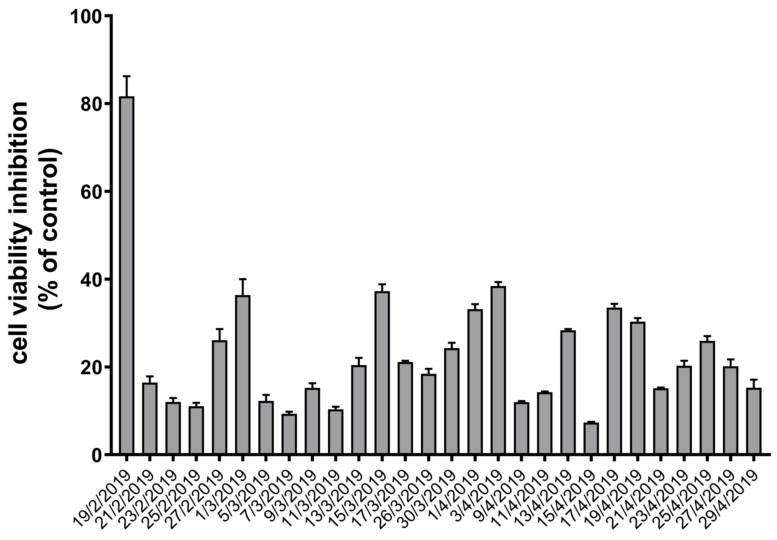
As regards the cytotoxicity data obtained by the MTT test on A549 cells (Figure 2), in all the samples analyzed, statistically significant inhibition of the NAD(P)H-dependent cellular oxidoreductase enzymes was observed following the exposure of the cells for 24 h to the aqueous extracts of PM10 compared to control cells. The dose-dependence of the response of the assay for the aqueous extracts was preliminarily assessed exposing A549 cells to different dilutions of the same extract, as reported in previous work [16]. The sample obtained on 19th February, which expressed the highest OPDTTV value, was able to induce cellular viability inhibition of about 80%. All the other samples showed values on average below 50% cell viability inhibition. Fourteen samples were in the range between 20% and 50% ascribable to slight cytotoxicity, while the remaining twelve samples did not exceed the 20% viability inhibition value.

3.3. Intracellular Oxidative Stress Induction by PM10 Exposure

In parallel to the cytotoxicity analysis, the cell-mediated oxidant generating capacity of PM10 was assessed on A549 cells exposed for 24 to the same aqueous extracts and then charged with CM-H2DCFDA. Preliminarily, the dose-response behavior of the assay was investigated exposing A549 cells to different dilutions of the same extract. The representative dose-response for one of the samples is reported in Figure 3A. A statistically significant dose-response change in the intracellular oxidative stress condition in A549 cells was observed with a percentage variation of the intracellular fluorescence of the probe (calculated with respect to the negative control) growing from about 6% (1:8 dilution) to 37% for the undiluted sample. This result demonstrates that PM10 mediates oxidative stress induction in the cells in a dose-dependent manner. Figure 3B shows a representative confocal image of control A549 cells (left image) and cells exposed to undiluted PM10 aqueous extract (left image). After 24 h exposure, the intracellular fluorescence intensity of the probe appeared clearly increased.
In Figure 4 (light bars), the intracellular oxidative stress recorded in A549 cells exposed to the aqueous extracts of all the PM10 samples is reported. The PM10 sample collected on 19/2/2020 showed the highest response, while all the others showed a percentage variation of the probe fluorescence ranging from 5 to 79% compared to control cells.
The induction of oxidative stress in cells after exposure to particulate matter has been reported also in other cellular systems [5,52,53]. Although the precise mechanisms underlying the oxidant generation capacity of PM following exposure of cells and tissues are not completely clarified, it is known that cell exposure to PM is associated with activation of NADPH oxidases, a family of enzymes generating O2- [14,54,55]. Moreover, metal cations in cells are known to catalyze the Fenton reaction, which can give rise to the Haber Weiss cycle in the presence of peroxide and superoxide ions, inducing the formation of hydroxyl radical [56]. The exposure to organic compounds present in PM has been described to impair electron transport at the mitochondrial level and to alter the mitochondrial functioning [21,57,58]. It is known that DCF fluorescence results from oxidation by potent oxidants, such as those arising from reactions catalyzed by metal ions and peroxidase and those derived from proton- and carbon dioxide-catalyzed decomposition of the reactive nitrogen species peroxynitrite [40]. Therefore, the fluorescence signal recorded in A549 cells exposed to the PM aqueous extracts could reflect the intracellular oxidative stress condition induced by redox-active metals associated with PM and/or the formation of reactive products from the direct reaction of CO2 with peroxynitrite, which is formed intracellularly by the reaction of NO with superoxide ion [59].
It is known that oxidative stress represents a major cause of DNA damage resulting in base modifications, base loss, and DNA strand breaks [58]. Therefore, in a subset of 6 samples, our study was deepened with the assessment of the genotoxic activity of PM10 on A549 cells exposed to PM10 aqueous extracts for 24 h using the comet assay, which represents the gold standard for DNA strand breaks assessment.
The results expressed as % of DNA in the comet tail are shown in Figure 4 by dark bars. The genotoxicity data showed very good correlations (Pearson r > 0.95) with the intracellular oxidative stress and with the cell mortality observed with the MTT test. The maximum value of % of DNA in the tail was observed for the sample of 19 February 2020. However, the number of data for genotoxicity is quite limited and this result should be confirmed with further studies.

3.4. Correlation of OP, Cytotoxicity, and Oxidative Stress Generating Capacity of PM10 with Particulate Concentration and Carbon Content

To assess the relationship between the acellular and cellular toxicity outcomes with the physical-chemical properties of PM10, the Pearson correlation analysis was performed (Table 2) to show the correlation levels between results of cellular and acellular assays with PM10 concentrations and carbon content. It is worth mentioning that OC and EC are well correlated (Pearson 0.93) and that TC (sum of OC and EC) is well correlated with OC (Pearson 0.99) and with EC (Pearson 0.94). PM10 is well correlated with OC and TC (Pearson 0.70). This indicates that, at the site studied, OC is a major component of aerosol and it has both a fraction of primary organ from combustion sources and a fraction of secondary origin (i.e., secondary organic aerosol formed in the atmosphere). The correlation of EC (primary origin from combustion sources) and PM10 (Pearson 0.59) indicates a non-negligible primary contribution of combustion sources at the site studied.
OPDDTV showed a statistically significant correlation with PM10 and with carbon content (both for OC and EC). Similar behavior was also observed for the cytotoxicity values normalized to the air sampled volume (MTTV) even if this parameter has a higher correlation with OC compared to that with EC. The mass normalized OPDTTM showed no significant correlation, while MTTM showed a negative correlation with PM10. To understand these results it is necessary to consider that particularly high PM10 concentration values are not always associated with high toxicity, as also discussed in previous work [16], because the chemical composition of the particulate itself, strongly correlated with sources, could heavily influence the cytotoxic outcome. In addition, it must be considered that advection of Saran dust events, characterized by particularly high concentrations of PM but associated with lower toxicity compared to combustion sources, can occur in the area and could be responsible for the observed results. In this dataset, an event of advection of African dust was identified at the end of April 2019 and this corresponds to the lowest values of OPDTTM observed on 25 April 2019 (Figure 1), confirming the low intrinsic OP values associated with this type of source [30]. The oxidative stress generating capacity (OSGCV) normalized in volume is well correlated with both PM10 and with carbon content while OSGCM does not show any significant correlation.
These results are in agreement with previous results obtained in other cellular systems. Park et al. [60] in several cell models, such as human airway epithelial cells (A549, H292, BEAS-2B, and SAEC) and Chinese hamster ovary (CHO-K1) cell lines, found a significant correlation between cytotoxicity and the carbon content of PM. Wang et al. [61], in the Chinese hamster ovary (CHO) cells, found a positive correlation between cytotoxicity and OC and particularly with water-soluble organic carbon (WSOC), but not with EC.
To investigate if there is any relationship between the outcomes of the cellular and acellular assays on the same samples, a comparison of the results obtained with the different tests was performed (Figure 5). The analysis was focused on data normalized in volume, which is a metric relevant for human exposure. The intracellular oxidative stress induced by PM is statistically correlated (Figure 5A) to the acellular oxidative potential, suggesting that the ability of PM10 to generate intracellularly reactive species is, in a certain way, related to its oxidative potential. Moreover, the oxidative potential is related to cytotoxicity (Figure 5B), which is a cellular endpoint with integrates all the potentially toxic effects that PM10 can exert in the cell. Cytotoxicity is related to the PM oxidative stress-inducing capacity (Figure 5C), suggesting that intracellular oxidative stress plays a key role in the multiple mechanisms underlying PM10 cytotoxicity. Figure 5 includes the sample of 19 February 2019 that seems to lead most of the correlation, however, if this sample is eliminated the correlations still remain statistically significant, even if weaker.
When the outcomes of cellular and acellular assays are compared normalized in mass (i.e., intrinsic values), the correlations become significantly weaker, suggesting that the different sources acting on the site produces particulate matter with different chemical composition leading to different toxicological potential. Thereby, chemical compositions will likely have different influences on the biological tests studied.
The analysis reported has some limitations. The first is that samples are taken between the end of winter and the beginning of spring and future studies could involve investigation of other seasons in which the weight of combustion sources (at least for biomass burning) could be more limited. Another aspect, that will be carried out in future studies already planned, is to investigate if these correlations are similar in other typology of sites (like urban and industrial sites) eventually adding information regarding chemical tracers of other PM sources in addition to carbon content.

4. Conclusions

A set of daily PM10 samples collected at an urban background site (ECO Environmental-Climate Observatory) in southern Italy was analyzed to determine cytotoxicity (using MTT test on A549 cells), genotoxicity (using the comet assay), and intracellular oxidative stress on A549 cells exposed for 24 h to aqueous extracts of PM10 samples. In addition, carbon content (OC and EC) of PM10 and acellular determination of oxidative potential with DTT assay were performed to compare results of acellular and cellular biological assays.
Results of cellular (OSGCV and MTTV) and acellular (OPDTTV) tests have been normalized in volume to be representative of human exposure and correlated with PM10 concentration and carbon content. A good correlation, statistically significant, was observed with both OC and EC. This suggests that combustion sources, mainly road traffic and biomass burning at this site, play an important role in determining cellular oxidative stress and cytotoxicity of PM10. When the outcomes of the assays are normalized in mass (i.e., intrinsic values depending on the contributions of the different sources), the correlations become weaker or negligible. Even if the number of data is limited, genotoxicity results, expressed as % of DNA in tails, are well correlated (Pearson r > 0.95) with OSGCV and MTTV, and a weaker, but statistically significant correlation was observed with OPDTTV. The OSGCV results are well correlated with the cell mortality observed with the MTTV test and, a lower, but still statistically significant correlation is observed between MTTV and OPDDTV. It was found a statistically significant correlation between acellular OPDTTV assay and intracellular oxidative stress OSGCV results. These results support the idea that the oxidative potential of PM could be an indicator of cytotoxicity of PM. When acellular and cellular assays of oxidative potential are compared, it seems that cellular assays are more representative of cytotoxicity. However, these results should be validated with further studies done in different seasons (at the same site) and different sites (for example urban and industrial areas).
When the outcomes of cellular and acellular assays are compared normalized in mass (i.e., intrinsic values), the correlations become significantly weaker suggesting that the different sources acting on the site produces particulate matter with multiple toxicological potential influencing differently the biological tests studied. Future studies could involve an analysis of correlation with chemical tracers of different sources, in addition to carbon content.

Author Contributions

Conceptualization, M.G.L., D.C. and M.R.G.; measurement campaigns M.C. and A.D.; toxicological analysis, M.G.L., R.C. and M.E.G.; oxidative potential analysis, M.R.G., A.R.D.B. and M.P.R.; writing—review and editing, M.G.L. and D.C. All authors have read, commented, and agreed to the published version of the manuscript. All authors have read and agreed to the published version of the manuscript.

Funding

This research was funded by project PAPER (Paper Analyzer for Particulate Exposure Risk), funded within POR Puglia FESR-FSE 2014-2020—Asse prioritario 1—Azione 1.6—Bando Innonetwork—Aiuti a sostegno delle attività di R&S, grant number PH3B166.

Institutional Review Board Statement

Not applicable.

Informed Consent Statement

Not applicable.

Data Availability Statement

The data presented in this study are available on request from the corresponding author.

Conflicts of Interest

The authors declare no conflict of interest.

References

  1. Anderson, J.O.; Thundiyil, J.G.; Stolbach, A. Clearing the air: A review of the effects of particulate matter air pollution on human health. JMT 2012, 8, 166–175. [Google Scholar] [CrossRef] [Green Version]
  2. Liu, S.; Zhou, Y.; Liu, S.; Chen, X.; Zou, W.; Zhao, D.; Li, X.; Pu, J.; Huang, L.; Chen, J.; et al. Association between exposure to ambient particulate matter and chronic obstructive pulmonary disease: Results from a cross-sectional study in China. Thorax 2017, 72, 788–795. [Google Scholar] [CrossRef] [PubMed] [Green Version]
  3. Loomis, D.; Grosse, Y.; Lauby-Secretan, B.; Ghissassi, F.E.; Bouvard, V.; Benbrahim-Tallaa, L.; Guha, N.; Baan, R.; Mattock, H.; Straif, K. International Agency for Research on Cancer Monograph Working Group IARC. The carcinogenicity of outdoor air pollution. Lancet Oncol. 2013, 14, 1262–1263. [Google Scholar] [CrossRef]
  4. Amato, F.; Alastuey, A.; Karanasiou, A.; Lucarelli, F.; Nava, S.; Calzolai, G.; Severi, M.; Becagli, S.; Vorne, L.G.; Colombi, C.; et al. AIRUSE-LIFEC: A harmonized PM speciation and source apportionment in five southern European cities. Atmos. Chem. Phys. 2016, 16, 3289–3309. [Google Scholar] [CrossRef] [Green Version]
  5. Li, N.; Sioutas, C.; Cho, A.; Schmitz, D.; Misra, C.; Sempf, J.; Wang, M.; Oberley, T.; Froines, J.; Nel, A. Ultrafine particulate pollutants induce oxidative stress and mitochondrial damage. Environ. Health Perspect. 2003, 111, 455–460. [Google Scholar] [CrossRef] [PubMed]
  6. Jia, Y.Y.; Wang, Q.; Liu, T. Toxicity Research of PM2.5 Compositions in Vitro. Int. J. Environ. Res. Public Health 2017, 14, 232. [Google Scholar] [CrossRef] [Green Version]
  7. Nel, A. Air pollution related illness: Effects of particles. Science 2005, 308, 804–806. [Google Scholar] [CrossRef]
  8. Cheng, H.; Saffari, A.; Sioutas, C.; Forman, H.J.; Morgan, T.E.; Finch, C.E. Nanoscale particulate matter from urban traffic rapidly induces oxidative stress and inflammation in olfactory epithelium with concomitant effects on brain. Environ. Health Perspect. 2016, 124, 1537–1546. [Google Scholar] [CrossRef] [PubMed] [Green Version]
  9. Sies, H. Oxidative stress: A concept in redox biology and medicine. Redox Biol. 2015, 4, 180–183. [Google Scholar] [CrossRef] [Green Version]
  10. Leomanni, A.; Schettino, T.; Calisi, A.; Gorbi, S.; Mezzelani, M.; Regoli, F.; Lionetto, M.G. Antioxidant and oxidative stress related responses in the Mediterranean land snail Cantareus apertus exposed to the carbamate pesticide Carbaryl. Comp. Biochem. Physiol. B Biochem. Mol. Biol. 2015, 168, 20–27. [Google Scholar] [CrossRef]
  11. Ayres, J.G.; Borm, P.; Cassee, F.R.; Castranova, V.; Donaldson, K.; Ghio, A.; Harrison, R.M.; Hider, R.; Kelly, F.; Kooter, I.M.; et al. Evaluating the toxicity of airborne particulate matter and nanoparticles by measuring oxidative stress potential—A Workshop report and consensus statement. Inhal. Toxicol. 2008, 20, 75–99. [Google Scholar] [CrossRef]
  12. Xiao, G.G.; Wang, M.; Li, N.; Loo, J.A.; Nel, A.E. Use of proteomics to demonstrate a hierarchical oxidative stress responseto diesel exhaust particles in a macrophage cell line. J. Biol. Chem. 2003, 278, 50781–50790. [Google Scholar] [CrossRef] [Green Version]
  13. Jiang, H.; Ahmed, C.M.S.; Canchola, A.; Chen, J.Y.; Lin, Y.H. Use of Dithiothreitol Assay to Evaluate the Oxidative Potential of Atmospheric Aerosols. Atmosphere 2019, 10, 571. [Google Scholar] [CrossRef] [Green Version]
  14. Ghio, A.J.; Carraway, M.S.; Madden, M.C. Composition of air pollution particles and oxidative stress in cells, tissues, and living systems. J. Toxicol. Environ. Health B Crit. Rev. 2012, 15, 1–21. [Google Scholar] [CrossRef]
  15. Bates, J.T.; Fang, T.; Verma, V.; Zeng, L.; Weber, R.J.; Tolbert, P.E.; Abrams, J.Y.; Sarnat, S.E.; Klein, M.; Mulholland, J.A.; et al. Review of acellular assays of ambient particulate matter oxidative potential: Methods and relationships with composition, sources, and health effects. Environ. Sci. Technol. 2019, 53, 4003–4019. [Google Scholar] [CrossRef]
  16. Lionetto, M.G.; Guascito, M.R.; Caricato, R.; Giordano, M.E.; De Bartolomeo, A.R.; Romano, M.P.; Conte, M.; Dinoi, A.; Contini, D. Correlation of Oxidative Potential with Ecotoxicological and Cytotoxicological Potential of PM10 at an Urban Background Site in Italy. Atmosphere 2019, 10, 733. [Google Scholar] [CrossRef] [Green Version]
  17. Shi, T.; Schins, R.P.F.; Knaapen, A.M.; Kuhlbusch, T.; Pitz, M.; Heinrich, J.; Borm, P.J.A. Hydroxyl radical generation by election paramagnetic resonance as a new method to monitor ambient particulate matter composition. J. Environ. Monit. 2003a, 5, 550–556. [Google Scholar] [CrossRef]
  18. Shi, T.; Knaapen, A.M.; Begerow, J.; Birmilli, W.; Borm, P.J.A.; Schins, R.P.F. Temporal variation of hydroxyl radical generation and 8-hydroxy-2′-deoxyguanosine formation by coarse and fine particulate matter. Occup. Environ. Med. 2003b, 60, 315–321. [Google Scholar] [CrossRef] [PubMed] [Green Version]
  19. Mudway, I.; Fuller, G.W.; Green, D.; Dunster, C.; Kelly, F.J. Quantifying the London Specific Component of PM10 Oxidative Activity; Report Defra Department of Environmental Food and Rural Affairs; DEFRA: London, UK, 2011. [Google Scholar]
  20. Cho, A.K.; Sioutas, C.; Miguel, A.H.; Kumagai, Y.; Schmitz, D.A.; Singh, M.; Eiguren-Fernandez, A.; Froines, J.R. Redox activity of airborne particulate matter at different sites in the Los Angeles Basin. Environ. Res. 2005, 99, 40–47. [Google Scholar] [CrossRef] [PubMed]
  21. Hiura, T.S.; Li, N.; Kaplan, R.; Horwitz, M.; Seagrave, J.C.; Nel, A.E. The role of a mitochondrial pathway in the induction of apoptosis by chemicals extracted from diesel exhaust particles. J. Immunol. 2000, 165, 2703–2711. [Google Scholar] [CrossRef] [Green Version]
  22. Crobeddu, B.; Aragao-Santiago, L.; Bui, L.C.; Boland, S.; Squiban, A.B. Oxidative potential of particulate matter 2.5 as predictive indicator of cellular stress. Environ. Pollut. 2017, 230, 125–133. [Google Scholar]
  23. Daellenbach, K.R.; Uzu, G.; Jiang, J.; Cassagnes, L.-E.; Leni, Z.; Vlachou, A.; Stefenelli, G.; Canonaco, F.; Weber, S.; Segers, A.; et al. Sources of particulate-matter air pollution and its oxidative potential in Europe. Nature 2020, 587, 414–419. [Google Scholar] [CrossRef]
  24. Øvrevik, J. Oxidative Potential Versus Biological Effects: A Review on the Relevance of Cell-Free/Abiotic Assays as Predictors of Toxicity from Airborne Particulate Matter. Int. J. Mol. Sci. 2019, 20, 4772. [Google Scholar] [CrossRef] [Green Version]
  25. Mosmann, T. Rapid colorimetric assay for cellular growth and survival: Application to proliferation and cytotoxicity assays. J. Immunol. Methods 1983, 65, 55–63. [Google Scholar] [CrossRef]
  26. Hsiao, W.L.W.; Moa, Z.Y.; Fang, M.; Shi, X.M.; Wang, F. Cytotoxicity of PM2.5 and PM2.5–10 ambient air pollutants assessed by the MTT and the Comet assays. Mutat. Res. 2000, 471, 45–55. [Google Scholar] [CrossRef]
  27. Faraji, M.; Nodehi, R.N.; Naddafi, K.; Pourpak, Z.; Alizadeh, Z.; Rezaei, S.; Mesdaghinia, A. Cytotoxicity of airborne particulate matter (PM10) from dust storm and inversion conditions assessed by MTT assay. J. Air Pollut. Health 2018, 3, 135–142. [Google Scholar]
  28. Arto, S.; Sillanpaa, M.; Halinen, A.I.; Happo, M.S.; Hillamo, R.; Brunekreef, B.; Katsouyanni, K.; Sunyer, J.; Hirvonen, M.R. Heterogeneities in inflammatory and cytotoxic responses of RAW 264.7 macrophage cell line to urban air coarse, fine, and ultrafine particles from six European sampling campaigns. Inhal. Toxicol. 2007, 19, 213–225. [Google Scholar]
  29. Singh, N.P.; McCoy, M.T.; Tice, R.R.; Schneider, E.L. A simple technique for quantification of low levels of DNA damage in individual cells. Exp. Cell. Res. 1988, 175, 184–189. [Google Scholar] [CrossRef] [Green Version]
  30. Collins, A.R. The Comet Assay-principles, applications and limitations. Methods Mol. Biol. 2003, 203, 163–167. [Google Scholar]
  31. Foster, K.A.; Oster, C.G.; Mayer, M.M.; Avery, M.L.; Audus, K.L. Characterization of the A549 Cell Line as a Type II Pulmonary Epithelial Cell Model for Drug Metabolism. Exp. Cell Res. 1998, 243, 359–366. [Google Scholar] [CrossRef]
  32. Yi, S.; Zhang, F.; Qu, F.; Ding, W. Water-insoluble fraction of airborne particulate matter (PM10) induces oxidative stress in human lung epithelial A549 cells. Environ. Toxicol. 2012, 29, 226–233. [Google Scholar] [CrossRef] [PubMed]
  33. Wang, B.; Li, K.; Jin, W.; Lu, Y.; Zhang, Y.; Shen, G.; Wang, R.; Shen, H.; Li, W.; Huang, Y.; et al. Properties and Inflammatory Effects of Various Size Fractions of Ambient Particulate Matter from Beijing on A549 and J774A.1 Cells. Environ. Sci. Technol. 2013, 47, 10583–10590. [Google Scholar] [CrossRef]
  34. Cesari, D.; Merico, E.; Dinoi, A.; Marinoni, A.; Bonasoni, P.; Contini, D. Seasonal variability of carbonaceous aerosols in an urban background area in Southern Italy. Atmos. Res. 2018, 200, 97–108. [Google Scholar] [CrossRef]
  35. Dinoi, A.; Cesari, D.; Marinoni, A.; Bonasoni, P.; Riccio, A.; Chianese, E.; Tirimberio, G.; Naccarato, A.; Sprovieri, F.; Andreoli, G.; et al. Inter-Comparison of Carbon Content in PM2.5 and PM10 Collected at Five Measurement Sites in Southern Italy. Atmosphere 2017, 8, 243. [Google Scholar] [CrossRef] [Green Version]
  36. Conte, M.; Merico, E.; Cesari, D.; Dinoi, A.; Grasso, F.M.; Donateo, A.; Guascito, M.R.; Contini, D. Long-term characterisation of African dust advection in south-eastern Italy: Influence on fine and coarse particle concentrations, size distributions, and carbon content. Atmos. Res. 2020, 233, 104690. [Google Scholar] [CrossRef]
  37. Merico, E.; Cesari, D.; Dinoi, A.; Gambaro, A.; Barbaro, E.; Guascito, M.R.; Giannossa, L.C.; Mangone, A.; Contini, D. Inter-comparison of carbon content in PM10 and PM2.5 measured with two thermo-optical protocols on samples collected in a Mediterranean site. Environ. Sci. Pollut. Res. 2019, 26, 29334–29350. [Google Scholar] [CrossRef] [PubMed]
  38. Chirizzi, D.; Cesari, D.; Guascito, M.R.; Dinoi, A.; Giotta, L.; Donateo, A.; Contini, D. Influence of Saharan dust outbreaks and carbon content on oxidative potential of water-soluble fractions of PM2.5 and PM10. Atmos. Environ. 2017, 163, 1–8. [Google Scholar] [CrossRef]
  39. Latronico, S.; Giordano, M.E.; Urso, E.; Lionetto, M.G.; Schettino, T. Effect of the flame retardant Tris (1,3-dichloro-2-propyl) Phosphate (TDCPP) on Na+-K+-ATPase and Cl- transport in HeLa cells. Toxicol. Mech. Methods 2018, 28, 599–606. [Google Scholar] [CrossRef]
  40. Forman, H.J.; Augusto, O.; Brigelius-Flohe, R.; Dennery, P.A.; Kalyanaraman, B.; Ischiropoulos, H.; Mann, G.E.; Radi, R.; RobertsII, L.J.; Vina, J.; et al. Even free radicals should follow some rules: A Guide to free radical research terminology and methodology. Free Radic. Biol. Med. 2015, 78, 233–235. [Google Scholar] [CrossRef] [PubMed]
  41. Giordano, M.E.; Caricato, R.; Lionetto, M.G. Concentration dependence of the antioxidant and prooxidant activity of Trolox in HeLa cells: Involvement in the induction of Apoptotic Volume Decrease. Antioxidants 2020, 9, 1058. [Google Scholar] [CrossRef]
  42. Contini, D.; Genga, A.; Cesari, D.; Siciliano, M.; Donateo, A.; Bove, M.C.; Guascito, M.R. Characterisation and source apportionment of PM10 in an urban background site in Lecce. Atmos. Res. 2010, 95, 40–54. [Google Scholar] [CrossRef]
  43. Contini, D.; Cesari, D.; Donateo, A.; Chirizzi, D.; Belosi, F. Characterization of PM10 and PM2.5 and Their Metals Content in Different Typologies of Sites in South-Eastern Italy. Atmosphere 2014, 5, 435–453. [Google Scholar] [CrossRef] [Green Version]
  44. Romano, S.; Perrone, M.R.; Becagli, S.; Pietrogrande, M.C.; Russo, M.; Caricato, R.; Lionetto, M.G. Ecotoxicity, genotoxicity, and oxidative potential tests of atmospheric PM10 particles. Atmos. Environ. 2020, 221, 117085. [Google Scholar] [CrossRef]
  45. Sandrini, S.; Fuzzi, S.; Piazzalunga, A.; Prati, P.; Bonasoni, P.; Cavalli, F.; Bove, M.C.; Calvello, M.; Cappelletti, D.; Colombi, C.; et al. Spatial and seasonal variability of carbonaceous aerosol across Italy. Atmos. Environ. 2014, 99, 587–598. [Google Scholar] [CrossRef]
  46. Cesari, D.; Donateo, A.; Conte, M.; Merico, E.; Giangreco, A.; Giangreco, F.; Contini, D. An inter-comparison of PM2.5 at urban and urban background sites: Chemical characterization and source apportionment. Atmos. Res. 2016, 174–175, 106–119. [Google Scholar] [CrossRef]
  47. Janssen, N.A.H.; Yang, A.; Strak, M.; Steenhof, M.; Hellack, B.; Gerlofs-Nijland, M.E.; Kuhlbusch, T.; Kelly, F.; Harrison, R.; Brunekreef, B.; et al. Oxidative potential of particulate matter collected at sites with different source characteristics. Sci. Total Environ. 2014, 472, 572–581. [Google Scholar] [CrossRef] [PubMed] [Green Version]
  48. Cesari, D.; Merico, E.; Grasso, F.M.; Decesari, S.; Belosi, F.; Manarini, F.; De Nuntiis, P.; Rinaldi, M.; Volpi, F.; Gambaro, A.; et al. Source apportionment of PM2.5 and of its oxidative potential in an industrial suburban site in south Italy. Atmosphere 2019, 10, 758. [Google Scholar] [CrossRef] [Green Version]
  49. Perrone, M.R.; Bertoli, I.; Romano, S.; Russo, M.; Rispoli, G.; Pietrogrande, M.C. PM2.5 and PM10 oxidative potential at a Central Mediterranean Site: Contrasts between dithiothreitol-and ascorbic acid-measured values in relation with particle size and chemical composition. Atmos. Environ. 2019, 210, 143–155. [Google Scholar] [CrossRef]
  50. Pietrogrande, M.C.; Russo, M.; Zagatti, E. Review of PM oxidative potential measured with acellular assays in urban and rural sites across Italy. Atmosphere 2020, 10, 626. [Google Scholar] [CrossRef] [Green Version]
  51. Massimi, L.; Ristorini, M.; Simonetti, G.; Frezzini, M.A.; Astolfi, M.L.; Canepari, S. Spatial mapping and size distribution of oxidative potential of particulate matter released by spatially disaggregated sources. Environ. Pollut. 2020, 266, 115271. [Google Scholar] [CrossRef]
  52. Voelkel, K.; Krug, H.F.; Diabate, S. Formation of reactive oxygen species in rat epithelial cells upon stimulation with fly ash. J. Biosci. 2003, 28, 51–55. [Google Scholar] [CrossRef] [PubMed]
  53. Li, R.; Wang, Y.; Qiu, X.; Xu, F.; Chen, R.; Gu, W.; Zhang, L.; Yang, S.; Cai, Z.; Liu, C. Difference on oxidative stress in lung epithelial cells and macrophages induced by ambient fine particulate matter (PM2.5). Air Qual. Atmos. Health 2020, 13, 789–796. [Google Scholar] [CrossRef]
  54. Kampfrath, T.; Maiseyeu, A.; Ying, Z.; Shah, Z.; Deiuliis, J.A.; Xu, X.; Kherada, N.; Brook, R.D.; Reddy, K.M.; Padture, N.P.; et al. Chronic fine particulate matter exposure induces systemic vascular dysfunction via NADPH oxidase and TLR4 pathways. Circ. Res. 2011, 108, 716–726. [Google Scholar] [CrossRef] [Green Version]
  55. Xu, X.; Yavar, Z.; Verdin, M.; Ying, Z.; Mihai, G.; Kampfrath, T.; Wang, A.; Zhing, M.; Lippermann, M.; Chen, L.C.; et al. Effect of early particulate air pollution exposure on obesity in mice: Role of p47phox. Arterioscler. Thromb. Vasc. Biol. 2010, 30, 2518–2527. [Google Scholar] [CrossRef] [Green Version]
  56. Imlay, J.A.; Chin, S.M.; Linn, S. Toxic DNA damage by hydrogen peroxide through the Fenton reaction in vivo and in vitro. Science 1988, 240, 640–642. [Google Scholar] [CrossRef] [PubMed]
  57. Xia, T.; Korge, P.; Weiss, J.N.; Li, N.; Venkatesen, M.I.; Sioutas, C.; Nel, A. Quinones and aromatic chemical compounds in particulate matter induce mitochondrial dysfunction: Implications for ultrafine particle toxicity. Environ. Health Perspect. 2004, 112, 1347–1358. [Google Scholar] [CrossRef] [PubMed]
  58. Gonzalez-Hunt, C.P.; Wadhwa, M.; Sanders, L.H. DNA damage by oxidative stress: Measurement strategies for two genomes. Curr. Opin. Toxicol. 2018, 7, 87–94. [Google Scholar] [CrossRef]
  59. Packer, P.; Beckman, J.S.; Liaudet, L. Nitric oxide and peroxynitrite in health and disease. Physiol. Rev. 2007, 87, 315–424. [Google Scholar] [CrossRef] [PubMed] [Green Version]
  60. Park, M.; Joo, H.S.; Lee, K.; Jang, M.; Kim, S.D.; Kim, I.; Borlaza, L.J.S.; Lim, H.; Shin, H.; Chung, K.H.; et al. Differential toxicities of fine particulate matters from various sources. Sci. Rep. 2018, 8, 17007. [Google Scholar] [CrossRef]
  61. Wang, Y.; Plewab, M.J.; Mukherjee, U.K.; Verma, V. Assessing the cytotoxicity of ambient particulate matter (PM) using Chinese hamster ovary (CHO) cells and its relationship with the PM chemical composition and oxidative potential. Atmos. Environ. 2018, 179, 132–141. [Google Scholar] [CrossRef]
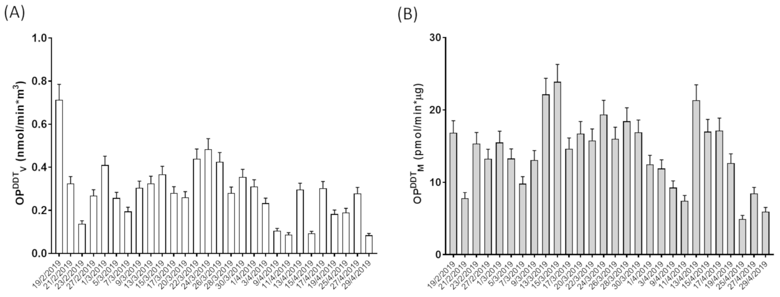
Figure 1. Oxidative potential expressed as OPDTTV (A) and OPDTTM (B) measured in aqueous extracts of the PM10 samples.
Atmosphere 12 00464 g001
Figure 2. Cytotoxicity of PM10 expressed as % cell viability inhibition assessed by the MTT test on A549 cells exposed for 24 h to PM10 aqueous extracts.
Atmosphere 12 00464 g002
Figure 3. (A) Cell-mediated oxidant generating capacity of PM10 (expressed as percentage variation of fluorescence intensity vs. negative control) in A549 cells exposed for 24 h to increasing dilutions of the aqueous extract obtained from PM10 collected on 20/3/2019; (B) Representative confocal images of control and PM10 exposed (24 h) A549 cells charged with the probe CM-H2DCFDA.
Atmosphere 12 00464 g003
Figure 4. Intracellular oxidative stress (expressed as % variation of CM-H2DCFDA vs. negative control) measured in A549 cells exposed for 24 h to the PM10 aqueous extracts are shown by light gray bars. Data are referred to the entire set of samples. Genotoxicity (expressed as % of DNA in the tail of the comets) measured by the A549 cells exposed for 24 h to the PM10 aqueous extracts are shown by dark gray bars. Genotoxicity data are referred to as a subset of samples.
Atmosphere 12 00464 g004
Figure 5. Relationship between the outcomes of volume-normalized OP, cytotoxicity, and oxidative stress-inducing capacity of PM10.
Atmosphere 12 00464 g005
Table 1. Values of PM10 concentrations, carbon content, cytotoxicity (assessed by MTT test), and oxidative potential (OP), referred to as the sampled volume (OPDTTV) or sampled mass (OPDTTM).
PM10
(µg/m3)
OC
(µg/m3)
EC
(µg/m3)
TC
(µg/m3)
MTT
(% inhibition)
OPDDTV
(nmol/min*m3)
OPDDTM
(pmol/min*µg)
Average21.03.40.74.021.80.314.2
(min-max)(5.5–42.4)(0.9–13.8)(0.1–2.0)(1.2–15.8)(5.3–81.6)(0.1–0.7)(4.9–23.9)
Median19.52.70.64.020.20.315.0
(25th–75th)(14.5–26.1)(1.8–4.1)(0.3–0.8)(2.2–4.8)(11.3–29.2)(0.2–0.3)(10.3–17.0)
Table 2. Pearson’s correlation coefficients between toxicological indicators and PM10 concentrations and carbon content. OPDTTV and OPDTTM represent the OP value referred to the inhaled air or the sampled PM10 mass, respectively. MTTV and MTTM correspond to the cell mortality value assessed by the MTT test and referred to the sampled air volume or the sampled PM10 mass, respectively. N = numbers of pairs. ** indicates cases with p < 0.01.
PM10 (µg/m3)OC (µg/m3)EC (µg/m3)TC (µg/m3)N of Pairs
OPDTTV0.6843 **0.8209 **0.8179 **0.8274 **28
OPDTTM−0.16790.20420.33870.224128
MTTV0.45450.8708 **0.8304 **0.8715 **28
MTTM−0.47650.28380.29460.287128
OSGCV0.5443 **0.6477 **0.7223 **0.6631 **28
OSGCM−0.38770.04640.16740.063128
Publisher’s Note: MDPI stays neutral with regard to jurisdictional claims in published maps and institutional affiliations.