Review

Non-Invasive Methods of Quantifying Heat Stress Response in Farm Animals with Special Reference to Dairy Cattle

by
Veerasamy Sejian
1,*,
Chikamagalore Gopalakrishna Shashank
2,
Mullakkalparambil Velayudhan Silpa
3,
Aradotlu Parameshwarappa Madhusoodan
4,
Chinnasamy Devaraj
2 and
Sven Koenig
3
1
Rajiv Gandhi Institute of Veterinary Education and Research, Pondicherry 605008, India
2
Centre for Climate Resilient Animal Adaptation Studies, ICAR-National Institute of Animal Nutrition and Physiology, Adugodi, Bangalore 560030, India
3
Institute of Animal Breeding and Genetics, Justus Liebig University Giessen, Ludwigstraße 21b, 35390 Giessen, Germany
4
Temperate Animal Husbandry Division, ICAR-Indian Veterinary Research Institute, Mukteswar Campus, Nainital 263138, India
*
Author to whom correspondence should be addressed.
Atmosphere 2022, 13(10), 1642; https://doi.org/10.3390/atmos13101642
Submission received: 17 September 2022 / Revised: 2 October 2022 / Accepted: 3 October 2022 / Published: 9 October 2022
(This article belongs to the Section Biometeorology)

Abstract

:
Non-invasive methods of detecting heat stress magnitude for livestock is gaining momentum in the context of global climate change. Therefore, the objective of this review is to focus on the synthesis information pertaining to recent efforts to develop heat stress detection systems for livestock based on multiple behavioral and physiological responses. There are a number of approaches to quantify farm animal heat stress response, and from an animal welfare point of view, these can be categorized as invasive and non-invasive approaches. The concept of a non-invasive approach to assess heat stress primarily looks into behavioral and physiological responses which can be monitored without any human interference or additional stress on the animal. Bioclimatic thermal indices can be considered as the least invasive approach to assess and/or predict the level of heat stress in livestock. The quantification and identification of the fecal microbiome in heat-stressed farm animals is one of the emerging techniques which could be effectively correlated with animal adaptive responses. Further, tremendous progress has been made in the last decade to quantify the classical heat stress endocrine marker, cortisol, non-invasively in the feces, urine, hair, saliva and milk of farm animals. In addition, advanced technologies applied for the real-time analysis of cardinal signs such as sounds through microphones, behavioral images, videos through cameras, and data stalking body weight and measurements might provide deeper insights towards improving biological metrics in livestock exposed to heat stress. Infrared thermography (IRT) can be considered another non-invasive modern tool to assess the stress response, production, health, and welfare status in farm animals. Various remote sensing technologies such as ear canal sensors, rumen boluses, rectal and vaginal probes, IRT, and implantable microchips can be employed in grazing animals to assess the quantum of heat stress. Behavioral responses and activity alterations to heat stress in farm animals can be monitored using accelerometers, Bluetooth technology, global positioning systems (GPSs) and global navigation satellite systems (GNSSs). Finally, machine learning offers a scalable solution in determining the heat stress response in farm animals by utilizing data from different sources such as hardware sensors, e.g., pressure sensors, thermistors, IRT sensors, facial recognition machine vision sensors, radio frequency identification, accelerometers, and microphones. Thus, the recent advancements in recording behavior and physiological responses offer new scope to quantify farm animals’ heat stress response non-invasively. These approaches could have greater applications in not only determining climate resilience in farm animals but also providing valuable information for defining suitable and accurate amelioration strategies to sustain their production.

1. Introduction

Adaptation physiology can be defined as any biological response to environmental stress by an individual organism resulting in an improved ability to cope with a changing environment. Behavioral and physiological adaptive responses of farm animals to a changing climate can have a significant impact on livestock systems’ profitability and production. Considering the fact that by 2050, demand for livestock products will increase significantly [1], climate change and its impacts on the biosphere might become tough challenges for the livestock sector. Therefore, precision dairy farming tools are of paramount importance in order to maintain the volume and quality of livestock products in the event of an increase in global temperature. With the advancements of the technologies in the present era, the adaptation and utilization of these technologies to observe and measure the responses of livestock to the current climate changing scenario might play a significant role in improving both livestock productivity and animal welfare.
When measuring stress in farm animals, invasive methods involving animal restraint or close human contact are frequently used; these methods may elevate the stress levels of animals even further [2]. Additionally, these procedures are time-consuming, subjective, and labor-intensive, rendering them inappropriate for use in rationally evaluating stress in farm animals [3]. Currently, with the changing climatic scenario, the real-time measurement of heat stress data has become increasingly important in terms of the goal of precision livestock husbandry [4]. With the goal of better guiding dairy farm heat abatement decisions, significant efforts have been made to use more sensitive physiological indicators. Non-invasive smart systems such as biosensors and wearable technologies combined with advanced statistical models such as machine learning and technologies such as artificial intelligence (AI) play a major role in achieving the above goal. The measurement of the responses of animals through these techniques inevitably leads to the development of a series of thresholds for assessing heat stress.
To gain an insight into the early identification of heat stress in livestock, the present review focused on recent efforts to develop heat stress detection systems non-invasively for livestock based on multiple physiological and behavioral responses. The main objective was to summarize heat stress responses and methods to quantify the same in livestock. Special emphasis was given to highlight the advances in non-invasive technologies along with the application of machine learning and neural networks from both physiological and environmental viewpoints, aiming to gain insights about various sensors and indicators to assess heat stress early in livestock, thus helping farmers to increase production through sustainable ways.

2. Methods to Quantify Heat Stress Response

As mentioned in the earlier section, animals exhibit several responses to heat stress. Assessing these responses can prove beneficial to quantifying heat stress and thereby enforcing suitable amelioration and mitigation strategies. There are a number of approaches to quantify heat stress, which in the current scenario with increasing animal welfare concern, can be considered as invasive and non-invasive approaches.

2.1. Invasive Approaches to Quantify Heat Stress

When exposed to hot and/or stressful climatic conditions, animals exhibit a number of metabolic, cellular and molecular changes which can also be accompanied with production-related losses [5]. Assessing these changes is considered to provide a vital indication of heat stress impact in livestock. Invasive approaches to assess heat stress involves interference with tissues, blood, or any structures of an animal to gain an in-depth insight into the mechanisms involved. The conventional methods of recording classical physiological responses in farm animals such as the use of a rectal thermometer to measure body temperature and the use of a stethoscope to measure heart rate and respiration rate can be grouped as invasive methodologies.
Hematological profiles can depict health statuses in animals and can serve as good indicators to assess heat stress, as it can reflect a number of metabolic activities [6]. In a study led by Morar et al. [7], heat stress was reported to have a significant impact on the hematological profile in Holstein dairy cows. The significantly lower hemoglobin (Hg) concentration and hematocrit (Ht) along with higher reticulocyte and white blood cell (WBC) counts were associated with heat stress. However, Attia [6] reported significantly increased RBC counts, Hb concentrations, and packed cell volumes (PCVs) in heat-stressed Zarabi goats in Egypt. The decreased Hb concentration and RBC count were associated with hemodilution and/or erythrocyte destruction, which were associated with increased water intake in animals during heat stress [8], while the increased PCV and hemoconcentration were hypothesized to be consequences of higher evaporative cooling [6]. Thus, the hematological profile in livestock was proved to be influenced by heat stress; however, its use to assess the degree of heat stress still remains questionable [7].
The evaluation of biochemical profiles is another widely used methodology to assess the impact of heat stress on livestock. A number of biochemical variables have been reported to be altered during heat stress. Chaudhary et al. [9] studied the blood biochemical profile of Surti buffaloes exposed to various seasons. The authors stated that an increase in the Temperature Humidity Index (THI) resulted in significant declines in serum glucose and cholesterol levels, while the same increase significantly increased serum alanine aminotransferase (ALT), creatinine, blood urea nitrogen, sodium (Na), potassium (K), manganese (Mn), copper (Cu), and zinc (Zn). Further, Aleena et al. [10] assessed the adaptability of three indigenous goat breeds, Malabari, Osmanabadi, and Salem Black, on exposure to heat stress based on their blood biochemical profile. Alterations in blood biochemical variables such as total protein and serum glucose have also been reported in broiler chicks [11]. Heat-stress-induced factors such as reductions in feed intake, altered energy metabolism, metabolism disorders, and altered liver function have been stated to be the major causes for alterations in the biochemical profiles of livestock [9].
The estimation of the hormonal profile in heat-stressed animals is another vital method to assess the stress impact for their significant role in neuro-endocrine responses [12]. This invasive approach to quantify heat stress is again of high importance, as it considers certain classical heat stress markers such as cortisol, triiodothyronine (T3), and thyroxine (T4). In a study to assess the impact of heat stress, nutritional stress, and their combination (combined stresses) in Malpura sheep, cortisol, T3, and T4 were proposed to be potential biomarkers [13]. Similarly in goats, cortisol levels were found to be significantly increased due to heat stress [6]. Apart from these, alterations in hormones such as thyroid-stimulating hormone (TSH), progesterone, estradiol, follicle-stimulating hormone (FSH), inhibin, luteinizing hormone (LH), growth hormone, etc., have also been reported to be associated with heat stress in livestock [14,15].
Another vital approach to assess heat stress in livestock is by looking into the changes occurring at the cellular and molecular level. A number of advanced biotechnological applications and tools have been used to assess the impact of heat stress on the cellular and molecular level. Gene expression studies to compare the relative expression profile of a number of adaptive, metabolic, productions-related and immune-response-associated genes have been screened in livestock [16]. Most of these studies are conducted on tissues such as PBMC (Tarai buffalo, [17]), liver (Malabari goat, [18]), meat (Osmanabadi and Salem Black goat, [19]), lymph nodes (Malabari goat, [20]), and other organs (thymus, bursa of Fabricius, and spleen in broilers, [21]). Several approaches using next-generation sequencing technologies have also aided the assessment of the impact of heat stress in animals. The study led by Garner et al. [22] revealed new insights into cellular adaptations in Holstein Friesian cows exposed to heat stress. The authors adopted the transcriptomics approach to identify differentially expressed genes that were altered due to heat stress in PBMC and milk somatic cells. In another study, Halli et al. [23] identified 31 suggestive single-nucleotide polymorphisms that were closely associated to 62 potential candidate genes using genomic animal models from genotyped Holstein cows on the basis of time-lagged heat stress interactions for milk production traits. All these stated approaches aid in assessing the impact of heat stress in animals and also in comparing their adaptability to heat stress.

2.2. Non-Invasive Approaches

With the rising concern for animal welfare, researchers are encouraged to opt for methodologies that are non-invasive, which would additionally reduce the stress caused to the animal. This area is slowly gaining attention, with several innovations already being brought about. Most of the behavioral (postural adjustments, rumination time, drinking frequency, etc.) and physiological (respiration rate, body temperature, heart rate, pulse rate, etc.) responses exhibited by animals have good correlation with heat stress. The concept of a non-invasive approach to assess heat stress primarily looks into responses which can be monitored without any human interference or additional stress on the animal [5].
Bioclimatic thermal indices can be considered as the least invasive approach to assess and/or predict the level of heat stress in livestock [24]. This is also the most widely implemented tool; rather, any heat stress study would be incomplete without considering any one of the established bioclimatic indices. The incorporation of such indices to measure the impact of heat stress on cattle began way back in the 1940s [25]. Since then, a number of indices based on meteorological variables have been developed in livestock and poultry, which are widely associated with physiological and/or production related responses [24,26]. The Temperature Humidity Index (THI) is one such index that has been widely used by researchers. This index is formulated by incorporating air temperature and relative humidity [24].
Behavioral responses, being among the first response exhibited by animals to combat heat stress, are among the prime non-invasive indicators to estimate the impact of heat stress in animals. Behavioral coping strategies are stated to be altered based on the level of heat stress experienced by an animal [27]. In a study conducted to assess the impact of heat stress in three indigenous goat breeds, Aleena et al. [28] observed that the drinking frequency of heat-stressed goats was significantly higher that of their respective controls. Likewise, heat-stressed cattle were found to have increased standing bouts along with drinking more water [29]. This was also accompanied with altered eating, decreased lying bouts, and agonistic behaviors [30]. This behavior might be due to the fact that upon standing, the body surface area is increased for heat loss through convection [31].
The respiratory dynamics act as a critical source to dissipate heat in livestock. Increased respiration rate and panting score are the classical physiological indicators of heat stress in animals [12]. Upon exposure to heat stress where the core body temperature increases beyond dissipation, animals, particularly sheep and poultry, adopt panting to efficiently dissipate the heat from the body [32]. These indicators could be recorded manually by recording the flank movements [6,28] or with the use of several advanced monitoring tools [33,34].
Another widely adopted non-invasive methodology to assess heat stress impact in livestock is body surface temperature. Infrared thermometry and infrared thermography (IRT) are the two technologies that are used to record body surface temperatures [24]. Though these methodologies have been well established, their applicability in the area of heat stress assessment in livestock has gradually been being established in recent years. The adoption of certain classical heat stress biomarkers using biological samples that are non-invasive is also another alternative. Fecal and hair cortisol estimations are the best-suited example for this approach. Based on the experiment conducted by Rees et al. [35], an increased concentration of the glucocorticoid metabolite 11,17-dioxoandrostanes (11,17-DOA) was stated to be associated with heat stress in Holstein Friesian cows. Likewise, Broin et al. [36] also concluded, based on their study on Rocky mountain goats, that fecal glucocorticoid metabolites and hair cortisol can be used as valid biomarkers for HPA-axis activity.
The assessment of heat stress in livestock has advanced to the nest level with the incorporation of automated monitoring systems. These are broadly composed of ‘on-animal sensors’ and ‘off-animal devices’ [30]. These sensors, being of varied types, can record a number of behavioral responses (eating, drinking and rumination activity, resting time, rumination time, and locomotory activity) and physiological responses (respiration rate, heart rate, body temperature, and rumen temperature) [30]. Further, these technologies can also enable automatic, continuous, and real-time heat stress monitoring. Although applications of these advanced technologies in heat-stressed animals, especially in species other than cattle, are limited, they are, however, gaining more reach due to their importance, especially in the current scenario wherein animal welfare is considered of equal importance.

3. Importance of Non-Invasive Methods to Quantify Heat Stress Response in Farm Animals

Most of the potential biomarkers to assess heat stress in livestock rely on invasive approaches, based on the biological samples required. However, recent studies have found an alternative to use non-invasive approaches, for instance, using fecal and hair samples as an alternative to plasma to estimate cortisol metabolites [5]. The advantage of using feces and hair samples for the estimation of cortisol and its metabolites is that they provide the relatively longer-term assessment of stress that an animal has been experiencing over a duration, rather than a short-term assessment (few minutes to hours) attained using blood sampling which is also invasive and requires animal handling. Moreover, the handling of animals, especially during stressful periods, may further aggravate the stress induced in animals [5].
Therefore, to circumvent the additional stress that arises due to handling and restraining, various remote sensing methods have been developed. These methodologies provide closely monitored and accurate data in real time which thereby allows for the prior detection of heat stress or even diseases [37]. The greatest benefit of utilizing such non-invasive technologies is the drastic reduction in the human interference (via animal handling), acquiring valid and accurate representations of thermal and animals’ status, all of which ultimately culminate towards improved animal welfare [38]. Moreover, the data obtained from these technologies have greater temporal and diurnal distribution that is physically and practically impossible to attain using manual measurements [39].
Therefore developing predictive model software using some of the eminent heat stress biomarkers, along with the incorporation of non-invasive technologies to record heat-stress-induced animal responses and also environmental data could be a milestone approach [38]. The data generated from such models could yield valid and sound findings that not only predict the occurrence of heat stress but can also aid in identifying thermo-tolerant animals.

4. Animal-Related Non-Invasive Methods to Quantify Heat Stress Responses in Farm Animals

In this segment, we shall have a peek at some animal-related, non-invasive techniques that have immense potential to be a reliable indicator for heat stress assessment. One of the emerging techniques includes the quantification and identification of fecal microbiomes in farm animals. Various studies have reported the relationship between heat stress, the hypothalamus–pituitary–adrenal (HPA) axis and microbial composition in animals [40,41,42,43]. The identification of microbiomes can be regarded as a new indicator for numerous effects of the brain–gut axis [44]. Considering animal welfare, the identification of fecal microbial composition might give a better perspective about the actual metabolism or activity influencing the adaptation of animal to heat stress. Techniques such as the 16s rRNA gene sequencing of hypervariable regions in fecal microbes, shotgun metagenomics, microbial transcriptome analysis, and whole-genome microbial sequencing might provide insights into the rumen and gut environment subjected to heat stress in farm animals. For instance, heat stress affected the fecal microbial composition in Holstein dairy cows. Further, the fecal microbial composition was negatively correlated with cortisol. Further, a few researchers established the impact of heat stress on the abundance of fecal microbes. Baek et al. [45] observed an increased abundance of Succinivibrio in cows exposed to heat stress; further, the same genus was associated with a respiratory score metric by Czech et al. [46]. Additionally, Czech et al. [46] confirmed the association between respiratory and drooling scores with Fibrobacteres. The same was reported as one of the significant phyla in pigs by He et al. [47]. These techniques can be better used in extensive livestock rearing systems to understand the health of an ecosystem by analyzing the biodiversity [48], but further research is necessary for the validation of the process, such as the collection of the sample, transportation, and processing to reduce the influence of the environment on fecal samples. In recent decades, more fecal biomarkers were identified to understand the impact of heat stress and ameliorate the same without hampering the welfare of animals.
With advancements in the above techniques, presently, various groups of researchers are working on environmental-DNA (e-DNA), where environmental-derived samples from different ecosystems such as soil, freshwater, marine, and terrestrial samples are collected and sequenced through various next-generation sequencing (NGS) technologies where studies answer the question of what is present in a given environment [49]. Thanks to the massive amounts of sequence data generated through NGS platforms, researchers have been able to observe subtle changes in community structures that may occur as a result of anthropogenic or natural environmental fluctuations [50]. Presently, most of this research is emerging in the field of wildlife biology, biodiversity monitoring, soil bacterial bio-mass, and marine biology [51,52,53]; in fact, few are concentrated on impact of climate change on ecosystems. More understanding in this field might open up the window to access more and more possibilities to understand the health, mechanisms, and strategies to ameliorate heat stress in farm animals.
The investigation of the endocrine response to stress in most species is through invasive methods. However, in the realm of animal welfare in the present era, stress markers such as cortisol can be measured non-invasively in the feces, urine, hair, saliva, and milk of farm animals. Among various non-invasive procedures, fecal cortisol measurement can also be considered as an established non-invasive approach to determine stress in farm animals [54]. With the rising concern for non-invasive methodologies, researchers have been considering the adoption of fecal samples as an alternative to blood samples to estimate metabolic glucocorticoids [35]. For instance, Palme et al. [55] worked on the effect of the infusion of adrenocorticotropic hormone (ACTH) and Dexamethasone and the biological relevance of fecal cortisol metabolites in cattle. Positive correlation was observed between a dose of injected ACTH and an increase in fecal cortisol metabolites, validating the relationship between ACTH and fecal cortisol. Further, the same authors worked on the application of fecal cortisol in transportation stress for 2 h in cattle. The authors recorded an increase in the 11,17-dioxoandrostane concentrations in the feces [55]. Interestingly, Rees et al. [35] observed an increase in fecal 11,17-dioxoandrostane in the acute stress group compared to the chronic heat stress group in dairy cows. According to the authors, this might have been due to a reduction in metabolic heat production via a reduction in concentrations in metabolic hormones. They further suggested fecal cortisol as a reliable indicator for acute stress in dairy cows. Several studies have also validated the reliability of fecal samples to estimate metabolic glucocorticoids in cattle [56], buffalo [57,58] sheep [59], and goats [36]. Such reports thereby validate the usage of fecal cortisol estimation as a vital non-invasive variable for heat stress detection in animals.
Urinary cortisol metabolite is excreted in an unbound form, and its concentration mainly depends on tubular and glomerular function [60]. The urinary cortisol metabolite concentration is usually measured as a function of creatinine to balance the error that will arise from the dilution of urine excretion in connection with water consumption by the animal [61,62]. Since urine accumulation happens over several hours, rapid changes in the hormone profile do not alter the urinary cortisol level. It holds to be an amalgamated index of cortisol production over time [63,64]. By causing minimal trouble to the animals, urinary cortisol metabolite estimation holds to be the preferable non-invasive tool to quantify heat stress in farm animals. In addition to being a reliable indicator for chronic stress along with hair cortisol, it may also provide a valid estimation of acute stress following an ACTH challenge [62]. Urinary cortisol metabolite estimation is the most widely used method for analyzing samples collected from horses suspected of doping after racing [64,65]. Only cortisol metabolites can be detected in urine, not the hormone in its natural state. During the interpretation of results, it should be noted that the hormone in question participates in many normal functions of the system and not only in response to stress. Considering the above facts and advantages, many studies conducted proved urinary cortisol metabolite estimation as a valid method for quantifying heat stress.
Hair cortisol analysis holds to be a very simple and useful technique to evaluate heat stress in farm animals. It is considered to be a long-term indicator of the amount of stress one animal has endured over the period of time. In the process of figuring out the possible ways to understand the movement and incorporation of hormones into hair, the conclusion was reached that it happens during the active growth period, when there is a close association of the hair follicle with the surrounding capillaries [36,66]. The first successful hair ACTH challenge and validation of EIA for hair cortisol in the case of ungulate was performed in Rocky mountain goats, and it was found that the result also varies according to the hair type [36].
Hair sampling proves to be an ideal alternative for many reasons. The main advantages are its stability and long-term storage ability at room temperature. Restraining may be of concern for few species, but the stress evolved during that particular event will not interfere with the results [67].
Salivary cortisol can be considered as one of the most accurate reflections of stress [68], and it is increasingly used nowadays. It possesses a strong positive relationship with the plasma cortisol concentration [69]. Salivary cortisol contains the biologically active portion of plasma cortisol, and this is because of the passive diffusion of plasma cortisol into the lumen of salivary glands [70]. It is observed that the response of the salivary cortisol concentration to stress is relatively higher in comparison to the plasma cortisol concentration [68] and is considered to be a superior measure of quantifying stress response [68]. The salivary cortisol concentration is most often reported to be only 10% of the plasma cortisol concentration [68]. The major disadvantage of salivary cortisol estimation revolves around the fact that this method requires a considerable amount of restraining, which does not make it a minimally invasive method and means it is difficult to group it as a completely non-invasive technique. Moreover, there is no evidence to suggest that the stress caused during restraining will not be a hindering element in the interpretation of results [68].
The milk cortisol estimation method will be of maximum use in lactating animals. Milk sampling can be performed without much handling in lactating animals and is justified from an animal welfare perspective. Fox et al. [71] reported that cortisol is transferred from blood to milk within 4 h, which may get delayed in the absence of the continued activation of the hypothalamic–pituitary–adrenal (HPA) axis [72]. The concentration of milk cortisol is less than the plasma cortisol concentration and ranges between 500 pg/mL and 10 ng/mL [73]. The literature supporting milk cortisol as a method to quantify heat stress is sparse. Further investigations need to be made in this field to further widen the knowledge. In addition to the abovementioned methods of cortisol analysis, there is also future scope for the estimation of cortisol using ear wax as a non-invasive technique with much more intensified results [60]. Although non-invasive methodologies have immense potential from the animal welfare perspective, they possess some limitations, and moreover, no single measure of stress can be considered to be perfect [74]. For instance, [75] stated that the milk cortisol concentration may not reliably detect the degree of stress response in dairy cows. The authors observed that the milk cortisol concentration in cows that were exposed to moderate stressors but had a sufficient length of recovery period before milking was not well represented when compared to that of the plasma cortisol concentration. Nevertheless, non-invasive methodologies can be a potential tool when the abilities and limitations of each approach is recognized.

5. Advances Associated with Non-Invasive Methods of Assessing Heat Stress Response in Farm Animals

Driven by this thought, more emphasis is needed for the development of various specific-sensor-based technologies that can monitor the normal physiological changes and determine which technological advances can improve livestock productivity, animal welfare, and the economic status of farmers.
Recently, the pace has increased in the development of new non-invasive methods to assess and counteract the responses to the heat stress in farm animals such as image processing and remote-sensing-based technologies, sensor technologies such as biosensors, and wearable technologies which can evaluate and predict the responses by animals to environmental stimuli [76] such as heat stress. The real-time analysis of cardinal signs such as sounds through microphones, behavioral images, videos through cameras, and data stalking body weight and measurements might provide deeper insights towards improving biological metrics in livestock exposed to heat stress [77]. Speaking of advances in different types of sensors and technologies, we have global positioning systems (GPSs) to monitor positioning and timing in animals exposed to heat stress, different sensors such as accelerometers, inertial measurement units (IMUs), heart rate monitors, echocardiograms, jaw sensors, contact loggers, oxygen sensors, inclinometers, and pitch and roll sensors which measure the motion of a particular part or complete body, providing insights about grazing, ruminating, heart rate, respiration rate, behavior and other physiological responses in animals during heat stress. Few advances in non-invasive technologies are listed in Table 1 along with methodology, technology, and accuracy.
The mentioned technologies have been proposed or used in animals and humans for different scenarios, but the implementation of those technologies towards the assessment of heat stress on animals might give an insight about different effects of heat stress on livestock or coping mechanisms implied by livestock during exposure.
Advancements in automated monitoring or recording sensors have a huge advantage in reducing labor capacity, allowing for more efficient work, an increased number of observation frequency, and precision data collection, but these advancements require specialized instrumentation and more research towards validating the data obtained.

6. Infrared Thermal Image Applications in Assessing Thermo-Tolerance in Farm Animals

Routine assessment methods for stress in animals such as blood sampling are invasive, cause additional stress to farm animals, and even affect the measurement of interest. This necessitates the development of new non-invasive tools for the assessment of the stress response and welfare of farm animals across the globe. Infrared thermography (IRT) can be considered a better tool among the other non-invasive tools used to assess stress response, production, health, and welfare in farm animals [5,103]. Since 60% of heat dissipation occurs in the range of infrared, this method could be used as an indicator of stress response [104,105].
Measuring the body temperature by means of rectal temperature is a commonly used method to assess the health of farm animals which may cause additional stress to the animals. An IRT camera detects the heat emissions from the body through infrared sensors and displays them as a thermogram of pixels varying in colors or shades, which represents different infrared temperatures of the animal. The IRT images may indicate changes in core body temperature as well as blood flow changes induced by increased body temperature related to environmental heat stress, which leads to changes in the skin surface temperatures of the animals [74,106]. Each thermal image of user-targeted body surface areas are to be analyzed in terms of the minimum, maximum, and average IRT using relevant software. However, changes in the maximum IRT of body surface areas are better associated with changes in the core body temperature, physiological stress responses, changes in the udder and lactation, and metabolism than the average or minimum IRT values [107,108,109]. Therefore, non-invasive measurement tools and the method of collecting data at a distance such as IRT might be useful to measure heat stress and facilitate further improvement in the production, health, and welfare of farm animals.
Cardoso et al. [110] used IRT in addition to physiological and hematological traits to evaluate the thermal tolerance of five cattle breeds, namely Gir, Girolando, Nelore, Sindhi, and Indubrasil breeds. They established that eye and head (brain) surface temperature were most affected during exposure to environmental temperature and humidity. Usually, temperature around the eye region is considered for IRT, because of a lack of hair around that area [111,112]. In addition, the lacrimal caruncle area is innervated with rich capillaries [113]. Henceforth, the eye could be considered as an ideal location for evaluating blood flow changes [111,112]. In most studies, when considering eye temperature as a means to estimate the stress response, a characteristic drop in eye temperature in response to a stressor was observed, followed by increase in temperature above the baseline level [112].
Small ruminants such as goat and sheep are considered as well adopted to wider temperature ranges. Infrared thermography was shown to be efficient in determining the thermal stress in sheep with medium to high associations between IRT and traditional heat tolerance methods such as respiration rate and rectal temperature [114]. They concluded that IRT of the flank, rump, and nose regions are important variables for heat tolerance assessment in lambs. Paim et al. [115] observed that the skin temperature of lambs in three genetic groups (Santa Inês, Ile de France × Santa Inês, and Dorper × Santa Inês) increased with an increase in climatic indices. Pamungkas et al. [116] used IRT to assess the optimum physiological conditions, particularly body temperature, of young Sapera dairy goats.
Brcko et al. [117] employed IRT to assess the thermotolerance capacity of female buffaloes reared in a hot and humid climate and to correlate rectal temperature (RT) with that of different body areas IRT. They concluded that IRT was an efficient tool in evaluating the heat stress of female buffaloes reared in hot and humid climates, since the RT was positively correlated with the IRT of the eye and cheek.
Brown-Brandl et al. [118] used IRT to assess heat tolerance in finishing pigs. They found that the surface temperatures of pigs were significantly affected by ambient environmental temperature and concluded that IRT images can be successfully used to determine the optimum temperature of pigs. Infrared thermography has been used to evaluate the heat stress condition and identify the hottest and coldest surface body areas of pigs. IRT could be the best tool to assess pig housing facilities, stress conditions, and animal welfare [119].
The skin surface temperatures of broiler chicken recorded as infrared thermal imaging strongly correlated with core body temperature, which revealed the thermal status of birds; thus, IRT could be used to improve the diagnoses of environmental stress in a flock [120]. However, IRT has some limitations that need to be considered while employing it. Direct sunlight and wind current might affect the thermogram, so that should be avoided. Dirt, any foreign materials, and moisture on the hair coat would affect the emissivity and conductivity, as well as alter heat loss from the skin surface to the environment. Other factors that should be considered when using IRT are weather conditions, feeding time, milking, rumination, and lying time [74].

7. Sensor-Based Applications in Assessing Heat Stress Response in Grazing Animals

The sensor-based, non-invasive, and real-time automated measurement of physiological and behavioral traits in livestock is the new arena to explore for researchers and academicians. These promising technologies allows one to closely monitor body temperature and aids in the early prediction of heat stress and diseases without human interference, keeping animal welfare standards at the highest level. Thus, the accurate thermal status level of livestock can be determined though sensor-based technologies without the risk of handling and restraint [38]. Various remote sensing technologies such as ear canal sensors, rumen boluses, rectal and vaginal probes, infrared thermography (IRT) or thermal imaging, and implantable microchips [121,122,123] can be employed in grazing animals to assess the heat stress. Behavioral responses and activity alterations to heat stress can be monitored using accelerometers, Bluetooth technology, GPS, or GNSS [124]. Animal welfare is of paramount importance in employing remote sensing technologies to measure and monitor physiological and behavioral responses to heat stress in grazing animals.

7.1. Rumen/Reticular Boluses

Rumen/reticular boluses consist of temperature sensors, a battery, chip, and antenna. They enable the real-time collection of rumen temperature (RuT) data via wireless transmission [37]. These boluses are orally administered and are lodged in the reticulum or at the rumeno-reticluar junction through gravity. They are commonly employed in cattle for the remote measurement of core body temperature and thus in turn can be used to assess heat stress. As RuT is affected by feed and water intake as well as by their frequency, correlating these parameters with heat stress events in grazing animals paves the way for management [125].

7.2. Subcutaneous Implantable Devices

The continuous measurement of body temperature in grazing livestock can be accomplished through subcutaneous implantable devices such as microchips. These devices are placed under the skin, and real-time temperature measurement is conveyed to a handheld receiver. These are exclusively used in sheep to measure the core body temperature and to relate it with the environmental temperature [126].

7.3. Rectal and Vaginal Probes

Thermal sensors such as rectal and vaginal probes facilitate the accurate measurement of core body temperature in heat-stressed animals without affecting their grazing behavioral pattern [127]. Rectal and vaginal areas are well insulated; these probes are minimally invasive and record core body temperature accurately and consistently [128]. These are extensively used in sheep husbandry [129]. The expulsion of probes during defecation, micturition, and parturition needs to be considered.

7.4. GPS Technology

The spatial behavioral patterns of grazing livestock can be sensed remotely through the monitoring of animal movement via GPS technology. Shade-seeking preferences in grazing livestock during heat stress can be mapped through GPS data [130]. With the aid of GPS data, shade and water provision management can be accomplished for grazing animals during heat stress periods.

7.5. Accelerometer

Behavioral monitoring in grazing animals aids in the early detection of heat stress and related illness, which can be used to modify management strategies, and thus, the efficiency of farm production can be improved. To monitor behavior in grazing animals, “accelerometers” come in handy. Accelerometers are minute and lightweight apparatus with the least interference to the natural behavior of grazing animals [131]. Accelerometers measure linear acceleration along the axes and thus precisely determine animals’ movement. Accelerometers are used in conjunction with Global Positioning Systems (GPSs) to track various behaviors such as grazing, lying down, foot movement, standing, rumination, kicking, and running [132]. Thus, accelerometers could be a promising remote sensing technology for the identification of heat-stress-related behavioral alterations in extensive animal production systems.

7.6. Bioacoustics

Bioacoustics technology can be employed to analyze vocalization patterns in grazing animals as well as in broiler production. This is the best non-invasive remote sensing technology for the determination of the welfare of animals and birds during heat stress [133]. The research related to bioacoustics technology applications in the livestock sector is at the primordial stage and has huge scope for exploration in heat stress studies.
To conclude, sensor-based technologies aim to provide the accurate and real-time measurement of grazing animals’ behavioral and physiological alterations to heat stress. These technologies are non-invasive and provide precise, continuous, remote, and real-time data. Animal welfare takes the highest priority, as handling and restraining is negligible while applying these technologies at the farm level. These pools of technologies along with automation and big data analytics are the future perspectives of heat stress studies. Figure 1 depicts an overview of some of the non-invasive methodologies to quantify heat stress responses in farm animals.

8. Applications of Machine Learning in Heat Stress Assessment in Farm Animals

Machine learning (ML) is a subset of artificial intelligence (AI) and computer science that is concerned with the use of data and algorithms to improve performance or to make accurate predictions [134]. Data science is a rapidly expanding area that relies heavily on ML. In data mining projects, data inputs and statistical methods are used to train algorithms to make proper divisions, characterizations, and predictions, uncovering key insights within projects. Decisions made based on these insights can then have a direct impact on key growth indicators in fields such as agriculture and allied sectors. The key outcome of ML is generalizability; the algorithm’s ability to correctly anticipate new data based on previously acquired rules [135].
Recently, a concept named Precision Livestock Farming (PLF) has been implemented to improve the farming process. In traditional livestock farming, the producer decides a factor based on his experiences, whereas in PLF, such decisions are based on quantitative data, such as liters of milk per milking, respiratory rate during the afternoon, behavior during heat stress, methane production, and many more aspects. In addition, quantitative data can be obtained in real time [136]. To study real-time complex and huge data, PLF depends on systems such as ML, control systems, and information and communication technologies [137]. ML has been used in different fields due to its versatility and ability to derive a model from available data [138]; however, because ML is not always the best match, traditional linear models, such as logistic regression, are still used to make predictions in some cases [139]. In some cases, different ML alternatives can be compared, but it can be difficult to predict which strategy will yield the best results [140]. Many ML models exist and may be suitable to predict the variable of interest. In nutshell, a trial-and-error strategy can be utilized to determine the most appropriate procedure for each prediction [141]. As mentioned previously, to process and discover abnormalities in the data that impact the production of animals, such as the effects of heat stress on livestock production, ML plays a major role in extracting huge data from various sensors put on a large population of animals and further processes it. ML can offer a scalable solution, utilizing data from different sources such as hardware sensors, e.g., pressure sensors, thermistors, infrared thermal imaging sensors, facial recognition machine vision sensors, Radio Frequency Identification, accelerometers, microphones, etc. [142]; functional data such as climate variables, weight estimates, physiological parameters (respiration rate, pulse rate, rectal temperature, and skin temperature), animal behavior, feed intake, and water intake assisted with biochemical and endocrine parameters.
The idea of introducing sensors on the farm is to provide decision-making information to the farmer. Through the GPS, in pasture-based dairy cows, it is easy to distinguish between grazing, resting, and walking through temporal positioning. Different models such as JRip, J48, and random forest all classified resting with an accuracy of 0.85 or more, while all models failed to accurately categorize grazing behavior (accuracy: 0.16–0.72). Additionally, researchers used GPS locations to forecast cow behavior and effectively detected transition moments between walking, grazing, and resting [143]. Similar behavior analysis studies were carried out using accelerometers mounted on the cow’s neck and leg [144]. Further, a neural network model was used on data from radiofrequency identification (RFID) sensors along with automatic milking system data to track cows. Recently, by combining background reduction and inter-frame difference models, Guo et al. [145] built a machine vision model for calf behavior recognition, and the detection rate was over 90%. These behavioral models can be implemented in heat stress studies to predict the changes in behaviors when animals are exposed to heat stress.
Several climate variables play a major role in causing heat stress, such as air temperature, solar radiation, relative humidity, and wind speed. Ranking these variables according to their impact on animals can be achieved through ML. In a study carried out by Gorczyca [146], where four models (penalized linear regression, random forests, gradient boosted machines, and neural network models) were utilized, a random forest model was accurate in predicting physiological responses (core temperature, skin temperature, and respiration rate) during heat stress. Further, these authors stated that air temperature has the largest effect on physiological responses, followed by solar radiation and by the interaction of air temperature and relative humidity during heat stress; wind speed and relative humidity were negligible heat stressors to cows. To assess the physiological responses, Gorczyca and Gebremedhin [147] used non-linear models such as neural networks and random forest and claimed these models are top predicting models for respiration rate, skin temperature, and vaginal temperature (R2: 0.61, 0.85, and 0.472, respectively).
In another study by Kim and Hidaka [148], infrared thermography (IRT) was used along with RGB (red, green, blue) images to observe breathing patterns in cattle. The Mask R-CNN algorithm was used to determine breathing rates and breathing patterns and to detect the region of interest, in this case, the nose. Temperature fluctuations around the nose during respiration were monitored through IRT to determine the respiration rate per minute in animals. The author proposed that the R-CNN algorithm has an accuracy of 76% in detecting cattle nose and infrared thermography, and a deep learning algorithm can be used to determine breathing patterns whenever animals are exposed to different stressors. Using these algorithms, farmers may determine when to deploy heat stress mitigation techniques by computing thresholds for environmental factors. One of the subsets of ML, neural networks were applied to assess the level of thermal stress in feedlot cattle, considering both weather and animal factors, by Sousa et al. [149] in Nellore cattle. The results suggested that the neural model has a good predictive ability, which had an R2 of 0.72, while the normal regression model had an R2 of 0.57. The results suggested that the neural model has a good predictive ability, with an R2 of 0.72, while the regression model yielded an R2 of 0.57. The neural model predicted thermal stress and was well correlated with the measured rectal temperature (94.35%), and it was significantly better than the THI method’s performance. Another non-invasive method of recognizing the stress in animals is through livestock vocalization.
Milk yield decreases particularly during heat stress, especially in high-producing dairy cattle, which have higher metabolic turnovers [150]. Benni et al. [151] predicted the response of milch cattle to heat stress, where they proposed a generalized additive model with mixed effects that was suitable for describing each cow’s response to crucial THI circumstances on milk production. Furthermore, they asserted that this analysis can contribute to improving herd management during heat stress conditions and identify the cows most likely to be suffering during heat stress to implement some specific actions for these animals in terms of cooling treatments, the enhancement of the feeding strategies, and attention to their specific welfare conditions. Another study by Bovo et al. [152] revealed that the Random Forest Model can detect a decrease in milk yield in cows due to heat stress effects caused by extremely hot temperatures. Indeed, the model’s average relative error in the forecasts is approximately 18% when considering a single daily yield but drops to just 2% when considering the total milk output during the test days.

9. Conclusions

This review highlights the different non-invasive methodologies used to quantify heat stress response in farm animals. Non-invasive methodologies primarily operate to quantify stress response based on changes associated with behavioral and physiological responses. The technologies associated with the estimation of the classical endocrine biomarker, cortisol, from hair, feces, urine, saliva, and milk could provide useful information to accurately assess the magnitude of heat stress in farm animals. The available literature also suggests that an IRT facility could provide precise animal surface temperature measurements useful for interpreting the climate resilience potential in farm animals. The applications of GPS and GNSS are making greater inroads into understanding behavioral and activity alterations under grazing conditions, and such information is very useful to assess animal welfare during heat stress exposure. Although few techniques such as applications of sensors and machine learning are in the infancy stage, they are still showing promising results in quantifying the heat stress response with relative accuracy in farm animals. However, more accuracy and refinements are needed in these technologies to have wider applications in the grazing animals. Such refinement could provide valuable information for policy makers to identify the intervening points for developing appropriate amelioration strategies to sustain livestock production in the changing climate scenario.

Author Contributions

V.S and S.K. designed the overall outline and concept of the review. V.S., C.G.S., M.V.S., A.P.M. and C.D. wrote various subheadings of the manuscript. C.G.S. and V.S. worked in generating and editing the figure for the manuscript. V.S., C.D and S.K. reviewed and edited the manuscript. All authors have read and agreed to the published version of the manuscript.

Funding

This research received no external funding.

Institutional Review Board Statement

Not applicable.

Informed Consent Statement

Not Applicable.

Data Availability Statement

Not applicable.

Conflicts of Interest

The authors declare no conflict of interest.

References

  1. Thornton, P.K. Livestock Production: Recent Trends, Future Prospects. Philos. Trans. R. Soc. B Biol. Sci. 2010, 365, 2853–2867. [Google Scholar] [CrossRef] [PubMed] [Green Version]
  2. Jorquera-Chavez, M.; Fuentes, S.; Dunshea, F.R.; Warner, R.D.; Poblete, T.; Jongman, E.C. Modelling and Validation of Computer Vision Techniques to Assess Heart Rate, Eye Temperature, Ear-Base Temperature and Respiration Rate in Cattle. Animals 2019, 9, 1089. [Google Scholar] [CrossRef] [PubMed] [Green Version]
  3. Chen, Y.; Arsenault, R.; Napper, S.; Griebel, P. Models and Methods to Investigate Acute Stress Responses in Cattle. Animals 2015, 5, 1268–1295. [Google Scholar] [CrossRef] [PubMed]
  4. Shu, H.; Wang, W.; Guo, L.; Bindelle, J. Recent Advances on Early Detection of Heat Strain in Dairy Cows Using Animal-Based Indicators: A Review. Animals 2021, 11, 980. [Google Scholar] [CrossRef]
  5. Idris, M.; Uddin, J.; Sullivan, M.; McNeill, D.M.; Phillips, C.J.C. Non-Invasive Physiological Indicators of Heat Stress in Cattle. Animals 2021, 11, 71. [Google Scholar] [CrossRef]
  6. Attia, N. Physiological, Hematological and Biochemical Alterations in Heat Stressed Goats. Benha Vet. Med. J. 2016, 31, 56–62. [Google Scholar] [CrossRef]
  7. Morar, D.; Ciulan, V.; Simiz, F.; Moț, T.; Hutu, I.; Văduva, C. Effect of Heat Stress on Haematological Parameters in Dairy Cows. In Proceedings of the Lucrari Stiintifice: Medicina Veterinara Timisoara (Scientifical Papers: Veterinary Medicine Timisoara), Timisoara, Romania, 29 May 2018; Imprimeria Mirton Timisoara: Timisoara, Romania, 2018; Volume LI, pp. 65–70. [Google Scholar]
  8. Omran, F.I.; Ashour, G.; Youssef, M.M.; Shafie, M.M. Responses of Hematology, Blood Metabolites, Mineral Ions and Hormonal Profile to Heat Stress for Egyptian Buffalo-Calves. Egypt. J. Agric. Res. 2011, 89, 1129–1140. [Google Scholar] [CrossRef]
  9. Chaudhary, S.S.; Singh, V.K.; Upadhyay, R.C.; Puri, G.; Odedara, A.B.; Patel, P.A. Evaluation of Physiological and Biochemical Responses in Different Seasons in Surti Buffaloes. Vet. World 2015, 8, 727–731. [Google Scholar] [CrossRef]
  10. Aleena, J.; Sejian, V.; Krishnan, G.; Bagath, M.; Pragna, P.; Bhatta, R. Heat Stress Impact on Blood Biochemical Response and Plasma Aldosterone Level in Three Different Indigenous Goat Breeds. J. Anim. Behav. Biometeorol. 2020, 8, 266–275. [Google Scholar] [CrossRef]
  11. Feizi, A.; Dadian, F.; Asadzadehmajdi, S. The Effect of Heat Stress on Some Blood Parameters, Biochemical Values and Humoral Immunity in Broiler Chickens. Vet. Clin. Pathol. Q. Sci. J. 2012, 6, 1621–1627. [Google Scholar]
  12. Sejian, V.; Bhatta, R.; Gaughan, J.B.; Dunshea, F.R.; Lacetera, N. Review: Adaptation of Animals to Heat Stress. Animal 2018, 12, s431–s444. [Google Scholar] [CrossRef]
  13. Maurya, V.P.; Sejian, V.; Kumar, D.; Naqvi, S.M.K. Impact of Heat Stress, Nutritional Stress and Their Combinations on the Adaptive Capability of Malpura Sheep under Hot Semi-Arid Tropical Environment. J. Anim. Behav. Biometeorol. 2019, 7, 31–38. [Google Scholar] [CrossRef]
  14. Bernabucci, U.; Lacetera, N.; Baumgard, L.H.; Rhoads, R.P.; Ronchi, B.; Nardone, A. Metabolic and Hormonal Acclimation to Heat Stress in Domesticated Ruminants. Animal 2010, 4, 1167–1183. [Google Scholar] [CrossRef] [Green Version]
  15. Pragna, P.; Sejian, V.; Soren, N.M.; Bagath, M.; Krishnan, G.; Beena, V.; Devi, P.I.; Bhatta, R. Summer Season Induced Rhythmic Alterations in Metabolic Activities to Adapt to Heat Stress in Three Indigenous (Osmanabadi, Malabari and Salem Black) Goat Breeds. Biol. Rhythm Res. 2018, 49, 551–565. [Google Scholar] [CrossRef]
  16. Sejian, V.; Bagath, M.; Krishnan, G.; Rashamol, V.P.; Pragna, P.; Devaraj, C.; Bhatta, R. Genes for Resilience to Heat Stress in Small Ruminants: A Review. Small Rumin. Res. 2019, 173, 42–53. [Google Scholar] [CrossRef]
  17. Manjari, R.; Yadav, M.; Ramesh, K.; Uniyal, S.; Rastogi, S.K.; Sejian, V.; Hyder, I. HSP70 as a Marker of Heat and Humidity Stress in Tarai Buffalo. Trop. Anim. Health Prod. 2015, 47, 111–116. [Google Scholar] [CrossRef]
  18. Angel, S.P.; Bagath, M.; Sejian, V.; Krishnan, G.; Bhatta, R. Expression Patterns of Candidate Genes Reflecting the Growth Performance of Goats Subjected to Heat Stress. Mol. Biol. Rep. 2018, 45, 2847–2856. [Google Scholar] [CrossRef]
  19. Archana, P.R.; Sejian, V.; Ruban, W.; Bagath, M.; Krishnan, G.; Aleena, J.; Manjunathareddy, G.B.; Beena, V.; Bhatta, R. Comparative Assessment of Heat Stress Induced Changes in Carcass Traits, Plasma Leptin Profile and Skeletal Muscle Myostatin and HSP70 Gene Expression Patterns between Indigenous Osmanabadi and Salem Black Goat Breeds. Meat Sci. 2018, 141, 66–80. [Google Scholar] [CrossRef]
  20. Rashamol, V.P.; Sejian, V.; Bagath, M.; Krishnan, G.; Beena, V.; Bhatta, R. Effect of Heat Stress on the Quantitative Expression Patterns of Different Cytokine Genes in Malabari Goats. Int. J. Biometeorol. 2019, 63, 1005–1013. [Google Scholar] [CrossRef]
  21. Hirakawa, R.; Nurjanah, S.; Furukawa, K.; Murai, A.; Kikusato, M.; Nochi, T.; Toyomizu, M. Heat Stress Causes Immune Abnormalities via Massive Damage to Effect Proliferation and Differentiation of Lymphocytes in Broiler Chickens. Front. Vet. Sci. 2020, 7, 46. [Google Scholar] [CrossRef]
  22. Garner, J.B.; Chamberlain, A.J.; Vander Jagt, C.; Nguyen, T.T.T.; Mason, B.A.; Marett, L.C.; Leury, B.J.; Wales, W.J.; Hayes, B.J. Gene Expression of the Heat Stress Response in Bovine Peripheral White Blood Cells and Milk Somatic Cells in Vivo. Sci. Rep. 2020, 10, 19181. [Google Scholar] [CrossRef]
  23. Halli, K.; Vanvanhossou, S.F.; Bohlouli, M.; König, S.; Yin, T. Identification of Candidate Genes on the Basis of SNP by Time-Lagged Heat Stress Interactions for Milk Production Traits in German Holstein Cattle. PLoS ONE 2021, 16, e0258216. [Google Scholar] [CrossRef]
  24. Wijffels, G.; Sullivan, M.; Gaughan, J. Methods to Quantify Heat Stress in Ruminants: Current Status and Future Prospects. Methods 2021, 186, 3–13. [Google Scholar] [CrossRef]
  25. Rhoad, A.O. The Iberia Heat Tolerance Test for Cattle. Trop. Agric. 1944, 21, 162–164. [Google Scholar]
  26. Rashamol, V.P.; Sejian, V.; Pragna, P.; Lees, A.M.; Bagath, M.; Krishnan, G.; Gaughan, J.B. Prediction Models, Assessment Methodologies and Biotechnological Tools to Quantify Heat Stress Response in Ruminant Livestock. Int. J. Biometeorol. 2019, 63, 1265–1281. [Google Scholar] [CrossRef] [Green Version]
  27. Herbut, P.; Hoffmann, G.; Angrecka, S.; Godyń, D.; Vieira, F.M.C.; Adamczyk, K.; Kupczyński, R. The Effects of Heat Stress on the Behaviour of Dairy Cows—A Review. Ann. Anim. Sci. 2021, 21, 385–402. [Google Scholar] [CrossRef]
  28. Aleena, J.; Sejian, V.; Bagath, M.; Krishnan, G.; Beena, V.; Bhatta, R. Resilience of Three Indigenous Goat Breeds to Heat Stress Based on Phenotypic Traits and PBMC HSP70 Expression. Int. J. Biometeorol. 2018, 62, 1995–2005. [Google Scholar] [CrossRef] [PubMed]
  29. Allen, J.D.; Hall, L.W.; Collier, R.J.; Smith, J.F. Effect of Core Body Temperature, Time of Day, and Climate Conditions on Behavioral Patterns of Lactating Dairy Cows Experiencing Mild to Moderate Heat Stress. J. Dairy Sci. 2015, 98, 118–127. [Google Scholar] [CrossRef] [Green Version]
  30. Islam, M.A.; Lomax, S.; Doughty, A.; Islam, M.; Jay, O.; Thomson, P.; Clark, C. Automated Monitoring of Cattle Heat Stress and Its Mitigation. Anim. Sci. 2021, 2, 737213. [Google Scholar] [CrossRef]
  31. Herbut, P.; Angrecka, S. The Effect of Heat Stress on Time Spent Lying by Cows in a Housing System. Ann. Anim. Sci. 2018, 18, 825–833. [Google Scholar] [CrossRef] [Green Version]
  32. Habibu, B.; Yaqub, L.S.; Dzenda, T.; Kawu, M.U. Sensitivity, Impact and Consequences of Changes in Respiratory Rate During Thermoregulation in Livestock—A Review. Ann. Anim. Sci. 2019, 19, 291–304. [Google Scholar] [CrossRef] [Green Version]
  33. Pastell, M.; Kaihilahti, J.; Aisla, A.-M.; Hautala, M.; Poikalainen, V.; Ahokas, J. A System for Contact-Free Measurement of Respiration Rate of Dairy Cows. J. Prec. Livest. Farm 2007, 7, 105–109. [Google Scholar]
  34. Milan, H.F.M.; Maia, A.S.C.; Gebremedhin, K.G. Technical Note: Device for Measuring Respiration Rate of Cattle under Field Conditions1. J. Anim. Sci. 2016, 94, 5434–5438. [Google Scholar] [CrossRef] [PubMed]
  35. Rees, A.; Fischer-Tenhagen, C.; Heuwieser, W. Effect of Heat Stress on Concentrations of Faecal Cortisol Metabolites in Dairy Cows. Reprod. Domest. Anim. 2016, 51, 392–399. [Google Scholar] [CrossRef]
  36. Dulude-de Broin, F.; Côté, S.D.; Whiteside, D.P.; Mastromonaco, G.F. Faecal Metabolites and Hair Cortisol as Biological Markers of HPA-Axis Activity in the Rocky Mountain Goat. Gen. Comp. Endocrinol. 2019, 280, 147–157. [Google Scholar] [CrossRef] [PubMed]
  37. Koltes, J.E.; Koltes, D.A.; Mote, B.E.; Tucker, J.; Hubbell, D.S., III. Automated Collection of Heat Stress Data in Livestock: New Technologies and Opportunities. Transl. Anim. Sci. 2018, 2, 319–323. [Google Scholar] [CrossRef] [Green Version]
  38. Lewis Baida, B.E.; Swinbourne, A.M.; Barwick, J.; Leu, S.T.; van Wettere, W.H.E.J. Technologies for the Automated Collection of Heat Stress Data in Sheep. Anim. Biotelemetry 2021, 9, 4. [Google Scholar] [CrossRef]
  39. Kearton, T.R.; Doughty, A.K.; Morton, C.L.; Hinch, G.N.; Godwin, I.R.; Cowley, F.C. Core and Peripheral Site Measurement of Body Temperature in Short Wool Sheep. J. Therm. Biol. 2020, 90, 102606. [Google Scholar] [CrossRef] [PubMed]
  40. Tannock, G.W.; Savage, D.C. Influences of Dietary and Environmental Stress on Microbial Populations in the Murine Gastrointestinal Tract. Infect. Immun. 1974, 9, 591–598. [Google Scholar] [CrossRef] [Green Version]
  41. Bailey, M.T.; Dowd, S.E.; Galley, J.D.; Hufnagle, A.R.; Allen, R.G.; Lyte, M. Exposure to a Social Stressor Alters the Structure of the Intestinal Microbiota: Implications for Stressor-Induced Immunomodulation. Brain. Behav. Immun. 2011, 25, 397–407. [Google Scholar] [CrossRef] [Green Version]
  42. Zhang, P.; Yan, T.; Wang, X.; Kuang, S.; Xiao, Y.; Lu, W.; Bi, D. Probiotic Mixture Ameliorates Heat Stress of Laying Hens by Enhancing Intestinal Barrier Function and Improving Gut Microbiota. Ital. J. Anim. Sci. 2017, 16, 292–300. [Google Scholar] [CrossRef] [Green Version]
  43. Davis, M.Y.; Zhang, H.; Brannan, L.E.; Carman, R.J.; Boone, J.H. Rapid Change of Fecal Microbiome and Disappearance of Clostridium Difficile in a Colonized Infant after Transition from Breast Milk to Cow Milk. Microbiome 2016, 4, 53. [Google Scholar] [CrossRef] [Green Version]
  44. Chen, S.; Wang, J.; Peng, D.; Li, G.; Chen, J.; Gu, X. Exposure to Heat-Stress Environment Affects the Physiology, Circulation Levels of Cytokines, and Microbiome in Dairy Cows. Sci. Rep. 2018, 8, 14606. [Google Scholar] [CrossRef]
  45. Baek, Y.C.; Choi, H.; Jeong, J.-Y.; Lee, S.D.; Kim, M.J.; Lee, S.; Ji, S.-Y.; Kim, M. The Impact of Short-Term Acute Heat Stress on the Rumen Microbiome of Hanwoo Steers. J. Anim. Sci. Technol. 2020, 62, 208–217. [Google Scholar] [CrossRef] [Green Version]
  46. Czech, B.; Szyda, J.; Wang, K.; Luo, H.; Wang, Y. Fecal Microbiota and Their Association with Heat Stress in Bos Taurus. BMC Microbiol. 2022, 22, 171. [Google Scholar] [CrossRef]
  47. He, J.; Guo, H.; Zheng, W.; Xue, Y.; Zhao, R.; Yao, W. Heat Stress Affects Fecal Microbial and Metabolic Alterations of Primiparous Sows during Late Gestation. J. Anim. Sci. Biotechnol. 2019, 10, 84. [Google Scholar] [CrossRef]
  48. Hajibabaei, M.; Shokralla, S.; Zhou, X.; Singer, G.A.C.; Baird, D.J. Environmental Barcoding: A Next-Generation Sequencing Approach for Biomonitoring Applications Using River Benthos. PLoS ONE 2011, 6, e17497. [Google Scholar] [CrossRef] [Green Version]
  49. Shokralla, S.; Spall, J.L.; Gibson, J.F.; Hajibabaei, M. Next-Generation Sequencing Technologies for Environmental DNA Research: Next-Generation Sequencing for Environmental DNA. Mol. Ecol. 2012, 21, 1794–1805. [Google Scholar] [CrossRef]
  50. Fierer, N.; Breitbart, M.; Nulton, J.; Salamon, P.; Lozupone, C.; Jones, R.; Robeson, M.; Edwards, R.A.; Felts, B.; Rayhawk, S.; et al. Metagenomic and Small-Subunit RRNA Analyses Reveal the Genetic Diversity of Bacteria, Archaea, Fungi, and Viruses in Soil. Appl. Environ. Microbiol. 2007, 73, 7059–7066. [Google Scholar] [CrossRef] [PubMed] [Green Version]
  51. Bohmann, K.; Evans, A.; Gilbert, M.T.P.; Carvalho, G.R.; Creer, S.; Knapp, M.; Yu, D.W.; de Bruyn, M. Environmental DNA for Wildlife Biology and Biodiversity Monitoring. Trends Ecol. Evol. 2014, 29, 358–367. [Google Scholar] [CrossRef]
  52. Jo, T.; Murakami, H.; Yamamoto, S.; Masuda, R.; Minamoto, T. Effect of Water Temperature and Fish Biomass on Environmental DNA Shedding, Degradation, and Size Distribution. Ecol. Evol. 2019, 9, 1135–1146. [Google Scholar] [CrossRef] [Green Version]
  53. Onley, I.R.; Moseby, K.E.; Austin, J.J. Genomic Approaches for Conservation Management in Australia under Climate Change. Life 2021, 11, 653. [Google Scholar] [CrossRef]
  54. Hoffmann, G.; Silpa, M.V.; Mylostyvyi, R.; Sejian, V. Non-Invasive Methods to Quantify the Heat Stress Response in Dairy Cattle. In Climate Change and Livestock Production: Recent Advances and Future Perspectives; Sejian, V., Chauhan, S.S., Devaraj, C., Malik, P.K., Bhatta, R., Eds.; Springer: Singapore, 2021; pp. 85–98. ISBN 9789811698361. [Google Scholar]
  55. Palme, J.; Robia, C.H.; Messmann, S.; Hofer, J.; Mostl, E.; Palme, R.; Robia, C.H.; Messmann, S.; Hofer, J.; Mostl, E. Measurement of Faecal Cortisol Metabolites in Ruminants: A Non-Invasive Parameter of Adrenocortical Function. Wien. Tierärztliche Mon. 1999, 86, 237–241. [Google Scholar]
  56. Idris, M. Behavioural and Physiological Responses of Beef Cattle to Hot Environmental Conditions. Ph.D. Thesis, The University of Queensland, Brisbane, QLD, Australia, 2020. [Google Scholar]
  57. Ganswindt, A.; Tordiffe, A.S.W.; Stam, E.; Howitt, M.J.; Jori, F. Determining Adrenocortical Activity as a Measure of Stress in African Buffalo (Syncerus Caffer) Based on Faecal Analysis. Afr. Zool. 2012, 47, 261–269. [Google Scholar] [CrossRef]
  58. Gholib, G.; Pampang, F.H.R.A.; Lubis, T.M.; Adam, M.; Jalaluddin, M.; Razali, R.; Azhar, A.; Karmil, T.F. Non-Invasive Measurement of Cortisol Metabolite in Feces of Toraya Buffalo by Using Enzyme Immunoassay Technique. In Proceedings of the E3S Web of Conferences; Gholib, G., Sutriana, A., Engelhardt, A., Duboscq, J., Sahara Zamzami, R., Eds.; EDP Sciences: Les Ulis, France, 2020; Volume 151, p. 01061. [Google Scholar]
  59. Weaver, S.J.; Hynd, P.I.; Ralph, C.R.; Hocking Edwards, J.E.; Burnard, C.L.; Narayan, E.; Tilbrook, A.J. Chronic Elevation of Plasma Cortisol Causes Differential Expression of Predominating Glucocorticoid in Plasma, Saliva, Fecal, and Wool Matrices in Sheep. Domest. Anim. Endocrinol. 2021, 74, 106503. [Google Scholar] [CrossRef]
  60. Isaac, A.; Ibrahim, Y.; Andrew, A.; Edward, D.; Solomon, A. The Cortisol Steroid Levels as a Determinant of Health Status in Animals. J. Proteomics Bioinform. 2017, 10, 277–283. [Google Scholar] [CrossRef]
  61. Pol, F.; Courboulay, V.; Cotte, J.-P.; Martrenchar, A.; Hay, M.; Mormède, P. Urinary Cortisol as an Additional Tool to Assess Thewelfare of Pregnant Sows Kept in Two Types of Housing. Vet. Res. 2002, 33, 13–22. [Google Scholar] [CrossRef] [Green Version]
  62. Mohan, N.H.; Nath, A.; Thomas, R.; Kumar, S.; Banik, S.; Das, A.K.; Das, R.K.; Sarma, D.K. Relationship between Plasma, Saliva, Urinary and Faecal Cortisol Levels in Pigs. Indian J. Anim. Sci. 2020, 90, 768–772. [Google Scholar] [CrossRef]
  63. Lasley, B.L. Methods for Evaluating Reproductive Function in Exotic Species. Adv. Vet. Sci. Comp. Med. USA 1985, 30, 209–228. [Google Scholar]
  64. Morrow, C.J.; Kolver, E.S.; Verkerk, G.A.; Matthews, L.R. Urinary Cortisol: An Indicator of Stress in Dairy Cattle. In Proceedings of the New Zealand Society of Animal Production; New Zealand Society of Animal Production: Hamilton, New Zealand, 2000; Volume 60, pp. 218–221. [Google Scholar]
  65. Popot, M.A.; Houghton, E.; Ginn, A.; Jones, M.; Teale, P.; Samuels, T.; Lassourd, V.; Dunnett, N.; Cowan, D.A.; Bonnaire, Y.; et al. Cortisol Concentrations in Post Competition Horse Urine: A French and British Survey. Equine Vet. J. 1997, 29, 226–229. [Google Scholar] [CrossRef]
  66. Pragst, F.; Balikova, M.A. State of the Art in Hair Analysis for Detection of Drug and Alcohol Abuse. Clin. Chim. Acta 2006, 370, 17–49. [Google Scholar] [CrossRef]
  67. Wiechers, D.-H.; Brunner, S.; Herbrandt, S.; Kemper, N.; Fels, M. Analysis of Hair Cortisol as an Indicator of Chronic Stress in Pigs in Two Different Farrowing Systems. Front. Vet. Sci. 2021, 8, 605078. [Google Scholar] [CrossRef]
  68. Cook, N.J. Review: Minimally Invasive Sampling Media and the Measurement of Corticosteroids as Biomarkers of Stress in Animals. Can. J. Anim. Sci. 2012, 92, 227–259. [Google Scholar] [CrossRef]
  69. Riek, A.; Schrader, L.; Zerbe, F.; Petow, S. Comparison of Cortisol Concentrations in Plasma and Saliva in Dairy Cattle Following ACTH Stimulation. J. Dairy Res. 2019, 86, 406–409. [Google Scholar] [CrossRef]
  70. Dzviti, M.; Mapfumo, L.; Muchenje, V. Relationship between Saliva and Blood Cortisol in Handled Cows. Asian-Australas. J. Anim. Sci. 2019, 32, 734–741. [Google Scholar] [CrossRef] [PubMed]
  71. Fox, L.; Butler, W.R.; Everett, R.W.; Natzke, R.P. Effect of Adrenocorticotropin on Milk and Plasma Cortisol and Prolactin Concentrations. J. Dairy Sci. 1981, 64, 1794–1803. [Google Scholar] [CrossRef]
  72. Sgorlon, S.; Fanzago, M.; Guiatti, D.; Gabai, G.; Stradaioli, G.; Stefanon, B. Factors Affecting Milk Cortisol in Mid Lactating Dairy Cows. BMC Vet. Res. 2015, 11, 259. [Google Scholar] [CrossRef] [Green Version]
  73. Ito, T.; Aoki, N.; Tsuchiya, A.; Kaneko, S.; Akiyama, K.; Uetake, K.; Suzuki, K. Detection of Stress Hormone in the Milk for Animal Welfare Using QCM Method. J. Sens. 2017, 2017, 6486891. [Google Scholar] [CrossRef] [Green Version]
  74. Stewart, M.; Webster, J.; Schaefer, A.; Cook, N.; Scott, S. Infrared Thermography as a Non-Invasive Tool to Study Animal Welfare. Anim. Welf. 2005, 14, 319–325. [Google Scholar]
  75. Verkerk, G.A.; Phipps, A.M.; Matthews, L.R. Milk Cortisol Concentrations as an Indicator of Stress in Lactating Dairy Cows. Proc. N. Z. Soc. Anim. Prod. 1996, 56, 4. [Google Scholar]
  76. Neethirajan, S. The Role of Sensors, Big Data and Machine Learning in Modern Animal Farming. Sens. Bio-Sens. Res. 2020, 29, 100367. [Google Scholar] [CrossRef]
  77. Neethirajan, S. Recent Advances in Wearable Sensors for Animal Health Management. Sens. Bio-Sens. Res. 2017, 12, 15–29. [Google Scholar] [CrossRef] [Green Version]
  78. Strutzke, S.; Fiske, D.; Hoffmann, G.; Ammon, C.; Heuwieser, W.; Amon, T. Technical Note: Development of a Noninvasive Respiration Rate Sensor for Cattle. J. Dairy Sci. 2019, 102, 690–695. [Google Scholar] [CrossRef] [PubMed] [Green Version]
  79. Jorquera-Chavez, M.; Fuentes, S.; Dunshea, F.R.; Warner, R.D.; Poblete, T.; Unnithan, R.R.; Morrison, R.S.; Jongman, E.C. Using Imagery and Computer Vision as Remote Monitoring Methods for Early Detection of Respiratory Disease in Pigs. Comput. Electron. Agric. 2021, 187, 106283. [Google Scholar] [CrossRef]
  80. Bar, D.; Kaim, M.; Flamenbaum, I.; Hanochi, B.; Toaff-Rosenstein, R.L. Technical Note: Accelerometer-Based Recording of Heavy Breathing in Lactating and Dry Cows as an Automated Measure of Heat Load. J. Dairy Sci. 2019, 102, 3480–3486. [Google Scholar] [CrossRef] [Green Version]
  81. Mazgaoker, S.; Ketko, I.; Yanovich, R.; Heled, Y.; Epstein, Y. Measuring Core Body Temperature with a Non-Invasive Sensor. J. Therm. Biol. 2017, 66, 17–20. [Google Scholar] [CrossRef]
  82. Guschlbauer, M.; Maul, A.C.; Yan, X.; Herff, H.; Annecke, T.; Sterner-Kock, A.; Böttiger, B.W.; Schroeder, D.C. Zero-Heat-Flux Thermometry for Non-Invasive Measurement of Core Body Temperature in Pigs. PLoS ONE 2016, 11, e0150759. [Google Scholar] [CrossRef]
  83. Cuthbertson, H.; Tarr, G.; González, L.A. Methodology for Data Processing and Analysis Techniques of Infrared Video Thermography Used to Measure Cattle Temperature in Real Time. Comput. Electron. Agric. 2019, 167, 105019. [Google Scholar] [CrossRef]
  84. Abecia, J.A.; Pascual-Alonso, M.; Aguayo-Ulloa, L.A.; Maria, G.A. Comparison of Several Devices to Measure Body Temperature in Sheep. Precis. Livest. Farming 2015, 221–229. [Google Scholar]
  85. Peng, D.; Chen, S.; Li, G.; Chen, J.; Wang, J.; Gu, X. Infrared Thermography Measured Body Surface Temperature and Its Relationship with Rectal Temperature in Dairy Cows under Different Temperature-Humidity Indexes. Int. J. Biometeorol. 2019, 63, 327–336. [Google Scholar] [CrossRef]
  86. Pohl, A.; Heuwieser, W.; Burfeind, O. Technical Note: Assessment of Milk Temperature Measured by Automatic Milking Systems as an Indicator of Body Temperature and Fever in Dairy Cows. J. Dairy Sci. 2014, 97, 4333–4339. [Google Scholar] [CrossRef] [PubMed]
  87. Klefot, J.M.; Murphy, J.L.; Donohue, K.D.; O’Hara, B.F.; Lhamon, M.E.; Bewley, J.M. Development of a Noninvasive System for Monitoring Dairy Cattle Sleep. J. Dairy Sci. 2016, 99, 8477–8485. [Google Scholar] [CrossRef]
  88. Unakafov, A.M.; Möller, S.; Kagan, I.; Gail, A.; Treue, S.; Wolf, F. Using Imaging Photoplethysmography for Heart Rate Estimation in Non-Human Primates. PLoS ONE 2018, 13, e0202581. [Google Scholar] [CrossRef] [Green Version]
  89. Wu, H.-Y. Eulerian Video Processing and Medical Applications. Ph.D. Thesis, Massachusetts Institute of Technology, Cambridge, MA, USA, 2012. [Google Scholar]
  90. Balakrishnan, G.; Durand, F.; Guttag, J. Detecting Pulse from Head Motions in Video. In Proceedings of the IEEE Conference on Computer Vision and Pattern Recognition 2013; IEEE: Piscataway, NJ, USA; pp. 3430–3437.
  91. Kovács, L.; Kézér, F.L.; Jurkovich, V.; Kulcsár-Huszenicza, M.; Tőzsér, J. Heart Rate Variability as an Indicator of Chronic Stress Caused by Lameness in Dairy Cows. PLoS ONE 2015, 10, e0134792. [Google Scholar] [CrossRef]
  92. Khatate, P.; Savkar, A.; Patil, C.Y. Wearable Smart Health Monitoring System for Animals. In Proceedings of the 2018 2nd International Conference on Trends in Electronics and Informatics (ICOEI), Tirunelveli, India, 2 December 2018; IEEE: Piscataway, NJ, USA, 2018; pp. 162–164. [Google Scholar]
  93. Kitajima, K.; Oishi, K.; Miwa, M.; Anzai, H.; Setoguchi, A.; Yasunaka, Y.; Himeno, Y.; Kumagai, H.; Hirooka, H. Effects of Heat Stress on Heart Rate Variability in Free-Moving Sheep and Goats Assessed with Correction for Physical Activity. Front. Vet. Sci. 2021, 8, 658763. [Google Scholar] [CrossRef] [PubMed]
  94. Giovanetti, V.; Decandia, M.; Molle, G.; Acciaro, M.; Mameli, M.; Cabiddu, A.; Cossu, R.; Serra, M.G.; Manca, C.; Rassu, S.P.G.; et al. Automatic Classification System for Grazing, Ruminating and Resting Behaviour of Dairy Sheep Using a Tri-Axial Accelerometer. Livest. Sci. 2017, 196, 42–48. [Google Scholar] [CrossRef]
  95. Barker, Z.E.; Vázquez Diosdado, J.A.; Codling, E.A.; Bell, N.J.; Hodges, H.R.; Croft, D.P.; Amory, J.R. Use of Novel Sensors Combining Local Positioning and Acceleration to Measure Feeding Behavior Differences Associated with Lameness in Dairy Cattle. J. Dairy Sci. 2018, 101, 6310–6321. [Google Scholar] [CrossRef] [PubMed] [Green Version]
  96. Zehner, N.; Umstätter, C.; Niederhauser, J.J.; Schick, M. System Specification and Validation of a Noseband Pressure Sensor for Measurement of Ruminating and Eating Behavior in Stable-Fed Cows. Comput. Electron. Agric. 2017, 136, 31–41. [Google Scholar] [CrossRef]
  97. Williams, L.R.; Moore, S.T.; Bishop-Hurley, G.J.; Swain, D.L. A Sensor-Based Solution to Monitor Grazing Cattle Drinking Behaviour and Water Intake. Comput. Electron. Agric. 2020, 168, 105141. [Google Scholar] [CrossRef]
  98. Barnes, A.L.; Wickham, S.L.; Admiraal, R.; Miller, D.W.; Collins, T.; Stockman, C.; Fleming, P.A. Characterization of Inappetent Sheep in a Feedlot Using Radio-Tracking Technology1. J. Anim. Sci. 2018, 96, 902–911. [Google Scholar] [CrossRef] [Green Version]
  99. Gholib, G.; Wahyuni, S.; Wahyudi, A.; Silalahi, K.S.; Akmal, M.; Sabri, M.; Nugraha, T.P. Validation of Commercial ELISA Kit for Non-Invasive Measurement of Cortisol Concentrations and the Evaluation of the Sampling Time of Blood and Fecal Sample in Aceh Cattle. E3S Web Conf. 2020, 151, 01007. [Google Scholar] [CrossRef]
  100. Allwin, B.; Jayathangaraj, M.G.; Palanivelrajan, M.; Raman, M. Evaluation of Endogenous Faecal Cortisol as a Non Invasive Assessment of Stress in Free Ranging Wild Pigs (Sus Scrofa). Indian J. Vet. Anim. Sci. Res. 2015, 44, 89–92. [Google Scholar]
  101. Hirschenhauser, K.; Spreitzer, K.; Lepschy, M.; Kotrschal, K.; Möstl, E. Excreted Corticosterone Metabolites Differ between Two Galliform Species, Japanese Quail and Chicken, between Sexes and between Urine and Faecal Parts of Droppings. J. Ornithol. 2012, 153, 1179–1188. [Google Scholar] [CrossRef]
  102. Rey, J.; Atxaerandio, R.; Ruiz, R.; Ugarte, E.; González-Recio, O.; Garcia-Rodriguez, A.; Goiri, I. Comparison between Non-Invasive Methane Measurement Techniques in Cattle. Animals 2019, 9, 563. [Google Scholar] [CrossRef] [PubMed] [Green Version]
  103. De Moura, D.J.; de Maia, A.P.A.; Vercellino, R.d.A.; Medeiros, B.B.L.; Sarubbi, J.; Griska, P.R. Uso da termografia infravermelha na análise da termorregulação de cavalo em treinamento. Eng. Agríc. 2011, 31, 23–32. [Google Scholar] [CrossRef] [Green Version]
  104. Schaefer, A.L.; Cook, N.J.; Church, J.S.; Basarab, J.; Perry, B.; Miller, C.; Tong, A.K.W. The Use of Infrared Thermography as an Early Indicator of Bovine Respiratory Disease Complex in Calves. Res. Vet. Sci. 2007, 83, 376–384. [Google Scholar] [CrossRef] [PubMed]
  105. De Diego, A.C.P.; Sánchez-Cordón, P.J.; Pedrera, M.; Martínez-López, B.; Gómez-Villamandos, J.C.; Sánchez-Vizcaíno, J.M. The Use of Infrared Thermography as a Non-Invasive Method for Fever Detection in Sheep Infected with Bluetongue Virus. Vet. J. 2013, 198, 182–186. [Google Scholar] [CrossRef] [PubMed]
  106. Oikonomou, G.; Trojacanec, P.; Ganda, E.K.; Bicalho, M.L.S.; Bicalho, R.C. Association of Digital Cushion Thickness with Sole Temperature Measured with the Use of Infrared Thermography. J. Dairy Sci. 2014, 97, 4208–4215. [Google Scholar] [CrossRef] [PubMed] [Green Version]
  107. Metzner, M.; Sauter-Louis, C.; Seemueller, A.; Petzl, W.; Klee, W. Infrared Thermography of the Udder Surface of Dairy Cattle: Characteristics, Methods, and Correlation with Rectal Temperature. Vet. J. 2014, 199, 57–62. [Google Scholar] [CrossRef]
  108. Byrne, D.T.; Berry, D.P.; Esmonde, H.; McGovern, F.; Creighton, P.; McHugh, N. Infrared Thermography as a Tool to Detect Hoof Lesions in Sheep. Transl. Anim. Sci. 2019, 3, 577–588. [Google Scholar] [CrossRef] [Green Version]
  109. Uddin, J.; McNeill, D.M.; Lisle, A.T.; Phillips, C.J.C. A Sampling Strategy for the Determination of Infrared Temperature of Relevant External Body Surfaces of Dairy Cows. Int. J. Biometeorol. 2020, 64, 1583–1592. [Google Scholar] [CrossRef]
  110. Cardoso, C.C.; Peripolli, V.; Amador, S.A.; Brandão, E.G.; Esteves, G.I.F.; Sousa, C.M.Z.; França, M.F.M.S.; Gonçalves, F.G.; Barbosa, F.A.; Montalvão, T.C.; et al. Physiological and Thermographic Response to Heat Stress in Zebu Cattle. Livest. Sci. 2015, 182, 83–92. [Google Scholar] [CrossRef] [Green Version]
  111. Dai, F.; Cogi, N.H.; Heinzl, E.U.L.; Dalla Costa, E.; Canali, E.; Minero, M. Validation of a Fear Test in Sport Horses Using Infrared Thermography. J. Vet. Behav. 2015, 10, 128–136. [Google Scholar] [CrossRef]
  112. Sutherland, M.A.; Worth, G.M.; Dowling, S.K.; Lowe, G.L.; Cave, V.M.; Stewart, M. Evaluation of Infrared Thermography as a Non-Invasive Method of Measuring the Autonomic Nervous Response in Sheep. PLoS ONE 2020, 15, e0233558. [Google Scholar] [CrossRef] [PubMed]
  113. Stewart, M.; Webster, J.R.; Verkerk, G.A.; Schaefer, A.L.; Colyn, J.J.; Stafford, K.J. Non-Invasive Measurement of Stress in Dairy Cows Using Infrared Thermography. Physiol. Behav. 2007, 92, 520–525. [Google Scholar] [CrossRef] [PubMed]
  114. McManus, C.; Bianchini, E.; Paim, T.D.P.; De Lima, F.G.; Neto, J.B.; Castanheira, M.; Esteves, G.I.F.; Cardoso, C.C.; Dalcin, V.C. Infrared Thermography to Evaluate Heat Tolerance in Different Genetic Groups of Lambs. Sensors 2015, 15, 17258–17273. [Google Scholar] [CrossRef] [PubMed] [Green Version]
  115. Paim, T.d.P.; Martins, R.F.S.; Cardoso, C.; Dallago, B.; Louvandini, H.; McManus, C. Thermal Comfort Index and Infrared Temperatures for Lambs Subjected to Different Environmental Conditions. Sci. Agric. 2014, 71, 356–361. [Google Scholar] [CrossRef]
  116. Pamungkas, F.A.; Purwanto, B.P.; Manalu, W.; Yani, A.; Sianturi, R.G. Use of Infrared Thermography for Identifying Physiological and Hematological Conditions of Young Sapera Dairy Goats. J. Ilmu Ternak Dan Vet. 2020, 25, 120–130. [Google Scholar] [CrossRef]
  117. Brcko, C.C.; da Silva, J.A.R.; Martorano, L.G.; Vilela, R.A.; de Nahúm, B.S.; Silva, A.G.M.; Barbosa, A.V.C.; Bezerra, A.S.; de Lourenço, J.B., Jr. Infrared Thermography to Assess Thermoregulatory Reactions of Female Buffaloes in a Humid Tropical Environment. Front. Vet. Sci. 2020, 7, 180. [Google Scholar] [CrossRef]
  118. Brown-Brandl, T.M.; Eigenberg, R.A.; Purswell, J.L. Determining Heat Tolerance in Finishing Pigs Using Thermal Imaging; ASABE: St. Joseph, MI, USA, 2012; p. 3. [Google Scholar]
  119. Ricci, G.D.; da Silva-Miranda, K.O.; Titto, C.G. Infrared Thermography as a Non-Invasive Method for the Evaluation of Heat Stress in Pigs Kept in Pens Free of Cages in the Maternity. Comput. Electron. Agric. 2019, 157, 403–409. [Google Scholar] [CrossRef]
  120. Giloh, M.; Shinder, D.; Yahav, S. Skin Surface Temperature of Broiler Chickens Is Correlated to Body Core Temperature and Is Indicative of Their Thermoregulatory Status1 1Contribution from the Agricultural Research Organization, the Volcani Center, Bet Dagan, Israel No. 575/10. Poult. Sci. 2012, 91, 175–188. [Google Scholar] [CrossRef] [PubMed]
  121. Burdick, N.C.; Carroll, J.A.; Dailey, J.W.; Randel, R.D.; Falkenberg, S.M.; Schmidt, T.B. Development of a Self-Contained, Indwelling Vaginal Temperature Probe for Use in Cattle Research. J. Therm. Biol. 2012, 37, 339–343. [Google Scholar] [CrossRef]
  122. Lees, A.M.; Lees, J.C.; Lisle, A.T.; Sullivan, M.L.; Gaughan, J.B. Effect of Heat Stress on Rumen Temperature of Three Breeds of Cattle. Int. J. Biometeorol. 2018, 62, 207–215. [Google Scholar] [CrossRef] [PubMed]
  123. Torrao, N.A.; Hetem, R.S.; Meyer, L.C.R.; Fick, L.G. Assessment of the Use of Temperature-Sensitive Microchips to Determine Core Body Temperature in Goats. Vet. Rec. 2011, 168, 328. [Google Scholar] [CrossRef]
  124. Taylor, D.B.; Schneider, D.A.; Brown, W.Y.; Price, I.R.; Trotter, M.G.; Lamb, D.W.; Hinch, G.N.; Taylor, D.B.; Schneider, D.A.; Brown, W.Y.; et al. GPS Observation of Shelter Utilisation by Merino Ewes. Anim. Prod. Sci. 2011, 51, 724–737. [Google Scholar] [CrossRef]
  125. AlZahal, O.; AlZahal, H.; Steele, M.A.; Van Schaik, M.; Kyriazakis, I.; Duffield, T.F.; McBride, B.W. The Use of a Radiotelemetric Ruminal Bolus to Detect Body Temperature Changes in Lactating Dairy Cattle. J. Dairy Sci. 2011, 94, 3568–3574. [Google Scholar] [CrossRef]
  126. Beatty, D.T.; Barnes, A.; Fleming, P.A.; Taylor, E.; Maloney, S.K. The Effect of Fleece on Core and Rumen Temperature in Sheep. J. Therm. Biol. 2008, 33, 437–443. [Google Scholar] [CrossRef]
  127. Taylor, N.A.S.; Tipton, M.J.; Kenny, G.P. Considerations for the Measurement of Core, Skin and Mean Body Temperatures. J. Therm. Biol. 2014, 46, 72–101. [Google Scholar] [CrossRef] [Green Version]
  128. Vickers, L.A.; Burfeind, O.; von Keyserlingk, M.A.G.; Veira, D.M.; Weary, D.M.; Heuwieser, W. Technical Note: Comparison of Rectal and Vaginal Temperatures in Lactating Dairy Cows. J. Dairy Sci. 2010, 93, 5246–5251. [Google Scholar] [CrossRef]
  129. Pent, G.J.; Fike, J.H.; Kim, I. Ewe Lamb Vaginal Temperatures in Hardwood Silvopastures. Agrofor. Syst. 2021, 95, 21–32. [Google Scholar] [CrossRef]
  130. Bailey, D.W.; Trotter, M.G.; Knight, C.W.; Thomas, M.G. Use of GPS Tracking Collars and Accelerometers for Rangeland Livestock Production Research1. Transl. Anim. Sci. 2018, 2, 81–88. [Google Scholar] [CrossRef]
  131. Yang, C.-C.; Hsu, Y.-L. A Review of Accelerometry-Based Wearable Motion Detectors for Physical Activity Monitoring. Sensors 2010, 10, 7772–7788. [Google Scholar] [CrossRef] [PubMed]
  132. Umstätter, C.; Waterhouse, A.; Holland, J.P. An Automated Sensor-Based Method of Simple Behavioural Classification of Sheep in Extensive Systems. Comput. Electron. Agric. 2008, 64, 19–26. [Google Scholar] [CrossRef]
  133. Corkery, G.; Ward, S.; Kenny, C.; Hemmingway, P. Incorporating Smart Sensing Technologies into the Poultry Industry. J. Worlds Poult. Res. 2013, 3, 106–128. [Google Scholar]
  134. IBM. What Is Machine Learning? Available online: https://www.ibm.com/in-en/cloud/learn/machine-learning (accessed on 16 September 2022).
  135. Domingos, P. A Few Useful Things to Know about Machine Learning. Commun. ACM 2012, 55, 78–87. [Google Scholar] [CrossRef] [Green Version]
  136. García, R.; Aguilar, J.; Toro, M.; Pinto, A.; Rodríguez, P. A Systematic Literature Review on the Use of Machine Learning in Precision Livestock Farming. Comput. Electron. Agric. 2020, 179, 105826. [Google Scholar] [CrossRef]
  137. Banhazi, T.M.; Lehr, H.; Black, J.L.; Crabtree, H.; Schofield, P.; Tscharke, M.; Berckmans, D. Precision Livestock Farming: An International Review of Scientific and Commercial Aspects. Int. J. Agric. Biol. Eng. 2012, 5, 1–9. [Google Scholar] [CrossRef]
  138. Kotsiantis, S.B. Supervised Machine Learning: A Review of Classification Techniques. In Proceedings of the 2007 Conference on Emerging Artificial Intelligence Applications in Computer Engineering: Real Word AI Systems with Applications in eHealth, HCI, Information Retrieval and Pervasive Technologies, 10 June 2007; IOS Press: Amsterdam, The Netherlands; pp. 3–24.
  139. Ghafouri-Kesbi, F.; Rahimi-Mianji, G.; Honarvar, M.; Nejati-Javaremi, A.; Ghafouri-Kesbi, F.; Rahimi-Mianji, G.; Honarvar, M.; Nejati-Javaremi, A. Predictive Ability of Random Forests, Boosting, Support Vector Machines and Genomic Best Linear Unbiased Prediction in Different Scenarios of Genomic Evaluation. Anim. Prod. Sci. 2016, 57, 229–236. [Google Scholar] [CrossRef]
  140. White, B.J.; Amrine, D.E.; Larson, R.L. Big data analytics and precision animal agriculture symposium: Data to Decisions. J. Anim. Sci. 2018, 96, 1531–1539. [Google Scholar] [CrossRef]
  141. Libbrecht, M.W.; Noble, W.S. Machine Learning Applications in Genetics and Genomics. Nat. Rev. Genet. 2015, 16, 321–332. [Google Scholar] [CrossRef] [PubMed]
  142. Neethirajan, S. Transforming the Adaptation Physiology of Farm Animals through Sensors. Animals 2020, 10, 1512. [Google Scholar] [CrossRef] [PubMed]
  143. Williams, M.L.; Mac Parthaláin, N.; Brewer, P.; James, W.P.J.; Rose, M.T. A Novel Behavioral Model of the Pasture-Based Dairy Cow from GPS Data Using Data Mining and Machine Learning Techniques. J. Dairy Sci. 2016, 99, 2063–2075. [Google Scholar] [CrossRef] [PubMed] [Green Version]
  144. Benaissa, S.; Tuyttens, F.A.M.; Plets, D.; de Pessemier, T.; Trogh, J.; Tanghe, E.; Martens, L.; Vandaele, L.; Van Nuffel, A.; Joseph, W.; et al. On the Use of On-Cow Accelerometers for the Classification of Behaviours in Dairy Barns. Res. Vet. Sci. 2019, 125, 425–433. [Google Scholar] [CrossRef] [PubMed] [Green Version]
  145. Guo, Y.; He, D.; Chai, L. A Machine Vision-Based Method for Monitoring Scene-Interactive Behaviors of Dairy Calf. Animals 2020, 10, 190. [Google Scholar] [CrossRef] [PubMed] [Green Version]
  146. Gorczyca, M.T. Machine Learning Applications for Monitoring Heat Stress in Livestock. Master’s Thesis, Cornell University, Ithaca, NY, USA, 2019. [Google Scholar]
  147. Gorczyca, M.T.; Gebremedhin, K.G. Ranking of Environmental Heat Stressors for Dairy Cows Using Machine Learning Algorithms. Comput. Electron. Agric. 2020, 168, 105124. [Google Scholar] [CrossRef]
  148. Kim, S.; Hidaka, Y. Breathing Pattern Analysis in Cattle Using Infrared Thermography and Computer Vision. Animals 2021, 11, 207. [Google Scholar] [CrossRef] [PubMed]
  149. De Sousa, R.V.; da Rodrigues, A.V.S.; de Abreu, M.G.; Tabile, R.A.; Martello, L.S. Predictive Model Based on Artificial Neural Network for Assessing Beef Cattle Thermal Stress Using Weather and Physiological Variables. Comput. Electron. Agric. 2018, 144, 37–43. [Google Scholar] [CrossRef]
  150. Nardone, A.; Ronchi, B.; Lacetera, N.; Ranieri, M.S.; Bernabucci, U. Effects of Climate Changes on Animal Production and Sustainability of Livestock Systems. Livest. Sci. 2010, 130, 57–69. [Google Scholar] [CrossRef]
  151. Benni, S.; Pastell, M.; Bonora, F.; Tassinari, P.; Torreggiani, D. A Generalised Additive Model to Characterise Dairy Cows’ Responses to Heat Stress. Animal 2020, 14, 418–424. [Google Scholar] [CrossRef] [PubMed]
  152. Bovo, M.; Agrusti, M.; Benni, S.; Torreggiani, D.; Tassinari, P. Random Forest Modelling of Milk Yield of Dairy Cows under Heat Stress Conditions. Animals 2021, 11, 1305. [Google Scholar] [CrossRef]
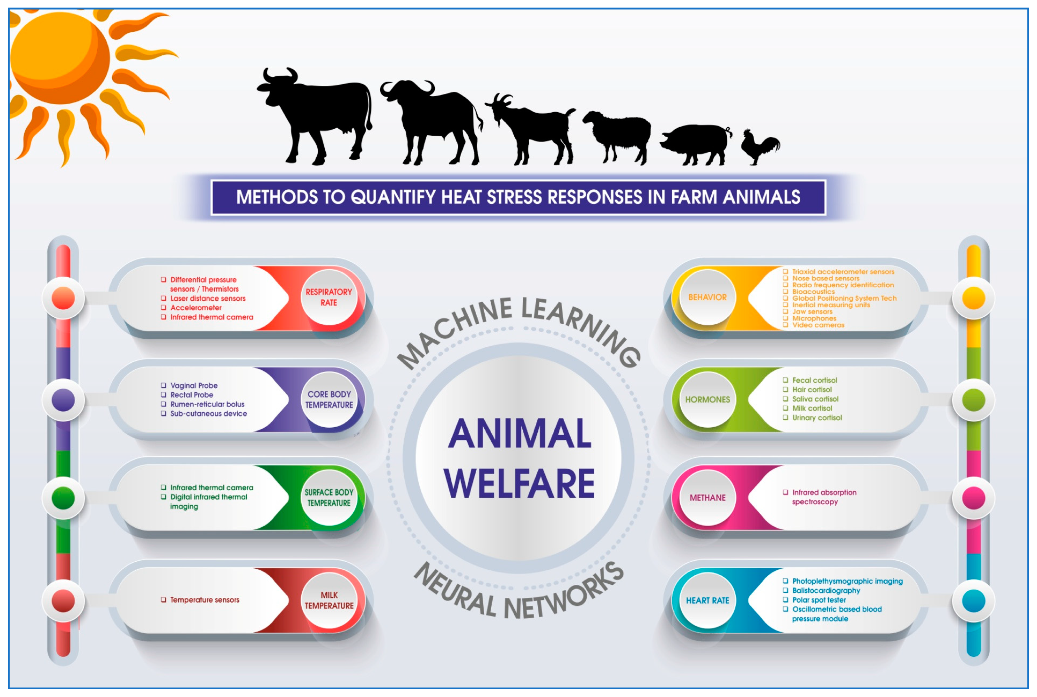
Figure 1. Different non-invasive methods to quantify heat stress response in farm animals.
Atmosphere 13 01642 g001
Table 1. Advanced technologies that can be used for assessment of heat stress in livestock.
IndicatorMethodologyTechnologySpeciesContinuousAccuracyReferences
Respiration rateMeasuring nasal exhalation pressureDifferential pressure sensorCattleUnknown[78]
Differences in temperature near nostrilsTemperature sensors/thermistorCattle±0.15 °C[34]
Counting radial movement of the flank areaLaser distance sensorCattleUnknown[33]
Measuring temperature changes in nostrils during respirationInfrared thermography + RGB sensorsCattle, pigs±2%[2,79]
Counting back and forward motion of the body during pantingAccelerometerCattleUnknown[80]
Core body temperatureComparing the heat flux between core body temperature and the skin surface“Dräger” Double Sensor (DS)HumansUnknown[81]
Comparing the differences in core body temperature using a thermosensoric patchZero-heat-flux thermometersPigsUnknown[82]
Body surface temperatureMeasuring eye and muzzle temperatureDigital infrared thermal imagingCattle, sheep±2 °C[83,84]
Measuring ear, cheek, forehead, flank, rump, and udder surface temperaturePortable Infrared cameraCattle±2 °C[85]
Milk temperatureMeasuring milk temperature of lactating cows Temperature SensorsCattleUnknown[86]
Sleeping intervalMeasuring body movement during REM and NREM sleepAccelerometer-based sleep deviceCattle93.7 ± 0.7% for wake behavior and 92.2 ± 0.8% for sleep-like behavior[87]
Heart rate/pulse rateOptical measurement of blood volume changes in the microvascular bed of tissuePhotoplethysmographic Imaging (PPGI)Non-human primates±1%[88]
Measuring the whole-body recoil forces leads to changes in displacements and vibrations of the body surfaceEulerian Video Magnification/Ballistocardiography (BCG)HumansUnknown[89,90]
Measures very small electrical impulses emitted by the heartPolar Spot tester (PST)CattleUnknown[91]
Measurement of oscillations during inflation and deflation in limbsOscillometric-based blood pressure moduleDogsUnknown[92]
Counting the variation over time of the period between consecutive heartbeatsAccelerometerSheep, goatsUnknown[93]
Grazing and ruminating behaviorDetects animals’ jaw movement by measuring the accelerationsTri-axial accelerometer sensorSheep96% for grazing, 95% for ruminating, and 94% for resting[94]
Assessing the feeding behavior of animalsThe wireless sensor system (network sensors) + mobile sensorsCattle2.66 m (range = 0.57–5.95 m)[95]
Water drinking behaviorMeasuring the pressure changes in noseband sensor and Tri-axial accelerometer sensorNoseband sensorCattle0.98[96]
The sensor will record the electronic tag when the animal arrives near water space, and the water meter measures the amount of water consumedRadio Frequency Identification (RFID) + water flow meterCattle, sheep95%[97,98]
CortisolAnimal feces is collected, and it will be further processed for extraction of cortisolEnzyme-linked
immunosorbent assay (ELISA)
Cattle, pigs, chicken, sheep99,76 ± 3.77%[59,99,100,101]
MethaneThe device is pointed towards nostril of cows; the device measures the density of the air column between device and animal’s nostrilInfrared absorption spectroscopy using a semiconductor laser for CH4 detectionCattleSensitivity = 95.4% and specificity = 96.5%[102]
(% = Percentage, m = meters, °C = Degree Celsius).
Publisher’s Note: MDPI stays neutral with regard to jurisdictional claims in published maps and institutional affiliations.

