Article

Intrinsic Characteristics and Biological Effects of Standard Reference Indoor Dust SRM® 2585 and Its Inhalable Subfractions PM10 and PM2.5

1
Normandie Univ, UNICAEN, UNIROUEN, ABTE, 14000 Caen, France
2
Centre François Baclesse, Boulevard Général Harris, CEDEX 05, 14076 Caen, France
3
Normandie Univ, UNICAEN, UNIROUEN, ABTE, 76000 Caen, France
4
Unité de Chimie Environnementale et Interactions sur le Vivant (UCEIV), Université du Littoral Côte d’Opale, UR 4492, SFR Condorcet FR CNRS 3417, 59140 Dunkerque, France
*
Author to whom correspondence should be addressed.
Atmosphere 2022, 13(11), 1818; https://doi.org/10.3390/atmos13111818
Submission received: 30 September 2022 / Revised: 26 October 2022 / Accepted: 29 October 2022 / Published: 1 November 2022
(This article belongs to the Special Issue Chemical Composition and Toxicology of Indoor Particulate Matter)

Abstract

:
Standard Reference Material® (SRM®) 2585 can be considered as a model sample of settled indoor dust. At least 168 adsorbed chemicals were identified and quantified, generating a complex and potentially toxic mixture. Inhalable fractions PM10 and PM2.5 were isolated and compared to SRM® 2585 for their intrinsic properties and their respiratory toxicity using pertinent normal human bronchoepithelial (NHBE) cells. The intrinsic oxidative potential (OP) of SRM® 2585 and its subfractions was measured in acellular conditions, together with ROS generation and endotoxin content. For SRM® 2585 and PM10, cellular responses were evaluated on NHBE cells after 72 h exposure (1.1 to 110 µg/mL). The presence of endotoxins associated with an intrinsic prooxidant potential did not lead to the induction of an inflammatory response. Interestingly, cytokine production was decreased, strongly for IL-1β and to a lesser extent for IL-6. Only PM10 induced an acute toxicity. In parallel, mutagenicity was evaluated from organic extracts. Mutagenic profiles indicated the concomitant presence of nitro-PAHs in addition to the previously reported PAHs. This could be of concern for long-term health effects in a context of chronic exposure.

1. Introduction

Indoor air pollution is typically a complex aerosol, composed of a gaseous phase associated with various kinds of particles: mineral (mainly issued from erosion processes), carbonated (mainly issued from combustion processes) or biological (with pollens, moulds, bacteria, viruses, dust mites, etc.). Chemicals could then be adsorbed onto the surface of these particles, which act as transporters. The composition of this aerosol varies very widely over time, including over short periods, which limits the representativeness of real-time sampling. Alternatively, the settled dust integrates the particles initially in suspension in the air over a period depending on the frequency of cleaning. In addition, these settled dusts can be resuspended and inhaled, depending on their particle size.
SRM® 2585 namely ‘Organic contaminants in house dust’ is a commercially available sample of settled dust collected from houses, motels, cleaning services and hotels in the USA [1]. At least 168 molecules belonging to seven chemical families such as polycyclic aromatic hydrocarbons (PAH) congeners, polychlorinated biphenyl (PCB) congeners, chlorinated pesticides, polybrominated diphenyl ether (PBDE) congeners, synthetic musks, perfluorinated alkyl acids (PFAAs), hexabromocyclododecane (HBCDD) isomers, and phosphorous flame retardants, have been already identified, quantified and/or certified from this sample by the National Institute of Standards and Technology (NIST) and updated in 2018 [1]. SRM® 2585 is thus used as a reference material for the evaluation of analytical methods for these chemicals [2]. Despite the known toxic properties (genotoxicity, endocrine disruption, pro-oxidative and pro-inflammatory effects) of some precited chemical families, this sample was poorly investigated for its toxic potential [3,4]. In these previous studies, the authors used organic extracts representative of the diversity of the compounds adsorbed by the dust, of their interactions and therefore of the modulation of their respective toxicities, which are difficult to predict or to model. However, the use of organic extracts does not reflect the bioavailability of these compounds under real exposure conditions.
The main health effects associated with indoor air pollutants result from their inhalation. The depth of penetration into the respiratory tract depends on the particle size. Particles with an aerodynamic diameter around 10 µm (PM10) lead to tracheobronchial deposition while the smaller particles (PM2.5) reach the alveoli. Thus, these two inhalable fractions were separated from the whole SRM® 2585, and their intrinsic toxic potential was evaluated and compared. First, the presence of biological contaminants likely to contribute to the toxic effects, namely endotoxins and fungi, was investigated. Then, due to the well-established involvement of oxidative mechanisms in particle-associated health effects [5,6,7,8], the intrinsic oxidative potential (OP) of SRM® 2585 and its subfractions was measured in acellular conditions, together with reactive oxygen species (ROS) generation capabilities.
Thereafter, using a relevant model for the assessment of respiratory toxicity, i.e., primary normal human bronchoepithelial (NHBE) cells, biological responses to SRM® 2585 and its PM10 subfraction exposures were evaluated in vitro through viability and proinflammatory potential measurements. In parallel, organic extracts of these two dust samples were prepared and tested for their mutagenic potential on Salmonella typhimurium (Ames test).
The aim of this study was to identify relevant intrinsic characteristics of a representative sample of domestic settled dust and of its two inhalable derived subfractions. In a second step, in order to create bridges between intrinsic properties and potential toxic effects, biomarkers of toxicity, such as viability, inflammation, and genotoxicity were determined in vitro.

2. Materials and Methods

2.1. SRM® 2585 and Granulometric Separation

The Standard Reference Material® SRM® 2585 ‘Organic contaminants in house dust’ was referenced by the National Institute of Standards and Technology (NIST, Gaithersburg, MD, USA). It was obtained from indoor dust collected from homes, cleaning services, motels, and hotels, then sterilised using gamma irradiation. The particle size fraction lesser than 100 µm was separated using vibrating stainless steel sieve apparatus. Analytical composition was determined for 168 organic contaminants, naturally present in this dust. Mass fraction was certified, i.e., with a highest confidence in its accuracy, for selected PAHs (n = 33), PCB congeners (n = 30), chlorinated pesticides (n = 4) and PBDE congeners (n = 16). Reference or information on mass fraction values were also given for additional compounds issued from the four previous chemical families (n = 33; n = 12; n = 10, and n = 12 respectively), but also for selected polycyclic musks (n = 5), perfluorinated alkyl acids (PFAAs n = 8) and flame retardants (n = 5). For more details, see the certificate of analysis [1].
Granulometric separation was performed in order to isolate PM10 and PM2.5 inhalable fractions. Three units of SRM® 2585 (10 g per unit) were aerosolised using a Topas 410U powder disperser (Topas GmbH, Germany) then separated through a cyclone BGI GK4.162 model (Mesa labs, France) to collect the PM10 fraction. For the PM2.5 fraction, two distinct units of SRM® 2585 were aerosolized on Tapas 410U disperser and separated using a GK2.05 (KTL) cyclone model (Mesa labs, France). Finally, the corresponding fractions were collected on Isopore membrane 0.2 µm (Millipore, France).

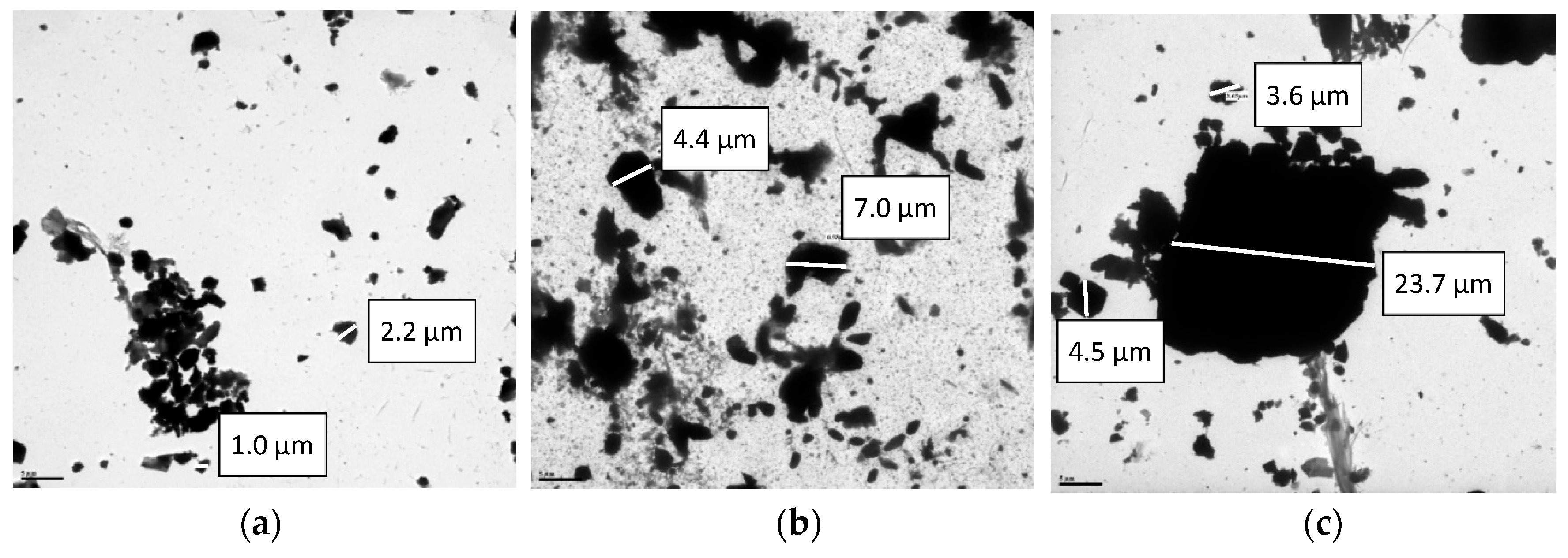
2.2. Transmission Electron Microscopy (TEM)

Suspension of SRM® 2585, PM10 or PM2.5 were put on formvar carbon coated grids previously glow discharged (ELMO CORDOUAN, France) and observed with a transmission electron microscope 1011 (JEOL, Japan) equipped with camera Orius 200 (GATAN, USA) and digital micrograph (GATAN, USA) at the Electron Facility Center CMAbio3 (EMerode, UNICAEN, France).

2.3. Diesel Exhaust Particles (DEP)

DEP were originated from a direct injection turbocharged intercooled diesel engine used with a reference diesel fuel containing less than 50 ppm of sulphur, without any after-treatment process [9].

2.4. Endotoxin Measurements

The quantification of endotoxin in SRM® 2585 and its PM10 and PM2.5 subfractions was conducted from 1 mg/mL suspensions in PBS, using the Limulus Amebocyte Lysate (LAL) method [10], following the manufacturer’s instructions (Endosafe®, Charles-River, USA).

2.5. Microbiological Contamination

The microbial content of the whole SRM® 2585 was evaluated using the Warcup method [11]. MEA (Malt Extract Agar) and TSA (Tryptone Soy Agar) plates were incubated at 25, 30, and 37 °C, and regularly checked for one month to monitor bacterial and fungal growth.

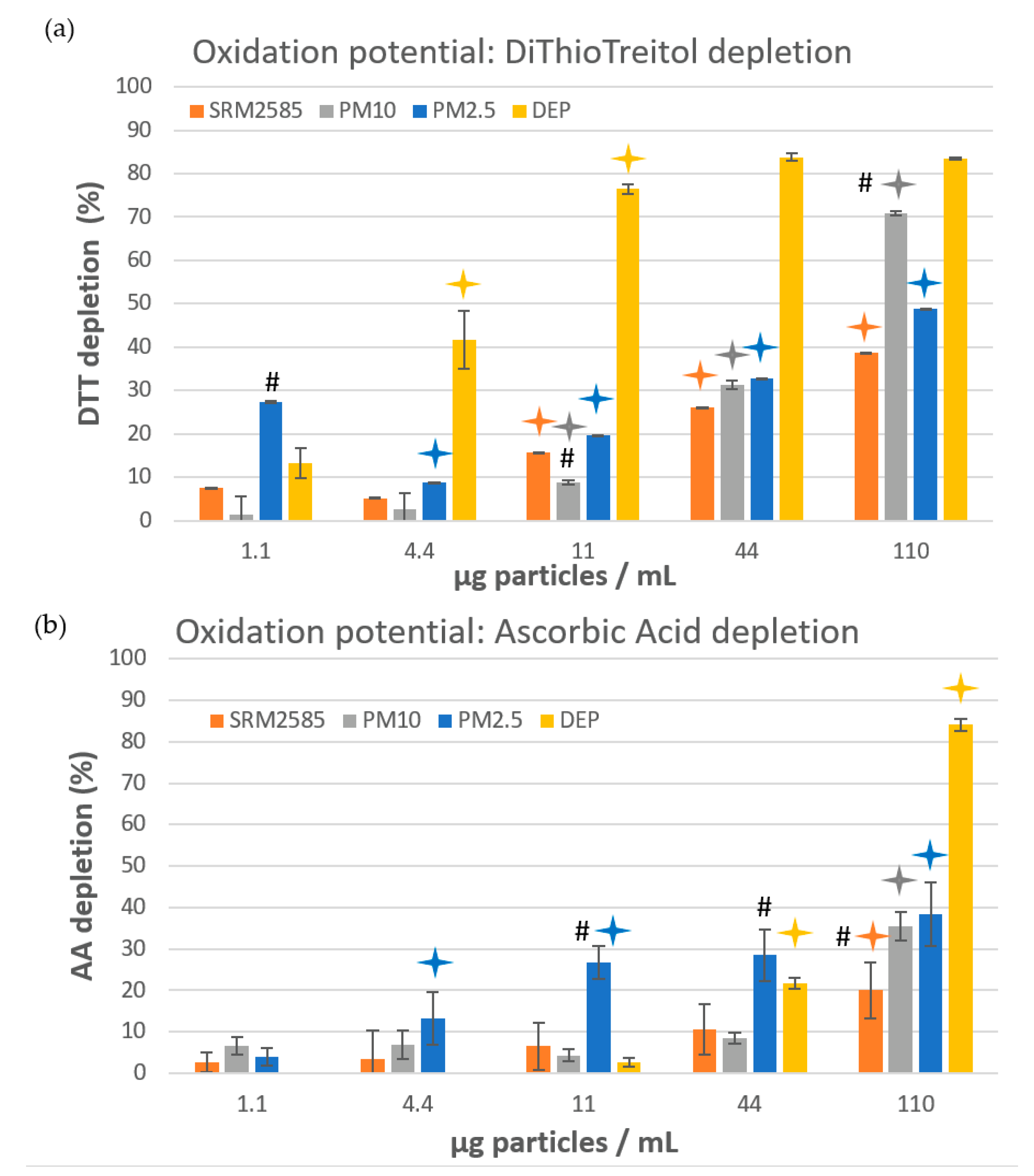
2.6. Oxidative Potential Measurement

According to Martin de Lagarde et al. [12], the intrinsic oxidative potential of SRM® 2585 and its PM10 and PM2.5 subfractions was determined with an ascorbic acid (AA) depletion assay and with a dithiothreitol (DTT) assay. The particles were tested using a range of final concentration from 1.1 to 110 µg/mL. These concentrations correspond to 0.5 µg/cm2 to 50 µg/cm2 and are consistent with the levels usually found in the literature [4,8,13]. For comparative purposes, diesel exhaust particles (DEP) were tested in the same conditions. Each sample was analysed in three independent experiments.

2.6.1. Ascorbic Acid Depletion Assay

Particles were incubated for 4 h at 37 °C in an ascorbic acid solution (200 µM) prepared in 0.9% NaCl at pH 7.4. Samples were then centrifuged 5 min (12,000× g, 4 °C) to remove particles and the supernatants were filtered (Nylon 0.45 µm syringe filter). The final concentrations of AA were quantified with an Agilent High Pressure Liquid Chromatography Infinity II equipped with DAD detector (Agilent Technologies, USA) and a Poroshell 120 EC-C18 (3 × 150 mm, 2.7 µm) column (Agilent Technologies, USA) equipped with guard EC-C18 (3 mm) maintained at 25 °C. The mobile phase (potassium hydrogen phosphate 10 mM, pH 3) flow rate was 0.6 mL/min. UV data were collected at 220 nm. Percentage of depletion was calculated as described below:
OPAA = 100 − (AA concentration in the sample/AA concentration in the blank) × 100.

2.6.2. Dithiothreitol Assay (DTT Assay)

Particles were incubated with 200 µM DTT in 0.10 M phosphate buffer (0.08 M Na2HPO4 and 0.02 M NaH2PO4, pH 7.4, Chelex-treated) in a 96-well plate for 1 h at 37 °C. Plates were then centrifuged for 15 min at 2100× g and 4 °C. Equal volumes of supernatant and of 2.5 mM 5,5′-dithiobis-(2-nitrobenzoic acid) (DTNB) were mixed and absorbance was measured at 412 nm (Clariostar, BMG Labtech, France). The percentage of depletion was calculated as described below:
OPDTT = 100 − (DTT concentration in the sample/DTT concentration in the blank) × 100.

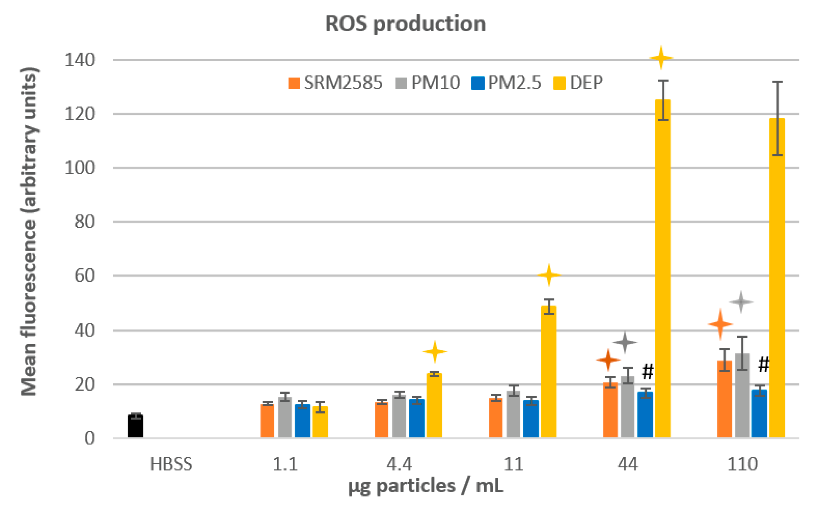
2.7. Intrinsic ROS Production

0.5 mL of 2′,7′-dichlorofluorescein diacetate (DCFDA) 1 mM in ethanol was chemically deacetylated by addition of 2 mL NaOH 10 mM, leading to DCFH production. After 30 min in the dark and at room temperature, this solution was neutralised with 10 mL phosphate buffer 25 mM, pH 7.2. Then, 1.6 mL Hank’s balanced salt solution (HBSS) was added to 400 µL DCFH. Sixty microliters of particle suspension (1.1 to 110 µg/mL) was added to 2 mL of DCFH/HBSS solution, vortexed and distributed in a 96-well plate (150 µL/well). Green fluorescence intensity was kinetically measured using an IncuCyte® device (Essen Bioscience, UK). The reported results were obtained after 90 min, corresponding to the maximum of the recorded signal. Measurements were conducted in triplicates, through two independent experiments.

2.8. Organic Extract Preparation

Organic extracts (OE) were obtained, according to Badran et al. [14]. Briefly, 262 mg of SRM® 2585 or its PM10 fraction was placed in a glass vial, with 10 mL dichloromethane (DCM), and subjected to a 20 min sonication assisted extraction step at 20 °C. The obtained solution was centrifuged at 1000× g for 20 min at 20 °C. Then, the solution containing extracted organic compounds was carefully removed and transferred into a second vial and 2 additional ultrasonic cycles were performed on the remaining PM using fresh DCM. Finally, supernatants were gathered, and DCM was evaporated under nitrogen flux. The residues were dissolved in dimethylsulfoxide (DMSO) and kept frozen until used for mutagenicity testing.

2.9. Ames Test

The mutagenicity of the two organic extracts was evaluated on 4 Salmonella typhimurium tester strains (overnight cultures), namely TA98, YG1041, TA100, and TA102. The preincubation protocol was applied [9], without (−S9mix) and with (+S9mix) an exogenous microsomal metabolization system (Aroclor-induced S9, Trinova Biochem, Germany). OE (10 µL) was mixed with Salmonella strain (100 µL) and either PBS (100 µL; −S9mix conditions) or S9mix (100 µL; +S9mix conditions) in a sterile tube and shaken for 1 h at 37 °C. Top agar (2 mL) was then added. Tubes were vortexed and poured onto Petri dishes containing VBE medium. After a 48 h incubation at 37 °C, revertant colonies were automatically counted (Ames software, Noesis, France). For each sample, three concentrations (1, 10, and 40 µg-equivalent particles/plate) were tested, in triplicate. Toxicity (bactericidal effect) was evaluated in parallel through microscopic observation of the background lawn density. Results were expressed as the mean ratio of induced/spontaneous revertants, determined through at least two independent experiments.

2.10. NHBE Cell Culture

Normal human bronchoepithelial cells (NHBE) were purchased frozen from ATCC® (PCS-300-010, LGC Standards SARL, France). They were issued from a 14-year-old Hispanic/Latino male donor. Cells were cultured at 37 °C with 5% CO2 atmosphere, on airway epithelial cell basal medium supplemented with components provided in bronchial epithelial cell growth kit (ATCC® PCS-300-040, LGC Standards SARL, Molsheim, France) and 1% antibiotic–antimycotic solution (Fisher Scientific, Illkirch, France). Growth media were changed every 48 h. When they reached 80% confluence, cells were detached using TrypLe Expess solution (Fisher Scientific SAS, France) then re-seeded in a new flask with fresh growth media. Exposures were performed on cells cultivated at passage 3 or 4.

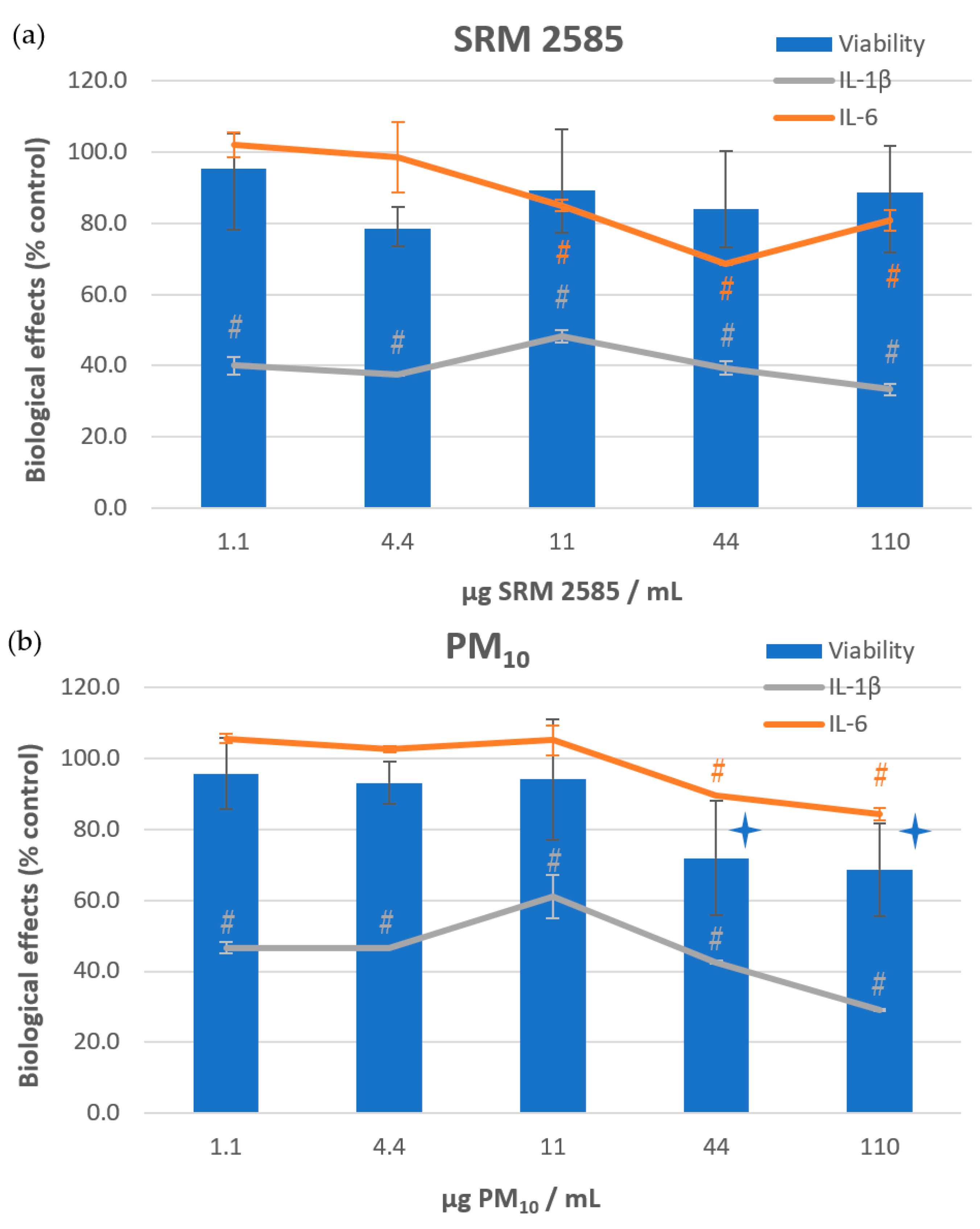
2.11. Viability and Inflammatory Response Determination

NHBE were seeded in 96-well cell culture plates at 7500 cells/well. Twenty-four hours later, suspensions of SRM® 2585 or its PM10 subfraction were added (1.1 to 110 µg/mL), for a 72 h exposure. Each condition was tested in triplicate and repeated through 2 independent experiments. After exposure, the culture media were removed and immediately frozen for further cytokine analysis.

2.11.1. Viability

Cells were then rinsed with PBS and resazurin (Presto Blue™ cell viability reagent; Invitrogen, France) was added. After 2 h at 37 °C, fluorescence intensity was measured on each well, using a microplate reader (Synergy H1, Biotek, Agilent, USA). Cell viability was calculated as follows:
Mean   fluo .   Measured   after   exposure     mean   fluo .   Blank × 100 Mean   fluo .   Measured   for   negative   control     mean   fluo   blank

2.11.2. Inflammation Response

Cytokine excretion was measured in frozen culture media for IL-6, TNF-α, and IL-1β. ELISA MAX™ Deluxe Set human kits were purchased from Biolegend (The Netherlands) and used according to the manufacturer’s instructions.

2.12. Statistical Analysis

Statistical analyses were performed on raw data using Student’s t test with a minimal significant threshold of p ≤ 0.05.

3. Results

Granulometric separation of the PM10 fraction led to the recovery of 693 mg representing 2.9% of the initial mass of SRM® 2585 (3 units of 10 g each). For PM2.5 fraction, 30.8 mg (i.e., 0.15%) were isolated from 20.2 g (2 units) of the SRM® 2585. This limited amount was used for the determination of intrinsic properties only. The obtention of a differentiated granulometry was assessed by TEM as shown in Figure 1.
No microbiological development was observed on the versatile MEA and TSA media over a period of 1 month, regardless of the applied temperature (25 °C, 30 °C, or 37 °C). Endotoxin levels ranged from 24.3 to 36.20 EU/mg. It was notably increased (+49%) for the smallest fraction PM2.5 compared to the whole SRM® 2585 (Table 1).
Intrinsic oxidative potential (OP) was then measured for the three samples and compared with the OP of diesel exhaust particles (DEP) available in the laboratory and issued from a previous study dealing with their toxic effects [9]. The antioxidant depletion method was used in acellular conditions, both towards ascorbic acid (OPAA) and dithiothreitol (OPDTT) (Figure 2). OP varied in a dose-dependent manner for the three dust samples and at the two highest doses, OPDTT was higher compared to OPAA.
The subfractions tended to be more pro-oxidant than the whole SRM® 2585 sample, especially the PM2.5 when tested between 4.4 and 44 µg/mL for OPAA and the PM10 at the highest dose for OPDTT. DEP were clearly more pro-oxidant than indoor dust for OPDTT, whereas for OPAA, only the highest dose of DEP differed from the indoor dust behaviour.
Intrinsic ROS production was estimated using the chemically deacetylated DCFDA probe (i.e., DCFH derivative), that may fluoresce in the presence of ROS. Compared to the negative control (HBSS), the fluorescence intensity was weakly but significantly modified in the presence of dust (Figure 3). A significant dose-effect relationship was observed only for SRM® 2585 and PM10 fraction, at the two highest doses. For a given dose, the particle size did not influence ROS production, except for PM2.5, which remained significantly lower at the two highest doses. Depending on the tested dose, DEP induced between 1.5- and 7.7-fold higher ROS levels compared to the various indoor dust samples.
Viability of NHBE cells was measured after 72 h exposure to SRM® 2585 and its PM10 fraction (PM2.5 were not tested due to the small amount available) (Figure 4). A limited but significant reduction in NHBE viability was noted with the PM10 fraction only, when tested at the two highest doses (72% and 68% of viability at 44 and 110 µg/mL respectively). From the same experiments, cytokine production was measured in supernatants for IL-6, TNF-α, and IL-1β (Figure 4). No TNF-α production was observed in NHBE cells. IL-1β release was drastically limited whatever the dose and the type of particles tested. IL-6 excretion was also reduced, but to a lesser extent, and only by 10 to 30% after exposure to SRM® 2585 from 11 µg/mL and to PM10 from 44 µg/mL.
Mutagenic activity was evaluated from organic extracts (OE) obtained with SRM® 2585 and PM10 fraction (due to the small amount of PM2.5 available, it was not possible to prepare the corresponding OE) (Table 2). Four Salmonella typhimurium tester strains were used, complementarily covering different mechanisms of action. Indirect mutagens were characterised after metabolic activation performed by addition of S9mix issued from Aroclor-induced rat liver. Significant responses, corresponding to at least a doubling of the spontaneous revertant level, are indicated in red in Table 2. TA98 and YG1041 both revert by a frameshift mechanism. No direct response was noted in TA98, but upon S9mix addition, a significant and dose-dependent mutagenicity was observed for the two OEs, with a higher response for PM10. YG1041 overexpresses nitroreductase and O-acetyltransferase enzymes, and thus was specifically designed for the detection of nitroaromatics (w/o S9mix) and aminoaromatics (with S9mix) [15]. Strong and comparable direct mutagenicity was observed for the two OEs, again associated with a clear dose-effect relationship. In the presence of S9mix, mutagenicity was slightly decreased at the highest dose tested, but tended to be reinforced at the lowest dose, especially for PM10.
TA100 and TA102 revert through a base pair substitution mechanism. In TA100, a reduction of the background lawn density indicated that the two OE were toxic, at the highest doses only. With the subsequent dilutions, toxicity was abolished, but no significant response was observed. Upon S9mix addition, a clear but not significant increase of the induced revertant number was observed at the highest dose. No change in the number of revertants was reported for TA102.

4. Discussion

Household dust is a reservoir of a mixture of chemicals emitted both by various indoor sources and from infiltrated outdoor pollutions. Settled dusts can be considered as integrating, over a cumulative period, a series of pollutants initially in suspension in the air. SRM® 2585 is a representative settled dust and its chemical composition for organic compounds has been carefully analysed by the NIST [1]. At least 168 chemicals have already been identified, some of them known for their toxic properties. In order to characterize their potential toxicity after inhalation, intrinsic physicochemical and biological properties of these particles were first investigated, and compared with those of its inhalable subfractions PM10 and PM2.5. Then, the response of NHBE cells was evaluated after 72 h exposure to SRM® 2585 and its PM10 subfraction.
The dry particle size separation was carried out using two cyclonic devices. TEM analysis showed that the isolated fractions have retained their respective size characteristics, with limited aggregation.
No microbiological development was observed upon one-month culture of SRM® 2585, which indicates that the initial sterilisation of dust samples with gamma rays [1] was efficient in avoiding the development of subsequent contamination during long storage. However, alternatively, by destroying the microorganisms originally present in the dust, this preservative irradiation also precludes the assessment of a possible contribution of microbiota to intrinsic dust toxicity.
Indeed, after lysis or destruction of gram-negative bacteria, lipopolysaccharide (LPS) is released from their outer membrane and distributed into the surrounding environment as endotoxin. This compound is known to induce inflammatory mechanisms in tissues or cell cultures [16]. Endotoxin exposure is thus a risk factor for respiratory diseases [17] such as asthma, COPD or fibrosis. Shamsollahi et al. [18] recently reviewed numerous studies that measured endotoxin levels in domestic settled dust. Focusing on studies that collected dust from ground only, a very large range of endotoxin levels was nevertheless reported, i.e., from 0.041 to 1698 EU/mg dust. The mean amount, when reported, also varied from 9.24 to 79.6 EU/mg dust, depending on the studies. No clear differences were observed between rural and urban studies. Thus, by comparison, SRM® 2585 and its subfractions displayed intermediate endotoxin levels, quite similar to those reported by Leung et al. [19], Tischer et al. [20], and Thorne et al. [21] but lower than those measured by Bose et al. [16], Horick et al. [22], Lawson et al. [23], and Litonjua et al. [24]. Recently, Sun et al. [17] studied the household determinants of biocontaminant exposures, including endotoxin. Among the 290 urban houses included, endotoxin levels varied widely, from 7626 to 258 917 EU/g dust, although collected from the living room floor only. Authors reported that the presence of carpeting, of pets, but also low floor cleaning frequency, home age, and indoor relative humidity were the major determinants for indoor biocontamination. They also observed large regional variations. From an analytical point of view, the reactivity of endotoxins to the LAL test may also vary depending on the bacterial strain from which they originate, but also on the attachment or not of endotoxin to the cell membrane [25]. All these aspects contribute to the wide variations in endotoxin levels observed in the different sampled houses. For its part, the SRM results from many pooled samples that integrates these variation criteria.
Liu et al. [26] mentioned that dust size can modulate the concentration of adsorbed chemicals. Endotoxin content was not drastically modified after size segregation of SRM® 2585, although it raised 1.5-fold for the PM2.5. This observation could be linked to the increase in the specific surface area for finer particles which leads, at equivalent mass, to a possible accumulation of adsorbed ambient compounds [27]. However, this effect seems to depend on the type of dust, since for dairy dust, the endotoxin level was 14.5 times higher for PM10 compared to PM>10 [28].
Given the important contribution of oxidative mechanisms in particle toxicity, the intrinsic oxidative potential (OP) and ROS generation capability of SRM® 2585 and its subfractions were then determined in acellular conditions. Three tests were performed displaying distinct sensitivities towards various families of pro-oxidant compounds. These tests are thus considered complementary [29,30]. The depletion of antioxidants such as ascorbic acid (AA) and dithiotreithol (DTT) were measured. DTT is considered sensitive to organic compounds, mainly polycyclic aromatic hydrocarbons (PAHs), quinones [31,32], or soluble transition metals [33,34], whereas AA is more especially sensitive to Cu and Mn [35]. Lastly, DCFH probe detects preferably reactive oxygen species (ROS) and reactive nitrogen species (RNS) and reacts significantly with H2O2 and transition metals [29,36]. Our results showed that OPDTT measured for DEP, rich in PAHs issued from combustion and metals issued from engine wear [37], effectively depleted DTT to a larger extent than dust samples. Nevertheless, OPDTT remained significant for dust particles, which is consistent with the analytical quantification of various PAHs on SRM® 2585. Metals were not quantified by the NIST for SRM® 2585, but the dust used for its the preparation is a mix of 70% SRM® 2583 (‘Trace Elements in Indoor Dust, Nominal 90 mg/kg Lead’) and 30% SRM® 2584 (‘Trace Elements in Indoor Dust, Nominal 1% Lead’) for which, besides lead (Pb), various metals were measured. Based on analytical data displayed in their respective certificates of analysis [38,39] and on their proportions in SRM® 2585, it could therefore be roughly estimated that minimal concentration of Fe, Mg and Al was about 5–7 mg/g in SRM® 2585, Zn was around 0.8 mg/g, whereas Cu and Ni were near 0.1 mg/g dust. Surprisingly, only limited OPAA were observed from dust. ROS and RNS production were also very limited for dust, compared to DEP. These complementary data indicate that intrinsic prooxidant properties of SRM® 2585, mainly observed for DTT, could thus be primarily attributed to the PAHs fraction. Based on the NIST analysis [1], 66 PAHs were identified, corresponding to a total mass concentration of 37.9 µg/g. In contrast, metals appear to contribute only marginally to the intrinsic OP of dust.
The biological effects consecutive to inhalation of such domestic dust were then investigated in vitro after exposure of pertinent primary normal human bronchial epithelial (NHBE) cells. Viability was slightly but significantly impaired only for PM10 after 72 h exposure at 44 and 110 µg/mL. Zhang et al. [3] reported that organic extract prepared from SRM® 2585, and corresponding to 0.6 mg of dust did not modify liver (HepG2) cell viability after 72 h exposure. Marques dos Santos et al. [4] observed a dose-dependent mitochondrial dysfunction (ATP production and respiration) towards various pulmonary cell lines, after 24 h exposure to SRM® 2585 extract, but the impact on viability was not reported. HepG2 cells were also tested and appeared more resistant to these perturbations. Using differentiated NHBE cells isolated from brush biopsy, Hawley et al. [28] did not observe any variation in viability after 2 h of air-to-cell exposure to dairy dust. For comparative purposes with well-studied DEP particles, Kim et al. [13] reported that, despite their high content in PAHs and metals, these particles did not affect survival of nasal fibroblasts after 72 h exposure until the concentration reached 400 μg/mL. In contrast, using NHBE cells, LD50 of DEP was calculated between 109 and 140 µg/mL after only 1 h exposure [40], indicating the higher sensitivity of normal cells versus the established cell line. Thus, even if limited, the acute effects of PM10 fraction isolated from SRM® 2585 on NHBE viability must be considered, since they occurred at doses comparable to DEP LD50 for this cell model.
Due to the presence of endotoxins and to the OPDTT, the cellular inflammatory response was also investigated after 72 h exposure. Surprisingly, cytokine levels were found decreased for SRM® 2585 as well as for PM10, except for IL-6 at the lowest doses. IL-1β was the most drastically reduced compared to controls, whatever the dust exposure conditions. Based on the detailed values displayed in their supplementary data, Marques dos Santos et al. [4] observed such drastic decrease of IL-6 (and IL-8) levels in pulmonary cell lines 16HBE14o- after 24 h exposure to an organic extract of SRM® 2585 in submerged conditions. No major changes were noted in a distinct pulmonary NuLi-1 cell line. Kang et al. [41] previously reported such reduced production of IL-1β (and IL-8), associated with decreased mRNA expression, 24 h after exposure of U937 cells to indoor dust. However, no associated mechanism has been proposed. Among the complex mixture of compounds adsorbed on indoor dust such as SRM® 2585, it could be hypothesized that some of them partly inhibited the secretion of some cytokines. By contrast, IL-6, IL-8, as well as TNF-α transcript levels were found significantly increased in differentiated NHBE cells after a short exposure (2 h) to dairy dust [28]. More recently, using RAW 264.7 cells exposed to sand dust, Olgun et al. [42] indicated that optimal kinetics for IL-6 production was obtained after 4 h exposure whereas 24 h was necessary for TNF-α. Thus, the exposure duration applied in the present study (72 h) may not correspond to an optimal cytokine secretion kinetics. This later appeared to be both cytokine- and cell type-dependent and requires further investigation.
A significant genotoxic potential was also demonstrated, involving only the two strains that reverse according to a frameshift mechanism. Hence, significant responses of TA98 used with S9 fraction can be considered as characteristic of the PAHs. Moreover, a stronger response was observed in YG1041 w/o S9mix, indicating the probable presence of nitro-PAHs. This family of compounds was not included in the NIST analysis but is known for its powerful genotoxic properties [43]. As previously observed with endotoxin, particle size also influenced genotoxicity which was reinforced for the PM10 fraction. Its increased specific area could again contribute to this observation.
The absence of responses towards TA102 which is sensitive, notably, to prooxidants [44] indicated that the oxidative potential of PAHs (OPDTT) was not efficient enough to significantly impact this strain. The potential contribution of metals could not be taken into account from organic extracts.

5. Conclusions

SRM® 2585, and more specifically its inhalable subfractions, have intrinsic properties that can contribute to the induction of toxic cellular responses. Endotoxins were found at intermediate levels compared to the literature and this dust had prooxidative properties that were mainly attributable to the organic fraction and especially to PAHs. Metals, although not directly measured on these samples, appeared to be weakly involved.
Regarding the cell toxicity assessment, acute exposure to the PM10 fraction reduced the viability of the NHBE model, in contrast to the overall SRM®. This underlines the importance of associating a relevant cell model, physiologically compatible with the particle size of the inhalable fraction tested. The presence of endotoxins associated with an intrinsic prooxidant potential did not lead to the induction of an inflammatory response. Interestingly, excreted cytokine levels were decreased, very strongly and for all conditions for IL-1β and to a lesser extent for IL-6 at the highest doses only. The underlying mechanisms of these decreases remain to be identified.
Organic extracts exhibited a mutagenicity, in accordance with the mixture of PAHs adsorbed on the particles. In addition, the mutagenic profiles indicated the concomitant presence of nitro-PAHs. This could be of particular concern for long-term health effects, since some of these chemicals are known to be genotoxic and carcinogenic. This is why repeated exposures will be further performed and genotoxicity markers analysed. In parallel, the PM2.5 fraction will be isolated again to allow the study of its toxicity on a cell model corresponding to the alveolar stage, which is relevant from a physiological point of view.

Author Contributions

Funding acquisition, I.P. and V.A.; investigation, C.E.H., T.R.-F., V.S., A.V., D.G. and V.A.; methodology, I.P. and V.A.; project administration, V.A.; supervision, V.A.; writing—original draft, V.A.; writing—review and editing, I.P. All authors have read and agreed to the published version of the manuscript.

Funding

This research was funded by l’Agence de l’environnement et de la maîtrise de l’énergie (ADEME), grant number 20ESC0002. C.E.L. was funded by Région Normandie.

Institutional Review Board Statement

Not applicable.

Informed Consent Statement

Not applicable.

Acknowledgments

Thanks to Didier GOUX (Biological Microscopy Facility Center, CMAbio3, Normandie Univ, UNICAEN) for technical TEM support for microscopy observations and to Sophie Pricot for proofreading the English. Thanks to E. Brotin (ImpedenCELL platform, UNICAEN, France) for access to the IncuCyte® S3 device, as part of the European project “DILEMI”, co-funded by the Normandy County Council, the European Union within the framework of the Operational Programme ERDF/ESF 2014-2020.

Conflicts of Interest

The authors declare no conflict of interest. The funders had no role in the design of the study; in the collection, analyses, or interpretation of data; in the writing of the manuscript; or in the decision to publish the results.

References

  1. NIST (National Institute of Standards and Technology). Certificate of Analysis for Standard Reference Material 2585 (SRM® 2585), Organic Contaminants in House Dust, Updated in 2018. Available online: https://www-s.nist.gov/srmors/view_detail.cfm?srm=2585 (accessed on 10 March 2019).
  2. Poster, D.L.; Kucklick, J.R.; Schantz, M.M.; Vanderpol, S.S.; Leigh, S.D.; Wise, S.A. Development of a house dust standard reference material for the determination of organic contaminants. Environ. Sci. Technol. 2007, 41, 2861–2867. [Google Scholar] [CrossRef] [PubMed]
  3. Zhang, J.; Abdallah, M.A.; Williams, T.D.; Harrad, S.; Chipman, J.K.; Viant, M.R. Gene expression and metabolic responses of HepG2/C3A cells exposed to flame retardants and dust extracts at concentrations relevant to indoor environmental exposures. Chemosphere 2016, 144, 1996–2003. [Google Scholar] [CrossRef] [PubMed]
  4. Marques Dos Santos, M.; Fei, M.T.P.; Li, C.; Jia, S.; Snyder, S.A. Cell-line and culture model specific responses to organic contaminants in house dust: Cell bioenergetics, oxidative stress, and inflammation endpoints. Environ. Int. 2022, 167, 107403. [Google Scholar] [CrossRef] [PubMed]
  5. Ghio, A.J.; Carraway, M.S.; Madden, M.C. Composition of air pollution particles and oxidative stress in cells, tissues, and living systems. J. Toxicol. Environ. Health B Crit. Rev. 2012, 15, 1–21. [Google Scholar] [CrossRef]
  6. Crobeddu, B.; Aragao-Santiago, L.; Bui, L.C.; Boland, S.; Baeza Squiban, A. Oxidative potential of particulate matter 2.5 as predictive indicator of cellular stress. Environ. Pollut. 2017, 230, 125–133. [Google Scholar] [CrossRef]
  7. Valavanidis, A.; Vlachogianni, T.; Fiotakis, K.; Loridas, S. Pulmonary oxidative stress, inflammation and cancer: Respirable particulate matter, fibrous dusts and ozone as major causes of lung carcinogenesis through reactive oxygen species mechanisms. Int. J. Environ. Res. Public Health 2013, 10, 3886–3907. [Google Scholar] [CrossRef]
  8. Hammond, J.; Maher, B.A.; Gonet, T.; Bautista, F.; Allsop, D. Oxidative Stress, cytotoxic and inflammatory effects of urban ultrafine road-deposited dust from the UK and Mexico in human epithelial lung (Calu-3) cells. Antioxidants 2022, 11, 1814. [Google Scholar] [CrossRef]
  9. André, V.; Barraud, C.; Capron, D.; Preterre, D.; Keravec, V.; Vendeville, C.; Cazier, F.; Pottier, D.; Morin, J.P.; Sichel, F. Comparative mutagenicity and genotoxicity of particles and aerosols emitted by the combustion of standard vs. rapeseed methyl ester supplemented bio-diesel fuels. Impact of after treatment devices: Oxidation Catalyst and Particulate Filter. Mutat. Res. 2015, 777, 33–42. [Google Scholar] [CrossRef] [Green Version]
  10. Milton, D.K.; Gere, R.J.; Feldman, H.A.; Greaves, I.A. Endotoxin measurement: Aerosol sampling and application of a new Limulus method. Am. Ind. Hyg. Assoc. J. 1990, 51, 331–337. [Google Scholar] [CrossRef]
  11. Warcup, J.H. The soil-plate method for isolation of fungi from soil. Nature 1950, 166, 117–118. [Google Scholar] [CrossRef]
  12. Martin de Lagarde, V.; Rogez-Florent, T.; Cazier, F.; Dewaele, D.; Cazier-Dennin, F.; Ollivier, A.; Janona, M.; Achard, S.; André, V.; Monteil, C.; et al. Oxidative potential and in vitro toxicity of particles generated by pyrotechnic smokes in human small airway epithelial cells. Ecotoxicol. Environ. Saf. 2022, 239, 113637. [Google Scholar] [CrossRef] [PubMed]
  13. Kim, J.A.; Cho, J.H.; Park, I.H.; Shin, J.M.; Lee, S.A.; Lee, H.M. Diesel exhaust particles upregulate interleukins IL-6 and IL-8 in nasal fibroblasts. PLoS ONE 2016, 11, e0157058. [Google Scholar] [CrossRef] [PubMed] [Green Version]
  14. Badran, G.; Ledoux, F.; Verdin, A.; Abbas, I.; Roumie, M.; Genevray, P.; Landkocz, Y.; Lo Guidice, J.M.; Garçon, G.; Courcot, D. Toxicity of fine and quasi-ultrafine particles: Focus on the effects of organic extractable and non- extractable matter fractions. Chemosphere 2020, 243, 125440. [Google Scholar] [CrossRef] [PubMed]
  15. Hagiwara, Y.; Watanabe, M.; Oda, Y.; Sofuni, T.; Nohmi, T. Specificity and sensitivity of Salmonella Typhimurium YG1041 and YG1042 strains possessing elevated levels of both nitroreductase and acetyltransferase activity. Mutat. Res. 1993, 291, 171–180. [Google Scholar] [CrossRef]
  16. Bose, S.; Rivera-Mariani, F.; Chen, R.; Williams, D.; Belli, A.; Aloe, C.; McCormack, M.C.; Breysse, P.N.; Hansel, N.N. Domestic exposure to endotoxin and respiratory morbidity in former smokers with COPD. Indoor Air 2016, 26, 734–742. [Google Scholar] [CrossRef] [Green Version]
  17. Sun, L.; Miller, J.D.; Van Ryswyk, K.; Wheeler, A.J.; Héroux, M.E.; Goldberg, M.S.; Mallach, G. Household determinants of biocontaminant exposures in Canadian homes. Indoor Air 2022, 32, e12933. [Google Scholar] [CrossRef]
  18. Shamsollahia, H.R.; Ghoochania, M.; Jaafarib, J.; Moosavic, A.; Sillanpääd, M.; Alimohammadia, M. Environmental exposure to endotoxin and its health outcomes: A systematic review. Ecotox. Environ. Saf. 2019, 174, 236–244. [Google Scholar] [CrossRef]
  19. Leung, T.F.; Wong, Y.S.; Chan, I.H.S.; Yung, E.; Wong, C.K.; Lam, C.W.K.; Wong, G.W.K. Indoor determinants of endotoxin and dust mite exposures in Hong Kong homes with asthmatic children. Int. Arch. Allergy Immunol. 2010, 152, 279–287. [Google Scholar] [CrossRef]
  20. Tischer, C.; Gehring, U.C.; Chen, M.; Kerkhof, M.; Koppelman, G.; Sausenthaler, S.; Herbarth, O.; Schaaf, B.; Lehmann, I.; Krämer, U.; et al. Respiratory health in children, and indoor exposure to (1,3)-β-D-glucan, EPS mould components and endotoxin. Eur. Respir. J. 2011, 37, 1050–1059. [Google Scholar] [CrossRef] [Green Version]
  21. Thorne, P.S.; Kulhánková, K.; Yin, M.; Cohn, R.; Arbes, S.J., Jr.; Zeld, D.C. Endotoxin exposure is a risk factor for asthma. The national survey of endotoxin in United States housing. Am. J. Respir. Crit. Care Med. 2005, 172, 1371–1377. [Google Scholar] [CrossRef]
  22. Horick, N.; Weller, E.; Milton, D.K.; Gold, D.R.; Li, R.; Spiegelman, D. Home endotoxin exposure and wheeze in infants: Correction for bias due to exposure measurement error. Environ. Health Perspect. 2006, 114, 135–140. [Google Scholar] [CrossRef] [PubMed] [Green Version]
  23. Lawson, J.A.; Dosman, J.A.; Rennie, D.C.; Beach, J.; Newman, S.C.; Senthilselvan, A. Relationship of endotoxin and tobacco smoke exposure to wheeze and diurnal peak expiratory flow variability in children and adolescents. Respirology 2002, 16, 332–339. [Google Scholar] [CrossRef] [PubMed]
  24. Litonjua, A.A.; Milton, D.K.; Celedon, J.C.; Ryan, L.; Weiss, S.T.; Gold, D.R. A longitudinal analysis of wheezing in young children: The independent effects of early life exposure to house dust endotoxin, allergens, and pets. J. Allergy Clin. Immunol. 2002, 110, 736–742. [Google Scholar] [CrossRef] [PubMed]
  25. Duquenne, P.; Marchand, G.; Duchaine, C. Mesure des endotoxines dans les aérosols biologiques aux postes de travail. INRS Note Sci. Tech. 2011, 293, 64. [Google Scholar]
  26. Liu, R.; He, R.; Cui, X.; Ma, L.Q. Impact of particle size on distribution, bioaccessibility, and cytotoxicity of polycyclic aromatic hydrocarbons in indoor dust. J. Hazard. Mater. 2018, 357, 341–347. [Google Scholar] [CrossRef]
  27. Cao, Z.G.; Yu, G.; Chen, Y.S.; Cao, Q.M.; Fiedler, H.; Deng, S.B.; Huang, J.; Wang, B. Particle size: A missing factor in risk assessment of human exposure to toxic chemicals in settled indoor dust. Environ. Int. 2012, 49, 24–30. [Google Scholar] [CrossRef]
  28. Hawley, B.; Schaeffer, J.; Poole, J.A.; Dooley, G.P.; Reynolds, S.; Volckens, J. Differential response of human nasal and bronchial epithelial cells upon exposure to size-fractionated dairy dust. J. Toxicol. Environ. Health A 2015, 78, 583–594. [Google Scholar] [CrossRef] [Green Version]
  29. Sauvain, J.J.; Rossi, M.J.; Riediker, M. Comparison of three acellular tests for assessing the oxidation potential of nanomaterials. Aerosol Sci. Technol. 2013, 47, 218–227. [Google Scholar] [CrossRef] [Green Version]
  30. Fang, T.; Verma, V.; Bates, J.T.; Abrams, J.; Klein, M.; Strickland, M.J.; Sarnat, S.E.; Chang, H.H.; Mulholland, J.A.; Tolbert, P.E.; et al. Oxidative potential of ambient watersoluble PM2.5 in the southeastern United States: Contrasts in sources and health associations between ascorbic acid (AA) and dithiothreitol (DTT) assays. Atmos. Chem. Phys. 2016, 16, 3865–3879. [Google Scholar] [CrossRef] [Green Version]
  31. Chung, M.Y.; Lazaro, R.A.; Lim, D.; Jackson, J.; Lyon, J.; Rendulic, D.; Hasson, A.S. Aerosol-borne quinones and reactive oxygen species generation by particulate matter extracts. Environ. Sci. Technol. 2006, 40, 4880–4886. [Google Scholar] [CrossRef]
  32. McWhinney, R.D.; Badali, K.; Liggio, J.; Li, S.M.; Abbatt, J.P.D. Filterable redox cycling activity: A comparison between diesel exhaust particles and secondary organic aerosol constituents. Environ. Sci. Technol. 2013, 47, 3362–3369. [Google Scholar] [CrossRef] [PubMed]
  33. Visentin, M.; Pagnoni, A.; Sarti, E.; Pietrogrande, M.C. Urban PM2.5 oxidative potential: Importance of chemical species and comparison of two spectrophotometric cell-free assays. Environ. Pollut. 2016, 219, 72–79. [Google Scholar] [CrossRef] [PubMed]
  34. Charrier, J.G.; Anastasio, C. On dithiothreitol (DTT) as a measure of oxidative potential for ambient particles: Evidence for the importance of soluble transition metals. Atmos. Chem. Phys. 2012, 12, 9321–9333. [Google Scholar] [CrossRef] [Green Version]
  35. Stoeger, T.; Takenaka, S.; Frankenberger, B.; Ritter, B.; Karg, E.; Maier, K.; Schulz, H.; Schmid, O. Deducing in vivo toxicity of combustion-derived nanoparticles from a cell-free oxidative potency assay and metabolic activation of organic compounds. Environ. Health Perspect. 2008, 117, 54–60. [Google Scholar] [CrossRef] [PubMed] [Green Version]
  36. King, L.E.; Weber, R.J. Development and testing of an online method to measure ambient fine particulate reactive oxygen species (ROS) based on the 2′,7′-dichlorofluorescin (DCFH) assay. Atmos. Meas. Tech. 2013, 6, 1647–1658. [Google Scholar] [CrossRef] [Green Version]
  37. Vattanasit, U.; Navasumrit, P.; Khadka, M.B.; Kanitwithayanun, J.; Promvijit, J.; Autrup, H.; Ruchirawat, M. Oxidative DNA damage and inflammatory responses in cultured human cells and in humans exposed to traffic-related particles. Int. J. Hyg. Environ. Health 2014, 217, 23–33. [Google Scholar] [CrossRef] [PubMed]
  38. NIST (National Institute of Standards and Technology). Certificate of Analysis for Standard Reference Material 2583 (SRM®2583), Trace Elements in Indoor Dust, Nominal 90 mg/kg Lead. Available online: https://www-s.nist.gov/srmors/view_detail.cfm?srm=2583 (accessed on 5 September 2022).
  39. NIST (National Institute of Standards and Technology). Certificate of Analysis for Standard Reference Material 2584 (SRM®2584), Trace Elements in Indoor Dust, Nominal 1% Lead. Available online: https://www-s.nist.gov/srmors/view_detail.cfm?srm=2584 (accessed on 5 September 2022).
  40. Matsuo, M.; Shimada, T.; Uenishi, R.; Sasaki, N.; Sagai, M. Diesel exhaust particle-induced cell death of cultured normal human bronchial epithelial cells. Biol. Pharm. Bull. 2003, 26, 438–447. [Google Scholar] [CrossRef] [Green Version]
  41. Kang, Y.; Cheung, K.C.; Wong, M.H. The use of cytokine array to examine cytokine profiles of two human cell lines exposed to indoor dust. Toxicol. Lett. 2010, 199, 301–307. [Google Scholar] [CrossRef]
  42. Olgun, N.S.; Morrisa, A.M.; Stefaniakb, A.B.; Bowersb, L.N.; Kneppb, A.K.; Dulinga, M.G.; Mercera, R.R.; Kashona, M.L.; Fedana, J.S.; Leonard, S.S. Biological effects of inhaled hydraulic fracturing sand dust. III. Cytotoxicity and pro-inflammatory responses in cultured murine macrophage cells. Toxicol. Appl. Pharmacol. 2020, 408, 115281. [Google Scholar] [CrossRef]
  43. Umbuzeiro, G.A.; Franco, A.; Martins, M.H.; Kummrow, F.; Carvalho, L.; Schmeiser, H.H.; Leykauf, J.; Stiborova, M.; Claxton, L.D. Mutagenicity and DNA adduct formation of PAH, nitro-PAH, and oxy-PAH fractions of atmospheric particulate matter from São Paulo, Brazil. Mutat. Res. 2008, 652, 72–80. [Google Scholar] [CrossRef]
  44. Levin, D.E.; Hollstein, M.; Christman, M.F.; Schwiers, E.A.; Ames, B.N. A new Salmonella tester strain (TA102) with A. T base pairs at the site of mutation detects oxidative mutagens. Proc. Natl. Acad. Sci. USA 1982, 79, 7445–7449. [Google Scholar] [CrossRef] [PubMed]
Figure 1. Transmission electron microscopy (TEM) (a): PM2.5; (b): PM10; (c): SRM® 2585. Bars on the bottom left = 5 µm.
Atmosphere 13 01818 g001
Figure 2. Oxidative potential of SRM® 2585, PM10, and PM2.5 isolated fractions based on their ability to deplete antioxidants such as (a) dithiothreitol (DTT) and (b) ascorbic acid (AA). Diesel exhaust particles (DEP) were used for comparative purposes. n = 3 independent experiments. Atmosphere 13 01818 i001: significant difference between 2 consecutive doses (dose-effect relationship), p ≤ 0.05. #: for a given dose, significant difference between this sample and the two others (granulometric effect), p ≤ 0.05.
Atmosphere 13 01818 g002
Figure 3. Intrinsic ROS production by SRM® 2585, PM10, and PM2.5-isolated fractions, based on their ability to induce green fluorescence in the presence of deacetylated DCFDA probe. Diesel exhaust particles (DEP) were used for comparative purposes. HBSS = negative control. Atmosphere 13 01818 i001: significant difference between 2 consecutive doses (dose-effect relationship), p ≤ 0.05. #: For a given dose, significant difference between this sample and the two others (granulometric effect), p ≤ 0.05.
Atmosphere 13 01818 g003
Figure 4. Biological effects measured on NHBE cells after 72 h exposure to either (a) SRM® 2585 or (b) PM10 fraction. Viability and cytokine production (IL-6, IL-1β and TNF-α) were evaluated. Each condition was tested in two independent experiments, in triplicate for viability and in duplicate for cytokine production. Results are expressed relative to negative control. Student’s test was performed on raw data. Significant reduction of NHBE viability (Atmosphere 13 01818 i002) or of cytokine excretion (#) (p ≤ 0.05).
Atmosphere 13 01818 g004
Table 1. Endotoxin levels measured in SRM® 2585 dust, and in PM10 and PM2.5 isolated fractions.
SRM® 2585PM10PM2.5
EU/mg dust24.326.636.2
Table 2. Mutagenicity of organic extracts obtained from SRM® 2585 and from its PM10 fraction.
StrainS9mixNegative Control (Mean Spontaneous rev/Plate) SRM® 2585PM10 Fraction
Doses (µg Equivalent Particles/Plate)
40 µg10 µg1 µg40 µg10 µg1 µg
Mutagenic Index
= Mean Induced Revertants/Mean Spontaneous Revertants
TA98w/o35 ± 81.21.21.11.61.11.3
with32 ± 92.31.21.13.21.61.2
YG1041w/o46 ± 69.33.01.210.03.71.2
with44 ± 158.42.61.58.63.82.6
TA100w/o112 ± 120.61.00.90.70.81.0
with119 ± 141.51.11.01.51.21.0
TA102w/o445 ± 251.01.01.01.11.01.0
with478 ± 201.01.01.01.01.01.0
Mutagenic responses are in bold, whereas toxic responses (reduced density of the background lawn) are in italic. Positive controls were 2-nitrofluorene (2-NF) 1 µg/plate (−S9mix, 235 ± 37 rev./plate) and 2-aminofluorene (2-AF) 1.5 µg/plate (+S9mix, 367 ± 42 rev./plate) for TA98; 2-NF 0.1 µg/plate (−S9mix, 800 ± 121 rev./plate); and 2-AF 0.75 µg/plate (+S9mix, 1952 ± 108 rev./plate) for YG1041; sodium azide 1.5 µg/plate (−S9mix, 606 ± 187 rev./plate) for TA100 and terbutylhydroperoxyde 2 µmol/plate for strain TA102 (−S9mix, 1763 ± 487 rev./plate).
Publisher’s Note: MDPI stays neutral with regard to jurisdictional claims in published maps and institutional affiliations.

