Communication

Comparison of Lichens and Mosses as Biomonitors of Airborne Microplastics

1
Department of Life Sciences, University of Siena, 53100 Siena, Italy
2
School of Environment, Trent University, Peterborough, ON K9L 0G2, Canada
*
Author to whom correspondence should be addressed.
Atmosphere 2023, 14(6), 1007; https://doi.org/10.3390/atmos14061007
Submission received: 12 April 2023 / Revised: 31 May 2023 / Accepted: 8 June 2023 / Published: 10 June 2023
(This article belongs to the Special Issue Biomonitoring - an Effective Tool for Air Pollution Assessment)

Abstract

:
The atmosphere is an important pathway for microplastic (MP) transport; however, observations are limited, as traditional sampling methods are generally labor-intensive. Biological monitors (biomonitors) have been widely used as a simple alternative to determine the abundance or presence of anthropogenic pollutants. Here, we compared the effectiveness of co-located lichen and moss species as biomonitors of the atmospheric deposition of microplastics. Samples of the epiphytic lichen Evernia prunastri and the epigeic moss Pseudoscleropodium purum were collected from five remote areas of central Italy. A total of 154 MPs were found across all samples, 93.5% of which were fibers and 6.5% were fragments. The accumulation of MPs for lichens (range of 8–12 MP/g) was significantly lower than for mosses (12–17 MP/g), which might be related to their structural characteristics or habitat positions (epiphytic versus epigeic). Nonetheless, higher accumulation facilitates analytical determination and provides greater separation from the limit of detection, suggesting that mosses are preferred over lichens for studying the deposition of airborne MPs. This study further suggests that biomonitoring may be an effective tool to assess the spatial distribution of atmospheric microplastics, which is a key requirement for the development of waste mitigation policies.

1. Introduction

Microplastics (MPs, plastic pieces < 5 mm) are generally produced as a consequence of photodegradation and biological, mechanical, thermo-oxidative, and hydrolytic degradations of plastic debris in the environment [1,2,3]. Microplastics have been found globally across all environmental compartments: aquatic [4,5,6], terrestrial [7,8,9], and atmospheric [10,11,12], as well as in plant [13,14] and animal [15,16,17] tissue, and even in human blood [18,19]. However, research has primarily focused on the marine environment, and studies on the presence of MPs in the atmosphere are scarce, despite the growing recognition of its importance as a transport pathway.
Microplastics in the atmosphere are mostly derived from fabric textiles [20,21,22], tire wear particles [23], and the construction industry [24]. MPs have been found in urban [10,25,26] and remote areas as a consequence of long-range atmospheric transport [12,24,27,28,29] and also at high atmospheric elevations [30]. Further, many studies have noted the dominance of plastic microfibers over fragments, especially in remote regions [25,27,28,29], suggesting preferential transport mechanisms.
This ubiquity of MPs in the atmosphere means that humans are potentially at risk. Humans can be exposed to MPs through inhalation, ingestion, and dermal contact, with potential consequences such as lung inflammation, immune and metabolic issues, DNA damage, and oxidative stress, causing issues in the respiratory, digestive, immune, reproductive, and nervous systems [31,32,33,34,35]. All of these health risks show the urgency of increasing our knowledge about the level of airborne MPs.
Lichens and mosses are well-established biomonitors of atmospheric pollution by potentially toxic elements (PTEs), nitrogen, persistent organic pollutants, and radionuclides, as they are cost-effective and are a simple alternative to instrument deposition monitoring [36,37]. Moreover, they possess a high capacity to effectively capture pollutants from the atmosphere, due to their structural (physical) characteristics. However, only a very few studies have used lichens [38,39] and mosses [40,41,42] to assess the deposition of airborne MPs. The aim of this study was to evaluate if co-located lichens and mosses have the same ability to accumulate MPs, specifically focusing on the epiphytic (tree-inhabiting) lichen Evernia prunastri (L.) Ach. and the epigeic (soil-inhabiting) pleurocarpous moss Pseudoscleropodium purum (Hedw.) M. Fleisch. Given that our aim was to compare the effectiveness of lichen and moss rather than the polymeric composition of atmospheric deposition, we focused our analysis on microplastic count only. To the best of our knowledge, this is the first study comparing the suitability of mosses and lichens for the assessment of the atmospheric deposition of anthropogenic MPs.

2. Materials and Methods
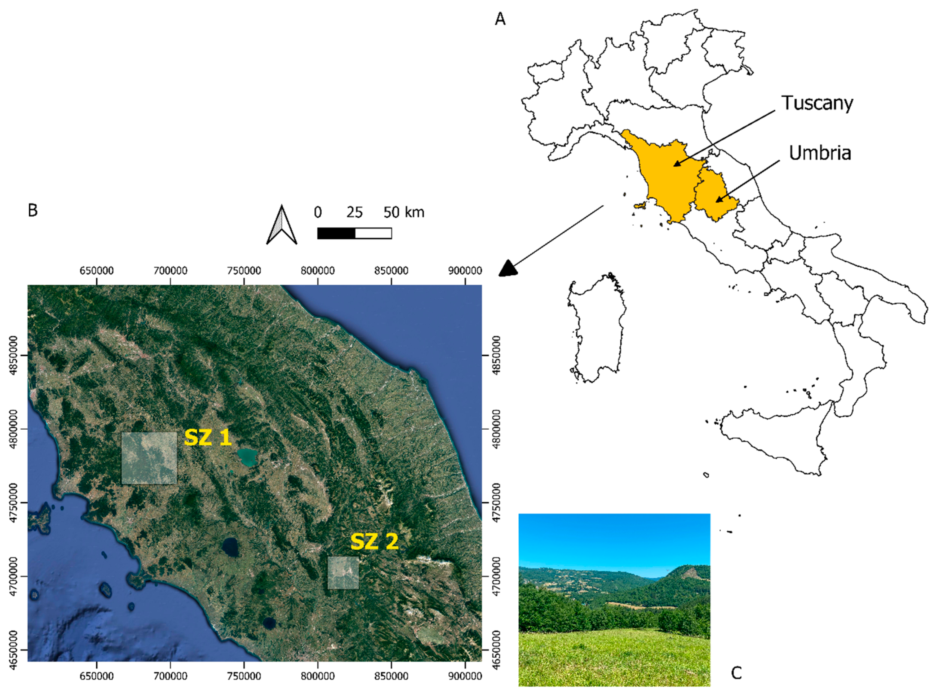
2.1. Study Area

The study was carried out at five remote sites in central Italy, three in Tuscany and two in Umbria (Figure 1). Site selection focused on remote areas (i.e., distance from urban centers) that received similar levels of regional background pollutant deposition. All study sites were based on prior studies that used similar background areas remote from local sources of pollution [43]. The elevation of the sites ranged from 440–1140 m asl. The climate was Mediterranean, with mild winters and warm, dry summers; mean annual temperature was 13–14 °C, and annual rainfall was 600–1000 mm [44].

2.2. Sample Collection

The epiphytic lichen Evernia prunastri and the epigeic moss Pseudoscleropodium purum (Figure 2) were selected as study species, as they have been widely used in biomonitoring studies to assess the atmospheric deposition of airborne pollutants [39,45]. They are widely distributed, easily identified, and were both present at all five study sites. From June–August 2022, lichen thalli were randomly collected from tree (Quercus cerris) branches as composites from a minimum of three trees, and moss samples were randomly collected in nearby open areas (50 m by 50 m), at least 3 m away from tree canopies, as composites from a minimum of five points. All samples were placed into paper bags, which were tightly closed to avoid contamination, and stored in a dry place until analysis. Five subsamples from the individual lichen and moss composite samples at each study site were subsequently analyzed for microplastics.

2.3. Microplastic Analysis

In the laboratory, air-dried (residual water < 10%) whole lichen thalli and the green parts of moss samples (roughly corresponding to the last 2–3 years of growth) were digested using the wet peroxide oxidation method [38,40]. Samples were then vacuum-filtered onto cellulose filter papers (Whatman Grade 1, Maidstone, UK, 1001-090, 11 µm) and placed into glass petri dishes for storage. The filter papers were examined for MPs under a stereomicroscope (Eurotek OXTL101TUSB, Eurotek, Inc, Eatontown, NJ, USA) equipped with a digital camera (MDCE-5C, NINGBO YONGXIN OPTICS CO., LTD. Ningbo, China), following a five-criteria method [38,39,40]. Microplastics were further verified using a hot needle test; if a particle melted or curled under the presence of a hot needle, it was counted as a microplastic. All MPs were classified (fiber or fragment) and measured using the open-source image-processing software ImageJ. In general, visual analysis was limited to particles >50 µm. Overall, 50 subsamples (25 lichens and 25 mosses; two species each at five sites, with five replicates) were analyzed.
Strict quality-control procedures were followed to ensure that contamination was minimized during sampling and analysis. Laboratory contamination was routinely controlled using analytical process blanks. All solutions were vacuum-filtered prior to use in the extraction process. All laboratory glassware used during digesting and filtering were covered with aluminum foil to prevent airborne contamination, and all glassware were rinsed in triplicate with filtered, deionized water. Surfaces were wiped down with paper towels and deionized water between the digestion of each sample. Digestion (process) blanks were vacuum-filtered using filtered, deionized water in place of sample media and analyzed for microplastic contamination. Finally, cotton clothing was worn during the collection of the moss and lichen samples and the laboratory extraction of microplastics.

2.4. Statistical Analysis
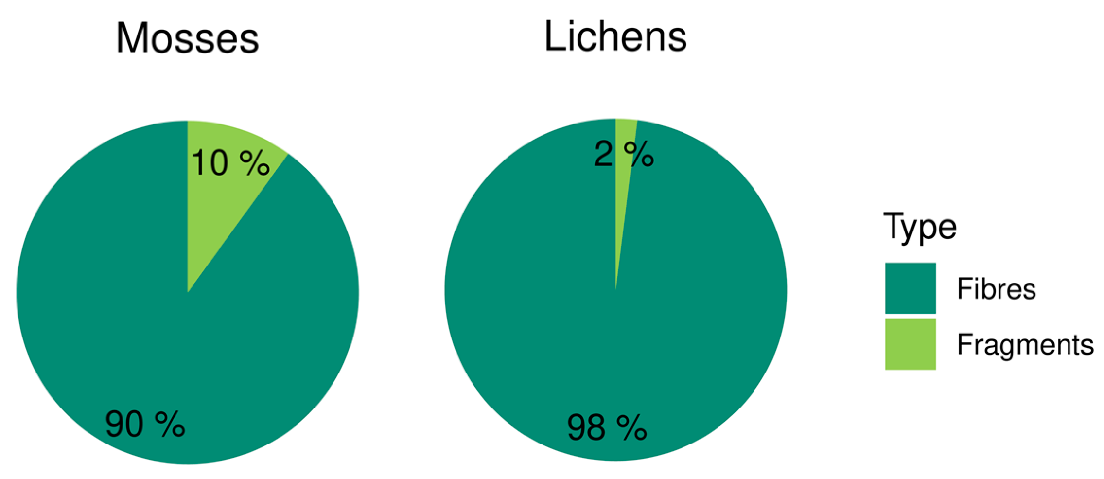
A linear mixed-effect model (LMEM) was fitted to check the differences between moss and lichen samples, with the type of biomonitor (i.e., lichen or moss) as the fixed factor and the site as the random factor. The significance of the LMEM (p < 0.05) was checked with the analysis of deviance (type II Wald chi-square). For model validation, scatterplots of the residual and fitted values were used to check for homoscedasticity (see Supplementary Materials Figure S1), and the Shapiro–Wilk test was used to check for data normality. All calculations were run with the R software [46].

3. Results

Overall, a total of 154 MPs were found across all subsamples (n = 50) from the five study sites; 62 MPs were found in lichens, and 92 MPs were found in mosses. The vast majority (93.5%) of MPs were classified as fibers, and only 6.5% were classified as fragments (see Supplementary Materials Figure S2). In mosses, the MP proportion consisted of 90% fibers and 10% fragments, while in lichens, it was 98% fibers and 2% fragments (Figure 3).
Across all of the five study sites, the mean MP concentrations (per gram of dry mass) ranged from 7.9–12.5 in lichens and 12.5–21.2 in mosses; fibers ranged from 7.9–11.7 in lichens and 9.5–19.6 in mosses; fragments ranged from 0–0.8 in lichens and 0–4.0 in mosses. The mean concentration of MPs across all sites was ~50% higher for moss (14.5 MP/g), compared to lichen (9.7 MP/g). Further variability (relative standard deviation) between sites was higher for moss (26%), compared with lichen (18%), suggesting that lichens had a lower capacity to trap MPs (Table 1). Mean MP concentrations at the Tuscan sites (lichen: 8.7 MP/g, moss: 12.6 MP/g; sites 1–3) were slightly lower than Umbria (lichen: 11.4 MP/g, moss: 17.4 MP/g; sites 4 and 5); however, there was no significant difference among the sites.
The fiber length distributions ranged from 147–4461 µm in lichens and 139–4075 µm in mosses (Table 1; Figure 4). The fragment length distributions ranged from 500–653 µm in lichens and 292–592 µm in mosses (Figure 5).
All of the above features (concentrations of MPs, fibers, and fragments and fragment lengths) were significantly different (p < 0.05) between lichen and moss (higher in moss, except for fragment length), with the only exception being fiber length.

4. Discussion

Our results suggested that the moss P. purum accumulated a higher number of airborne MPs than the lichen E. prunastri. There are several possible explanations for this outcome, including the limitations in our study design. First of all, the study lichen was epiphytic, meaning that it was, at least partially, sheltered by the tree canopy from spring to autumn (however, the tree canopy could increase the scavenging of atmospheric MPs [47], potentially leading to higher concentrations in lichens), while the moss was epigeic and was collected in open areas at least 3 m from the nearest tree canopy. Second, the structural characteristics of the two cryptogams might play an important role. The pleurocarpous branched stems with leaflets, the wider growth of mosses on the substrate, and its higher surface-to-mass ratio might make it more efficient at intercepting and retaining airborne MPs.
A study comparing the capacity of bioaccumulation of PTEs in epigeic mosses and epiphytic lichens showed higher concentrations of elements (subjected to long-range atmospheric transport) in moss [48]. Similar studies showed higher contents of PTEs and polycyclic aromatic hydrocarbons in mosses [49,50]. Adamo et al. [51] emphasized the role of the higher surface area of a moss species, compared to a lichen species, to explain the higher concentrations of trace elements in mosses. Nevertheless, despite these differences in the ability to accumulate airborne pollutants, all these studies clearly showed that both mosses and lichens can be used to monitor their atmospheric deposition. Our study fell within this case: irrespective of absolute values, both organisms clearly indicated that the remote areas investigated were subjected to the deposition of atmospheric MPs.
Another important parameter to consider is the age of the biomonitor. Although the growth rate can be influenced by a wide array of factors, including species-specific differences, which we have not assessed experimentally, we can assume that the lichen was older than the analyzed moss portion. However, this should imply a higher accumulation, since longer and slower growth means longer exposure to wind, rainfall, biomass loss, MPs, etc. In order to overcome this problem, we suggest the use of lichen and moss bags, i.e., samples taken from a remote/background site and transplanted to the study area, referring the final values to the starting conditions [52].
Only a few studies have used lichens and mosses to evaluate the deposition of atmospheric microplastics. A study at a landfill dumping site in central Italy showed an average accumulation of 79 MP/g in the foliose epiphytic lichen Flavoparmelia caperata (L.) Hale. However, at greater distances from the landfill (i.e., at remote sites), the concentration dropped to 7–13 MP/g [38]. In a remote area of northern Italy, lichen bags of E. prunastri exposed for 3 months showed 12–24 MP/g [39]. Roblin and Aherne [40] found 15–30 MP/g in mosses collected from remote areas of Ireland, while Bertrim and Aherne [41] reported an average of 3 MP/g in moss from a remote area of Ontario, Canada. Capozzi et al. [42] found an average of 53–87 MPs in moss samples collected at rural sites in southern Italy, close to the city of Naples. Our results were largely consistent with or at the lower end of the range for studies in background regions in Europe [38,39,40], with lichen concentrations at 7.9–12.5 MP/g and moss at 12.5–21.2 MP/g (Table 1).
Our results were further consistent with all other similar studies [38,39,40,41,42], as fibers dominated over fragments. This was likely caused by the differences in the settling velocity of MP types, due to their weight, with fibers having a low mass-to-surface ratio and, consequently, being more easily transported over longer distances or more easily attached and retained by biomonitors, compared to fragments. This outcome was further supported by investigations using methods other than biomonitors, which yielded comparable results. For example, Ding et al. [53] demonstrated that 88–100% of the MPs in the atmosphere over the northwestern Pacific Ocean were fibers; the study took place on a cruise ship, with an offshore distance of 400 km. Another study that investigated MPs in Antarctic snow found that 60% were fibers [29]. The use of synthetic textiles was shown to be a major contributor to airborne microfibers (MFs), which were among the dominant types of MPs found at or close to inhabited regions, as shown for Ross Island (Antarctica), where the research facility and its field equipment constituted the primary origin of MFs [29].
According to our findings, MFs with a length < 500 µm were prevalent in both lichens and mosses, emphasizing their potential as indicators of long-distance MF transport. As MFs decrease in size, they become and remain airborne more easily, and thus, they are more likely to be transported over greater distances. This suggests that shorter MFs may be widespread in remote regions.

5. Conclusions

Our study shows that both lichen and moss species are effective biomonitors of the deposition of airborne microplastics. Our results further suggest that epigeic mosses may serve as a more effective indicator of atmospheric microplastic contamination, compared with epiphytic lichens, due to their more favorable structure and their open habitat position. However, we note our caution in identifying an optimum species for biomonitoring of atmospheric microplastics, given the limitations of comparing an epiphytic lichen to an epigeic moss. To shed further light on the effectiveness of using lichens and mosses, we recommend that future research compare a parallel exposure of transplants of both organisms.

Supplementary Materials

The following supporting information can be downloaded at: https://www.mdpi.com/article/10.3390/atmos14061007/s1, Figure S1: Linear mixed-effect model plot performed in R (lme4 package). Figure S2: Example images of the extracted microplastics from lichen and moss samples (A—blue microfiber, L = 1147 µm; B—fragment, L = 379 µm; L—length).

Author Contributions

S.L. conceived the research; M.J. and L.G. performed the experiments; M.J. and S.L. analyzed the data; M.J. wrote the article; S.L. and J.A. supervised the text. All authors have read and agreed to the published version of the manuscript.

Funding

This research received no external funding.

Institutional Review Board Statement

Not applicable.

Informed Consent Statement

Not applicable.

Data Availability Statement

Not applicable.

Acknowledgments

We are grateful to Sonia Ravera (University of Palermo) for her valuable contribution of moss and lichen samples collected in the Umbria region.

Conflicts of Interest

The authors declare no conflict of interest.

References

  1. Cai, L.; Wang, J.; Peng, J.; Tan, Z.; Zhan, Z.; Tan, X.; Chen, Q. Characteristic of microplastics in the atmospheric fallout from Dongguan city, China: Preliminary research and first evidence. Environ. Sci. Pollut. Res. 2017, 24, 24928–24935. [Google Scholar] [CrossRef] [PubMed]
  2. Andrady, A.L. Microplastics in the marine environment. Mar. Pollut. Bull. 2011, 62, 1596–1605. [Google Scholar] [CrossRef]
  3. Webb, H.K.; Arnott, J.; Crawford, R.J.; Ivanova, E.P. Plastic degradation and its environmental implications with special reference to poly (ethylene terephthalate). Polymers 2012, 5, 1. [Google Scholar] [CrossRef] [Green Version]
  4. Wright, S.L.; Thompson, R.C.; Galloway, T.S. The physical impacts of microplastics on marine organisms: A review. Environ. Pollut. 2013, 178, 483–492. [Google Scholar] [CrossRef]
  5. Nerland, I.L.; Halsband, C.; Allan, I.; Thomas, K.V. Microplastics in Marine Environments: Occurrence, Distribution and Effects; Norwegian Institute for Water Research: Oslo, Norway, 2014. [Google Scholar]
  6. Yang, H.; Chen, G.; Wang, J. Microplastics in the marine environment: Sources, fates, impacts and microbial degradation. Toxics 2021, 9, 41. [Google Scholar] [CrossRef] [PubMed]
  7. Rillig, M.C. Microplastic in terrestrial ecosystems and the soil? Environ. Sci. Technol. 2012, 46, 6453–6454. [Google Scholar] [CrossRef]
  8. de Souza Machado, A.A.; Kloas, W.; Zarfl, C.; Hempel, S.; Rillig, M.C. Microplastics as an emerging threat to terrestrial ecosystems. Glob. Chang. Biol. 2018, 24, 1405–1416. [Google Scholar] [CrossRef] [PubMed] [Green Version]
  9. He, D.; Luo, Y. Microplastics in terrestrial environments. In Emerging Contaminants and Major Challenges; Springer: Cham, Switzerland, 2020; pp. 87–130. [Google Scholar] [CrossRef]
  10. Dris, R.; Gasperi, J.; Rocher, V.; Saad, M.; Renault, N.; Tassin, B. Microplastic contamination in an urban area: A case study in Greater Paris. Environ. Chem. 2015, 12, 592–599. [Google Scholar] [CrossRef]
  11. Dris, R.; Gasperi, J.; Saad, M.; Mirande, C.; Tassin, B. Synthetic fibers in atmospheric fallout: A source of microplastics in the environment? Mar. Pollut. Bull. 2016, 104, 290–293. [Google Scholar] [CrossRef]
  12. Allen, S.; Allen, D.; Phoenix, V.R.; Le Roux, G.; Durántez Jiménez, P.; Simonneau, A.; Binet, S.; Galop, D. Atmospheric transport and deposition of microplastics in a remote mountain catchment. Nat. Geosci. 2019, 12, 339–344. [Google Scholar] [CrossRef] [Green Version]
  13. Qi, Y.; Yang, X.; Pelaez, A.M.; Lwanga, E.H.; Beriot, N.; Gertsen, H.; Garbeva, P.; Geissen, V. Macro-and micro-plastics in soil-plant system: Effects of plastic mulch film residues on wheat (Triticum aestivum) growth. Sci. Total Environ. 2018, 645, 1048–1056. [Google Scholar] [CrossRef]
  14. Li, J.; Yu, S.; Yu, Y.; Xu, M. Effects of microplastics on higher plants: A review. Bull. Environ. Contam. Toxicol. 2022, 109, 241–265. [Google Scholar] [CrossRef] [PubMed]
  15. Li, J.; Green, C.; Reynolds, A.; Shi, H.; Rotchell, J.M. Microplastics in mussels sampled from coastal waters and supermarkets in the United Kingdom. Environ. Pollut. 2018, 241, 35–44. [Google Scholar] [CrossRef]
  16. Nelms, S.E.; Barnett, J.; Brownlow, A.; Davison, N.J.; Deaville, R.; Galloway, T.S.; Lindeque, P.K.; Santillo, D.; Godley, B.J. Microplastics in marine mammals stranded around the British coast: Ubiquitous but transitory? Sci. Rep. 2019, 9, 1075. [Google Scholar] [CrossRef] [Green Version]
  17. Carlin, J.; Craig, C.; Little, S.; Donnelly, M.; Fox, D.; Zhai, L.; Walters, L. Microplastic accumulation in the gastrointestinal tracts in birds of prey in central Florida, USA. Environ. Pollut. 2020, 264, 114633. [Google Scholar] [CrossRef]
  18. Hwang, J.; Choi, D.; Han, S.; Choi, J.; Hong, J. An assessment of the toxicity of polypropylene microplastics in human derived cells. Sci. Total Environ. 2019, 684, 657–669. [Google Scholar] [CrossRef]
  19. Çobanoğlu, H.; Belivermiş, M.; Sıkdokur, E.; Kılıç, Ö.; Çayır, A. Genotoxic and cytotoxic effects of polyethylene microplastics on human peripheral blood lymphocytes. Chemosphere 2021, 272, 129805. [Google Scholar] [CrossRef] [PubMed]
  20. Napper, I.E.; Thompson, R.C. Release of synthetic microplastic plastic fibres from domestic washing machines: Effects of fabric type and washing conditions. Mar. Pollut. Bull. 2016, 112, 39–45. [Google Scholar] [CrossRef] [Green Version]
  21. Henry, B.; Laitala, K.; Klepp, I.G. Microfibres from apparel and home textiles: Prospects for including microplastics in environmental sustainability assessment. Sci. Total Environ. 2019, 652, 483–494. [Google Scholar] [CrossRef] [PubMed]
  22. De Falco, F.; Di Pace, E.; Cocca, M.; Avella, M. The contribution of washing processes of synthetic clothes to microplastic pollution. Sci. Rep. 2019, 9, 6633. [Google Scholar] [CrossRef] [PubMed] [Green Version]
  23. Munyaneza, J.; Jia, Q.; Qaraah, F.A.; Hossain, M.F.; Wu, C.; Zhen, H.; Xiu, G. A review of atmospheric microplastics pollution: In-depth sighting of sources, analytical methods, physiognomies, transport and risks. Sci. Total Environ. 2022, 822, 153339. [Google Scholar] [CrossRef]
  24. Wang, J.; Liu, Q.; Zhang, C.; Wang, Y.; Yang, F.; Zhao, Y.; Jiang, Y. Microplastics shift macrobenthic community structures near a coastal nuclear power plant under construction in North East China. J. Hazard. Mater. 2022, 437, 129335. [Google Scholar] [CrossRef] [PubMed]
  25. Wright, S.L.; Ulke, J.; Font, A.; Chan, K.L.A.; Kelly, F.J. Atmospheric microplastic deposition in an urban environment and an evaluation of transport. Environ. Int. 2020, 136, 105411. [Google Scholar] [CrossRef]
  26. Abbasi, S.; Jaafarzadeh, N.; Zahedi, A.; Ravanbakhsh, M.; Abbaszadeh, S.; Turner, A. Microplastics in the atmosphere of Ahvaz City, Iran. J. Environ. Sci. 2023, 126, 95–102. [Google Scholar] [CrossRef] [PubMed]
  27. Roblin, B.; Ryan, M.; Vreugdenhil, A.; Aherne, J. Ambient atmospheric deposition of anthropogenic microfibers and microplastics on the western periphery of Europe (Ireland). Environ. Sci. Technol. 2020, 54, 11100–11108. [Google Scholar] [CrossRef]
  28. Brahney, J.; Mahowald, N.; Prank, M.; Cornwell, G.; Klimont, Z.; Matsui, H.; Prather, K.A. Constraining the atmospheric limb of the plastic cycle. Proc. Natl. Acad. Sci. USA 2021, 118, e2020719118. [Google Scholar] [CrossRef]
  29. Aves, A.R.; Revell, L.E.; Gaw, S.; Ruffell, H.; Schuddeboom, A.; Wotherspoon, N.E.; LaRue, M.; McDonald, A.J. First evidence of microplastics in Antarctic snow. Cryosphere 2022, 16, 2127–2145. [Google Scholar] [CrossRef]
  30. González-Pleiter, M.; Edo, C.; Aguilera, Á.; Viúdez-Moreiras, D.; Pulido-Reyes, G.; González-Toril, E.; Osuna, S.; de Diego-Castilla, G.; Leganés, F.; Fernández-Piñas, F.; et al. Occurrence and transport of microplastics sampled within and above the planetary boundary layer. Sci. Total Environ. 2021, 761, 143213. [Google Scholar] [CrossRef] [PubMed]
  31. Warheit, D.B.; Hart, G.A.; Hesterberg, T.W.; Collins, J.J.; Dyer, W.M.; Swaen, G.M.H.; Castranova, V.; Soiefer, A.I.; Kennedy, G.L. Potential pulmonary effects of man-made organic fiber (MMOF) dusts. Crit. Rev. Toxicol. 2001, 31, 697–736. [Google Scholar] [CrossRef]
  32. Prata, J.C. Airborne microplastics: Consequences to human health? Environ. Pollut. 2018, 234, 115–126. [Google Scholar] [CrossRef] [PubMed]
  33. Chen, G.; Feng, Q.; Wang, J. Mini-review of microplastics in the atmosphere and their risks to humans. Sci. Total Environ. 2020, 703, 135504. [Google Scholar] [CrossRef]
  34. Yao, X.; Luo, X.S.; Fan, J.; Zhang, T.; Li, H.; Wei, Y. Ecological and human health risks of atmospheric microplastics (MPs): A review. Environ. Sci. Atmos. 2022, 2, 921–942. [Google Scholar] [CrossRef]
  35. Sangkham, S.; Faikhaw, O.; Munkong, N.; Sakunkoo, P.; Arunlertaree, C.; Chavali, M.; Mousazadeh, M.; Tiwari, A. A review on microplastics and nanoplastics in the environment: Their occurrence, exposure routes, toxic studies, and potential effects on human health. Mar. Pollut. Bull. 2022, 181, 113832. [Google Scholar] [CrossRef]
  36. Loppi, S.; Pirintsos, S.A. Epiphytic lichens as sentinels for heavy metal pollution at forest ecosystems (central Italy). Environ. Pollut. 2003, 121, 327–332. [Google Scholar] [CrossRef]
  37. Bargagli, R.; Monaci, F.; Borghini, F.; Bravi, F.; Agnorelli, C. Mosses and lichens as biomonitors of trace metals. A comparison study on Hypnum cupressiforme and Parmelia caperata in a former mining district in Italy. Environ. Pollut. 2002, 116, 279–287. [Google Scholar] [CrossRef]
  38. Loppi, S.; Roblin, B.; Paoli, L.; Aherne, J. Accumulation of airborne microplastics in lichens from a landfill dumping site (Italy). Sci. Rep. 2021, 11, 4564. [Google Scholar] [CrossRef] [PubMed]
  39. Jafarova, M.; Contardo, T.; Aherne, J.; Loppi, S. Lichen biomonitoring of airborne microplastics in Milan (N Italy). Biology 2022, 11, 1815. [Google Scholar] [CrossRef]
  40. Roblin, B.; Aherne, J. Moss as a biomonitor for the atmospheric deposition of anthropogenic microfibres. Sci. Total Environ. 2020, 715, 136973. [Google Scholar] [CrossRef]
  41. Bertrim, C.; Aherne, J. Moss Bags as Biomonitors of Atmospheric Microplastic Deposition in Urban Environments. Biology 2023, 12, 149. [Google Scholar] [CrossRef] [PubMed]
  42. Capozzi, F.; Sorrentino, M.C.; Cascone, E.; Iuliano, M.; De Tommaso, G.; Granata, A.; Giordano, S.; Spagnuolo, V. Biomonitoring of Airborne Microplastic Deposition in Semi-Natural and Rural Sites Using the Moss Hypnum cupressiforme. Plants 2023, 12, 977. [Google Scholar] [CrossRef] [PubMed]
  43. Vannini, A.; Canali, G.; Pica, M.; Nali, C.; Loppi, S. The water content drives the susceptibility of the lichen Evernia prunastri and the moss Brachythecium sp. to high ozone concentrations. Biology 2020, 9, 90. [Google Scholar] [CrossRef]
  44. Pavan, V.; Antolini, G.; Barbiero, R.; Berni, N.; Brunier, F.; Cacciamani, C.; Cagnati, A.; Cazzuli, O.; Cicogna, A.; De Luigi, C.; et al. High resolution climate precipitation analysis for north-central Italy, 1961–2015. Clim. Dyn. 2019, 52, 3435–3453. [Google Scholar] [CrossRef]
  45. Ares, Á.; Fernández, J.Á.; Aboal, J.R.; Carballeira, A. Study of the air quality in industrial areas of Santa Cruz de Tenerife (Spain) by active biomonitoring with Pseudoscleropodium purum. Ecotoxicol. Environ. Saf. 2011, 74, 533–541. [Google Scholar] [CrossRef]
  46. R Core Team. R: A Language and Environment for Statistical Computing; R Foundation for Statistical Computing: Vienna, Austria, 2022; Available online: https://www.R-project.org/ (accessed on 4 April 2023).
  47. McCune, D.C.; Boyce, R.L. Precipitation and the transfer of water, nutrients and pollutants in tree canopies. Trends Ecol. Evol. 1992, 7, 4–7. [Google Scholar] [CrossRef]
  48. Adamo, P.; Bargagli, R.; Giordano, S.; Modenesi, P.; Monaci, F.; Pittao, E.; Spagnuolo, V.; Tretiach, M. Natural and pre-treatments induced variability in the chemical composition and morphology of lichens and mosses selected for active monitoring of airborne elements. Enviro. Pollut. 2008, 152, 11–19. [Google Scholar] [CrossRef] [PubMed]
  49. Vingiani, S.; De Nicola, F.; Purvis, W.O.; Concha-Grana, E.; Muniategui-Lorenzo, S.; López-Mahía, P.; Giordano, S.; Adamo, P. Active biomonitoring of heavy metals and PAHs with mosses and lichens: A case study in the cities of Naples and London. Water Air Soil Pollut. 2015, 226, 240. [Google Scholar] [CrossRef]
  50. Giordano, S.; Adamo, P.; Sorbo, S.; Vingiani, S. Atmospheric trace metal pollution in the Naples urban area based on results from moss and lichen bags. Environ. Pollut. 2005, 136, 431–442. [Google Scholar] [CrossRef]
  51. Adamo, P.; Crisafulli, P.; Giordano, S.; Minganti, V.; Modenesi, P.; Monaci, F.; Pittao, E.; Tretiach, M.; Bargagli, R. Lichen and moss bags as monitoring devices in urban areas. Part II: Trace element content in living and dead biomonitors and comparison with synthetic materials. Environ. Pollut. 2007, 146, 392–399. [Google Scholar] [CrossRef] [PubMed]
  52. Loppi, S.; Ravera, S.; Paoli, L. Coping with uncertainty in the assessment of atmospheric pollution with lichen transplants. Environ. Forensics 2019, 20, 228–233. [Google Scholar] [CrossRef]
  53. Ding, J.; Sun, C.; He, C.; Zheng, L.; Dai, D.; Li, F. Atmospheric microplastics in the Northwestern Pacific Ocean: Distribution, source, and deposition. Sci. Total Environ. 2022, 829, 154337. [Google Scholar] [CrossRef]
Figure 1. Study area in central Italy showing the (A) map of Italy with the Tuscan and Umbria regions highlighted; (B) sampling zones in Tuscany—SZ1, with three sampling sites (N 43.64353° E 011.97958°; N 43.34555° E 11.15181°; N 43.337333° E 11.171333°), and in Umbria—SZ2, with two sampling sites (N 42.4844759° E 12.707810°; N 42.4846855° E 12.707860°); and (C) an image of a sampling site.
Atmosphere 14 01007 g001
Figure 2. Epiphytic lichen species Evernia prunastri (L.) Ach. (A) and epigeic pleurocarpous moss species Pseudoscleropodium purum (Hedw.) M. Fleisch (B).
Atmosphere 14 01007 g002
Figure 3. Proportions of fibers and fragments in moss and lichen samples from the five study sites.
Atmosphere 14 01007 g003
Figure 4. Frequency distributions of microfiber lengths (n = 144).
Atmosphere 14 01007 g004
Figure 5. Frequency distributions of fragment lengths (n = 10).
Atmosphere 14 01007 g005
Table 1. Mean (±standard error) numbers of MPs, fibers, and fragments accumulated (per gram of dry matter) by the fruticose lichen Evernia prunastri (L) and the moss Pseudoscleropodium purum (M), along with fiber lengths (µm) at five remote sites of Central Italy (Tuscany 1–3; Umbria 4 and 5).
SiteMicroplasticsFibersFragmentsFiber Length (µm)
LMLMLMLM
18.6 ± 3.812.5 ± 5.68.6 ± 3.812.5 ± 5.60 ± 00 ± 02286 ± 6601822 ± 470
27.9 ± 3.512.7 ± 5.67.9 ± 3.511.8 ± 5.30 ± 00.8 ± 0.41129 ± 357469 ± 130
39.5 ± 4.212.6 ± 5.69.5 ± 4.211.8 ± 5.30 ± 00.8 ± 0.42844 ± 8211272 ± 446
412.5 ± 5.613.5 ± 6.111.7 ± 5.29.5 ± 4.20.8 ± 0.44.0 ± 1.82157 ± 5572033 ± 564
510.2 ± 4.621.2 ± 9.510.2 ± 4.619.6 ± 8.80 ± 01.6 ± 0.72025 ± 5691631 ± 340
Disclaimer/Publisher’s Note: The statements, opinions and data contained in all publications are solely those of the individual author(s) and contributor(s) and not of MDPI and/or the editor(s). MDPI and/or the editor(s) disclaim responsibility for any injury to people or property resulting from any ideas, methods, instructions or products referred to in the content.

