Article

Occurrences of Deposited Polycyclic Aromatic Hydrocarbons in Wax of Plant Leaves Using Laser Scanning Microscopy and Gas Chromatography–Mass Spectrometry

by
Ming Yang
1,
Qingyang Liu
2,*,
Shili Tian
1,
Zheng Yang
3,
Yifan Yang
1,
Peng Shao
1,* and
Yanju Liu
1,*
1
Beijing Center for Physical and Chemical Analysis, Institute of Analysis and Testing, Beijing Academy of Science and Technology, Beijing 100089, China
2
College of Biology and the Environment, Nanjing Forestry University, Nanjing 210037, China
3
Beijing Milu Ecological Research Center, Beijing 100076, China
*
Authors to whom correspondence should be addressed.
Atmosphere 2024, 15(10), 1165; https://doi.org/10.3390/atmos15101165
Submission received: 31 August 2024 / Revised: 21 September 2024 / Accepted: 27 September 2024 / Published: 29 September 2024

Abstract

:
The knowledge of the deposition fate of ambient polycyclic aromatic hydrocarbons (PAHs) on plant leaves is limited. To fill in this knowledge gap, this study strives to observe the intermolecular complex between the polycyclic aromatic hydrocarbons and epicuticular wax of plant leaves using laser scanning microscopy. Epicuticular wax refers to a type of organic mixture that covers the outermost layer of leaves. The leaves of 20 tree species were collected in Beijing in July 2023. The concentrations of 31 PAHs were quantified by gas chromatography–mass spectrometry. Furthermore, the intermolecular complexes of polycyclic aromatic hydrocarbons and epicuticular wax were found with a fluorescence spectrofluorometer and laser scanning microscopy. The levels of total PAHs across 20 tree species ranged from 12.4 ng g−1 to 68.4 ng g−1. Differences in the amounts of total PAHs across tree leaves were observed, which may be ascribed to the differences in leaf surface across tree types. The higher concentration of low-molecular-weight PAHs was found in leaves compared to high-molecular-weight PAHs. The fluorescence spectrofluorometer identified the formation of a new intermolecular complex with fluorescence emission at an excitation wavelength of 340 nm between PAHs and epicuticular wax relative to PAHs. We used laser scanning microscopy with a 405 nm laser for excitation to observe the new intermolecular complex of PAHs and epicuticular wax on the stomata and epicuticular wax of leaves. This study found the intermolecular complex of PAHs on the surface of leaves in situ, which provides important information about the deposition fate of ambient PAHs.

1. Introduction

Polycyclic aromatic hydrocarbons (PAHs) have been listed as teratogenic, carcinogenic, and mutagenic substances by the World Health Organization (WHO) [1,2,3]. The occurrences of PAHs are derived from incomplete combustion, which includes that of fossil fuels such as petroleum and coal, as well as wood, natural gas, crop straw, etc. [4,5,6]. Most PAHs exist in the ambient environment through the emission from sources including mobile sources, coal combustion, and biomass burning [4,7]. China is estimated to be responsible for approximately 20% of the total global emissions of PAHs [8,9,10]. The US Environmental Protection Agency (EPA) announced 16 congeners of PAHs as being prioritized to control toxic substances [11]. In 2005, the European Food Safety Agency (EFSA) proposed 16 congeners of PAHs, including 8 congeners of PAHs in the priory control list of PAHs derived from the EPA and 8 new congeners of PAHs [12,13,14]. Moreover, there are other non-priority congeners of PAHs of concern in the atmosphere [13,15]. A method for the simultaneous measurement of 24 priority and 7 non-priority PAHs in plant leaves using gas chromatography–mass spectrometry with ultrasonic extraction was established in our prior study [16]. Among these priory and non-priority congeners of PAHs, benzo(a)pyrene (BaP) is recognized as a Group-I carcinogen by the International Agency for Research on Cancer [7]. In North China, the annual average level of particulate benz(a)pyrene (BaP) was found to vary from 1.1 to 14.3 ng m−3, exceeding the threshold of 1.0 ng m−3 recommended by the World Health Organization [17]. High concentrations of BaP may result in potential health risks to public health and ecotoxicological effects on ecosystems [18,19]. A previous study estimated that the incremental lifetime cancer risk (ILCR) associated with exposures to PAHs was 3.1 × 10⁻⁵, which is greater than the recommended safety level (10−6) [17]. In addition, PAHs are a type of persistent organic pollutant that exist in the environment and are difficult to decompose using traditional chemical mitigation technologies [20,21,22].
The ecological remediation strategy is recommended for mitigating PAHs in the environment because it is a cost-effective and eco-friendly approach [23,24]. Plant leaves could serve as filters to reduce the concentration of PAHs [19,25,26]. A prior study by Klingberg et al. [27] documented the accumulation of PAHs in Quercus palustris and Pinus nigra located in areas of Gothenburg, Sweden. They observed a significant association between gaseous PAH concentrations in leaves and the air. The levels of PAHs were greater in 3-year-old black pine needles than in 1-year-old black pine needles [27]. Gaseous PAHs can enter plant leaves through the stomata and be transferred to the internal tissues of the leaves [28,29]. Our prior study indicated that there are seven main pathways associated with the gene transcription levels of leaves in response to the exposures of PAHs higher than an environmentally relevant concentration of PAHs [30]. The main pathways include flavone and flavonol biosynthesis, glyoxylate and dicarboxylate metabolism, RNA polymerase, ribosome biogenesis in eukaryotes, porphyrin metabolism, photosynthesis–antenna proteins, and photosynthesis. In contrast, environmentally relevant concentrations of PAHs exhibited no significant gene transcription levels in the leaves of Rosa chinensis Jacq. during the 7-day exposure period [30]. The lipid waxes of leaves are found to be important substances in combination with ambient PAH enrichment for physical adsorption [31,32]. Yang et al. [28] observed a negative relationship between the wax contents and total concentration of 16 PAHs in Cinnamomum camphora. Tian et al. [33] compared the differences in the accumulation of PAHs by leaves across eight plants in Shanghai, China. They demonstrated the differences in the absorption ability of PAHs across eight plants because of the variations in the morphology and physiological characteristics of leaves. The leaf morphology and physiological characteristics, including surface roughness, stomatal density, and polar components, were associated with the retention of low-molecular-weight PAHs in eight plant leaves. The content of wax played a dominant role in the accumulation of medium- and high-molecular-weight PAHs in eight plant leaves [33]. These studies explored the role of wax in the accumulation of PAHs using traditional chemical analysis methods [28,33,34]. They extracted wax from leaves using organic solvents and quantitatively weighed the mass [33,34]. Then, they explored the relationships between the content of wax and the concentration of PAHs and found that the wax could retain PAHs [33,34]. Prigioniero and his colleagues [35] employed optical microscopy to investigate the role of leaf surface functional traits in the accumulation of PAHs in Mediterranean evergreen trees. They used bright fields and the epifluorescence of optical microscopy to estimate the number of stomata and stomatal surfaces per leaf area. The study showed that leaf surface functional traits were associated with the accumulation of PAHs; in contrast, the uptakes of different fractions of PAHs were generally weakly related to different leaf functional traits. Though several of the above studies documented the interactions between the wax of leaves and PAHs, limited work interprets the reasons leading to the interactions between the wax of leaves and PAHs and observes the interaction complex in situ. This attempt could advance the understanding of the deposition fate of ambient PAHs on leaves and fill in the knowledge gap for the underlying mechanism of ecological remediation to reduce PAHs using tree leaves.
Based on the above, we hypothesize that the formation of a new intermolecular complex occurred in the deposition process of ambient PAHs on plant leaves. Thus, the formation of a new intermolecular complex was identified using a fluorescence spectrofluorometer at an excitation wavelength of 340 nm. In addition, the laser scanning microscopy technique was employed to observe the intermolecular complex of wax in leaves and PAHs in situ. This study could provide adsorption evidence between wax in leaves and PAHs at the molecular scale. The finding is beneficial to the phyto-remediation of PAHs in the ambient environment using tree leaves.

2. Materials and Methods

2.1. Chemicals

Mixed standards of 24 PAHs at 500 mg L−1 were used for a stock standard solution for PAH quantification in leaves, which includes naphthalene, acenaphthylene, acenaphthene, fluorine, phenanthrene, anthracene, fluoranthene, pyrene, benzo(a)anthracene, chrysene, benzo(b)fluoranthene, benzo(k)fluoranthene, benzo(e)pyrene, indeno(1,2,3-cd)pyrene, dibenzo(a,h)anthracene, benzo(g,h,i)perylene, benzo(c)phenanthrene, 7,12-dimethylbenz(a)anthracene, benzo(j)fluoranthene, benzo(e)pyrene, 3-methylcholanthrene, picene, dibenzo(a,l)pyrene, and dibenzo(a,i)pyrene. Two substitutes including 2-fluorophenyl and terphenyl-d14 concentrated at 4000 mg L−1 were adopted to evaluate the efficiencies of extraction processes. A solution of five compounds including naphthalene-d8, acenaphthene-d10, phenanthrene-d10, chrysene-d12, and perylene-d12 at 4000 mg L−1 was selected as the internal standard.

2.2. Leaf Samples Collection

The leaves of 20 trees were harvested along the sides of west third-ring highways (116°18′14″ N, 39°54′2″ E) in Beijing during the period of July 2022. The trees include Chaenomeles speciosa, Prunus cerasifera, Fraxinus chinensis, Ailanthus altissima, Salix babylonica L., Jasminum nudiflorum, Populus tomentosa, Acer mono Maxim., Robinia pseudoacacia, Prunus persica, Salix matsudana, Paulownia fortune, Sophora japonica, Lonicera maackii, Ulmus pumila L., Viburnum sargentii, Ginkgo biloba L., Platanus × acerifolia, Broussonetia papyrifera, and Prunus triloba. In the collection of leaves, three mature and healthy trees of similar sizes and ages were selected for each tree species. Intact leaves of similar ages were collected from the direction of the highways. Approximately 100 leaves were collected from three parallel trees [16]. After collection, the leaves were kept in a polyethylene bag and shipped to a laboratory at 4 °C.

2.3. Measurements of PAHs in Leaf Samples

About 200 g of leaf samples was crushed thoroughly with a high-speed grinder and then mixed evenly. The powders were sealed in a clean polyethylene bag and kept at −18 °C. Before extraction, 5 g of a leaf sample was prepared and placed in a 100 mL centrifuge tube. Then, 10 μL of substitute solution (2-fluorophenyl and terphenyl-d14) at 20 μg mL−1 was added into the 100 mL centrifuge tube. After 30 min, 5 g of anhydrous sodium sulfate and the mixtures of n-hexane and dichloromethane at a volume ratio of 1:1 were mixed with leaf samples. The leaf samples were extracted ultrasonically for 30 min in a water bath with a temperature lower than 30 °C. After extraction, the leaf samples were centrifuged at 10,000 r min−1 for 3 min. The supernatants were extracted from the centrifuge tube and kept in a concentration vessel. The extraction process was repeated twice.
The moisture content in the supernatants of the extraction solution was removed using anhydrous sodium sulfate. The supernatants of the extraction solution were mixed with 5 g of anhydrous sodium sulfate and filtrated through a layer of glass wool on a glass funnel. The extraction solution was dropped into a concentrated container. Further, 2 mL of n-hexane and dichloromethane at a volume ratio of 1:1 was used to wash the glass wool on a glass funnel three times. The wash solution was collected and mixed with the extraction solution in a concentrated container. At a temperature of 20 °C, the extraction solution was concentrated to 2 mL with a multi-sample parallel evaporator (Q-101, BUCHI, Flawil, Switzerland). Then, 10 mL of n-hexane was added into 2 mL of extraction solution, and it was concentrated to 2 mL for further purification.
We used a solid-phase extraction column to purify the extraction solution. A solid-phase extraction column of 800 mg silica gel and 1200 mg neutral alumina was fixed on a solid-phase extraction device and then washed with 5 mL of dichloromethane and 10 mL of n-hexane in sequence. Then, the sample extraction solution was added to the solid-phase extraction column and washed with 10 mL of dichloromethane and n-hexane at a volume ratio of 1:4. The eluent was collected in a container and then concentrated to 1 mL with the evaporator. Before quantitative measurement, 1.0 μL of internal standards including naphthalene-d8, acenaphthene-d10, phenanthrene-d10, chrysene-d12, and perylene-d12 at 20 μg mL−1 was mixed with 1 mL of extraction solution.
The PAHs in leaf samples were measured using a gas chromatography–mass spectrometry QP2010Ultra (GC–MS QP2010Ultra, Shimadzu, Kyoto, Japan). The GC–MS QP2010Ultra included a DB-EUPAH capillary column (20 m × 0.18 mm × 0.14 μm, Agilent, Palo Alto, CA, USA) and a mass spectrometer with an electron impact (EI) ion source with an electron energy of 70 eV in the selected ion monitoring (SIM) mode. The temperature program for gas chromatography was set at an initial temperature of 70 °C for 2 min and increased to 280 °C with a heating rate of 10 °C min−1 and kept at 280 °C for 5 min. Next, the temperature was increased to 320 °C at a heating rate of 5 °C min−1 and kept constant for 10 min. The temperature of the injection port was 260 °C. The flow rate of the carrier gas using high-purity helium was 1.0 mL min−1. The injection volume was set to 1.0 μL without split injection. The temperatures of the ion source, fourth-stage rod, and transmission line were maintained at 240 °C, 150 °C, and 280 °C, respectively. Using the optimized method, a good linear relationship between 1 and 500 g mL−1 was found with a correlation coefficient greater than 0.99. The detection limit and quantitative detection limit were estimated to range from 0.2 to 0.7 ng g−1 and 0.8 to 2.8 ng g−1 for individual PAHs, respectively. The relative deviation of parallel samples was found to be within 15%, and the recovery rates using spiked experiments were in the range of 71–98% [16].

2.4. Observation of PAHs on Plant Leaves through Laser Scanning Microscopy

The leaves were extracted in 5 mL of dichloromethane for 30 min ultrasonically at 30 °C and filtered through 0.45 μm pore syringe filters to remove insoluble substances. The supernatants were kept as the solution of epicuticular wax. The photoluminescence spectra of epicuticular wax in the extracts of leaf samples and the mixtures of PAHs and epicuticular wax in the extracts of leaf samples were recorded with a Perkin-Elmer LS 55 fluorescence spectrofluorometer at an excitation wavelength of 340 nm. The observations of PAHs on plant leaves were carried out using a Zeiss LSM 710 laser scanning microscope equipped with a 405 nm laser for excitation.

2.5. Statistical Analysis

The concentrations of PAHs are described as the mean and standard deviation using the results from at least three independent experiments. The Wilcoxon rank-sum test was used to determine the mean differences in the levels of PAHs across different groups because the dataset has a non-normal distribution based on the assessment of the Shapiro–Wilk test. We performed a post hoc test using the Bonferroni methodology to adjust p-values for pairwise comparisons. Significant differences were considered at an adjusted p-value of <0.05. All statistical analyses were performed with SPSS V26.0.

3. Results and Discussion

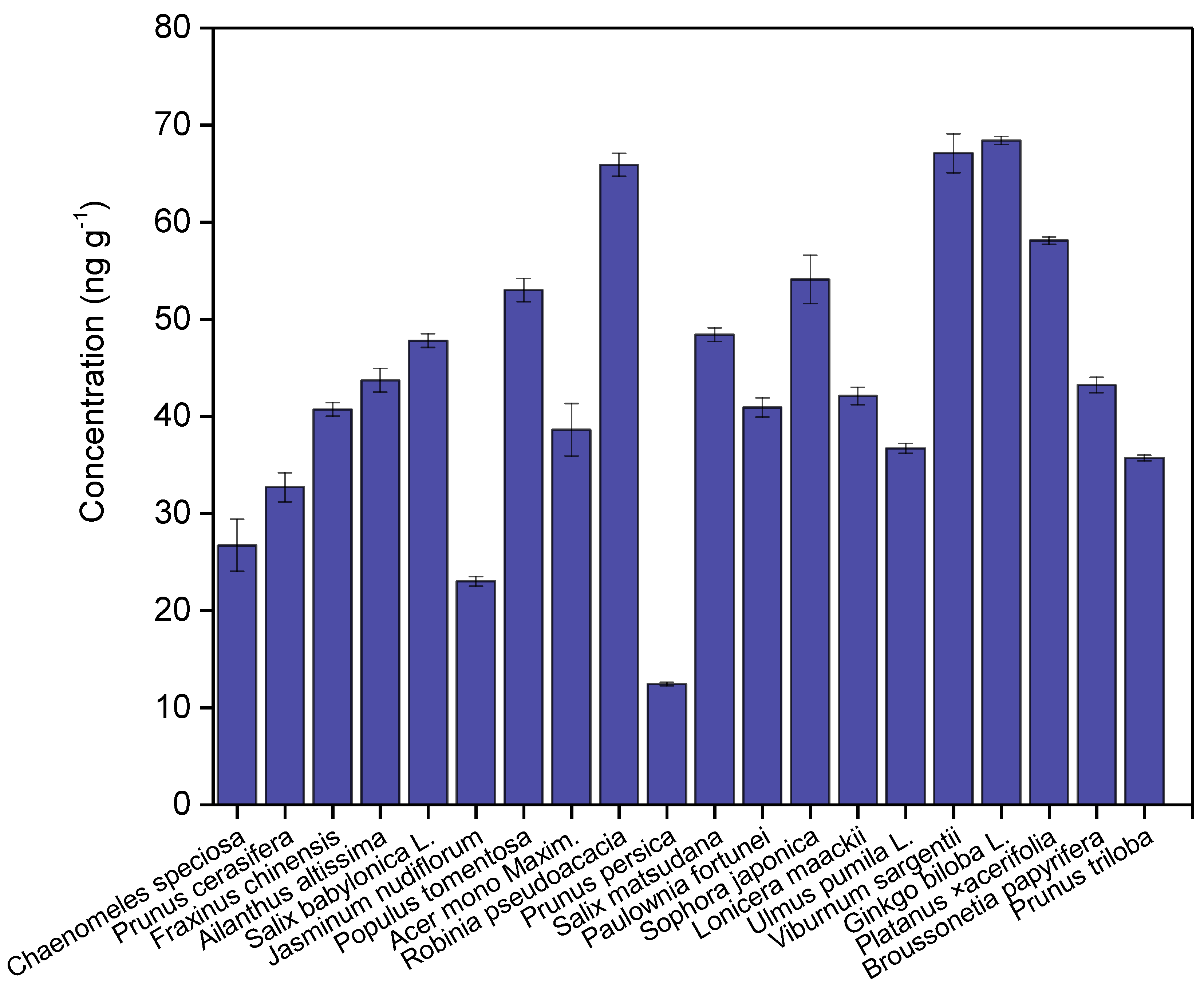
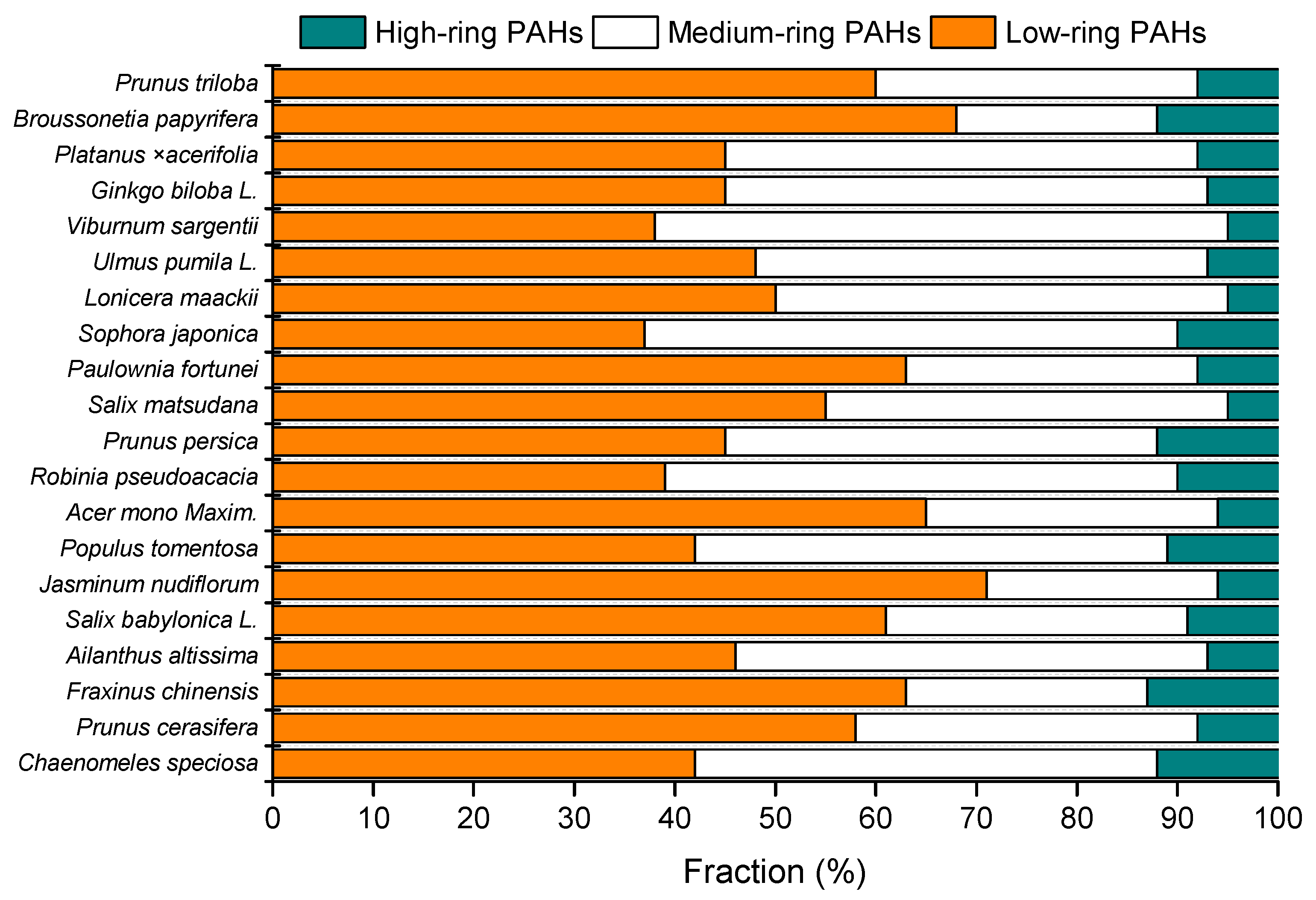
3.1. Differences in the Accumulation of PAHs across Species

The highest concentration of total PAHs was found to be 68.4 ng g−1 in the leaves of Ginkgo biloba L., followed by Viburnum sargentii (67.1 ng g−1), Robinia pseudoacacia (65.9 ng g−1), Platanus × acerifolia (58.1 ng g−1), Sophora japonica (54.1 ng g−1), Populus tomentosa (53.1 ng g−1), Salix matsudana (48.4 ng g−1), Salix babylonica L. (47.8 ng g−1), Ailanthus altissima (43.7 ng g−1), Broussonetia papyrifera (43.2 ng g−1), Lonicera maackii (42.1 ng g−1), Paulownia fortune (40.9 ng g−1), Fraxinus chinensis (40.7 ng g−1), Acer mono Maxim. (38.6 ng g−1), Ulmus pumila L. (36.7 ng g−1), Prunus triloba (35.7 ng g−1), Prunus cerasifera (32.7 ng g−1), Chaenomeles speciosa (26.7 ng g−1), Jasminum nudiflorum (23 ng g−1), and Prunus persica (12.4 ng g−1) (Figure 1).
The adsorption amounts of PAHs on leaves are associated with several environmental factors, which include local temperature, wind speed, wind direction, humidity, air compartment structure, the ages of leaves, and emission sources [27,28]. Leaves across the 20 species of trees were collected using the same selection criterion under similar environmental factors and ages of leaves (1 year old). We compared the mean levels of total PAHs across the 20 species of trees using the Wilcoxon rank-sum test. Our results indicated that significant differences in the mean levels of total PAHs across 20 species of trees were found (p = 0.003). Our results supporting prior findings indicated that there were differences in the accumulation of PAHs across different species of trees [27,28,35]. The mean concentration of individual PAHs in the leaves of 20 trees is summarized in Table S1. Different distribution characteristics of PAH congeners were observed across the leaves of 20 trees, indicating that there were differences in the accumulation of PAHs. Among 20 trees, the occurrences of naphthalene, fluorene, fluoranthene, and pyrene were detected in the leaves of 20 trees with an average concentration in the range of 2.0–7.8 ng g−1, while most PAH congeners, including acenaphthylene, acenaphthene, anthracene, benzo(g,h,i)perylene, benzo(c)phenanthrene, benz(a)anthracene, benzo(k)fluoranthene, 7,12-dimethylbenz(a)anthracene, benzo(j)fluoranthene, benz(e)pyrene, perylene, 3-methylcholanthrene, dibenz(a,h)anthracene, picene, benzo(g,h,i)pery, dibenz(a,l)pyrene, dibenz(a,e)pyrene, coronene, dibenz(a,i)pyrene, and dibenz(a,h)pyrene, were not detected (<0.5 ng g−1) in the leaves of 20 trees (Table S1). The highest number of detected species of PAH congeners was eleven in an individual tree among 20 species of trees, while the lowest number of detected species of PAH congeners was five in the leaves of Prunus persica. The contribution of low-ring (i.e., the sum of two-ring and three-ring) PAHs accounted for the total PAHs in the range from 37% to 71%, while medium-ring (i.e., four-ring) and high-ring PAHs (i.e., the sum of five-ring and six-ring) contributed to the total PAHs ranging from 20% to 47% and from 5% to 13%, respectively (Figure 2).
The average concentration of phenanthrene was observed to vary from 10.0 to 41.3 ng g−1 in 18 trees, while phenanthrene was not detected (<0.5 ng g−1) in the leaves of Chaenomeles speciosa and Prunus persica. The highest average concentration of Retene (5.5 ± 0.6 ng g−1) among 20 trees was found in the leaves of Chaenomeles speciosa. Fluoranthene exhibited the highest average concentration (10.9 ± 0.2 ng g−1) in Platanus × acerifolia. Chrysene was detected in the leaves of Viburnum sargentii with the highest mean concentration of 35.7 ± 2.0 ng g−1, while the mean concentration of chrysene was lower than 8.0 ng g−1. Prior studies documented that PAHs could remain on the leaf surface and accumulate in cuticular wax [28,35]. High-molecular-weight PAHs were more likely to be washed off from leaves in comparison to low-molecular-weight PAHs [34]. The detected amount of PAHs in the interior of leaves decreased with an increasing molecular weight [34]. The fact that there were more low-molecular-weight PAHs than high-molecular-weight PAHs on leaves in this study supported prior findings [34]. The differences in the accumulation of PAH congeners across the leaves of 20 tree species could be ascribed to the differences in cuticular wax across leaves, as well as surface roughness, stomatal density, and lipid components [34,35].
We grouped 20 species of trees into two types, which are deciduous shrubs and deciduous broad-leaved trees. The deciduous shrubs include Jasminum nudiflorum, Lonicera maackii, and Viburnum sargentii, while the deciduous broad-leaved trees include Populus tomentosa, Acer mono Maxim., Robinia pseudoacacia, Fraxinus chinensis, Ailanthus altissima, Salix babylonica L., Salix matsudana, Paulownia fortune, Sophora japonica, Ulmus pumila L., Ginkgo biloba L., Platanus × acerifolia, Broussonetia papyrifera, Chaenomeles speciosa, Prunus cerasifera, Prunus persica, and Prunus triloba [36,37]. The average concentrations of total PAHs were found to be 53 ± 9 ng g−1 (deciduous shrubs) and 44 ± 8 ng g−1 (deciduous broad-leaf trees). No significant concentration of total PAHs between deciduous shrubs and deciduous broad-leaf trees (p = 0.08) was observed, which suggested that the two types of trees could be properly used to filter ambient PAHs.

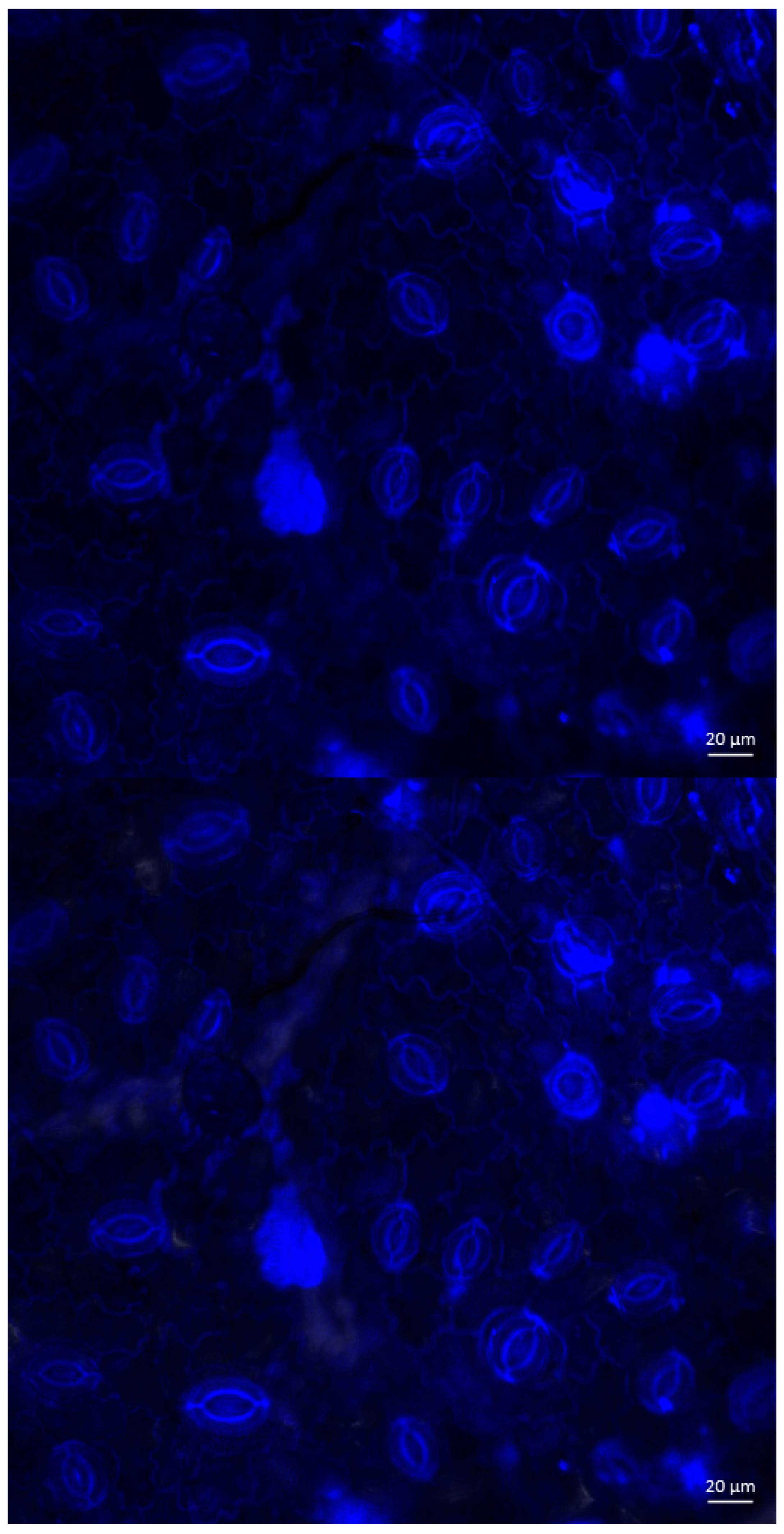
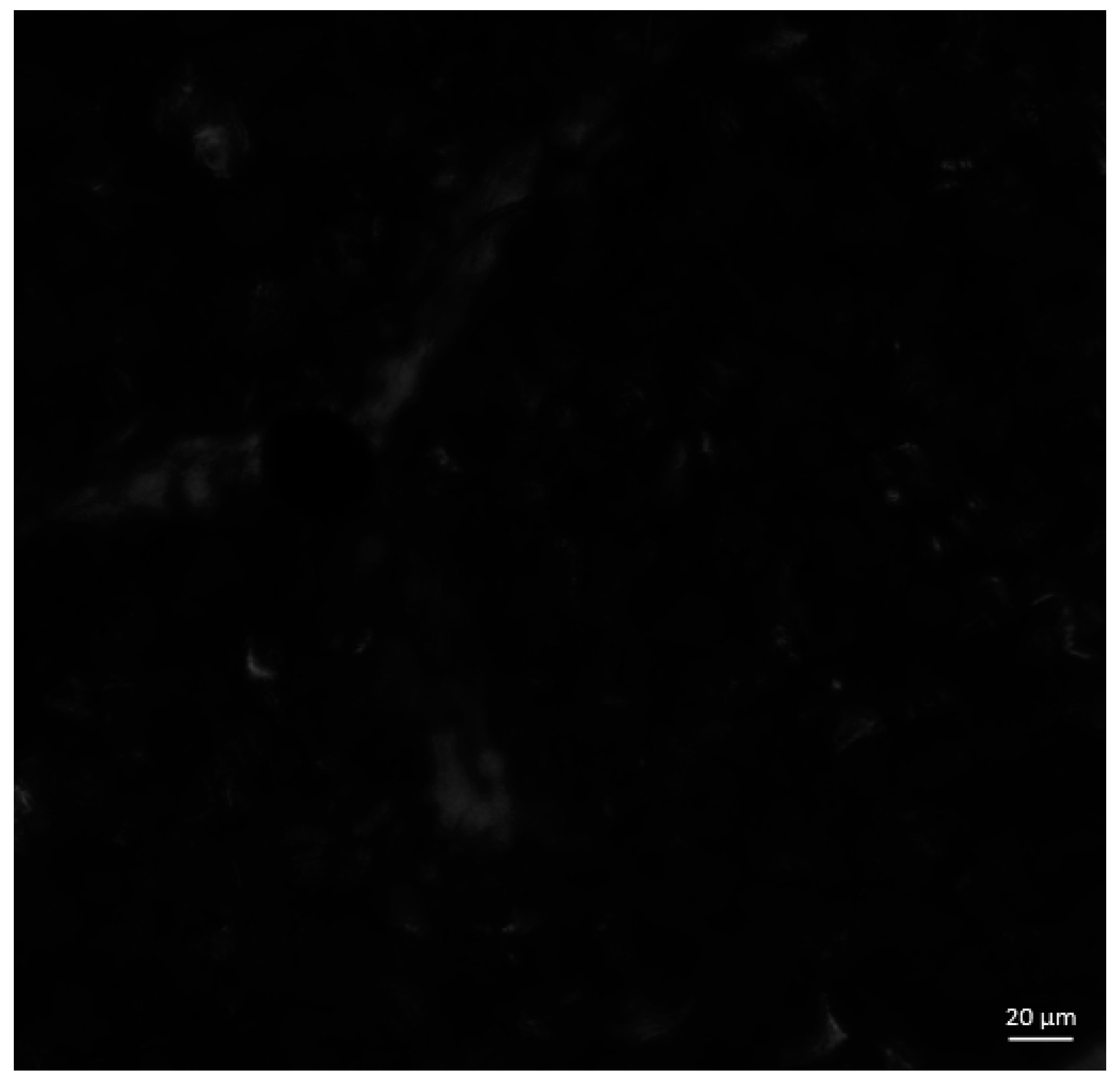
3.2. Observation of PAHs on Plant Leaves

The epicuticular wax in the extracts of leaf samples exhibited fluorescence emission spectra from 420 nm to 560 nm, which could be ascribed to fluorescence emission from the mixtures of chlorophyll a and hypoxanthine. The mixtures of PAHs and epicuticular wax in the extracts of leaf samples did not have a fluorescence emission of 420–460 nm nor an overlay with that of epicuticular wax in the range of 480–500 nm (Figure S1). Makarska-Bialokoz [38] found that xanthine compounds can interact with the porphyrin ring of chlorophyll to quench fluorescence emissions. Since the mixtures of PAHs have similar chemical structures as those of xanthine compounds, it was thus expected that the fluorescence quenching effect occurred from 420 to 450 nm with the formation of additional specific binding complexes. In addition, the intensity of fluorescence emission spectra in the mixtures of PAHs and epicuticular wax increased, ranging from 500 to 520 nm relative to that of epicuticular wax. The increases in fluorescence emission from 500 to 520 nm demonstrated a small red-shift in the intermolecular complexes relative to that of epicuticular wax [38]. The red-shift fluorescence emission is related to the formation of hydrogen bonding interactions in the intermolecular system [38]. The accumulations of PAHs on the leaves of deciduous broad-leaved trees with the observations from laser scanning microscopy are shown in Figure 3. As shown in Figure 3, the physiological status of the stomata in the leaves was not changed during the exposure to PAHs, which supported the prior finding that leaves could uptake and transport PAHs [27,30,33]. Several areas with relatively higher fluorescence emission intensity were observed using laser scanning microscopy with 405 nm excitation. The result derived from laser scanning microscopy was consistent with that of photoluminescence spectra, which indicated that the intermolecular complexes between PAHs and epicuticular wax had high fluorescence emission intensity. The red-shift fluorescence emission of intermolecular complexes can be observed without the interfaces of the surface of leaves.
Our study sought to identify the intermolecular complex of PAHs and epicuticular wax with a fluorescence spectrofluorometer and locate the occurrences of the intermolecular complex using laser scanning microscopy during the adsorption process of PAHs by leaves. The findings from this study could provide an understanding of the adsorption mechanism of PAHs by the epicuticular wax of plant leaves at the molecular scale in situ.

4. Conclusions

This study presented new data on PAH accumulation across tree species and novel insight into the interactions between airborne PAHs and the epicuticular wax of tree leaves. The concentration of 31 PAHs showed a large variation across 20 species of trees. In particular, low-molecular-weight PAHs were detected, and high-molecular-weight PAHs were mostly undetected (<0.5 ng g−1), while the concentration of low-molecular-weight PAHs was found to be higher than that of the detected high-molecular-weight PAHs in most tree leaf samples. The results from this study inferred that the intermolecular complex of PAHs and epicuticular wax was formed through the observation from photoluminescence spectra and laser scanning microscopy in situ. This study suggested that epicuticular wax played an important role in removing ambient PAHs. The findings could promote the understanding of species differences in the ecological remediation of ambient PAHs using trees. Our study provided improved insights into the removal of PAHs by tree leaves. Future studies may focus on the elucidation of the degradation process of the intermolecular complex of PAHs and epicuticular wax in leaves.

Supplementary Materials

The following supporting information can be downloaded at https://www.mdpi.com/article/10.3390/atmos15101165/s1, Figure S1: The fluorescence emission spectra of epicuticular wax in the extracts of leaf samples (blue line) and the mixtures of PAHs and epicuticular wax in the extracts of leaf samples (red line) with the excitation spectrum monitored at 340 nm. Table S1: The level of individual PAHs in 20 types of tree leaves.

Author Contributions

Q.L. and Y.L. conceived and designed the experiments. M.Y., Q.L., S.T., Z.Y., Y.Y. and P.S. performed the experiments. M.Y., Q.L., P.S. and Y.L. analyzed the data. M.Y., Q.L., P.S. and Y.L. drafted the manuscript, Q.L., P.S. and Y.L. reviewed and edited the manuscript. All authors have read and agreed to the published version of the manuscript.

Funding

This work was supported by the Beijing Natural Science Foundation (8232025).

Institutional Review Board Statement

Not applicable.

Informed Consent Statement

Not applicable.

Data Availability Statement

The data are included in the main text and Supplementary Materials.

Conflicts of Interest

The authors declare no conflicts of interest.

References

  1. Hümmler, A.; Bayer, V.J.; Achten, C. Unravelling mixed sources of polycyclic aromatic hydrocarbons (PAH) in urban soils by visual characterization of anthropogenic substrates and coal particles, 71 PAH and alkylated PAH patterns. Environ. Pollut. 2024, 342, 123029. [Google Scholar] [CrossRef] [PubMed]
  2. Zhang, Y.; Meliefste, K.; Hu, W.; Portengen, L.; Rothman, N.; Reiss, B.; Li, J.; Xu, J.; Ning, B.; Liu, D.; et al. Expanded PAH analysis of household air pollution in a rural region of China with high lung cancer incidence. Environ. Pollut. 2024, 361, 124717. [Google Scholar] [CrossRef] [PubMed]
  3. Patnana, D.P.; Chandra, B.P.; Chaudhary, P.; Sinha, B.; Sinha, V. Optimized LC-MS/MS method for simultaneous determination of endocrine disruptors and PAHs bound to PM2.5: Sources and health risk in 357 Indo-Gangetic Plain. Atmos. Environ. 2022, 290, 119363. [Google Scholar] [CrossRef]
  4. Ali-Taleshi, M.S.; Moeinaddini, M.; Riyahi Bakhtiari, A.; Feiznia, S.; Squizzato, S.; Bourliva, A. A one-year monitoring of spatiotemporal variations of PM2.5-bound PAHs in Tehran, Iran: Source apportionment, local and regional sources origins and source-specific cancer risk assessment. Environ. Pollut. 2021, 274, 115883. [Google Scholar] [CrossRef]
  5. Faridi, S.; Yousefian, F.; Roostaei, V.; Harrison, R.M.; Azimi, F.; Niazi, S.; Naddafi, K.; Momeniha, F.; Malkawi, M.; Moh’d Safi, H.A.; et al. Source apportionment, identification and characterization, and emission inventory of ambient particulate matter in 22 Eastern Mediterranean Region countries: A systematic review and recommendations for good practice. Environ. Pollut. 2022, 310, 119889. [Google Scholar] [CrossRef] [PubMed]
  6. Nim, N.; Morris, J.; Tekasakul, P.; Dejchanchaiwong, R. Fine and ultrafine particle emission factors and new diagnostic ratios of PAHs for peat swamp forest fires. Environ. Pollut. 2023, 335, 122237. [Google Scholar] [CrossRef] [PubMed]
  7. Wu, Y.; Zhang, H.; Zhang, H.; Zeng, T.; Qiao, N.; Shi, Y.; Zhang, N.; Luo, W.; Lu, S. Risks and sources of atmospheric particulate-bound polycyclic aromatic hydrocarbons (AP-PAHs) in seven regions of China: A review. Urban Clim. 2024, 57, 102108. [Google Scholar] [CrossRef]
  8. Wang, J.; Xia, L.; Wang, R. Provincial emission inventory of atmospheric polycyclic aromatic hydrocarbons (PAHs) in China. Atmos. Environ. 2024, 334, 120729. [Google Scholar] [CrossRef]
  9. Wang, D.-Q.; Jia, S.-M.; Liu, L.-Y.; Zhang, Z.-F.; Zhu, F.-J.; Ma, W.-L. Pollution characteristics, source apportionment and absorption spectra of size-resolved PAHs in atmospheric particles in a cold megacity of China. J. Hazard. Mater. 2024, 473, 134643. [Google Scholar] [CrossRef]
  10. Sun, J.; Shen, Z.; Niu, X.; Yu, J.; Zhang, Y.; Liu, S.; Niu, X.; Zhang, Y.; Xu, H.; Li, X.; et al. PM2.5 source profiles from typical Chinese commercial cooking activities in northern China and its influences on bioreactivity of vascular smooth muscle cells (VSMCs). Atmos. Environ. 2020, 239, 117750. [Google Scholar] [CrossRef]
  11. EPA. Polynculear Aromatic Hydrogens, SW-846 Method 8310; United States Environmental Protection Agency: Washington, DC, USA, 1999. [Google Scholar]
  12. Magalhães, K.M.; Carreira, R.S.; Rosa Filho, J.S.; Rocha, P.P.; Santana, F.M.; Yogui, G.T. Polycyclic aromatic hydrocarbons (PAHs) in fishery resources affected by the oil spill in Brazil: Short-term environmental health and seafood safety. Mar. Pollut. Bull. 2022, 175, 113334. [Google Scholar] [CrossRef] [PubMed]
  13. Badyda, A.J.; Rogula-Kozłowska, W.; Majewski, G.; Bralewska, K.; Widziewicz-Rzońca, K.; Piekarska, B.; Rogulski, M.; Bihałowicz, J.S. Inhalation risk to PAHs and BTEX during barbecuing: The role of fuel/food type and route of exposure. J. Hazard. Mater. 2022, 440, 129635. [Google Scholar] [CrossRef]
  14. Zapelini de Melo, A.P.; Hoff, R.B.; Molognoni, L.; de Oliveira, T.; Daguer, H.; Manique Barreto, P.L. Disasters with oil spills in the oceans: Impacts on food safety and analytical control methods. Food Res. Int. 2022, 157, 111366. [Google Scholar] [CrossRef] [PubMed]
  15. Ahad, J.M.E.; Macdonald, R.W.; Parrott, J.L.; Yang, Z.; Zhang, Y.; Siddique, T.; Kuznetsova, A.; Rauert, C.; Galarneau, E.; Studabaker, W.B.; et al. Polycyclic aromatic compounds (PACs) in the Canadian environment: A review of sampling techniques, strategies and instrumentation. Environ. Pollut. 2020, 266, 114988. [Google Scholar] [CrossRef] [PubMed]
  16. Yang, M.; Tian, S.; Liu, Q.; Yang, Z.; Yang, Y.; Shao, P.; Liu, Y. Determination of 31 polycyclic aromatic hydrocarbons in plant leaves using internal standard method with ultrasonic extraction-gas chromatography-mass spectrometry. Toxics 2022, 10, 634. [Google Scholar] [CrossRef] [PubMed]
  17. Shen, G.; Du, W.; Zhuo, S.; Yu, J.; Tao, S. Improving regulations on residential emissions and non-criteria hazardous contaminants—Insights from a field campaign on ambient PM and PAHs in North China Plain. Environ. Sci. Policy 2019, 92, 201–206. [Google Scholar] [CrossRef]
  18. Gautam, K.; Anbumani, S. Chapter 19—Ecotoxicological effects of organic micro-pollutants on the environment. In Current Developments in Biotechnology and Bioengineering; Varjani, S., Pandey, A., Tyagi, R.D., Ngo, H.H., Larroche, C., Eds.; Elsevier: Amsterdam, The Netherlands, 2020; pp. 481–501. [Google Scholar]
  19. Tiwari, M.; Sahu, S.K.; Pandit, G.G. Distribution of PAHs in different compartment of creek ecosystem: Ecotoxicological concern and human health risk. Environ. Toxicol. Pharmacol. 2017, 50, 58–66. [Google Scholar] [CrossRef]
  20. Dhokale, B.; Saeed, Z.M.; Ali, W.A.; Mohamed, S. Chapter 10—Recent advances in quantification and remediation technologies for toxic PAH mitigation from the environment. In Advances in Nano and Biochemistry; Morajkar, P., Naik, M., Eds.; Academic Press: New York, NY, USA, 2023; pp. 233–258. [Google Scholar]
  21. Mishra, A.; Kumari, M.; Swati; Kumar, R.; Iqbal, K.; Thakur, I.S. Persistent organic pollutants in the environment: Risk assessment, hazards, and mitigation strategies. Bioresour. Technol. Rep. 2022, 19, 101143. [Google Scholar] [CrossRef]
  22. Bolan, S.; Padhye, L.P.; Mulligan, C.N.; Alonso, E.R.; Saint-Fort, R.; Jasemizad, T.; Wang, C.; Zhang, T.; Rinklebe, J.; Wang, H.; et al. Surfactant-enhanced mobilization of persistent organic pollutants: Potential for soil and sediment remediation and unintended consequences. J. Hazard. Mater. 2023, 443, 130189. [Google Scholar] [CrossRef]
  23. Udom, G.J.; Frazzoli, C.; Ekhator, O.C.; Onyena, A.P.; Bocca, B.; Orisakwe, O.E. Pervasiveness, bioaccumulation and subduing environmental health challenges posed by polycyclic aromatic hydrocarbons (PAHs): A systematic review to settle a one health strategy in Niger Delta, Nigeria. Environ. Res. 2023, 226, 115620. [Google Scholar] [CrossRef]
  24. Song, Q.; Li, X.; Zhao, Z.; Li, D.; Li, Y.; Hou, N. Insights into ecotoxicity effects of PAHs and ecosystem responses of remediation strategies under cold stress: From PAHs degradation to ecological restoration regulated by signal molecular. Chem. Eng. J. 2023, 475, 146042. [Google Scholar] [CrossRef]
  25. Mukhopadhyay, S.; Dutta, R.; Das, P. A critical review on plant biomonitors for determination of polycyclic aromatic hydrocarbons (PAHs) in air through solvent extraction techniques. Chemosphere 2020, 251, 126441. [Google Scholar] [CrossRef]
  26. Robin, S.L.; Marchand, C. Polycyclic aromatic hydrocarbons (PAHs) in mangrove ecosystems: A review. Environ. Pollut. 2022, 311, 119959. [Google Scholar] [CrossRef] [PubMed]
  27. Klingberg, J.; Strandberg, B.; Sjöman, H.; Taube, M.; Wallin, G.; Pleijel, H. Polycyclic aromatic hydrocarbon (PAH) accumulation in Quercus palustris and Pinus nigra in the urban landscape of Gothenburg, Sweden. Sci. Total Environ. 2022, 805, 150163. [Google Scholar] [CrossRef] [PubMed]
  28. Brennan, A.; Moreno Jiménez, E.; Alburquerque, J.A.; Knapp, C.W.; Switzer, C. Effects of biochar and activated carbon amendment on maize growth and the uptake and measured availability of polycyclic aromatic hydrocarbons (PAHs) and potentially toxic elements (PTEs). Environ. Pollut. 2014, 193, 79–87. [Google Scholar] [CrossRef] [PubMed]
  29. Mandal, M.; Das, S.; Roy, A.; Rakwal, R.; Jones, O.A.H.; Popek, R.; Agrawal, G.K.; Sarkar, A. Interactive relations between plants, the phyllosphere microbial community, and particulate matter pollution. Sci. Total Environ. 2023, 890, 164352. [Google Scholar] [CrossRef]
  30. Tian, S.; Liu, Q.; Qu, J.; Yang, M.; Ma, Q.; Liu, J.; Shao, P.; Liu, Y. Whole-transcriptome analysis on the leaves of 433 Rosa chinensis Jacq. under exposure to polycyclic aromatic hydrocarbons. Toxics 2023, 11, 610. [Google Scholar] [CrossRef] [PubMed]
  31. Wang, Y.; Zhang, Z.; Xu, Y.; Rodgers, T.F.M.; Ablimit, M.; Li, J.; Tan, F. Identifying the contributions of root and foliage gaseous/particle uptakes to indoor plants for phthalates, OPFRs and PAHs. Sci. Total Environ. 2023, 883, 163644. [Google Scholar] [CrossRef] [PubMed]
  32. Sun, H.; Guo, S.; Nan, Y.; Ma, R. Direct determination of surfactant effects on the uptake of gaseous parent and alkylated PAHs by crop leaf surfaces. Ecotoxicol. Environ. Saf. 2018, 154, 206–213. [Google Scholar] [CrossRef]
  33. Tian, L.; Yin, S.; Ma, Y.; Kang, H.; Zhang, X.; Tan, H.; Meng, H.; Liu, C. Impact factor assessment of the uptake and accumulation of polycyclic aromatic hydrocarbons by plant leaves: Morphological characteristics have the greatest impact. Sci. Total Environ. 2019, 652, 1149–1155. [Google Scholar] [CrossRef]
  34. Bakker, M.I.; Koerselman, J.W.; Tolls, J.; Kollöffel, C. Localization of deposited polycyclic aromatic hydrocarbons in leaves of Plantago. Environ. Toxicol. Chem. 2001, 20, 1112–1116. [Google Scholar] [CrossRef]
  35. Prigioniero, A.; Zuzolo, D.; Niinemets, Ü.; Postiglione, A.; Mercurio, M.; Izzo, F.; Trifuoggi, M.; Toscanesi, M.; Scarano, P.; Tartaglia, M.; et al. Particulate matter and polycyclic aromatic hydrocarbon uptake in relation to leaf surface functional traits in Mediterranean evergreens: Potentials for air phytoremediation. J. Hazard. Mater. 2022, 435, 129029. [Google Scholar] [CrossRef]
  36. Eamus, D.; Prior, L. Ecophysiology of trees of seasonally dry tropics: Comparisons among phenologies. In Advances in Ecological Research; Academic Press: New York, NY, USA, 2001; Volume 32, pp. 113–197. [Google Scholar]
  37. Bertini, A.; Niccolini, G.; Gennari, R.; Lozar, F.; Menichetti, E.; Natalicchio, M.; Dela Pierre, F. Terrestrial and marine dynamics on the brink of the Messinian salinity crisis: A wet scenario from the northern Mediterranean. Glob. Planet. Change 2024, 233, 104362. [Google Scholar] [CrossRef]
  38. Makarska-Bialokoz, M. Spectroscopic evidence of xanthine compounds fluorescence quenching effect on water-soluble porphyrins. J. Mol. Struct. 2015, 1081, 224–232. [Google Scholar] [CrossRef]
Figure 1. The concentration of the total PAHs in 20 tree leaves.
Atmosphere 15 01165 g001
Figure 2. Distribution of PAH congeners in 20 tree leaves.
Atmosphere 15 01165 g002
Figure 3. Laser scanning microscopy images of deciduous broad-leaved trees with a 405 nm laser for excitation. Top row, 405 nm excitation. Middle row, overlay. Bottom row, bright field.
Atmosphere 15 01165 g003aAtmosphere 15 01165 g003b
Disclaimer/Publisher’s Note: The statements, opinions and data contained in all publications are solely those of the individual author(s) and contributor(s) and not of MDPI and/or the editor(s). MDPI and/or the editor(s) disclaim responsibility for any injury to people or property resulting from any ideas, methods, instructions or products referred to in the content.

