Article

Impact of Nanoparticles as an Air Pollutant on Angulin-1/Lipolysis-Stimulated Lipoprotein Receptor in Asthma

1
Department of Internal Medicine, Soonchunhyang University Bucheon Hospital, 170 Jomaru-ro, Bucheon-si 14584, Republic of Korea
2
Department of Internal Medicine, Korean Armed Forces Capital Hospital, Seongnam 13574, Republic of Korea
*
Author to whom correspondence should be addressed.
Atmosphere 2024, 15(12), 1532; https://doi.org/10.3390/atmos15121532
Submission received: 31 October 2024 / Revised: 13 December 2024 / Accepted: 18 December 2024 / Published: 20 December 2024
(This article belongs to the Special Issue Research on Air Pollution and Human Exposures)

Abstract

:
Background: The tricellular tight junction protein angulin-1/lipolysis-stimulated lipoprotein receptor (LSR) is linked to numerous signal transduction pathways that govern gene expression, epithelial cell function, and morphogenesis. The effect of titanium dioxide (TiO2) on LSR and asthma remains unknown. The objective of the present study was to evaluate the impact of TiO2 on LSR expression in asthma. Methods: A TiO2-induced animal model of asthma was established using BALB/c mice and cell lines using normal human bronchial epithelial (NHBE) lung cells and we examined LSR, RAGE, and TGFβ expression using this model. Additionally, we analyzed plasma-LSR concentrations and their correlation with clinical variables in asthma patients and control subjects. Results: The LSR concentrations in patients with asthma were lower compared to controls, and were correlated with lung function and inflammatory cell ratio. In NHBE cells treated with Derp1, LSR protein expression was reduced and changed by exposure to TiO2, whereas TGFβ expression was increased and changed. In mouse lungs, LSR expression was significantly reduced in OVA mice and changed in OVA/TiO2 mice. Conclusion: Circulating LSR levels were decreased and correlated with clinical variables in patients with asthma, and they were influenced by TiO2 exposure in mice, suggesting the potential involvement of LSR in asthma pathogenesis.

1. Introduction

Both short and prolonged exposures to air pollutants incite and worsen respiratory diseases, including asthma and chronic obstructive pulmonary disorder (COPD) [1,2,3]. More than 99% of the world’s population is exposed to harmful concentrations of air pollutants, underlining the problem of air pollutants as a major public health priority worldwide [4]. Air pollution adversely affects various bodily systems, suggesting there may be common pathways through which temporary and long-standing exposure to air pollution impact health [5,6,7]. Exposure to particulate matter and NO2 can lead to systemic oxidative stress [8], inflammatory reaction [9], and immune-mediated response [10], contributing to pathophysiology across various organs. Exposure to air pollutants is related to increased asthma-related mortality risk [11]. Outrageous concentrations of outdoor air pollution have direct inflammatory and irritant effects on the airway epithelium and, at lower concentrations, lead to airway hyperresponsiveness and inflammation, both observed in asthma [12]. The pathological mechanisms of air pollutants include airway remodeling, oxidative damage, immune response induction, and sensitization to aeroallergens [12].
Epithelial cells form a continuous selective barrier that divides the external and internal environments. Tight junctions (TJs) have a key role in conserving and directing this barrier [13,14]. TJs form a wall linking the apical and basolateral domains of polarized cells and promote bi-facial signal transmission between the intracellular and extracellular environments [13,14]. TJs contain both transmembrane and cytoplasmic structural portions. They are also related to numerous chemotaxis pathways that direct gene expression, epithelial cell multiplication, differentiation, and morphogenesis [15]. TJs include bicellular and tricellular TJs (tTJs) [16,17]. tTJs are located at the convergence of bicellular TJs, where three junctions composed of epithelial cells come across polarized epithelia [18]. Tricellulin is a molecular component of tTJs [18], and angulin-1/lipolysis-stimulated lipoprotein receptor (LSR) is an integral membrane protein localized at tTJs [19]. tTJs seal the intercellular space at the meeting point of three epithelial cells. Although tricellulin and angulin family membrane proteins have been identified as constituents of tTJs, the molecular mechanism of tTJ formation remains unknown [20].
Given the role of tight junctions in maintaining epithelial integrity and the impact of air pollutants on airway inflammation, understanding how LSR is affected could provide new insights into asthma pathogenesis [1,19,20]. However, there are few data [21] on the influence of air pollutants on LSR in asthma pathogenesis. To clarify the mechanisms by which air pollution impacts asthma, we focused on TJ proteins. The objective of this study was to measure LSR expression levels in an asthmatic mouse model with and without exposure to air pollution, and to evaluate the relationship between LSR levels and clinical variables in patients with asthma.

2. Materials and Methods

2.1. Study Subjects

All subjects had a clinical diagnosis of asthma according to the Global Initiative for Asthma (GINA) guidelines [22] that was supported by one or more of the following criteria. This study was approved by the institutional review board of Soonchunhyang University Hospital (approval No. SCHBC 2017-12-013-003). The biospecimen and clinical data were consented to the biobank of Soonchunhyang University Bucheon Hospital. The clinical characteristics of the study population are presented in Table 1. We downloaded and collated real-time air pollutant concentrations and meteorological data for 2018 and 2020 from Airkorea.

2.2. Animals

The ethical approval for this study was approved by the Institutional Animal Care and Use Committee in Soonchunhyang University Bucheon Hospital (approval No. SCHBC-Animal-2020-06). At the age of 6 weeks, female BALB/c mice were sensitized and challenged with ovalbumin (OVA) or TiO2 as previously described [23]. Mice in the TiO2 nanoparticle groups were administered 200 µg/m3 nanoparticles by inhalation at 1 h before OVA challenge daily for 3 days. On day 24, mice were anesthetized with 2.5 mg/kg tiletamine and xylazine (Zoletil and lumpum; Bayer Korea Co., Ltd., Seoul, Republic of Korea) and AHR were assessed following challenges with 0, 6.25, 12.5, or 50 mg/mL methacholine (Sigma-Aldrich, St. Louis, MO, USA). Bronchoalveolar lavage fluid (BALF) was collected, and lung tissue was harvested for protein extraction, Diff-Quick staining, hematoxylin and eosin (H&E), immunohistochemical (IHC) staining and confocal imaging.

2.3. Cell Culture

Normal human bronchial primary epithelial cells (NHBE) were purchased from Lonza (Lonza, Basel, Switzerland, cat#. CC-2540). NHBE cells were plated at 3000 cells/cm2 in culture 75 cm2 flasks in bronchial epithelial cell growth medium supplemented with the BEGM BulletKit™ (Bronchial Epithelial Cell Growth Medium) (Lonza, cat#: CC-3170) and cultured at 37 °C in a 5% CO2 incubator. NHBE cells (second-passage) with density 1.5 × 10⁶ cells/mL were seeded on a 6-well plate with BEGM medium. At 24 h prior to the experiment, the medium was changed to BEBM basal medium. The cells were exposed to 100 μg/mL TiO2 and 10 μg/mL house dust mite Dermatophagoides pteronyssinus 1 (Der p1). Additionally, control cell lines were not exposed to TiO2. Cells were treated with different doses and at different times, with the cell culture parameters and experimental conditions presented in Table 2.

2.4. Western Blot

Protein extracts from mouse lung tissue were collected following previously described methods [23]. The proteins were separated by SDS-PAGE and transferred to polyvinylidene fluoride (PVDF) membranes.
Membranes were blocked with 5% skim milk in 0.1% Tween 20-TBS for 1 h at room temperature, then incubated overnight at 4 °C with antibodies against LSR (1:1000, Cell Signaling, Danvers, MA, USA), RAGE (1:1000, Santa Cruz, Biotechnology, CA, USA), and TGFβ (1:1000, Santa Cruz).
After incubation with HRP-conjugated secondary antibodies, detection was carried out using EzWestLumi plus (ATTO Corporation, Tokyo, Japan). Relative protein levels were quantified by densitometric analysis after normalization to β-actin (Sigma-Aldrich).

2.5. Immunohistochemistry

Mouse lung tissue sections were deparaffinized and rehydrated in an ethanol series. The IHC staining of LSR (1:400, Cell Signaling, Danvers, MA, USA) was carried out as described previously [23]. The images were captured light microscopy (Olympus DP Controller 70).

2.6. Immunofluorescence

Mouse lung sections were incubated with LSR, followed by incubation with donkey polyclonal anti-rabbit IgG H&L (Alexa Fluor 488) (ab150073, Abcam, Cambridge, CA, MA, USA). Nuclei were counterstained with 4′,6-diamidino-2-phenylindole (DAPI) (Ab104139, Abcam, Cambridge, MA, USA). The images were generated using the Leica image browser (Leica Microsystems, Milton Keynes, UK). Images were analyzed using the Image J program (National Institutes of Health, Bethesda, MD, USA), and stain density was quantified by averaging the arbitrary density numbers of LSR from 6 to 8 fields.

2.7. Enzyme-Linked Immunosorbent Assays

Levels of LSR, high mobility group box protein 1 (HMGB1), and receptor for advanced glycation end products (RAGE) in plasma samples from asthma patients were quantified by enzyme-linked immunosorbent assay (ELISA; BT LAB, Mybiosource, San Diego, CA, USA, and Invitrogen, Carlsbad, CA, USA). IL-1β, IL-4, and TNF-α concentrations in mouse lung protein samples were determined by ELISA (R&D Systems, Inc., Minneapolis, MN, USA). Per manufacturer guidelines, the lower detection limits were set at 1.54 pg/mL for LSR, 19.5 pg/mL for HMGB1, 3 pg/mL for RAGE, 2.31 pg/mL for IL-1β, 2.0 pg/mL for IL-4, and 1.88 pg/mL for TNF-α.

2.8. Statistical Analysis

Statistical analyses were conducted using SPSS version 22 (IBM, Chicago, IL, USA). Group differences were assessed employing two-sample t-tests for normally distributed data, Mann–Whitney tests for skewed data, and Pearson’s χ2 tests for categorical data. Correlations between outcome measures were assessed by calculating Spearman correlation coefficients. p-values < 0.05 were considered statistically significant.

3. Results

3.1. Patients with Asthma Characteristics

Forty-two patients with asthma (mean age, 57 years) and nine control subjects (mean age, 53 years) were recruited, and their clinical characteristics are presented in Table 1. Compared to control subjects, patients with asthma exhibited significantly lower FEV1 (L), FEV1% predicted, and FEV1/FVC ratio values, indicating impaired lung function. Additionally, total IgE and eosinophil counts were markedly higher in the asthma group, consistent with a heightened allergic response. Elevated neutrophil counts in asthma patients further underscore the activation of immune pathways commonly associated with asthma pathophysiology.

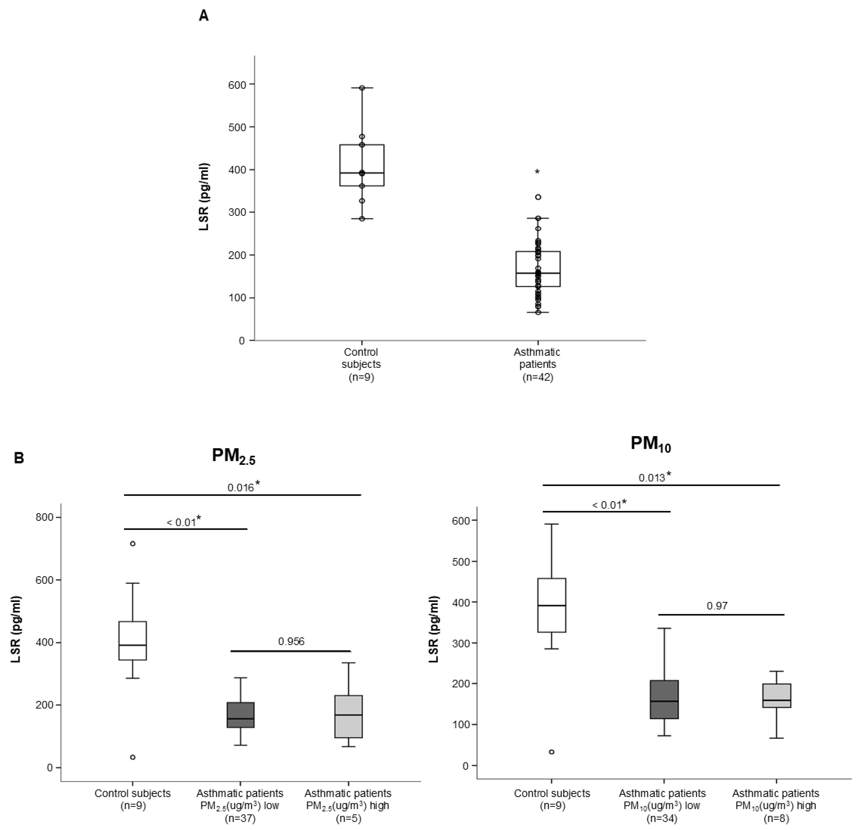
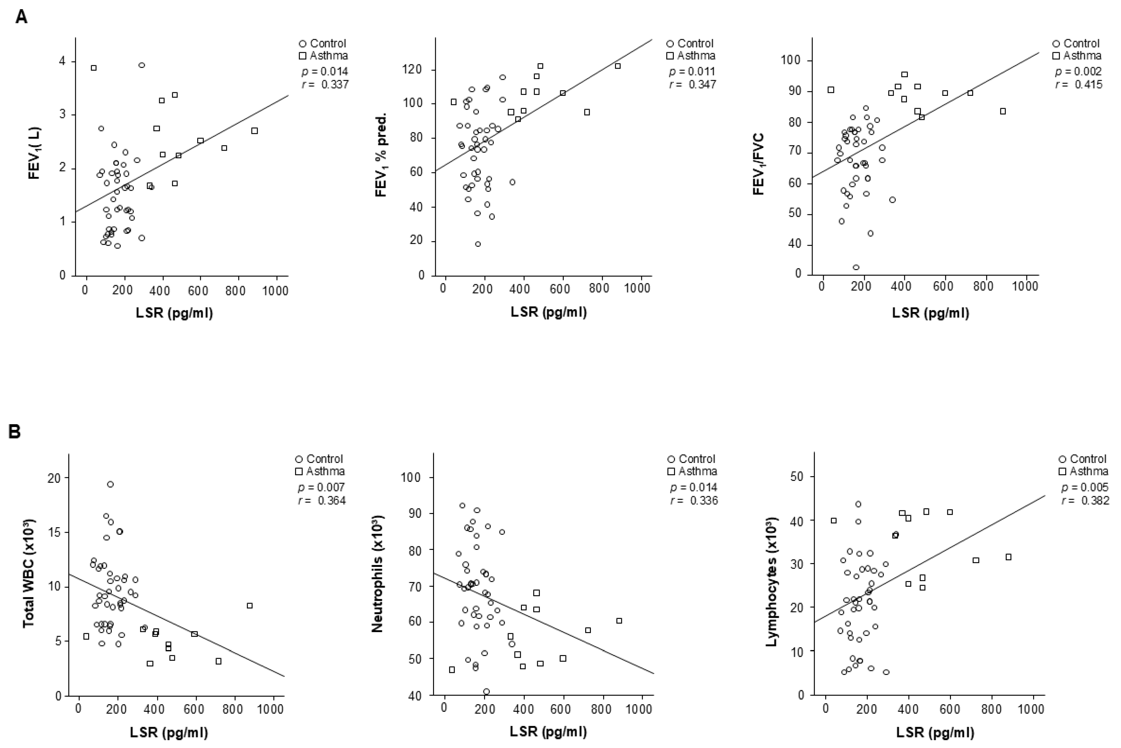
3.2. Tricellular TJ Protein LSR Levels Related to the Clinical Variables of Patients with Asthma

The LSR level had lower concentrations in the plasma from asthmatic patients (n = 42) than in that of control subjects (n = 9) (Figure 1A). This reduction aligns with previous findings linking tricellular tight junction dysfunction to inflammatory airway diseases. The LSR levels in asthmatic patients according to atmospheric PM2.5 and PM10 concentration were not significantly different (Figure 1B), suggesting that intrinsic disease mechanisms may play a more dominant role in regulating LSR. LSR concentration was correlated with FEV1 (L) (r = 0.337, p = 0.014), FEV % pred. (r = 0.347, p = 0.011), FEV1/FVC (r = 0.415, p = 0.002), WBC (r = −0.364, p = 0.007), and blood lymphocyte percent (r = 0.382, p = 0.005) in controls and patients with asthma (Figure 2A,B). These findings underscore the clinical relevance of LSR in reflecting asthma severity and immune dysregulation.

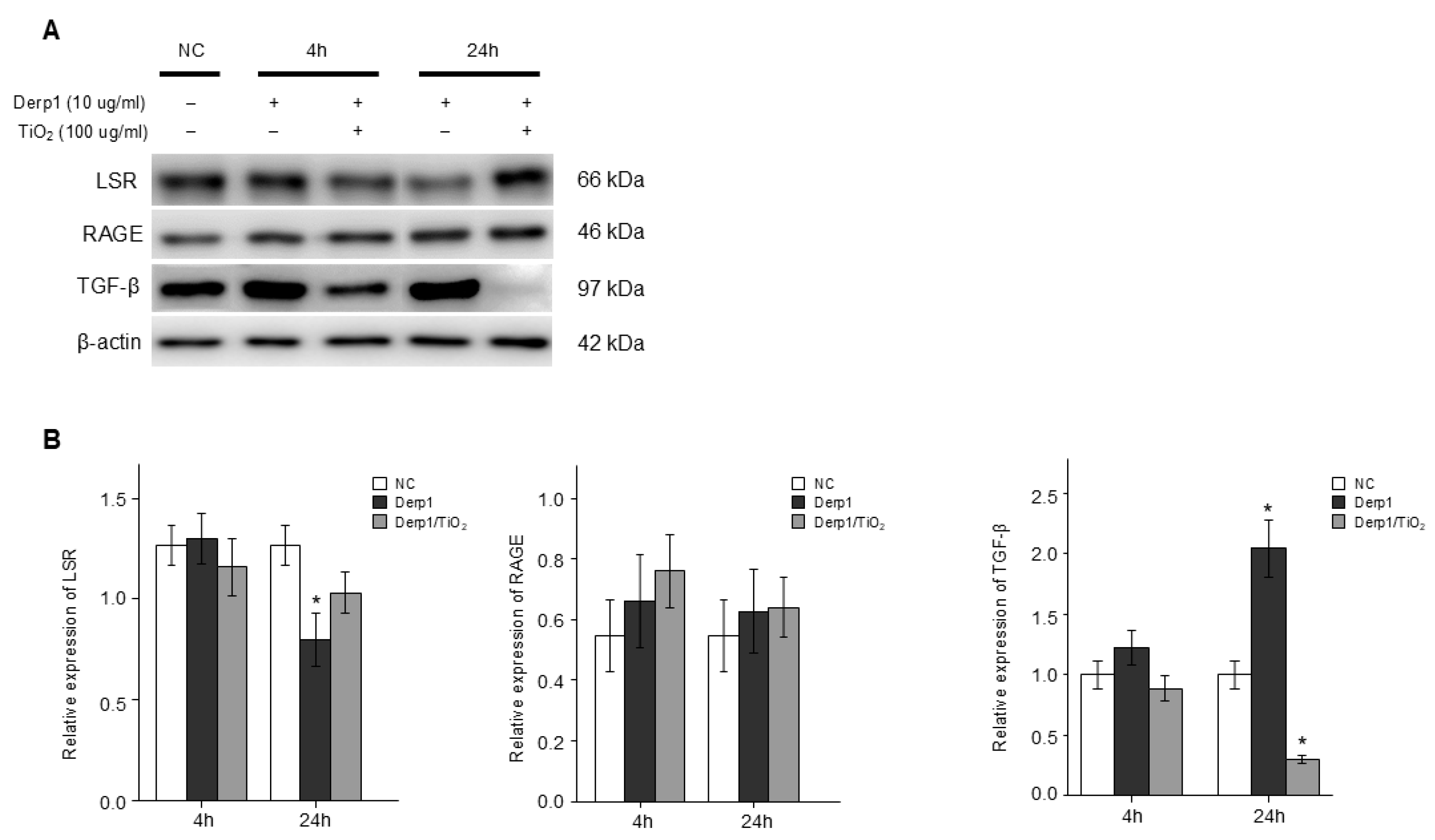
3.3. Change in RAGE and LSR, and TGFβ in NHBE Cells Treated to Derp1 and Exposed to TiO2

Although the LSR gene expression was significantly reduced in NHBE cells treated with Derp1 for 24 h, indicating a direct impact of allergens on tight junction proteins, interestingly, exposure to TiO2 modulated this reduction, highlighting potential protective or compensatory effects of the particulate material. Conversely, the TGFβ protein expression in NHBE cells treated to Derp1 had increased, and decreased with TiO2 exposure (Figure 3A,B). RAGE protein expression in NHBE cells treated to Derp1 did not change. These results suggest a complex interplay between asthma-related triggers and air pollutants in regulating epithelial tight junction integrity and inflammatory mediators.

3.4. OVA and TiO2-Activated Inflammation and Airway Responsiveness in Mice

There was increased AHR in the OVA mice and OVA/TiO2 mice compared to control mice (Figure 4B). These results indicate heightened airway reactivity, a hallmark of asthma, particularly exacerbated by TiO2 exposure. The OVA mice and OVA/TiO2 mice showed an increase in inflammatory cells in BALF compared to control mice (Figure 4C). These findings highlight the role of allergens and pollutants in promoting airway inflammation.

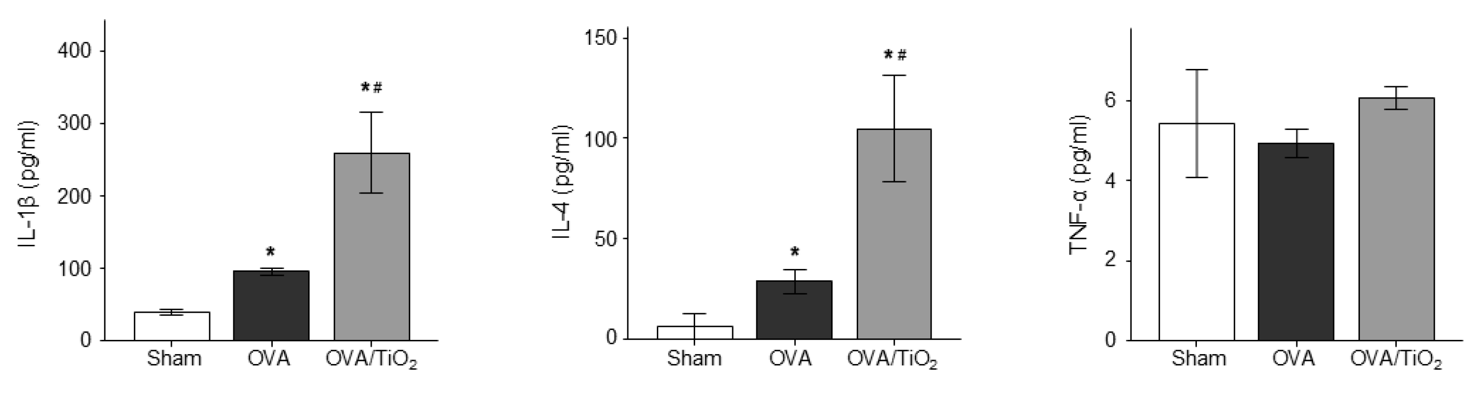
3.5. OVA and TiO2-Activated Cytokine Changes in Mice

Upon histologic examination, the OVA mice and OVA/TiO2 mice exhibited an increase in stimulating cell infiltration and exudative changes in the peribronchial layers and intraluminal areas of the bronchi (Figure 4C). This structural evidence further supports the functional data of airway inflammation and hyperresponsiveness observed in the OVA and OVA/TiO2 groups.
Although the TNF-α levels were not different between the three groups, the OVA mouse group had resulted in an increase in IL-1β and IL-4, and a greater increase in OVA/TiO2 mice (Figure 5) by ELISA using protein lysates from the lungs. These findings indicate that TiO2 exposure amplifies allergen-induced cytokine production, which may contribute to the observed exacerbation of airway inflammation.

3.6. Change in RAGE, LSR and TGFβ in the Lungs of OVA Mice and OVA/TiO2 Mice

OVA mice had resulted in a decrease in LSR and in increase in RAGE and TGFβ (Figure 6A,B). Moreover, OVA/TiO2 mice resulted in a change in RAGE, LSR and TGFβ (Figure 6A,B). Further alterations in these markers were observed in OVA/TiO2 mice, corroborating the in vitro findings. Additionally, IHC staining and immunofluorescence revealed that LSR expression had decreased in the lungs of OVA mice compared with control mice, and changed in OVA/TiO2 mice (Figure 6C–E). These results emphasize the critical role of tricellular tight junction proteins, particularly LSR, in maintaining epithelial barrier integrity in the context of asthma and pollutant exposure.

4. Discussion

Epithelial cells serve as protective barriers, shielding the internal milieu of the organism from the external environment, while concurrently delineating and sustaining distinct fluid-filled compartments within various organs. TJs limit solute leakage via the transcellular pathway, contributing to the barrier function of epithelial cells [24,25,26]. To maintain the full function of epithelial cells and paracellular barriers, there must be no extracellular area at tricellular contacts (TCs), i.e., the meeting point of three cells [18,20,27,28,29,30].
tTJs are supported by tricellulin and angulin family proteins [19]. Tricellulin is a four-transmembrane protein, anatomically similar to occludin [31]. Angulins are type I integral membrane proteins with a single immunoglobulin-like zone [19,32]. Tricellulin and angulins are located alongside the central sealing elements of tTJs [19,31]. Because angulins work on tricellulin to TCs [19,32], the angulin–tricellulin axis has a key role in tTJ formation [16]. Despite a significant body of research clarifying the physiological roles of tight junction-associated proteins, the exact specified mechanisms responsible for the formation of tTJs (tricellular tight junctions) remain incompletely understood. LSR is associated with the plasma membrane seal at TCs, separate from tricellulin and claudins [20].
There have been no papers on the role of LSR in asthma. Therefore, the current study examined the roles of LSR in asthma using a mouse model and blood from asthma patients. LSR concentrations were reduced in the plasma of asthmatic patients, and were associated with clinical variables such as lung function, white blood cell count, and blood lymphocyte proportion. Similarly, LSR gene expression levels were decreased in asthmatic mice. These findings indicate that LSR has an important involvement in asthma pathogenesis, and suggest that LSR may be a marker related to asthma and allergic inflammation.
HMGB1, RAGE, and toll-like receptor 4 (TLR4) are lead pathways for the evolution of cigarette smoke-induced lung inflammation. HMGB1 is a typical damage-associated molecular pattern protein, which mainly applies its activity by binding to RAGE and TLR4, and is implicated in airway inflammation [33,34]. RAGE, as a member of the immunoglobulin superfamily, is highly expressed in the lung (mostly in type I alveolar epithelial cells) but presents low expression in other organs and cells; it plays an important role in lung maturation and function [7]. Under certain pathological situations, RAGE is increased in response to the accumulation of ligands [35]. In the current study, we found that RAGE gene expression levels were increased in asthmatic mice, and changed by air pollutant exposure, and HMGB1 levels were not different in asthmatic patients and control subjects, suggesting that LSR and RAGE together are involved in airway inflammation in asthma.
Air pollution represents a considerable risk factor for human health, and may cause many lung and respiratory diseases. Air pollutants can be categorized as either major (emitted directly into the atmosphere) or subsidiary (formed within the atmosphere) air pollutants [36]. Asthma is a multiplex disease condition incited and worsened by increased exposure to various chemical, physical, and biological agents from diverse indoor and outdoor sources [37,38]. For example, asthma symptoms such as coughing, wheezing, and shortness of breath have been linked to short-term duration exposure to ambient PM2.5 and PM10 in intended prospective cohorts, particularly in children with atopy [39].
In addition, exposure to traffic-related air pollution was related to both continual and new-onset asthma in adults in a cohort study [40]. Moreover, living less than 200 m from a main roadway was correlated with greater odds of developing new asthma in middle-aged people who had had no asthma by age 45 [40]. In the same study, asthmatic patients at 45 years had a growing risk of persistent asthma up to 53 years if they lived less than 200 m from a main roadway compared with asthmatic patients living farther than 200 m from a main roadway [40]. Several studies have also found that short-term exposure to PM2.5 produced by combustion can cause asthma attacks and exacerbate COPD [1,3,41,42,43]. In our study, OVA mice showed an increase in IL-4 and IL1-β, but not TNF-α, and a greater increase in mice following TiO2 exposure as one of the air pollutants, suggesting that air pollutants can be a factor for asthma exacerbation.
One limitation of the study was the small size of the patient group who had asthma.
This study had limitations. First, it did not estimate cell barrier function, such as transepithelial electrical resistance. Second, it relied on data with a small number of control subjects. Regardless, the present findings highlight the participation of LSR in asthma pathogenesis and asthma phenotypes under air pollutant exposure.

5. Conclusions

In the current study, we examined the influences of air pollution on LSR in asthma pathogenesis. Plasma LSR levels were significantly diminished in asthmatic patients (166.05 ± 62.24 pg/mL) compared to control subjects (447.11 ± 213.99 pg/mL, p < 0.05). Moreover, LSR protein levels were decreased, and TGFβ increased, in the asthmatic mouse lungs. Both LSR and TGF-β levels in the asthmatic mice were altered by exposure to the air pollutant TiO2, suggesting that air pollutant exposure can affect asthma phenotypes. Additional studies are required to evaluate the precise role of LSR in air pollutant-induced airway inflammation.

Author Contributions

Study conception and design: A.-S.J. and D.H.; animal experiments: D.H., M.-H.A. and P.-H.L.; data collection: A.-S.J., D.H., M.-H.A. and P.-H.L.; analysis and interpretation of results: A.-S.J., D.H., M.-H.A., P.-H.L., J.-H.K., Y.N., A.-R.B. and S.P.; draft manuscript preparation: A.-S.J. and D.H. All authors have read and agreed to the published version of the manuscript.

Funding

This research was supported by Basic Science Research Program through the National Research Foundation of Korea (NRF) funded by the Ministry of Science and ICT (NRF-2020R1A2C1006506) and Soonchunhyang University.

Institutional Review Board Statement

This study was approved by the institutional review board of Soonchunhyang University Hospital (approval No. SCHBC 2017-12-013-003).

Informed Consent Statement

Informed consent was obtained from all subjects involved in the study.

Data Availability Statement

This article contains all of the data generated or evaluated throughout the study.

Conflicts of Interest

The authors declare no conflicts of interest.

References

  1. Bălă, G.P.; Râjnoveanu, R.M.; Tudorache, E.; Motișan, R.; Oancea, C. Air Pollution Exposure—The (In)visible Risk Factor for Respiratory Diseases. Environ. Sci. Pollut. Res. Int. 2021, 28, 19615–19628. [Google Scholar] [CrossRef] [PubMed]
  2. Jacquemin, B.; Siroux, V.; Sanchez, M.; Carsin, A.E.; Schikowski, T.; Adam, M.; Bellisario, V.; Buschka, A.; Bono, R.; Brunekreef, B.; et al. Ambient Air Pollution and Adult Asthma Incidence in Six European Cohorts (ESCAPE). Environ. Health Perspect. 2015, 123, 613–621. [Google Scholar] [CrossRef] [PubMed]
  3. Park, J.; Kim, H.J.; Lee, C.H.; Lee, H.W. Impact of Long-Term Exposure to Ambient Air Pollution on the Incidence of Chronic Obstructive Pulmonary Disease: A Systematic Review and Meta-Analysis. Environ. Res. 2021, 194, 110703. [Google Scholar] [CrossRef] [PubMed]
  4. World Health Organization. WHO Global Air Quality Guidelines: Particulate Matter, Ozone, Nitrogen Dioxide, Sulfur Dioxide and Carbon Monoxide; World Health Organization: Geneva, Switzerland, 2021. [Google Scholar]
  5. Lee, P.H.; Park, S.; Lee, Y.G.; Choi, S.M.; An, M.H.; Jang, A.S. The Impact of Environmental Pollutants on Barrier Dysfunction in Respiratory Disease. Allergy Asthma Immunol. Res. 2021, 13, 850–862. [Google Scholar] [CrossRef]
  6. Park, S.; Lee, P.H.; Baek, A.R.; Park, J.S.; Lee, J.; Part, S.W.; Kim, D.J.; Jang, A.S. Association of the Tight Junction Protein Claudin-4 with Lung Function and Exacerbations in Chronic Obstructive Pulmonary Disease. J. Chron. Obs. Pulmon. Dis. 2021, 6, 2735–2740. [Google Scholar] [CrossRef]
  7. Lee, Y.G.; Lee, P.H.; Choi, S.M.; An, M.H.; Jang, A.S. Effects of Air Pollutants on Airway Diseases. J. Environ. Res. Public Health 2021, 18, 9905. [Google Scholar] [CrossRef]
  8. Lodovici, M.; Bigagli, E. Oxidative Stress and Air Pollution Exposure. J. Toxicol. 2011, 2011, 487074. [Google Scholar] [CrossRef]
  9. Xu, Z.; Wang, W.; Liu, Q.; Li, Z.; Lei, L.; Ren, L.; Deng, F.; Guo, X.; Wu, S. Association Between Gaseous Air Pollutants and Biomarkers of Systemic Inflammation: A Systematic Review and Meta-Analysis. Environ. Pollut. 2022, 292, 118336. [Google Scholar] [CrossRef]
  10. Glencross, D.A.; Ho, T.R.; Camiña, N.; Hawrylowicz, C.M.; Pfeffer, P.E. Air Pollution and Its Effects on the Immune System. Free Radic. Biol. Med. 2020, 151, 56–68. [Google Scholar] [CrossRef]
  11. Guarnieri, M.; Balmes, J.R. Outdoor Air Pollution and Asthma. Lancet 2014, 383, 1581–1592. [Google Scholar] [CrossRef]
  12. Liu, Y.; Pan, J.; Zhang, H.; Chunxiang, S.; Li, G.; Peng, Z.; MA, J.; Zhou, Y.; Zhang, L. Short-Term Exposure to Ambient Air Pollution and Asthma Mortality. Am. J. Respir. Crit. Care Med. 2019, 200, 24–32. [Google Scholar] [CrossRef] [PubMed]
  13. Garcia, M.A.; Nelson, W.J.; Chavez, N. Cell-Cell Junctions Organize Structural and Signaling Networks. Cold Spring Harb. Perspect. Biol. 2018, 10, a029181. [Google Scholar] [CrossRef] [PubMed]
  14. Kohno, T.; Konno, T.; Kojima, T. Role of Tricellular Tight Junction Protein Lipolysis-Stimulated Lipoprotein Receptor (LSR) in Cancer Cells. Int. J. Mol. Sci. 2019, 20, 3555. [Google Scholar] [CrossRef] [PubMed]
  15. Matter, K.; Balda, M.S. Signalling to and from Tight Junctions. Nat. Rev. Mol. Cell Biol. 2003, 4, 225–236. [Google Scholar] [CrossRef]
  16. Furuse, M.; Izumi, Y.; Oda, Y.; Higashi, T.; Iwamoto, N. Molecular Organization of Tricellular Tight Junctions. Tissue Barriers 2014, 2, e28960. [Google Scholar] [CrossRef]
  17. Guillot, C.; Lecuit, T. Mechanics of Epithelial Tissue Homeostasis and Morphogenesis. Science 2013, 340, 1185–1189. [Google Scholar] [CrossRef]
  18. Ikenouchi, J.; Furuse, M.; Furuse, K.; Sasaki, H.; Tsukita, S. Tricellulin Constitutes a Novel Barrier at Tricellular Contacts of Epithelial Cells. J. Cell Biol. 2005, 171, 939–945. [Google Scholar] [CrossRef]
  19. Masuda, S.; Oda, Y.; Sasaki, H.; Ikenouchi, J.; Higashi, T.; Akashi, M.; Nishi, E.; Furuse, M. LSR Defines Cell Corners for Tricellular Tight Junction Formation in Epithelial Cells. J. Cell Sci. 2011, 124, 548–555. [Google Scholar] [CrossRef]
  20. Sugawara, T.; Furuse, K.; Otani, T.; Wakayama, T.; Furuse, M. LSR Seals Tricellular Contacts Independently of Tricellulin and Claudins. J. Cell Biol. 2021, 220, e202005062. [Google Scholar] [CrossRef]
  21. Amieva, M.R.; Vogelmann, R.; Covacci, A.; Tompkins, L.S.; Nelson, W.J.; Falkow, S. Disruption of the Epithelial Apical-Junctional Complex by Helicobacter pylori CagA. Science 2003, 300, 1430–1434. [Google Scholar] [CrossRef]
  22. Kaplan, A.; Boven, J.; Ryan, D.; Tsiligianni, I.; Sinthia, B.A. GINA 2020: Potential Impacts, Opportunities and Challenges for Primary Care. J. Allergy Clin. Immunol. Pract. 2021, 9, 1516–1519. [Google Scholar] [CrossRef] [PubMed]
  23. Lee, Y.G.; Lee, S.H.; Hong, J.; Lee, P.H.; Jang, A.S. Titanium Dioxide Particles Modulate Epithelial Barrier Protein, Claudin 7 in Asthma. Mol. Immunol. 2021, 132, 209–216. [Google Scholar] [CrossRef] [PubMed]
  24. Tsukita, S.; Furuse, M.; Itoh, M. Multifunctional Strands in Tight Junctions. Nat. Rev. Mol. Cell Biol. 2001, 2, 285–293. [Google Scholar] [CrossRef]
  25. Van Itallie, C.M.; Anderson, J.M. Architecture of Tight Junctions and Principles of Molecular Composition. Semin. Cell Dev. Biol. 2014, 36, 157–165. [Google Scholar] [CrossRef]
  26. Zihni, C.; Mills, C.; Matter, K.; Balda, M.S. Tight Junctions: From Simple Barriers to Multifunctional Molecular Gates. Nat. Rev. Mol. Cell Biol. 2016, 17, 564–580. [Google Scholar] [CrossRef]
  27. Wade, J.B.; Karnovsky, M.J. The Structure of the Zonula Occludens. A Single Fibril Model Based on Freeze-Fracture. J. Cell. Biol. 1974, 60, 168–180. [Google Scholar] [CrossRef]
  28. Walker, D.C.; MacKenzie, A.; Hulbert, W.C.; Hogg, J.C. A Re-Assessment of the Tricellular Region of Epithelial Cell Tight Junctions in Trachea of Guinea Pig. Acta Anat. 1985, 122, 35–38. [Google Scholar] [CrossRef]
  29. Staehelin, L.A. Further Observations on the Fine Structure of Freeze-Cleaved Tight Junctions. J. Cell Sci. 1973, 13, 763–786. [Google Scholar] [CrossRef]
  30. Staehelin, L.A.; Mukherjee, T.M.; Williams, A.W. Freeze-Etch Appearance of the Tight Junctions in the Epithelium of Small and Large Intestine of Mice. Protoplasma 1969, 67, 165–184. [Google Scholar] [CrossRef]
  31. Ikenouchi, J.; Sasaki, H.; Tsukita, S.; Furuse, M.; Tsukita, S. Loss of Occludin Affects Tricellular Localization of Tricellulin. Mol. Biol. Cell 2008, 19, 4687–4693. [Google Scholar] [CrossRef]
  32. Higashi, T.; Tokuda, S.; Kitajiri, S.; Masuda, S.; Nakamura, H.; Oda, Y.; Furuse, M. Analysis of the ‘Angulin’ Proteins LSR, ILDR1 and ILDR2—Tricellulin Recruitment, Epithelial Barrier Function and Implication in Deafness Pathogenesis. J. Cell Sci. 2013, 126, 966–977. [Google Scholar] [CrossRef] [PubMed]
  33. Chen, L.; Sun, X.; Zhong, X. Role of RAGE and its Ligand HMGB1 in the Development of COPD. Postgrad. Med. 2022, 134, 763–775. [Google Scholar] [CrossRef] [PubMed]
  34. Lin, L.; Li, J.; Song, Q.; Cheng, W.; Chen, P. The Role of HMGB1/RAGE/TLR4 Signaling Pathways in Cigarette Smoke-Induced Inflammation in Chronic Obstructive Pulmonary Disease. Immun. Inflamm. Dis. 2022, 10, e711. [Google Scholar] [CrossRef] [PubMed]
  35. Yonchuk, J.G.; Silverman, E.; Bowler, R.P.; Agusti, A.; Lomas, D.A.; Miller, B.E.; Singer, R.T.; Mayer, R.J. Circulating Soluble Receptor for Advanced Glycation End Products (sRAGE) as a Biomarker of Emphysema and the RAGE Axis in the Lung. Am. J. Respir. Crit. Care Med. 2015, 192, 785–792. [Google Scholar] [CrossRef]
  36. Bowatte, G.; Lodge, C.J.; Knibbs, L.D.; Erbas, B.; Perret, J.L.; Jalaludin, B.; Morgan, G.G.; Bui, D.S.; Giles, G.G.; Hamilton, G.S.; et al. Traffic Related Air Pollution and Development and Persistence of Asthma and Low Lung Function. Environ. Int. 2018, 113, 170–176. [Google Scholar] [CrossRef]
  37. Paterson, C.A.; Sharpe, R.A.; Taylor, T.; Morrissey, K. Indoor PM2.5, VOCs and Asthma Outcomes: A Systematic Review in Adults and Their Home Environments. Environ. Res. 2021, 202, 111631. [Google Scholar] [CrossRef]
  38. Misiukiewicz-Stepien, P.; Paplinska-Goryca, M. Biological Effect of PM10 on Airway Epithelium-Focus on Obstructive Lung Diseases. Clin. Immunol. 2021, 227, 108754. [Google Scholar] [CrossRef]
  39. Mannucci, P.M.; Harari, S.; Martinelli, I.; Franchini, M. Effects on Health of Air Pollution: A Narrative Review. Intern. Emerg. Med. 2015, 10, 657–662. [Google Scholar] [CrossRef]
  40. Adar, S.D.; Filigrana, P.A.; Clements, N.; Clements, N.; Peel, J.L. Ambient Coarse Particulate Matter and Human Health: A Systematic Review and Meta-Analysis. Curr. Environ. Health Rep. 2014, 1, 258–274. [Google Scholar] [CrossRef]
  41. Singh, D.; Agusti, A.; Anzueto, A.; Barnes, P.J.; Bourbeau, J.; Celli, B.R.; Criner, G.J.; Frith, P.; Halpain, D.M.G.; Han, M.; et al. Global Strategy for the Diagnosis, Management, and Prevention of Chronic Obstructive Lung Disease: The GOLD Science Committee Report. Eur. Respir. J. 2019, 53, 1900164. [Google Scholar] [CrossRef]
  42. Tian, Y.; Xiang, X.; Juan, J.; Song, J.; Cao, Y.; Huang, C.; Li, M.; Hu, Y. Short-Term Effects of Ambient Fine Particulate Matter Pollution on Hospital Visits for Chronic Obstructive Pulmonary Disease in Beijing, China. Environ. Health 2018, 17, 21. [Google Scholar] [CrossRef]
  43. Nishida, C.; Yatera, K. The Impact of Ambient Environmental and Occupational Pollution on Respiratory Diseases. Int. J. Environ. Res. Public Health 2022, 19, 2788. [Google Scholar] [CrossRef]
Figure 1. (A) LSR level in control subject and asthmatic patients. (B) LSR levels in control subjects and asthmatic patients by level of PM 2.5 and PM 10 μg/m3. * p < 0.05, control subjects vs. asthmatic patients.
Atmosphere 15 01532 g001
Figure 2. Relationship of LSR with (A) FEV1 (L) (r = 0.337, p = 0.014), FEV1% predicted (r = 0.347, p = 0.011) and FEV1/FVC (r = 0.415, p = 0.002). (B) WBC (r = −0.364, p = 0.007), neutrophils (r = −0.336, p = 0.014), and blood lymphocyte proportion (r = 0.382, p = 0.005) in control subjects and patients with asthma.
Atmosphere 15 01532 g002
Figure 3. LSR, RAGE, and TGFβ protein determined by Western blot in NHBE cells treated with 10  μg/mL Dermatophagoides pteronyssinus 1 (Derp1) and 100 μg/mL TiO2 for 4, and 24 h. (A) NHBE cells protein level of RAGE, TGFβ and LSR as determined by Western blot. (B) Densitometric data from the Western blots. Densitometry was determined with 3 immunoblots and normalized to β-actin. Data are expressed as means ± SD. Scale bar means 100 μm. * p < 0.05 vs. normal control (NC).
Atmosphere 15 01532 g003
Figure 4. (A) Experiment protocol of TiO2 exposure model. (B) Airway hyperresponsiveness (AHR) and inflammation in OVA mice. (C) Total and differential cell count in bronchoalveolar lavage fluid (BALF). BALF was collected on day 25 and cell differentials determined. Values are means ± SEM (n = 6 mice/group). * p < 0.05 vs. sham. # p < 0.05 vs. OVA.
Atmosphere 15 01532 g004
Figure 5. Lung protein levels of IL-1β, IL-4 and TNF-α as determined by ELISA. Data are expressed as means ± SD. * p < 0.05 vs. sham. # p < 0.05 vs. OVA.
Atmosphere 15 01532 g005
Figure 6. (A) Lung protein level of RAGE, TGFβ and LSR as determined by Western blot. (B) Densitometric data from the Western blots. Densitometry was determined with 3 immunoblots and normalized to β-actin. (C) Hematoxylin & eosin (H&E)-stained lung tissue and LSR immunohistochemical staining of mouse lung tissue sections. (D) Inflammatory index and quantification of LSR expression. (E) Immunofluorescence of LSR in lung tissue. Data are expressed as means ± SD. Scale bar means 100 μm. * p < 0.05 vs. sham.
Atmosphere 15 01532 g006
Table 1. Clinical characteristics in control subjects and patients with asthma.
VariablesControl SubjectsAsthmatic Patients
No. of subjects942
Sex (male/female)2/714/28
Age (at initial visit), years53 (48–62)57 (52–65)
Smoking status (NS/ES/SM)9/0/028/10/4 *
Cigarettes smoked, pack. years-20 (10–35)
Lung functionFEV1, (L)104.7 ± 4.9375.2 ± 17.15 *
FEV1, % pred.106 ± 9.9775.1 ± 21.7 *
FEV1/FVC88.4 ± 4.867.9 ± 11.2 *
BMI, kg/m225.5 ± 2.1627.3 ± 4.83
PC20, mg/mL-9.43 ± 8.91
Total IgE, kU-433.7 ± 1259.6
Skin test positive, %0 (0.0%)8 (19.2%)
Blood WBC/uL4333.3 ± 1958.99789.29 ± 3369.2 *
Blood eosinophil, %1.4 ± 0.962.38 ± 4.34
Blood neutrophil, %56.5 ± 7.9868.5 ± 12.38 *
Blood lymphocyte, %35.1 ± 7.9421.6 ± 9.83 *
Data expressed as mean ± SD. BMI; body mass index, ES; ex-smoker, FEV1; forced expiratory volume in one second, FVC; forced vital capacity, NS; non-smoker, SM; smoker, PC20 methacholine; the concentration of methacholine required to decrease the FEV1 by 20%, * p < 0.05 compared with control subjects.
Table 2. Cell culture parameters and experimental conditions.
ParameterDetails
Cell typeNHBE (normal human bronchial epithelial) cells
Growth mediumBEGM (bronchial epithelial growth medium)
SupplementsBovine pituitary extract (BPE), human epidermal growth factor (hEGF), granulocyte-aggregating 1000 (GA-1000), retinoic acid, hydrocortisone, insulin, transferrin, triiodothyronine
Initial seeding density3500 cells/cm2
Incubation conditions37 °C, 5% CO2
Treatment conditionsDerp1 (10 μg/mL), TiO2 (100 μg/mL)
Time4, 24 h
Disclaimer/Publisher’s Note: The statements, opinions and data contained in all publications are solely those of the individual author(s) and contributor(s) and not of MDPI and/or the editor(s). MDPI and/or the editor(s) disclaim responsibility for any injury to people or property resulting from any ideas, methods, instructions or products referred to in the content.

