Article

Chemical Composition of PM2.5-0.3 and PM0.3 Collected in Southern Lebanon and Assessment of Their Toxicity in BEAS-2B Cells

1
Unité de Chimie Environnementale et Interactions sur le Vivant (UCEIV UR4492), University Littoral Côte d’Opale, 59140 Dunkerque, France
2
IMPact de l’Environnement Chimique sur la Santé (IMPECS ULR4483), Institut Pasteur de Lille, CHU Lille, University Lille, 59000 Lille, France
3
Lebanese Atomic Energy Commission, National Council for Scientific Research (CNRS-L), Beirut P.O. Box 11-8281, Lebanon
4
Centre Commun de Mesures, University Littoral Côte d’Opale, 59140 Dunkerque, France
*
Author to whom correspondence should be addressed.
These authors contributed equally to this work, and should be considered as co-first authors.
Atmosphere 2024, 15(7), 811; https://doi.org/10.3390/atmos15070811 (registering DOI)
Submission received: 8 May 2024 / Revised: 23 June 2024 / Accepted: 4 July 2024 / Published: 6 July 2024
(This article belongs to the Special Issue Air Pollution Exposure and Health Impact Assessment (2nd Edition))

Abstract

:
Fine particles (PM2.5) have generally been reported as the major contributor to the adverse health effects of air pollution. Lebanon is characterized by a high density of transport, the production of electricity by generators, and a problem of uncontrolled incineration of household waste. For the purpose of this paper, the physico-chemical properties of fine (PM2.5-0.3) and quasi-ultrafine (PM0.3) particulate matter sampled in Southern Lebanon, were studied. Then, an evaluation and comparison of the toxicity of the different extracted fractions from PM (i.e., native PM2.5-0.3 vs. organic extractable matter fraction (OEM2.5-0.3), and non-extractable matter fraction (NEM2.5-0.3)) was performed. Also, an examination of the toxicity of PM0.3 was conducted indirectly through the evaluation of the OEM0.3 harmfulness. The physico-chemical analysis showed that PM0.3 was much more concentrated than PM2.5-0.3 in organic compounds such as polycyclic aromatic hydrocarbons (PAHs) (28-fold) and their nitrated (N-PAHs, 14-fold) and oxygenated (O-PAHs, 10-fold) derivatives. Normal human bronchial epithelial cells (BEAS-2B) were exposed to PM2.5-0.3, its derived fractions (i.e., OEM2.5-0.3 and NEM2.5-0.3), and OEM0.3 before evaluating the global cytotoxicity, metabolic activation of organic compounds, genotoxicity, and inflammatory response. Different responses were observed depending on the considered fraction of particles. The global cytotoxicity showed a pronounced response related to ATP and LDH activities after exposure to the quasi-ultrafine organic extractable matter fraction (OEM0.3). There was no significant induction of the AhR cell-signaling pathway by NEM2.5-0.3. Despite the apparent difference in the kinetics of induction of the toxicological endpoints under study, OEM0.3 provoked a higher overall cytotoxicity and genotoxicity than OEM2.5-0.3 and total PM2.5-0.3. Taken together, these results clearly showed that the finest particles are more damaging to BEAS-2B cells than PM2.5-0.3 because they are richer in organic compounds, thereby inducing more remarkable toxic effects.

1. Introduction

Ranked as the fifth most crucial mortality factor, fine atmospheric particulate matter (PM2.5) constitutes one of the main air pollutants that cause millions of deaths every year worldwide [1,2]. According to the World Health Organization (WHO), around 4.2 million people have died from exposure to outdoor air pollution, and those deaths are mainly linked to heart disease, stroke, chronic obstructive pulmonary disease (COPD), lung cancer, and acute respiratory infections in children. Classified in Group 1 in terms of its carcinogenicity to humans by the International Agency of Research on Cancer (IARC) in 2013, PM2.5 can be formed from precursors by chemical reactions in the atmosphere or emitted directly by natural/anthropogenic sources. It usually consists of a complex and heterogeneous mixture with a direct impact on air quality and human health. PM2.5 is generally composed of an inorganic (i.e., ions, metals, carbonaceous species), organic (polycyclic aromatic hydrocarbons (PAHs), volatile organic compounds (VOC), polychlorobiphenyls (PCB), polychlorinated dibenzo-p-dioxins (PCDD), polychlorinated dibenzofurans (PCDF)), and biological part (bacteria, fungi) [2,3,4,5]. PM2.5 can induce adverse health effects in humans. It can penetrate deeply into the lung, irritating and corroding the alveolar wall and consequently impairing lung function. PM2.5 deposits mainly on the bronchial pseudostratified epithelial layer, which provides a chemically, immunologically, and mechanically protective barrier against environmental insults; it can also reach the lower airways. It can be cleared by macrophage-mediated phagocytosis. Nonetheless, the finest particles, especially the ultrafine ones, or their inorganic and organic components, may reach the underlying cells and the blood circulation, therefore exerting their adverse health effects on other organs [6,7,8]. Until now, limited research has investigated the respective impacts of the different fractions of PM2.5-0.3 (organic extractable matter fraction (OEM2.5-0.3) and non-extractable matter fraction (NEM2.5-0.3). Consequently, the comprehension of cellular mechanisms responsible for inducing toxicity associated with exposure to these distinct particle fractions remains limited, especially in the context of BEAS-2B cells.
In vitro toxicology methods have been widely used over the last two decades. They represent a relevant approach for determining and describing the underlying mechanisms involved in the toxicity triggered by air pollution derived from PM2.5 [4,9,10,11,12]. Recent studies, at cellular and molecular scales, have demonstrated that PM2.5 induces oxidative stress by generating high levels of reactive oxygen species (ROS) and also provokes DNA damage, pro-inflammatory response, cell cycle deregulation, and regulated cell death [4,9,10,13]. The inorganic fraction of PM2.5 can cause some of these mechanisms, but others have been more related to the organic fraction. It has been determined that the most toxic PM2.5 components are metals, PAHs, and carbonaceous particles [14]. Several studies evidenced that the co-presence of organic compounds and other inorganic elements may determine the biological effects of PM2.5 [4,9,15,16]. Badran et al. also supported the view that organic compounds associated with the quasi-ultrafine fraction (PM0.3) were the main drivers of fine PM genotoxicity and phase I and II enzymes involved in the metabolic activation of PAHs. This underscores the importance of improving the knowledge of the toxicological impact of ultrafine (PM0.1) and quasi-ultrafine (PM0.3) particles [4].
This work aimed to examine the toxicological impact of PM2.5-0.3 and PM0.3 collected in south Lebanon from the city of Deir AlZahrani on normal human bronchial epithelial cells (BEAS-2B). This city accommodates a diverse array of industries, with certain sectors specializing in aluminum manufacturing and handling, while others are dedicated to the production of real estate materials such as construction supplies and plumbing equipment. Deir AlZahrani is characterized by a significant number of chimneys, indicative of numerous factories within the area capable of emitting particulate matter. This is compounded by emissions from power generators. These factors promote the emission of toxic organic compounds such as PAHs and subsequently increase the environmental risks associated with PM2.5. To our knowledge, no toxicological studies have been carried out in southern Lebanon focusing on the sources of PM2.5 and PM0.3 and their respective adverse effects on human lung cells. Although the toxicity of atmospheric particles is well known in the literature, there is a lack of data on the fraction within PM responsible for derived toxic effects. Are fine or ultrafine particles implicated? Is it the entirety of particles or their organic constituents? Therefore, there is an urgent need for new toxicological research to enhance the understanding of the underlying mechanisms of toxicity induced by different chemical fractions of PM2.5 from Southern Lebanon: organic (OEM2.5-0.3 and OEM0.3) and inorganic (NEM2.5-0.3). The collection site of the present study differs from that of our previous work focusing on Beirut [4]. Indeed, Beirut is characterized by high levels of fine particle pollution, primarily attributable to diesel-powered generators and road traffic. While some studies have been previously conducted in Lebanon, they focused on Beirut and North Lebanon [17,18,19,20,21], and, to the best of our knowledge, this work is the first one focusing on air quality in Southern Lebanon. So, our study represents the first analysis of the air quality in the Deir AlZahrani agglomeration.

2. Materials and Methods

2.1. Sampling Site

Fine (PM2.5-0.3) and quasi-ultrafine (PM0.3) particles were collected in Deir AlZahrani (33°25′14.29″ N; 35°27′17.9784″ E), a semi-rural city, located in the south of Lebanon, 75 km from the capital, Beirut (Lebanon). Deir AlZahrani belongs to the district of Nabatiyeh, with 10,000 inhabitants. This area has several industries that fabricate and handle aluminum, while others produce real estate supplies. It also features many fireplaces and diesel-electric generators. The village is crossed by a main highway that links Beirut with Southern Lebanon and is therefore characterized by heavy road traffic (Figure 1). In addition, at 8 km from the sampling site, the Valley of Kfour, known to be the biggest in the region, has several open-cast industrial activities such as tire melting.
The sampling point was situated on the roof of a 3-story building in Deir AlZahrani, Nabatiyeh, and the PM sampling was performed continuously from the 1st of January to the 3rd of March 2017. It was achieved using a high volume five-stage plus backup cascade impactor (model 235, 68 m3/h, Tisch, Cincinnati, OH, USA), allowing the separation of collected particles according to their equivalent aerodynamic diameter. PM2.5-0.3 was collected on impaction plates (stages 2–5), while PM0.3 was retained on a quartz fiber filter (Pallflex® Tissuquartz 2500 QAT-UP, Pall, Putnam, NY, USA) placed after the impaction system. The system was changed every 5 days. Plates and backup filters were dried in a laminar flow hood for 48 h. PM2.5-0.3 were obtained by brushing the plates, and individual PM2.5-0.3 samples were gathered and carefully homogenized to form an average PM2.5-0.3 sample. Similarly, average PM0.3 samples were constituted by producing a proportional mix of pieces of the individual backup filters. Samples were weighted and finally stored at −20 °C. Sampling details have been described elsewhere [4].

2.2. Physico-Chemical Analysis of the Collected PM

Numerous analyses were performed to determine the chemical composition of the collected PM. Table A1 summarizes the different types of physico-chemical analysis tests and the samples concerned. Several tests were conducted to determine the inorganic composition of the collected PM, such as X-ray diffraction (XRD), scanning electronic microscopy-energy dispersive X-ray spectroscopy (SEM-EDX) analysis, quantification of major and trace elements using inductively coupled plasma-atomic emission spectroscopy (ICP-AES), and quantification of water-soluble ions using ionic chromatography (IC). Organic compounds such as PAHs, N- and O-PAHs, PCDD, PCDF, and PCB were also quantified, using gas chromatography-mass spectrometry (GC-MS) for PAHs and high-resolution GC–high-resolution MS (HRGC–HRMS) for the other organic compounds. Paraffin, also known as alkanes, was also analyzed using GC–MS. The details related to the methodology are widely explained elsewhere [4].

2.3. Sample Preparation for Toxicological Studies

In order to study the implications of each PM fraction in the induction of toxic effects, the toxicity of the whole PM2.5-0.3, its OEM2.5-0.3, and NEM2.5-0.3 were evaluated and compared. In addition, evaluation and comparison of the toxicity of the OEM2.5-0.3 and OEM0.3 were performed. Regarding the preparation of the samples, 200 mg of native PM2.5-0.3 and their equivalent of this mass in PM0.3 filters were placed in glass centrifuge tubes containing 10 mL of dichloromethane (DCM) and subjected to sonication (10 min, 20 °C). After extraction, the mixture was centrifuged at 1000 g, 20 °C for 10 min, allowing the separation of the extracted organic part from the PM or filters freed from this fraction. The supernatant was then recovered, and the extraction was repeated 3 times successively with fresh DCM to ensure the recovery of all the extractable organic compounds. For each sample, the solutions resulting from successive extractions of PM by DCM were combined, and the DCM was evaporated under a nitrogen stream and replaced with dimethyl sulfoxide (DMSO), which is compatible with toxicity studies.
At the end of the extraction, OEM2.5-0.3 (supernatants) were obtained from PM2.5-0.3 in addition to NEM2.5-0.3 (kept in the centrifugation tube). From the PM0.3 collected on filters, only OEM0.3 was prepared. Concentrations of the final OEM2.5-0.3 and OEM0.3 solution were 98.1 and 96.4 mg PM-equivalent/mL (PM-eq/mL), respectively. For more details, please see [4].

2.4. In Vitro Toxicity of PM2.5

2.4.1. Cell Culture and Exposure

Normal human bronchial epithelial BEAS-2B cells, and an Ad12-SV40 hybrid virus-transformed human bronchial epithelial cell line (ATCC® CRL-9609™) were cultured as published elsewhere [22]. Just before reaching the confluence (<80%), BEAS-2B cells were exposed to the different fractions of PM: native PM2.5-0.3, NEM2.5-0.3, OEM2.5-0.3, and OEM0.3, prepared in LHC-9 culture medium, at concentrations ranging from 3 to 96 µg PM/cm2 or μg PM-eq/cm2. Negative controls were represented by BEAS-2B incubated either with LHC-9 culture medium when exposed to PM and NEM or with dimethylsulfoxide (DMSO) at 0.5% (v/v) when exposed to OEM. In addition, benzo[a]pyrene (BaP) (25 mM), 1-nitropyrene (1-NPyr) (25 mM), and 9 fluorenone (9-FluO) (25 mM) were used as positive controls of PAHs, O-, and N-PAHs effects, respectively.

2.4.2. Global Cytotoxicity Evaluation: Intracellular Adenosine Triphosphate (ATP) and Extracellular Lactate Dehydrogenase (LDH) Assays

Quantification of ATP, indicating the presence of metabolically active cells, was performed using the CellTiter-Glo® Luminescent Cell Viability Assay purchased from Promega. In addition, the global cytotoxic effects were evaluated through the quantification of LDH, which was carried out using the Cytotoxicity Detection Kit (LDH), purchased from Sigma-Aldrich (St. Louis, MO, USA). For these tests, BEAS-2B cells were seeded on a 96-well plate and exposed to PM2.5-0.3, OEM2.5-0.3, NEM2.5-0.3, and OEM0.3 at different concentrations (3, 6, 12, 24, 48, and 96 µg PM/cm2 or μg PM-eq/cm2) for 24 and 72 h. At the end of exposure, intracellular ATP and extracellular LDH were quantified according to the supplier’s instructions.

2.4.3. Metabolic Activation of PAHs: Evaluation of the Aryl Hydrocarbon Receptor (AHR) Cell Signaling Pathway

The gene expression of enzymes implicated in the metabolic activation of PAHs was evaluated using quantitative real-time PCR (TaqManTM). BEAS-2B cells were exposed for 6 and 24 h to the different fractions of PM at 3 and 12 µg/cm2 or μg PM-eq/cm2. At the end of exposure, total RNA was isolated using the miRNeasy Mini Kit (Qiagen, Courtaboeuf, France). After the reverse transcription of 1 µg of total RNA in single-stranded cDNA using the High-Capacity cDNA Reverse Transcription Kit (Thermofisher scientific, Illkirch-Graffenstaden, France), gene expression relative quantitation was carried out using Taqman™ gene expression assays: 18S ribosomal RNA (RNA18S) (Housekeeping gene, Hs9999990), AHR (Hs00169233), aryl-hydrocarbon receptor repressor (AhRR) (Hs01005075_m1), aryl-hydrocarbon receptor nuclear translocator (ARNT) (Hs01121918_m1), cytochrome P4501A1 (CYP1A1) (Hs01054797_g1), cytochrome P4501B1 (CYP1B1) (Hs00164383_m1), epoxyde hydrolase 1 (EPHX-1) (Hs01116806_m1), and glutathione S-transferase alpha 4 (GSTA4) (Hs01119249_m1). The StepOnePlus™ Real-Time PCR System and the Expression Suite Software v2.3 (ThermoFisher scientific) were used (for more details see [9]).

2.4.4. PM-Induced Genotoxicity

The capacity of PM and its different fractions to induce genotoxicity was evaluated through the Milliplex Map Kit-7-plex DNA damage/Genotoxicity based on the Luminex xMAP technology (Merck-Millipore, Saint-Quentin-en-Yvelines, France). The Milliplex Kit is used to detect changes in phosphorylated checkpoint kinase 1 (CHK-1) (Ser 345), phosphorylated checkpoint kinase 2 (CHK-2) (Thr68), phosphorylated H2A histone family member X (H2AX) (Ser 139), and phosphorylated protein 53 (pP53) (Ser 15), as well as total protein levels of ataxia telangiectasia and Rad3-related protein (ATR), mouse double minute 2 homolog (MDM2), and protein 21 (P21) in cell lysates, according to the manufacturer’s recommendations.

2.4.5. Inflammation: TNF-α and IL-6 Secretion

Tumor necrosis factor-alpha (TNF-α) and interleukin-6 (IL-6) secretion were evaluated and quantified in the cell-free culture supernatants of BEAS-2B cells using MILLIPLEX® MAP Human Cytokine/Chemokine Magnetic Bead Panel-Immunology Multiplex Assay (Merck-Millipore, Saint-Quentin-en-Yvelines, France). This assay used lipopolysaccharides (LPS) as a positive control of cytokine secretion.

2.5. Statistical Analysis

Results were normalized to control and presented as mean values and standard deviations. For each incubation time, results from BEAS-2B cells exposed to PM2.5-0.3, OEM2.5-0.3, NEM2.5-0.3, and OEM2.5-0.3 were compared to those obtained in non-exposed cells using the Mann–Whitney U test (SPSS software, v26). Significant differences were reported with p values < 0.05.

3. Results and Discussion

3.1. PM2.5 Atmospheric Concentration

The site’s average daily PM2.5 atmospheric concentration was 26 µg/m3. This concentration exceeds the new daily guideline value recommended by the WHO in 2021, which is 5 µg/m3. The concentration of PM2.5 collected in the same period in the southern suburb of Beirut was much higher (54 µg/m3). This can be explained by the more intense road traffic at this site, the number of inhabitants, which is higher in Beirut, and the number of private diesel generators used to resolve the electricity crisis. In Lebanon, the annual average of PM2.5 often exceeds the WHO guidelines [23]. A significant relationship between short-term variations in ambient concentrations of PM2.5 and emergency hospital admissions in Beirut has been demonstrated, and the exposure of the Lebanese population to fine particles for up to 48 h can repress DNA repair mechanisms [24,25]. It was shown that Lebanese rural/suburban zones are more polluted than urban ones due to the usage of more traditional heating equipment and chemical products in agriculture, such as pesticides and fertilizers [26].

3.2. The Chemical Analysis of the Collected PM

3.2.1. Elemental, Water-Soluble Ions, and Total Carbon

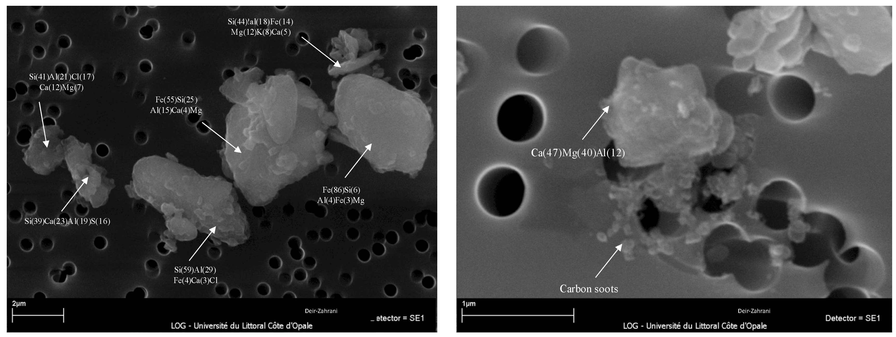
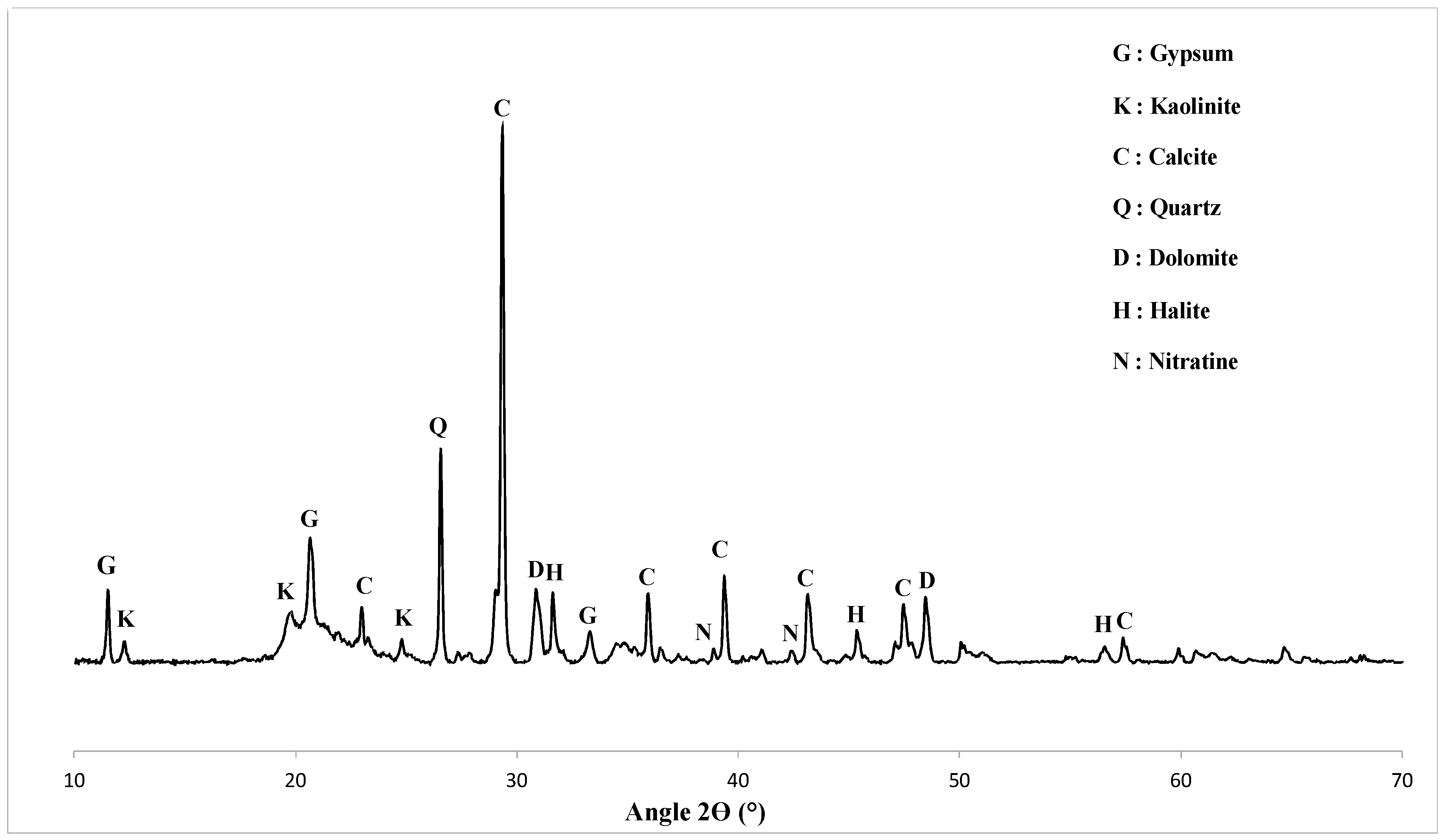
Table 1 represents the average concentrations of major and trace elements, water-soluble ions, and total carbon in fine (PM2.5-0.3) and quasi-ultrafine particles (PM0.3). Results showed that Ca and NO3 were the most abundant elements in both PM2.5-0.3 (24.46% and 22.74%) and PM0.3 (20.40% and 19.32%), respectively. In addition, elements and ions such as Al, SO42−, Fe, NH4+, Mg, Cl, K, and Na were predominant in both PM2.5-0.3 and PM0.3, with different percentages. Scanning electronic microscopy (SEM) analysis also showed the presence of silica (Si), which seems to be predominant in PM2.5-0.3 particles (Figure A1). Other elements and ions such as Ti, Cu, P, Mn, Sr, Ba, Pb, Zn, Cr, Sn, Mo, NO3, SO42−, NH4+, and Cl were also detected in both PM2.5-0.3 and PM0.3, with a percentage ranging from 0.1% to 1%. Sn (0.01%), Co (0.005%), As (0.005%), and Cd (0.002%) were only detected in the quasi-ultrafine fraction (PM0.3). The predominance of elements such as Ca, Al, Fe, Mg, and Ti can be related to the resuspension of soil dust [27]. From XRD analysis, Ca appears in the form of calcite and gypsum. The presence of Ca in high quantities could also be linked to the soil type in Lebanon, which is calcareous. XRD analysis also showed the presence of kaolinite, one of the characteristic minerals of the Lebanese soil type [4,28] (Figure A2). It can explain the presence of Al in PM in addition to the possible influence of Al factories close to the sampling site. On the other hand, elements influenced by vehicular emissions, such as Ba, Ni, Cu, and Zn, were observed. The presence of these elements is linked to the road traffic and probably to the existence of a principal highway in Deir AlZahrani, connecting the capital to all the cities of the South. Particularly, Cu is related to brake emissions, and Zn is linked to tire emissions [29] and the use of lubricating oil [30]. Otherwise, two major ions, NO3 and SO42−, were detected in both types of PM. These ions could originate from the gas-to-particle conversion of NOx and SO2 precursor gases and long-range transport [4,31].

3.2.2. Organic Composition

The 16 priority PAHs listed by the United States Environmental Protection Agency (US EPA) were detected in both PM2.5-0.3 and (mostly) PM0.3 (Table 2). Indeed, the content of PAHs in PM0.3 was about 28 times higher compared to that in PM2.5-0.3 (1007.3 µg/g of PAHs in PM0.3 versus 35.8 µg/g of PAHs in PM2.5-0.3). This difference is the result of anthropogenic activities and typical combustion processes that mainly emit dUFP [4]. In addition, the most abundant PAHs were BbF, InPyr, BghiP, Chry, BaP, and BbK. These PAHs emissions are generally linked to combustion sources, including gasoline motor vehicles, biomass burning, and industrial processes [32]. To better investigate the emission sources of these PAHs, diagnostic ratios were calculated based on already published studies [33,34]. The Fla/(Fla + Pyr) ratio was lower than 0.5 for both PM samples, showing the predominance of diesel emissions (<0.5) [33,34]. In addition, the Fla/Pyr ratio was greater than 0.6, which suggests that the emissions could be related to road traffic (Table A2). Moreover, to evaluate the carcinogenic risk of environmental exposure to these PAHs, concentrations were converted to a BaP-toxic equivalent factor (BaP-TEQ), using factors published elsewhere [35,36]. The potential carcinogenicity of these PM2.5 (PM2.5-0.3 + PM0.3) was equal to 3 ng/m3 BaP-TEQ, higher than the BaP WHO guideline (0.12 ng/m3; cancer risk 1/100,000). The highest BaP-TEQ values were for dibenz[a,h]anthracène: 1.6 ng/m3 BaP-TEQ, Benzo[a]Pyrene: 0.6 ng/m3 BaP-TEQ, Benzo[b]fluorenthene: 0.3 ng/m3 BaP-TEQ, and Indeno [1,2,3-c,d]Pyrene: 0.25 ng/m3 BaP-TEQ. Although this value exceeded the BaP-TEQ values set by the WHO, these differences showed and validated the influence of the presence of important industrial activities and traffic in Deir AlZahrani. An even higher value was previously detected for particles sampled in the same period but on another site in Lebanon (in the southern suburb of Beirut; 8 ng/m3 BaP-TEQ) [4]. These differences show and validate the influence of the presence of more dynamic industrial activities and busier traffic in the suburbs of Beirut.
O- and N-PAHs: 5 O-PAHs and 5 N-PAHs were detected in PM2.5-0.3, and 6 O-PAHs/9 N-PAHs in PM0.3 (Table 2). The total concentrations of N- and O-PAHs were higher in PM0.3 vs. PM2.5-0.3 (O-PAHs: 136 µg/g vs. 9.2 µg/g, respectively; N-PAHs: 13 µg/g vs. 1.3 µg/g, respectively). Among the detected O-PAHs, the most abundant in PM2.5-0.3 were 1,8-naphtalic anhydre (3.7 µg/g; 40% of the total O-PAHs content); followed by 9,10-anthraquinone (2.9 µg/g; 32%) and 7H-benz(de)anthracen-7-one (1.1 µg/g; 12%). However, in PM0.3, the most abundant O-PAHs was 1,8-naphtalic anyhdre (63 µg/g; 46%); followed by 7H-benz(de)anthracen-7-one (33.5 µg/g; 25%); benz(a)anthracen-7,12-dione (18.3 µg/g; 13%), which was not detected in the case of PM2.5-0.3; and 9,10-anthraquinone (11 µg/g; 8%). Concerning the N-PAHs, the major component in PM2.5-0.3 was 1-nitropyrene (0.4 µg/g; 34%), followed by 2-nitrofluorene (0.3 µg/g; 29%) and 6-nitrochrysene (0.3 µg/g; 23%). However, in PM0.3, the most abundant N-PAHs was 7-nitrobenz(a)anthracene, which is not detected in PM2.5-0.3 (2.6 µg/g; 20%); followed by 9-nitroanthracene (2.5 µg/g; 19%); 3-nitrofluoranthene (1.8 µg/g; 14%), also not detected in the PM2.5-0.3; 2-nitroanthracene (1.3 µg/g; 10%); and 6-nitrochrysene (1 µg/g; 8%). It should be noted that 9-nitroanthracene, 1-nitropyrene, 2-nitrofluoranthene, and 3-nitrofluoranthene were shown to be 25% as potent as BaP [37].
Compared to PAHs, O- and N-PAHs have a higher molecular weight, lower vapor pressure, and a higher tendency to be adsorbed onto particulate matter [38,39]. Moreover, these components can be emitted directly into the atmosphere. Also, they can be formed as secondary pollutants via heterogeneous or homogeneous photo-oxidation reaction of PAHs with atmospheric oxidants, photolysis, and thermal transformation [40,41,42]. The emission sources of each O- and N-PAHs are poorly identified and documented in the bibliography. Their determination is challenging because of the coexistence of their primary and/or secondary sources. However, some studies have tried to associate compounds with emission sources, such as the case of 1,8-Naphthalic anhydride, which was reported as a major O-PAHs emitted by residential wood combustion [43,44,45]. Unlike anhydrous1,8-naphthalic emitted directly from a combustion source, the formation of quinones, including 9,10- Anthraquinone, has been demonstrated in certain studies as products of the oxidation of PAHs by photolysis or reactions with O3, NO3, and OH [39,46,47,48]. Among N-PAHs, 1-NP is formed and emitted directly into the atmosphere during fossil fuel combustion at high temperatures and has been proposed as a chemical marker for diesel exhaust particles [49]. IARC also categorized it in Group 2 A (probably carcinogenic to humans). Concerning 9-NAnt, it has been reported to be emitted from direct sources, predominantly diesel engines. It can also be formed in the atmosphere because of reactions with OH radicals followed by heterogeneous nitration reactions with anthracene during the day and reactions with NO3 at night [50].
PCB, PCDD, and PCDF: All these chemical compounds were also analyzed and quantified in both PM2.5-0.3 and PM0.3. The results showed that PM2.5-0.3 was more concentrated in PCDD and PCB than PM0.3. However, PM0.3 was more concentrated in PCDD and PCB indicators than PM2.5-0.3 (Table 3). Concentrations of PCDD, PCDF, and PCB were also quantified on two other sites in Lebanon (Zouk Mikael and Fiaa) by [51]. The total concentrations of these compounds in the whole PM2.5 were much higher in Deir AlZahrani compared to the two other sites (PCDD: 399.43 fg/m3 vs. 81.1 ± 38.7 and 52.0 ± 21.7 fg/m3; PCDF: 333.33 fg/m3 vs. 82.5 ± 35.1 and 71.8 ± 36.7 fg/m3; PCB: 7043.95 fg/m3 vs. 454.8 ± 202 and 586.3 ± 128 fg/m3, respectively). These compounds are generally produced from anthropogenic activities such as forest fires or domestic and hospital waste incineration [52]. In this study, the presence of PCDD, PCDF, and PCB could be attributed to the uncontrolled domestic waste incineration in the Kfour valley, which is relatively close to the sampling site.
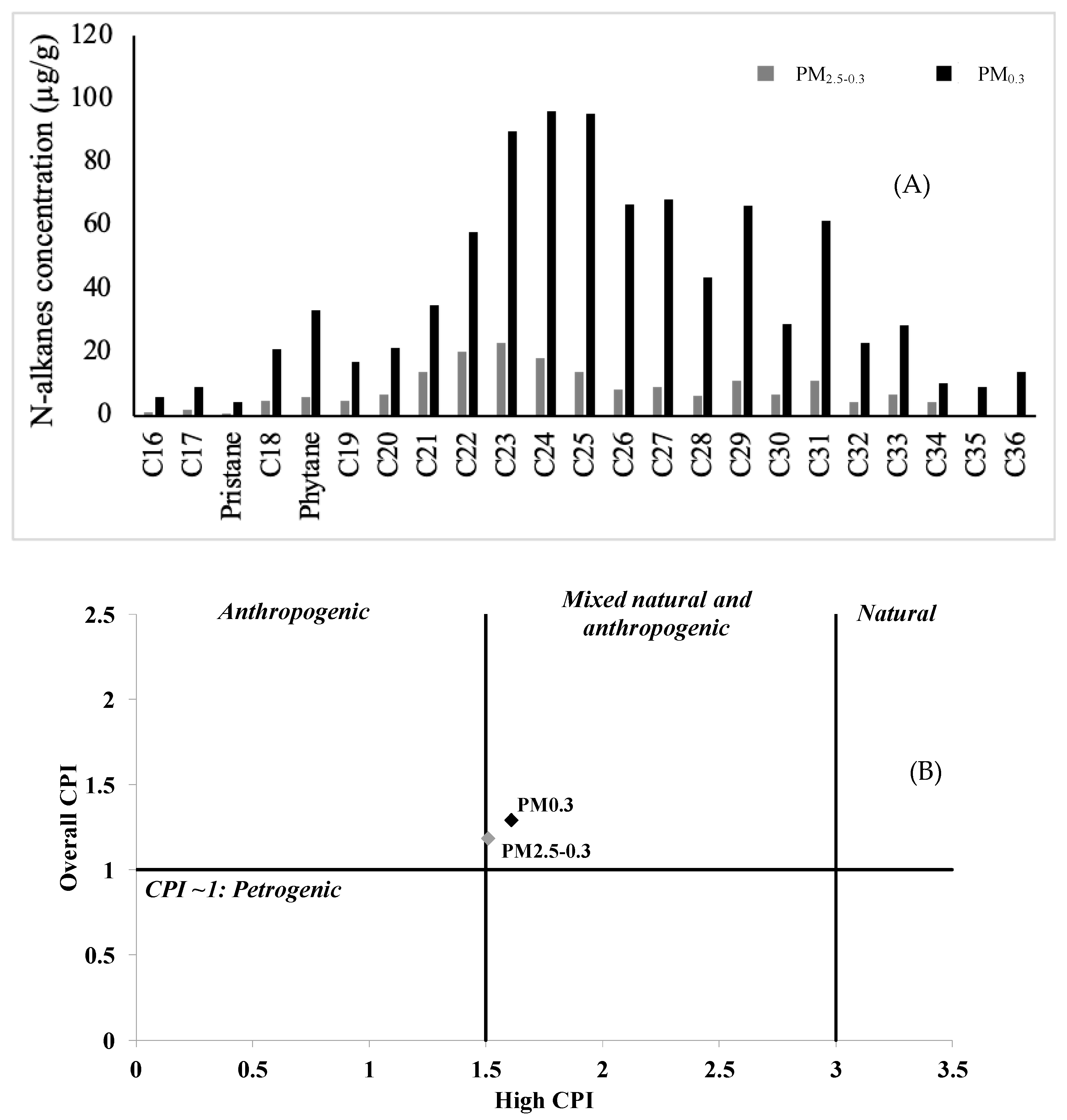
Paraffins (n-alkanes): Paraffins, also known as n-alkanes, were quantified in PM2.5-0.3 and PM0.3, and the results are presented in Figure A3. Concentrations of paraffins were about 5 times higher in PM0.3 (906 µg/g; 14 ng/m3) than in PM2.5-0.3 (181 µg/g; 2.7 ng/m3). This is also true for other organic compounds, such as PAHs and their derivatives.
The carbon preference index (CPI), defined as the concentration ratio of odd to even carbon number n-alkanes and the carbon maximum number (Cmax), has been widely used in the literature to evaluate biogenic and anthropogenic contributions to organic aerosol [4,53,54,55]. Cmax for PM2.5-0.3 was C34 and C36 for PM0.3, indicating that the n-alkanes primarily originated from terrestrial plants, especially epicuticular waxes. Values of overall and high CPI were equal to 1.18 and 1.51 for PM2.5-0.3, respectively, and to 1.61 and 1.29 for PM0.3, respectively. These data suggested that the n-alkanes detected in the two samples originated from both natural and anthropogenic sources (1.5 < high CPI < 3 and overall CPI > 1).

3.3. Evaluation of PM2.5 Toxicity:

3.3.1. ATP Production and LDH Activity

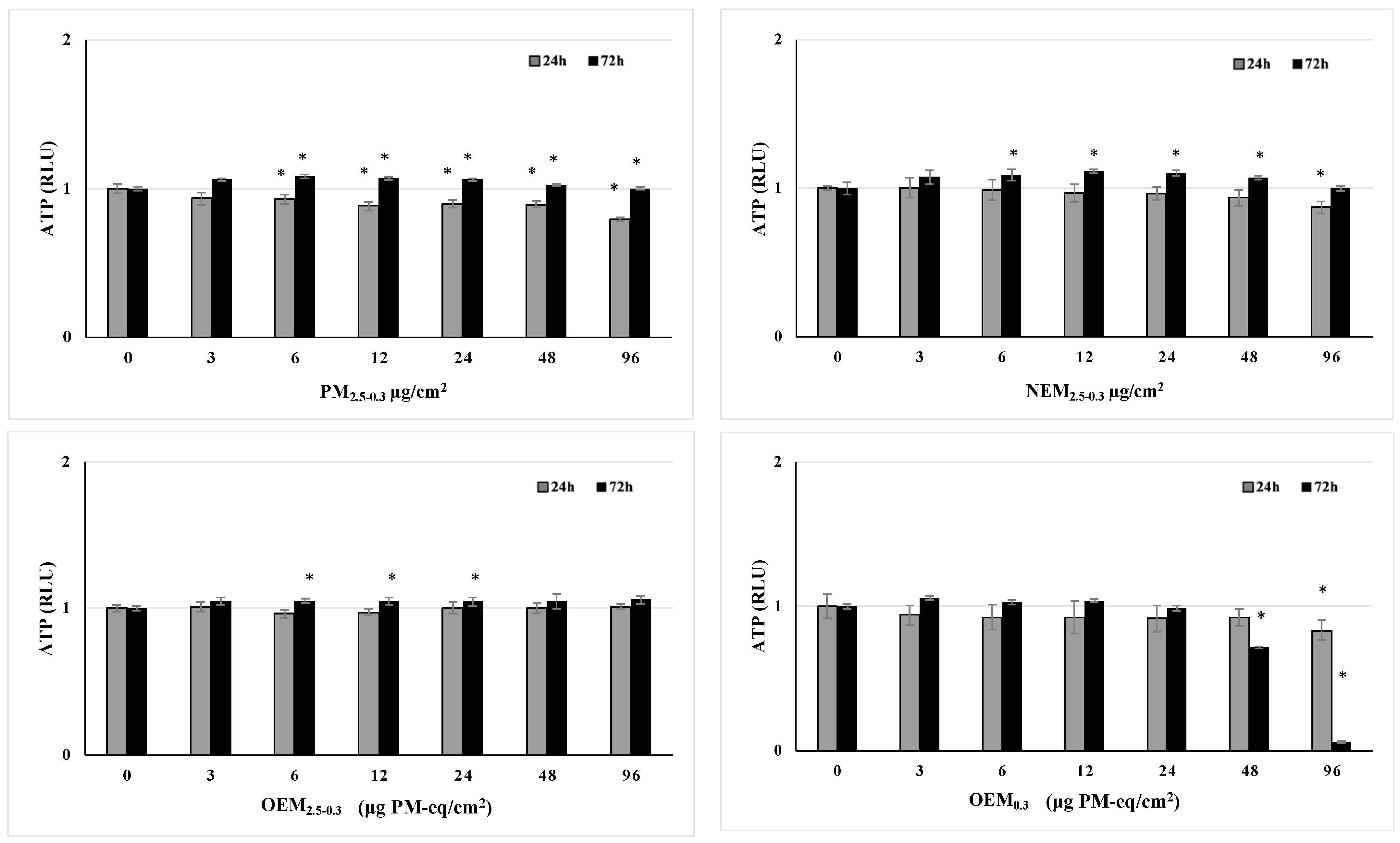
In this study, cell viability was evaluated through the quantification of intracellular ATP produced by mitochondria, which are special double-membraned intracellular compartments, popularly known as the ‘powerhouses of the cell’ because they provide ATP used to fuel cellular biochemical reactions [56]. Alterations in cellular metabolism are linked to impaired mitochondrial functions such as a decrease in ATP production and an increase in reaction oxygen species production [57]. In this study, BEAS-2B cells were exposed to the different prepared suspensions (PM2.5-0.3, OEM2.5-0.3, NEM2.5-0.3, and OEM0.3) at different concentrations (3, 6, 12, 24, 48, and 96 µg/cm2 or μg PM-eq/cm2) for 24 and 72 h (Figure 2). A significant decrease in ATP production was observed following BEAS-2B exposure to 6, 12, 24, 48, and 96 µg/cm2 of PM2.5-0.3 for 24 and 72 h.
These data showed that PM2.5-0.3 affected cellular metabolism, resulting in a decrease in the cellular ATP concentration at short times (24 h), and this perturbation was more pronounced when exposed to the highest concentration of PM2.5-0.3 (96 µg/cm2). However, after 72 h of exposure, BEAS-2B cells were trying to compensate for the decrease in ATP and to eliminate PM2.5-0.3, which can explain the increase in cellular ATP. In addition, exposure of BEAS-2B to the NEM2.5-0.3 and OEM2.5-0.3 fractions in the same conditions showed different cellular responses compared to the whole PM2.5-0.3. Concerning the exposure to the NEM2.5-0.3 fraction, a significant increase of the cellular ATP was observed following 72 h of exposure to 6, 12, 24, and 48 μg PM-eq/cm2, in addition to a significant decrease following 24 h of exposure to the highest concentration (96 μg PM-eq/cm2). Regarding OEM2.5-0.3, only a significant increase was observed following the exposure to 6, 12, 24, and 48 μg PM-eq/cm2 for 72 h. Taken together, these results showed different cellular responses when exposed to the whole PM vs. its fractions (NEM2.5-0.3 and OEM2.5-0.3). In our conditions, alteration in cellular metabolism was mostly observed in the case of the native PM2.5-0.3, and the effect was shown to be dose-dependent in the case of PM2.5-0.3 (for 24 and 72 h) and NEM2.5-0.3 (for 72 h). Regarding OEM0.3, a decrease in cellular ATP was observed following 24 h of exposure to 96 Eq. PM/cm2 and 72 h to 48 and 96 Eq. PM/cm2. These results showed a time and concentration-dependent effect of OEM0.3. In addition, the effect of OEM0.3 was more pronounced than that of OEM2.5-0.3. [4] demonstrated similar findings following the exposure of BEAS-2B cells to OEM2.5-0.3 and OEM0.3 collected from Beirut. This difference could be related to the organic composition of these two fractions, which are more concentrated in OEM0.3 (cf. 3.2.2. organic composition).
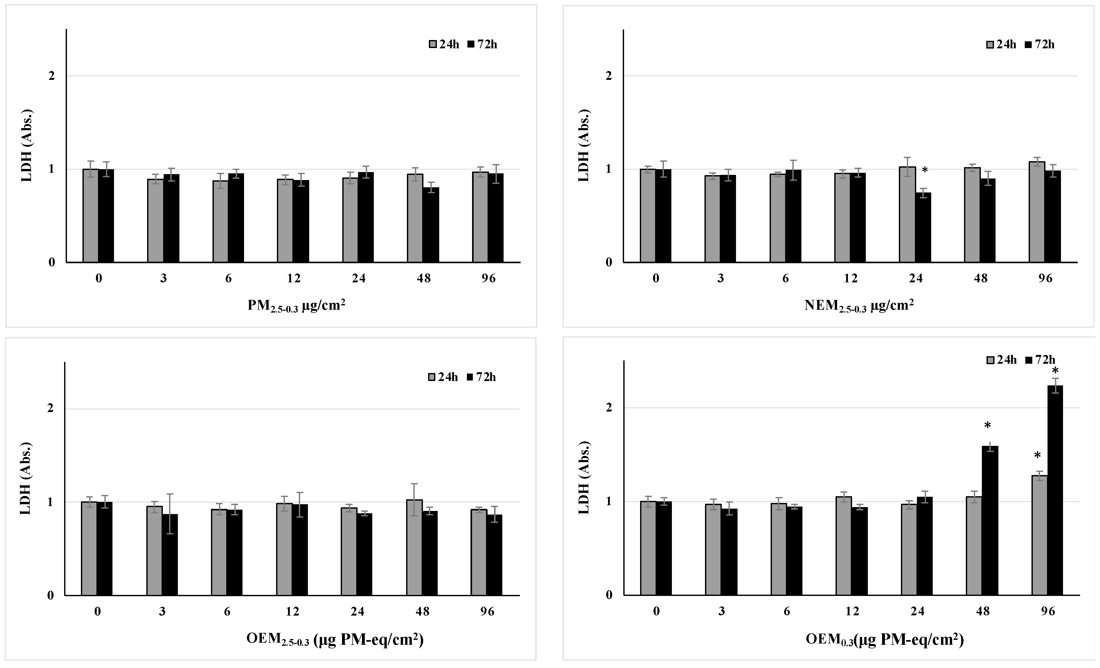
Global cytotoxicity was also established by studying LDH activities in cell-free culture supernatants (Figure 3). The data show that the exposure of cells to different pollutants induces a decrease in the activity of LDH after exposure to OEM0.3, 24 and 72 h to 96 µg PM/cm2 or μg PM-eq/cm2 and only 72 h to 48 Eq. PM/cm2 showed slight and transitory cytotoxicity after their exposure to the highest concentrations of OEM0.3. Like ATP activities, the effect of OEM0.3 was more pronounced than that of OEM2.5-0.3, related to the presence of more organic compounds and explained by chemical organic fraction characterization.

3.3.2. Metabolic Activation of PAHs

PAHs represent toxic compounds, and their toxicity occurs after their metabolic activation. Depending on their nature, metabolites obtained after this activation will interact with cellular macromolecules and induce dysfunction in the body. Briefly, once in the cell, the PAHs binds to its cytoplasmic receptor, the AhR, inducing its activation and migration into the nucleus, where it will bind with another nuclear protein called ARNT (AhR nuclear translocator). Thus, the new complex is formed, is bonded to specific regions of DNA called “xenobiotic response elements” (ERX) and induces the expression of specific target genes involved in the PAHs metabolization process (e.g., CYP1A1, CYP1B1, EPHX1, and GSTA4).
In this study, we evaluated the expression of the above-mentioned genes in addition to AHRR, which functions as a negative feedback modulator by repressing AhR-dependent gene expression following BEAS-2B exposure to C1 = 3 and C2 = 12 µg/cm2 or μg eqPM/cm2 for 6 and 24 h. The results are represented in Table 4. Exposure of BEAS-2B to the prepared fractions showed different responses concerning the gene expression of AHR, ARNT, AHRR, CYP1A1, CYP1B1, EPHX1, and GSTA-4. First, after 6 h of exposure to the whole PM2.5-0.3, significant increases in the gene expression of AHRR and CYP1B1 were observed for the highest concentration (C2 = 12 µg/cm2). Then, after 24 h of exposure, a significant increase in the gene expression of CYP1A1 was observed when exposed to C1. Moreover, all the genes studied showed a significant increase in gene expression when exposed to a C2 concentration for 24 h. These results showed that BEAS-2B cells represented metabolically competent cells, since they were able to phagocyte the PM2.5-0.3 and induce the metabolic activation of organic compounds, as supported by other authors [4,9,58,59]. However, the cellular response to the NEM2.5-0.3 and OEM2.5-0.3 fractions was entirely different from that of PM2.5-0.3. No significant increase in the expression of any of the studied genes was observed following exposure to these two fractions. This is quite normal in the case of NEM2.5-0.3, which must not contain any organic compounds following DCM extraction. Nevertheless, the absence of induction of the metabolic activation pathway by OEM2.5-0.3, which normally contained all the organic compounds extractable with DMSO, could be somewhat disturbing. However, it can be assumed that this absence could be due to the difference between the bioavailability of the organic compounds in the organic extract versus that in the total particles. Here, two hypotheses can be drawn. The first one is based on earlier metabolic activation of PAHs adsorbed on the PM, as the particles could represent a vector facilitating the entry of PAHs into the cells. The second one is based on the earlier bioactivation of PAHs in the organic extract, where they were free in the medium and could also penetrate easily and quickly, unlike when the PAHs were adsorbed on the particles and also required more time to be detached from the core particles to enter the biotransformation cycle [4,60,61].
Concerning the effect of the OEM0.3, the results showed a significant increase in AHRR and CYP1B1 gene expression after 6 h of exposure to C2, of CYP1A1 after 24 h of exposure to C1, and of ARNT, CYP1A1, CYP1B1, and GSTA4 after 24 h of exposure to C2. The results therefore showed a difference in the cellular response to the two organic extracts OEM0.3 and OEM2.5-0.3. This could be explained by an earlier or prolonged metabolic activation of OEM0.3 vs. OEM2.5-0.3, since it contained many more organic compounds (28-fold increases of PAHs in PM0.3 vs. PM2.5-0.3) [4].
Taken together, these results showed the capacity of organic compounds to induce the AHR metabolic activation pathway in BEAS-2B cells. This induction was only shown by fractions containing organic compounds (whole PM2.5-0.3 and OEM). Knowing that the concentration of organic compounds is the same in PM2.5-0.3 and OEM, it was important to consider that the induction of this pathway by each fraction is linked to the bioavailability of the organic compounds in each fraction rather than their concentrations. [4] demonstrated similar findings following exposure of BEAS-2B cells to PM2.5-0.3 and OEM0.3, with more pronounced results observed for particles from Beirut compared to those from Deir AlZahrani. This variance aligns with findings regarding the chemical composition of PM2.5-0.3 and PM0.3 collected in Beirut by [4]. Particles collected in Beirut exhibited a higher concentration of toxic organic compounds compared to those sampled in Deir AlZahrani [4].

3.3.3. Genotoxicity Induced by Atmospheric PM

PM2.5 is classified as carcinogenic to humans [62]. Since its composition is extremely complex, it was challenging to identify which fraction from the fine particles could be mainly responsible for the genotoxic effects. In this context, we have studied protein expression of several biomarkers involved in the genotoxicity pathway, such as phosphorylated p-CHK1, p-CHK2, p-H2AX, and p-P53, and total proteins, ATR, MDM2, and P21 following the exposure of BEAS-2B to two concentrations C1 = 3 and C2 = 12 µg/cm2 for 24 and 72 h (Table 5). The results showed that exposure to the entire PM2.5-0.3 induced only a significant decrease in phosphorylated H2AX in BEAS-2B exposed for 24 h to C1 and C2. In addition, BEAS-2B exposed to the NEM2.5-0.3 fraction also reduced its ATR expression when exposed to C1 for 72 h and in phosphorylated H2AX when exposed for 72 h to C2. Conversely, no effect was observed when BEAS-2B cells were exposed to OEM2.5-0.3. However, a significant protein expression increase was observed only when the cells were exposed to OEM0.3. Results showed a significant increase in ATR and CHK1 by BEAS-2B exposed to C2 for 72h; of P21, P53, and MDM2 by BEAS-2B exposed to C2 for 24 and 72h; and for H2AX by BEAS-2B exposed to C2 for 72 h. These results showed that the extractable organic fraction of PM0.3, the most concentrated in organic compounds, was the only one capable of inducing the expression of proteins related to cell cycle arrest and DNA damage under the studied conditions. On the other hand, the decrease in these biomarkers in the case of exposure to PM2.5-0.3 and its two fractions could be attributed to a greater repair activity of the cells. The same results concerning the decrease in protein expression have been recently supported by other authors [4,9,59,63]. Keeping DNA stable in eukaryotic cells involves a network of processes such as copying DNA, solving DNA issues, and managing cell cycles. The ATM and ATR kinases were key in overseeing this network when DNA was damaged. Both in vivo and in vitro investigations indicated that ATM and ATR exhibited distinct DNA damage-specific responses. ATM predominantly responded to double-stranded DNA breaks (DSBs), while ATR’s activation encompassed a wider range of DNA damage, including DSBs and various lesions that disrupt DNA replication. These results showed a significant increase in ATR expression after exposure to OEM0.3, and subsequently CHK1, P21, P53, MDM2, and H2AX expression, involved in cell cycle arrest, apoptosis, and DNA damage repair [4,64,65]. [4] showed also that the quasi-ultrafine particles were the most genotoxic due to the predominance of organic compounds such as PAHs in a fraction of this size [4].

3.3.4. Inflammation: TNF-α and IL-6 Secretion

The inhalation and deposition of PM2.5 in the lungs trigger oxidative stress and inflammatory responses that provokes a wide range of cytokine secretion [66,67,68]. As PM represents a mixture of organic and inorganic components, an evaluation of the capacity of each fraction (PM2.5-0.3, NEM2.5-0.3, OEM2.5-0.3, and OEM0.3) to stimulate the production of inflammatory cytokines was carried out. BEAS-2B cells were exposed to two concentrations C1 = 3 µg/cm2 or μg eqPM/cm2 and C2 = 12 µg/cm2 or μg eqPM/cm2 of each suspension, for 6 and 24 h (Figure 4). Results showed the capacity of all fractions to stimulate the production of two cytokines, TNF-α and IL-6, at 6 h rather than at 24 h. The organic extracts (OEM) containing only organic compounds, as well as the NEM containing the inorganic fraction, both provoke the production of IL-6 and TNF-α by BEAS-2B. Numerous experimental studies conducted both in vitro and in vivo have highlighted the detrimental pulmonary health impacts caused by PM2.5 derived from air pollution. These effects predominantly arose from PM2.5 capacity to instigate essential mechanisms of toxicity, notably oxidative stress-induced inflammation in the airways [4,10,69,70]. Exposure of BEAS-2B, NHBE, and particularly COPD-DHBE to PM2.5 showed a cytosolic ROS overproduction, inducing oxidative damage and activating oxygen-sensitive NRF2 and NF-kB signaling pathways [69]. Accordingly, exposure to a mixture of PAHs and diesel exhaust particles induced an increase in gene and protein expressions of IL-6 and TNF-α [68,71,72]. In addition, a previous study demonstrated that exposure to certain metals (Ag, Al, Ba, K, Li, Ni, Sn, and V) was significantly associated with increased inflammatory markers such as IL-6 as well as oxidative stress markers [73]. Taken together, the already published studies together with this one show that all the components of PM can participate in the inflammatory process [66,74,75].

4. Conclusions

The main objectives of this study were to compare the physico-chemical properties and toxicity of fine and quasi-ultrafine particles and to target the fraction within PM responsible for the known toxic effects.
To achieve these objectives, PM2.5-0.3 and PM0.3 were sampled in Southern Lebanon’s Deir AlZahrani, and an extraction of the different parts of PM2.5-0.3 was carried out while obtaining native PM2.5-0.3, OEM2.5-0.3, and NEM2.5-0.3. The chemical analysis of both PM2.5-0.3 and PM0.3 showed the presence of elements related to the resuspension of soil dust (Ca, Al, Fe, Mg, and Ti) in addition to other elements, related to traffic emissions (Zn and Cu). In addition, the concentration of organic compounds such as PAHs, O-, and N-PAHs was higher in PM0.3 than PM2.5-0.3, showing the influence of anthropogenic activities and combustion on the emission of ultrafine particles.
Toxicological studies showed that the induction of the metabolic activation of PAHs was delayed by PM2.5-0.3 rather than OEM2.5-0.3. This delay is probably related to the bioavailability of organic compounds. Furthermore, only OEM0.3 was able to induce an increase in the protein expression of ATR, P21, P53, MDM2, and H2AX, all implicated in the genotoxic pathway. This could be related to the organic composition of PM0.3, which is more concentrated in PAHs and derivatives, compared to PM2.5-0.3.
These results indicate that UFPs seem to be of great concern to human health because they are concentrated in organic compounds and much more toxic than fine particles. These PM2.5-0.3 and their toxicity must be taken with caution into consideration when setting emission limits for concentrations of particles.

Author Contributions

Conceptualization, G.B, M.C., A.V., I.A., O.S., F.C., M.R., D.C., J.-M.L.G., F.L. and G.G; methodology, G.B., M.C., A.V., I.A., F.L. and G.G.; software, G.B., M.C., A.V., I.A., F.C., F.L. and G.G.; validation, A.V., I.A., M.R., D.C., F.L. and G.G.; formal analysis, G.B. and M.C.; investigation, G.B., M.C., A.V., I.A., F.L. and G.G.; writing—original draft preparation, G.B. and M.C.; writing—review and editing, A.V., I.A., F.L. and G.G.; supervision, A.V, I.A., F.L. and G.G.; project administration, M.R., D.C., F.L. and G.G.; funding acquisition, A.V., I.A., F.L. and G.G. All authors have read and agreed to the published version of the manuscript.

Funding

The research described in this article was financially supported by the National Council for Scientific Research of Lebanon (CNRS-L) and the Agence Universitaire de la Francophonie (AUF) (3R-Lebanon Program), the Unité de Chimie Environnementale et Interactions sur le Vivant (UCEIV-EA4492) and IMPacts de l’Environnement Chimique sur la Santé (IMPECS-ULR4483). UCEIV and IMPECS participate in the CLIMIBIO project, which is financially supported by the Hauts-de-France Region Council, the French Ministry of Higher Education and Research, and the European Regional Development Funds. The authors would like to acknowledge the National Council for Scientific Research of Lebanon (CNRS-L) and the ULCO for the doctoral fellowships of G.B and M.C.

Institutional Review Board Statement

Not Applicable.

Informed Consent Statement

Not applicable.

Data Availability Statement

The data presented in this study are available on request from the corresponding author. The data are not publicly available as it is a part of a thesis study of a student.

Acknowledgments

The authors would like to thank Adam Hachimi (Micropollutants Technologie, Saint-Julien-les-Metz, France) for the determination of dioxins, furans, and PCB within air pollution PM2.5-0.3 and PM0.3, Dorothée Dewaele and Paul Genevray (Centre Commun de Mesures, ULCO, Dunkerque, France) for their help in the ICP–AES and CHN analysis, and Yann Landkocz (UCEIV-EA4492, ULCO, Dunkerque, France) for his help in the N-PAHs and O-PAHs analysis. Ghidaa Badran dedicates this paper to the people of her hometown, Deir AlZahrani, hoping that this first publication will be followed by other scientific studies that will urge the authorities to take action and implement measures for a cleaner environment in this city.

Conflicts of Interest

The authors declare no conflicts of interest.

Abbreviations

1-NPyr1-nitropyrene
9-FluO9-Fluorenone
AceAcenaphthalene
AcyAcenaphthylene
AntAnthracene
ATPAdenosine triphosphate
BaABenz[a]anthracene
BaPBenzo[a]pyrene
BbFBenzo[b]fluoranthene
BghiPBenzo[g,h,i]perylene
BkFBenzo[k]fluoranthene
ChrChrysene
DahADibenz[a,h]anthracene
FlaFluoranthene
FluFluorene
InPyIndeno[1,2,3-c,d]pyrene
LDHLactate dehydrogenase
N-PAHsnitrated-PAHs
NapNaphthalene
OEMorganic extractable matter
NEMnon-extractable matter
O-PAHsoxygenated-PAHs
PAHspolycyclic aromatic hydrocarbons
PhePhenanthrene
PM2.5-0.3particles with an equivalent aerodynamic diameter between 0.3 and 2.5 mm
PM0.3particles with an equivalent aerodynamic diameter below 0.3 mm (quasi-ultrafine particles)
PyrPyrene
ROSreactive oxygen species;

Appendix A

Table A1. A summary of the different physico-chemical analyses.
Physico-Chemical AnalysisTechniquesAnalyzed Samples
PM2.5-0.3PM0.3
PM morphology and individual particle compositionScanning electron microscopy coupled with energy-dispersive X-ray (SEM-EDX)-
Cristalline phasesX-ray diffraction -
Elements and water-soluble ions Inductively coupled plasma atomic emission spectroscopy and ionic chromatography
Organic compounds PAHsGas chromatography–mass spectrometry (GC–MS)
N- and O-PAHsHigh-resolution gas chromatography and high-resolutionmass spectrometry
PCDD, PCDF, and PCBHigh-resolution gas chromatography and high-resolutionmass spectrometry
n-alkanesGas chromatography–mass spectrometry (GC–MS)
✓: Applied to.
Figure A1. Particles (PM2.5-0.3) observed by scanning electron microscopy coupled with energy-dispersive X-ray (left) and individual particle composition (right) (mass % into brackets). Si: silicium. Al: aluminum. Fe: iron. S: sulfur. Ca: calcium. Mg: magnesium. K: potassium. Cl: chlorine.
Atmosphere 15 00811 g0a1
Figure A2. X-ray diffraction patterns of PM2.5 collected at Deir AlZahrani.
Atmosphere 15 00811 g0a2
Table A2. PAHs diagnostic ratios calculated for PM2.5-0.3 and PM0.3 collected at Deir Al Zahrani and compared to the bibliography to determine their possible emission sources.
PAHs Diagnosis RatioPM2.5-0.3PM0.3 Characteristic Ratio Values and Sources
InPy/(InPy+B(g,h,i)Pe (a)0.590.500.18: Cars 0.62: Wood burning 0.35–0.70: Diesel burning
Fla/(Fla+Pyr) (a)0.470.45<0.5: Gasoline >0.5: Diesel
B[a]P/(B[a]P+Chry) (b)0.290.38>0.35: Vehicular emission0.2–0.35: Coal combustion
B[b]F/B[k]F (a)4.012.89>0.5: Diesel
B[a]P/B[g,h,i]Pe (a)0.530.470.5–0.6: Traffic emission >1.25: Brown coal or lignite
Anth/(Anth+Phe) (b)0.090.30>0.1: Pyrogenic<0.1: Petrogenic
InPy/B(g,h,i)Pe (a)1.421.00<0.4: Gasoline ~1: Diesel
Fla/(Fla+Pyr) (b)0.480.46<0.4: Petrogenic0.4–0.5: Fossil fuel combustion >0.5: Grass wood coal combustion
Fla/Pyr (b)0.940.84<0.6: Non-traffic emission >0.6: Traffic emission
CPAHs/TPAHs (a)0.920.95≈1 (Combustion)
(a): [33]; (b): [34].
Figure A3. N-alkane concentrations (µg/g) determined in PM2.5-0.3 and in PM0.3 (A), and identification of n-alkane sources using overall CPI and high CPI in PM2.5-0.3 and PM0.3 (B).
Atmosphere 15 00811 g0a3

References

  1. Cohen, A.J.; Brauer, M.; Burnett, R.; Anderson, H.R.; Frostard, J.; Estep, K.; Balakrishnan, K.; Brunekreef, B.; Dandona, L.; Dandona, R.; et al. Estimates and 25-year trends of the global burden of disease attributable to ambient air pollution: An analysis of data from the Global Burden of Diseases Study 2015. Lancet 2017, 389, 1907–1918. [Google Scholar] [CrossRef] [PubMed]
  2. Raaschou-Nielsen, O.; Antonsen, S.; Agerbo, E.; Hvidtfeldt, U.A.; Geels, C.; Frohn, L.M.; Christensen, J.H.; Sigsgaard, T.; Brandt, J.; Pedersen, C.B. PM2.5 air pollution components and mortality in Denmark. Environ. Int. 2023, 171, 107685. [Google Scholar] [CrossRef] [PubMed]
  3. IARC Working Group on the Evaluation of Carcinogenic Risks to Humans. Outdoor air pollution. IARC Monogr. Eval. Carcinog. Risks Hum. 2016, 109, 9–444. [Google Scholar]
  4. Badran, G.; Ledoux, F.; Verdin, A.; Abbas, I.; Roumie, M.; Genevray, P.; Landkocz, Y.; Lo Guidice, J.-M.; Garçon, G.; Courcot, D. Toxicity of fine and quasi-ultrafine particles: Focus on the effects of organic extractable and non-extractable matter fractions. Chemosphere 2020, 243, 125440. [Google Scholar] [CrossRef] [PubMed]
  5. Shen, F.; Zheng, Y.; Niu, M.; Zhou, F.; Wu, Y.; Wang, J.; Zhu, T.; Wu, Y.; Wu, Z.; Hu, M.; et al. Characteristics of biological particulate matters at urban and rural sites in the North China Plain. Environ. Pollut. 2019, 253, 569–577. [Google Scholar] [CrossRef] [PubMed]
  6. Xing, Y.F.; Xu, Y.H.; Shi, M.H.; Lian, Y.X. The impact of PM2.5 on the human respiratory system. J. Thorac. Dis. 2016, 8, 69–74. [Google Scholar] [CrossRef]
  7. Cooper, D.M.; Loxham, M. Particulate matter and the airway epithelium: The special case of the underground? Eur. Respir. Rev. 2019, 28, 190066. [Google Scholar] [CrossRef] [PubMed]
  8. Park, S.R.; Lee, J.W.; Kim, S.K.; Yu, W.J.; Lee, S.J.; Kim, D.; Kim, K.W.; Jung, J.W.; Hong, I.S. The impact of fine particulate matter (PM) on various beneficial functions of human endometrial stem cells through its key regulator SERPINB2. Exp. Mol. Med. 2021, 53, 1850–1865. [Google Scholar] [CrossRef]
  9. Abbas, I.; Badran, G.; Verdin, A.; Ledoux, F.; Roumie, M.; Lo Guidice, J.-M.; Courcot, D.; Garçon, G. In vitro evaluation of organic extractable matter from ambient PM2.5 using human bronchial epithelial BEAS-2B cells: Cytotoxicity, oxidative stress, pro-inflammatory response, genotoxicity, and cell cycle deregulation. Environ. Res. 2019, 171, 510–522. [Google Scholar] [CrossRef]
  10. Badran, G.; Verdin, A.; Grare, C.; Abbas, I.; Achour, D.; Ledoux, F.; Roumie Cazier, F.; Courcot, D.; Jean-Marc Lo Guidice, J.-M.; Garçon, G. Toxicological appraisal of the chemical fractions of ambient fine (PM2.5-0.3) and quasi-ultrafine (PM0.3) particles in human bronchial epithelial BEAS-2B cells. Environ. Pollut. 2020, 263, 114620. [Google Scholar] [CrossRef]
  11. Pang, P.; Huang, W.; Luo, X.S.; Chen, Q.; Zhao, Z.; Tang, M.; Hong, Y.; Chen, J.; Li, H. In-vitro human lung cell injuries induced by urban PM2.5 during a severe air pollution episode: Variations associated with particle components. Ecotoxicol. Environ. Saf. 2020, 206, 111406. [Google Scholar] [CrossRef] [PubMed]
  12. Cristaldi, A.; Conti, G.O.; Pellitteri, R.; La Cognata, V.; Copat, C.; Pulvirenti, E.; Grasso, A.; Fiore, M.; Cavallaro, S.; Dell’Albani, P.; et al. In vitro exposure to PM2.5 of olfactory Ensheathing cells and SH-SY5Y cells and possible association with neurodegenerative processes. Environ. Res. 2024, 241, 117575. [Google Scholar] [CrossRef] [PubMed]
  13. Barzgar, F.; Sadeghi-Mohammadi, S.; Aftabi, Y.; Zarredar, H.; Shakerkhatibi, M.; Sarbakhsh, P.; Gholampour, A. Oxidative stress indices induced by industrial and urban PM2.5-bound metals in A549 cells. Sci. Total Environ. 2023, 877, 162726. [Google Scholar] [CrossRef] [PubMed]
  14. Wang, Q.; Liu, S. The Effects and Pathogenesis of PM2.5 and Its Components on Chronic Obstructive Pulmonary Disease. Int. J. Chron. Obs. Pulmon Dis. 2023, 18, 493–506. [Google Scholar] [CrossRef] [PubMed]
  15. Honda, A.; Fukushima, W.; Oishi, M.; Tsuji, K.; Sawahara, T.; Hayashi, T.; Kudo, H.; Kashima, Y.; Takahashi, K.; Sasaki, H.; et al. Effects of Components of PM2.5 Collected in Japan on the Respiratory and Immune Systems. Int. J. Toxicol. 2017, 36, 153–164. [Google Scholar] [CrossRef] [PubMed]
  16. Rodriguez-Cotto, R.I.; Ortiz-Martinez, M.G.; Rivera-Ramirez, E.; Mateus, V.L.; Amaral, B.S.; Jiménez-Vélez, B.D.; Gioda, A. Particle pollution in Rio de Janeiro, Brazil: Increase and decrease of pro-inflammatory cytokines IL-6 and IL-8 in human lung cells. Environ. Pollut. 2014, 194, 112–120. [Google Scholar] [CrossRef] [PubMed]
  17. Afif, C.; Chélala, C.; Borbon, A.; Abboud, M.; Adjizian-Gérard, J.; Farah, W.; Jambert, C.; Zaarour, R.; Badaro Saliba, N.; Perros, P.E.; et al. SO2 in Beirut: Air quality implication and effects of local emissions and long-range transport. Air Qual. Atmos. Health 2008, 1, 167–178. [Google Scholar] [CrossRef]
  18. Borgie, M.; Dagher, Z.; Ledoux, F.; Verdin, A.; Cazier, F.; Martin, P.; Hachimi, A.; Shirali, P.; Greige-Gerges, H.; Courcot, D. Comparison between ultrafine and fine particulate matter collected in Lebanon: Chemical characterization, in vitro cytotoxic effects and metabolizing enzymes gene expression in human bronchial epithelial cells. Environ. Pollut. Barking Essex 2015, 205, 250–260. [Google Scholar] [CrossRef] [PubMed]
  19. Kfoury, A.; Ledoux, F.; Khoury, B.E.; Nakat, H.E.; Nouali, H.; Cazier, F.; Abi-Aad, E.; Aboukaïs, A. A study of the inorganic chemical compositionof atmospheric particulate matterin the region of Chekka, North Lebanon. Leban. Sci. J. 2009, 10, 3–16. [Google Scholar]
  20. Melki, P.N.; Ledoux, F.; Aouad, S.; Billet, S.; El Khoury, B.; Landkocz, Y.; Abdel-Massih, R.M.; Courcot, D. Physicochemical characteristics, mutagenicity and genotoxicity of airborne particles under industrial and rural influences in Northern Lebanon. Environ. Sci. Pollut. Res. Int. 2017, 24, 18782–18797. [Google Scholar] [CrossRef]
  21. Waked, A.; Afif, C.; Seigneur, C. Assessment of source contributions to air pollution in Beirut, Lebanon: A comparison of source-based and tracer-based modeling approaches. Air Qual. Atmos. Health 2015, 8, 495–505. [Google Scholar] [CrossRef]
  22. Leclercq, B.; Alleman, L.Y.; Perdrix, E.; Riffault, V.; Happillon, M.; Strecker, A.; Lo-Guidice, J.-M.; Garçon, G.; Coddeville, P. Particulate metal bioaccessibility in physiological fluids and cell culture media: Toxicological perspectives. Environ. Res. 2017, 156, 148–157. [Google Scholar] [CrossRef] [PubMed]
  23. Waked, A.; Seigneur, C.; Couvidat, F.; Kim, Y.; Sartelet, K.; Afif, C.; Borbon, A.; Formenti, P.; Sauvage, S. Modeling air pollution in Lebanon: Evaluation at a suburban site in Beirut during summer. Atmos. Chem. Phys. 2013, 13, 5873–5886. [Google Scholar] [CrossRef]
  24. Melki, P. Health Impact of Airborne Particulate Matter in Northern Lebanon: From a Pilot Epidemiological Study to Physico-Chemical Characterization and Toxicological Effects Assessment. Ph.D. Thesis, Université du Littoral Côte d’Opale, Université de Balamand, Tripoli, Liban, 2017. [Google Scholar]
  25. Nakhlé, M.M.; Farah, W.; Ziadé, N.; Abboud, M.; Salameh, D.; Annesi-Maesanno, I. Short-term relationships between emergency hospital admissions for respiratory and cardiovascular diseases and fine particulate air pollution in Beirut, Lebanon. Environ. Monit. Assess. 2015, 187, 196. [Google Scholar] [CrossRef] [PubMed]
  26. Faour, A.; Abboud, M.; Germanos, G.; Wehbeh, F. Assessment of the exposure to PM2.5 in different Lebanese microenvironments at different temporal scales. Environ. Monit. Assess. 2023, 195, 21. [Google Scholar] [CrossRef] [PubMed]
  27. Rincon, G.; Morantes Quintana, G.; Gonzalez, A.; Buitrago, Y.; Gonzalez, J.C.; Molina, C.; Jones, B. PM2.5 exceedances and source appointment as inputs for an early warning system. Environ. Geochem. Health 2022, 44, 4569–4593. [Google Scholar] [CrossRef] [PubMed]
  28. Al-Zubaidi, A.; Yanni, S.; Bashour, I. Potassium status in some Lebanese soils. Leban. Sci. J. 2008, 9, 81–97. [Google Scholar]
  29. Gietl, J.; Lawrence, R.; Thorpe, A.; Harrison, R. Identification of brake wear particles and derivation of a quantitative tracer for brake dust at a major road. Atmos. Environ. 2010, 44, 141–146. [Google Scholar] [CrossRef]
  30. Voutsa, D.; Samara, C. Labile and bioaccessible fractions of heavy metals in the airborne particulate matter from urban and industrial areas. Atmos. Environ. 2002, 36, 3583–3590. [Google Scholar] [CrossRef]
  31. Borgie, M.; Ledoux, F.; Dagher, Z.; Verdin, A.; Cazier, F.; Courcot, L.; Shirali, P.; Greige-Gerges, H.; Courcot, D. Chemical characteristics of PM2.5-0.3 and PM0.3 and consequence of a dust storm episode at an urban site in Lebanon. Atmos. Res. 2016, 180, 274–286. [Google Scholar] [CrossRef]
  32. INERIS. Hydrocarbures Aromatiques Polycycliques. Guide méthodologique. Acquisition des Données D’entrée des Modèles Analytiques ou Numériques de Transfert dans les Sols et les Eaux Souterraines. Rapport D’étude n°66244-DESP-R01, 2005. p. 99p. Available online: https://transpol.ineris.fr/sites/default/files/cr1/66244-DESP-R02.pdf (accessed on 16 July 2018).
  33. Ravindra, K.; Sokhi, R.; Vangrieken, R. Atmospheric polycyclic aromatic hydrocarbons: Source attribution, emission factors and regulation. Atmos. Environ. 2008, 42, 2895–2921. [Google Scholar] [CrossRef]
  34. Tobiszewski, M.; Namieśnik, J. PAH diagnostic ratios for the identification of pollution emission sources. Environ. Pollut. 2012, 162, 110–119. [Google Scholar] [CrossRef]
  35. Kumar, B.; Verma, V.K.; Kumar, S.; Sharma, C.S.; Akolkar, A.B. Benzo(a)Pyrene Equivalency and Source Identification of Priority Polycyclic Aromatic Hydrocarbons in Surface Sediments from Yamuna River. Polycycl. Aromat. Compd. 2020, 40, 396–411. [Google Scholar] [CrossRef]
  36. Nisbet, I.C.T.; LaGoy, P.K. Toxic equivalency factors (TEFs) for polycyclic aromatic hydrocarbons (PAHs). Regul. Toxicol. Pharmacol. 1992, 16, 290–300. [Google Scholar] [CrossRef]
  37. Durant, J.L.; Busby, W.F.; Lafleur, A.L.; Penman, B.W.; Crespi, C.L. Human cell mutagenicity of oxygenated, nitrated and unsubstituted polycyclic aromatic hydrocarbons associated with urban aerosols. Mutat. Res./Genet. Toxicol. 1996, 371, 123–157. [Google Scholar] [CrossRef]
  38. Goldfarb, J.L.; Suuberg, E.M. Vapor pressures and thermodynamics of oxygen-containing polycyclic aromatic hydrocarbons measured using Knudsen effusion. Env. Toxicol. Chem. 2008, 27, 1244–1249. [Google Scholar] [CrossRef]
  39. Vione, D.; Barra, S.; de Gennaro, G.; Rienzo, M.; Gilardoni, S.; Perrone, M.; Pozzoli, L. Polycyclic Aromatic Hydrocarbons in the Atmosphere: Monitoring, Sources, Sinks and Fate. II: Sinks and Fate. Ann. Di Chim. 2004, 94, 257–268. [Google Scholar] [CrossRef]
  40. Atkinson, R.; Arey, J. Mechanisms of the Gas-Phase Reactions of Aromatic Hydrocarbons and Pahs with Oh and No 3 Radicals. Polycycl. Aromat. Compd. 2007, 27, 15–40. [Google Scholar] [CrossRef]
  41. Bandowe, B.A.M.; Meusel, H. Nitrated polycyclic aromatic hydrocarbons (nitro-PAHs) in the environment–A review. Sci. Total Environ. 2017, 581–582, 237–257. [Google Scholar] [CrossRef]
  42. Walgraeve, C.; Demeestere, K.; Dewulf, J.; Zimmermann, R.; Van Langenhove, H. Oxygenated polycyclic aromatic hydrocarbons in atmospheric particulate matter: Molecular characterization and occurrence. Atmos. Environ. 2010, 44, 1831–1846. [Google Scholar] [CrossRef]
  43. Bruns, E.A.; Krapf, M.; Orasche, J.; Huang, Y.; Zimmermann, R.; Drinovec, L.; Močnik, G.; El-Haddad, I.; Slowik, J.G.; Dommen, J.; et al. Characterization of primary and secondary wood combustion products generated under different burner loads. Atmos. Chem. Phys. 2015, 15, 2825–2841. [Google Scholar] [CrossRef]
  44. Gullet, B.K.; Touati, A.; Hays, M.D. PCDD/F, PCB, HxCBz, PAH, and PM Emission Factors for Fireplace and Woodstove Combustion in the San Francisco Bay Region. Environ. Sci. Technol. 2003, 37, 1758–1765. [Google Scholar] [CrossRef]
  45. Orasche, J.; Schnelle-Kreis, J.; Schön, C.; Hartmann, H.; Ruppert, H.; Arteaga-Salas, J.M.; Zimmermann, R. Comparison of Emissions from Wood Combustion. Part 2: Impact of Combustion Conditions on Emission Factors and Characteristics of Particle-Bound Organic Species and Polycyclic Aromatic Hydrocarbon (PAH)-Related Toxicological Potential. Energy Fuels 2013, 27, 1482–1491. [Google Scholar] [CrossRef]
  46. Kojima, Y.; Inazu, K.; Hisamatsu, Y.; Okochi, H.; Baba, T.; Nagoya, T. Influence of secondary formation on atmospheric occurrences of oxygenated polycyclic aromatic hydrocarbons in airborne particles. Atmos. Environ. 2010, 44, 2873–2880. [Google Scholar] [CrossRef]
  47. Lara, S.; Villanueva, F.; Cabañas, B.; Sagrario, S.; Aranda, I.; Soriano, J.A.; Martin, P. Determination of policyclic aromatic compounds, (PAH, nitro-PAH and oxy-PAH) in soot collected from a diesel engine operating with different fuels. Sci. Total Environ. 2023, 900, 165755. [Google Scholar] [CrossRef] [PubMed]
  48. Sekar, M.; Praveenkumar, P. Critical review on the formations and exposure of polycyclic aromatic hydrocarbons (PAHs) in the conventional hydrocarbon-based fuels: Prevention and control strategies. Chemosphere 2024, 350, 141005. [Google Scholar] [CrossRef] [PubMed]
  49. Scheepers, P.T.J.; Micka, V.; Muzyka, V.; Anzion, R.; Dahmann, D.; Poole, J.; Bos, R.P. Exposure to Dust and Particle-associated 1-Nitropyrene of Drivers of Diesel-powered Equipment in Underground Mining. Ann. Occup. Hyg. 2003, 47, 379–388. [Google Scholar] [CrossRef] [PubMed]
  50. Bamford, H.; Baker, D. Nitro-polycyclic aromatic hydrocarbon concentrations and sources in urban and suburban atmospheres of the Mid-Atlantic region. Atmos. Environ. 2003, 37, 2077–2091. [Google Scholar] [CrossRef]
  51. Fadel, M.; Ledoux, F.; Afif, C.; Courcot, D. Human health risk assessment for PAHs, phthalates, elements, PCDD/Fs, and DL-PCBs in PM2.5 and for NMVOCs in two East-Mediterranean urban sites under industrial influence. Atmos. Pollut. Res. 2022, 13, 101261. [Google Scholar] [CrossRef]
  52. Urban, J.D.; Wikoff, D.S.; Bunch, A.T.G.; Harris, M.A.; Haws, L.C. A review of background dioxin concentrations in urban/suburban and rural soils across the United States: Implications for site assessments and the establishment of soil cleanup levels. Sci. Total Environ. 2014, 466–467, 586–597. [Google Scholar] [CrossRef]
  53. Bi, X.; Sheng, G.; Peng, P.; Chen, Y.; Zhang, Z.; Fu, J. Distribution of particulate- and vapor-phase n-alkanes and polycyclic aromatic hydrocarbons in urban atmosphere of Guangzhou, China. Atmos. Environ. 2003, 37, 289–298. [Google Scholar] [CrossRef]
  54. Chen, Y.; Cao, J.; Zhao, J.; Xu, H.; Arimoto, R.; Wang, G.; Han, Y.; Shen, Z.; Li, G. n-Alkanes and polycyclic aromatic hydrocarbons in total suspended particulates from the southeastern Tibetan Plateau: Concentrations, seasonal variations, and sources. Sci. Total Environ. 2014, 470–471, 9–18. [Google Scholar] [CrossRef] [PubMed]
  55. Simoneit, B.R.T. 2019 A review of biomarker compounds as source indicators and tracers for air pollution. Environ. Sci. Pollut. Res. 1999, 6, 159–169. [Google Scholar] [CrossRef] [PubMed]
  56. Chakrabarty, R.P.; Chandel, N.S. Beyond ATP, new roles of mitochondria. Biochemist 2022, 44, 2–8. [Google Scholar] [CrossRef] [PubMed]
  57. Bhatti, J.S.; Bhatti, G.K.; Reddy, P.H. Mitochondrial dysfunction and oxidative stress in metabolic disorders—A step towards mitochondria based therapeutic strategies. Biochim. Biophys. Acta (BBA) 2017, 1863, 1066–1077. [Google Scholar] [CrossRef]
  58. Gualtieri, M.; Ovrevik, J.; Mollerup, S.; Asare, N.; Longhin, E.; Dahlman, H.-J.; Camatini, M.; Holme, J.A. Airborne urban particles (Milan winter-PM2.5) cause mitotic arrest and cell death: Effects on DNA, mitochondria, AhR binding and spindle organization. Mutat. Res. 2011, 713, 18–31. [Google Scholar] [CrossRef] [PubMed]
  59. Longhin, E.; Holme, J.A.; Gutzkow, K.B.; Arlt, V.; Kukab, G.; Camatini, M.; Gualtieri, M. Cell cycle alterations induced by urban PM2.5 in bronchial epithelial cells: Characterization of the process and possible mechanisms involved. Part. Fibre Toxicol. 2013, 10, 63. [Google Scholar] [CrossRef] [PubMed]
  60. Garçon, G.; Gosset, P.; Maunit, B.; Zerimech, F.; Creusy, C.; Muller, J.-F.; Shirali, P. Influence of iron ((Fe2O3)-Fe-56 or (Fe2O3)-Fe-54) in the upregulation of cytochrome P4501A1 by benzo[a]pyrene in the respiratory tract of Sprague-Dawley rats. J. Appl. Toxicol. JAT 2004, 24, 249–256. [Google Scholar] [CrossRef]
  61. Garçon, G.; Gosset, P.; Zerimech, F.; Grave-Descampiaux, B.; Shirali, P. Effect of Fe(2)O(3) on the capacity of benzo(a)pyrene to induce polycyclic aromatic hydrocarbon-metabolizing enzymes in the respiratory tract of Sprague-Dawley rats. Toxicol. Lett. 2004, 150, 179–189. [Google Scholar] [CrossRef]
  62. Loomis, D.; Huang, W.; Chen, G. The International Agency for Research on Cancer (IARC) evaluation of the carcinogenicity of outdoor air pollution: Focus on China. Chin. J. Cancer 2014, 33, 189–196. [Google Scholar] [CrossRef]
  63. Bouquet, F.; Muller, C.; Salles, B. The loss of gammaH2AX signal is a marker of DNA double strand breaks repair only at low levels of DNA damage. Cell Cycle 2006, 5, 1116–1122. [Google Scholar] [CrossRef]
  64. Phan, L.M.; Rezaeian, A.H. ATM: Main Features, Signaling Pathways, and Its Diverse Roles in DNA Damage Response, Tumor Suppression, and Cancer Development. Genes 2021, 12, 845. [Google Scholar] [CrossRef]
  65. Marechal, A.; Zoo, L. DNA Damage Sensing by the ATM and ATR Kinases. Cold Spring Harb. Perspect. Biol. 2013, 5, a012716. [Google Scholar] [CrossRef]
  66. Fu, H.; Liu, X.; Li, W.; Zu, Y.; Zhou, F.; Shou, Q.; Ding, Z. PM2.5 Exposure Induces Inflammatory Response in Macrophages via the TLR4/COX-2/NF-κB Pathway. Inflammation 2020, 43, 1948–1958. [Google Scholar] [CrossRef]
  67. Pope, C.A.; Bhatnagar, A.; McCracken, J.P.; Abplanalp, W.; Conklin, D.J.; O’Toole, T. Exposure to Fine Particulate Air Pollution Is Associated With Endothelial Injury and Systemic Inflammation. Circ. Res. 2016, 119, 1204–1214. [Google Scholar] [CrossRef] [PubMed]
  68. Wang, H.; Song, L.; Ju, W.; Wang, X.; Dong, L.; Zhang, Y.; Ya, P.; Yang, C.; Li, F. The acute airway inflammation induced by PM2.5 exposure and the treatment of essential oils in Balb/c mice. Sci. Rep. 2017, 7, 44256. [Google Scholar] [CrossRef]
  69. Leclercq, B.; Kluza, J.; Antherieu, S.; Sotty, J.; Alleman, L.Y.; Perdrix, E.; Loyens, A.; Coddeville, P.; Lo Guidice, J.-M.; Marchetti, P.; et al. Air pollution-derived PM2.5 impairs mitochondrial function in healthy and chronic obstructive pulmonary diseased human bronchial epithelial cells. Environ. Pollut. 2018, 243, 1434–1449. [Google Scholar] [CrossRef]
  70. Sotty, J.; Kluza, J.; De Sousa, C.; Tardivel, M.; Anthérieu, S.; Alleman, L.-Y.; Canivet, L.; Perdrix, E.; Loyens, A.; Marchetti, P.; et al. Mitochondrial alterations triggered by repeated exposure to fine (PM2.5-0.18) and quasi-ultrafine (PM0.18) fractions of ambient particulate matter. Environ. Int. 2020, 142, 105830. [Google Scholar] [CrossRef]
  71. Rojas, G.A.; Saavedra, N.; Saavedra, K.; Hevia, M.; Morales, C.; Lanas, F.; Salazar, L.A. Polycyclic Aromatic Hydrocarbons (PAHs) Exposure Triggers Inflammation and Endothelial Dysfunction in BALB/c Mice: A Pilot Study. Toxics 2022, 10, 497. [Google Scholar] [CrossRef] [PubMed]
  72. Totlandsdal, A.I.; Cassee, F.R.; Schwarze, P.; Refsnes, M.; Låg, M. Diesel exhaust particles induce CYP1A1 and pro-inflammatory responses via differential pathways in human bronchial epithelial cells. Part. Fibre Toxicol. 2010, 7, 41. [Google Scholar] [CrossRef] [PubMed]
  73. Liu, L.; Urch, B.; Szyszkowicz, M.; Evans, G.; Speck, M.; Van Huang, A.; Leingartner, K.; Shutt, R.H.; Pelletier, G.; Gold, D.R.; et al. Metals and oxidative potential in urban particulate matter influence systemic inflammatory and neural biomarkers: A controlled exposure study. Environ. Int. 2018, 121, 1331–1340. [Google Scholar] [CrossRef] [PubMed]
  74. He, M.; Ichinose, T.; Yoshida, S.; Ito, T.; He, C.; Yoshida, Y.; Arashidani, K.; Takano, H.; Sun, G.; Shibamoto, T. PM2.5-induced lung inflammation in mice: Differences of inflammatory response in macrophages and type II alveolar cells. J. Appl. Toxicol. 2017, 37, 1203–1218. [Google Scholar] [CrossRef] [PubMed]
  75. Wang, L.; Wei, L.Y.; Ding, R.; Feng, Y.; Li, D.; Li, C.; Malko, P.; Syed-Mortadza, C.; Wu, W.; Yaling, Y.; et al. Predisposition to alzheimer’s and age-related brain pathologies by PM2.5 exposure: Perspective on the roles of oxidative stress and TRPM2 channel. Front. Physiol. 2020, 11, 155. [Google Scholar] [CrossRef]
Figure 1. The location of the sampling site in Southern Lebanon: Deir AlZahrani.
Atmosphere 15 00811 g001
Figure 2. Adenosine triphosphate (ATP) concentrations in BEAS-2B cells, normalized to control. Cells were exposed for 24 and 72 h to increasing concentrations (3, 6, 12, 24, 48, and 96 μg PM-eq/cm2) of PM2.5-0.3 (upper left), organic extractable matter from PM2.5-0.3 (OEM2.5-0.3) (lower left), non-extractable matter fraction from PM2.5-0.3 (NEM2.5-0.3) (upper right), and organic extractable matter from the quasi-ultrafine fraction (OEM0.3) (lower right). The results are described by their means and standard deviations (16 replicates for non-exposed cells and 8 replicates for exposed cells (Mann–Whitney U-test vs. controls (non-exposed cells); * = p < 0.05).
Atmosphere 15 00811 g002
Figure 3. Lactate dehydrogenase (LDH) concentrations in supernatants of BEAS-2B cells, normalized to control. Cells were exposed for 24 and 72 h to increasing concentrations (3, 6, 12, 24, 48, and 96 μg/cm2 or μg PM-eq/cm2) of PM2.5-0.3 (upper left), organic extractable matter from PM2.5-0.3 (OEM2.5-0.3) (lower left), non-extractable matter fraction from PM2.5-0.3 (NEM2.5-0.3) (upper right), and organic extractable matter from the quasi-ultrafine fraction (OEM0.3) (lower right). The results are described by their means and their standard deviations (8 replicates for non-exposed cells and 8 replicates for exposed cells (Mann–Whitney U-test vs. controls (non-exposed cells); * = p < 0.05).
Atmosphere 15 00811 g003
Figure 4. Inflammation response through IL-6 and TNF-α quantification in BEAS-2B cells exposed for 6 and 24 h to different treatment: PM2.5-0.3, organic extractable matter (OEM2.5-0.3), non-extractable matter fraction (NEM2.5-0.3), and organic extractable matter from the quasi-ultrafine fraction (OEM0.3) at C1 = 3 and C2 = 12 μg/cm2 or μg eqPM/cm2. The results are described by their means and standard deviations (8 replicates for controls and 8 replicates for exposed cells (Mann–Whitney U-test vs. controls (non-exposed cells); * = p < 0.05). Variations were normalized and compared to the negative control. *: p < 0.05 (Mann–Whitney test).
Atmosphere 15 00811 g004
Table 1. Elements, water-soluble ions, and total carbon concentrations in PM2.5-0.3 and PM0.3 collected at Deir AlZahrani site.
Species (mg/g)PM2.5-0.3(%)PM0.3(%)
Ca87248520
NO381238019
Al4312266
SO42−3510164
Fe226133
NH4+1954410
Mg164256
Cl92154
K103154
Na825714
ThTi20.710.2
Cu20.620.5
P0.90.220.5
Mn0.40.10.20.05
Sr0.30.080.20.05
Ba0.30.080.20.05
Pb0.20.0610.2
Zn0.20.0610.2
Cr0.10.030.080.02
V0.10.030.40.1
Sn<DL *-0.040.01
Ni0.10.030.20.05
Mo0.10.0310.2
Co<DL *-0.020.005
As<DL *-0.020.005
Cd<DL * -0.010.002
Total carbon185317
* DL: detection limit.
Table 2. Concentrations of PAHs, N-PAHs, and O-PAHs (in µg/g) detected in PM2.5-0.3 and PM0.3 collected at Deir AlZahrani.
AbbreviationPM2.5-0.3 PM0.3
PAHs (µg/g)
NaphthaleneNap0.11
AcenaphthyleneAcy0.061
AcenaphtheneAce0.010.3
FluoreneFlu0.070.6
PhenantrenePhe211
AnthraceneAnt0.051
FluorantheneFla328
PyrenePyr227
Benz[a]anthraceneBaA250
ChryseneChry463
Benzo[b]fluorantheneBbF9268
Benzo[k]fluorantheneBbK251
Benzo[a]pyreneBaP254
Indeno[1,2,3-c,d]pyreneInPyr4226
Dibenz[a,h]anthraceneDahA0.429
Benzo[g,h,i]peryleneBghiP5196
ΣPAHs 35.81007.3
N-PAHs (µg/g)
2-nitrofluorene 2-NFlu 0.40.9
6-nitrochrysene 6-NChry 0.31
7-nitrobenz(a)anthracene 7-NBaA <DL *3
3-nitrofluoranthene 3-NFluor <DL *2
1-nitropyrene 1-NPyr 0.41
1,3-dinitronaphthalene 1.3-DNNap <DL *<LD *
5-nitroacenaphthene 5-NAce 0.10.7
9-nitroanthracene 9-NAnt 0.13
3-nitrophenanthrene 3-NPhe <DL *1
2-nitroanthracene 2-NAnt <DL *1
ΣN-PAHs 1.313
O-PAHs (µg/g)
9-fluorenone 9-FluO 0.75
9,10-anthraquinone 9,10-AntQ 311
Benzo(a)fluorenone BaFluO 0.75
7H-benz(de)anthracen-7-one 1,9-BAntO 133
Benz(a)anthracen-7,12-dione 7,12-BaAQ <LD *18
1,8-naphtalic anhydre 463
1-naphtaldehyde <LD *<LD *
Phenanthrene-9-carboxyaldehyde <LD *<LD *
ΣO-PAHs 9.2136
* DL: detection limit.
Table 3. Concentrations of PCDD, PCDF, and PCB (ng/g and fg/m3 of PM) detected in PM2.5-0.3 and PM0.3 collected at Deir AlZahrani.
PM2.5-0.3 PM0.3
PCDDng/gng/g
2,3,7,8 TCDD0.020.04
1,2,3,7,8 PeCDD0.070.2
1,2,3,4,7,8 HCDD0.090.2
1,2,3,6,7,8 HCDD0.30.4
1,2,3,7,8,9 HCDD0.20.3
1,2,3,4,6,7,8,9 HpCDD33
OCDD179
ΣPCDD2013
PCDF
2,3,8,7 TCDF0.30.4
1,2,3,7,8 PeCDF0.20.6
2,3,4,7,8 PeCDF0.41
1,2,3,4,7,8 HCDF0.51
1,2,3,6,7,8 HCDF0.41
2,3,4,6,7,8 HCDF0.52
1,2,3,7,8,9 HCDF0.10.4
1,2,3,4,6,7,8 HpCDF25
1,2,3,4,7,8,9 HpCDF0.30.7
OCDF55
ΣPCDF917
PCB
PCB 810.080.2
PCB7722
PCB 1230.70.6
PCB 1181917
PCB 1140.80.2
PCB 105107
PCB 1260.090.2
PCB 16721
PCB 15644
PCB 1570.80.3
PCB 1690.090.3
PCB 18911
ΣPCB4033
PCB indicators
PCB 282890
PCB524474
PCB1012643
PCB1384534
PCB1535439
PCB1804228
ΣPCB indicators239308
Table 4. Gene expression of AHR, AHRR, ARNT, CYP1A1, CYP1B1, EPHX, and GSTA4 in BEAS-2B cells exposed for 6 and 24 h to different treatment: PM2.5-0.3, organic extract matter (OEM2.5-0.3), non-extractable matter fraction (NEM2.5-0.3), and organic extractable matter from the quasi-ultrafine fraction (OEM0.3) at C1 = 3 and C2 = 12 μg/cm2 or μg eqPM/cm2. These values are depicted as mean values and standard deviations of 3 replicates for controls and 3 replicates for exposed cells (Mann–Whitney U-test vs. controls, *: p < 0.05).
AhrAhRRARNTCYP1A1CYP1B1EPHX1GSTA4
6 h24 h6 h24 h6 h24 h6 h24 h6 h24 h6 h24 h6 h24 h
(−) control1.001.001.001.001.001.001.001.001.001.001.001.001.001.00
±0.02±0.02±0.08±0.04±0.02±0.06±0.07±0.03±0.04±0.13±0.12±0.02±0.10±0.14
PM2.5-0.3C10.871.741.61.110.931.371.003.70 *1.592.04 *0.541.450.681.49
±0.07±0.34±0.33±0.33±0.09±0.29±0.13±0.94±0.06±0.36±0.07±0.57±0.11±0.57
C21.45±2.69 *2.48 *2.18 *0.872.69 *1.573.45 *2.35 *4.96 *0.863.10 *0.890.63 *
0.26±0.61±0.64±0.64±0.04±0.70±0.14±0.68±0.52±1.35±0.08±0.51±0.04±0.14
NEM2.5-0.3C10.930.961.371.160.931.061.071.221.131.660.901.331.111.30
±0.18±0.03±0.37±0.37±0.09±0.60±0.2±0.24±0.28±0.58±0.25±0.54±0.33±0.34
C20.871.570.901.150.901.550.831.871.111.640.841.350.821.22
±0.10±0.55±0.23±0.23±0.05±0.39±0.09±0.34±0.28±0.51±0.01±0.32±0.09±0.22
OEM2.5-0.3C10.650.940.910.970.881.281.021.000.751.480.781.060.880.93
±0.12±0.45±0.04±0.04±0.06±0.40±0.18±0.12±0.06±0.13±0.02±0.30±0.01±0.14
C20.611.221.051.190.991.400.901.631.351.880.951.800.951.20
±0.13±0.59±0.35±0.35±0.03±0.57±0.02±0.36±0.48±0.87±0.26±0.25±0.01±0.59
OEM0.3C10.801.331.261.270.871.381.260.932.84 *0.841.331.180.831.53
±0.10±0.23±0.62±0.62±0.11±0.23±0.36±0.03±0.32±0.12±0.38±0.20±0.09±0.28
C21.461.702.11 *1.701.180.652.11 *1.213.91 *3.40 *2.81 *1.811.502.21 *
±0.51±0.33±0.37±0.37±0.33±0.03±0.85±0.54±0.42±0.98±0.81±0.57±0.28±0.80
BaP25 µM0.9 ± 0.131.50 0.942.50 *1.051.63 *1.037.70 *0.696.02 *0.821.70 *0.811.46
*±0.1±0.2±0.93±0.2±0.10±0.2±1.64±0.26±1.57±0.06±0.50±0.2±0.26
1.50 *±0.125 µM4.18 *2.48 *3.15 *1.642.36 *1.342.28 *1.86 *2.55 *1.67 *2.44 *1.753.04 *1.29
±0.4±0.72±1.05±0.11±0.54±0.60±0.35±0.46±0.67±0.17±0.45±0.26±1±0.2
9-FluO25 µM277.7 *152.1 *61.3 *30.2 *134.8 *117.0 *37.3 *18.1 *46.4 *17.1 *114.5 *58.1 *202.7 *94.9 *
±64±25±12.9±5.23±39±27±3.71±3.14±16.1±3.16±19±9.65±34±18
Background color, Significative expression (*: p < 0.05).
Table 5. Genotoxicity: protein expression of ataxia telangiectasia and Rad3-related protein (ATR), phosphorylated checkpoint kinase 1 (p-CHK-1), phosphorylated checkpoint kinase 2 (p-CHK-2), phosphorylated H2A histone family member X (p-H2AX), total proteins of mouse double minute 2 homolog (MDM2), total protein 21 (P21), and phosphorylated protein 53 (p-P53), in BEAS-2B cells exposed for 6 and 24 h to different treatment: PM2.5-0.3, organic extractable matter (OEM2.5-0.3), non-extractable matter fraction (NEM2.5-0.3), and organic extractable matter from the quasi-ultrafine fraction (OEM0.3) at C1 = 3 and C2 = 12 μg/cm2 or μg eqPM/cm2 and positive controls (BaP, 1-NPyr and 9-FluO). These values are depicted as mean values and standard deviations of 3 replicates for controls and 3 for exposed cells (Mann–Whitney U test vs. controls, *: p < 0.05).
ATRCHK1CHK2P21P53MDM2H2AX
24 h72 h24 h72 h24 h72 h24 h72 h24 h72 h24 h72 h24 h72 h
(−) CONTROL1.001.001.001.001.001.001.001.001.001.001.001.001.001.00
±0.07±0.14±0.08±0.15±0.09±0.12±0.10±0.24±0.17±0.20±0.11±0.12±0.12±0.13
PM2.5-0.3C10.921.190.871.050.871.110.691.080.890.960.961.180.68 *0.81
±0.06±0.04±0.39±0.06±0.03±0.02±0.001±0.04±0.06±0.09±0.88±0.20±0.12±0.03
C20.941.020.861.150.851.180.501.271.081.061.041.350.63 *1.10
±0.14±0.03±0.13±0.37±0.14±0.4±0.09±0.65±0.13±0.35±0.16±0.37±0.14±0.48
NEM2.5-0.3C11.220.76 *1.131.031.221.060.871.091.350.58 *1.170.901.110.47 *
±0.2±0.05±0.26±0.11±0.27±0.08±0.18±0.33±0.3±0.11±0.26±0.10±0.23±0.45
C21.030.810.961.080.991.050.560.701.270.461.080.990.990.46 *
±0.16±0.16±0.26±0.21±0.27±0.21±0.16±0.20±0.43±0.26 *±0.22±0.24±0.22±0.12
OEM2.5-0.3C11.081.011.121.411.161.521.090.881.381.151.131.110.920.82
±0.21±0.20±0.34±0.32±0.37±0.35±0.2±0.20±0.63±0.40±0.30±0.21±0.24±0.38
C21.090.871.171.060.981.031.310.61 *1.341.090.830.901.000.49
±0.15±0.12±0.18±0.10±0.11±0.08±0.12±0.04±0.19±0.09±0.72±0.18±0.09±0.05
OEM0.3C10.891.090.861.350.921.381.001.081.101.020.940.960.860.49
±0.08±0.19±0.14±0.18±0.10±0.23±0.12±0.10±0.08±0.17±0.11±0.11±0.16±0.14
C20.993.41 *1.150.78 *1.200.652.45 *20.21 *1.78 *9.10 *1.26 *3.45 *0.751.03
±0.07±0.60±0.14±0.09±0.12±0.03±0.26±4.5±0.39±3.7±0.09±1.22±0.10±0.12 *
BAP25 µM11.11.231.56 *1.34 *1.65 *1.42 *0.941.241.030.881.404.29 *3.51 *
±0.224±0.15±0.2±0.16±0.19±0.17±0.08±0.38±0.20±0.11±0.15±0.20±0.47±0.56
1-Npyr25 µM1.060.931.161.243.63 *1.311.190.651.150.800.950.881.71 *1.42 *
±0.01±0.33±0.08±0.21±0.29±0.27±0.24±0.15±0.03±0.193±0.07±0.26±0.09±0.17
9-FluO25 µM10.881.031.371.041.460.830.461.060.791.170.941.121.51
±0.209±0.14±0.21±0.32±0.22±0.37±0.27±0.11±0.25±0.15±0.26±0.25±0.29±0.63
Background color, Significative expression (*: p < 0.05).
Disclaimer/Publisher’s Note: The statements, opinions and data contained in all publications are solely those of the individual author(s) and contributor(s) and not of MDPI and/or the editor(s). MDPI and/or the editor(s) disclaim responsibility for any injury to people or property resulting from any ideas, methods, instructions or products referred to in the content.