Article

Kinetic Models of Disinfection with Sodium Hypochlorite and Peracetic Acid of Bacteria Isolated from the Effluent of a WWTP

by
Dulce Brigite Ocampo-Rodríguez
1,
Gabriela A. Vázquez-Rodríguez
1,
José Antonio Rodríguez
1,
María del Refugio González Sandoval
2,
Ulises Iturbe-Acosta
3,
Sylvia Martínez Hernández
3 and
Claudia Coronel-Olivares
1,*
1
Área Académica de Química, Universidad Autónoma del Estado de Hidalgo, Carr. Pachuca-Tulancingo Km. 4.5, Mineral de la Reforma 42184, Hidalgo, Mexico
2
Área Académica de Ingeniería y Arquitectura, Universidad Autónoma del Estado de Hidalgo, Carr. Pachuca-Tulancingo Km. 4.5, Mineral de la Reforma 42184, Hidalgo, Mexico
3
Área Académica de Biología, Universidad Autónoma del Estado de Hidalgo, Carr. Pachuca-Tulancingo Km. 4.5, Mineral de la Reforma 42184, Hidalgo, Mexico
*
Author to whom correspondence should be addressed.
Water 2023, 15(11), 2019; https://doi.org/10.3390/w15112019
Submission received: 21 April 2023 / Revised: 22 May 2023 / Accepted: 24 May 2023 / Published: 26 May 2023
(This article belongs to the Section Wastewater Treatment and Reuse)

Abstract

:
The disinfection of wastewater is a treatment that allows for its reuse. However, not all pathogenic microorganisms or their resistant structures, e.g., endospores, cysts, or oocysts, are eliminated in conventional treatments. This work compared the removal efficacy of sodium hypochlorite (NaClO) and peracetic acid (PAA) on three strains of bacteria isolated from the tertiary effluent of a wastewater treatment plant. The results of the inactivation kinetics showed that complete inactivation of S. pasteuri was achieved with both NaClO (>12 log, t = 5–10 min, 15–30 mg/L) and PAA (~9 log, t = 20–30 min, 15 mg/L). Likewise, with higher concentration of both disinfectants, the inactivation time decreased. K. pneumoniae showed greater resistance to PAA (3 log, t = 30 min) than to NaClO (8 log, t = 15 min). B. subtilis endospores showed resistance to NaClO (3 log, t = 60–100 min, 15 mg/L); however, PAA was more effective (~4 log, t = 45–100 min), with 15 mg/L regarding these latter four kinetics. The inactivation curves of these strains showed concave and linear tendencies with tail effects, fitting the Weibull and Geeraerd models. Both the inactivation kinetics and the models established for vegetative cells and endospores in this study are conclusive to understand the differences between these biological forms and, consequently, their ability to survive disinfection.

Graphical Abstract

1. Introduction

The aim of disinfection is to remove a diversity of pathogens present in water, as well as their different biological forms, such as cysts, oocysts, spores, or endospores, which allows protection of human and animal health [1,2]. Disinfection treatments in wastewater treatment plants (WWTPs) allow direct reuse of the effluent in aquaculture, aquifer recharge, lake filling, recreational artificial canals, ornamental fountains, vehicle washing, park and garden irrigation, or indirect reuse through the irrigation of medians, golf courses, supply of fire hydrants, hydraulic safety barriers, and cemeteries, among others [3,4].
Chlorine and its derivatives are used in more than two-thirds of WWTPs worldwide, as they can inactivate a series of microorganisms present in wastewater, and degrade organic pollutants [5]. In addition, chlorination is a low-investment, cost-effective, and easy-to-manage treatment [2]. NaClO is a strong oxidizing agent with a broad antimicrobial spectrum [6]. It degrades proteins and enzymes involved in carbohydrate metabolism by oxidizing sulfhydryl groups [7,8]. In addition, it damages cellular components such as walls and membranes, interrupts their functions, and affects DNA [9].
Generally, the doses of NaClO used in WWTPs for disinfection of wastewater vary from 5 to 30 mg/L, with contact times of 1 to 30 min, obtaining a removal up to 7 log [10]. However, the dose and contact time depend on the nature of the microorganisms that are to be inactivated. Kampf [11] reported doses of 25 mg·min/L for Staphylococcus aureus, and 50,000 mg·min/L for Enterococcus faecalis, but in the case of biofilms, doses greater than 325,000 mg·min/L are required. Destiani and Templeton [12] applied doses higher than 30 mg·min/L for the elimination of antibiotic-resistant genes, though there are studies with concentrations up to 40 mg/L for the elimination of antibiotic-resistant bacteria [4]. One disadvantage is the generation of disinfection byproducts (DBPs) through the reaction between chlorine or its derivatives with organic matter, which have carcinogenic potential [2,13] and threaten ecosystems and public health [5].
PAA is a disinfectant with a broad spectrum of antimicrobial activity; it is efficient against bacteria, viruses, fungi, and spores [14,15]. It has recently been proposed as an alternative to those disinfectants based on chlorine because it does not generate carcinogenic byproducts, and has even been used in various industries, e.g., food and beverage processing, brewing, pharmaceutical, pulp and paper, as well as water cooling systems [16]. Additionally, it is less toxic to aquatic animals and plants, can be easily implemented, and is cost-effective [17,18]. PAA disrupts the chemiosmotic function of the cell by breaking or dislocating cell walls, and by altering the lipoproteins of the cytoplasmic membrane and transport systems [19]. Both doses and contact times vary depending on the treatment train. Secondary effluents require 0.6–10 mg/L, with exposure times of 10 to 60 min, and for tertiary effluents, doses of 1.5–15 mg/L are used, with exposure times of 10 to 36 min [20]. However, longer contact times are required to inactivate viruses and protists [18], since the reported disinfectant exposure (C·t) values in primary and secondary effluents reach up to 3000 mg·min/L [14].
On the one hand, to evaluate the design of a disinfection system and, therefore, its performance, both the microbiological inactivation rates and kinetic models are important elements that explain how the disinfectant is consumed during the contact time [21]. There are a variety of kinetic models; however, not all of them can explain the deviations observed in the inactivation curves. The curves can be linear, with lag phase (shoulder) or tailing-off phenomena. The combination of these features sometimes results in sigmoid curves [21]. The most used models to describe the variety of inactivation curves are Chick [22], Chick–Watson [23], Chick–Watson delay [24], Hom and its modification [25], Selleck [26], Geeraerd [27], and Weibull [28]. It is crucial to note that for a model to be properly validated, it must be able to accurately predict inactivation data for the disinfectant used in a variety of situations [29].
On the other hand, disinfection treatments are designed using the C·t value, which is based on Chick–Watson inactivation kinetics [21]. The C·t determines the efficacy of the process, and is ideal for interpreting disinfection kinetics and obtaining unified values for comparing the removal of microorganisms [30,31].
Currently, WWTPs face new challenges because disinfection resistance has been demonstrated in some biological forms, such as protist cysts and oocysts, fungal spores, as well as bacterial endospores [32]. Continuous exposure of bacteria to disinfectants selects for adaptations and tolerance of bacteria to them. These adaptive mechanisms reduce the effectiveness of disinfectants and pose a serious threat to human health and ecosystems [33]. There are several studies on chlorine-resistant bacteria (CRB); these bacteria can survive or remain viable in the presence of high doses of chlorine and residual chlorine [9,34]. The most reported genera of CRB are Mycobacterium, Legionella, Pseudomonas, Sphingomonas, Bacillus, Staphylococcus, Clostridium, and Enterococcus [9]. Some studies report that chlorination leads to the release and proliferation of antibiotic-resistant bacteria in the receiving aquatic bodies [19], since chlorination can induce the release of antibiotic resistance genes from damaged cells [4,9].
The use of PAA and chlorine derivatives has been commonly studied to inactivate water quality indicators such as Escherichia coli [18,35,36,37,38], fecal and total coliforms [39], Enterococcus spp. [7,18,40,41], Salmonella spp., Campylobacter [40,42], some coliphages, viruses [18,40,43], and even antibiotic resistance genes [10,44]. Certified strains are mostly used in these studies. It is evident that studies on strains isolated from the environment are needed, which can lead to different results than on certified strains.
The aim of this research was to model and compare the inactivation kinetics of bacteria isolated from the effluent of a WWTP using NaClO and PAA. To do this, strains were isolated, characterized, and identified. Vegetative cells were obtained from two strains, and endospores from another one. The inactivation kinetics of these strains, with both disinfectants at different doses and times, were studied to compare their removal efficiency, and finally, kinetic models were obtained.

2. Materials and Methods

2.1. Sampling and Characterization of the Effluent

Five samples of tertiary effluent, treated with chlorine gas, were taken from a municipal WWTP located in Pachuca (Hidalgo, Mexico), from August 2019 to February 2020. This plant uses an activated sludge process and receives a flow rate of 100 L/s. The samples were collected in 1 L sterilized polypropylene containers with 10% w/v sodium thiosulfate (MEYER™, Mexico) to neutralize chlorine. The following parameters were determined in situ using the multiparameter device HANNA HI 9828 (Woonsocket, RI, USA): pH, oxidation–reduction potential (ORP), dissolved oxygen (DO), electrical conductivity (EC), dissolved total solids (DTS), and salinity (practical salinity units, PSU). Total suspended solids (TSS), chemical oxygen demand (COD), and biochemical oxygen demand (BOD5) were obtained by the weekly report of the WWTP, as conventional gravimetric, open reflux, and iodometric method, respectively. In addition, the residual free chlorine was measured using the DPD technique with a La Motte Company free chlorine and pH analyzer kit (Chestertown, MD, USA).

2.2. Isolation, Characterization, and Identification of Strains

Serial decimal dilutions were obtained from the effluent samples. They were spread plated on CM agar (agar, 20 g/L; peptone, 10 g/L; yeast extract, 10 g/L; NaCl, 5 g/L; pH 7 ± 0.2) [45]. The plates were incubated at 37 °C for 24 h. Different colony-forming units (CFUs) were isolated and identified. Pure cultures of these CFUs were characterized based on their colonial morphology on solid medium (nutrient agar, NA, DIBICO®, Cuautitlán Izcalli, Mexico) and in liquid medium (nutrient broth, NB, DIBICO®, Cuautitlán Izcalli, Mexico). Then, Gram and endospore staining (ES) were performed, as well as the following biochemical tests: utilization of citrate (C), hydrolysis of casein (HC), salt tolerance (6.5% and 10%) (ST), Voges–Proskauer (VP), methyl red (MR), indole (I), hydrogen sulfide production (H2S), lysine decarboxylation (LD), lysine deamination (LA), hydrolysis of starch (HS), gelatin liquefaction (GL), and catalase (CT) [46]. The certified strain Bacillus subtilis (CDBB-1009, Mexico City, Mexico) was used as a control. Species-level identification was performed using the Bruker Daltonik MALDI Biotyper™ system (Billerica, MA, USA) [47] on cultures with less than 24 h of incubation on NA. Taxonomic identification was performed according to the names assigned by the NCBI (National Center for Biotechnology Information) code [48].

2.3. Vegetative Cell Preparation

Two isolated strains were activated in 4 mL of sterile NB and incubated at 37 °C for 24 h. Then, cells were washed three times with an isotonic saline solution 0.85% NaCl, and resuspended in 5 mL of the same solution. The viability and quantity of cells (CFU/mL) were analyzed using the Miles and Misra method [49].

2.4. Endospore Preparation

The endospore suspension of one of the isolated strains was prepared according to Rochelle et al. [50]. For this purpose, the strain was activated in 10 mL of NB at 37 °C for 24 h. Then, 5 mL of this culture was added to 50 mL of sporulation medium (MgSO4·H2O, 280 mg/L; KCl, 1.11 g/L; FeSO4·7H2O, 3.1 mg/L; NB, 8.9 g/L) supplemented with MnSO4 (1 mM). The medium was incubated for five days at 37 °C and 125 rpm on an orbital shaker. The resulting suspension was centrifuged at 3500 rpm for 25 min, and the endospores were washed three times with phosphate buffer (KH2PO4, 0.3 mM; MgCl2, 2 mM; pH 7.2). The final pellet was resuspended in 10 mL of phosphate buffer and heated at 80 °C for 10 min to eliminate vegetative cells [30]. The viability and quantity of endospores (endospores/mL) were analyzed using the Miles and Misra method [49].

2.5. Inactivation Kinetics

2.5.1. Vegetative Cells

Inactivation assays of two isolated strains were separately conducted in Erlenmeyer flasks with 150 mL of sterilized water. They were inoculated with 5 mL of vegetative cell solution and the pH was adjusted to 7.5. The flasks were then placed on an orbital shaker (125 rpm) at room temperature. The doses of disinfectants were 15 and 30 mg/L for NaClO, and 5 and 15 mg/L for PAA. Aliquots of 11 mL were taken at different contact times (0, 5, 10, 15, and 30 min), and sodium thiosulfate (10% w/v, MEYER™, Mexico City, Mexico) was added to stop the action of the disinfectants [51]. Free chlorine and residual PAA were measured by the colorimetric DPD method (4500-Cl G) at 515 and 530 nm, respectively [18,52]. To obtain unified values that allowed the comparison of bacterial removal, C·t values were obtained by the product of the residual chlorine or PAA (mg/L) and the contact time (min). For microbiological analyses, serial decimal dilutions (10−1–10−12) were obtained, and 100 µL of the last three dilutions was spread plated in duplicate on NA agar. The plates were incubated at 37 °C for 24 h, and the CFUs were counted; the results were expressed as CFU/mL. From these data, the total reduction values were obtained, and reported as units of inactivation UI (log N/N0, where N0 is the number of CFU/mL at the start of the test, and N is the number of CFU/mL at the contact time).

2.5.2. Endospores

Inactivation assays of endospores from one of the isolated strains were conducted under the same conditions as the vegetative cells, although different contact times (0, 10, 20, 30, 45, 60, and 100 min) were used. As in the previous section, C·t values and units of inactivation (log N/N0) were obtained.

2.6. Kinetic Models

For each strain, the log of CFU/mL vs. contact time was plotted. From the obtained curves, the Chick model (1) [22], Weibull model (2) [28], and Geeraerd model (3) [13] were evaluated.
Nt = N0 · e (−Kmax · t)
Log (Nt/N0) = (−t/Δ) p
Nt= (N0 − Nres) · e (−Kmax · t) + Nres
where Nt is the number of microorganisms at time t; N0 is the number of microorganisms at time 0; Kmax is the lethality constant (min−1); Nres is the number of resistant microorganisms; Δ is the kinetic constant (decimal reduction time); and p is the kinetic constant (concave curves, p < 1; convex curves, p > 1).
Subsequently, the best-fit model was selected according to the coefficient of determination (R2 > 0.9, obtained by the program). The models and kinetic parameters for each strain were obtained using the Solver tool and the GInaFiT plugin version 1.6 (Geeraerd and Van Impe Inactivation Model Fitting Tool) [53] in the Microsoft® Office Excel® program.

2.7. Statistical Analysis

To compare the doses used for each disinfectant, a Kolmogorov–Smirnov analysis was performed to check the data distribution. Then, a t-Student test was carried out with 95% confidence to establish significant differences between the means of the inactivations achieved with the doses used for each disinfectant. The SigmaStat 3.5 software (Witzenhausen, Germany) was used for this analysis.

3. Results and Discussion

3.1. Characterization of the Effluent

The mean values and standard deviations of the physicochemical parameters of the tertiary effluent are shown in Table 1. The average residual free chlorine was below 2 mg/L. The mean values of temperature, pH, TSS, and COD comply with the maximum permissible limits stipulated for discharge into national water bodies [3]. Despite meeting the required quality for the reuse of treated water, the mean values of temperature, pH, and DO values were ideal for the isolation of mesophilic, neutrophilic, and aerobic microorganisms. The ORP, EC, TDS, COD, and BOD5 parameters showed to be suitable for the potential growth of microorganisms; some of them fit with the accepted values of water quality according to the Mexican regulation for treated water.

3.2. Isolation, Characterization, and Identification of Strains

Three strains of different colonial morphology were isolated, which were named 1EB9, 1EB10, and 5EE7. The characterization in solid and liquid media is described in Table 2 and Table 3. In addition, the results of the staining, cell shapes, and biochemical tests are shown in Table 4. The results evidenced the diversity of bacteria present in the effluent from the WWTP, and the variety of their metabolism. According to Vos et al. [46], strain 5EE7 is presumptive of the Bacillus genus because the results of the biochemical tests match completely with the certified strain.
The results of the identification at the species level by Maldi-Biotyper for these strains are shown in Table 5. A score higher than 2 indicates the confidence level of the result, which confirms the name of the genus and species.
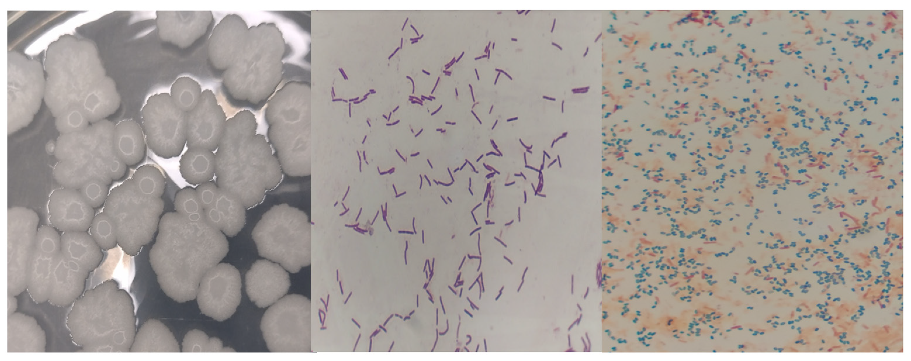
According to Vos et al. [46], S. pasteuri are coccus-shaped cells with a diameter of 0.5–1.5 µm, Gram-positive, and occur singly, in pairs, or clusters. Their cell wall contains peptidoglycan and teichoic acid, and colonies are usually yellow, raised, glistening, and smooth. K. pneumoniae are straight rod-shaped cells of 0.3–1.0 × 0.6–6.0 µm, Gram-negative, and occur singly, in pairs, or short chains, surrounded by a thick polysaccharide capsule. Depending on the strain and the media composition, this strain forms colonies that are more or less dome-shaped, shiny, and sticky to variable degrees. Finally, B. subtilis are rod-shaped cells with 0.7 − 0.8 × 2.0 − 3.0 µm, Gram-positive, and occur singly or in pairs. They are endospore-forming cells. Their colony morphology is variable, ranging from round to irregular, with a wavy margin. All of these data extracted from the literature agree with the results obtained herein (Figure 1, Figure 2 and Figure 3).
S. pasteuri has not been reported in similar studies concerning treated wastewater. K. pneumoniae has been reported in drinking water, wastewater (distribution system), and rivers [54]. Additionally, K. pneumoniae has a capsule capable of protecting the cell, and is considered resistant to cephalosporins [55] and carbapenem [56]. Therefore, it is important to mention that this species is multidrug-resistant and a frequent nosocomial pathogen, causing infections with high rates of morbidity and mortality (up to 50%) [57]. Finally, the genus Bacillus has also been reported as resistant to chlorination, and is used as an indicator for disinfection studies [9,58].

3.3. Inactivation Kinetics

The inactivation tests of S. pasteuri, K. pneumoniae, and B. subtilis with NaClO began at concentrations of 1 × 10¹² CFU/mL, 1 × 10¹⁰ CFU/mL, and 1 × 10¹² endospores/mL, respectively. Meanwhile, in the PAA inactivation assays, the concentrations were 1 × 10⁹ CFU/mL, 1 × 10⁸ CFU/mL, and 1 × 10¹¹ endospores/mL, respectively. Table 6 summarizes the total inactivation units and C·t values obtained. According to Ocampo-Rodríguez et al. [10], C·t values are reported when a 4 log or 99.99% reduction is achieved. With a concentration of 15 mg/L of NaClO, the C·t values that achieved this inactivation for S. pasteuri, K. pneumoniae, and B. subtilis were 3, 7.13, and 418.74 mg·min/L, respectively; with a concentration of 30 mg/L, they were 3.32, 17.61, and 91.87 mg·min/L, respectively.
The PAA was more effective against vegetative cells of S. pasteuri and the endospores of B. subtilis, as at the lower tested concentration (5 mg/L) and contact times of t = 10 and 60 min respectively, a 99.99% removal (4-log reduction) was achieved for both. However, NaClO (15 mg/L) was found to be more effective for the inactivation of K. pneumoniae within 5 min.
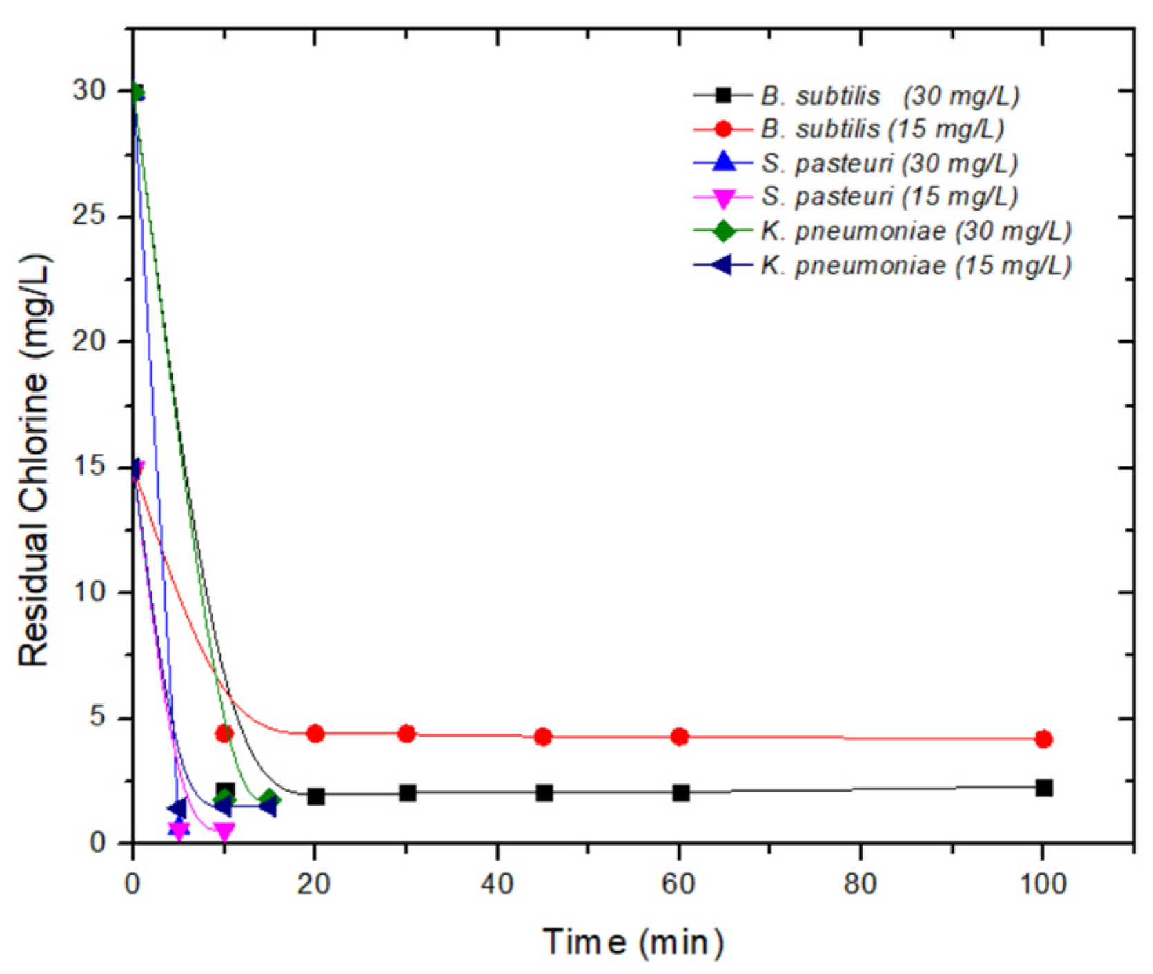
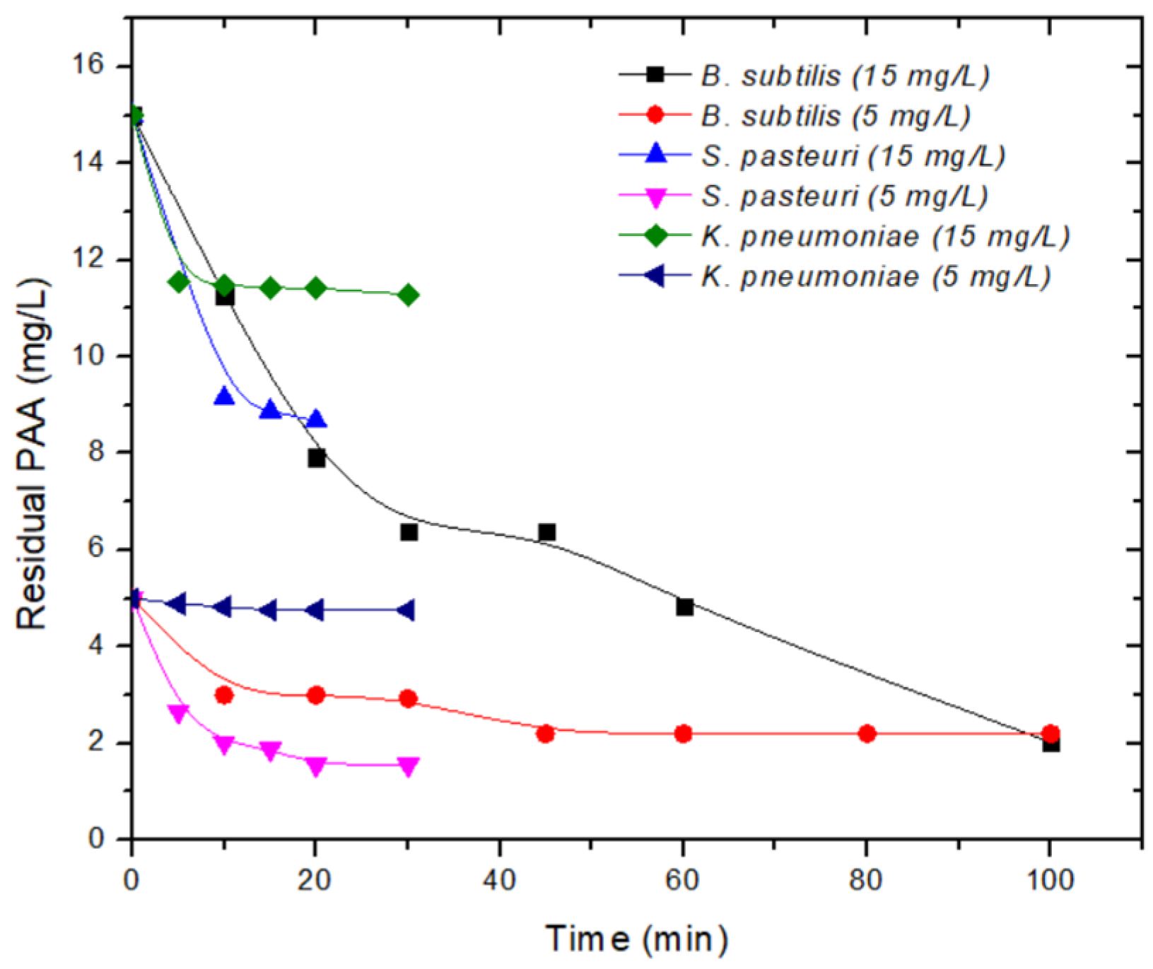
In the PAA assays, when using a concentration of 5 mg/L, the C·t values for S. pasteuri and B. subtilis were 20.13 and 99 mg·min/L, respectively; with a concentration of 15 mg/L, they were 91.37 and 287.31 mg·min/L, respectively. For K. pneumoniae, the 4-log reduction was not achieved with any PAA dose. Figure 4 and Figure 5 show the residual NaClO and PAA results from the disinfection assays. The residual chlorine is within the limits recommended by the World Health Organization (WHO) for water disinfection. However, residual doses of PAA in disinfection trials are not reported in the literature. The commonly employed concentrations for this latter disinfectant are typically lower than 12 mg/L.
Inactivation of less than 6 log of different Staphylococcus species has been reported with PAA; however, with NaClO it can range from 4 to 6 log, depending on the species [59]. The use of PAA has been reported for Gram-positive and Gram-negative bacteria with concentrations lower than 11 mg/L, which achieves inactivation of up to 5 log [60]. This is consistent with the inactivation results obtained in this research. It was observed that with the use of both disinfectants, at higher doses, the units of inactivation of S. pasteuri increased, and for NaClO, the contact time decreased, resulting in lower C·t values.
As S. pasteuri is a Gram-positive bacteria, it would be expected to show greater resistance to disinfectants due to the characteristics of its cell wall [61]. However, it was shown that this species had the greatest sensitivity to disinfectants, at both doses.
In the case of K. pneumoniae, as a typical Gram-negative bacteria, it showed greater sensitivity to NaClO because free chlorine interacts with nucleophilic structures (hemes and porphyrins), proteins (containing iron and sulfur), purine and pyrimidine bases, sulfhydryl groups, amines, and amino acids [62]. K. pneumoniae showed greater resistance to PAA, as indicated by the high C·t values. This can be explained because bacteria can develop resistance to disinfectants through enzymatic catalysis, gene expression, and efflux pumps that are used to expel various toxic substances from inside the cell [33,63]. The qac genes are the most common ones that lead to disinfectant tolerance, because they encode resistance to organic cations with different structures, such as the qacE gene in K. pneumoniae, located in the plasmid (pR751) [33]. In another study, it was shown that these species exhibit resistance to biguanides [64], because, in Gram-negative bacteria, it is more difficult for antibacterial agents to penetrate the interior of the bacterial cell due to the asymmetric structure of lipopolysaccharides in their outer membrane [33]. There is great interspecific variability of resistance to disinfectants and, contrary to common belief, Gram-positive strains are not more resistant than Gram-negative strains [60].
According to the results obtained, B. subtilis endospores showed greater resistance to PAA, and similar inactivation units were obtained with both doses used. Inactivation of B. subtilis endospores with chlorine derivatives has been reported in the literature, mainly at different doses and different pH. For example, with the B. subtilis (ATCC 6633) strain, an inactivation of 3 log was achieved, with a C·t of 127 mg·min/L (pH 5.6, initial chlorine of 2 mg/L) [65]. Reductions of 1–2 log with C·t values of 467–671 mg·min/L (gas chlorine: 0.5–1 mg/L, pH 7.2) have been reported with the same strain [66]. C·t values of 103–386 mg·min/L were reported to inactivate 2 log at pH 5.6–8.2 [67]. Li et al. [30] reported a C·t of 140 mg·min/L to inactivate 4.5 log of the same strain. Additionally, it is clear that pH directly influences endospore inactivation, since HClO (pKa = 7.4) is more effective than ClO (pKa = 7.22) [68]. The C·t values obtained with NaClO in this study occurred within the reported ranges. However, increasing the dose facilitates the elimination of the endospores of the isolated strain, with lower C·t values.
In studies with certified strains, unlike the strains isolated in this study, it has been shown that at least 300 mg/L of PAA is needed to achieve an inactivation of 6 log of endospores from the B. subtilis (PS533) strain [69]. In the case of B. subtilis subsp. globigii (ATCC 9372) endospores, this same concentration achieves an inactivation of 3.5 log [59]. The literature has presented the minimal sporicidal concentration of PAA against Bacillus strains, which varies between 168 and 1344 mg/L [70]. These concentrations are considerably higher than those used in this study. The resistance to PAA of B. subtilis strain isolated from an endoscope washer–disinfector has also been demonstrated, due to the fact that the cell releases exopolymeric substances that play a protective role in deactivating oxidizing agents [71]. The strain of B. subtilis isolated from the WWTP also shows resistance to this disinfectant; however, it is necessary to perform further assays with higher doses of PAA. Additionally, it is required to measure the residual PAA to obtain C·t values, and compare them to the results of this study.

3.4. Kinetic Models

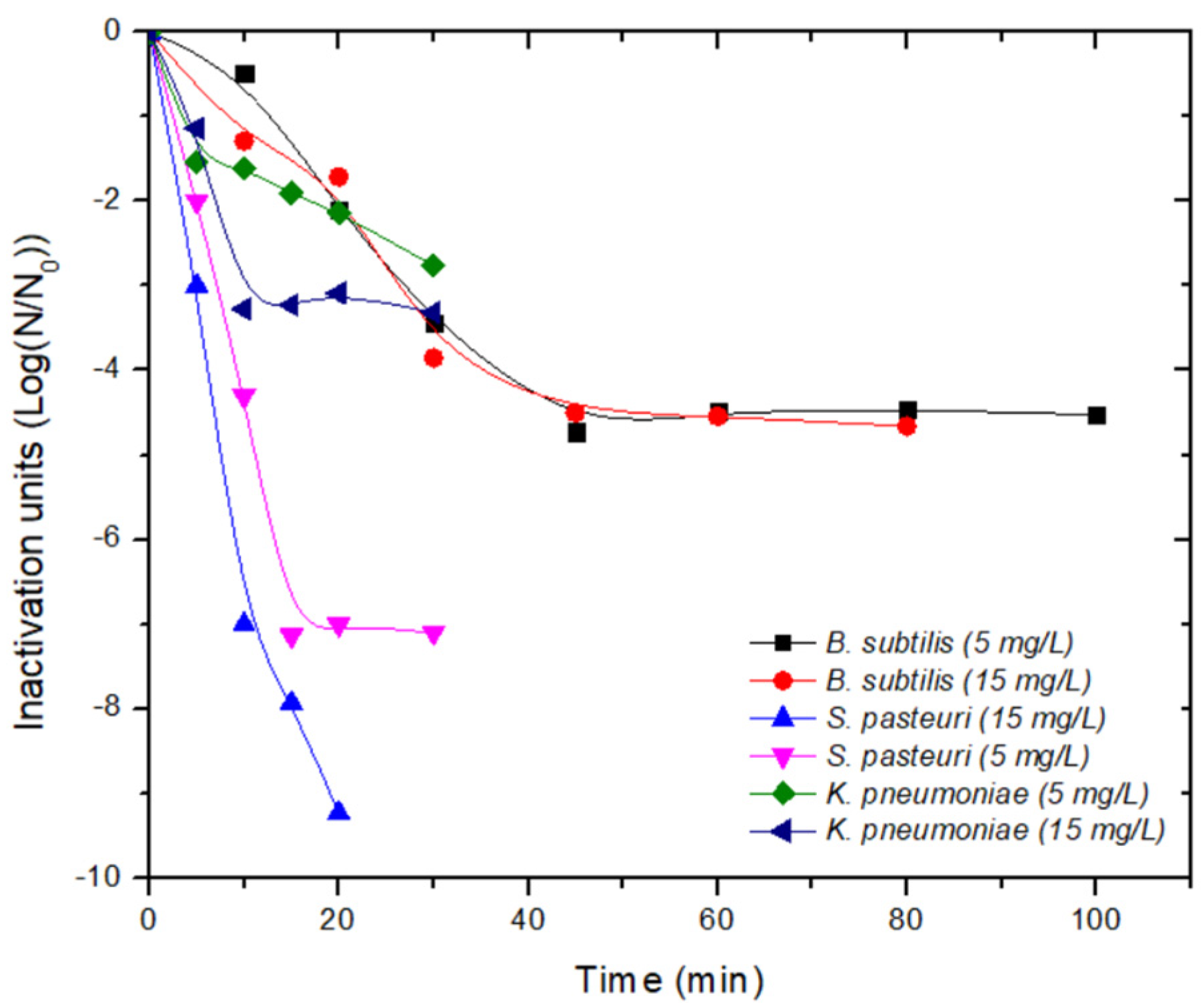
Figure 6 and Figure 7 depict the inactivation of S. pasteuri and K. pneumoniae cells, as well as B. subtilis endospores, using NaClO and PAA, respectively. For S. pasteuri, linear trends were observed, while for K. pneumoniae, concave trends were observed. B. subtilis endospores showed a linear effect followed by a tail effect. These effects were also observed in the PAA assays, but 15 min later than when using NaClO. Based on these observations, kinetic models were selected to describe these effects. The kinetic models complement the designing and implementing of the disinfection process, although it is important to consider the DBPs formation, the disinfectant demand, and the reactor hydraulics [31]
The GInaFiT complement of the Excel program allows the selection of kinetic models based on the inactivation curves. Table 7 shows the kinetic coefficients of the models tested for each species and doses of both disinfectants. To select the best-fit model, one quality index, the coefficient of determination (R2 > 0.9000), was considered.
It was observed that the Chick model is not suitable for describing the inactivation of the strains isolated in this study, which are resistant to chlorine. Additionally, the evaluation at longer contact times allowed the determination of the effect of both disinfectants on the tested bacterial species.
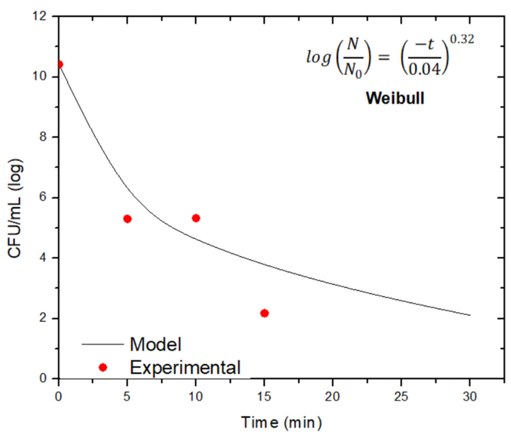
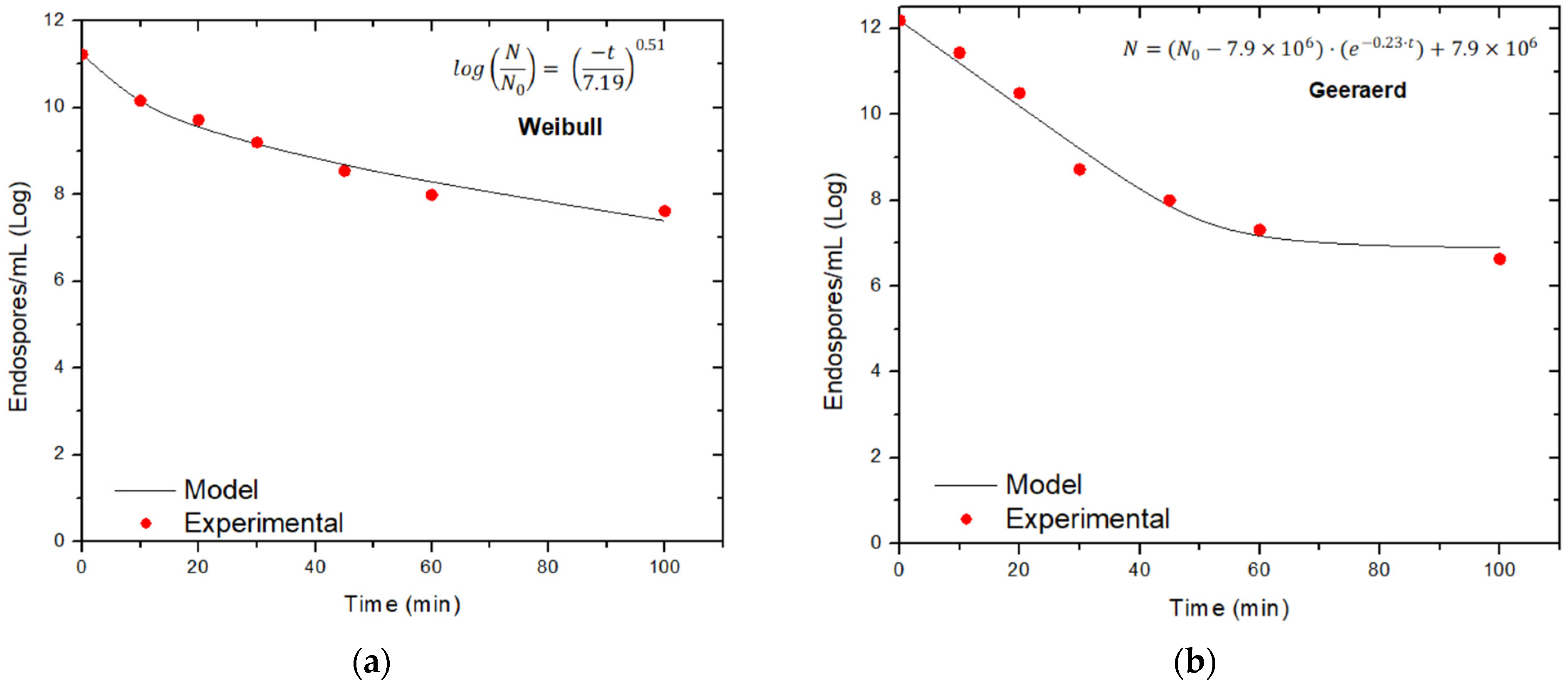
Figure 8, Figure 9, Figure 10, Figure 11 and Figure 12 show the fitted models with their equations. Due to the rapid inactivation of S. pasteuri with NaClO (15 and 30 mg/L), and of K. pneumoniae with NaClO (30 mg/L), the kinetic model that can describe their inactivation at these concentrations could not be obtained. In addition, at least six data points are required to obtain the model with the GInaFiT software. The disinfection of K. pneumoniae with 15 mg/L of NaClO showed a concave curve, which is properly described by the Weibull model, while the B. subtilis inactivation with 15 and 30 mg/L of the same disinfectant was adequately described by the Weibull and Geeraerd models, respectively.
The chlorine inactivation of endospores of B. subtilis certified strains has been described using the Chick–Watson delay model [30,65,66,67,72]. In those assays, doses lower than 10 mg/L of Cl2 were used, and the model was obtained by a graph of IU (log N/N0) vs. C·t, which describes a shoulder effect followed by a linear trend, which is different from what is presented in this study (Figure 3 and Figure 4). According to Peleg [29], models are based on fitting their equations to experimental data, rather than their ability to predict dynamic inactivation patterns. Moreover, it has been shown that inactivation kinetics can vary depending on the culture media from which the endospores were obtained [73,74].
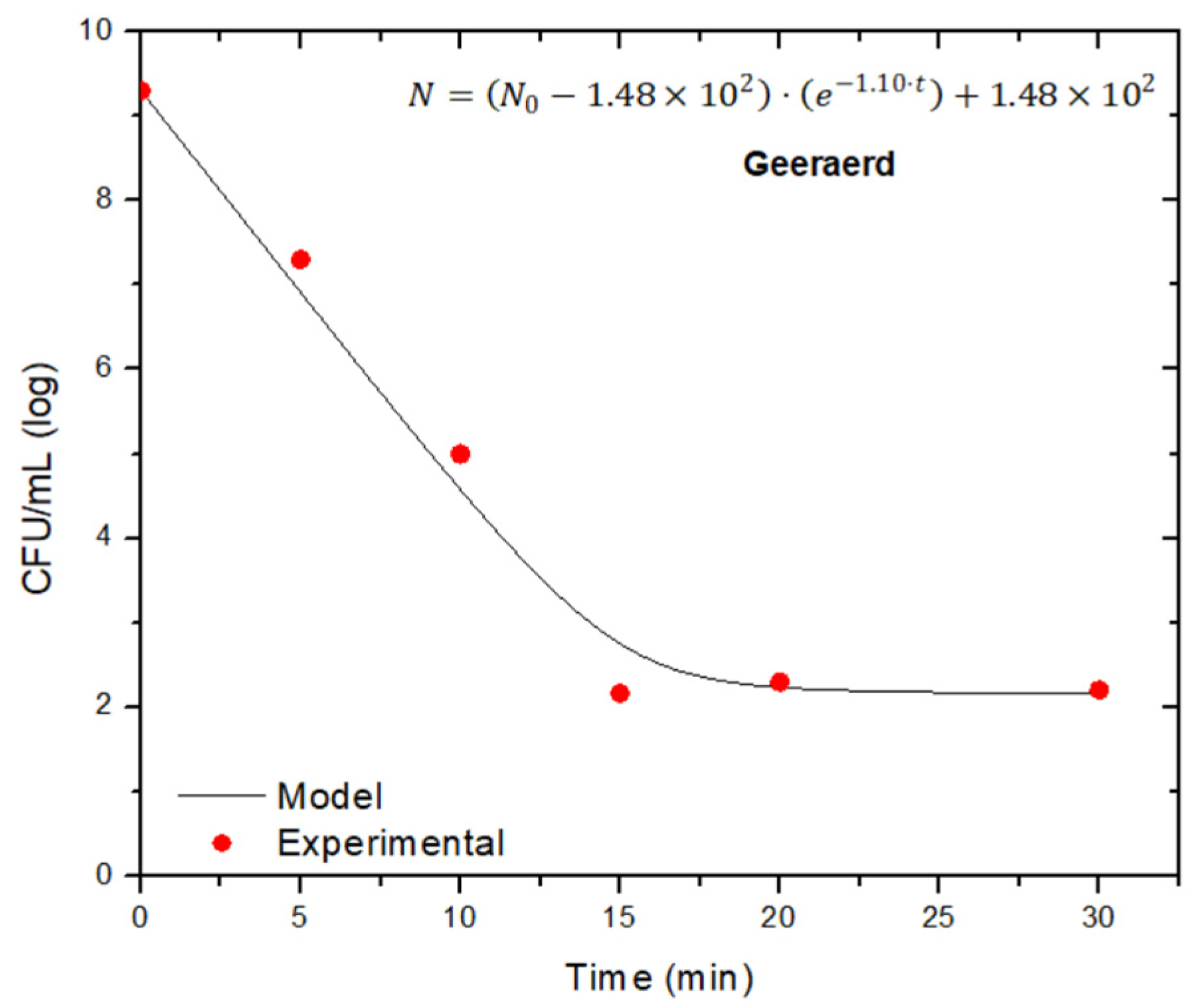
The disinfection of S. pasteuri, K. pneumoniae, and B. subtilis with PAA can be described using the Geeraerd model. However, due to the rapid inactivation of S. pasteuri at 15 mg/L, a kinetic model was not obtained, because samples must be collected in less than five minutes. To the best of our knowledge, no inactivation kinetics of S. pasteuri have been reported in the literature, so the model proposed in this study would be the first to describe inactivation with PAA. In the case of K. pneumoniae, inactivation with UV radiation and H2O2 has also been described using the Geeraerd model [75].
The disinfection of B. subtilis endospores with PAA has also been described using the Weibull model [76], while inactivation using pulsed light has been modeled with the Geeraerd equation [77]. Both studies used the GInaFiT tool to obtain the kinetic models. However, variations in the experimental conditions of different inactivation assays make it difficult to compare the efficacy of disinfectants and their inactivation models [67].

3.5. Statistical Analysis

According to the Kolmogorov–Smirnov test, the data showed a normal distribution. The t-Student test indicated that there were no significant differences between the NaClO doses for the inactivation of K. pneumoniae (t = 0.404, dof = 4, p = 0.707) and B. subtilis endospores (t = −0.0546, dof = 12, p = 0.957). In addition, the t-Student test indicated that there were no significant differences between the concentrations of PAA for S. pasteuri (t = 0.743, dof = 8, p = 0.479), K. pneumoniae (t = −1.581, dof = 10, p = 0.145), and B. subtilis (t = 0.0553, dof = 12, p = 0.957).
Based on the statistical analyses, low concentrations of NaClO (15 mg/L) and PAA (5 mg/L) can be used in the disinfection processes carried out in the studied WWTP, as there were no significant differences in the inactivation of the isolated bacteria of the three species when using higher doses. It is important to highlight that at low doses of Cl2, such as the one used at the studied WWTP and the available residual chlorine, chlorine-resistant bacteria in the tertiary effluent can occur.

4. Conclusions

Currently, WWTPs are facing new challenges, including resistance to chlorine, the most common treatment. The results of this work showed that it is possible to find a diversity of microorganisms in WWTP effluents, despite the addition of a disinfectant, particularly of species endowed with resistance structures such as endospores. This work demonstrates this resistance of both Gram-negative and Gram-positive bacteria. Three strains were isolated from the tertiary effluent of a WWTP, and were identified as S. pasteuri, K. pneumoniae, and B. subtilis. In this study, the effectiveness of two disinfectants, one widely used throughout the world, and the other emerging for wastewater treatment, is assessed as a potential method to enhance the quality of treated water.
Disinfection tests were performed with vegetative cells of S. pasteuri and K. pneumoniae, as well as with endospores of B. subtilis. Further tests with wild-type strains isolated from the environment are necessary, as certified strains often respond differently to disinfection. K. pneumoniae was found to be more resistant to PAA than to NaClO in the inactivation kinetics, whereas PAA was more effective at lower doses against endospores of B. subtilis. Therefore, PAA has a broad spectrum of antimicrobial action and is effective against spores, fungi, viruses, and bacteria. Due to its simplicity of use and low cost, it has recently been suggested as an alternative disinfectant to those based on chlorine.
The kinetic models of inactivation can be a helpful tool in the disinfection of water resources, and can make it easier to compare the effectiveness of different disinfectants used in water treatment. The different initial concentrations of the disinfectants interfere with the obtained inactivation curves due to the response of the bacteria to these oxidizing agents and, therefore, the models obtained for each disinfectant may vary. The inactivation of S. pasteuri, K. pneumoniae, and B. subtilis with NaClO and PAA showed concave and linear curves with a tail effect, respectively, which can mostly be described using the Weibull and Geeraerd models that suggest variable levels of resistance.
Further studies are needed to investigate the inactivation of microbial consortia with emerging disinfectants, also considering common physicochemical parameters and the WWTP’s effluent characteristics, as well as the potential generation of disinfection byproducts. Due to the resistance of microorganisms to disinfection processes, it is recommended to replace conventional treatments with advanced treatments, such as simultaneous or sequential treatments, to guarantee a better microbiological quality of treated water.

Author Contributions

Conceptualization, D.B.O.-R. and C.C.-O.; methodology, D.B.O.-R., J.A.R., S.M.H. and C.C.-O.; data curation, D.B.O.-R., G.A.V.-R., J.A.R. and M.d.R.G.S.; supervision, C.C.-O.; writing—original draft preparation, D.B.O.-R., U.I.-A., G.A.V.-R. and C.C.-O.; writing—review and editing, D.B.O.-R., U.I.-A., G.A.V.-R., M.d.R.G.S., S.M.H., J.A.R. and C.C.-O. All authors have read and agreed to the published version of the manuscript.

Funding

This research received no external funding.

Data Availability Statement

This research has no complementary data to what is here published.

Acknowledgments

D.B.O.-R. acknowledges the PhD scholarship provided by the Consejo Nacional de Ciencia y Tecnología (CONACYT-Mexico).

Conflicts of Interest

The authors declare no conflict of interest.

References

  1. Collivignarelli, M.C.; Abbá, A.; Benigna, I.; Sorlini, S.; Torretta, V. Overview of the main Disinfection Processes for Wastewater and Drinking water Treatment Plants. Sustainability 2018, 10, 86. [Google Scholar] [CrossRef]
  2. Collivignarelli, M.C.; Abbá, A.; Miino, M.C.; Caccamo, F.F.; Torretta, V.; Rada, E.C.; Sorlini, S. Disinfection of wastewater by UV-Based Treatment for Reuse in a Circular Economy Perspective. Where are we at? Int. J. Environ. Res. Public Health 2021, 18, 77. [Google Scholar] [CrossRef] [PubMed]
  3. DOF-Diario Oficial de la Federación. Norma Oficial Mexicana NOM-001-SEMARNAT-2021, Que Establece Los Límites Permisibles de Contaminantes en Las Descargas de Aguas Residuales en Cuerpos Receptores Propiedad de la Nación. 11 de Marzo de 2022. Available online: https://www.dof.gob.mx/nota_detalle.php?codigo=5645374&fecha=11/03/2022#gsc.tab=0 (accessed on 11 May 2023).
  4. Umar, M. From Conventional Disinfection to Antibiotic Resistance Control-Status of the Use of Chlorine and UV Irradiation during Wastewater Treatment. Int. J. Environ. Res. Public Health. 2022, 19, 1636. [Google Scholar] [CrossRef] [PubMed]
  5. Luongo, G.; Previtera, L.; Ladhari, A.; Di Fabio, G.; Zarrelli, A. Peracetic acid vs. Sodium Hypochlorite: Degradation and Transformation of Drugs in Wastewater. Molecules. 2020, 25, 2294. [Google Scholar] [CrossRef] [PubMed]
  6. Köhler, A.T.; Rodloff, A.C.; Labahn, M.; Reinhardt, M.; Truyen, U.; Speck, S. Efficacy of sodium hypochlorite against multidrug-resistant gram-negative bacteria. J. Hosp. Infect. 2018, 100, e40–e46. [Google Scholar] [CrossRef] [PubMed]
  7. Ersoy, Z.G.; Dinc, O.; Cinar, B.; Gedik, S.T.; Dimoglo, A. Comparative evaluation of disinfection mechanism of sodium hypochlorite, chlorine dioxide, and electroactivated water on Enterococcus faecalis. LWT-Food Sci. Technol. 2019, 102, 205–213. [Google Scholar] [CrossRef]
  8. Ziemba, C.; Larivé, O.; Deck, S.; Huisman, T.; Morgenroth, E. Comparing the anti-bacterial performance of chlorination and electrolysis post-treatments in a hand washing water recycling system. Water Res. X 2019, 2, 100020. [Google Scholar] [CrossRef] [PubMed]
  9. Luo, L.W.; Wu, Y.H.; Yu, T.; Wang, Y.H.; Chen, G.Q.; Tong, X.; Bai, Y.; Xu, C.; Wang, H.B.; Ikuno, N.; et al. Evaluating method and potential risks of chlorine-resistant bacteria (CRB): A review. Water Res. 2021, 188, 116474. [Google Scholar] [CrossRef]
  10. Ocampo-Rodríguez, D.B.; Vázquez-Rodríguez, G.A.; Martínez-Hernández, S.; Iturbe-Acosta, U.; Coronel-Olivares, C. Desinfección del agua: Una revisión a los tratamientos convencionales y avanzados con cloro y ácido peracético. Ing. Agua. 2022, 26, 185–204. [Google Scholar] [CrossRef]
  11. Kampf, G. Sodium Hypochlorite. In Antiseptic Stewardship Biocide Resistance and Clinical Implications; Springer: Cham, Switzerland, 2018; pp. 161–210. [Google Scholar]
  12. Destiani, R.; Templeton, M.R. Chlorination and ultraviolet disinfection of antibiotic-resistant bacteria and antibiotic resistance genes in drinking water. AIMS Environ. Sci. 2019, 6, 222–241. [Google Scholar] [CrossRef]
  13. Gilca, A.F.; Teodosiu, C.; Fiore, S.; Musteret, C.P. Emerging disinfection by-products: A review on their occurrence and control in drinking water treatment processes. Chemosphere 2020, 259, 127476. [Google Scholar] [CrossRef]
  14. Kitis, M. Disinfection of wastewater with peracetic acid: A review. Environ. Int. 2004, 30, 47–55. [Google Scholar] [CrossRef] [PubMed]
  15. Kampf, G. Peracetic Acid. In Antiseptic Stewardship Biocide Resistance and Clinical Implications; Springer: Cham, Switzerland, 2018; pp. 63–98. [Google Scholar]
  16. Henao, L.D.; Turolla, A.; Antonelli, M. Disinfection by-products formation and ecotoxicological effects of effluents treated with peracetic acid: A review. Chemosphere 2018, 213, 25–40. [Google Scholar] [CrossRef] [PubMed]
  17. Sun, P.; Zhang, T.; Mejia-Tickner, B.; Zhang, R.; Cai, M.; Huang, C. Rapid disinfection by peracetic acid combined with UV irradiation. Environ. Sci. Technol. Lett. 2018, 5, 400–404. [Google Scholar] [CrossRef]
  18. Hassaballah, A.H.; Bhatt, T.; Nyitrai, J.; Dai, N.; Sassoubre, L. Inactivation of E. coli, Enterococcus spp., somatic coliphage, and Cryptosporidium parvum in wastewater by peracetic acid (PAA), sodium hypochlorite, and combined PAA-ultraviolet disinfection. Environ. Sci. Water Res. Technol. 2020, 6, 197–209. [Google Scholar] [CrossRef]
  19. Campo, N.; De Flora, C.; Maffettone, R.; Manoli, K.; Sarathy, S.; Santoro, D.; Gonzalez-Olmos, R.; Auset, M. Inactivation kinetics of antibiotic-resistant Escherichia coli in secondary wastewater effluents by peracetic and performic acids. Water Res. 2019, 169, 115227. [Google Scholar] [CrossRef] [PubMed]
  20. Luukkonen, T.; Pehkonen, S.O. Peracids in water treatment: A critical review. Crit. Rev. Environ. Sci. Technol. 2017, 47, 1–39. [Google Scholar] [CrossRef]
  21. Gyürék, L.L.; Finch, G.R. Modeling Water Treatment Chemical Disinfection Kinetics. J. Environ. Eng. 1998, 124, 783–793. [Google Scholar] [CrossRef]
  22. Chick, H. An investigation of the laws of disinfection. Epidemiol. Infect. 1908, 8, 92–158. [Google Scholar] [CrossRef]
  23. Watson, H.E. A note on the variation of the rate of disinfection with change in the concentration of the disinfectant. Epidemiol. Infect. 1908, 8, 536–542. [Google Scholar] [CrossRef]
  24. Rennecker, J.L.; Mariñas, B.J.; Owens, J.H.; Rice, E.W. Inactivation of Cryptosporidium parvum oocysts with ozone. Water Res. 1999, 33, 2481–2488. [Google Scholar] [CrossRef]
  25. Hom, L.W. Kinetics of chlorine disinfection in an ecosystem. J. Sanit. Eng. Div. 1972, 98, 183–194. [Google Scholar] [CrossRef]
  26. Selleck, R.E.; Saunier, B.M.; Collins, H.F. Kinetics of bacterial deactivation with chlorine. J. Environ. Eng. Div. 1978, 104, 1197–1212. [Google Scholar] [CrossRef]
  27. Geeraerd, A.H.; Herremans, C.H.; Van Impe, J.F. Structural model requirements to describe microbial inactivation during a mild heat treatment. Int. J. Food Microbiol. 2000, 59, 185–209. [Google Scholar] [CrossRef] [PubMed]
  28. Mafart, P.; Couvert, O.; Gaillard, S.; Leguérinel, I. On calculating sterility in thermal preservation methods: Weibull frequency distribution model application. Int. J. Food Microbiol. 2002, 72, 107–113. [Google Scholar] [CrossRef]
  29. Peleg, M. Microbial Dose-Response Curves and Disinfection Efficacy Models Revisited. Food Eng. Rev. 2021, 13, 305–321. [Google Scholar] [CrossRef]
  30. Li, G.Q.; Huo, Z.Y.; Wu, Q.Y.; Lu, Y.; Hu, H.Y. Synergistic effect of combined UV-LED and chlorine treatment on Bacillus subtilis spore inactivation. Sci. Total Environ. 2018, 639, 1233–1240. [Google Scholar] [CrossRef]
  31. Manoli, K.; Sarathy, S.; Maffettone, R.; Santoro, D. Detailed modeling and advanced control for chemical disinfection of secondary effluent wastewater by peracetic acid. Water Res. 2019, 153, 251–262. [Google Scholar] [CrossRef]
  32. Kong, J.; Lu, Y.; Ren, Y.; Chen, Z.; Chen, M. The virus removal in UV irradiation, ozonation, and Chlorination. Water Cycle 2021, 2, 23–31. [Google Scholar] [CrossRef]
  33. Tong, C.; Hu, H.; Chen, G.; Li, Z.; Li, A.; Zhang, J. Disinfectant resistance in bacteria: Mechanisms, spread, and resolution strategies. Environ. Res. 2021, 195, 110897. [Google Scholar] [CrossRef]
  34. Wang, J.; Sui, M.; Yuan, B.; Li, H.; Lu, H. Inactivation of two Mycobacteria by free chlorine: Effectiveness, influencing factors, and mechanisms. Sci. Total Environ. 2019, 648, 271–284. [Google Scholar] [CrossRef] [PubMed]
  35. Garg, A.; Narasimman, L.M.; Hogg, J.; Nutter, A.; Mahoney, G. Wastewater Disinfection with peracetic acid. Proc. Water Environ. Fed. 2016, 13, 1798–1808. [Google Scholar] [CrossRef]
  36. Collivignarelli, M.C.; Abbá, A.; Alloisio, G.; Gozio, E.; Benigna, I. Disinfection in wastewater treatment plants: Evaluation of effectiveness and Acute toxicity effects. Sustainability 2017, 9, 1704. [Google Scholar] [CrossRef]
  37. McFadden, M.; Loconsole, J.; Schockling, A.J.; Nerenberg, R.; Pavissich, J.P. Comparing peracetic acid and hypochlorite for disinfection of combined sewer overflows Effects of suspended-solids and pH. Sci. Total Environ. 2017, 599, 533–539. [Google Scholar] [CrossRef]
  38. Ragazzo, P.; Chiucchini, N.; Piccolo, V.; Spadolini, M.; Carrer, S.; Zanon, F.; Ronald, G. Wastewater disinfection: Long-term laboratory and full-scale studies on performic acid in comparison with peracetic acid and chlorine. Water Res. 2020, 184, 116169. [Google Scholar] [CrossRef]
  39. Freitas, B.O.; Leite, L.S.; Daniel, L.A. Chlorine and peracetic acid in decentralizes wastewater treatment: Disinfection, oxidation and odor control. Process Saf. Environ. Protect. 2021, 146, 620–628. [Google Scholar] [CrossRef]
  40. Koivunen, J.; Heinonen-Tanski, H. Inactivation of enteric microorganisms with chemical disinfectants, UV irradiation, and combined chemical/Uv treatments. Water Res. 2005, 39, 1519–1526. [Google Scholar] [CrossRef]
  41. Dang, T.T.; Imai, T.; Le, T.V.; Nguyem, D.K.; Higuchi, T.; Kanno, A.; Yamamoto, K.; Sekine, M. Synergistic effect of pressurized carbon dioxide and sodium hypochlorite on the inactivation of Enterococcus sp. in seawater. Water Res. 2016, 106, 204–213. [Google Scholar] [CrossRef]
  42. Bonetta, S.; Pignata, C.; Bonetta, S.; Amagliani, G.; Brandi, G.; Gilli, G.; Carraro, E. Comparison of UV, Peracetic Acid and Sodium Hypochlorite Treatment in the Disinfection of Urban Wastewater. Pathogens 2021, 10, 182. [Google Scholar] [CrossRef]
  43. Dunkin, N.; Weng, S.C.; Schwab, K.J.; McQuarrie, J.; Bell, K.Y.; Jacangelo, J.G. Comparative Inactivation of Murine Norovirus and MS2 Bacteriophage by Peracetic Acid and Monochloramina in Municipal Secondary Wastewater Effluent. Environ. Sci. Technol. 2017, 51, 2972–2981. [Google Scholar] [CrossRef]
  44. Eramo, A.; Medina, W.R.M.; Fahrenfeld, N.L. Peracetic acid disinfection kinetics for combined sewer overflows: Indicator organisms, antibiotic resistance genes, and microbial community. Environ. Sci. Water Res. Technol. 2017, 3, 1061–1072. [Google Scholar] [CrossRef] [PubMed]
  45. Atlas, R.M. Handbook of Microbiological Media, 4th ed.; Taylor & Francis Group: Washington, DC, USA, 2010; p. 428. [Google Scholar]
  46. Vos, P.; Garrity, G.; Jones, D.; Krieg, N.; Ludwig, W.; Rainey, F.; Whitman, W. Bergey’s Manual of Systematic Bacteriology, 2nd ed.; Springer: Dordrecht, The Netherlands; Berlin/Heidelberg, Germany; New York, NY, USA, 2009; pp. 21–127, 392–420. [Google Scholar]
  47. Oviaño, G.M.; Rodríguez, S.B.; Caballero, P.J.D.; Muñoz, B.J.L. Aplicaciones de la Espectrometría de Masas MALDI-TOF en Microbiología Clínica; Sociedad Española de Enfermedades Infecciosas y Microbiología Clínica: Madrid, Spain, 2019; pp. 9–11. [Google Scholar]
  48. Schoch, C.L.; Ciufo, S.; Domrachev, M.; Hotton, C.L.; Kannan, S.; Khovanskaya, R.; Leipe, D.; Mcveigh, R.; O'Neill, K.; Robbertse, B.; et al. NCBI Taxonomy: A comprehensive updateoncuration, resources and tools. Database: J. Biol. Databases Curation 2020, 2020, baaa062. [Google Scholar] [CrossRef] [PubMed]
  49. Miles, A.A.; Misra, S.S.; Irwin, J.O. The estimation of the bactericidal power of the blood. Epidemiol. Infect. 1938, 38, 732–749. [Google Scholar] [CrossRef] [PubMed]
  50. Rochelle, P.A.; Blatchley, E.R., III; Chan, P.S.; Scheible, O.K.; Shen, C. Challenge Organisms for Inactivation of Viruses by Ultraviolet Treatment; Water Research Foundation: Denver, CO, USA, 2010; pp. 16–18. [Google Scholar]
  51. Adeyemo, F.E.; Singh, G.; Reddy, P.; Bux, F.; Stenström, T.A. The efficiency of chlorine and UV in the inactivation of Cryptosporidium and Giardia in wastewater. PLoS ONE. 2019, 14, e0216040. [Google Scholar] [CrossRef]
  52. APHA. American Public Health Association. Method 4500-CL: Standard Methods for the Examination of Water and Wastewater, 22nd ed.; American Public Health Association: Washington, DC, USA, 2012; pp. 4–67. [Google Scholar]
  53. Geeraerd, A.H.; Valdramidis, V.P.; Impe, J.F. GInaFiT, a freeware tool to assess non-log-linear microbial survivor curves. Int. J. Food Microbiol. 2005, 102, 95–105. [Google Scholar] [CrossRef]
  54. Geldreich, E.E.; Rice, E.W. Occurrence, Significance, and Detection of Klebsiella in Water Systems. J. Am. Water Work. Assoc. 1987, 79, 74–80. [Google Scholar] [CrossRef]
  55. Vargas, J.A.L.; Toro, L.M.E. K. pneumoniae: ¿la nueva “superbacteria”? Patogenicidad, epidemiología y mecanismos de resistencia. Iatreia 2010, 23, 157–165. [Google Scholar]
  56. Serna-Galvis, E.A.; Vélez-Peña, E.; Osorio-Vargas, P.; Jiménez, A.N.; Salazar-Ospina, L.; Guaca-González, Y.M.; Torres-Palma, R.A. Inactivation of carbapenem-resistant Klebsiella pneumoniae by photo-Fenton: Residual effect, gene evolution and modifications with citric acid and persulfate. Water Res. 2019, 161, 354–363. [Google Scholar] [CrossRef]
  57. Popa, L.I.; Gheorghe, I.; Barbu, I.C.; Surleac, M.; Paraschiv, S.; Marutescu, L.; Popa, M.; Piracalabioru, G.G.; Talapan, D.; Nita, M.; et al. Multidrug Resistant Klebsiella pneumoniae ST101 Clone Survival Chain From Inpatients to Hospital Effluent After Chlorine Treatment. Front. Microbiol. 2021, 11, 610296. [Google Scholar] [CrossRef]
  58. Paes, F.A.; Hissa, D.C.; Angelim, A.L.; Pinto, A.W.; Grangeiro, T.B.; Melo, V.M.M. Diversity of a Chlorine-Resistant Bacillus Population Isolated from a Wastewater Treatment Station. Water Environ. Res. 2012, 84, 274–281. [Google Scholar] [CrossRef]
  59. Sagripanti, J.L.; Bonifacino, A. Comparative Sporicidal Effects of Liquid Chemical Agents. Appl. Environ. Microbiol. 1996, 62, 545–551. [Google Scholar] [CrossRef] [PubMed]
  60. Bridier, A.; Briandet, R.; Thomas, V.; Dubois-Brissonnet, F. Comparative biocidal activity of peracetic acid, benzalkonium chloride, and ortho-phthalaldehyde on 77 bacterial strains. J. Hosp. Infect. 2011, 78, 208–213. [Google Scholar] [CrossRef] [PubMed]
  61. Mir, J.; Morató, J.; Ribas, F. Resistance to chlorine of freshwater bacterial strains. J. Appl. Microbiol. 1997, 87, 7–18. [Google Scholar] [CrossRef] [PubMed]
  62. Nizer, W.S.C.; Inkovskiy, V.; Overhage, J. Surviving Reactive chlorine Stress: Responses of Gram-Negative Bacteria to Hypochlorous Acid. Microorganisms 2020, 8, 1220. [Google Scholar] [CrossRef]
  63. Jiménez, L.C.; Rojas-Jiménez, K. Resistencia a desinfectantes y su relación con la resistencia a los antibióticos. Acta Médica Costarriencense 2020, 62, 7–12. [Google Scholar]
  64. Bock, L.J.; Wand, M.E.; Sutton, J.M. Varying activity of chlorhexidine-based disinfectants against Klebsiella pneumoniae clinical isolates and adapted strains. J. Hosp. Infect. 2016, 93, 42–48. [Google Scholar] [CrossRef]
  65. Cho, M.; Chung, H.; Yoon, J. Quantitative Evaluation of the synergistic Sequential Inactivation of Bacillus subtilis Spores with Ozone Followed by Chlorine. Environ. Sci. Technol. 2003, 37, 2134–2138. [Google Scholar] [CrossRef]
  66. Choi, Y.; Byun, S.H.; Jang, H.J.; Kim, S.E.; Choi, Y. Comparison of disinfectants for drinking water: Chlorine gas vs. On-site generated chlorine. Environ. Eng. Res. 2022, 27, 200543. [Google Scholar] [CrossRef]
  67. Cho, M.; Kim, J.H.; Yoon, J. Investigating synergism during sequential inactivation of Bacillus subtilis spores with several disinfectants. Water Res. 2006, 40, 2911–2920. [Google Scholar] [CrossRef]
  68. Drogui, P.; Daghrir, R. Chlorine for water disinfection: Properties, applications, and health effects. In CO2 Sequestration, Biofuels and Depollution; Lichtfouse, E., Schwarzbauer, J., Robert, D., Eds.; Springer International Publishing: Cham, Switzerland, 2015; pp. 1–32. [Google Scholar]
  69. Setlow, B.; Korza, G.; Blatt, K.M.S.; Fey, J.P.; Setlow, P. Mechanism of Bacillus subtilis spore inactivation by and resistance to supercritical CO2 plus peracetic acid. J. Appl. Microbiol. 2015, 120, 57–69. [Google Scholar] [CrossRef]
  70. Alasari, A.; Valverde, M.; Roques, C.; Michel, G.; Cabassud, C.; Aptel, P. Sporicidal properties of peracetic acid and hydrogen peroxide, alone and in combination, in comparison with chlorine and formaldehyde for ultrafiltration membrane disinfection. Can. J. Microbiol. 1993, 39, 52–60. [Google Scholar] [CrossRef] [PubMed]
  71. Martin, D.J.H.; Denyer, S.P.; McDonnell, G.; Maillard, J.Y. Resistance and cross-resistance to oxidizing agents of bacterial isolates from endoscope washer disinfectors. J. Hosp. Infect. 2008, 69, 377–383. [Google Scholar] [CrossRef] [PubMed]
  72. Larson, M.A.; Mariñas, B.J. Inactivation of Bacillus subtilis spores with ozone and monochloramine. Water Res. 2003, 37, 833–844. [Google Scholar] [CrossRef] [PubMed]
  73. Aronson, A.; Fitz-James, P. Structure and Morphogenesis of the Bacterial Spore Coat. Bacteriol. Rev. 1976, 40, 360–402. [Google Scholar] [CrossRef] [PubMed]
  74. Leaper, S. Synergistic killing of spores of Bacillus subtilis by peracetic acid and alcohol. J. Food Technol. 1984, 19, 355–360. [Google Scholar] [CrossRef]
  75. Aguilar, S.; Rosado, D.; Moreno-Andrés, J.; Cartuche, L.; Cruz, D.; Acevedo-Merino, A.; Nebot, E. Inactivation of a wild isolated Klebsiella pneumoniae by photo-chemical processes: UV-C, UV-C/H2O2, and UV-C/H2O2/Fe3+. Catal. Today 2017, 313, 94–99. [Google Scholar] [CrossRef]
  76. Leggett, M.J.; Schwarz, J.S.; Burke, P.A.; Mcdonnell, G.; Denyer, S.P.; Maillard, J.Y. Resistance to and killing by the sporicidal microbicide peracetic acid. J. Antimicrob. Chemother. 2015, 70, 773–779. [Google Scholar] [CrossRef]
  77. Artíguez, M.L.; Marañon, I.M. Inactivation of spores and vegetative cells of Bacillus subtilis and Geobacillus stearothermophilus by pulsed light. Innov. Food Sci. Emerg. Technol. 2015, 28, 52–58. [Google Scholar] [CrossRef]
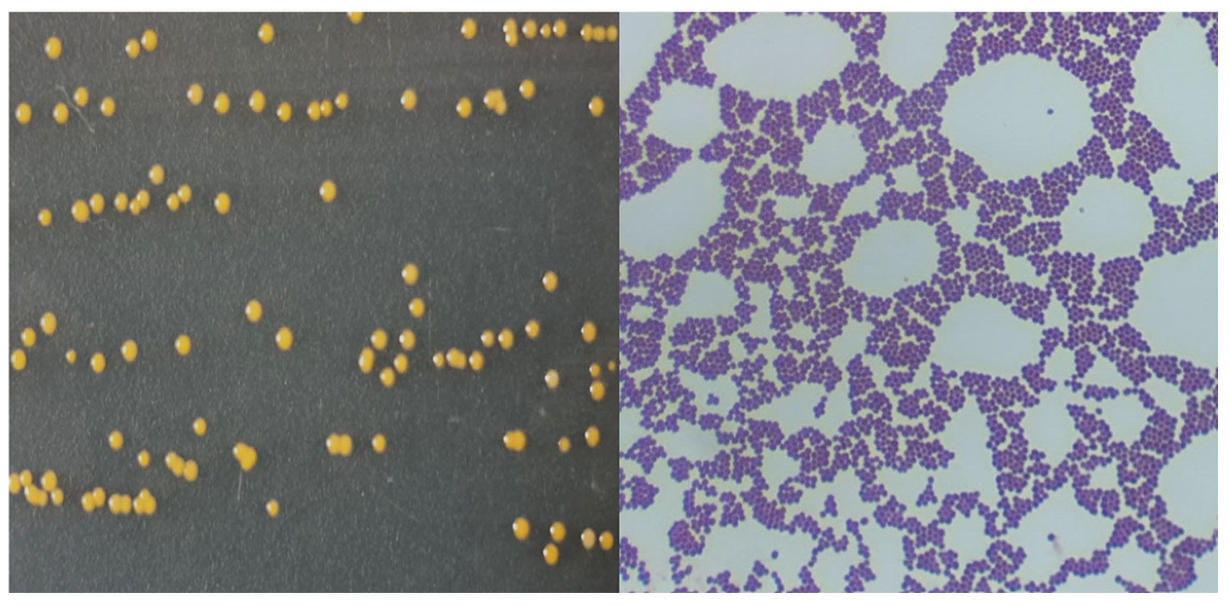
Figure 1. Colony morphology in NA and Gram stain of isolated Staphylococcus pasteuri.
Water 15 02019 g001
Figure 2. Colony morphology in NA and Gram stain of isolated Klebsiella pneumoniae.
Water 15 02019 g002
Figure 3. Colony morphology in NA, Gram stain, and endospores of isolated Bacillus subtilis.
Water 15 02019 g003
Figure 4. Residual chlorine from disinfection assays of bacteria with NaClO (Water 15 02019 i001 and Water 15 02019 i002 endospores of Bacillus subtilis).
Water 15 02019 g004
Figure 5. Peracetic acid residual from disinfection assays of isolated bacteria with PAA (Water 15 02019 i003 and Water 15 02019 i004 endospores of Bacillus subtilis).
Water 15 02019 g005
Figure 6. Inactivation of the isolated bacteria with NaClO (Water 15 02019 i005 and Water 15 02019 i006 endospores of Bacillus subtilis).
Water 15 02019 g006
Figure 7. Inactivation of the isolated bacteria with PAA (Water 15 02019 i007 and Water 15 02019 i008 endospores of Bacillus subtilis).
Water 15 02019 g007
Figure 8. Inactivation of isolated Klebsiella pneumoniae with 15 mg/L NaClO.
Water 15 02019 g008
Figure 9. Inactivation of endospores of isolated Bacillus subtilis with (a) 15 mg/L and (b) 30 mg/L NaClO.
Water 15 02019 g009
Figure 10. Inactivation of isolated Staphylococcus pasteuri with 5 mg/L PAA.
Water 15 02019 g010
Figure 11. Inactivation of isolated Klebsiella pneumoniae with (a) 5 mg/L and (b) 15 mg/L PAA.
Water 15 02019 g011
Figure 12. Inactivation of isolated endospores of Bacillus subtilis with (a) 5 mg/L and (b) 15 mg/L PAA.
Water 15 02019 g012
Table 1. Physiochemical characterization of the WWTP effluent.
ParameterMean ValueStandard Deviation (n = 5)
T (°C)20.71.71
pH7.540.57
ORP128102
DO (mg/L)15.726
EC (µS/cm)1833.894.1
TDS (mg/L)916.846.8
Salinity (PSU)0.930.05
TSS (mg/L) 113.66.07
COD (mg/L) 131.69.96
BOD5 (mg/L) 116.94.42
Note(s): 1 Values reported by the WWTP. ORP: oxidation–reduction potential; DO: dissolved oxygen; EC: electrical conductivity; TDS: total dissolved solids; PSU: practical salinity units; TSS: total suspended solids; COD: chemical oxygen demand; BOD5: biochemical oxygen demand.
Table 2. Colonial plate morphology of strains isolated from the WWTP.
StrainShapeMarginElevationColorTexture
1EB9Circular EntireFlatYellowSmooth
1EB10Circular EntireDrop-likeWhiteSmooth
5EE7Concentric UndulateUmbonateWhiteRough
Table 3. Growth in liquid medium of strains isolated from the WWTP.
StrainSurface GrowthOpacitySedimentAmount
Sediment
1EB9NullTranslucentCompactScarce
1EB10NullOpaqueCompactAbundant
5EE7PellicleTransparentGranularAbundant
Table 4. Biochemical tests and staining of strains isolated from the WWTP.
StrainFGramESCHCST
(6.5%) (10%)
VPMRIH2SLDLAHSGLCT
1EB9Spheres++++++
1EB10Rod+++++ +r
5EE7Rod++++++++++
Bacillus subtilis 1Rod++++++++++
Note(s): F: cell form; ES: endospore staining; C: citrate; HC: hydrolysis of casein; ST: salt tolerance; VP: Voges–Proskauer; MR: methyl red; I: indole; H2S: H2S production; LD: lysine decarboxylation; LA: lysine deamination; HS: hydrolysis of starch; GL: gelatin liquefaction; CT: catalase; r: retarded. 1 Certified strain Bacillus subtilis CDBB-1009; (+): positive result; (−): negative result.
Table 5. Results of species-level identification of strains isolated from the WWTP.
StrainNameScoreNCBI Code
1EB9Staphylococcus pasteuri2.2345972-Staphylococcus pasteuri DSM 10657 DSM
1EB10Klebsiella pneumoniae2.3872407-Klebsiella pneumoniae ssp. pneumoniae 9295_1 CHB
5EE7Bacillus subtilis2.45135461-Bacillus subtilis ssp. subtilis DSM 10T DSM
Table 6. Results of disinfection assays using NaClO and PAA (T = 25 °C, pH 7.5) of the isolated bacteria.
StrainStaphylococcus pasteuriKlebsiella pneumoniaeBacillus subtilis1
Disinfectant C (mg/L)T
(min)
IU
(log N/N0)
C·t (mg·min/L)T
(min)
IU
(log N/N0)
C·t
(mg·min/L)
T
(min)
IU
(log N/N0)
C·t (mg·min/L)
NaClO1510−12.705.685−5.127.13100−3.6418.74
15−8.2540.50
305−13.513.3210−4.6517.6145−4.1991.87
15−9.1226.78100−5.56229.78
PAA510−4.320.1330−2.76142.7560−4.48132
30−7.1046.67100−4.53220
1510−7.0091.3730−3.31338.3245−4.5287.31
20−9.23173.59100−4.66202.56
Note(s): C: initial concentration; T: contact time; IU: Inactivation units. 1 Endospores.
Table 7. Kinetic models best fitted for the disinfection of the isolated bacteria using NaClO and PAA.
Disinfection with NaClO
ModelChick
Log-Linear Regression
Weibull
Concave
Geeraerd
Log-Linear + Tail
StrainCKmaxR2Δplog (N0)R2Kmaxlog (Nres)log (N0)R2
Klebsiella pneumoniae150.490.58390.040.3210.350.90622.363.4810.420.8705
Bacillus subtilis150.080.88127.190.5111.260.98660.137.7410.970.9822
300.130.854310.320.7711.610.90150.236.9012.260.9783
Disinfection with PAA
ModelChick
Log-Linear Regression
Weibull
Concave
Geeraerd
Log-Linear + Tail
StrainCKmaxR2Δplog (N0)R2Kmaxlog (Nres)log (N0)R2
Staphylococcus pasteuri50.580.78530.480.519.490.89581.102.179.510.9930
Klebsiella pneumoniae50.180.82272.410.388.740.97670.276.158.360.8659
150.240.62821.080.3710.380.84360.787.2710.720.9683
Bacillus subtilis50.110.70902.450.460.250.85080.286.4411.310.9858
150.110.69932.050.4411.210.87050.286.4511.140.9749
Note(s): C: concentration initial; R2: coefficient of determination; N0: number of microorganisms at time 0; Kmax: lethality constant (min−1); Nres: resistant microorganisms; Δ: time for the first decimal reduction; p: shape parameter. p > 1, convex curves; p < 1, concave curves are obtained.
Disclaimer/Publisher’s Note: The statements, opinions and data contained in all publications are solely those of the individual author(s) and contributor(s) and not of MDPI and/or the editor(s). MDPI and/or the editor(s) disclaim responsibility for any injury to people or property resulting from any ideas, methods, instructions or products referred to in the content.