Article

Two New Species, Mallomonas baicalensis sp. nov. and M. grachevii sp. nov. (Synurales Chrysophyceae), Found under the Ice of Lake Baikal

Limnological Institute, Siberian Branch of the Russian Academy of Sciences, 3 Ulan-Batorskaya, 664033 Irkutsk, Russia
*
Author to whom correspondence should be addressed.
Water 2023, 15(12), 2250; https://doi.org/10.3390/w15122250
Submission received: 12 May 2023 / Revised: 3 June 2023 / Accepted: 14 June 2023 / Published: 15 June 2023
(This article belongs to the Special Issue Biogeography and Speciation of Aquatic Organisms)

Abstract

:
Two new species of silica-scaled chrysophytes (order Synurales) from the genus Mallomonas and sections Striatae Mallomonas baicalensis sp. nov. and M. grachevii sp. nov. found at the bottom surface of the ice of Lake Baikal, with a structure of siliceous scales, are described using electron microscopy. The main and unique distinctive feature of M. baicalensis is its dome with a long spine and the slightly asymmetrical shape of its scales, regardless of their position on the cell. We could not find the bristles, and if there are spines on the dome, we can assume that they may be missing. The main distinguishing feature of M. grachevii is the presence of a secondary layer on the shield except at the angle of the V-rib, in which an area without the secondary layer, a “window”, is present with numerous pores, and the first transverse rib closest to the dome is thickened. Among the Mallomonas species from the section Striatae, only M. siveri and M. baicalensis have a group of rimmed pores in the corner of the V-rib. As a result of our research, the number of Mallomonas species of the section Striatae found in Lake Baikal has increased to eight, of which, in addition to the new species, only one species is widespread, and the rest are rare, previously foundin oligotrophic/mountain water bodies.

1. Introduction

Baikal is the deepest and one of the largest freshwater lakes on Earth. It is located in the temperate climate zone, extends within 51°29′–55°46′ N and 103°43′–109°58′ E and contains 23,000 km3 of water. The lake is covered by ice from January to May. A feature of the ice of the lake is its transparency combined with a large thickness. Ice reaches its maximum thickness (50–100 cm) in the end of March or in the beginning of April [1]. However, snow on the ice surface significantly reduces the penetration of solar radiation through the ice [2,3,4]. The mineralization of Lake Baikal’s water is low, and the sum of ions is about 96 mg∙L−1;they belong to the hydrocarbonate class and the calcium group [5,6,7]. The content of the main ions (hydrocarbonates, sulfates, chlorides, calcium, magnesium, sodium, and potassium) in the deep zone of the lake is stable seasonally and interannually [5,7,8]. Mineralization in the water layer adjacent to the ice increases during the period of ice growth, leading, as a result, to the emergence of convection [9]. The maximum oxygen content (about 14.5 mg∙L−1) is associated with the period of under-ice development of phytoplankton at the end of March–April [6]; in under-ice water, it is 13.4–14.3 mg∙L−1 The content of biogenic elements in the Baikal pelagial is low (Si 0.31–1.0 mg·L−1; NO3 0.22–0.36 mg·L−1), which characterizes the lake as oligotrophic [10]. Seasonal changes in the content of nutrients are observed mainly in the upper 100-meter water layer and have two maxima (January–February and the second half of June–July) and two minima (April and August–September) [5,11]. The concentrations of nutrients in the under-ice water were determined previously at the stations of the South Basin of the lake in the littoral and pelagial and vary within the following limits: Si 0.63–0.68 mg∙L−1; PO43− 0.008–0.054 mg∙L−1; and NO3 0.11–0.35 mg∙L−1 [12,13].
Previously, 25 species of silica-scaled chrysophytes were found in Lake Baikal [14], including 8 species of the genus Mallomonas Perty, 2 of which belong to the section Striatae Asmund & Kristiansen: the widespread M. striata var. striata and the rare species M. striata var. getseniae Voloshko, typical for oligotrophic/mountain water bodies. A new species from this section, M. sibirica, was recently described in the lake [15]. The main part of the species of silica-scaled chrysophytes was recorded in Lake Baikal during the period of ice-free open water. Two peaks of their development, spring and autumn, were shown [14], and the dynamics of the formation of different chrysophyte stomatocysts during the year were studied [16,17]. Only two species of silica-scaled chrysophytes of the order Synurales, Mallomonas vannigera Asmund and Synura petersenii Korshikov, were found in the ice of Southern Baikal [18]. However, high-throughput sequencing of total DNA from South Basin of the lake samples taken during the ice period (March, April) showed an abundance of chrysophyte OTUs of unknown taxonomic affiliation, different from OTUs from open water samples (May, September) (Figure 5f in ref [19]).
In this work, in order to identify new taxa of scaly chrysophytes, we analyzed samples taken in March 2022 by divers from the bottom surface of ice at 14 stations in Lake Baikal throughout from south to north, and identified new species of the genus Mallomonas from the section Striatae. Mallomonas is the most diverse genus in the order Synurales and currently includes over 272 species [20], which are an important component of phytoplankton communities in freshwater ecosystems. The genus comprises widespread, unicellular siliceous flagellates. These organisms, along with other representatives of the silica-scaled chrysophytes, convert silicic acid dissolved in water into the shield elements of a species-specific structure of scales and bristles. The members of the Mallomonas section Striatae are characterized by having struts on the scale shield and craspedodontic bristles [21,22]. The Striatae section mainly includes species from oligotrophic/mountainous waters, as well as rare species [23,24,25,26].

2. Materials and Methods
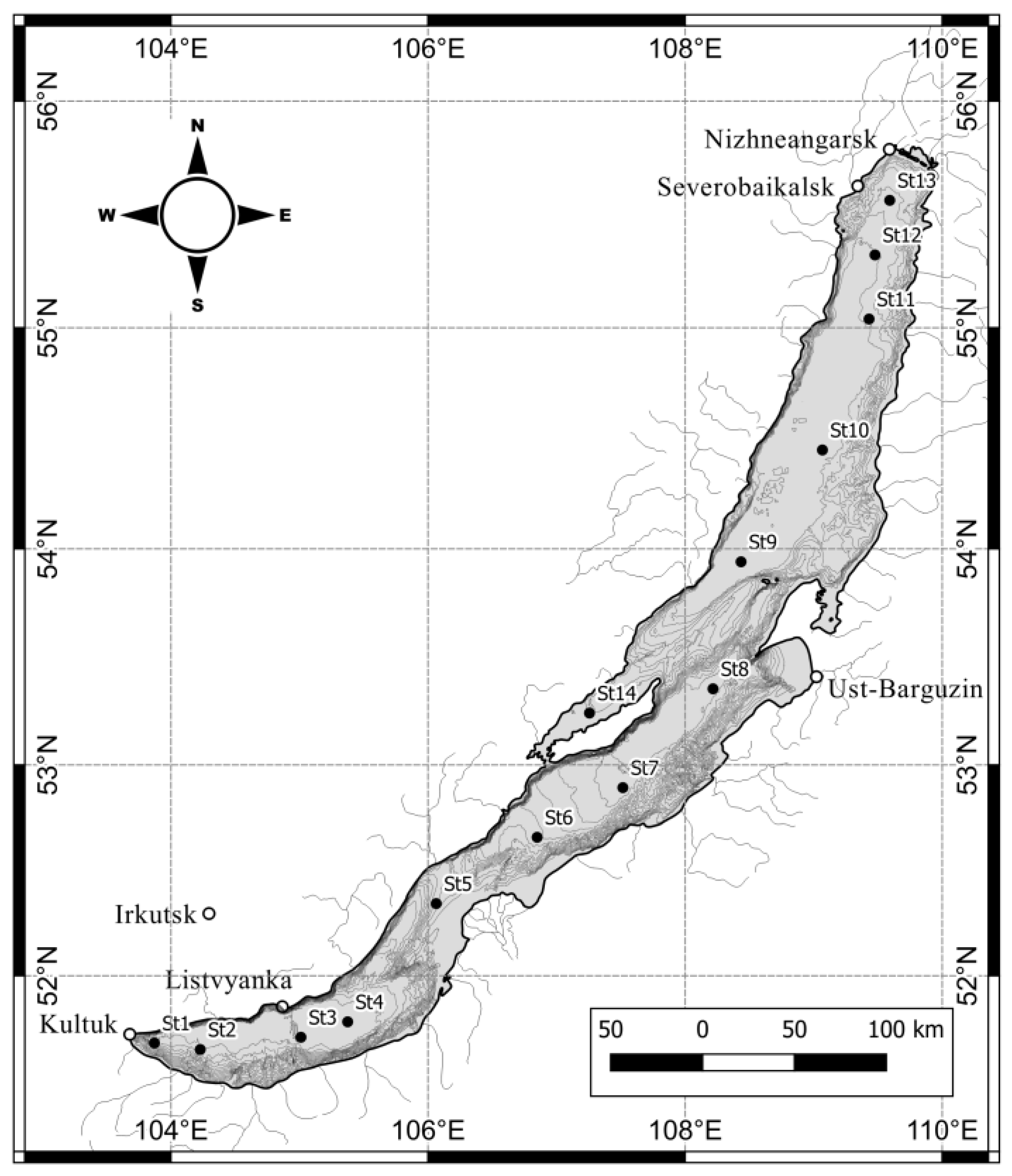
2.1. Study Site

The material for the study was sampled in March 2022 from the bottom surface of ice at 13 stations of the pelagial of Lake Baikal and 1from the strait (Figure 1, Table 1).

2.2. Water Parameters

Measurement of the hydrophysical characteristics of the under-ice water layer, temperature and electrical conductivity was determined with a CTD probe RinkoAQQ-177 (JFE, Tokyo, Japan) (temperature, T: measurement range from −3 to 45 °C, resolution 0.001 °C and measurement accuracy ±0.01 °C; electrical conductivity, EC: measurement range 0–2000 µS∙cm−1, resolution 0.1 µS∙cm−1 and measurement accuracy ±2 µS∙cm−1; photoactive radiation, PAR: measurement range 0–5000 µmol m−2·s−1, resolution 0.1 µmol m−2·s−1 and measurement accuracy ±4%). The conductivity of water was recalculated to 25 °C. Probing at the sampling point was carried out through a hole prepared in advance in the ice, 40 × 40 cm in size. The hole was made using a hand-held ice drill and an ice saw. The CTD probe was lowered using an automatic winch equipped with a cable from the water surface to a depth of 50 m. The measured parameters were displayed in real time on the screen of a portable computer.
The pH value was measured by the potentiometric method on an “Expert-pH” pH meter (Econix Expert, Moscow, Russia) (pH measurement range from 0 to 14; limits of permissible basic absolute measurement error pH = 0.05). The mineral forms of biogenic elements were determined after filtration using membrane cellulose acetate filters with a pore size of 0.45 μm (Vladisart, Vladimir, Russia). The content of biogenic elements was measured with a PE-5400VI spectrophotometer (Ecohim, Moscow, Russia): nitrates were measured using salicylic sodium, detection limit 0.1 mg∙L−1 [27]; silicon in the form of silicomolybdic heteropoly acid, detection limit 0.1 mg∙L−1 [28]; and phosphates as phosphomolybdenum complex, detection limit 0.010 mg∙L−1 [29]. Determination of chemical oxygen demand (COD) was carried out in unfiltered water via the photometric method [30] with a detection limit of 5 mg O∙L−1.

2.3. Scale Chrysophyte Sampling and Investigation

We studied water samples from the bottom surface of ice. The samples were taken by divers using 150 mL syringes.
For scanning electron microscopy (SEM), 20 mL from 200 mL of the samples was precipitated by filtration onto a filter with a diameter of 13 mm and pore diameter of 0.8 μm (Whatman Part of GE HealthCare, Chicago, IL, USA). Then, 20 mL of 70% ethanol was passed through the filter. The filter with the studied material dried at room temperature was attached to the SEM stub with double tape, coated with gold in an SDC 004 vacuum evaporator (SD 004 Balzers, Liechtenstein) and examined using a QUANTA 200 SEM (FEI Company; Hillsboro, OR, USA).
For transmission electron microscopy (TEM), the collected samples of 500 mL were filtered through 3 µm filters in a filter unit (“REATREK-Filter”, Obninsk-3, Russia) with a diameter of 47 mm, (Vladipor, Vladimir, Russia). Precipitate on the filter material was washed from the filter with 500 μL of Baikal water, and 500 μL of 8% paraformaldehyde was added to fixation. Then, 30 µL from the fixed sample was transferred to a 1.5 mL Eppendorf tube and centrifuged using a MiniSpin centrifuge (Eppendorf, Humburg, Germany) at 13,400 rpm for 10 minutes. The supernatant was removed using a pipette, and distilled water was added to the precipitate. The procedure was performed three times. Next, 30% H2O2 was added to the precipitate, and the sample was heated in a thermostat (Termit, “DNA-Technology”, Moscow, Russia) at 80 °C for 5 h. Thereafter, the precipitate was washed with distilled water and centrifuged. The procedure was performed five times. The washed samples were pipetted onto a grid covered with Formvar support, dried at room temperature, examined in a Leo 906 E TEM (Zeiss, Jena, Germany) at 80 kV and imaged with a MegaView II digital camera (Olympus Soft Imaging Solutions, Münster, Germany).

3. Results

We have discovered two new species of the genus Mallomonas, which, according to the structure of the scales, we attributed to the section Striatae.
Mallomonas baicalensis Bessudova sp. nov. (Figure 2 and Figure 3).
Description: Cells from rounded (10–20 µm) to oval (13–18 × 16–23 µm) shape (Figure 2A). The body scales are oval, 3.4–4.5 × 2.1–2.7 µm. All scale sare slightly asymmetrical, possessing a smooth dome with a spine. The shield area of the scale is patterned with a series of 6–11 regularly spaced transverse ribs lying on the basal plate. The V-rib is rounded and slightly hooded. A group of numerous rimmed pores (up to 46) is located on the posterior part of the shield in the angle of the V-rib (Figure 3E,C,G,I). The distal ends of the arms of the V-rib are continuous with the anterior submarginal ribs. The anterior flanges are smooth and narrow. The posterior flange is smooth. The posterior rim is smooth. The caudal scales, 2.9–3.3 × 1.55–2.0 µm, show the same basic design as the body scales, except for the less-developed dome with a long spine (Figure 3I). There were no bristles observed on the cells.
Holotype specimen: A portion of a single gathering of cellson the SEM stub No. 18041 is stored in the herbarium of the Limnological Institute, Siberian Branch of the Russian Academy of Sciences, Irkutsk. Figure 2 and Figure 3 show representative scales of the holotype specimen.
Type Locality: Lake Baikal, Latitude/Longitude:53°13′ N/107°45′ E
Epithet: Species name derived from “Baikal”, the lake from which this taxon was described.
Distribution: This species was found in the type locality only. pH = 7.65–8.08, T = 0.1–0.3 °C and EC25 = 140.8–153.2 μS·cm−1 (see Table 1).
Mallomonas grachevii Bessudova sp. nov. (Figure 4 and Figure 5).
Description: Dried whole cells range in size 10–14 × 15–20 µm (Figure 4A,B). The body scales are oval, 3.6–4.6 × 2.3–2.6 µm. Body scales have arounded dome. (Figure 4C,D). Dome ornamentation consists of a series of 8–11 parallel ribs, and sometimes ribs are connected (Figure 5A). The ribs of the dome, located on the right and left sides, are usually connected to the transverse rib on the shield closest to the dome (Figure 4D,E and Figure 5H). The shield is covered with a secondary layer. In the corner of the V-rib, there is a “window”, an area lacking the secondary siliceous layer, with numerous pores (Figure 5B,F). The shield of the scales is patterned with a series of 6–9 regularly spaced transverse ribs. The first transverse rib closest to the dome is usually thickened (Figure 4D and Figure 5A,G). The V-rib is rounded. The distal ends of the arms of the V-rib are continuous with the anterior submarginal ribs. The anterior flanges are narrow but well defined and separated from the shield by the submarginal ribs. The posterior flange and posterior rim are narrow and smooth. Apical scales 4–4.3 × 2–2.2 have a smaller dome, less-developed transverse ribs and more pronounced anterior flanges in comparison with the body scales (Figure 5H,I). Caudal scales 3.1–3.5 × 1.9–2.1 have the same basic design as body scales but are smaller in size (Figure 5G). Bristles are straight or slightly curved, 4.9–12.3 µm, with longitudinal ribs and without serration, with an acute tip (Figure 4C,E and Figure 5C,D).
Holotype specimen: A portion of a single gathering of cells on the SEM stub No. 18041 is stored in the herbarium of the Limnological Institute, Siberian Branch of the Russian Academy of Sciences, Irkutsk. Figure 4 and Figure 5 show representative scales of the holotype specimen.
Type Locality: Lake Baikal, Latitude/Longitude: 53°13′ N/107°45′ E.
Epithet: The species is named after DrSc Mikhail Alexandrovich Grachev (1939–2022), a famous scientist who introduced a wide range of analytical methods to the study of Lake Baikal.
Distribution: This species was found in the type locality only. pH = 7.65–8.08, T = 0.04–0.6 °C and EC25 = 140.8–153.2 μS·cm−1 (see Table 1).

4. Discussion

The section Striatae includes species with domed scales bearing craspedodontic bristles and transverse ribs of the shield [22]. The exceptions are Mallomonas kristiansenii Wujek & Bicudo, M. actinoloma Takahashi in Asmund & Takahashi, M. actinoloma var. nadiensis Dürrschmidt, M. rutifera Dürrschmidt and M. cristata Dürrschmidt, which do not have transverse ribs of the shield, but the structure of their scales or bristles is characteristic of the representatives of this section [22]. We classify M. baicalensis and M. grachevii within the section Striatae due to the presence of the well-developed transverse ribs on the shield. M. baicalensis differs significantly from all representatives of the section Striatae. A distinctive feature of M. baicalensis is its dome with a spine, which is a unique feature of the genus Mallomonas. This species also hasa slightly asymmetrical shape of all scales, regardless of their position on the cell. Of particular interest is the potential absence of bristles in M. baicalensis. To date, only two species have been discovered (Mallomonas insignis Penard and Mallomonas adamas Harris & Bradley) which lack bristles [31,32,33]. M. baicalensis is most closely related to M. siveri Němcová & Kristiansen. These species are similar in that they possess a rounded V-rib and a group of rimmed pores in the angle of the V-rib [34]. In both taxa, the distal ends of the arms of the V-rib curve become continuous with the anterior submarginal ribs, the shield is marked with about eight regularly spaced transverse ribs and the anterior flanges and posterior flanges are smooth. The scales of M. baicalensis differ from the scales of M. siveri in size. M. siveri has scales of 2.7–3.3 × 1.3–2.0 µm [34], while the scales of M. baicalensis are larger, 2.9–4.5 × 1.55–2.7 µm. All scales of M. baicalensis are slightly asymmetrical, while only individual scales of M. sivieri have asymmetry. M. baicalensis have a smooth dome with a spine, while the scales of M. siveri do not have spines. M. baicalensis is distinguished by a large amount of numerous rimmed pores (up to 46) in the corner in the angle of the V-rib, while in scales of M. sivieri their amount varies between 6 and 10 [34]. In both species, the distal ends of the arms of the V-rib bend and become continuous with the anterior submarginal ribs. However, M. siveri have weakly developed, sometimes undiscernible submarginal ribs on the rest of the anterior flange. Some M. baicalensis scales on the flanges have rudiments of ribs extending from the anterior rib (Figure 3F). M. baicalensis has narrow anterior flanges and rather wide posterior flanges with wide posterior rim. M. siveri has wider anterior flanges and narrow posterior flanges, in comparison with M. baicalensis.
The main distinguishing feature of M. grachevii is the presence of a secondary layer on the shield except the angle of the V-rib, in which an area without the secondary layer, the “window”, is present. This area contains numerous pores. One more distinguishing feature is the connection of the dome ribs, located on the right and left sides, with the transverse rib on the shield, adjacent to the dome. This transverse rib is usually thickened in comparison with other ones located on the shield. M. grachevii is most closely related to M. pseuocratis Dürrschmidt, M. asmundiae (Wujek & van der Veer) Nicholls and M. flora Harris & Bradley. These species have strong ribs on the dome, anterior submarginal ribs and narrow anterior flanges. The scales of taxa differ in shape and size. The scales of the taxa M. pseudocratis, M. flora and M. grachevii have an oval shape, while the scales of M. asmundiae differ in lateral incurvings [22,35]. The scales of M. grachevii (3.2–4.6 × 1.9–2.5 µm) are most similar in size to M. pseudocratis (3.8–4.7 × 1.8–2.5 µm). The scales M. asmundiae (4.6–11.5 × 2.7–4 µm) and M. flora (4–6 × 2.3–4 µm) are larger [22,35]. The scales of all four taxa are similar in their domes, which are prominent and have curved ribs. The scales of M. flora, M. asmundiae and M. grachevii are similar, in that the ribs on the dome are oriented in different directions in the left and right portions of the dome. The ribs on the dome of the scales of M. pseudocratis, as a rule, are transversely curved. The scales of taxa differ in the number of transverse ribs on the dome; the scales of M. flora are marked by about 6 concentric ribs [36], while the scales of M. asmundiae are marked by about 18 [35], and the scales of M. pseudocratis are marked by 8–11. The scales of all four taxa are similar in the presence of transverse ribs on the shield; however, they differ in their number. The scales of M. grachevii (6–9) have the least number of ribs on the shield, followed by M. pseudocratis (10–15) [22] and M. flora (9–13) [22], and the largest number of transverse ribs are seen in the scales of M. asmundiae (7–33) [35]. It is worth noting that the thickening of the first transverse rib in the scales of M. grachevii is also found in the scales of M. flora var. palermii Vigna and is a diagnostic feature of this variety. The scales of all taxa are similar in the presence of a V-rib which is hooded. However, the scales of M. flora clearly differ from the three taxa by the presence of the rounded angle of the hood and small teeth. In addition, the scales of M. asmundiae have a well-developed hood and, in the larger scales, may cover as much as 20% of the shield area [35]. On the scales of M. grachevii and M. asmundiae in the corner of the V-rib, there are numerous pores. The scales of M. flora differ due to the presence of a flower-like pattern in the angle of the V-rib. The scales of M. pseudocratis have proximity with a patch of pores and one of the ribs is interrupted there. The V-rib continues on the anterior submarginal ribs of the scales of M. grachevii, M. flora and M. asmundiae. The scales of M. pseudocratis are distinguished by winged anterior submarginal ribs [35]. The scales of M. grachevii are differentiated by narrow but well-defined anterior flanges. The scales of M. pseudocratis have anterior flanges that are weakly developed. The anterior flanges of the scales of M. asmundiae can be very wide and merge with the distal extension of the shield to form wing like structures on either side of the dome. The scales of M. flora have anterior flanges with struts. M. grachevii differs in the presence of narrow and smooth posterior flanges. The scales of the other taxa have struts on the posterior flanges. The bristles of the new taxa, as well as the other three taxa, are curved, but they differ from the rest through their unserrated longitudinal ribs. The bristles of M. asmundiae, M. flora and M. pseudocratis are dentate.
For a long time, the section Striatae included 15 taxa: M. striata var. striata Asmund, M. striata var. serrata Harris & Bradley, M. flora, M. flora var. palermii, M. cratis Harris & Bradley, M. pseudocratis, M. asmundiae, M. pseudocratis var. deltaensis Siver & Vigna, M. actinoloma, M. actinoloma var. maramuresensis Péterfi & Momeu, M. actinoloma var. nadiensis, M. cristata, M. rutifera, M. verrucosa Vigna and M. kristiansenii Wujek & Bicudo [22]. Recent studies have supplemented the species diversity of the section with eight more species: M. striata var. balonovii Voloshko, M. striata var. getseniae Voloshko [23], M. siveri [34], M. pechlaneri Němcová & Rott [24], M. marina Jeong, Kim, Jo, Kim, Siver & Shin [37], M. voloshkoae Gusev, Němcová & Kulikovskiy [25], M. punctostriata Gusev & Kulikovskiy [38] and M. altaica Gusev & Martynenko [26].
Including 2 undescribed species, as well as the above-described M. baicalensis and M. grachevii, the section now includes 27 species. Among the representatives of the section Striatae, there are both widespread and rare species. Rare species have previously been shown to be confined to oligotrophic/mountain water bodies [26]. To date, eight species of the section Striatae have been found in Lake Baikal: M. striata var. striata, M. striata var. balonovii, M. striata var. getseniae, M. voloshkoae, M. pechlaneri and M. sibirica [15] (Figure 6), as well as M. baicalensis and M. grachevii.
The species M. striata var. balonovii, M. striata var. getseniae, M. voloshkoae and M. pechlaneri are rare species characteristic of oligotrophic/mountain water bodies, the last two being found in the pelagial of the lake for the first time.
Most often, the scales of M. striata var. getseniae are found in the spring–summer period both in a silicified state and “fresh” with an undisturbed siliceous structure. Single scales of the species with bristles have also been found in the investigated subglacial samples (see Figure 6D,H,I). M. voloshkoae, M. pechlaneri, M. sibirica and M. striata var. balonovii are typical only of the spring–summer period; during the ice period, their scales were not found. In scales of M. striata var. balonovii, only silicified scales were found. The new species M. baicalensis and M. grachevii were found as whole cells and individual scales during the ice period; single scales were found in the spring–summer period. Thus, the described new species are typical for the under-ice (March) period, along with a rare species from the section Striatae M. striata var. getseniae.
By the end of the February–March period, organisms of the under-ice community have already begun to actively develop in Lake Baikal at the water–ice interface [18,34,35,36,37]. They are attached to the underside of the ice or intensively multiply in intermediate spaces filled with water inside the ice [18,39]. The subglacial community of Lake Baikal is characterized by the development of diatoms [12,39,40,41,42,43] and dinophytes [13,44,45,46,47,48,49,50].
Among chrysophytes, Dinobryon cylindricum O.E. Imhof, Mallomonas vannigera and Synura petersenii presented with in ice in Lake Baikal [18]. Additionally, we found two new species, M. baicalensis and M. grachevii, at the bottom surface of the ice. Despite the similarity of habitat parameters at the stations from south to north (see Table 1), these two species had a patchy distribution, associated either with the asynchronous passage of life cycles at different stations, or with a relatively low rate of cell migration compared to the size of Lake Baikal (see Figure 1).

5. Conclusions

M. baicalensis and M. grachevii have been observed at the same environmental parameters, but their distribution on the bottom surface of the ice of Lake Baikal was uneven, despite similar conditions throughout the lake from south to north (see Figure 1, Table 1).
At present, Lake Baikal is the only water body in which eight taxa of the genus Mallomonas section Striatae, including two undescribed taxons, have been found. This fact characterizes the uniqueness of Lake Baikal.

Author Contributions

A.B., A.F. and D.H., electron microscopy; A.B., collection of literature, interpreting the results and writing the first version of the manuscript; M.M. and M.S., sampling and hydrochemical analysis; M.B. and Y.Z., sampling and filtration; I.K., under-ice sampling; Y.L., review and editing. All authors took part in writing the manuscript. All authors have read and agreed to the published version of the manuscript.

Funding

The study is supported by projects FWSR-2021-0008, No. 121032300186-9 (Limnological Institute—electron microscopy, species identification and description) and FWSR-2021-0005, No. 121032300224-8 (Limnological Institute—expedition, sampling and water parameters).

Data Availability Statement

The data that support the findings of this study are available from the corresponding authors upon reasonable request.

Acknowledgments

The authors express their gratitude to Andrey Fedotov for organizing and conducting the expedition, as well as for under-ice sampling, and to Evgeny Gusev for valuable advice in describing the species. The microscopy studies were performed at the Electron Microscopy Center of the Shared Research Facilities “Ultramicroanalysis” of Limnological Institute, https://www.lin.irk.ru/copp/ (accessed on 24 March 2023).

Conflicts of Interest

The authors declare no conflict of interest.

References

  1. Baikal., A.; Galazy, G.I. (Eds.) Federal Agency for Geodesy and Cartography: Moscow, Russia, 1993; 160p.
  2. Shimaraev, M.N. Hydrometeorological factors and oscillations of Baikal plankton abundance. In Limnology of Pre-Delta Areas of Lake Baikal; Nauka: Leningrad, Russia, 1971; pp. 259–267. [Google Scholar]
  3. Granin, N.G.; Jewson, D.H.; Gnatovsky, R.Y.; Levin, L.A.; Zhdanov, A.A.; Gorbunova, L.A.; Tsekhanovsky, V.V.; Doroschenko, L.M.; Mogilev, N.Y. Turbulent mixing under ice and the grow the of diatoms in Lake Baikal. Verh. Internat. Verein. Limnol. 2000, 27, 2812–2814. [Google Scholar]
  4. Jewson, D.H.; Granin, N.G.; Zhdanov, A.A.; Gnatovsky, R.Y. Effect of snow depth on under-ice irradiantce and growth of Aulacoseira baicalensis in Lake Baikal. Aquat. Ecol. 2009, 43, 673–679. [Google Scholar] [CrossRef]
  5. Grachev, M.A.; Domysheva, V.M.; Khodzher, T.V.; Korovyakova, N.V.; Golobokova, L.P.; Vereshagina, A.L.; Granin, N.G.; Gnatovsky, R.Y.; Kostornova, T.Y. Deep water of Lake Baikal as a standard of fresh water. Chem. Sust. Develop. 2004, 12, 417–429. [Google Scholar]
  6. Khodzher, T.V.; Domysheva, V.M.; Sorokovikova, L.M.; Sakirko, M.V.; Tomberg, I.V. Current chemical composition of LakeBaikal water. Inland Waters 2017, 7, 250–258. [Google Scholar] [CrossRef]
  7. Domysheva, V.M.; Sorokovikova, L.M.; Sinyukovich, V.N.; Onishchuk, N.A.; Sakirko, M.V.; Tomberg, I.V.; Zhuchenko, N.A.; Golobokova, L.P.; Khodzher, T.V. Ionic composition of water in Lake Baikal, its tributaries, and the Angara river source during the modern period. Russ. Meteorol. Hydrol. 2019, 44, 687–694. [Google Scholar] [CrossRef]
  8. Grachev, M.A. On Recent State of Ecological System of Lake Baikal; SB RAS: Novosibirsk, Russia, 2002; 153p. [Google Scholar]
  9. Granin, N.G.; Dzhuson, D.; Gnatovsky, R.Y.; Levin, L.A.; Zhdanov, A.A.; Averin, A.I.; Gorbunova, L.A.; Tsekhanovskiy, V.V.; Doroshchenko, L.F.; Min’ko, N.P.; et al. Turbulent mixing of Lake Baikal waters in the layer immediately adjacent to the ice and its role in the development of diatoms. Rep. Russ. Acad. Sci. 1999, 366, 835–839. [Google Scholar]
  10. Domysheva, V.M.; Usoltseva, M.V.; Sakirko, M.V.; Pestunov, D.A.; Shimaraev, M.N.; Popovskaya, G.I.; Panchenko, M.V. Spatial distribution of carbon dioxide fluxes, biogenic elements, and phytoplankton biomass in the pelagic zone of Lake Baikal in spring 2010–2012. Atmos. Ocean. Opt. 2014, 27, 539–545. [Google Scholar] [CrossRef]
  11. Votinsev, K.K. Hydrochemistry of Lake Baikal; Izdatelstvo Akademii Nauk SSSR: Moscow, Russia, 1961; 312p. [Google Scholar]
  12. Bashenkhaeva, M.V.; Zakharova, Y.R.; Petrova, D.P.; Khanaev, I.V.; Galachyants, Y.P.; Likhoshway, Y.V. Sub-ice microalgal and bacterial communities in fresh water Lake Baikal. Russia Microb. Ecol. 2015, 70, 751–765. [Google Scholar] [CrossRef] [PubMed]
  13. Bashenkhaeva, M.V.; Galachyants, Y.P.; Khanaev, I.V.; Sakirko, M.V.; Petrova, D.P.; Likhoshway, Y.V.; Zakharova, Y.R. Comparative analysis of free-living and particle-associated bacterial communities of Lake Baikal during the ice-covered period. J. Great Lakes Res. 2020, 46, 508–518. [Google Scholar] [CrossRef]
  14. Bessudova, A.; Domysheva, V.M.; Firsova, A.D.; Likhoshway, Y.V. Silica-scaled chrysophytes of Lake Baikal. Acta Biol. Sib. 2017, 3, 47–56. [Google Scholar] [CrossRef] [Green Version]
  15. Bessudova, A.; Firsova, A.D.; Tomberg, I.V.; Bayramova, E.; Hilkhanova, D.; Bedoshvili, E.L.; Bashenkhaeva, M.V.; Kopyrina, L.I.; Zakharova, Y.R.; Likhoshway, Y.V. Two new species of silica-scaled chrysophytes Mallomonas kicherica and M. sibirica inwater bodies of Eastern Siberia, Russia. Phytotaxa 2023, in press. [Google Scholar]
  16. Firsova, A.D.; Kuzmina, A.E.; Tomberg, I.V.; Potemkina, T.G.; Likhoshway, Y.V. Sezonnaya dinamica formirovaniya stomatocyst chrysophytovych vodorosley v planktone Yuzhnogo Baikala. Izv. RAN Seriya Biol. 2008, 5, 589–596. [Google Scholar]
  17. Firsova, A.D.; Bessudova, A.; Likhoshway, Y.V. Chrysophycean stomatosysts in tributaries of northern limit of Lake Baikal. Acta Biol. Sib. 2018, 4, 25–44. [Google Scholar] [CrossRef] [Green Version]
  18. Bondarenko, N.A.; Belykh, O.I.; Golobokova, L.P.; Artemyeva, O.V.; Logacheva, N.F.; Tikhonova, I.V.; Lipko, I.A.; Kostornova, T.Y.; Parfenova, V.V.; Khodzher, T.V.; et al. Stratified distribution of nutrients and extremophile biota with in fresh water icecovering the surface of Lake Baikal. J. Microbiol. 2012, 50, 8–16. [Google Scholar] [CrossRef] [PubMed]
  19. Mikhailov, I.S.; Galachyants, Y.P.; Bukin, Y.S.; Petrova, D.P.; Bashenkhaeva, M.V.; Sakirko, M.V.; Blinov, V.V.; Titova, L.A.; Zakharova, Y.R.; Likhoshway, Y.V. Seasonal succession and coherence among bacteria and microeukaryotes in Lake Baikal. Microb. Ecol. 2022, 84, 404–422. [Google Scholar] [CrossRef]
  20. Guiry, M.D.; Guiry, G.M. AlgaeBase; World-Wide Electronic Publication; National University of Ireland: Galway, Ireland, 2023; Available online: https://www.algaebase.org (accessed on 10 March 2023).
  21. Asmund, B.; Kristiansen, J. The Genus Mallomonas (Chrysophyceae). A taxonomic survey based on the ultrastructure of silica scales and bristles. Opera Bot. 1986, 85, 1–128. [Google Scholar]
  22. Kristiansen, J.; Preisig, H.R. Chrysophyta and Haptophyta algae, 2nd part: Synurophyceae. In Süsswasser Flora von Mitteleuropa (Freshwater Flora of Central Europe); Springer: Berlin, Germany, 2007; pp. 1–252. [Google Scholar]
  23. Voloshko, L.N. New taxa of the genus Mallomonas (Chrysophyta, Synurophyceae) from lakes of the Polar Ural. Russ. Bot. J. 2009, 94, 1068–1076. [Google Scholar]
  24. Nemcová, Y.; Rott, E. Diversity of silica-scaled chrysophytes inhigh-altitude alpinesites (North Tyrol, Austria) including a description of Mallomonas pechlaneri sp. nov. Cryptogam. Algol. 2018, 39, 63–83. [Google Scholar] [CrossRef]
  25. Gusev, E.; Němcová, Y.; Kulikovskiy, M. Mallomonas voloshkoae sp. nov. (Synurales, Chrysophyceae) and distribution of M. pechlaneri in mountain lakes of Siberia. Phytotaxa 2022, 530, 221–229. [Google Scholar] [CrossRef]
  26. Gusev, E.; Martynenko, N. Silica-scaled chrysophytes of Teletskoye lake and Adjacent area with a description of a new species from the genus Mallomonas. Diversity 2022, 14, 1040. [Google Scholar] [CrossRef]
  27. ISO 7890-3: 1988; Water Duality—Determination of Nitrate-Part 3: Spectrometric Method Using Sulfosalicylic Acid. International standard ISO: Geneva, Switzerland, 2004.
  28. Wetzel, R.; Likens, G. Limnological Analysis, 3rd ed.; Springer: New York, NY, USA, 2000; pp. 57–112. [Google Scholar]
  29. ISO 6878: 2004; Water Quality—Determination of Phosphorus—Ammonium Molybdate Spectrometric Method. International standard ISO: Geneva, Switzerland, 2004.
  30. ISO 15705: 2002; Water Quality—Determination of the Chemical Oxygen Demand Index (ST-COD)—Small-Scale Sealed-Tube Method. International standard ISO: Geneva, Switzerland, 2002.
  31. Lavau, S.; Saunders, G.W.; Wetherbee, R. A phylogenetic analysis of the Synurophyceae using molecular data and scale case morphology. J. Phycol. 1997, 33, 135–151. [Google Scholar] [CrossRef]
  32. Siver, P.A.; Wolfe, A.P.; Rohlf, F.J.; Shin, W.; Jo, B.Y. Combining geometric morphometrics, molecular phylogeny, and micropaleontology to assess evolutionary patterns in Mallomonas (Synurophyceae: Heterokontophyta). Geobiology 2013, 11, 127–138. [Google Scholar] [CrossRef]
  33. Siver, P.A. Synurophyte algae. In Freshwater Algae of North America: Ecology and Classification., 2nd ed.; Academic Press: Boston, MA, USA, 2015; pp. 607–651. [Google Scholar]
  34. Němcová, Y.; Bulant, P.; Kristiansen, J. Mallomonas solea-ferrea and Mallomonas siveri (Chrysophyceae/Synu-ophyceae): Twonew taxa from the Western Cape (South Africa). Nova Hedwig. 2011, 93, 375–384. [Google Scholar] [CrossRef]
  35. Nicholls, K.N. Form variation in Mallomonas asmundiue and a description of Mallomonas sphagniphila sp. nov. (series Comconticae, Mallomonadaceae). Can. J. Bot. 1987, 65, 627–634. [Google Scholar] [CrossRef]
  36. Harris, K.; Bradley, D.E. A taxonomic study of Mallomonas. J. Gen. Microbiol. 1960, 22, 750–777. [Google Scholar] [CrossRef] [Green Version]
  37. Jeong, M.; Kim, J.I.; Jo, B.Y.; Kim, H.S.; Siver, P.A.; Shin, W. Surviving the marine environment: Two new species of Mallomonas (Synurophyceae). Phycologia 2019, 58, 276–286. [Google Scholar] [CrossRef]
  38. Gusev, E.; Kulikovskiy, M. Two new species of genus Mallomonas from swamp localities in Vietnam. Phytotaxa 2020, 468, 121–129. [Google Scholar] [CrossRef]
  39. Obolkina, L.A.; Bondarenko, N.A.; Doroshenko, L.F.; Molozhavaya, O.A. Finding of a cryophilic community in Lake Baikal. DAN 2000, 371, 815–817. [Google Scholar]
  40. Popovskaya, G.I. Ecological monitoring of phytoplankton in Lake Baikal. Aquat. Ecosyst. Health. Manag. 2000, 3, 215–225. [Google Scholar] [CrossRef]
  41. Bordonsky, G.S.; Bondarenko, N.A.; Obolkina, L.A.; Timoshkin, O.A. Ice communities of Baikal. Priroda 2003, 7, 22–24. [Google Scholar]
  42. Bashenkhaeva, M.V.; Zakharova, Y.R.; Galachyants, Y.P.; Khanaev, I.V.; Likhoshway, Y.V. Bacterial communities during theperiod of massive under-ice dinoflagellate development in Lake Baikal. Microbiology 2017, 86, 524–532. [Google Scholar] [CrossRef]
  43. Bondarenko, N.A.; Timoshkin, O.A.; Röpstorf, P.; Melnik, N.G. Theunder-ice and bottom periods in the life of Aulacoseira baicalensis (K. Meyer) Simonsen, a principal Lake Baikal alga. Hydrobiol. J. 2006, 568, 107–109. [Google Scholar] [CrossRef]
  44. Skabitchevsky, A.P. On the biology of Melosira baicalensis (K. Meyer) Wisl. Russ. Hydrobiol. J. 1929, 8, 93–114. [Google Scholar]
  45. Yasnitsky, V.N. Results on observations of Baikal plankton in the region of biological station during 1926–1928. Proc. Biol. Geogr. 1930, 4, 191–234. [Google Scholar]
  46. Izmest’eva, L.R.; Moore, M.V.; Hampton, S.E. Seasonal dynamics of common phytoplankton in Lake Baikal. Proc. Russ. Acad. Sci. Sci. Cent. 2006, 8, 191–196. [Google Scholar]
  47. Bedoshvili, E.L.; Bondarenko, N.A.; Sakirko, M.V.; Khanaev, I.V.; Likhoshway, Y.V. Changes of colony length in the planktonic diatom Aulacoseira baicalensis at different stage sofits annual cyclein Lake Baikal. Hydrobiol. J. 2007, 43, 81–89. [Google Scholar] [CrossRef]
  48. Kozhova, O.M.; Izmest’eva, L.R. Lake Baikal: Evolution and Biodiversity; Backhuys Publish: Leiden, The Netherlands, 1998; 447p. [Google Scholar]
  49. Pomazkina, G.V.; Belykh, O.I.; Domysheva, V.M.; Sakirko, M.V.; Gnatovsky, R.Y. Structure and dynamics of phytoplankton of Southern Baikal (Russia). Intern. J. Algae 2010, 12, 64–79. [Google Scholar] [CrossRef]
  50. Annenkova, N.V. Phylogenetic relations of the dinoflagellate Gymnodinium baicalense from Lake Baikal. Cent. Eur. J. Biol. 2013, 8, 366–373. [Google Scholar] [CrossRef]
Figure 1. Maps cheme of sampling in Lake Baikal.
Water 15 02250 g001
Figure 2. Mallomonas baicalensis sp. nov. (A) A cell covered with scales; (BD) a group of scales from the type habitat; (E) scales with an aberrant rib pattern. Scale bars: (CE) 2 μm; (B) 5 μm; (A) 20 μm. SEM: (AE).
Water 15 02250 g002
Figure 3. M. baicalensis sp. nov., scales from the type habitat. (A) An apical scale; (BG) body scales; (F) a body scale, with rudiments of ribs extending from the anterior rib on the flanges; (H) a low silicified body scale without ribs on the shield; (I) a caudal scale without ribs and with a small dome and a long spine. Scale bars: 2 μm. SEM: (A,EG,I). TEM: (BD,H).
Water 15 02250 g003
Figure 4. Mallomonas grachevii sp. nov., scales from the type habitat. (A,B) Dried whole cells; (C,E) a group of scales with bristles from the type habitat; (D) a group of scales from the type habitat. Scale bars: 2 μm. SEM: (AE).
Water 15 02250 g004
Figure 5. M. grachevii sp. nov., scales from the type habitat. (A) Body scales with ribs connected on the dome; (B) body scales with a clearly visible “window” with numerous pores; (C) bottom surface of a scale with bristle; (D) a body scale with bristle; scales with an aberrant rib pattern; (E) scale with an aberrant rib pattern; (F) pores in the corner of the V-rib; (G) caudal scale; (H) apical scale with less developed transverse ribs; (I) bottom surface of a scale. Scale bars: (A,B,EI) 2 μm; (D) 5 μm; (C) 10 μm. SEM: (A,CG,I). TEM: (B,H).
Water 15 02250 g005
Figure 6. Rare species Mallomonas section Striatae from Lake Baikal. (A,B) Mallomonas pechlaneri, body scales; (C) Mallomonas striata var. balonovii, body scales; (D) Mallomonas voloshkoae, body scale (E) Mallomonas striata var. getseniae, body scales; (F) M. sibirica, body scales. Scale bars: (AF) 2 μm. SEM: (B,DF). TEM: (A,C).
Water 15 02250 g006
Table 1. Ecological conditions and distributional records for M. baicalensis and M. grachevii. Site numbers correspond to those in Figure 1. Occurrence of new species: single scales (+); frequent scales, clusters of scales (++); whole cells (+++); not found (N.f.).
St. No.Coordinates
N/E
Date of Sampling
dd.mm.yy
Time of Sampling
hh:mm
Ice Thickness, cmSnow Cover Thickness, cmT, °CpHEC25,
μS·cm−1
PAR,
μmol·m−2·s−1
Si,
mg·L−1
PO43−,
mg·L−1
NO3,
mg·L−1
COD,
mgO·L−1
M. baicalensisM. grachevii
1.51° 40.578′/
103° 52.309′
17.03.2214:2563120.0447.80141.159.8080.470.0280.416.6N.f.+
2.51° 38.710′/
104° 13.715′
17.03.2218:3745210.0497.82141.30.00.510.0230.446.5N.f.N.f.
3.51° 42.262′/
105° 00.720′
18.03.2211:406390.0737.94142.5154.6260.530.0250.427.0N.f.N.f.
4.51° 46.731′/
105° 22.528′
19.03.2209:526440.2747.99141.2152.80.530.0230.366.5+N.f.
5.52° 20.722′/
106° 03.870′
19.03.2214:336570.6518.08141.896.1770.600.0210.367.4++++++
6.52° 39.590′/
106° 50.978′
19.03.2218:038540.2437.93141.50.00.600.0280.406.7+N.f.
7.52° 53.630′/
107° 31.001′
20.03.2211:34673.50.1877.71140.998.1740.550.0230.365.3++++
8.53° 21.278′/
108° 13.078′
20.03.2216:548570.1097.65143.37.4760.540.0240.385.7++++++
9.53° 56.472′/
108° 26.178′
23.03.229:208850.1477.95140.8364.9880.450.0200.314.2++++++
10.54° 27.052′/
109° 04.164′
21.03.2211:0584110.1077.80141.0117.2140.530.0240.425.4N.f.+
11.55° 02.388′/
109° 25.939′
22.03.2212:568480.1028.04146.176.4150.600.0250.406.1N.f.+
12.55° 19.487′/
109° 28.707′
21.03.2215:308680.0687.92143.033.6290.550.0260.396.2+++++
13.55° 33.968′/
109° 35.597′
22.03.2209:3483100.1107.94153.247.8390.620.0220.384.1++++
14.53° 14.500′/
107° 15.416′
23.03.2215:307850.3258.04141.836.0310.500.0310.299.4+N.f.
Disclaimer/Publisher’s Note: The statements, opinions and data contained in all publications are solely those of the individual author(s) and contributor(s) and not of MDPI and/or the editor(s). MDPI and/or the editor(s) disclaim responsibility for any injury to people or property resulting from any ideas, methods, instructions or products referred to in the content.

