Article

Identification of Antibiotic Resistance Gene Hosts in Treatment Wetlands Using a Single-Cell Based High-Throughput Approach

1
Department of Environmental Biotechnology, Helmholtz Centre for Environmental Research—UFZ, 04318 Leipzig, Germany
2
Medical Microbiology and Parasitology, Faculty of Medicine, University of Buenos Aires-National Council for Scientific and Technological Research (IMPaM, UBA-CONICET), 1121 Ciudad Autónoma de Buenos Aires, Argentina
3
Institute for Biological Interfaces (IBG-5), Karlsruhe Institute of Technology, 76344 Eggenstein-Leopoldshafen, Germany
4
Environmental and Biotechnology Centre, Helmholtz Centre for Environmental Research—UFZ, 04318 Leipzig, Germany
5
Research Unit REVERSAAL, National Research Institute for Agriculture, Food and the Environment (INRAE), 69625 Villeurbanne, France
*
Author to whom correspondence should be addressed.
Water 2023, 15(13), 2432; https://doi.org/10.3390/w15132432
Submission received: 6 June 2023 / Revised: 25 June 2023 / Accepted: 28 June 2023 / Published: 30 June 2023
(This article belongs to the Section Water Quality and Contamination)

Abstract

:
Determining the prevalence of antimicrobial resistance (AMR) in non-clinical settings is vital for better management of the global AMR crisis. Untreated and even treated wastewaters are important sources that release AMR into the environment. Methodologically, it is difficult to generate a comprehensive in situ profile of antibiotic resistance gene hosts. Here, we used epicPCR (emulsion, paired isolation, and concatenation PCR) as a cultivation-independent method to reveal the host profiles of the AMR indicator genes intI1, sul1, sul2, and dfrA1 in two constructed wetlands treating municipal wastewater. Overall, the epicPCR analysis revealed a profile of AMR indicator gene hosts that is consistent with literature data from cultivation-based approaches. Most carriers of antibiotic resistance (AR) genes and likely of class 1 integrons belonged to the Gammaproteobateria, particularly the Burkholderiaceae and Rhodocyclaceae families, followed by members of the Campylobacterota, Desulfobacterota, and Firmicutes. The analysis also identified several novel hosts for the indicator genes widely distributed in the wetlands, including the genera Legionella and Ralstonia. Therefore, the application of epicPCR has produced an expanded insight into the in situ indicator gene host profile, while highlighting the role of the environment as a reservoir for AMR.

Graphical Abstract

1. Introduction

Infectious diseases caused by antibiotic-resistant bacteria are one of the major health burdens of our time. To contain the spread of these bacteria and the antibiotic resistance (AR) genes they carry (collectively, antimicrobial resistance or AMR), the One Health approach advocates for the inclusion of environmental compartments in surveillance and prevention efforts [1,2]. The release of treated and untreated wastewater into surface waters is a substantial pathway for the distribution of AMR via the environment [3,4]. Monitoring the prevalence of AMR in wastewater is therefore an important part of evaluating the risk to human and animal health posed by the spread of AMR in the environment [5].
The antibiotic resistance genes that are commonly detected in waste and surface waters include sul1, sul2, and dfrA1, all of which confer resistance against inhibitors of folate biosynthesis [3,6]. The sul1 gene is part of the classical class 1 integron, an important carrier in AR gene dispersal [7]. Another defining feature of that integron type is the intI1 gene, which codes for an integrase that can recombine the AR gene cassette content of the integron. The association of intI1 with AMR has led to its use as an indicator of anthropogenic pollution in water bodies [8,9]. Class 1 integrons have predominantly been found in Gammaproteobacteria [10]. A recent publication listed 104 bacterial species from 44 genera as carriers of that integron [11]. Two thirds of the genera belong to the Pseudomonadota (almost always Gammaproteobacteria), with the remainder being members of the Actinobacteriota, Bacteroidota, Campylobacterota, and Firmicutes. Gammaproteobacteria also account for by far the largest proportion of integrons detected in sequence databases. Approximately one fifth of all sequenced genomes of this class contain at least one integron of the class 1 or a similar type [12]. Yet, the information on prevalence in sequenced genomes does not necessarily translate 1:1 to a specific environmental site. Generating a comprehensive in situ host profile of class 1 integrons and AR genes, including sul1&2 and dfrA1, is not a trivial task. The classical cultivation-dependent approach is subject to bias, as many bacteria do not grow readily in the laboratory. Shotgun metagenomics, as a cultivation-independent approach, can detect AR genes in a microbial community, but has limitations in assigning respective hosts due to the frequent association of these genes with mobile genetic elements [13,14,15]. Host profiling can be achieved using recently developed molecular biology methods. Proximity DNA ligation methods such as Hi-C involve the metagenomic sequencing of DNA that has been cross-linked prior to extraction from its host [15]. However, the generation of one metagenome per sample renders the approach expensive if the research aim is to capture temporal and spatial variations in host profiles. An alternative method is epicPCR (emulsion, paired isolation, and concatenation polymerase chain reaction), which generates fusion PCR amplicons of parts of the 16S rRNA gene and another target gene of single cells imbedded in polyacrylamide beads [16]. The fusion products are then isolated and sequenced in parallel. So far, the method has been applied to reveal bacterial hosts of various genes associated with AMR, including intI1 and sul1, in environmental samples [17,18,19,20,21]. A variant of epicPCR was used to identify bacterial hosts of beta-lactamases in human gut microbiomes [22].
Here, we used epicPCR to identify hosts of intI1, sul1&2, and dfrA1 in two treatment wetlands. Such wetlands are nature-based solutions that are increasingly applied as a cost-effective option to treat domestic and industrial wastewater [23,24]. Removal of bacteria and chemical pollutants introduced with the influent, including antibiotics, can be achieved with similar or even better efficiency than activated sludge-based treatment systems, although with longer hydraulic residence times [25,26,27]. In a previous study on the fate of AMR in the same treatment wetlands investigated herein, we observed increases in integron integrase activity and intI1 and sul abundance, which were likely caused by cellular stress [28]. There was no evidence of an influence of folate biosynthesis inhibitors on the prevalence of AMR. Increases in intI1 and sul1 abundance, apparently independent of such inhibitors, have also been detected in rivers downstream of wastewater treatment plants [29,30,31]. It is uncertain which bacteria are involved in such increases. In the wetlands, we found mostly Aeromonas spp., Escherichia coli, and Pseudomonas spp., as hosts of the indicator genes, using cultivation-dependent profiling. The present application of epicPCR aimed to further explore the host profiles.

2. Materials and Methods

2.1. Study Site and Sampling Regime

The pilot-scale horizontal subsurface flow treatment wetlands investigated in this study are located at the Langenreichenbach Ecotechnology Research Facility, 42 km north-east of Leipzig, Germany (51.50613 N, 12.89758 E). The design and operation of the wetlands have previously been described in detail [32]. In brief, the wetlands have a surface area of 5.6 m2 and are named H50p for Horizontal, 50 cm depth, and planted, and HAp for Horizontal, Aerated, and planted. Their main vegetation is the common reed, Phragmites australis. Wastewater from the surrounding rural residential villages is pretreated in a septic tank and pumped from there to H50p and HAp, as the sole source of water other than precipitation.
Samples were taken monthly from May through July 2017 for method development, and in August 2017 for data generation. The conventional water characteristics (temperature, pH, dissolved oxygen, and inorganic nitrogen species) and relevant microbiological data (AR gene abundances and 16S rRNA profiles) of the sampling period have been previously reported [28,33]. There was only small variation in the conventional and microbiological water characteristics during the sampling period. One-liter samples were taken from the septic tank, the effluent, and from inside the treatment wetlands at 0.12, 0.25, 0.50, 0.75 of the fractional length and 0.50 of the fractional depth via permanently installed tubes. The samples were chilled and transported within 1–2 h to the laboratory for further processing.

2.2. epicPCR

The following protocol is based on previous studies with some modifications [16,18]. Table S1 shows all of the primers used for fusion PCR, (semi)-nestedPCR, and the introduction of adapter sequences for Illumina sequencing [34,35,36,37]. Almost all primers were previously used either in epicPCR or in quantitative PCR. The exception was the forward primer for the amplification of dfrA1, which was newly designed using NCBI’s online tool Primer-BLAST, with the reference sequence downloaded from GenBank (accession number 3244905). The establishment of the overall method, including optimized PCR conditions, was performed using a Citrobacter sp. strain S22 isolated from the treatment systems for sul1, sul2 and dfrA1 [28], and Pseudomonas plecoglossicida M135 for intI1 [38]. Cell numbers in the assays were determined using a Coulter counter (Beckman Coulter, Indianapolis, IN, USA). Correct amplification of the intended epicPCR fusion product was ensured via agarose gel electrophoresis and Sanger sequencing (Macrogen, Amsterdam, The Netherlands).
Samples were first inspected via microscopy. When cell aggregates were present, samples were passed through a cell strainer (40 μm pore size; Falcon, NY, USA). Cells in all samples were counted via DAPI (4′,6-diamidin-2-phenylindol) staining using a fluorescence microscope (Axioscope, Zeiss, Oberkochen, Germany), and then pelleted in a benchtop centrifuge (Eppendorf, Hamburg, Germany) at 8000 rpm for 15 min at room temperature. The volume of the respective sample depended on the sampling site, and ranged from 2 to 180 mL to obtain approximately 1–2 × 10⁸ cells per 100 µL epicPCR assay. Cell numbers were determined with a Coulter counter. After centrifugation, the pellets containing the cells were carefully resuspended using vortexing and pipetting in 100 μL of nuclease free water. The absence of cell aggregates in the suspension was checked for using microscopy. Then, the cell suspension was amended with 100 μL of 30% acrylamide/bis-acrylamide and 25 μL of ammonium persulfate (APS), and gently vortexed. After addition of 600 μL mineral oil, the mixtures were vortexed for 30 s at 3000 rpm. Then, 25 μL of tetramethylethylenediamine (TEMED) was added, followed by vortexing for 45–60 s at 3000 rpm. The mixture was kept at room temperature for 90 min to allow polymerization. To collect the polyacrylamide beads, 800 μL of diethyl ether was added, whereupon a precipitate was formed. After removing the diethyl ether and adding 1 mL of nuclease-free water, mixing, and centrifuging for 30 s at 12,000× g, three layers formed: a bottom layer containing the beads, a middle layer containing a mixture of oil and water, and an upper layer of oil. The oil layer was discarded. This step of mixing water into the bead suspension was repeated until no more three-layer-formation was visible. The water layer was discarded and 1 mL of TK buffer (20 mM Tris-HCl, pH 7.5, 60 mM KCl) was added to the beads. The bead suspension was passed through a cell strainer (40 μm pore size) with the flow-through containing the beads. The absence of cell aggregates inside the beads was checked for using microscopy.
For cell lysis, the beads were distributed into 250 μL aliquots; 2 μL of Ready-LyseTM Lysozyme Solution (4 × 10⁶ U, Cambio, Cambridge, UK) was added to each aliquot and incubated overnight at 37 °C. Aliquots were pooled and centrifuged at 12,000× g for 30 s. The supernatant was discarded. Then, the TK buffer was used to obtain a total volume of 400 μL in which the beads were resuspended. Next, 100 μL of proteinase K (>600 U/mL, Thermo Fisher Scientific, Waltham, MA, USA) and 4 μL of Triton X-100 were added to the beads and incubated at 37 °C for 30 min and 95 °C for 10 min. Samples were aliquoted into 1 mL and centrifuged at 12,000× g for 30 s. The beads were resuspended with TK buffer to obtain a total volume of 500 μL.
For fusion PCR, 90 μL of the polyacrylamide beads, four 2 mm glass beads, 900 μL ABIL emulsion oil (Evonik, Essen, Germany), and 110 μL of the following mix was added per microcentrifuge tube: 40 μL of HF buffer for S7 Fusion High-Fidelity DNA Polymerase (Mobidiag), 5 μL of 10 mM dNTPs, 20 μL of primer mixture (forward and reverse primers at 10 μM, fusion primer at 0.1 μM), and 16 μL of S7 Fusion High-Fidelity DNA Polymerase (Mobidiag, Espoo, Finland). As in Hultman et al. [18], blocking primers [16] were not used, because unwanted chimeras could not be detected via gel electrophoresis and Sanger sequencing using our final fusion PCR conditions for the four AMR indicator genes. The fusion PCR mixture was emulsified by vortexing at 3000 rpm for 1 min and aliquoted into 60 μL. The PCR program run was as follows: 98 °C for 30 s, 25 cycle of [98 °C for 5 s, 55 °C for 30 s, 72 °C for 30 s], 72 °C for 5 min. After PCR, 2 μL of 50 mM of ethylenediaminetetraacetic (EDTA) was added.
To break up the beads and purify the PCR fusion products, the bead suspensions were centrifuged at 13,000× g and 22 °C for 5 min, and the upper phase was discarded. To each bead sample, 1 mL of diethyl ether was added and vortexed gently. The suspensions were centrifuged for 1 min at 13,000× g and the upper phase was discarded. This step was repeated. This was followed by extraction with ethyl acetate and two more extractions with diethyl ether. Bead extractions were left open in a laminar flow hood for 10 min. Then, 100–150 μL of the lower phase was taken to purify the fusion PCR products using a NucleoSpin Gel and PCR Clean-up Kit (Macherey-Nagel, Düren, Germany). Quality and quantity controls were carried out through agarose gel electrophoresis, spectrophotometry (NanoDrop 1000, NanoDrop Technologies, Wilmington, DE, USA), and Sanger sequencing of selected products.
Then, the fusion products were subjected to (semi)-nested PCR. For the sul genes, a semi-nested approach was used, while a nested approach was applied for dfrA1 and intI1. Different volumes (4–6 μL) of the fusion products were added to the following mix: 5 μL of 5X HF buffer, 0.5 μL of 10 mM dNTP, 1.5 μL of 10 μM of the primers, 0.25 μL of 2 U/μL of S7 Fusion Polymerase, 15.5 μL ddH2O. PCR was performed following a cycling program: 98 °C for 30 s, depending on the target of 30–40 cycles of [98 °C for 10 s, 55 °C for 30 s, 72 °C for 30 s], and a final elongation step of 5 min at 72 °C. The PCR products were cleaned-up using ethanol precipitation and dissolved in 13 µL of EB buffer. The quality and quantity of the products were again checked via agarose gel electrophoresis, spectrophotometry, and Sanger sequencing of selected products.

2.3. Illumina Sequencing and Data Analysis

Illumina sequencing adapters were added in seven PCR cycles with NEBNext Multiplex Oligos, using a NEBNext Ultra II FS DNA Library Prep kit. PCR products were purified with SPRI beads (Beckman Coulter). Quantity controls were carried out using the Quan-iT PicoGreen dsDNA Assay Kit (Roche Diagnostics, Meylan, France) and a QuantiFluor-ST Fluorometer (Promega, Madison, WI, USA), while purity was assessed with a Bioanalyzer (Agilent Technologies, Santa Clara, CA, USA), respectively. Paired-end sequencing (2 × 300 bp) was carried out using an Illumina MiSeq-System (Illumina, San Diego, CA, USA). Illumina reads were analysed with fastQC [39] to check their quality. Forward and reverse reads were analysed individually. Forward reads were used to confirm the AMR part of the fusion product, and reverse reads were used for phylogenetic analysis. Sequencing data were analysed in R using the packages DADA2 [40], phyloseq [41], Biostrings [42] and ggplot2 [43]. Reverse raw reads containing 16S rRNA gene sequences were 300 nt long on average. Their quality was visualized with the function plotQualityProfile from the DADA2 package. All reverse reads were then trimmed to remove the primer and adapter sequence (24 nt) and truncated to 150 nt, because in some samples, the quality substantially decreased from that position onwards. Phylogenetic assignments are based on the Genome Taxonomy Database (GTDB) [44], with two exceptions, in which older synonyms and classifications are used for familiarity (Firmicutes as the phylum name and Comamonadaceae as family name).

3. Results and Discussion

3.1. AMR Gene Hosts along the Watercourse of the Treatment Wetlands

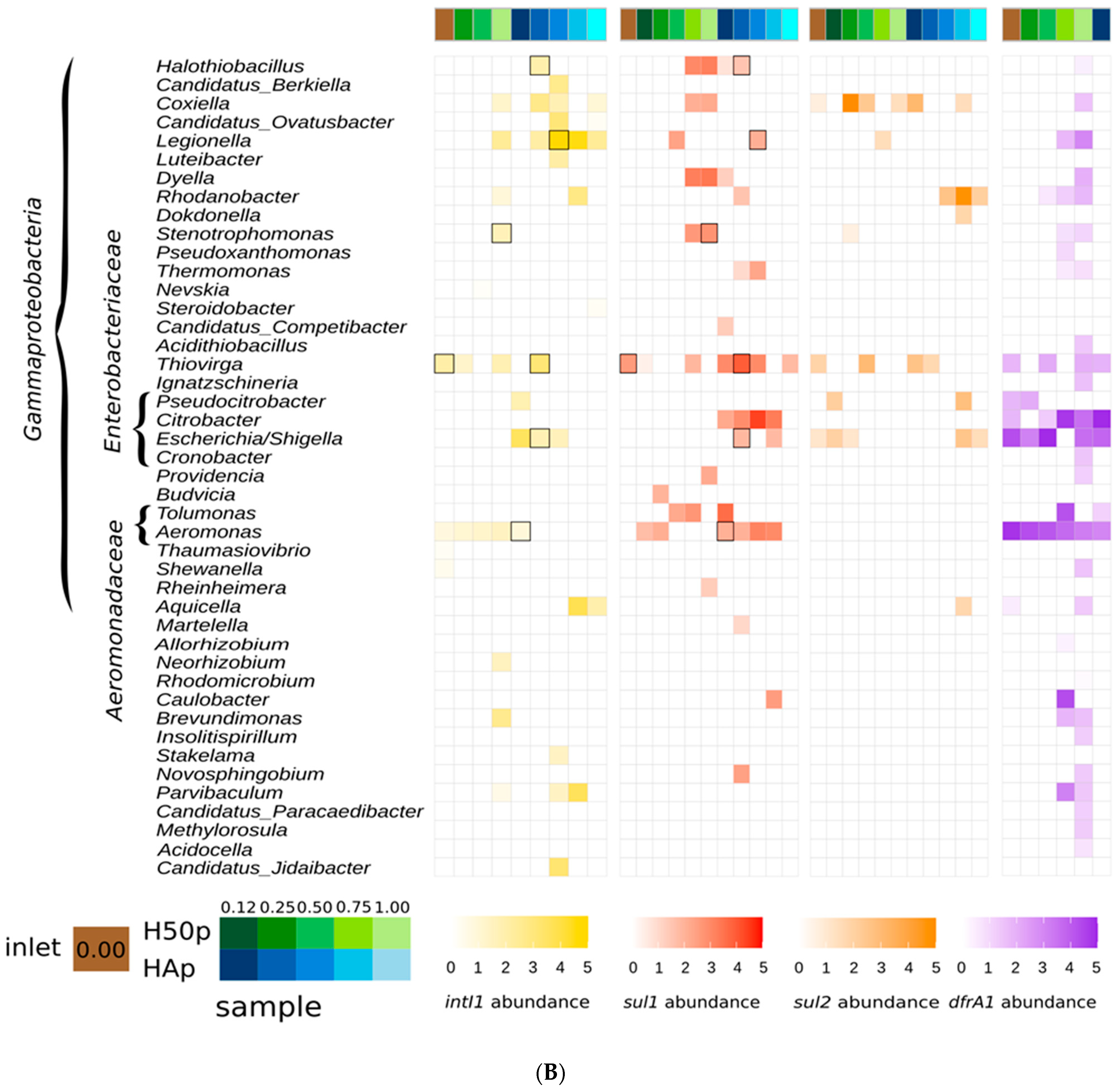
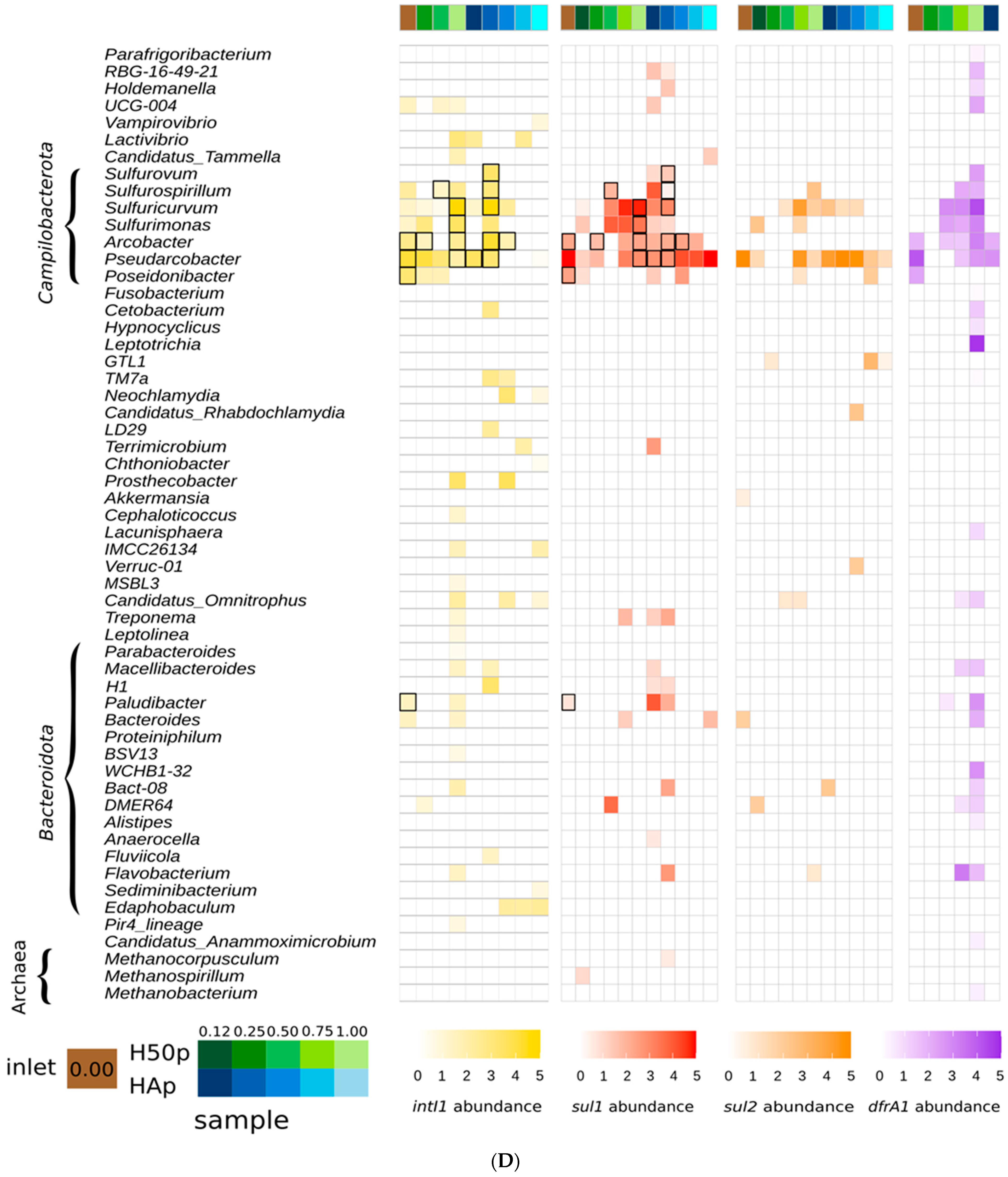
EpicPCR is semi-quantitative because it has the same biases as standard PCR, such as a different strength of primer binding to the target sequence, and there are taxon-specific differences in cell lysis efficiency. Therefore, in the following, we focus on the qualitative aspects of the results, i.e., the detection of specific AMR gene host rather than their presumptive abundance in the treatment wetlands. Relative abundances are nevertheless indicated in Figure 1A–D. In epicPCR analysis, false positive results can occur when cells with and without the target gene are present in the same polyacrylamide bead [16,45]. Since millions of beads are generated per epicPCR assay, such instances may be overlooked during microscopic inspection, as carried out here. In their study on improvements in epicPCR, Roman et al. [45] pointed out that it is currently impossible to differentiate between false positive artifacts and rare taxa found only in few samples. They assigned confidence levels according to the taxa detection frequency in biological replicates. We adapted their approach by showing all detected genera in Figure 1A–D, and listing genera found at more than 50% of the sampling sites in Table 1; this prevalence we consider a high-confidence detection. Only genera detected at two or more sampling sites are specifically discussed in the following (except for members of the Archaea, see Section 3.3).
After quality filtering, we obtained 850,050 to 1382,015 clean reads for the four genes. Targeting dfrA1 in HAp yielded high-quality results only for the first sampling site, which does not match the previous detection of the gene along the entire flow path of the wetland [28]. Establishing epicPCR for dfrA1 was more difficult in our laboratory than for the other indicator genes, and included the application of a new forward fusion primer. Further optimization work for dfrA1 is apparently needed in the future. In total, 209 genera were identified as carrying at least one of the AMR indicator genes in at least one sample (Figure 1A–D). Previously, ten genera harboring the studied genes were isolated from both treatment wetlands and the common inflow: Acinetobacter, Aeromonas, Arcobacter, Citrobacter, Escherichia, Flavobacterium, Pseudomonas, Providencia, Shewanella, and Stenotrophomonas [28]. All of these were also found in this study to be carriers of at least one of the target genes (with the qualifier that the Illumina reads did not allow for differentiation between Escherichia and Shigella). About half of all the detected genera (96/209) belonged to the Pseudomonadota (Figure 1A,B), especially to the gammaproteobacterial order Burkholderiales, which is consistent with reports on the taxonomic distribution of these indicator genes [10,11,12]. Among the Burkholderiales, there were 14 members of the Rhodocyclaceae that carried sul1, with some also harboring dfrA1 (Figure 1A). Members of this family are frequently found in WWTP systems [46]. Recently, a conjugative IncP-1 type plasmid with a class 1 integron was discovered in the genome of the Rhodocyclaceae genus Thauera [47]. This genus was detected as carrier of sul1 at 6 out of 11 of our sampling sites, supporting the hypothesis that Rhodocyclaceae may constitute a substantial reservoir of antibiotic resistance genes in WWTP systems [47]. Furthermore, 21 genera of the Burkholderiaceae, particularly close relatives of Comamonas (i.e., members of the family Comamonadaceae, according to the SILVA taxonomy), were found to carry sul1 and dfrA1, and to a lesser extent, intI1 and sul2 (Figure 1A). These families are known to harbor the AMR indicator genes [48]. Burkholderiaceae were also among the most common hosts of sul1 in a recent epicPCR study investigating WWTP [20]. Interestingly, the genus Ralstonia, a member of the Burkholderiaceae, was among the most prevalent hosts of int1, sul1, and dfrA1 in both treatment wetlands (Figure 1A and Table 1). To our knowledge, these genes have previously been found only in a few isolates and genomes of Ralstonia spp. [29,49]. The present finding is significant because several Ralstonia spp. are opportunistic pathogens that can be prevalent in drinking water sources [50,51]. Moreover, the folate biosynthesis inhibitors sulfamethoxazole and trimethoprim are among the best treatment options for Ralstonia spp. infections [50]. The last member of the Burkholderiales that we detected and would like to highlight is Laribacter (family Aquaspirillaceae), which was a carrier of sul1 in both HAp and H50p. Class 1 integrons and sul2 have been found in sequenced genomes of Laribacter hongkongensis [52], a potential cause of infectious diarrhea [53].
In addition, we found seven taxa of the gammaproteobacterial order Pseudomonadales, including Pseudomonas, carrying the AMR indicator genes in the wetlands (Figure 1A). Numerous Pseudomonas spp. strains carrying class 1 integrons have been described in the literature, and respective isolates were obtained from HAp and H50p throughout the flow path [28]. All four genes were also found in association with Acinetobacter (family Moraxellaceae). Acinetobacter spp. are ubiquitous in environmental habitats as well as in animals and humans, and can cause various diseases [54]. Etiological species include A. baumannii, a member of the ESKAPE group, which are six virulent and antibiotic-resistant bacterial pathogens that cause critical nosocomial infections [55]. There are numerous sequenced Acinetobacter spp. isolates with dfrA1 and sul resistances genes and class 1 integrons [56,57,58,59]. Our findings are in line with the notion that the environment is a substantial reservoir of antibiotic-resistant Acinetobacter spp. [54].
Furthermore, we discovered four taxa that have been described as symbionts of amoebae, namely Coxiella, Candidatus Berkiella, Candidatus Ovatusbacter, and Legionella (Figure 1B), the latter being the etiological agent of legionellosis. These taxa are not known to be typical carriers of the AMR indicator genes. In GenBank, we found only a partial sul2 in a sequenced L. pneumophila strain from hospital water associated with the 2016 outbreak of legionellosis in Flint, MI, USA [60]. Although folate biosynthesis inhibitors are currently not the drugs of choice for the treatment of legionellosis, the potential presence of class 1 integrons in the genus (see also Section 3.2) is of concern.
Members of the Enterobacteriaceae are well-known carriers of the four AMR indicator genes. Here, we found Escherichia/Shigella to be associated with each of the four genes, and other members of the Enterobacteriaceae with some of these genes (Figure 1B). Previously, each AMR indicator gene was detected in isolates of these taxa from both treatment wetlands [28]. While the epicPCR results match the literature data on host distribution of the AMR genes, they also demonstrate the detection limitation of the method. For example, the Citrobacter sp. strain used to develop the method for sul1, sul2 and dfrA1 was isolated from the wetlands, but epicPCR provided evidence only for Citrobacter spp. with sul1 and dfrA1, but not with sul2, in the treatment systems. Since certain hosts were not always detected despite being present, we did not attempt to estimate the horizontal gene transfer (HGT) of the AMR indicators, as has been published elsewhere [20]. Reliable evaluation of HGT would have required the analysis of more replicates, which was restricted by the labor required and the high costs of Phusion DNA polymerase.
Furthermore, epicPCR revealed the genus Aeromonas (family Aeromonadaceae) to be carriers of intI1 and sul1 at half of the sampling sites, and of dfrA1 at all sites (Figure 1B). No fusion product with sul2 was detected at any of the sites, although the gene was previously detected in nine distinct Aeromonas spp. isolates recovered from both wetlands [28]. Members of the genus Aeromonas are ubiquitous in freshwater habitats and can be also found in the clinical environment, and have been identified as carriers of the four AMR indicator genes [61,62]. Moreover, Aeromonadaceae were inferred in a Hi-C study to be a particularly prevalent reservoir of AR genes in a wastewater treatment plant [15].
Other carriers of the four genes in the wetlands belonged to the Desulfobacterota, including Trichlorobacter (also classified as Geobacter) and various sulfate-reducing bacteria, Bdellovibrionota (Bdellovibrio as carrier of intI1), and 39 genera of Firmicutes (Figure 1C). Firmicutes are known to harbor class 1 integrons [11]. Here, the detection of multiple Firmicutes genera is noteworthy, as these are Gram-positive bacteria for which the lysis of encapsulated single cells is often difficult. Incubation of polyacrylamide beads with Ready-Lyse lysozyme may have improved cell lysis [22]. Furthermore, members of the Campylobacterota were particularly prevalent, represented by the family Arcobacteraceae (Arcobacter, Poseidonibacter, Pseudoarcobacter) and the genera Sulfuricurvum, Sulfurimonas, Sulfurospirillum, and Sulfurovum (Figure 1D). The latter are sulfur-oxidizing bacteria that can occur in high absolute and relative abundance in the rhizosphere of wetland plants [63]. To our knowledge, they have not been previously identified as carriers of antibiotic resistance genes and class 1 integrons. In other isolated members of the Campylobacterota such as Campylobacter spp., class 1 integrons with various AR gene cassettes are commonly detected [64]. Campylobacteraceae including Arcobacter were also previously detected by epicPCR as hosts of intI1 and qacEΔ1 in the inflow of two urban WWTPs [18].

3.2. Potential Class 1 Integron Carriers in the Treatment Wetlands

Next, we searched for identical 16S rRNA fragments among the sequenced intI1 and sul1 fusion products from a given sample. While this approach does not provide conclusive evidence that a respective taxon at the sampling site contains at least one cell harboring a classical class 1 integron, it is nevertheless a good indication thereof. In total, we found identical 16S rRNA fragments belonging to 22 genera, indicated by black boxes in Figure 1A–D. The taxonomic pattern at the phylum level was quite similar to that described in the literature [11]. Thirteen (59%) of the genera belonged to the Gammaproteobacteria, seven genera to the Campylobacterota, and one each to the Bacteroidota and Desulfobacterota. Six of the Gammaproteobacteria genera have been frequently reported to be carriers of class 1 integrons, including Aeromonas, Escherichia/Shigella, and Pseudomonas, representatives of which had previously been isolated from the treatment wetlands [28]. The seven gammaproteobacterial genera that are not or only rarely described in the literature as carriers of class 1 integrons include Ralstonia and Legionella, which are among the taxa identified with high confidence (Table 1) and are briefly discussed in Section 3.1. Furthermore, Paludibacter, a member of the Bacteroidota discovered here, was recently suggested to be a strong candidate host of intI1 in a lake–river–lake system in Northern Italy [65].

3.3. Evidence for AMR Genes in Methanogenic Archaea

There were also epicPCR fusion products of sul1 and dfrA1 with 16S rRNA fragments of methanogenic Archaea in three samples (H50p at fractional length 0.12 and 1.00, and HAp at fractional length 0.12). Specifically, the sul1 products were linked with 16S rRNA fragments of Methanocorpusculum and Methanospirillum, and the dfrA1 product linked with a 16S rRNA fragment of Methanobacterium, all of which are members of the Methanobacteriota. Genes coding for tetrahydrofolate-synthesizing enzymes were discovered in some methanogenic Archaea [66], but the amino acid sequence identities are only around 30–40% of the products of sul1 and dfrA, and we have not found closer homologs in the sequenced archaeal genomes of GenBank and PATRIC [67]. We note that the epicPCR results need to be corroborated, since there were only three samples were positive for fusion products with archaeal 16S rRNA gene amplicons and only <20 reads each. Nevertheless, our findings are first evidence for inter-domain transfer of sul1 and dfrA1 into members of the Archaea. Horizontal transference between Archaea and Bacteria has been suggested for multiple other genes [68,69,70]. Furthermore, integrons, although not of the class 1 type, were recently discovered at a low frequency in metagenome-assembled genomes (MAGs) of Archaea, including members of the Methanobacteriota [71]. In the same study, it was shown that class 1 integron integrase harbored by an E. coli strain could incorporate archaeal gene cassettes into its integron via the archaeal attC recombination site. It is reasonable to hypothesize that archaeal integrase can incorporate bacterial gene cassettes, possibly taken up via transformation [72], into archaeal integrons, which could explain the apparent presence of sul1 and dfrA1 in the methanogenic Archaea observed here. Future epicPCR analysis could employ primers specifically targeting the archaeal 16S rRNA gene.

4. Conclusions

In this study, the epicPCR analysis revealed a profile of AMR indicator gene hosts highly consistent with literature data, including our previous cultivation-dependent investigation of the same treatment wetlands. Most carriers of the genes and apparently also of class 1 integrons belonged to the Gammaproteobateria, followed by members of the Desulfobacterota, Firmicutes and Campylobacterota. The analysis also identified several novel hosts of the indicator genes that were prevalent in the wetlands. Furthermore, the genes were found in genera with members known to cause disease in humans or domesticated animals, but also in genera without such medical or veterinary significance. The presence of the latter group of microorganisms underscores the function of the environment as a reservoir for AMR. Finally, the diversity of gene hosts observed here relates to the question of how best to address the spread of AMR in environmental compartments [3,4]. Limiting emission loads via improved wastewater treatment would be valuable, especially where risks of transmission are currently high [73]. However, we also note that different microbial hosts may have different survival and gene transmission potentials in the environment. This hampers the quantitative assessment of the effectiveness of AMR attenuation through wastewater treatment, as resistance genes may be propagated again, downstream of the treatment system, through vertical or horizontal transmission, if present in a suitable host. We are not the first to advocate this precautionary principle as a guide to better antibiotics stewardship, and hopefully as a pathway to curbing the spread of AMR worldwide [74,75,76].

Supplementary Materials

The following supporting information can be downloaded at: https://www.mdpi.com/article/10.3390/w15132432/s1, Table S1: Primers used in this study.

Author Contributions

Conceptualization, C.A.K., M.H. and J.A.M.; methodology, C.A.K., M.H. and I.M.; formal analysis, C.A.K.; investigation, C.A.K., M.H. and I.M.; resources, J.N.; writing—original draft preparation, C.A.K. and J.A.M.; writing—review and editing, C.A.K., J.N., A.-K.K. and J.A.M.; supervision, J.A.M.; project administration, J.A.M.; funding acquisition, C.A.K. and J.A.M. All authors have read and agreed to the published version of the manuscript.

Funding

This study was funded in part by the European Commission and the German Federal Ministry of Education and Research (BMBF; Grant Number 02WAP1616) within the framework of the collaborative international consortium NATURE, financed under the 2020 AquaticPollutants Joint call of the AquaticPollutants ERA-NET Cofund (GA NO 869178). ERA-NET is an integral part of the activities developed by the Water, Oceans and AMR JPIs. Further funding came from the Helmholtz Association of German Research Centers through its research program “PoF IV”, and from a doctoral stipend of the Henrich-Böll-Foundation to C.A.K.

Data Availability Statement

Sequence reads are available from the Sequence Read Archive (SRA) under accession number PRJNA859940.

Acknowledgments

The authors would like to thank Katy Bernhard for her support in the operation and sampling of the treatment system in Langenreichenbach, and Stephan Schreiber and John Vollmers for their help with Illumina sequencing and analysis.

Conflicts of Interest

The authors declare no conflict of interest. The funders had no role in the design of the study; in the collection, analyses, or interpretation of data; in the writing of the manuscript; or in the decision to publish the results.

References

  1. Collignon, P.; Beggs, J.J.; Walsh, T.R.; Gandra, S.; Laxminarayan, R. Anthropological and socioeconomic factors contributing to global antimicrobial resistance: A univariate and multivariable analysis. Lancet Planet. Health 2018, 2, e398–e405. [Google Scholar] [CrossRef] [PubMed]
  2. McEwen, S.A.; Collignon, P.J. Antimicrobial Resistance: A One Health Perspective. Microbiol. Spectr. 2018, 6. [Google Scholar] [CrossRef] [PubMed] [Green Version]
  3. Berendonk, T.U.; Manaia, C.M.; Merlin, C.; Fatta-Kassinos, D.; Cytryn, E.; Walsh, F.; Buergmann, H.; Sørum, H.; Norström, M.; Pons, M.-N.; et al. Tackling antibiotic resistance: The environmental framework. Nat. Rev. Microbiol. 2015, 13, 310–317. [Google Scholar] [CrossRef] [PubMed]
  4. Larsson, D.G.J.; Flach, C.-F. Antibiotic resistance in the environment. Nat. Rev. Genet. 2021, 20, 257–269. [Google Scholar] [CrossRef]
  5. Aarestrup, F.M.; Woolhouse, M.E.J. Using sewage for surveillance of antimicrobial resistance. Science 2020, 367, 630–632. [Google Scholar] [CrossRef]
  6. Pruden, A.; Pei, R.; Storteboom, H.; Carlson, K.H. Antibiotic Resistance Genes as Emerging Contaminants: Studies in Northern Colorado. Environ. Sci. Technol. 2006, 40, 7445–7450. [Google Scholar] [CrossRef]
  7. Mazel, D. Integrons: Agents of bacterial evolution. Nat. Rev. Genet. 2006, 4, 608–620. [Google Scholar] [CrossRef]
  8. Gillings, M.R.; Gaze, W.H.; Pruden, A.; Smalla, K.; Tiedje, J.M.; Zhu, Y.-G. Using the class 1 integron-integrase gene as a proxy for anthropogenic pollution. ISME J. 2014, 9, 1269–1279. [Google Scholar] [CrossRef]
  9. Ma, L.; Li, A.-D.; Yin, X.-L.; Zhang, T. The Prevalence of Integrons as the Carrier of Antibiotic Resistance Genes in Natural and Man-Made Environments. Environ. Sci. Technol. 2017, 51, 5721–5728. [Google Scholar] [CrossRef]
  10. Zhang, A.N.; Li, L.-G.; Ma, L.; Gillings, M.R.; Tiedje, J.M.; Zhang, T. Conserved phylogenetic distribution and limited antibiotic resistance of class 1 integrons revealed by assessing the bacterial genome and plasmid collection. Microbiome 2018, 6, 130. [Google Scholar] [CrossRef] [Green Version]
  11. Ghaly, T.M.; Gillings, M.R.; Penesyan, A.; Qi, Q.; Rajabal, V.; Tetu, S.G. The Natural History of Integrons. Microorganisms 2021, 9, 2212. [Google Scholar] [CrossRef]
  12. Néron, B.; Littner, E.; Haudiquet, M.; Perrin, A.; Cury, J.; Rocha, E.P.C. IntegronFinder 2.0: Identification and Analysis of Integrons across Bacteria, with a Focus on Antibiotic Resistance in Klebsiella. Microorganisms 2022, 10, 700. [Google Scholar] [CrossRef]
  13. Martínez, J.L.; Coque, T.M.; Baquero, F. What is a resistance gene? Ranking risk in resistomes. Nat. Rev. Genet. 2014, 13, 116–123. [Google Scholar] [CrossRef]
  14. Pärnänen, K.; Karkman, A.; Tamminen, M.; Lyra, C.; Hultman, J.; Paulin, L.; Virta, M. Evaluating the mobility potential of antibiotic resistance genes in environmental resistomes without metagenomics. Sci. Rep. 2016, 6, 35790. [Google Scholar] [CrossRef] [Green Version]
  15. Stalder, T.; Press, M.O.; Sullivan, S.; Liachko, I.; Top, E.M. Linking the resistome and plasmidome to the microbiome. ISME J. 2019, 13, 2437–2446. [Google Scholar] [CrossRef] [Green Version]
  16. Spencer, S.J.; Tamminen, M.V.; Preheim, S.P.; Guo, M.T.; Briggs, A.W.; Brito, I.L.; Weitz, D.A.; Pitkänen, L.K.; Vigneault, F.; Virta, M.P.; et al. Massively parallel sequencing of single cells by epicPCR links functional genes with phylogenetic markers. ISME J. 2015, 10, 427–436. [Google Scholar] [CrossRef] [Green Version]
  17. Dai, S.; He, Q.; Han, Z.; Shen, W.; Deng, Y.; Wang, Y.; Qiao, W.; Yang, M.; Zhang, Y. Uncovering the diverse hosts of tigecycline resistance gene tet(X4) in anaerobic digestion systems treating swine manure by epicPCR. Water Res. X 2023, 19, 100174. [Google Scholar] [CrossRef]
  18. Hultman, J.; Tamminen, M.; Pärnänen, K.; Cairns, J.; Karkman, A.; Virta, M. Host range of antibiotic resistance genes in wastewater treatment plant influent and effluent. FEMS Microbiol. Ecol. 2018, 94, fiy038. [Google Scholar] [CrossRef] [Green Version]
  19. Qi, Q.; Ghaly, T.M.; Penesyan, A.; Rajabal, V.; Stacey, J.A.; Tetu, S.G.; Gillings, M.R. Uncovering Bacterial Hosts of Class 1 Integrons in an Urban Coastal Aquatic Environment with a Single-Cell Fusion-Polymerase Chain Reaction Technology. Environ. Sci. Technol. 2023, 57, 4870–4879. [Google Scholar] [CrossRef]
  20. Wei, Z.; Feng, K.; Wang, Z.; Zhang, Y.; Yang, M.; Zhu, Y.-G.; Virta, M.P.J.; Deng, Y. High-Throughput Single-Cell Technology Reveals the Contribution of Horizontal Gene Transfer to Typical Antibiotic Resistance Gene Dissemination in Wastewater Treatment Plants. Environ. Sci. Technol. 2021, 55, 11824–11834. [Google Scholar] [CrossRef]
  21. Wei, Z.; Shen, W.; Feng, K.; Feng, Y.; He, Z.; Li, Y.; Jiang, C.; Liu, S.; Zhu, Y.-G.; Deng, Y. Organic fertilizer potentiates the transfer of typical antibiotic resistance gene among special bacterial species. J. Hazard. Mater. 2022, 435, 128985. [Google Scholar] [CrossRef] [PubMed]
  22. Diebold, P.J.; New, F.N.; Hovan, M.; Satlin, M.J.; Brito, I.L. Linking plasmid-based beta-lactamases to their bacterial hosts using single-cell fusion PCR. eLife 2021, 10, e66834. [Google Scholar] [CrossRef] [PubMed]
  23. Carvalho, P.N.; Arias, C.A.; Brix, H. Constructed wetlands for water treatment: New developments. Water 2017, 9, 397. [Google Scholar] [CrossRef] [Green Version]
  24. Dotro, G.; Langergraber, G.; Molle, P.; Nivala, J.; Puigagut, J.; Stein, O.; von Sperling, M. Treatment Wetlands; IWA Publishing: London, UK, 2017. [Google Scholar]
  25. Matamoros, V.; García, J.; Bayona, J.M. Organic micropollutant removal in a full-scale surface flow constructed wetland fed with secondary effluent. Water Res. 2008, 42, 653–660. [Google Scholar] [CrossRef]
  26. Sossalla, N.A.; Nivala, J.; Reemtsma, T.; Schlichting, R.; König, M.; Forquet, N.; van Afferden, M.; Müller, R.A.; Escher, B.I. Removal of micropollutants and biological effects by conventional and intensified constructed wetlands treating municipal wastewater. Water Res. 2021, 201, 117349. [Google Scholar] [CrossRef]
  27. Wu, S.; Carvalho, P.N.; Müller, J.A.; Manoj, V.R.; Dong, R. Sanitation in constructed wetlands: A review on the removal of human pathogens and fecal indicators. Sci. Total Environ. 2016, 541, 8–22. [Google Scholar] [CrossRef]
  28. Knecht, C.A.; Krüger, M.; Kellmann, S.; Mäusezahl, I.; Möder, M.; Adelowo, O.O.; Vollmers, J.; Kaster, A.-K.; Nivala, J.; Müller, J.A. Cellular stress affects the fate of microbial resistance to folate inhibitors in treatment wetlands. Sci. Total Environ. 2022, 845, 157318. [Google Scholar] [CrossRef]
  29. Fluit, A.C.; Bayjanov, J.R.; Aguilar, M.D.; Cantón, R.; Tunney, M.M.; Elborn, J.S.; van Westreenen, M.; Ekkelenkamp, M.B. Characterization of clinical Ralstonia strains and their taxonomic position. Antonie van Leeuwenhoek 2021, 114, 1721–1733. [Google Scholar] [CrossRef]
  30. Haenelt, S.; Wang, G.; Kasmanas, J.C.; Musat, F.; Richnow, H.H.; da Rocha, U.N.; Müller, J.A.; Musat, N. The fate of sulfonamide resistance genes and anthropogenic pollution marker intI1 after discharge of wastewater into a pristine river stream. Front. Microbiol. 2023, 14, 1058350. [Google Scholar] [CrossRef]
  31. Lee, J.; Ju, F.; Maile-Moskowitz, A.; Beck, K.; Maccagnan, A.; McArdell, C.S.; Molin, M.D.; Fenicia, F.; Vikesland, P.J.; Pruden, A.; et al. Unraveling the riverine antibiotic resistome: The downstream fate of anthropogenic inputs. Water Res. 2021, 197, 117050. [Google Scholar] [CrossRef]
  32. Nivala, J.; Headley, T.; Wallace, S.; Bernhard, K.; Brix, H.; van Afferden, M.; Müller, R.A. Comparative analysis of constructed wetlands: The design and construction of the ecotechnology research facility in Langenreichenbach, Germany. Ecol. Eng. 2013, 61, 527–543. [Google Scholar] [CrossRef]
  33. Kahl, S.; Nivala, J.; van Afferden, M.; Müller, R.A.; Reemtsma, T. Effect of design and operational conditions on the performance of subsurface flow treatment wetlands: Emerging organic contaminants as indicators. Water Res. 2017, 125, 490–500. [Google Scholar] [CrossRef]
  34. Edwards, U.; Rogall, T.; Blöcker, H.; Emde, M.; Böttger, E.C. Isolation and direct complete nucleotide determination of entire genes. Characterization of a gene coding for 16S ribosomal RNA. Nucleic Acids Res. 1989, 17, 7843–7853. [Google Scholar] [CrossRef] [Green Version]
  35. Grape, M.; Motakefi, A.; Pavuluri, S.; Kahlmeter, G. Standard and real-time multiplex PCR methods for detection of trimethoprim resistance dfr genes in large collections of bacteria. Clin. Microbiol. Infect. 2007, 13, 1112–1118. [Google Scholar] [CrossRef] [Green Version]
  36. Herlemann, D.P.; Labrenz, M.; Jürgens, K.; Bertilsson, S.; Waniek, J.J.; Andersson, A.F. Transitions in bacterial communities along the 2000 km salinity gradient of the Baltic Sea. ISME J. 2011, 5, 1571–1579. [Google Scholar] [CrossRef] [Green Version]
  37. Wang, N.; Yang, X.; Jiao, S.; Zhang, J.; Ye, B.; Gao, S. Sulfonamide-resistant bacteria and their resistance genes in soils fertilized with manures from Jiangsu Province, Southeastern China. PLoS ONE 2014, 9, e112626. [Google Scholar] [CrossRef] [Green Version]
  38. Adelowo, O.O.; Vollmers, J.; Mäusezahl, I.; Kaster, A.-K.; Müller, J.A. Detection of the carbapenemase gene blaVIM-5 in members of the Pseudomonas putida group isolated from polluted Nigerian wetlands. Sci. Rep. 2018, 8, 15116. [Google Scholar] [CrossRef] [Green Version]
  39. Wingett, S.W.; Andrews, S. FastQ Screen: A tool for multi-genome mapping and quality control. F1000Research 2018, 7, 1338. [Google Scholar] [CrossRef]
  40. Callahan, B.J.; McMurdie, P.J.; Rosen, M.J.; Han, A.W.; Johnson, A.J.A.; Holmes, S.P. DADA2: High-resolution sample inference from Illumina amplicon data. Nat. Methods 2016, 13, 581–583. [Google Scholar] [CrossRef] [Green Version]
  41. McMurdie, P.J.; Holmes, S. phyloseq: An R package for reproducible interactive analysis and graphics of microbiome census data. PLoS ONE 2013, 8, e61217. [Google Scholar] [CrossRef] [Green Version]
  42. Pagès, H.; Aboyoun, P.; Gentleman, R.; DebRoy, S. Biostrings: Efficient Manipulation of Biological Strings. R package version 2.54.0; 2019. Available online: https://bioconductor.org/packages/release/bioc/html/Biostrings.html (accessed on 1 May 2023).
  43. Kassambara, A. ggpubr: ‘ggplot2’ Based Publication Ready Plots. R package version 0.2; 2018. Available online: https://rpkgs.datanovia.com/ggpubr/ (accessed on 1 May 2023).
  44. Parks, D.H.; Chuvochina, M.; Chaumeil, P.-A.; Rinke, C.; Mussig, A.J.; Hugenholtz, P. A complete domain-to-species taxonomy for Bacteria and Archaea. Nat. Biotechnol. 2020, 38, 1079–1086. [Google Scholar] [CrossRef] [PubMed]
  45. Roman, V.L.; Merlin, C.; Virta, M.P.J.; Bellanger, X. EpicPCR 2.0: Technical and Methodological Improvement of a Cutting-Edge Single-Cell Genomic Approach. Microorganisms 2021, 9, 1649. [Google Scholar] [CrossRef]
  46. Wang, Z.; Li, W.; Li, H.; Zheng, W.; Guo, F. Phylogenomics of Rhodocyclales and its distribution in wastewater treatment systems. Sci. Rep. 2020, 10, 3883. [Google Scholar] [CrossRef] [PubMed] [Green Version]
  47. Lo, H.; Martínez-Lavanchy, P.M.; Goris, T.; Heider, J.; Boll, M.; Kaster, A.; Müller, J.A. IncP-type plasmids carrying genes for antibiotic resistance or for aromatic compound degradation are prevalent in sequenced Aromatoleum and Thauera strains. Environ. Microbiol. 2022, 24, 6411–6425. [Google Scholar] [CrossRef] [PubMed]
  48. Gerzova, L.; Videnska, P.; Faldynova, M.; Sedlar, K.; Provazník, I.; Cizek, A.; Rychlik, I. Characterization of Microbiota Composition and Presence of Selected Antibiotic Resistance Genes in Carriage Water of Ornamental Fish. PLoS ONE 2014, 9, e103865. [Google Scholar] [CrossRef]
  49. Adelowo, O.O.; Osuntade, A.I. Class 1 Integron, Sulfonamide and Florfenicol Resistance Genes in Bacteria from Three Unsanitary Landfills, Ibadan, Nigeria. J. Microbiol. Infect. Dis. 2019, 9, 34–42. [Google Scholar] [CrossRef] [Green Version]
  50. Ryan, M.P.; Adley, C.C. Ralstonia spp.: Emerging global opportunistic pathogens. Eur. J. Clin. Microbiol. Infect. Dis. 2013, 33, 291–304. [Google Scholar] [CrossRef] [Green Version]
  51. Vaz-Moreira, I.; Nunes, O.C.; Manaia, C.M. Ubiquitous and persistent Proteobacteria and other Gram-negative bacteria in drinking water. Sci. Total Environ. 2017, 586, 1141–1149. [Google Scholar] [CrossRef]
  52. Wang, L.; Fu, L.; Liu, Z.; Guo, H.; Wang, L.; Feng, M.; He, Y.; Chen, Q.; Hu, J. Comparative Analysis of Antimicrobial Resistance, Integrons, and Virulence Genes Among Extended-Spectrum β-Lactamase-Positive Laribacter hongkongensis from Edible Frogs and Freshwater Fish. Microb. Drug Resist. 2019, 25, 855–864. [Google Scholar] [CrossRef]
  53. Woo, P.C.; Kuhnert, P.; Burnens, A.P.; Teng, J.L.; Lau, S.K.; Que, T.-L.; Yau, H.-H.; Yuen, K.-Y. Laribacter hongkongensis: A potential cause of infectious diarrhea. Diagn. Microbiol. Infect. Dis. 2003, 47, 551–556. [Google Scholar] [CrossRef]
  54. Atrouni, A.E.; Joly-Guillou, M.L.; Ehamze, M.; Ekempf, M. Reservoirs of Non-baumannii Acinetobacter Species. Front. Microbiol. 2016, 7, 49. [Google Scholar] [CrossRef] [Green Version]
  55. Rice, L.B. Federal Funding for the Study of Antimicrobial Resistance in Nosocomial Pathogens: No ESKAPE. J. Infect. Dis. 2008, 197, 1079–1081. [Google Scholar] [CrossRef]
  56. Álvarez, V.E.; Quiroga, M.P.; Galán, A.V.; Vilacoba, E.; Quiroga, C.; Ramírez, M.S.; Centrón, D. Crucial Role of the Accessory Genome in the Evolutionary Trajectory of Acinetobacter baumannii Global Clone 1. Front. Microbiol. 2020, 11, 342. [Google Scholar] [CrossRef] [Green Version]
  57. Lee, K.; Yum, J.H.; Yong, D.; Lee, H.M.; Kim, H.D.; Docquier, J.-D.; Rossolini, G.M.; Chong, Y. Novel acquired metallo-beta-lactamase gene, bla(SIM-1), in a class 1 integron from Acinetobacter baumannii clinical isolates from Korea. Antimicrob Agents Chemother. 2005, 49, 4485–4491. [Google Scholar] [CrossRef] [Green Version]
  58. Liu, C.-C.; Tang, C.Y.; Chang, K.-C.; Kuo, H.-Y.; Liou, M.-L. A comparative study of class 1 integrons in Acinetobacter baumannii. Gene 2014, 544, 75–82. [Google Scholar] [CrossRef]
  59. Ramírez, M.S.; Quiroga, C.; Centrón, D. Novel Rearrangement of a Class 2 Integron in Two Non-Epidemiologically Related Isolates of Acinetobacter baumannii. Antimicrob. Agents Chemother. 2005, 49, 5179–5181. [Google Scholar] [CrossRef] [Green Version]
  60. Garner, E.; Brown, C.L.; Schwake, D.O.; Rhoads, W.J.; Arango-Argoty, G.; Zhang, L.; Jospin, G.; Coil, D.A.; Eisen, J.A.; Edwards, M.A.; et al. Comparison of Whole-Genome Sequences of Legionella pneumophila in Tap Water and in Clinical Strains, Flint, Michigan, USA, 2016. Emerg. Infect. Dis. 2019, 25, 2013–2020. [Google Scholar] [CrossRef] [Green Version]
  61. Gordon, L.; Cloeckaert, A.; Doublet, B.; Schwarz, S.; Bouju-Albert, A.; Ganière, J.-P.; Le Bris, H.; Le Flèche-Matéos, A.; Giraud, E. Complete sequence of the floR-carrying multiresistance plasmid pAB5S9 from freshwater Aeromonas bestiarum. J. Antimicrob. Chemother. 2008, 62, 65–71. [Google Scholar] [CrossRef] [Green Version]
  62. Schmidt, A.S.; Bruun, M.S.; Larsen, J.L.; Dalsgaard, I. Characterization of class 1 integrons associated with R-plasmids in clinical Aeromonas salmonicida isolates from various geographical areas. J. Antimicrob. Chemother. 2001, 47, 735–743. [Google Scholar] [CrossRef] [Green Version]
  63. Nguyen, P.M.; Arslan, M.; Kappelmeyer, U.; Mäusezahl, I.; Wiessner, A.; Müller, J.A. Spatial characterization of microbial sulfur cycling in horizontal-flow constructed wetland models. Chemosphere 2022, 309 Pt 1, 136605. [Google Scholar] [CrossRef]
  64. Lucey, B.; Crowley, D.; Moloney, P.; Cryan, B.; Daly, M.; O’Halloran, F.; Threlfall, E.J.; Fanning, S. Integronlike structures in Campylobacter spp. of human and animal origin. Emerg. Infect. Dis. 2000, 6, 50–55. [Google Scholar] [CrossRef] [PubMed]
  65. Corno, G.; Ghaly, T.; Sabatino, R.; Eckert, E.M.; Galafassi, S.; Gillings, M.R.; Di Cesare, A. Class 1 integron and related antimicrobial resistance gene dynamics along a complex freshwater system affected by different anthropogenic pressures. Environ. Pollut. 2023, 316, 120601. [Google Scholar] [CrossRef] [PubMed]
  66. Buchenau, B.; Thauer, R.K. Tetrahydrofolate-specific enzymes in Methanosarcina barkeri and growth dependence of this methanogenic archaeon on folic acid or p-aminobenzoic acid. Arch. Microbiol. 2004, 182, 313–325. [Google Scholar] [CrossRef] [PubMed]
  67. Antonopoulos, A.D.; Assaf, R.; Aziz, R.K.; Brettin, T.; Bun, C.; Conrad, N.; Davis, J.J.; Dietrich, E.M.; Disz, T.; Gerdes, S.; et al. PATRIC as a unique resource for studying antimicrobial resistance. Briefings Bioinform. 2017, 20, 1094–1102. [Google Scholar] [CrossRef] [Green Version]
  68. Frigaard, N.-U.; Martinez, A.; Mincer, T.J.; DeLong, E.F. Proteorhodopsin lateral gene transfer between marine planktonic Bacteria and Archaea. Nature 2006, 439, 847–850. [Google Scholar] [CrossRef]
  69. Koretke, K.K.; Lupas, A.N.; Warren, P.V.; Rosenberg, M.; Brown, J.R. Evolution of Two-Component Signal Transduction. Mol. Biol. Evol. 2000, 17, 1956–1970. [Google Scholar] [CrossRef] [Green Version]
  70. Nelson, K.E.; Clayton, R.A.; Gill, S.R.; Gwinn, M.L.; Dodson, R.J.; Haft, D.H.; Hickey, E.K.; Peterson, J.D.; Nelson, W.C.; Ketchum, K.A.; et al. Evidence for lateral gene transfer between Archaea and Bacteria from genome sequence of Thermotoga maritima. Nature 1999, 399, 323–329. [Google Scholar] [CrossRef]
  71. Ghaly, T.M.; Tetu, S.G.; Penesyan, A.; Qi, Q.; Rajabal, V.; Gillings, M.R. Discovery of integrons in Archaea: Platforms for cross-domain gene transfer. Sci. Adv. 2022, 8, eabq6376. [Google Scholar] [CrossRef]
  72. Fonseca, D.R.; Halim, M.F.A.; Holten, M.P.; Costa, K.C. Type IV-Like Pili Facilitate Transformation in Naturally Competent Archaea. J. Bacteriol. 2020, 202, e00355-20. [Google Scholar] [CrossRef]
  73. Larsson, D.G.J.; Andremont, A.; Bengtsson-Palme, J.; Brandt, K.K.; de Roda Husman, A.M.; Fagerstedt, P.; Fick, J.; Flach, C.-F.; Gaze, W.H.; Kuroda, M.; et al. Critical knowledge gaps and research needs related to the environmental dimensions of antibiotic resistance. Environ. Int. 2018, 117, 132–138. [Google Scholar] [CrossRef]
  74. European Parliament. Strategic Approach to Pharmaceuticals in the Environment. 2020. Available online: https://www.europarl.europa.eu/doceo/document/TA-9-2020-0226_EN.pdf (accessed on 15 May 2023).
  75. UN Environment Programme. Frontiers 2017: Emerging Issues of Environmental Concern. 2017. Available online: https://www.unenvironment.org/resources/frontiers-2017-emerging-issues-environmental-concern (accessed on 1 May 2023).
  76. WHO. Technical Brief on Water, Sanitation, Hygiene (WASH) and Wastewater Management to Prevent Infections and Reduce the Spread of Antimicrobial Resistance (AMR). 2020. Available online: https://www.who.int/water_sanitation_health/publications/wash-wastewatermanagement-to-prevent-infections-and-reduce-amr/en/ (accessed on 1 May 2023).
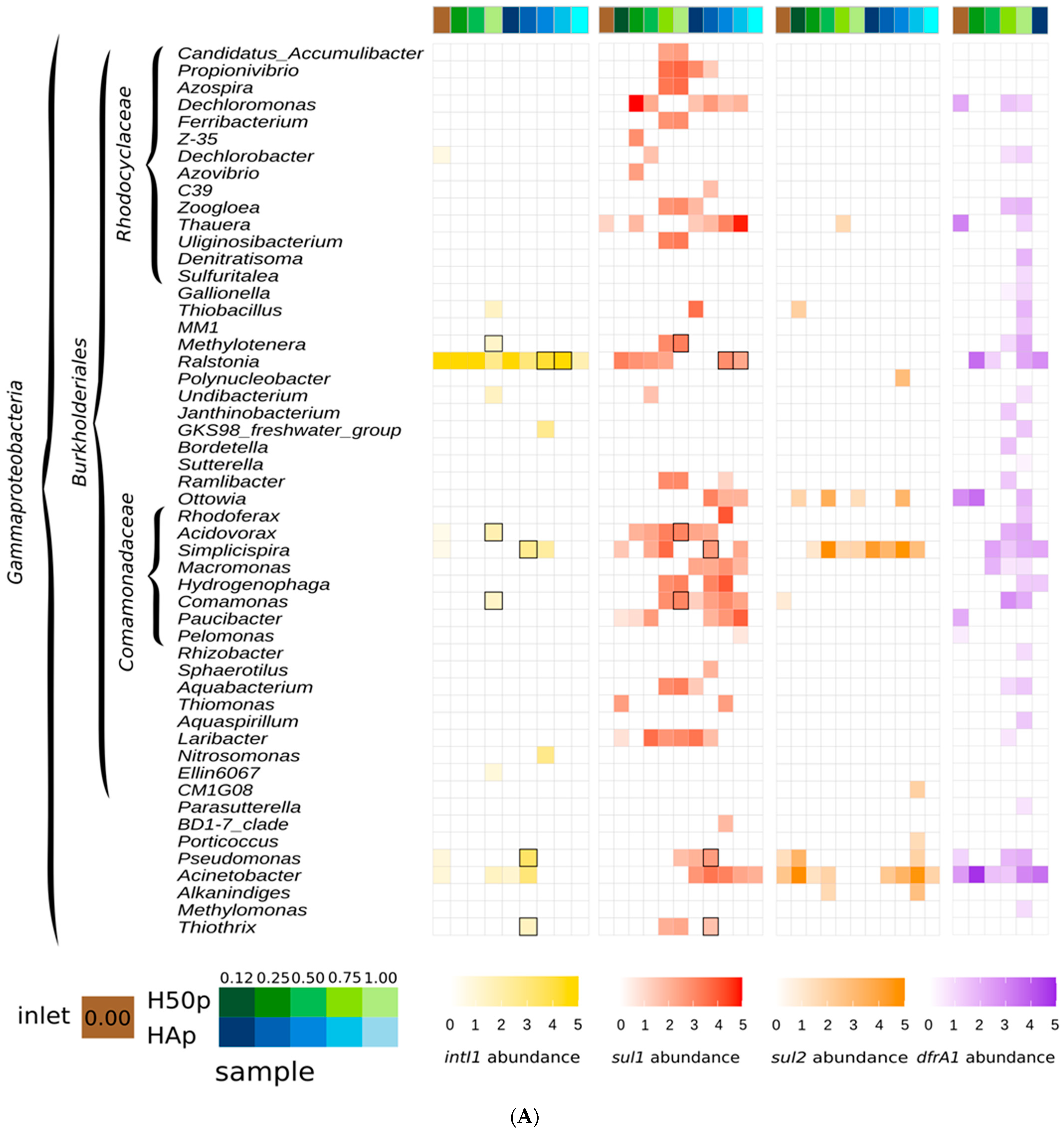
Figure 1. Genera detected as hosts of intI1 (yellow squares), sul1 (red), sul2 (orange), and dfrA1 (purple) in samples from the common inlet (brown) and the two treatment wetlands H50p (green) and HAp (blue). Relative abundances are Log10 normalized (Roman et al., 2021), indicated per color intensity. (A) Hosts belonging to the Gammaproteobacteria, including members of the families Rhodocylaceae and Comamonadaceae. (B) Further hosts belonging to the Gammaproteobacteria and other Proteobacteria. (C) Hosts belonging to the Desulfobacterota, Bdellovibrionota, and Firmicutes. (D) Hosts belonging to the Campylobacterota, Bacteroidota, and Archaea.
Water 15 02432 g001aWater 15 02432 g001bWater 15 02432 g001cWater 15 02432 g001d
Table 1. Genera found at >50% of the sampling sites as hosts of AMR indicator genes.
intI1sul1sul2dfrA1
Acidovorax
AcinetobacterAcinetobacterAcinetobacter
AeromonasAeromonas Aeromonas
Bdellovibrio
Citrobacter
Commamonas
CoxiellaCoxiella
Dechloromonas
Escherichia/Shigella
Laribacter
Legionella
Paucibacter
Protocatella
RalstoniaRalstonia Ralstonia
SimplicispiraSimplicispiraSimplicispira
SubdoligranulumSubdoligranulum
Thauera
ThiovirgaThiovirga Thiovirga
Trichlorobacter Trichlorobacter
Disclaimer/Publisher’s Note: The statements, opinions and data contained in all publications are solely those of the individual author(s) and contributor(s) and not of MDPI and/or the editor(s). MDPI and/or the editor(s) disclaim responsibility for any injury to people or property resulting from any ideas, methods, instructions or products referred to in the content.